n° 68 dec 2010 n - espcr · which precise lineage analysis is ... she showed that human...
TRANSCRIPT

ESP CRBULLETIN
N° 6
8 D
ec 2
010
Editorial Office: G. Ghanem (Editor), C. Meunier (Secretary), Laboratory of Oncology and Experimental Surgery (L.O.C.E.), Université Libre de Bruxelles, Institut J. Bordet, Rue Héger-Bordet 1, B – 1000 Brussels, Belgium. Phone: 32-2-541.32.96 E-Mail:[email protected]
E S
P C
R
B
U L
L E
T I
N
PUBL
ISH
ED B
Y TH
E EU
ROPE
AN S
OC
IETY
FO
R PI
GM
ENT
CEL
L RE
SEAR
CH
E
DIT
OR
:
G
. GH
ANEM
(Bru
ssel
s)
I
NTE
RN
ATI
ON
AL
F
. BEE
RMAN
N (L
ausa
nne)
, M. B
ÖH
M (M
ünst
er),
J. B
ORO
VAN
SKY
(Pra
gue)
, M. d
’ISC
HIA
(Nap
les)
, N. S
MIT
(Lei
den)
,
ED
ITO
RIA
L B
OA
RD
: JC
GAR
CIA
-BO
RRO
N (M
urci
a),
R. M
ORA
ND
INI (
Brus
sels
), A
. NAP
OLI
TAN
O (N
aple
s), M
. PIC
ARD
O (R
ome)
.
N° 68 Dec 2010
HHHAAAPPPPPPYYY NNNEEEWWW YYYEEEAAARRR 2 22000111111
CONTENT
Discussion, Letters to the editor, Reviews, Short communications, ...
Meeting report. Hinxton-Cambridge, UK History of pigment cell research in Europe (Prof. J. Borovansky)
Review of the literature
1. Chemistry of Melanins and other pigments (Prof A. Napolitano)
2. Biology of pigment cells and pigmentary disorders (Dr M. Picardo)
3. MSH, MCH, other hormones (Prof M. Böhm) 4. Photobiology (Dr N. Smit) 5. Neuromelanins (Prof M. d'Ischia) 6. Genetics, molecular and developmental biology
(Dr F. Beermann) 7. Tyrosinase, TRPs, other enzymes
(Prof JC. Garcia-Borron) 8. Melanosomes (Prof J. Borovansky) 9. Melanoma experimental, cell culture (Dr R. Morandini)
Announcements and related activities
- Calendar of events - Official List of ESPCR 2010 members
LETTER TO THE EDITOR DISCUSSION, REVIEW, SHORT COMMUNICATION, ...
ESPCR Meeting Report September, 4-7, Hinxton-Cambridge, UK
Session 1 : Development biology of pigment cells and MSCs Chair: Véronique Delmas and Takahiro Kunisada We see many reports describing multipotential stem cells possibly derived from neural crest cells found in the various part of the body. “Chromatoblast” concept reported by Robert Kerlsh (Univesity of Bath, UK) may hopefully bring some order in the discussion about these neural crest-related stem cells. He found that ablation of ltk kinase mostly affect iridophore cell fate, however, ltk expression marks all pigment cell lineages and Schwann cells. Using this kind of lineage markers in Zebrafish in which precise lineage analysis is possible during embryogenesis, dynamics of “chromatoblasts” (or any kind of neural crest stem cells) during progressive fate restriction of neural crest stem cells could be clarified. Melanocyte stem cells were functionally classified by Stephen Johnson (Washington Univ. Med. Sch., USA) in Zebrafish. Addition of melanocyotoxic MoTP to the early embryo ablate matured melanocytes in trunk region but the regenerated melanoblasts appeared within 4 days after MoTP washout, indicating the presence of adult-type melanocyte stem cells. While the addition of ErbB3 inhibitor AG1478 or erb3b defective picasso mutant strain develop normal melanocyte, MoTP-induced melanocyte regeneration was abolished by the addition of ErbB3 inhibitor or in erbb3 mutant. He therefore suggested that embryonic melanocytes develop directly, without proceeding through the ErbB-dependent adult type melanocyte stem cells. These analytical studies might eventually be combined to elucidate whole melanocyte cell lineage picture in which experimentally-tested real stem cells were nicely imposed. The requirement of BRAF and CRAF signaling during melanoblast development and homeostasis was presented by Agathe Valuet (Institut Curie, France). The double Braf and Craf knockout (KO) mice displayed normal pigmentation at birth, however following the first hair molting, the double KO mice exhibited progressive hair graying. After several hair cycles, analysis of the double KO mice revealead a disappearance of melanocyte stem cell in the bulge. It was previously show that the emergence of the melanocytes that produce the first hair requires KIT signalling whereas the presence of melanocyte stem cells in the bulge become independent of it. Taken together, the authors proposed an uncoupling between KIT and RAF signaling during melanocyte lineage development. To understand stem cells, quantitative analysis of the particular cell lineage seems to be essential and Lionel Larue (Institut Curie, France) presented mathematical models of melanocyte development during early embryogenesis in mouse skin. His model explained different behavior of dermal and epidermal melanocytes during development. Interestingly, beta catenin that profoundly affect proliferation of epidermal melanocyte precursors did not affect dermal ones, therefore a new key to the regulation of early melanocyte development must be held by the beta catenin function in developing melanocytes. Another mathematical model was presented by Richard Mort (MRC, United Kingdom) to analyse melanoblast behavior during embryonic development. The goal of the study was to explain how individual cellular properties and tissue expansion can contribute to the dispersal of the melanoblast population. The authors presented live imaging of EYFP-melanoblast migration in embryonic skin in an ex vivo system. Analysis of time lapse sequences revealed that melanoblasts are highly mobile and allowed measurement of the dynamics of their migration.
2077
Session 2: Genetics of pigment cell transcription Chair: Vittoria Schiaffino and Eirikur Steingrimsson Session 2 had 3 invited speakers and 3 selected presentations, which discussed various transcriptional regulatory pathways operating in the melanocyte lineage and in particular involving MC1R, Mitf, beta-catenin and Notch signalling. Zalfa Abdel-Malek (Cincinnati, USA) started the session by discussing the central role of MC1R in regulating human pigmentation and the UV response. She showed that human beta-defensin 3 (HBD3) acts as an antagonist of MC1R by blocking the effects of alpha-MSH in human melanocytes. As an alternative possibility based on mouse model phenotypes, Dr. G. Barsh in the audience underlined that HBD3 might actually act as a neutral agonist for MC1R, enhancing its constitutive activity. Zalfa also showed how transcription of MC1R can be affected by either alpha-MSH, ACTH, by cAMP signaling or by Endothelin 1 and bFGF. She also presented evidence, which suggest that MC1R may undergo different levels of desensitization in different melanocyte strains. This, however, was independent of the MC1R genotype and to the expression level of G protein-coupled receptor kinases (GRKs). Eirikur Steingrimsson (Reykjavik, Iceland) described the structure of the MITF protein and how it has elucidated important aspects of dimerization and DNA binding properties of the protein. The structure has also provided information on the biochemistry of the Mitf Mi-Wh mutation, a mutation with interesting phenotypic and genetic characteristics. Eirikur also discussed the acetylation of MITF and how it affects function and may lead to an oncogenic protein, in particular when modifying residues K243/248 within the helix-loop-helix domain. Francesca Pignoni (Syracuse, USA) described how she has used the powerful Drosophila genetic system for modeling the oncogenic activities of MITF in the Drosophila wing epithelium. In order to do this she has used the Gal4-UAS system to express MITF in different cells of the wing epithelium and then analyse effects on various aspects of cell function including proliferation, apoptosis, cell adhesion and migration. Her results show that in combination with activated RAS, MITF can act as an oncogene in the fly epithelium. In addition to this she has used deletion strains, siRNA strains, ENU mutagensis and the yeast-2-hybrid screen to search for modifiers and mediators of MITF function. Several genes were found in more than one of the screens, suggesting that they are true modifiers or mediators of MITF. Claudia Wellbrock (Manchester, UK) has investigated the controversy regarding the role of beta-catenin in melanoma. In contrast to other kind of cancers, nuclear beta-catenin in melanoma has been associated with both reduced overall survival as well as with good prognosis. Using melanoma cell lines with high and low levels of nuclear beta-catenin, she showed that in melanoma, high expression of nuclear beta-catenin is indicative for a non-invasive phenotype whereas low beta-catenin levels results in invasive cells. These effects of beta-catenin are at least partially mediated through effects on MITF gene expression. Thus, the activation of the same signaling pathway in different types of cancer might result in opposite effects, depending on cancer-type specific downstream targets. Genevieve Aubin-Houzelstein (Paris, France) discussed her analysis of the role of Strawberry Notch homolog 2 (Sbno2) in melanocytes and melanocyte stem cells. She used transgenic mice to overexpress Sbno2 in the melanocyte lineage and studied both coat color phenotype and fate of the melanocyte stem cells. Mice overexpressing Sbno2 are largely normally pigmented at birth but gradually lost pigment starting at 3 months of age. There was a reduction in melanocyte precursors from the bulge region with successive hair cycles, suggesting that Sbno2 overexpression affects stem cell maintenance. Interestingly, they also observed a ring of beta-galactosidase positive cells around several hair follicles, suggesting a cell fate switch of the melanocyte precursors. Finally, Bhushan Sarode (Lausanne, Switzerland) presented studies on the role of Notch1 and 2 receptors in melanoblast and melanocyte stem cells maintenance. Notch signaling plays critical role in many biological processes. Deletion of Notch1 and Notch2 receptors results in dose dependent hair graying, due to an elimination of melanocytes and melanocyte stem cells. The expression of a NotchIC transgene rescued this deficiency, however, not the deletion of RbpJK, thus proving further evidence
2078
that Notch signaling acts via RbpJK transcription factor in pigment cells. The identification of targets in melanocytes might indicate why melanocyte stem cells disappear in the absence of Notch. For that purpose he undertook a microarray strategy in order to identify downstream factors involved in this process. Session 3 : Pigmentary Disorders Chair: Prasad Kumarasinghe and Lluis Montoliu Sunday 5, September 2010, 8.45 AM-10.15 AM Albinism is characterized by hypopigmentation and affects 1 in 17000 human beings. It is a heterogeneous group of rare genetic conditions. Severe visual deficiencies are common among albinos therefore albinism affects the quality of life significantly. Lluís Montoliu spoke on the topic ‘Towards a universal genetic diagnosis of all types of albinism’. He described the various types of oculocutaneous albinism and ocular albinism, including Hermansky – Pudlak and Chediak Higashi syndromes. Up to 14 genes have been described to be associated with albinism. These genes encode proteins that are necessary for normal melanogenesis, melanin synthesis, melanosome function, and lysosome related organelle development and function. Over 500 mutations have been reported in genes associated with albinism. However, in most medical institutions or laboratories, genetic diagnostic tests are not available for definite categorization of types of albinism. CIBERER, the Biomedical Network Research Centre on Rare Diseases in Spain has attempted to develop a universal genetic diagnosis for all types of albinism. This approach uses Sequenom iPLEX methodology which combines automated array processing of DNA samples with mass spectrometry. This method can be used to differentiate up to 1000 alleles with known mutations from a given DNA sample. DNA obtained from 2 ml of saliva is sufficient for this analysis. It was an interesting and thought provoking presentation highlighting one of the relatively neglected pigmentary disorders. Rudolph Happle spoke on Patterns and mechanisms of pigmentary mosaicism. It was interesting analysis of various mechanisms of clinically evident pigmentary mosaicism. He explained the mosaicism patterns of narrow band Blashko lines, broad band types such as McCune Albright syndrome as well as checkerboard pattern and phylloid patterns. Patchy pattern without midline separation is postulated for giant melanocytic nevi. He also spoke of genomic versus epigenetic mosaicism. Genomic mosaics following Blashko’s lines include pigmentary mosaicism of Ito’s type. Cutis tricolor is a peculiar genomic mosaic characterized by paired hyper and hypopigmented macules reflecting twin spotting or allelic didymosis. Epigenetic mosaics include X inactivation patterns as observed in incontinentia pigmenti. Monoallelic expression (autosomal epigenetic mosaicism) has been proposed to explain an unusual familial constellation of pigmentary mosaicism following Blaschko’s lines. Canine autosomal trait ‘brindle’ was also described and its possible analogy to this syndrome was also debated. Happle’s vast experience in cases of pigmentary mosaicism was clearly evident in the presentation. This presentation was particularly useful for the clinicians and clinical geneticists. Caroline Le Poole spoke on’ HSP70i as the sole requirement for activating depigementation in vitiligo prone mice. Previously, they had reported the ability of HSP70i secreted by vitiligo melanocytes in increased amounts, to activate DC mediated cytotoxicity. HSP70i is also accelerated by TRP2 DNA vaccine induced T cell mediated depigmentation in C57BL/6 mice. They had performed a study to challenge the redundancy of HSP70i in vitiligo, using kockout mice lacking constitutive or inducible HSP70. Pmel1 mice transgenic for a T cell receptor reactive with gp100 were subject to vaccination with an expression vector encoding HSP70i versus empty vector DNA. HSP70i knockout mice depigmented in response to TRP2, the response to antigen challenge was impaired in HSP1 knockout mice. Their study suggests a crucial and non redundant role for inducible HSP70i in accelerating vitiligo. The study also demonstrated that vaccination with HSP70i was necessary and
2079
sufficient to induce depigmentation in Pmel1 transgenic mice. Le Poole and others suggest a crucial role of stress induced HSP70i in vitiligo, and in interfering with this may have significant impact on progress of depigmentation. Elisabeth Minder talked on the use of afamelanotide for treating patients affected by erythropoietic protoporphyria (EPP). The title of the talk was ‘Afamelanotide in erythropoietic protoporphyria, a randomized, placebo controlled multicentre phase III trial’. It was a study sponsored by the Clinuvel pharmaceutical company. Phototoxicity and intolerable pain in the skin following exposure to sunlight are main symptoms of EPP. Afamelanotide is a melanocortin 1 receptor agonist and an analogue of alpha MSH, and it has shown beneficial effects in reducing symptoms in EPP. The investigators of their study had administered afamelanotide during a long term clinical assay and collected clinical and laboratory data and evaluated adverse events. The study was a placebo controlled trial. Controlled release controlled release implant was administered every 60 days. The patients were asked to record daily, any adverse events, the time spent in the sun, and EPP related pain level on a standard pain scale (Lickert). Melanin density measurements and laboratory safety controls were done bimonthly. During afamelanotide treatment, EPP patients tolerated sun exposure for several hours a day. They remained pain free for most of the time and severer pain episodes were uncommon. Prolonged use of afamelanotide was well tolerated by all the patients according to the investigators. Overall it was an interesting presentation. If similar results are obtained in larger studies as well, the findings may pave the way to reduce or minimize pain on sun exposure in EPP patients world over. Session 4 : Melanocyte cell biology Chair: Graça Raposo and Miguel Seabra Session X was chaired by M. Seabra from Imperial College London and IGC, Portugal, and G. Raposo from the Department of Cell Biology (CNRS UMR144) in Institut Curie . The session consisted of 3 invited lectures, including those of the two chairs and 1 oral presentation selected from the abstracts. The goal of the session was to give an overview of current topics on the cell biology of the melanocyte and also on the biology of melanocyte interactions with keratinocytes and epithelial cells. Graça Raposo presented studies carried in her group in close collaboration with the group of Michael Marks at UPenn in Philadelphia. The studies presented aim to get novel insights onto the biogenesis of immature and mature melanosomes in melanocytic cells, models of eumelanogenesis. In particular she showed unpublished data revealing that proteins of the Tetraspanin family are involved in the endosomal sorting of Pmel17, a process required for the formation of the amyloid fibrillar sheets of premelanosomes. She also showed that late steps of melanogenesis are regulated by protein components whose encoding genes are mutated in the Hermansky Pudlak Syndrome. These late steps of melanosome maturation and melanin synthesis require the specialization of early recycling endosomes through the coordinated action of molecular Adaptors and a microtubule motor. The dialogue between endosomes and melanosomes could be potentially regulated by melanocyte-keratinocyte interactions. Following the first talk on melanocytes, Miguel Seabra presented very recent studies performed to investigate the mechanisms of melanosome transfer from melanocytes to keratinocytes and the fate of melanosomes within keratinocytes. Using high resolution electron microscopy of human skin, he proposed that melanosomes are exocytosed by melanocytes into the extracellular space and the melanosome core is subsequently taken up by keratinocytes. Within keratinocytes, the melanosome core particles were concentrated onto vesicles that rapidly gain late endosomal markers such as LAMP1, suggesting that keratinocyte uptake share similarities to phagosome maturation in macrophages. Janice Brissette (State University of New York Downstate Medical Center, New York City) presented studies of a novel class of epithelial cells, termed 'pigment recipients.' She showed that pigment recipients play an important role in the patterning of pigmentation, as they attract melanocytes and stimulate pigment transfer, thereby engineering their own pigmentation. Pigment recipients acquire their abilities via the Foxn1 transcription factor, which
2080
induces the recipients to transmit signals to the melanocytes. The signals appear to include diffusible proteins, such as Fgfs, and cell-bound proteins, such as Notch activators. Finally Mireille van Gele (Dept. of Dermatology, Ghent University Hospital, Belgium) presented a newly developped in vitro reconstructed skin model containing keratinocytes and melanocytes. This model is suitable to study the effect on pigmentation after knockdown of tyrosinase (key-enzyme in melanogenesis) or molecular components (MyoVa ; Rab27 ;melanophilin) involved in melanosome transport. Session 5 : Melanoma biology and therapeutics Chair: Richard Marais and Kyoung Chan Park Not Available Session 6 : Melanocytes, migration and melanoma Chair: Lionel Larue and Elisabeth Patton Erik Sahai (London Research Institute, UK) presented evidence of reversible phenotype switching in melanoma using intravital imaging for melanosomes combined with a Brn-2-GFP reporter construct. Sahai and colleagues identified a sub-population of cells containing little or no pigment and high levels of Brn2::GFP expression that are motile in the primary tumour and enter the vasculature. Significantly, the less differentiated state of motile and intravasated cells are not maintained at secondary sites, implying switching between states as melanoma cells metastasize. Thus, a subset of less differentiated cells exits the primary tumour but subsequently give rise to metastases that include a range of more differentiated and pigment producing cells. Corine Bertolotto (INSERM, France) presented evidence that long-term depletion of MITF in melanoma cells, triggers activation of the DNA damage response signaling pathway associated with an up-regulation of p53, and ultimately ends into cellular senescence-like phenotypes. Our findings uncover the existence of a lineage-restricted DNA damage response/p53 signaling pathway in melanoma that is inhibited by MITF to prevent senescence and favor melanoma cell proliferation. Liz Patton (MRC Human Genetics Unit, UK) presented the results of a series of small molecule screens to identify compounds that control melanocyte development in zebrafish. One class of compounds, the nitrofurans, selectively targeted differentiated and melanocyte stem cells (MSCs) in zebrafish development. Patton and colleagues also screened for suppressors of the nitrofuran MSC phenotype and identified the PRL3 phosphatase a new regulator of MSCs. Thus, small molecule screening in zebrafish has identified a series of chemical regulators of melanocyte development, that point to new molecular and therapeutic pathways. Marina Mione (IFOM, Milan) presented a new model of melanoma developed in zebrafish using the Gal4/UAS system to express oncogenic HRAS under the control of the kita promoter. In this model, melanoma developed very early (at 2 weeks) and did not require inactivation of tumor suppressors like p53 or PTEN. The study of melanoma initiating cells, aided by the presence of a GFP tagged oncogene and the use of chemicals to ablate a subpopulation of erbB3+ melanocyte stem cells (Hultman et al., 2009), showed that, in this model, melanoma originates mostly from differentiated larval melanocytes. Anna Golovko (Uppsala University, Sweden) presented evidence that STX17 is upregulated in Grey horse melanocytes and melanomas, with the full-length being the predominant form. Immuno-fluorescence studies showed a complete co-localization of the STX17 isoforms and their partial co-localization with the ER. Additional work from the laboratory has shown that STX17 mediates ERK signaling in Grey horse melanoma cell cultures (Jiang et al, submitted). Golovko and colleagues now show that ERK1/2 is activated already in melanocytes of Grey horses, which, together with the co-expression of high levels of STX17 in these cells, further strengthens STX17’s role in ERK activation
2081
and/or signaling. Session 7 : Senescence Chair: Robert Ballotti and Dorothy C Bennett Session 7 was centred on cell senescence (permanent growth-arrest following extensive proliferation or oncogene activation), in melanocytes and melanoma. Daniel Peeper, who gave the EMBO lecture, focused on oncogene-induced senescence (OIS) caused by BRAFE600, as proposed to occur in nevi. He reported the presence of IL6 and IL8 (part of the “senescence messaging secretome”) in benign lesions in vivo. The CDK inhibitor p16INK4A has previously been implicated in melanocyte senescence, is upregulated by BRAFE600 and is strongly expressed in naevi, but not by all naevus cells, suggesting a requirement for other factor(s) for the induction and maintenance of senescence in these cells. p15INK4B, another CDK inhibitor, was found to be greatly increased in abundance by BRAFE600, but neither p16 nor p15 (nor both) seemed to account fully for BRAFE600-induced senescence in human fibroblasts or melanocytes. Interestingly, studies of melanoma arising from a contiguous naevus led to the identification of PTEN as lost in the melanomas, suggesting the PI kinase pathway as regulating senescence. Indeed, BRAFE600 inhibited AKT3 expression in culture, and PTEN inhibition (resulting in AKT activation) prevented BRAFE600 induced senescence. Candidate gene and large-scale genomic approaches have been undertaken to identify new signalling networks involved in senescence bypass in BRAFE600 induced cancers. Maria Soengas gave a very innovative lecture dealing with the role of protein degradation pathways in melanoma development and resistance to chemotherapeutic treatments. Melanoma cells have an overactive metabolism and need efficient mechanisms of clearance of aggregate-prone proteins and damaged organelles. Sequestosome 1 (SQSTM1/p62), a multifunctional protein that links the proteasome to the autophagic machinery, was found to be overexpressed in melanoma compared to nevi. SQSTM1 silencing induced a senescence-like phenotype in melanoma and blocked tumour growth in mice. Furthermore, SQSTM1 localization was modified by treatment with chemotherapeutic agents. Interestingly, SQSTM1 silencing did not affect normal human melanocyte growth. Therefore, these data point to protein degradation pathways and more specifically to SQSTM1 as a potential target for new anti-melanoma drugs. Dot Bennett spoke about a network project with four other British groups and Cancer Research Technology, to develop methods for rapidly screening libraries of chemicals, siRNAs etc, for the ability to induce senescence in cancer cells. This might provide a therapeutic route for cancer types resistant to conventional treatments. Screening methods were initially developed using normal human fibroblasts, and then for A375P melanoma cells, using an Arrayscan digital reader. A screen of a bioactive chemical library with the fibroblasts yielded a number of “hits”, confirmed with biological and protein markers, and three of these also gave stable senescence-like arrest in melanoma cells and also in other types of cancer cells, giving encouraging support for this approach. Lastly, Susanne Schiffner from the group of Anja Bosserhoff gave an oral presentation. She reported on the diffusible factor MIA (melanoma inhibitory activity/protein), often secreted by melanoma cells. They found that it is also produced by most nevus cells, and by normal human senescent melanocytes. When overexpressed in normal human melanocytes, MIA was able to induce markers of senescence including �-galactosidase, p21 and p15. Likewise, knockdown of MIA in human melanocytes apparently delayed senescence. KO of MIA in mice could accelerate the appearance of melanomas. It was proposed that MIA might have some properties of an oncogene, being associated with melanoma cells, but could also contribute to senescence, perhaps as with oncogene-induced senescence.
2082
Session 8 : Signalling patways in melanocytes and melanoma cells Chairs: Colin Goding and Anja Bosserhoff The session opened with a talk from Alain Mauviel (Paris) on the role of Gli2, a mediator of Hedgehog signaling, in melanoma. He demonstrated that Gli2 expression is transcriptionally upregulated by TGF-ß signaling, in the most aggressive melanoma cell lines. GLI2 promoted melanoma migration and invasion by controlling a mesenchymal transition characterized by loss of expression of E-cadherin and increased MMP secretion. GLI2 knockdown in melanoma cells that express high levels of GLI2 significantly inhibited their invasive capacity and the development of experimental bone metastases in mice. GLI2 expression was shown to be heterogeneous within human melanoma lesions, and associated with tumor regions in which E-cadherin is lost, and was increased in distant metastases as compared to primary lesions. Consistent with this, GLI2 staining by immunohistochemistry of 37 primary melanoma tumors, demonstrated that GLI2 expression was associated with Clark level and with reduced distant metastasis-free survival. Also, GLI2-positive distant metastases were associated with dramatically reduced overall survival. The data presented provided support for a direct role for GLI2 in driving melanoma invasion and metastasis and indicate that GLI2 expression in melanoma tumor lesions may be a marker for poor prognosis. Vittoria Schiaffino (Milan) discussed the protein product of the ocular albinism gene, OA1, a G protein-coupled receptor localized to intracellular organelles, melanosomes and lysosomes. Recently Lopez et al., 2008, provided evidence for an OA1-mediated signaling pathway triggered by L-DOPA at the plasma membrane and suggested that tyrosine in the medium act as a kind of ligand, able to promote the constitutive internalization and downregulation of the receptor. However, the results from Vittoria’s laboratory indicated that amino acid starvation, including cell culturing in tyrosine-free medium, induces the general upregulation of exogenous transgenes, most likely by means of epigenetic modifications. Therefore, the effects of low tyrosine in the medium on the total amount of exogenous OA1 are neither OA1 specific nor tyrosine specific, and this amino acid does not appear to play any role on the intracellular localization of the receptor. Further work will therefore be necessary to identify the ligand for the OA 1 receptor. Jose Carlos Garcia Borron (Murcia) discussed the functional properties of natural human MC1R mutants. The major R151C, R160W and D294H MC1R alleles associated with red hair, fair skin (the RHC phenotype) and increased skin cancer risk encode for forms with decreased signaling through cAMP, yet they were shown activate the ERKs at least as effectively as wild-type MC1R. This disparate effect of the mutations on the two major signaling pathways originating from the MC1R was unexpected, since it was generally assumed that cAMP is responsible for ERK activation in MSH-stimulated melanocytic cells. After confirming that MC1R-mediated ERK activation in melanocytes and melanoma cells is independent on cAMP by a series of pharmacological studies, an alternative pathway linking MC1R to the ERKs was sought. Pharmacological interference, siRNA and functional reconstitution experiments showed that MC1R activation led to transactivation of the cKIT receptor, thus pointing to an unexpected link between MC1R and KIT signaling in melanocytes and melanoma. Preliminary data suggested that Src is also activated following MSH binding to the MC1R and is located upstream of cKIT in the pathway mediating MC-dependent ERK activation in melanocytic cells. Thus, the cAMP and ERK pathways are not activated sequentially by the MC1R, but rather they are triggered independently. Both pathway display different sensitivity to many natural mutations, with an imbalanced signaling in major RHC alleles. Ian Jackson spoke about his collaboration with MRC Harwell through which they have identified new mouse mutations that affect the switch between eumelanin and phaeomelanin synthesis during hair growth. One of them is a recessive mutation that increases the amount of phaeomelanin. The phenotype is very similar to that of a knockout of the Corin gene described recently by Enshell-Seijffers, Bruce Morgan and colleagues and the mutation maps to the Corin locus. It appears to be a loss of function caused by a genomic duplication leading to an exon duplication, and frame shift, in the
2083
mRNA. The second new mutation was dominant, and had the opposite phenotype; that is it had increased eumelanin. Mapping and sequencing revealed a point mutation in the G-protein alpha stimulatory subunit. Finally Yoko Funasaka spoke on the role of the metabotropic glutamate receptor subtype 1 (mGluR1), a G protein coupled receptor activated by glutamate that is functionally expressed in the central nervous system. She had shown previously that ectopic expression of mGluR1 in melanocytes is essential for both development and in vivo growth of melanoma in transgenic mice. Upon stimulation by glutamate, mGluR1 can couple to multiple signaling pathways through different G proteins, such as activation of phospholipase C (PLC) b that triggers IP3-mediated Ca2+ release and activation of PKC, an increase cytoplasmic cAMP, and activation of ERK1/2 MAP kinase. To understand the contribution of each signaling pathway to melanoma, she studied the effects of inhibitors of phospholipase C, PKC, MEK1/2, and cAMP phosphodiesterase, antagonists to Ca2+ and calmodulin, and an activator of adenylyl cyclase that were administered to transgenic mice engineered to develop melanoma. With the exception of the cAMP inducers, each inhibitor of PLC, PKC, Ca2+ release, calmodulin, and MEK1/2 inhibited melanoma development. Although administration of a glutamate release inhibitor suppressed melanoma growth, these signal transduction inhibitors only suppressed melanoma growth partially. The results indicate that for development of melanoma, activation of multiple signals is required, but for melanoma growth, inhibition of several signaling pathways can be compensated by other events downstream of mGluR1.
Session 9: Genetics of melanocyte and melanoma development Chair: Keith Hoek and Julia Newton Bishop This session began with a presentation by Patrik Ernfors (Karolinska Institute, Sweden) on “Migratory pathways and cellular mechanisms of melanocyte development”. Patrik described his studies of melanocyte development from the neural crest and showed data which challenges traditional theories of melanocyte origin: proposing that melanocytes may also evolve from Schwann cell precursors. This talk was followed by one given by Ze’ev Ronai (Sanford-Burnham, USA) who discussed an emerging role for the transcription factor ATF2 in regulating MITF and melanoma development. This ubiquitously expressed protein is crucial for AP1 dimerization and therefore regulates cell cycle progression through the transcriptional control of several key genes. Gloria Ribas (Fundacion Investigacion Hospital Clinico, Spain) then presented a candidate gene study designed to identify inherited pigment genes acting as susceptibility genes. An Illumina platform in which 384 SNPs in 65 genomic regions was used to screen 1294 Spanish melanoma cases and 1194 controls. The study confirmed roles for SLC45A2 and TYR in melanoma susceptibility. Marie-Dominique Galibert (CNRS UR6061 – IGDR, France) presented data which suggested that increased expression of TYRP1 plays a role in melanoma progression, showing that its knock-down in cell lines both precipitated cell cycle arrest and affected migration rates. Finally Aaron Smith (University of Queensland, Australia) talked about his work on NR4A nuclear receptors in melanocytes. NRF4 is a member of the nuclear receptor superfamily of transcriptional receptors, and Aaron showed evidence that the protein mediates normal and oncogenic signalling in melanocytes. Specifically his data support the view that NR4A gene expression is rapidly upregulated by wild type MC1R signaling and that it mediates MC1R-driven repair of UV-induced DNA damage. Session 10 : The sun, DNA damage and epidermal biology Chair: Markus Böhm and Yoko Funasaka By Markus Böhm This session consisted of 4 invited talks. In the first talk, Dr. David A. Gillespie (from Beatson Institute for Cancer Research, UK) introduced checkpoint kinase 1 (chk1) as an important regulator of
2084

experimental tumor development. Genetic ablation of chk1 in a mouse model of chemical epidermal carcinogenesis (employing DMBA as a tumor initiator and TPA as a tumor promoter) had different effects depending on whether chk1 deletion is homozygous or hemizygous. Complete loss of chk1 was incompatible with epithelial tumorigenesis while partial loss of function promoted benign-malignant tumor progression. Dr. Markus Böhm (University of Münster, Germany) highlighted modulatory effects of KdPT, a small tripeptide derivative of the last 3 C-terminal amino acids of alpha-MSH, on proinflammatory cytokine expression (IL-6 and IL-8) in normal human melanocytes and keratinocytes exposed to UVB irradiation. KdPT was previously shown not to mediate its effects via binding to MC1R. It suppressed IL-1beta-induced generation of reactive oxygen species and NF-kB signaling. Potential target structures and receptors for this small molecule, e. g. oligopeptide transporters, were discussed. In the next talk, Dr. Julia Newton-Bishop (from University of Leeds, UK) reported that sunburn and intermittent sun exposure are risk factors for melanoma incidence by meta-analysis of case-control studies. She reported evidence from a new-case-control study that suggested that higher levels of regular weekend sun exposure was associated with reduced risk of melanoma in the UK and discussed the hypothesis that vitamin D resulting from that sun exposure might be protective for melanoma. Furthermore she hypothesized that sun exposure may be both causal and protective for melanoma depending on phenotype and pattern of sun exposure, Finally, Dr. Nicolas Mouchet (from CNRS UMR6061, France) reported their study of whole genome microarrays in the skin of Caucasian patients with phototypes II or III five hours after irradiation by solar simulated light at 2 and 4 J/cm2 in vivo. The results indicated a strengthening of the inflammation process and up-regulation of the JAK-STAT pathway, and parallel to the p53 pathway, the p38 stress-responsive pathway was affected. In ex vivo assay using a specific inhibitor of p38 (SB203580), he identified new direct p38 target genes. Their findings provided further insight into the physiological response to UV, including cell-cell interactions and cross-talk effects. By Yoko Funasaka Dr. Newton-Bishop (from Universtity of Leeds, UK) reported that sunburn and intermittent sun exposure are risk factors for melanoma incidence by meta-analysis of case-control studies. She reported evidence from a new-case-control study that suggested that higher levels of regular weekend sun exposure was associated with reduced risk of melanoma in the UK and discussed the hypothesis that vitamin D resulting from that sun exposure might be protective for melanoma. Furthermore she hypothesized that sun exposure may be both causal and protective for melanoma depending on phenotype and pattern of sun exposure. Dr. Mouchet (from CNRS UMR6061, France) reported their study of whole genome microarrays in the skin of Caucasian patients with phototypes II or III five hours after irradiation by solar simulated light at 2 and 4 J/cm2 in vivo. The results indicated a strengthening of the inflammation process and up-regulation of the JAK-STAT pathway, and parallel to the p53 pathway, the p38 stress-responsive pathway was affected. In ex vivo assay using a specific inhibitor of p38 (SB203580), he identified new direct p38 target genes. Their findings provided further insight into the physiological response to UV, including cell-cell interactions and cross-talk effects. Workshop A – Pigment patterns – their formation and evolution Chair: Greg Barsh and Nick Mundy Not available
2085
Workshop B - Function and formation of the melanosome Chair: Josè Carlos Garcia-Borron, Marco d’Ischia The symposium featured two invited lectures (delivered by M. d’Ischia, J.N. Rodriguez-Lopez) and 3 short communications (presented by K. Beaumont, F. Giordano, and A. Napolitano). It was attended by several interested participants with stimulating discussions. The protective role of epidermal melanocytes against ultraviolet (UV) radiation and oxidative stress is believed to depend, among other factors, on functionally active MC1R receptors, competence for eumelanin synthesis and efficient melanosome transfer to keratinocytes. However, a central issue concerns the actual biological role and function of dopachrome tautomerase (Dct or Tyrp2), the enzyme responsible for the synthesis of 5,6-dihydroxyindole-2-carboxylic acid (DHICA) in the eumelanin pathway. In the opening lecture by Marco d’Ischia and coworkers new chemical data were reported showing that both 5,6-dihydroxyindole-2-carboxylic acid (DHICA) and its main circulating metabolite, 6-hydroxy-5-methoxyindole-2-carboxylic acid (6H5MICA) are potent H-atom donors, ferric ion reducing agents and OH radical scavengers, suggesting that these indoles may serve protective antioxidant functions distal to the site of eumelanin synthesis. These results provide a convincing background to propose that the actual significance of Dct is related to the dual role of DHICA as important determinant of eumelanin protective action and as diffusible antioxidant and chemical messenger, partly in the form of 6H5MICA. Human melanoma is characterized by its resistance to treatment by most chemotherapeutic agents, including antifolates. The group led by Jose Neptuno Rodriguez-Lopez at the University of Murcia is actively involved in the development of a second generation of low-toxicity antifolate drugs for the treatment of melanoma. Prof. Rodriguez-Lopez presented evidence showing that the ester-bonded gallate catechins isolated from green tea, epigallocatechin-3-gallate (EGCG) and epicatechin-3-gallate (ECG), are potent inhibitors of DHFR activity at concentrations found in the serum and tissues of green tea drinkers. However, the excellent anticancer properties of tea catechins are significantly limited by their poor bioavailability, which is related to both their low stability and their permeability characteristics. A synthetic 3,4,5-trimethoxybenzoyl analogue of ECG (TMECG) exhibited high and quite specific antiproliferative activity against malignant melanoma by acting as a prodrug selectively activated by tyrosinase. Upon activation, TMECG generated a stable quinone methide that strongly and irreversibly inhibited DHFR, and triggered apoptosis of melanoma cells. Dr Rodriguez-Lopez also presented promising results obtained in a melanoma animal model. Kimberley A. Beaumont, from the Institute for Molecular Bioscience, University of Queensland, presented a talk on the regulation of melanosome trafficking by Rab GTPases. The role of several members of this family such as Rab27a in the regulation of melanosome movement is well established and highlighted by the pigmentation phenoptype of mutant mice. However, the role of Rab GTPases in other steps in melanosome maturation, remains obscure. Dr Beaumont presented interesting new data on Rab17 which localizes to recycling endosomes and melanosomes, and whose expression is regulated by MITF. Analysis of melanosome number, morphology, maturation and subcellular location in melanoma cells treated with Rab17 siRNA suggest a role of Rab17 and the recycling endosome in melanosome release. It will be interesting to see if Rab17 also participates in melanosme transfer to keratinocytes. Francesca Giordano, from Graça Raposo’s lab at the Institut Curie in Paris, reported new data on the intracellular sorting of the OA1 protein. This is a unique G protein coupled receptor, most intriguing in that it is located within the melanosomes and lisosomes, with the putative ligand binding site oriented to the lumen of the organelle. The details of OA1 trafficking to these target organelles are mostly unknown, and therefore information on this topic is welcome. According to the data presented by Dr Giordano, OA1 can be ubiquitinated and its sorting and degradation requires functional Endosomal Sorting Complex Responsible for Transport (ESCRT) components that may recognize the ubiquitin-modified OA1 receptor. The mature of the putative ubiquitin E3 ligase responsible for OA1
2086

ubiquitination, as well as the details of the interaction of ubiquitinated OA1 and ESCRT remain to be determined, but in any case the talk opened a new perspective for the study of protein trafficking to the melanosome. Investigation of the main structural units of pheomelanins is of critical importance for an understanding of the basic photophysical and photochemical properties of these pigments. Alessandra Napolitano and her group reported the results of new biomimetic studies and oxidative degradation experiments showing for the first time that human red hair pheomelanin contains dihydroisoquinoline subunits linked to a benzothiazole ring. Model studies on isoquinoline-containing dimers isolated by biomimetic oxidation of 5-S-cysteinyldopa revealed absorption, chemical and spectral features closely resembling those of the pheomelanin polymer. These results fill an important gap in our knowledge of pheomelanin structure and provide a new basis to look at the mechanisms of UVA-induced phototoxicity and photocarcinogenesis in red haired individuals. Workshop C - Vitiligo Chair: Mauro Picardo and Alain Taïeb A. Taieb reviewed last year advances in the field and presented in more details the possible scenario of vitiligo with an initiation phase (silent) a clinical phase with an acceleration phase (inflammatory), and a sequellar phase, with several cycles throughout a patient’s life. This scenario is based on the recent breakthroughs in the understanding of other chronic inflammatory skin diseases such as atopic dermatitits. Animals models especially new mouse models reproducing the autoimmune/inflammatory phase of vitiligo are giving clues for possible interventions in humans especially anti interferon gamma strategies. However, RCT looking at immunosuppressant effects in histologically inflammatory vitiligo should first be implemented, and methotrexate would be a good candidate. M Picardo reviewed the latest findings in oxidative stress and lipid modifications in melanocytes grown in vitiligo patients, emphasizing an increased cholesterol level. A role of cholesterol on G protein receptors modulating ROS production is hypothesized. Recent literature confirms that antioxidant enzymes are upregulated in vitiligo melanocytes and that this increased basal level may limit responses to further stresses. The transcription factor nrf2 important to control antioxidant enzyme levels is modulated by alpha MSH signalling. The activation of PPAR gamma receptors by alpha MSH may also prove to be an interesting target for melanocyte function, since prostaglandins are ligands of these receptors. D Parsad showed recent findings of cultured melanocytes from the border of active lesions showing aspects compatible with senescence, leading to less adhesion to substrates. Hydroxycholesterol is implicated in senescence. A recent study on LXR-alpha indicates increased levels in perilesional skin together with decreased gelatinase levels. Y Gauthier showed recent data using the guinea pig model to induce marginal repigmentation with EDTA or 5 Fluorouracil, which seem to work by increasing intercellular spaces between keratinocytes within interfollicular epidermis, which allows a better migration of melanocytes. The effect of needling (creating a tunnel in the basal layer of the epidermis with a needle and secondary insufflation) was also presented. This presentation stimulated a larger discussion, especially on antioxidant role of EDTA and downregulation of cadherins and integrins. S Moretti reported on a study comparing two groups of nonsegmental vitiligo patients, one with biologic evidence of serum autoantbodies and another without, indicating that the Koebner phenomenon would be associated to the non autoimmune group. The concept of autoimmune vitiligo was largely discussed and the opportunity to put this issue on the global issues consensus conference for the next IPCC was envisaged. V Eleftheriadiou reviewed her work done at the centre for evidence based dermatology in Nottingham to set the priorities for clinical research in vitiligo. The survey indicated 660 treatments uncertainties submitted by 460 participants. A final Prioritisation Workshop ranked first a RCT in nonsegmental vitiligo with an immunosuppressant.
2087

HISTORY OF PIGMENT CELL RESEARCH IN EUROPE IVth and Vth European Workshop on Melanin Pigmentation (EWMP)
By Jan Borovansky & Patrick A. Riley
„Farewell,Edinburgh, and a´ your glittering wealth; Your Bernard´s Well, your Carlton hill, where every breeze is health; An´ spite o´ a´ your fresh sea-gales should ony chance to dee, It´s no for want o´ recipe, the doctor, or the fee.“ (Edinburgh by Caroline Oliphant (1766)) The IVth EWMP took place in Edinburgh on September 21-23, 1982. It was organized by Professor J.A.A. Hunter. It consisted of 37 lectures and 22 posters divided into 6 sessions:
1. Structure and function of the melanocyte (chaired by P. Fritsch and J.P. Ortonne) 2. Genetics of pigmentation (chaired by H. Kacser) 3. Hormonal control of melanogenesis (chaired by A.J.Thody) 4. Biology of malignant melanoma (chaired by J. Duchon and S.S. Bleehen) 5. Discussion of all posters (chaired by J.A.A. Hunter and P.A. Riley) 6. Treatment of malignant melanoma (chaired by N. Cascinelli and J.F. Smyth)
In recollection it was a windy meeting. The express train from London was delayed by several hours due to high winds, and in order to consult a street map it was necessary to seek shelter in a phone box. The workshop was held in the University of Edinburgh’s Department of Dermatology which was situated in the newly reconstructed Hospital. Most of the building was empty and, after sunset, gave a strong impression of a bleak and lonely house. Only the lights on the 3rd floor radiated warmth and gave impression to passing pilgrims that something important was happening. And indeed, the workshop continued the customary tradition of first class science and friendly festive interaction. During the Discussion of the posters the lights failed and the entire session was conducted in the pitch dark with an eerie wind howling behind the windows which deepened a special romantic mood.
Fig.1: Prof. J.A. Hunter (on the left) having just accepted the task of organising of the IVth EWMP reacted by immediately lighting a cigar (next to him are J.P. Ortonne and J. Duchon). The picture was taken in the cellar of a winery school in Melnik, Czechoslovakia, in September 1981).
2088
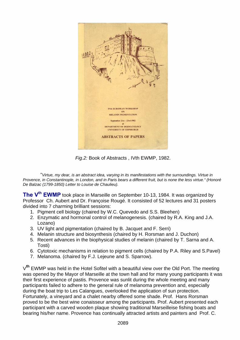
Fig.2: Book of Abstracts , IVth EWMP, 1982. ”Virtue, my dear, is an abstract idea, varying in its manifestations with the surroundings. Virtue in Provence, in Constantinople, in London, and in Paris bears a different fruit, but is none the less virtue.“ (Honoré De Balzac (1799-1850) Letter to Louise de Chaulieu). The Vth EWMP took place in Marseille on September 10-13, 1984. It was organized by Professor Ch. Aubert and Dr. Françoise Rougé. It consisted of 52 lectures and 31 posters divided into 7 charming brilliant sessions:
1. Pigment cell biology (chaired by W.C. Quevedo and S.S. Bleehen) 2. Enzymatic and hormonal control of melanogenesis. (chaired by R.A. King and J.A.
Lozano) 3. UV light and pigmentation (chaired by B. Jacquet and F. Serri) 4. Melanin structure and biosynthesis (chaired by H. Rorsman and J. Duchon) 5. Recent advances in the biophysical studies of melanin (chaired by T. Sarna and A.
Tosti) 6. Cytotoxic mechanisms in relation to pigment cells (chaired by P.A. Riley and S.Pavel) 7. Melanoma. (chaired by F.J. Lejeune and S. Sparrow).
Vth EWMP was held in the Hotel Sofitel with a beautiful view over the Old Port. The meeting was opened by the Mayor of Marseille at the town hall and for many young participants it was their first experience of pastis. Provence was sunlit during the whole meeting and many participants failed to adhere to the general rule of melanoma prevention and, especially during the boat trip to Les Calangues, overlooked the application of sun protection. Fortunately, a vineyard and a chalet nearby offered some shade. Prof. Hans Rorsman proved to be the best wine conaisseur among the participants. Prof. Aubert presented each participant with a carved wooden plaque showing traditional Marseilleise fishing boats and bearing his/her name. Provence has continually attracted artists and painters and Prof. C.
2089
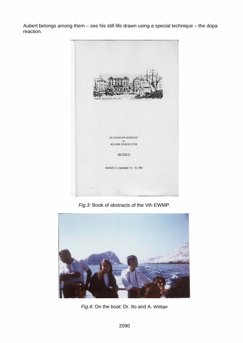
Aubert belongs among them – see his still life drawn using a special technique – the dopa reaction.
Fig.3: Book of abstracts of the Vth EWMP.
Fig.4: On the boat: Dr. Ito and A. Wittbjer
2090

Fig.5: Dopa flowers by C. Aubert.
2091
CURRENT LITERATURE ___________________________________
111... CCChhheeemmmiiissstttrrryyy ooofff MMMeeelllaaannniiinnnsss aaannnddd ooottthhheeerrr PPPiiigggmmmeeennntttsss (Pr A. Napolitano) A number of papers have appeared focused on the design and properties of melanin-based materials. Therefore these will be listed under a separate heading from now on. Among these, polydopamine films which have been studied as functional coatings for a variety of surfaces. Improved properties for these films in terms of transparency and ion permeability have been obtained by alternating deposition of dopamine-melanin particles with poly(diallyldimethylammonium chloride) (Bernsman et al ChemPhys Chem). Impedance spectra and zeta potential titration curves have also been recorded to get an insight into the electrical properties and surface chemistry of such films (Ball V Colloids and Surfaces) Polydopamine membranes were fabricated by dipping Stainless Steel substrates into alkaline dopamine solutions, fully characterized (by scanning electron microscope (SEM), energy dispersive X-ray spectroscopy (EDS), surface reflection Fourier transform IR spectrum (SR-FTIR), UV-visible spectrum) and the corrosion behaviour examined (Yu et al J. Molecular Structure). Full characterization of nanocomposite materials prepared by laponite promoted oxidation of L-Dopa has been reported (Jaber et al J. Chem Phys Lett). Hybrid materials have also been obtained from electrochemical polymerized polydopamine and single walled carbon nanotubes. Wrapping of the nanotubes with polymers resulted in a decrease of the impedance of such composite electrode and an increase of the rate of electron transfer from the electrolyte to the electrode ( Ben-Valid et al, Carbon). Another field receiving increasing interest is the exploitation of black pigments from plant sources which are now currently referred to as plant melanins. Extraction technologies have been set up for extraction of melanin from soy sauce waste (ren et al ) Auricularia auricula fruit bodies (Zou et al Innovative Food Science & Emerging Technologies) and black waxy corn (Ma et al Faming Zhuanli Shenqing). As usual, a variety of new melanogenesis inhibitory substances or plant extracts have been reported, among which of particular interest sulfurated derivatives of resorcinol with depigmenting, antioxidant and antibacterial properties (Poigny S Fr. demande). Structure, Reactivity and Properties - Beberok A, Buszman E, Zdybel M, Pilawa B, Wrzesniok D.
EPR examination of free radical properties of DOPA- melanin complexes with ciprofloxacin, lomefloxacin, norfloxacin and sparfloxacin. Chem Phys Letts (2010), 497(1-3):115-122.
- Jacob D, Shelton RL, Applegate BE.
Fourier domain Pump-Probe Optical Coherence Tomography imaging of melanin. Optics Express (2010), 18(12):12399-12410.
- Najder-Kozdrowska L, Pilawa B, Buszman E, Wieckowski AB, Swiatkowska L, Wrzesniok D, Wojtowicz W.
Triplet states in DOPA- melanin and in its complexes with kanamycin and copper Cu(II) ions. Acta Physica Polonica, A (2010), 118(4):613-618.
Melanin-based materials - Ball V.
Impedance spectroscopy and zeta potential titration of dopa- melanin films produced by oxidation of dopamine. Colloids and Surfaces, A: Physicochemical and Engineering Aspects (2010), 363(1-3):92-97.
- Ben-Valid S, Botka B, Kamaras K, Zeng A, Yitzchaik S.
Spectroscopic and electrochemical study of hybrids containing conductive polymers and carbon nanotubes. Carbon (2010), 48(10):2773-2781.
- Bernsmann F, Ersen O, Voegel J-C, Jan E, Kotov NA, Ball V.
2092
Melanin-Containing Films: Growth from Dopamine Solutions versus Layer-by-Layer Deposition. ChemPhysChem (2010), 11(15),3299-3305.
- Jaber M, Lambert J-F.
A New Nanocomposite: L-DOPA/Laponite. J Phys Chem Letts (2010), 1(1):85-88. - Jastrzebska M, Mroz I, Barwinski B, Wrzalik R, Boryczka S.
AFM investigations of self-assembled DOPA- melanin nanoaggregates. J Mat Sci (2010), 45(19):5302-5308. - Yu F, Chen S, Chen Y, Li H, Yang L, Chen Y, Yin Y.
Experimental and theoretical analysis of polymerization reaction process on the polydopamine membranes and its corrosion protection properties for 304 Stainless Steel. J Mol Structure (2010), 982(1-3):152-161.
Melanogenesis and its Modulation - Arung ET, Furuta S, Ishikawa H, Kusuma IW, Shimizu K, Kondo R.
Anti-melanogenesis properties of quercetin- and its derivative-rich extract from Allium cepa. Food Chem (2010), 124(3):1024-1028.
- Arung ET, Shimizu K, Tanaka H, Kondo R.
Melanin biosynthesis inhibitors from wood of Artocarpus heterophyllus: the effect of isoprenoid substituent of flavone with 4-substituted resorcinol moiety at B ring. Letts Drug Design & Discovery (2010), 7(8):602-605.
- Choi Y-M, Jun H-j, Dawson K, Rodriguez RL, Roh MR, Jun J, Choi C-H, Shim J-H, Lee CH, Lee SJ, Park K-H,
Lee S-J. Effects of the isoflavone puerarin and its glycosides on melanogenesis in B16 melanocytes. Eur Food ResTechnol (2010), 231(1):75-83.
- Chou T-H, Ding H-Y, Lin R-J, Liang J-Y, Liang C-H.
Inhibition of Melanogenesis and Oxidation by Protocatechuic Acid from Origanum vulgare (Oregano). J Nat Prod (2010), 73(11):1767-1774.
- Jeong ET, Jin MH, Kim M-S, Chang YH, Park SG.
Inhibition of melanogenesis by piceid isolated from Polygonum cuspidatum. Arch Pharm Res (2010), 33(9):1331-1338.
- Kim KD, Song MH, Yum EK, Jeon OS, Ju YW, Chang MS.
Melanogenesis inhibition by mono-hydroxycinnamic ester derivatives in B16 melanoma cells. Bull Korean Chem Soc (2010), 31(1):181-184.
- Koo J-H, Lee I-J, Yun S-K, Kim H-U, Park B-H, Park J-W.
Saponified Evening Primrose Oil Reduces Melanogenesis in B16 Melanoma Cells and Reduces UV-Induced Skin Pigmentation in Humans. Lipids (2010), 45(5):401-407.
- Lee CW, Kim HS, Kim HK, Kim J-W, Yoon JH, Cho Y, Hwang JK.
Inhibitory effect of panduratin A isolated from Kaempferia panduarata Roxb. on melanin biosynthesis. Phytother Res: PTR (2010), 24(11):1600-4.
- Lee EH, Lim Y-J, Ha SK, Kang TH, Koketsu M, Kang C, Kim SY, Park J-H.
Inhibitory effects of 5-chloroacetyl-2-piperidino-1,3-selenazole, a novel selenium-containing compound, on skin melanin biosynthesis. J Pharm Pharmacol (2010), 62(3):352-359.
- Matsui Y, Sugiyama K, Kamei M, Takahashi T, Suzuki T, Katagata Y, Ito T.
Extract of Passion Fruit (Passiflora edulis) Seed Containing High Amounts of Piceatannol Inhibits Melanogenesis and Promotes Collagen Synthesis. J Agr Food Chem (2010), 58(20): 11112-11118.
- Nakashima S, Matsuda H, Oda Y, Nakamura S, Xu F, Yoshikawa M.
Melanogenesis inhibitors from the desert plant Anastatica hierochuntica in B16 melanoma cells. Bioorg Med Chem (2010), 18(6): 2337-2345.
- Poigny S.
2093

Preparation of sulfurated derivatives of resorcinol as depigmenting, antioxidant and antibacterial agents for use in cosmetic and pharmaceutical compositions. Fr. Demande (2010), 42pp.
- Seo WD, Ryu YB, Curtis-Long MJ, Lee CW, Ryu HW, Jang KC, Park KH.
Evaluation of anti-pigmentary effect of synthetic sulfonylamino chalcone. Eur J Med Chem (2010), 45(5): 2010-2017.
- Shirasugi I, Kamada M, Matsui T, Sakakibara Y, Liu M-C, Suiko M.
Sulforaphane inhibited melanin synthesis by regulating tyrosinase gene expression in B16 mouse melanoma cells. Biosci Biotechnol Biochem (2010), 74(3):579-582.
- Yamaguchi Y, Hearing VJ, Maeda A, Morita A.
NADPH:quinone oxidoreductase-1 as a new regulatory enzyme that increases melanin synthesis. J Invest Dermatol (2010), 130(3): 645-7.
- Yu JS, Kim AK.
Effect of combination of taurine and azelaic acid on antimelanogenesis in murine melanoma cells. J Biomed Sci (2010), 17(Suppl. 1),
- Zhu Y-J, Qiu L, Zhou J-J, Guo H-Y, Hu Y-H, Li Z-C, Wang Q, Chen Q-X, Liu B.
Inhibitory effects of hinokitiol on tyrosinase activity and melanin biosynthesis and its antimicrobial activities. J Enz Inh Med Chem (2010), 25(6):798-803.
Plant and fungal pigments - Chai LYA, Netea MG, Sugui J, Vonk AG, van de Sande WWJ, Warris A, Kwon-Chung KJ, Jan K.
Aspergillus fumigatus Conidial Melanin Modulates Host Cytokine Response. Immunobiol (2010), 215(11): 915-920.
- Ma Q, Xu X, Lu W, Chen Y.
Method for extracting melanin from black waxy corn. Faming Zhuanli Shenqing (2010), 8pp. - Ren H, Wang X, Liu D.
Extraction technology of melanin from soy sauce waste and its biological function. Shipin Yu Fajiao Gongye (2010), 36(5): 156-160.
- Revankar SG, Sutton DA.
Melanized fungi in human disease. Clinical microbiology reviews (2010), 23(4): 884-928. - Sapkota K, Park S-E, Kim J-E, Kim S, Choi H-S, Chun H-S, Kim S-J.
Antioxidant and antimelanogenic properties of chestnut flower extract. Biosci Biotechnol Biochem (2010), 74(8):1527-1533.
- Ye M, Xu Q, Chen X, Yang L, Lin Y.
Total phenolic content and antioxidant activities of the extracellular melanin from Lachnum sp. YM-223. Junwu Xuebao (2010), 29(4): 608-611.
- Zou Y, Xie C, Fan G, Gu , Han Y.
Optimization of ultrasound-assisted extraction of melanin from Auricularia auricula fruit bodies. Innovative Food Science & Emerging Technologies (2010), 11(4): 611-615.
2094

222... BBBiiiooolllooogggyyy ooofff pppiiigggmmmeeennnttt ccceeellllllsss aaannnddd pppiiigggmmmeeennntttaaarrryyy dddiiisssooorrrdddeeerrrsss (Dr M. Picardo)
The Aryl hydrocarbon receptor (AhR) is a member of the family of basic-helix-loop-helix transcription factors. AhR is a transcription factor that is normally inactive; upon ligand binding AhR translocates to the nucleus, dimerizes with ARNT, enabling the complex to bind to DNA recognition sequences, and eventually initiates gene transcription. Since there are several recent evidences demonstrating that AhR activation affects cell proliferation, differentiation, and apoptosis, an increasing number of studies focus on AhR-dependent modulation of cell and organ-specific functions. Two distinct papers (Jux et al., Luecke et al.,) described the involvement of aryl hydrocarbon receptor in skin tanning. The paper from Jux et al., demonstrated that AhR plays a role in pigment formation by regulating the expression of genes coding for enzymes of the melanogenic pathway. UV light, the major physiological stimulus for melanogenesis, activates the aryl hydrocarbon receptor via a specific bioactive signal molecule that is formed in response to light. The study, from Luecke et al., using AhR-deficient mice showed a significantly weaker tanning than wild-type mice. Interestingly, in these mice, tyrosinase activity in the epidermis was lower as well. Tanning responses and tyrosinase activity, however, were normal in keratinocyte-specific conditional AhR knockout mice, indicating that release of melanogenic keratinocyte factors is unaffected by the UVB-AhR signaling pathway and that the diminished tanning response in AhR_/_ mice is confined to the level of melanocytes. Accordingly, the number of dihydroxyphenylalanin-positive melanocytes increased significantly less on UVB irradiation in AhR_/_ mice than in wild-type mice. This difference in melanocyte number was associated with a significantly reduced expression of stem cell factor-1 and c-kit in melanocytes of AhR_/_ mice. In conclusion, In conclusion both studies proposed a previously unknown role of the environmental signal sensor AhR links solar UVB radiation to skin pigmentation. Botton et al., demonstrated that the thiazolidinedione ciglitazone inhibited, independently of PPARgamma activation, melanoma cell growth. Interestingly, ciglitazone effects are mediated through the regulation of secreted factors. Q-PCR screening of several genes involved in melanoma biology reveals that ciglitazone inhibits expression of the CXCL1 chemokine gene. CXCL1 is overexpressed in melanoma and contributes to tumorigenicity. Ciglitazone induces a diminution of CXCL1 level in different human melanoma cell lines. This effect is mediated by the downregulation of microphthalmia-associated transcription factor, MITF, the master gene in melanocyte differentiation and involved in melanoma development. Further, recombinant CXCL1 protein is sufficient to abrogate thiazolidinedione effects such as apoptosis induction, whereas extinction of the CXCL1 pathway mimics phenotypic changes observed in response to ciglitazone. Finally, inhibition of human melanoma tumor development in nude mice treated with ciglitazone is associated with a strong decrease in MITF and CXCL1 levels. The study proposed an anti-melanoma approach involving an inhibition of the MITF/CXCL1 axis. Herraiz et al., described a new functional link between the stem cell factor receptor and Melanocortin 1 receptor (MC1R), a Gs protein-coupled receptor expressed in melanocytes. Upon stimulation by αMSH, MC1R triggers the cAMP and ERK1/ERK2 MAPK pathways. In mouse melanocytes, ERK activation by αMSH binding to Mc1r depends on cAMP, and melanocytes are considered a paradigm for cAMP-dependent ERK activation. However, human MC1R variants associated with red hair, fair skin [red hair color (RHC) phenotype], and increased skin cancer risk display reduced cAMP signaling but activate ERKs as efficiently as wild type in heterologous cells, suggesting independent signaling to ERKs and cAMP in human melanocytes. MC1R signaling activated the ERK pathway in normal human melanocytes and melanoma cells expressing physiological levels of endogenous RHC variants. ERK activation is comparable for wild-type and mutant MC1R and is independent on cAMP because it is neither triggered by stimulation of cAMP synthesis with forskolin nor blocked by the adenylyl cyclase inhibitor 2',5'-dideoxyadenosine. Pharmacological interference, small interfering RNA studies, expression profiles, and functional reconstitution experiments showed that αMSH-induced ERK activation resulted from Src tyrosine kinase-mediated transactivation of the stem cell factor receptor, a receptor tyrosine kinase essential for proliferation, differentiation, and survival of melanocyte precursors, thus demonstrating a functional link between the stem cell factor receptor and MC1R. The next goal for the translational research will arise from the regerative approach. Accordingly, relevant impact should be assigned to the definition of the definition and characterization of the stem compartment. Usually, for the melanocyte lineage the hair follicle is considered the stem reservoir. Recently, Ling et al published clear indications on the dermal stem melanocytes providing their phenotypic and functional features. The paper opens new perspectives on vitiligo pathogenesis and consequent molecular-oreinted approaches. Further new insights are provided by the genomewide study of Quan et al on the possible vitiligo gene association within MHC region. He identified a possible association with IBD. - Botton T, Puissant A, Cheli Y, Tomic T, Giuliano S, Fajas L, Deckert M, Ortonne JP, Bertolotto C, Tartare-Deckert S,
Ballotti R, Rocchi S. Ciglitazone negatively regulates CXCL1 signaling through MITF to suppress melanoma growth. Cell Death Differ. 2010 Jul 2.
- Herraiz C, Journé F, Abdel-Malek Z., Ghanem G, Jimenez-Cervantes C, Garcìa-Borròn JC.
Signaling from the Human Melanocortin 1 Receptor to ERK1 and ERK2 Mitogen-Activated Protein Kinases Involves Transactivation of cKIT. Mol Endocrinol. 2010 Nov 17.
2095

- Jux B, Kadow S, Luecke S, Rannug A, Krutmann J, Esser C.
The Aryl Hydrocarbon Receptor Mediates UVB Radiation-Induced Skin Tanning. J Invest Dermatol. 2010 Sep 23. - Ling L, Fukunaga-Kalabis, H Yu, X Xu, J Kong, J Lee and M Herlyn.
Human dermal stem cells differentiate into functional epidermal melanocytes. J Cell Sci 123(6):853-860. - Luecke S, Backlund M, Jux B, Esser C, Krutmann J, Rannug A.
The aryl hydrocarbon receptor (AHR), a novel regulator of human melanogenesis. Pigment Cell Melanoma Res. 2010 Dec;23(6):828-33.
- Quan C, Ren YQ, Xiang LH, et al. genome-wide association study for vitiligo identifies susceptibility loci at 6q27 and the MHC. Nat Gen 42(7):614-619, 2010.
2096

333... M MMSSSHHH,,, MMMCCCHHH,,, ooottthhheeerrr hhhooorrrmmmooonnneeesss,,, dddiiiffffffeeerrreeennntttiiiaaatttiiiooonnn (Pr M. Böhm)
What’s hot and new?A negative feedback loop for α-MSH-MC1R-mediated signaling identified in melanocytes. The intracellular amount of the ubiquitous signal transduction messenger cAMP, following activation of Gs-stimulating G protein-coupled receptors, is known to be controlled by phosphodiesterases. (PDEs). These enzymes orchestrate the magnitude, duration and subcellular localization of cAMP. Since cAMP is a crucial mediator within the α-MSH-MC1R-MITF pathway that regulates melanocytes differentiation, melanogenesis, proliferation as well as the dermal tanning response. In a recent research communicated by Khaled et al. (Genes Dev. 2010; 24: 2276-81) PDE4D3 was identified as a direct target of the MSH-cAMP-MITF pathway. Using gene ablation of this enzyme, the authors showed that PDE4D3 expression is MITF-dependent. By reporter-promoter assays as well as chromatin immunoprecipitation assays it could further delineated that MITF directly acts at PDE4D3 promoter to induce expression of its gene. Most interestingly, PDE4 inhibition synergized with forskolin to induce skin pigmentation in redhead/fair-skinned mice. In summary, the identified negative homeostatic of PDE4D3 pathway highlights a potent mechanism controlling melanocyte differentiation that may be amenable to pharmacologic manipulation for skin cancer prevention. Endogenous expression of chimeric MC1R-TUBB3 isoforms in human melanoma cells. Alternative splicing is a well known mechanism how higher eukaryotes increase their repertoire of proteins derived from a limited number of genes. In a recent paper by Dalziel et al. (Nucleic Acids Res. 2010, Epub ahead of print) an interesting additional mechanism, i. e. splicing between adjacent genes, was investigated with regard to the MC1R gene. Using HEK293 cells transfected with various MC1R minigenes, the MC1R gene was first identified to contain an unusual and highly complex poly(A) site (PAS) with rigid positional and structural arrangements of four individuals sequence components. Such structural arrangements were not identified within the MC4R gene. Interestingly, MC1R transcripts are not cleaved at the PAS and can create chimeric MC1R-TUBB3 transcripts due to the immediate downstream neighbour locus tubulin-β-III (TUBB3). Interestingly, these transcripts produced two distinct protein isoforms localizing to the plasma membrane and the endoplasmic reticulum. Melanoma cells expressing functional MC1R responded to α-MSH or activation of the stress response kinase p38-MAPK with a shift in expression from MC1R in favour of chimeric MC1R-TUBB3 isoforms. The authors propose that expression of such chimeric MC1R proteins may also equip normal human melanocytes with cellular phenotypes required as part of the pigmentation response. Signaling cross-talks between MC1R and cKIT in normal human melanocytes. In previous reports it was shown that in mouse melanocytes (melamoma cells) stimulation by α-MSH via cAMP activates the ERK1/ERK2 MAPK pathway. However, earlier work has suggested that this pathway may be different in normal human melanocytes. In a recent report by Herraiz et al. (Mol Endocrinol. 2010, Epub ahead of print) the impact of α-MSH and artificial cAMP inducers were examined on activation of ERK1/2 and the mast cell / stem cell factor cKIT was examined in normal human melanocytes expressing wild-type MC1R and MC1R variants associated with red hair and fair skin. Surprisingly, cells expressing MC1R variants were found to activate ERKs as efficiently as wild type in heterologous cells suggesting independent signaling to ERKs and cAMP in human melanocytes. In accordance with earlier work (e. g. Böhm et al., Cell Growth Diff. 1995; 6: 291-302) ERK1/2 activation was independent on cAMP because it was neither triggered by forskolin nor blocked by the adenylyl cyclase inhibitor 2',5'-dideoxyadenosine. Interestingly, pharmacological interference, small interfering RNA studies, expression profiles, and functional reconstitution experiments revealed that α-MSH-induced ERK activation resulted from Src tyrosine kinase-mediated transactivation of cKIT. The described transactivation phenomenon of the cKIT-ERK pathway in melanocytes expressing natural MC1R mutations appears to be unique and may point towards unexpected functional connection between the GPCR- and RTK-mediated signalling.
2097
444... PPPhhhoootttooobbbiiiooolllooogggyyy (Dr N. Smit)
The “indoor tanning business” once more is the topic of discussing in a number of papers that appeared in the recent literature (3, 7, 10, 12, 20, 34 and 37). Hery et al describe estimated annual percent changes (EAPCs) in the melanoma incidence in Iceland. EAPCs in men and women on the trunk have increased since 1954 and especially in women (aged less than 50) a sharp epidemic increase was observed after 1995. Use of sunbeds in Iceland expanded rapidly after 1985 and a connection therefore seems plausible. Cust et al analysed data from the Australian Melanoma Family Study and the association with sunbed use was especially strong for melanoma diagnosed at early age and with more frequent use. Lazovich et al studied a population of melanoma patients in Minnesota and found adjusted odds ratio for UVB-enhanced and primarily UVA-emitting devices of 2.86 and 4.44, respectively. Veierod et al discuss the beneficial effects of sun exposure for the production of vitamin D. They indicate that the mean irradiance from solariums is higher than that from the summer sun in Oslo and they recommend that restriction of solarium use is maintained. Grant and Schuitemaker describe that serum levels of 25-hydroxyvitamin D for optimal health would be 100-150 nmol/L. To increase the production to this level would only pose minimal increased risks of melanoma or skin cancer. Fisher (10) does not think that the skin vitamin D synthesis is a justifiable defense of indoor tanning and that strict regulation of this industry may offer one of the best ways of cancer prevention. Martinez-Levasseur et al studied sunburn and photoprotection in mammalian wildlife by photographic and histological surveys of different whale species and conclude that darker pigmentation is advantageous for the whales as in humans. Miyamura et al studied the benefits of facultative pigmentation . They found that unlike the UVB induced tanning, the pigmentation resulting from UVA treatment essentially provided no photoprotection. In this respect the findings of Mahmoud et al could be of interest since they describe pigment induced by visible light that also has different qualitative and quantitative properties than the pigment produced by UVA1 light in skin type IV-VI individuals. No pigmentation was induced in skin type II. The melanin type and content therefore will be important for the prevention of UV induced DNA damage. UVA lamps are known to generate reactive oxygen species. The paper by Jenkins et al describes a role for p16 as a regulator of oxidative stress. Melanocytes were found more susceptible to oxidative stress than fibroblasts and keratinocytes which might explain why the loss of p16 predisposes especially the melanocytes to cancerogenesis. Furthermore the paper by Wang et al in the PNAS describes the role of melanin in the production of ROS in melanocytes and the subsequent generation of oxidative DNA damage lesions. UVA induces much higher amount of such lesions in melanocytes than in fibroblasts probably due to the presence of melanin and the melanocytes showed a reduced repair capacity. The negative effects of (UVA)solaria, the role of the p16 tumor suppressor as a regulator of oxidative stress and the role of melanin mediating ROS production suggests an important role for the anti-oxidant defence of the pigment cells. In this light melanoma prevention may be achieved by antioxidants and dietary nutrients as proposed in the papers by Jensen et al and Sapkota et al. - Indoor tanning is strongly linked to melanoma risk. Harv.Womens Health Watch. 2010, 18:7. - An SM, Koh JS, Boo YC.
p-coumaric acid not only inhibits human tyrosinase activity in vitro but also melanogenesis in cells exposed to UVB. Phytother.Res. 2010, 24:1175-1180.
- Berwick M.
Invited commentary: a sunbed epidemic? Am.J.Epidemiol. 2010, 172:768-770. - Bzhalava D, Bray F, Storm H, Dillner J.
Risk of second cancers after the diagnosis of Merkel cell carcinoma in Scandinavia. Br.J.Cancer. 2010. - Chen N, Hu Y, Li WH, Eisinger M, Seiberg M, Lin CB.
The role of keratinocyte growth factor in melanogenesis: a possible mechanism for the initiation of solar lentigines. Exp.Dermatol. 2010, 19:865-872.
- Chiang HM, Lin JW, Hsiao PL, Tsai SY, Wen KC.
Hydrolysates of citrus plants stimulate melanogenesis protecting against UV-induced dermal damage. Phytother. Res. 2010.
- Cust AE, Armstrong BK, Goumas C, Jenkins MA, Schmid H, Hopper JL, Kefford RF, Giles GG, Aitken JF, Mann GJ.
Sunbed use during adolescence and early adulthood is associated with increased risk of early-onset melanoma. Int.J.Cancer. 2010.
- Dalziel M, Kolesnichenko M, das Neves RP, Iborra F, Goding C, Furger A.
2098
{alpha}-MSH regulates intergenic splicing of MC1R and TUBB3 in human melanocytes. Nucleic Acids Res. 2010. Significantly, treatment with the MC1R agonist alpha-MSH or activation of the stress response kinase p38-MAPK, both key molecules associated with ultraviolet radiation dermal insult and subsequent skin tanning, result in a shift in expression from MC1R in favour of chimeric MC1R-TUBB3 isoforms in cultured melanocytes. We propose that these chimeric proteins serve to equip melanocytes with novel cellular phenotypes required as part of the pigmentation response.
- Elias PM, Menon G, Wetzel BK, Williams JJ.
Barrier requirements as the evolutionary "driver" of epidermal pigmentation in humans. Am.J.Hum.Biol. 2010, 22:526-537. We summarize evidence here that epidermal interfollicular pigmentation in early hominids likely evolved in response to stress to the permeability barrier.
- Fisher DE, James WD.
Indoor tanning--science, behavior, and policy. N.Engl.J.Med. 2010, 363:901-903. - Gaddameedhi S, Kemp MG, Reardon JT, Shields JM, Smith-Roe SL, Kaufmann WK, Sancar A.
Similar nucleotide excision repair capacity in melanocytes and melanoma cells. Cancer Res. 2010, 70:4922-4930. We concluded that melanoma cells retain capacity for nucleotide excision repair, the loss of which probably does not commonly contribute to melanoma progression.
- Grant WB, Schuitemaker GE.
Health benefits of higher serum 25-hydroxyvitamin D levels in The Netherlands. J.Steroid Biochem.Mol.Biol. 2010, 121:456-458.
- Hay J, Dibonaventura M, Baser R, Press N, Shoveller J, Bowen D.
Personal attributions for melanoma risk in melanoma-affected patients and family members. J.Behav.Med. 2010. - Hery C, Tryggvadottir L, Sigurdsson T, Olafsdottir E, Sigurgeirsson B, Jonasson JG, Olafsson JH, Boniol M, Byrnes
GB, Dore JF, Autier P. A melanoma epidemic in Iceland: possible influence of sunbed use. Am.J.Epidemiol. 2010, 172:762-767.
- Horike N, Kumagai A, Shimono Y, Onishi T, Itoh Y, Sasaki T, Kitagawa K, Hatano O, Takagi H, Susumu T, Teraoka
H, Kusano K, Nagaoka Y, Kawahara H, Takemori H. Downregulation of SIK2 expression promotes the melanogenic program in mice. Pigment Cell Melanoma Res. 2010, 23:809-819.
- Hornle M, Peters N, Thayaparasingham B, Vorsmann H, Kashkar H, Kulms D.
Caspase-3 cleaves XIAP in a positive feedback loop to sensitize melanoma cells to TRAIL-induced apoptosis. Oncogene. 2010.
- Jenkins NC, Liu T, Cassidy P, Leachman SA, Boucher KM, Goodson AG, Samadashwily G, Grossman D.
The p16(INK4A) tumor suppressor regulates cellular oxidative stress. Oncogene. 2010. - Jensen JD, Wing GJ, Dellavalle RP.
Nutrition and melanoma prevention. Clin.Dermatol. 2010, 28:644-649. - Jo SJ, Kwon HH, Choi MR, Youn JI.
No Evidence for Increased Skin Cancer Risk in Koreans with Skin Phototypes III-V Treated with Narrowband UVB Phototherapy. Acta Derm.Venereol. 2010,10-0995.
- Lazovich D, Vogel RI, Berwick M, Weinstock MA, Anderson KE, Warshaw EM.
Indoor tanning and risk of melanoma: a case-control study in a highly exposed population. Cancer Epidemiol.Biomarkers Prev. 2010, 19:1557-1568.
- Lichte V, Dennenmoser B, Dietz K, Hafner HM, Schlagenhauff B, Garbe C, Fischer J, Moehrle M.
Professional risk for skin cancer development in male mountain guides--a cross-sectional study. J.Eur.Acad.Dermatol.Venereol. 2010, 24:797-804.
- Lin J, Yang Q, Wilder PT, Carrier F, Weber DJ.
The calcium-binding protein S100B down-regulates p53 and apoptosis in malignant melanoma. J.Biol.Chem. 2010, 285:27487-27498.
2099
Thus, a well known marker for malignant melanoma, S100B, likely contributes to cancer progression by down-regulating the tumor suppressor protein, p53.
- Mahmoud BH, Ruvolo E, Hexsel CL, Liu Y, Owen MR, Kollias N, Lim HW, Hamzavi IH.
Impact of long-wavelength UVA and visible light on melanocompetent skin. J.Invest Dermatol. 2010, 130:2092-2097.
- Martinez-Levasseur LM, Gendron D, Knell RJ, O'Toole EA, Singh M, cevedo-Whitehouse K.
Acute sun damage and photoprotective responses in whales. Proc.Biol.Sci. 2010. - Miyamura Y, Coelho SG, Schlenz K, Batzer J, Smuda C, Choi W, Brenner M, Passeron T, Zhang G, Kolbe L, Wolber
R, Hearing VJ. The deceptive nature of UVA tanning versus the modest protective effects of UVB tanning on human skin. Pigment Cell Melanoma Res. 2010,10-148X.
- Royle J, Baade P, Joske D, Fritschi L.
Risk of second cancer after lymphohematopoietic neoplasm. Int.J.Cancer. 2010. Our findings provide evidence of an increased risk of cancer, particularly ultraviolet radiation- and immunosuppression-related cancers, following a LHN in Australia.
- Sapkota K, Park SE, Kim JE, Kim S, Choi HS, Chun HS, Kim SJ.
Antioxidant and antimelanogenic properties of chestnut flower extract. Biosci.Biotechnol.Biochem. 2010, 74:1527-1533.
- Sekulic A, Colgan MB, Davis MD, DiCaudo DJ, Pittelkow MR.
Activating BRAF mutations in eruptive melanocytic naevi. Br.J.Dermatol. 2010, 163:1095-1098. The mechanism leading to development of EMN in this, and potentially other patients, may relate to synergistic mutagenic effects of thioguanines and ultraviolet (UV) A. Together with the documented importance of BRAF mutations in melanoma development and maintenance, these findings highlight the importance of UVA protection, especially in patients treated with thiopurines such as 6-mercaptopurine.
- Selfridge J, Song L, Brownstein DG, Melton DW.
Mice with DNA repair gene Ercc1 deficiency in a neural crest lineage are a model for late-onset Hirschsprung disease. DNA Repair (Amst). 2010, 9:653-660. Now, in an attempt to generate a model for UV-induced melanoma, we have used the floxed Ercc1 allele in combination with a Cre transgene under the control of the tyrosinase gene promoter to produce mice with Ercc1-deficient melanocytes that are hypersensitive to UV irradiation.
- Seo SJ, Fisher DE.
Melanocyte photobiology, ultraviolet radiation and melanoma. G.Ital.Dermatol.Venereol. 2010, 145:603-611. Although the picture is far from complete, the mechanisms by which melanocytes respond to ultraviolet radiation are beginning to be elucidated, and, as these pathways emerge, they offer new targets for chemopreventive and chemotherapeutic intervention.
- Tang MS.
Ultraviolet A light: potential underlying causes of melanoma. Future.Oncol. 2010, 6:1523-1526. - Tsai MH, Aki R, Amoh Y, Hoffman RM, Katsuoka K, Kimura H, Lee C, Chang CH.
GFP-fluorescence-guided UVC irradiation inhibits melanoma growth and angiogenesis in nude mice. Anticancer Res. 2010, 30:3291-3294. This color-coded tumor-host model is useful for evaluation of treatment efficacy on melanoma growth and angiogenesis, which are readily discernable with non-invasive color-coded fluorescent protein imaging. These results suggest that fluorescence-guided UVC irradiation is a promising therapeutic strategy for melanoma.
- Turnbull DJ, Parisi AV.
Dosimeter for the measurement of UV exposures related to melanoma induction. Phys.Med.Biol. 2010, 55:3767-3776.
- Veierod MB, Nilsen LT, Robsahm TE.
Solaria, vitamin D, and skin cancer. Tidsskr.Nor Laegeforen. 2010, 130:1818-1821. Recommendations on restricted solarium use should be maintained.
2100
- Wang HT, Choi B, Tang MS. Melanocytes are deficient in repair of oxidative DNA damage and UV-induced photoproducts. Proc.Natl.Acad.Sci.U.S.A. 2010, 107:12180-12185.
- Wang L, Shirure VS, Burdick MM, Wu S.
UVB-irradiation regulates VLA-4-mediated melanoma cell adhesion to endothelial VCAM-1 under flow conditions. Mol.Carcinog. 2010.
- Weinstock MA, Fisher DE.
Indoor ultraviolet tanning: what the data do and do not show regarding risk of melanoma and keratinocyte malignancies. J.Natl.Compr.Canc.Netw. 2010, 8:867-872.
- Xiao L, Aoshima H, Saitoh Y, Miwa N.
Fullerene-polyvinylpyrrolidone clathrate localizes in the cytoplasm to prevent Ultraviolet-A ray-induced DNA-fragmentation and activation of the transcriptional factor NF-kappaB. J.Cell Biochem. 2010, 111:955-966. Thus, the PVP-fullerene may be developed as a UV-protective agent with DNA-preservative effects owing to its combinative ability to molecules in the cytoplasm and cytomembrane, and then represses cellular oxidative stress and blocks abnormal signal pathways.
- Yamashita T, Negishi K, Hariya T, Yanai M, Iikura T, Wakamatsu S.
In vivo microscopic approaches for facial melanocytic lesions after quality-switched ruby laser therapy: time-sequential imaging of melanin and melanocytes of solar lentigo in Asian skin. Dermatol.Surg. 2010, 36:1138-1147.
- Yamate Y, Hiramoto K, Kasahara E, Jikumaru M, Sato EF, Inoue J, Inoue M.
Ultraviolet-A Irradiation to the Eye Modulates Intestinal Mucosal Functions and Properties of Mast Cells in the Mouse. Photochem.Photobiol. 2010,10-1097.
- Zykova TA, Zhu F, Vakorina TI, Zhang J, Higgins LA, Urusova DV, Bode AM, Dong Z.
T-LAK cell-originated protein kinase (TOPK) phosphorylation of Prx1 at Ser-32 prevents UVB-induced apoptosis in RPMI7951 melanoma cells through the regulation of Prx1 peroxidase activity. J.Biol.Chem. 2010, 285:29138-29146.
2101
555... NNNeeeuuurrrooommmeeelllaaannniiinnnsss (Pr M. d'Ischia) The biological roles of neuromelanin in relation to neuronal degeneration and the interaction of the pigment with metal ions are important themes in research on Parkinson’s disease. Tousi et al. (2010) addressed the effects of neuromelanin on astroglial cells, an issue that has remained so far little explored, and showed that the pigment inhibits TNF�-induced expression of interferon �inducible protein-10 (CXCL10). Although the actual relevance of these results to the in vivo situation remains to be demonstrated, the new data point to different roles of neuromelanin in the brain, being largely cytoprotective within dopaminergic neurons, stimulating on microglia when released from damaged neurons and inhibitory on inflammatory signaling in astroglial cells. Porter et al. (2010) investigated the mechanism of coordination of dopamine and 5,6-dihydroxyindole, a putative building block of neuromelanin, with ferric ions in the presence of various ligands. Data showed that both compounds can form strong complexes that account for the notorious iron binding properties of neuromelanin. Finally Chwiej (2010) reported use of X-ray fluorescence microscopy for two-dimensional elemental analysis of substantia nigra tissue and confirmed the statistically significant differences in elemental composition of Parkinsonian cells versus control nerve cells. - Barapatre N, Morawski M, Butz T, Reinert T.
Trace element mapping in Parkinsonian brain by quantitative ion beam microscopy. Nuclear Instruments & Methods in Physics Research, Section B: Beam Interactions with Materials and Atoms (2010), 268(11-12), 2156-2159. Abstract : The role of iron in the pathogenesis of the Parkinson's disease (PD) is a current subject of research in Neurochem., since an abnormal increase in iron is reported in the substantia nigra (SN) of Parkinsonian patients. A severe loss of the cells contg. dopamine in the SN in the PD has also drawn attention towards the function of a browny-black pigment called neuromelanin, which accumulates predominantly in these dopaminergic neurons. The neuromelanin has an ability to chelate metal ions, which, in free state, may cause considerable damage to cells by reacting with their lipid-rich membranes. However, it could also potentiate free radical prodn. if it releases the bound metal ions. The highly sensitive and non-destructive micro-PIXE method suits best to quantify and map the trace elements in the SN. The accuracy in charge measurement for such microanal. studies is of utmost importance for quant. anal. Since a Faraday cup is usually placed behind the thin biol. sample to measure the charge, the primary and the secondary electrons, knocked out from the sample by traversing ion beam, hamper an exact charge detn. Hence, a new non-interceptive technique was developed for precise charge measurement and for continuous monitoring of beam current.
- Chwiej J.
The use of cluster and discriminant analysis in the investigations of the role of trace metals in the pathogenesis of Parkinson's disease. Journal of Trace Elements in Medicine and Biology (2010), 24(2), 78-88. Abstract : X-ray fluorescence microscopy was applied for two-dimensional elemental anal. of substantia nigra (SN) tissue. The samples representing Parkinson's disease (PD) and control cases were examd. at HASYLAB beamline L and at ESRF beamline ID22. Two-dimensional mapping of P, S, Cl, K, Ca, Fe, Cu, Zn, Se and Br was done with the spatial resoln. of 15 and 5 �m. The masses per unit area of elements in neuromelanin reach nerve cells of SN were detd. The elemental data were processed using two multivariate techniques, namely cluster and discriminant anal. The statistical methods were used for data redn., both unsupervised and supervised classification as well as for the creation of a model that would simplify case identification based on the elemental anal. of SN tissue. The results of cluster anal. confirmed the statistical significance of the differences in elemental compn. of PD and control SN nerve cells. Based on the results of discriminant anal., the elements (P, Cl, Fe, Cu and Zn) that played the greatest role in the process of differentiation between neurons from examd. groups were detd.
- Lozano J, Munoz P, Nore BF, Le Doux S, Segura-Aguilar J.
Stable Expression of Short Interfering RNA for DT-Diaphorase Induces Neurotoxicity. Chemical Research in Toxicology 2010, vol. 23, no9, pp. 1492-1496. Abstract : DT-Diaphorase has been proposed to play a neuroprotective role in dopaminergic neurons by preventing aminochrome neurotoxicity. There are several studies supporting this idea, but in all studies, we used dicoumarol, an inhibitor of DT-diaphorase. We have designed and developed two siRNA to silence the expression of DT-diaphorase to study its role in aminochrome metab. We transduced RCSN-3 cells with retroviral particles contg. a pRetroSuper plasmid coding a siRNA for DT-diaphorase. The cells selected in the presence of puromycin generated a stable cell line RCSN-3Nq6 and RCSN-3Nq7 with low expression of DT-diaphorase (27% and 33% of wild type, resp.). A significant cell death was obsd. in RCSN-3 cells expressing siRNA Nq6 and Nq7 for DT-diaphorase when were incubated with 100 �M aminochrome during 48 (4- and 3.5-fold, resp.; P < 0.01). These results support the protective role of DT-diaphorase against aminochrome neurotoxicity in dopaminergic neurons contg. neuromelanin and show that Nq6 and Nq7 siRNA are very useful tools to study the role of DT-diaphorase in aminochrome metab.
2102
- Porter J, Arreguin S, Pierpont CG.
Ferric iron complexes of dopamine and 5,6-dihydroxyindole with nta, edda, and edta as ancillary ligands. Inorganica Chimica Acta (2010), 363(12), 2800-2803. Abstract : Coordination of the neurotransmitter dopamine (DA) and the metal-binding component of neuromelanin 5,6-dihydroxyindole (DHI) with ferric iron has been studied in aq. soln. in the presence of ancillary ligands contg. amine nitrogen and carboxylate oxygen donor sites. With nitrilotriacetic acid (nta) and ethylenediamine diacetic acid (edda) coligands, coordination of the catecholate ligands DA and DHI is obsd. to be complete at physiol. pH. The resulting complexes of DA have the characteristic two-component electronic spectrum obsd. characteristically for L4Fe(Cat) complexes. The spectrum obtained with DHI consists of a single broad absorption in the visible region. Both DA and DHI are able to coordinate with Fe3+ in the presence of edta, displacing carboxylate oxygen donors at pH values just above physiol. pH. These results demonstrate the strong affinity of DA and DHI for Fe3+, pointing to in vivo complex formation in neuronal mixts. at physiol. pH.
- Tousi NS, Buck DJ, Zecca L, Davis RL.
Neuromelanin inhibits CXCL10 expression in human astroglial cells. Neuroscience Letters (2010), 486(1), 47-50. Abstract : Increasing evidence indicates neuroinflammation is instrumental in the pathogenesis of Parkinson's disease (PD). In PD, there is selective degeneration of neuromelanin (NM)-contg. dopamine neurons. Neuromelanin is predominantly cytoprotective within dopaminergic neurons, whereas, NM released from damaged neurons activates microglia. However, the effects of NM on astroglial cells remain largely unknown. Astroglia are essential to neuronal homeostasis and responsive to injury, in part, through secretion of chemokines, including interferon � inducible protein-10 (CXCL10). Thus, we used an in vitro approach to identify the effects of NM on TNF�-induced CXCL10 expression in human astroglial cells. TNF�-induced CXCL10 expression was inhibited in NM exposed cells. Addnl., TNF�-induced NF-[n.1082]B activation was inhibited by NM. Given that CXCL10 expression is NF-[n.1082]B-dependent in human astroglial cells, these findings suggest that NM may inhibit CXCL10 expression, in part, through an NF-[n.1082]B-dependent mechanism. While the in vivo consequences of NM mediated effects on astroglial CXCL10 expression remain to be fully elucidated, insights obtained in this study further our understanding of the effects of NM on inflammatory signaling in human astroglial cells.
2103
666... GGGeeennneeetttiiicccsss,,, mmmooollleeecccuuulllaaarrr aaannnddd dddeeevvveeelllooopppmmmeeennntttaaalll bbbiiiooolllooogggyyy (Dr F. Beermann) - Alkhateeb A, Al-Dain MN, Qarqaz F.
SMOC2 gene variant and the risk of vitiligo in Jordanian Arabs. Eur J Dermatol 2010 (Epub, ahead of print). - Ambler CA, Watt FM.
Adult epidermal Notch activity induces dermal accumulation of T cells and neural crest derivatives through upregulation of jagged 1. Development 137: 3569-3579, 2010.
- Aoki H, Hara A, Motohashi T, Osawa M, Kunisada T.
Functionally distinct melanocytes populations revealed by reconstitution of hair follicles in mice. Pigment Cell Melanoma Res 2010 (Epub, ahead of print). Abstract: Hair follicle reconstitution analysis was used to test the contribution of melanocytes or their precursors to regenerated hair follicles. In this study, we first confirmed the process of chimeric hair follicle regeneration by both hair keratinocytes and follicular melanocytes. Then, as first suggested from the differential growth requirements of epidermal skin melanocytes and non-cutaneous or dermal melanocytes, we confirmed the inability of the latter to be involved as follicular melanocytes to regenerate hair follicles during the hair reconstitution assay. This clear functional discrimination between non-cutaneous or dermal melanocytes and epidermal melanocytes suggests the presence of 2 different melanocyte cell lineages, a finding that might be important in the pathogenesis of melanocyte-related diseases and melanomas.
- Arnheiter H.
The discovery of the microphthalmia locus and its gene, Mitf. Pigment Cell Melanoma Res 23: 729-735, 2010. - Bellei B, Pitisci A, Catricala C, Larue L, Picardo M.
Wnt/beta-catenin signaling is stimulated by alpha-Melanocyte-Stimulating Hormone in melanoma and melanocyte cells: implication in cell differentiation. Pigment Cell Melanoma Res 2010 (Epub, ahead of print).
- Bharti K, Debbache J, Wang X, Arnheiter H.
The basic-helix-loop-helix-leucine zipper gene Mitf: analysis of alternative promoter choice and splicing. Methods Mol Biol 647: 237-250, 2010.
- Chaki M, Sengupta M, Mondal M, Bhattacharya A, Mallick S, Bhadra R, Ray K.
Molecular and functional studies of tyrosinase variants among Indian Oculocutaneous Albinism Type 1 patients. J Invest Dermato 2010 (Epub, ahead of print).
- Choi W, Wolber R, Gerwat W, Mann T, Batzer J, Smuda C, Liu H, Kolbe L, Hearing VJ.
The fibroblast-derived paracrine factor neuregulin-1 has a novel role in regulating the constitutive color and melanocyte function in human skin. J Cell Sci 123: 3102-3111, 2010. Abstract: Interactions between melanocytes and neighboring cells in the skin are important in regulating skin color in humans. We recently demonstrated that the less pigmented and thicker skin on the palms and soles is regulated by underlying fibroblasts in those areas, specifically via a secreted factor (DKK1) that modulates Wnt signaling. In this study, we tested the hypothesis that dermal fibroblasts regulate the constitutive skin color of individuals ranging from very light to very dark. We used microarray analysis to compare gene expression patterns in fibroblasts derived from lighter skin types compared to darker skin types, with a focus on secreted proteins. We identified a number of genes that differ dramatically in expression and, among the expressed proteins, neuregulin-1, which is secreted by fibroblasts derived from dark skin, effectively increases the pigmentation of melanocytes in tissue culture and in an artificial skin model and regulates their growth, suggesting that it is one of the major factors determining human skin color.
- Curran K, Lister JA, Kunkel GR, Prendergast A, Parichy DM, Raible DW.
Interplay between Foxd3 and Mitf regulates cell fate plasticity in the zebrafish neural crest. Dev Biol 344: 107-118, 2010. Shortened abstract: Pigment cells of the zebrafish, Danio rerio, offer an exceptionally tractable system for studying the genetic and cellular bases of cell fate decisions. Cell lineage analyses using the photoconvertible marker, EosFP, revealed that both melanophores and iridophores develop from a mitfa+ precursor. Taken together, our data reveal a Foxd3/mitfa transcriptional switch that governs whether a bi-potent pigment precursor will attain either an iridophore or a melanophore fate.
- Dalziel M, Kolesnichenko M. das Neves RP, Iborra F, Goding C, Furger A.
2104

(alpha)-MSH regulates intergenic splicing of MC1R and TUBB3 in human melanocytes. Nucleic Acids Res 2010 (Epub, ahead of print).
- de Keizer PL, Packer LM, Szypowska AA, Riedl-Polderman PE, van den Broek NJ, de Bruin A, Dansen TB, Marais R,
Brenkman AB, Burgering BM. Activation of forkhead box O transcription factors by oncogenic BRAF promotes p21cip1-dependent senescence. Cancer Res 70: 8526-8536, 2010.
- Fukuzawa T.
Unusual development of light-reflecting pigment cells in intact and regenerating tail in the periodic albino mutant of Xenopus laevis. Cell Tissue Res 342: 53-66, 2010.
- Gargiulo A, Testa F, Rossi S, Di Iorio V, Fecarotta S, de Berardinis T, Iovine A, Magli A, Signorini S, Fazzi E,
Galantuomo MS, Fossarello M, Montefusco S, Ciccodicola A, Neri A, Macaluso C, Simonelli F, Surace EM. Molecular and clinical characterization of albinism in a large cohort of Italian patients. Invest Ophthalmol Vis Sci 2010 (Epub, ahead of print).
- Hayes BJ, Pryce J, Chamberlain AJ, Bowman PJ, Goddard ME.
Genetic architecture of complex traits and accuracy of genomic prediction: coat colour, milk-fat percentage, and type in Holstein cattle as contrasting model traits. PLoS Genet 6: e1001139, 2010.
- Horike N, Kumagai A, Shimono Y, Onishi T, Itoh Y, Sasaki T, Kitagawa K, Hatano O, Takagi H, Susumu T, Teraoka
H, Kusano K, Nagaoka Y, Kawahara H, Takemori H. Downregulation of SIK2 expression promotes the melanogenic program in mice. Pigment Cell Melanoma Res 23: 809-819, 2010.
- Hyter S, Bajaj G, Liang X, Barbacid M, Ganguli-Indra G, Indra AK.
Loss of nuclear receptor RXRalpha in epidermal keratinocytes promotes the formation of Cdk4-activated invasive melanomas. Pigment Cell Melanoma Res 23: 635-648, 2010. Abstract: Keratinocytes contribute to melanocyte transformation by affecting their microenvironment, in part through the secretion of paracrine factors. Here we report a loss of expression of nuclear receptor RXRalpha in epidermal keratinocytes during human melanoma progression. In the absence of keratinocytic RXRalpha, in combination with mutant Cdk4, cutaneous melanoma was generated that metastasized to lymph nodes in a bigenic mouse model. Expression of several keratinocyte-derived mitogenic growth factors (Et-1, Hgf, Scf, alpha-MSH and Fgf 2 ) was elevated in skin of bigenic mice, whereas Fas, E-cadherin and Pten, implicated in apoptosis, cellular invasion and melanomagenesis, respectively, were downregulated within the microdissected melanocytic tumors. We demonstrated that RXRalpha is recruited on the proximal promoter of both Et-1 and Hgf, possibly directly regulating their transcription in keratinocytes. These studies demonstrate the contribution of keratinocytic paracrine signaling during the cellular transformation and malignant conversion of melanocytes.
- Ito S, Wakamatsu K.
Human hair melanins: what we have learned and have not learned from mouse coat color pigmentation. Pigment Cell Melanoma Res 2010 (Epub, ahead of print).
- Johnson BA, Cole BS, Geisert EE, Ikeda S, Ikeda A.
Tyrosinase is the modifier of retinoschisis in mice. Genetics 2010 (Epub, ahead of print). - Khaled M, Levy C, Fisher DE.
Control of melanocyte differentiation by a MITF-PDE4D3 homeostatic circuit. Genes Dev 24: 2276-2281, 2010. Abstract: Cyclic AMP (cAMP) is a ubiquitous second messenger that regulates a variety of biological processes. The magnitude and duration of cAMP expression are regulated by both production and hydrolysis. Melanocyte-stimulating hormone (MSH) plays a crucial role in pigment cell differentiation via cAMP-regulated expression of the master transcription factor MITF. We report the identification of phosphodiesterase 4D3 as a direct target of the MSH/cAMP/MITF pathway. This creates a negative feedback loop that induces refractoriness to chronic stimulation of the cAMP pathway in melanocytes. This homeostatic pathway highlights a potent mechanism controlling melanocyte differentiation that may be amenable to pharmacologic manipulation for skin cancer prevention.
- Krispin S, Nitzan E, Kalcheim C.
The dorsal neural tube: a dynamic setting for cell fate decisions. Dev Neurobiol 70: 796-812, 2010. - Li Y, Zhu X, Yang L, Li J, Lian Z, Li N, Deng X.
2105
Expression and network analysis of genes related to melanocyte development in the Silky Fowl and White Leghorn embryos. Mol Biol Rep 2010 (Epub, ahead of print).
- Liao JL, Yu J, Huang K, Hu J, Diemer T, Ma Z, Dvash T, Yang XJ, Travis GH, Williams DS, Bok D, Fan G.
Molecular signature of primary retinal pigment epithelium and stem-cell-derived RPE cells. Hum Mol Genet 19: 4229-4238, 2010.
- Lu ZR, Seo E, Yan L, Yin SJ, Si YX, Qian GY, Park YD, Yang JM.
High-throughput integrated analyses for the tyrosinase-induced melanogenesis: microarray, proteomics and interactomics studies. J Biomol Struct Dyn 28: 259-276, 2010.
- Monahan KB, Rozenberg GI, Krishnamurthy J, Johnson SM, Liu W, Bradford MK, Horner J, DePinho RA, Sharpless
NE. Somatic p16(INK4a) loss accelerates melanomagenesis. Oncogene 29: 5809-5817, 2010.
- Mueller DW, Bosserhoff AK.
MicroRNA miR-196a controls melanoma-associated genes by regulating HOX-C8 expression. Int J Cancer 2010 (Epub, ahead of print).
- Newman M, Lardelli M.
A hyperactive sleeping beauty transposase enhances transgenesis in zebrafish embryos. BMC Res Notes 3: 282, 2010. Comment: A technical note on zebrafish transgenesis using the using a mitfa-amyloidbeta-GFP transgene.
- Paino F, Ricci G, De Rosa A, D'Aquino R, Laino L, Pirozzi G, Tirino V, Papaccio G.
Ecto-mesenchymal stem cells from dental pulp are committed to differentiate into active melanocytes. Eur Cell Mater 20: 295-305, 2010.
- Patel MP, Cribb Fabersunne CS, Yang YK, Kaelin CB, Barsh GS, Millhauser GL.
Loop-swapped chimeras of the agouti-related protein and the agouti signaling protein identify contacts required for melanocortin 1 receptor selectivity and antagonism. J Mol Biol 404: 45-55, 2010.
- Renugadevi K, Sil AK, Perumalsamy V, Sundaresan P.
Spectrum of candidate gene mutations associated with Indian familial oculocutaneous and ocular albinism. Mol Vis 16: 1514-1524, 2010.
- Schouwey K, Aydin IT, Radtke F, Beermann F.
RBP-Jkappa-dependent Notch signaling enhances retinal pigment epithelial cell proliferation in transgenic mice. Oncogene 2010 (Epub, ahead of print).
- Shirasugi I, Sakakibara Y, Yamasaki M, Nishiyama K, Matsui T, Liu MC, Suiko M.
Novel screening method for potential skin-whitening compounds by a luciferase reporter assay. Biosci Biotechnol Biochem. 2010 (Epub, ahead of print).
- Smith CL, Tallquist MD.
PDGF function in diverse neural crest cell populations. Cell Adh Migr 4 2010 (Epub, ahead of print). - Spritz RA.
The genetics of generalized vitiligo: autoimmune pathways and an inverse relationship with malignant melanoma. Genome Med 2: 78, 2010.
- Steingrimsson E.
Interpretation of complex phenotypes: Lessons from the Mitf gene. Pigment Cell Melanoma Res 2010 (Epub, ahead of print).
- Terzian T, Torchia EC, Dai D, Robinson SE, Murao K, Stiegmann RA, Gonzalez V, Boyle GM, Powell MB, Pollock
PM, Lozano G, Robinson WA, Roop DR, Box NF. p53 Prevents progression of nevi to melanoma predominantly through cell cycle regulation. Pigment Cell Melanoma Res 23: 781-794, 2010.
- Tobin DJ.
The cell biology of human hair follicle pigmentation. Pigment Cell Melanoma Res 2010 (Epub, ahead of print).
2106

- Trantow CM, Cuffy TL, Fingert J, Kuehn MH, Anderson M.
Microarray analysis of iris gene expression in mice with mutations influencing pigmentation. Invest Ophthalmol Vis Sci 2010 (Epub, ahead of print).
- Tu S, Johnson SL.
Clonal analyses reveal roles of organ founding stem cells, melanocyte stem cells and melanoblasts in establishment, growth and regeneration of the adult zebrafish fin. Development 137: 3931-3939, 2010 Abstract: In vertebrates, the adult form emerges from the embryo by mobilization of precursors or adult stem cells. What different cell types these precursors give rise to, how many precursors establish the tissue or organ, and how they divide to establish and maintain the adult form remain largely unknown. We use the pigment pattern of the adult zebrafish fin, with a variety of clonal and lineage analyses, to address these issues. Early embryonic labeling with lineage-marker-bearing transposons shows that all classes of fin melanocytes (ontogenetic, regeneration and kit-independent melanocytes) and xanthophores arise from the same melanocyte-producing founding stem cells (mFSCs), whereas iridophores arise from distinct precursors. Additionally, these experiments show that, on average, six and nine mFSCs colonize the caudal and anal fin primordia, and daughters of different mFSCs always intercalate to form the adult pattern. Labeled clones are arrayed along the proximal-distal axis of the fin, and melanocyte time-of-differentiation lineage assays show that although most of the pigment pattern growth is at the distal edge of the fin, significant growth also occurs proximally. This suggests that leading edge melanocyte stem cells (MSCs) divide both asymmetrically to generate new melanocytes, and symmetrically to expand the MSCs and leave quiescent MSCs in their wake. Clonal labeling in adult stages confirms this and reveals different contributions of MSCs and transient melanoblasts during growth. These analyses build a comprehensive picture for how MSCs are established and grow to form the pigment stripes of the adult zebrafish fins.
- Van Otterloo E, Li W, Bonde G, Day KM, Hsu MY, Cornell RA.
Differentiation of zebrafish melanophores depends on transcription factors AP2 alpha and AP2 epsilon. PLoS Genet 6, 2010.
- Wang Z, Coleman DJ, Bajaj G, Liang X, Ganguli-Indra G, Indra AK.
RXRalpha ablation in epidermal keratinocytes enhances UVR-induced DNA damage, apoptosis, and proliferation of keratinocytes and melanocytes. J Invest Dermatol 2010 (Epub, ahead of print).
- Wu GS, Luo HR, Dong C, Mastronardi C, Licinio J, Wong ML. Sequence polymorphisms of MC1R gene and their association with depression and antidepressant response. Psychiatr Genet 2010 (Epub, ahead of print).
- Zhang C, Song Y, Thompson DA, Madonna MA, Millhauser GL, Toro S, Varga Z, Westerfield M, Gamse J, Chen W,
Cone RD. Pineal-specific agouti protein regulates teleost background adaptation. Proc Natl Acad Sci U S A 2010 (Epub, ahead of print). Abstract: Background adaptation is used by teleosts as one of a variety of camouflage mechanisms for avoidance of predation. Background adaptation is known to involve light sensing by the retina and subsequent regulation of melanophore dispersion or contraction in melanocytes, mediated by alpha-melanocyte-stimulating hormone and melanin-concentrating hormone, respectively. Here, we demonstrate that an agouti gene unique to teleosts, agrp2, is specifically expressed in the pineal and is required for up-regulation of hypothalamic pmch and pmchl mRNA and melanosome contraction in dermal melanocytes in response to a white background. floating head, a mutant with defective pineal development, exhibits defective up-regulation of mch mRNAs by white background, whereas nrc, a blind mutant, exhibits a normal response. These studies identify a role for the pineal in background adaptation in teleosts, a unique physiological function for the agouti family of proteins, and define a neuroendocrine axis by which environmental background regulates pigmentation.
2107
777... T TTyyyrrrooosssiiinnnaaassseee,,, TTTRRRPPPsss,,, ooottthhheeerrr eeennnzzzyyymmmeeesss (Pr. J.C. Garcia-Borron)
- Chaki M, Sengupta M, Mondal M, Bhattacharya A, Mallick S, Bhadra R, Indian Genome Variation Consortium, Ray K.
Molecular and Functional Studies of Tyrosinase Variants Among Indian Oculocutaneous Albinism Type 1 Patients. J Invest Dermatol. 2010 Sep 23.
- Dalziel M, Kolesnichenko M, das Neves RP, Iborra F, Goding C, Furger A.
{alpha}-MSH regulates intergenic splicing of MC1R and TUBB3 in human melanocytes. Nucleic Acids Res. 2010 Nov 11. Alternative splicing enables higher eukaryotes to increase their repertoire of proteins derived from a restricted number of genes. However, the possibility that functional diversity may also be augmented by splicing between adjacent genes has been largely neglected. Here, we show that the human melanocortin 1 receptor (MC1R) gene, a critical component of the facultative skin pigmentation system, has a highly complex and inefficient poly(A) site which is instrumental in allowing intergenic splicing between this locus and its immediate downstream neighbour tubulin-β-III (TUBB3). These transcripts, which produce two distinct protein isoforms localizing to the plasma membrane and the endoplasmic reticulum, seem to be restricted to humans as no detectable chimeric mRNA could be found in MC1R expressing mouse melanocytes. Significantly, treatment with the MC1R agonist α-MSH or activation of the stress response kinase p38-MAPK, both key molecules associated with ultraviolet radiation dermal insult and subsequent skin tanning, result in a shift in expression from MC1R in favour of chimeric MC1R-TUBB3 isoforms in cultured melanocytes. We propose that these chimeric proteins serve to equip melanocytes with novel cellular phenotypes required as part of the pigmentation response.
- Garcia-Molina F, Munoz-Munoz JL, Garcia-Molina M, Garcia-Ruiz PA, Tudela J, García-Cánovas F, Rodriguez-Lopez
JN. Melanogenesis inhibition due to NADH. Biosci Biotechnol Biochem. 2010;74(9):1777-87. The effect of NADH on melanogenesis under aerobic conditions involves three types of reaction: (a) acting as tyrosinase substrate (a competitive substrate of L-tyrosine and L-DOPA), (b) irreversible inactivation acting as a suicide substrate of tyrosinase, and (c) non-enzymatic reduction of o-dopaquinone by NADH. Under anaerobic conditions, NADH irreversibly inhibits the enzymatic forms met-tyrosinase and deoxy-tyrosinase. In this paper, we kinetically characterize this coenzyme as it acts as a tyrosinase suicide substrate and propose a kinetic mechanism to explain its oxidation by tyrosinase. In addition, the compound is characterized as an irreversible inhibitor of met-tyrosinase and deoxy-tyrosinase.
- Herraiz C, Journé F, Abdel-Malek Z, Ghanem G, Jiménez-Cervantes C, García-Borrón JC.
Signaling from the Human Melanocortin 1 Receptor to ERK1 and ERK2 Mitogen-Activated Protein Kinases Involves Transactivation of cKIT. Mol Endocrinol. 2010 Nov 17. Melanocortin 1 receptor (MC1R), a Gs protein-coupled receptor expressed in melanocytes, is a major determinant of skin pigmentation, phototype and cancer risk. Upon stimulation by αMSH, MC1R triggers the cAMP and ERK1/ERK2 MAPK pathways. In mouse melanocytes, ERK activation by αMSH binding to Mc1r depends on cAMP, and melanocytes are considered a paradigm for cAMP-dependent ERK activation. However, human MC1R variants associated with red hair, fair skin [red hair color (RHC) phenotype], and increased skin cancer risk display reduced cAMP signaling but activate ERKs as efficiently as wild type in heterologous cells, suggesting independent signaling to ERKs and cAMP in human melanocytes. We show that MC1R signaling activated the ERK pathway in normal human melanocytes and melanoma cells expressing physiological levels of endogenous RHC variants. ERK activation was comparable for wild-type and mutant MC1R and was independent on cAMP because it was neither triggered by stimulation of cAMP synthesis with forskolin nor blocked by the adenylyl cyclase inhibitor 2',5'-dideoxyadenosine. Stimulation of MC1R with αMSH did not lead to protein kinase C activation and ERK activation was unaffected by protein kinase C inhibitors. Conversely, pharmacological interference, small interfering RNA studies, expression profiles, and functional reconstitution experiments showed that αMSH-induced ERK activation resulted from Src tyrosine kinase-mediated transactivation of the stem cell factor receptor, a receptor tyrosine kinase essential for proliferation, differentiation, and survival of melanocyte precursors, thus demonstrating a functional link between the stem cell factor receptor and MC1R. Moreover, this transactivation phenomenon is unique because it is unaffected by natural mutations impairing canonical MC1R signaling through the cAMP pathway.
- Huh S, Jung E, Lee J, Roh K, Kim JD, Lee J, Park D.
Mechanisms of melanogenesis inhibition by propafenone. Arch Dermatol Res. 2010 Sep;302(7):561-5. Melanogenesis is a physiological process that results in the synthesis of melanin pigments, which play a crucial protective role against skin photocarcinogenesis. The present study was conducted to determine the inhibitory effects of propafenone on melanogenesis and to elucidate the molecular events involved in the inhibition of melanogenesis by propafenone. To accomplish this, several experiments were conducted using human epidermal melanocyte cells. The
2108

melanin content and cAMP production were evaluated, and western blots for proteins involved in melanogenesis were conducted. The melanin content was significantly inhibited by propafenone in a concentration-dependent manner. To clarify the mechanism of the depigmenting property of propafenone, we examined the involvement of propafenone in cAMP signaling. In the cAMP production assay, the intracellular cAMP level was reduced by propafenone. The level of microphthalmia-associated transcription factor (MITF) protein, the upstream transcription factor of tyrosinase, was also reduced by propafenone. In addition, propafenone inhibited the expression of tyrosinase, TRP-1, and TRP-2. Taken together, the results of our study show that propafenone inhibits melanogenesis by suppressing cAMP production, which is involved in the expression of melanogenesis-related proteins and suggests that propafenone may be an effective inhibitor of hyperpigmentation.
- Ito S, Wakamatsu K.
Human hair melanins: what we have learned and have not learned from mouse coat color pigmentation. Pigment Cell Melanoma Res. 2010 Sep 14. doi: 10.1111/j.1755-1471.2010.00755.x. Hair pigmentation is one of the most conspicuous phenotypes in humans. Melanocytes produce two distinct types of melanin pigment: brown to black, indolic eumelanin and yellow to reddish brown, sulfur-containing pheomelanin. Biochemically, the precursor tyrosine and the key enzyme tyrosinase and the tyrosinase-related proteins are involved in eumelanogenesis, while only the additional presence of cysteine is necessary for pheomelanogenesis. Other important proteins involved in melanogenesis include P protein, MATP protein, α-MSH, agouti signaling protein (ASIP), MC1R (the receptor for MSH and ASIP), and SLC7A11, a cystine transporter. Many studies have examined the effects of loss-of-function mutations of those proteins on mouse coat color pigmentation. In contrast, much less is known regarding the effects of mutations of the corresponding proteins on human hair pigmentation except for MC1R polymorphisms that lead to pheomelanogenesis. This perspective will discuss what we have/have not learned from mouse coat color pigmentation, with special emphasis on the significant roles of pH and the level of cysteine in melanosomes in controlling melanogenesis. Based on these data, a hypothesis is proposed to explain the diversity of human hair pigmentation.
- Jux B, Kadow S, Luecke S, Rannug A, Krutmann J, Esser C.
The Aryl Hydrocarbon Receptor Mediates UVB Radiation-Induced Skin Tanning. J Invest Dermatol. 2010 Sep 23. Melanogenesis is the vital response to protect skin cells against UVB-induced DNA damage. Melanin is produced by melanocytes, which transfer it to surrounding keratinocytes. Recently, we have shown that the aryl hydrocarbon receptor (AhR) is part of the UVB-stress response in epidermal keratinocytes. UVB triggers AhR signaling by generating the AhR ligand 6-formylindolo(3,2-b)carbazole from tryptophan. We show here that normal murine melanocytes express functional AhR. Using standard UVB tanning protocols, AhR-deficient mice were shown to tan significantly weaker than wild-type mice; in these mice, tyrosinase activity in the epidermis was lower as well. Tanning responses and tyrosinase activity, however, were normal in keratinocyte-specific conditional AhR knockout mice, indicating that release of melanogenic keratinocyte factors is unaffected by the UVB-AhR signaling pathway and that the diminished tanning response in AhR(-/-) mice is confined to the level of melanocytes. Accordingly, the number of dihydroxyphenylalanin-positive melanocytes increased significantly less on UVB irradiation in AhR(-/-) mice than in wild-type mice. This difference in melanocyte number was associated with a significantly reduced expression of stem cell factor-1 and c-kit in melanocytes of AhR(-/-) mice. Thus, the environmental signal sensor AhR links solar UVB radiation to skin pigmentation.
- Khaled M, Levy C, Fisher DE.
Control of melanocyte differentiation by a MITF-PDE4D3 homeostatic circuit. Genes Dev. 2010 Oct 15;24(20):2276-81. Cyclic AMP (cAMP) is a ubiquitous second messenger that regulates a variety of biological processes. The magnitude and duration of cAMP expression are regulated by both production and hydrolysis. Melanocyte-stimulating hormone (MSH) plays a crucial role in pigment cell differentiation via cAMP-regulated expression of the master transcription factor MITF. We report the identification of phosphodiesterase 4D3 as a direct target of the MSH/cAMP/MITF pathway. This creates a negative feedback loop that induces refractoriness to chronic stimulation of the cAMP pathway in melanocytes. This homeostatic pathway highlights a potent mechanism controlling melanocyte differentiation that may be amenable to pharmacologic manipulation for skin cancer prevention.
- Lee DW, Kim HJ, Choi CH, Shin JH, Kim EK.
Development of a Protein Chip to Measure PKCβ Activity. Appl Biochem Biotechnol. 2010 Sep 28. Phosphorylation of proteins by kinases plays an important role in regulating cellular processes including melanin production in the skin cells. Protein kinase C β (PKCβ) is known to be involved in phosphorylating tyrosinase, the key enzyme of melanin production, regulating the skin pigmentation process. In melanogenesis, PKCβ activates the tyrosinase by phosphorylation of its two serine residues. In this study, phosphorylation activity by PKCβ was monitored on a protein chip for the screening of depigmenting agents. As a tyrosinase mimic, 11 or 30 amino acids of the C-terminal of tyrosinase was fused with maltose-binding protein (MBP). After immobilizing the MBP-fused PKCβ substrate peptide on epoxy-treated slide surface, PKCβ reaction mix was applied over the immobilized MBP-fused PKCβ substrate peptide. Phosphorylation was detected with anti-phosphoSer/Thr antibodies, followed by fluorescence-
2109
labeled second antibodies. Phosphorylation of MBP-30aa was observed on a protein chip, and this phosphorylation was inhibited by the PKC inhibitor (GF109203X). These results indicate the potential of PKCβ protein chip as a high-throughput screening tool in the screening of depigmenting agents.
- Luecke S, Backlund M, Jux B, Esser C, Krutmann J, Rannug A.
The aryl hydrocarbon receptor (AHR), a novel regulator of human melanogenesis. Pigment Cell Melanoma Res. 2010 Dec;23(6):828-33. Skin cancer, chloracne and hyperpigmentation have been associated with the exposure to environmental contaminants such as polychlorinated biphenyls, dioxins or polycyclic aromatic hydrocarbons. These compounds are xenobiotic high-affinity ligands for the aryl hydrocarbon receptor (AHR), a ligand-activated transcription factor with important physiological roles in, for example, the control of cell proliferation and inflammation. We show here that exposure of normal human melanocytes to the most potent dioxin, 2,3,7,8-tetrachlorodibenzo-p-dioxin (TCDD), results in activation of the AHR signaling pathway and an AHR-dependent induction of tyrosinase activity, the key enzyme of the melanogenic pathway. In accordance with the upregulation of tyrosinase enzyme activity, total melanin content was also elevated in TCDD-exposed melanocytes. Neither the induction of tyrosinase enzyme activity or of total melanin could be attributed to enhanced cell proliferation, but was rather due to the induction of tyrosinase and tyrosinase-related protein 2 gene expression. Thus, the AHR is able to modulate melanogenesis by controlling the expression of melanogenic genes.
- Panzella L, Napolitano A, D'Ischia M.
Is DHICA the key to dopachrome tautomerase and melanocyte functions ? Pigment Cell Melanoma Res. 2010 Sep 15. doi: 10.1111/j.1755-148X.2010.00771.x.
- Sato K, Takahashi H, Toriyama M. Depigmenting mechanism of NSAIDs on B16F1 melanoma cells. Arch Dermatol Res. 2010 Nov 16. The aim of the present work was to clarify the anti-melanogenic mechanism of non-steroidal anti-inflammatory drugs (NSAIDs). Mefenamic acid, diclofenac, and nimesulide were used in this study, and these drugs inhibit melanin synthesis in B16F1 melanoma cells. To elucidate the anti-melanogenic mechanism of NSAIDs, we performed western blotting analysis for melanogenic proteins, such as tyrosinase, TRP-1, and TRP-2. All NSAIDs used in this study inhibited tyrosinase protein level. Semi-quantitative RT-PCR analysis showed that the depigmentation effect of mefenamic acid and nimesulide might be due to the inhibition of tyrosinase gene transcription. These results indicate that NSAIDs inhibit α-MSH-enhanced melanin synthesis, and are candidate anti-melanogenic agents since they might be effective in hyperpigmentation disorders.
- Schallreuter KU, Salem MM, Hasse S, Rokos H.
The redox - biochemistry of human hair pigmentation. Pigment Cell Melanoma Res. 2010 Oct 19. doi: 10.1111/j.1755-148X.2010.00794.x. The biochemistry of hair pigmentation is a complex field involving a plethora of protein and peptide mechanisms. The in loco factory for melanin formation is the hair follicle melanocyte but it is common knowledge that melanogenesis results from a fine tuned concerted interaction between the cells of the entire dermal papilla in the anagen hair follicle. The key enzyme is tyrosinase to initiate the active pigmentation machinery. Hence, an intricate understanding from transcription of mRNA to enzyme activity including enzyme kinetics, substrate supply, optimal pH, cAMP - signalling is a must. Moreover, the role of reactive oxygen species (ROS) on enzyme regulation and functionality needs to be taken into account. So far our knowledge on the entire hair cycle relies on the murine model of the C57BL/6 mouse. Whether this data can be translated into humans still needs to be shown. This article aims to focus on the effect of H(2) O(2) -redox homeostasis on hair follicle pigmentation via tyrosinase, its substrate supply and signal transduction as well as the role of methionine sulfoxide repair via methionine sulfoxide reductases A&B (MSRA&B).
- Sendovski M, Kanteev M, Shuster Ben-Yosef V, Adir N, Fishman A.
Crystallization and preliminary X-ray crystallographic analysis of a bacterial tyrosinase from Bacillus megaterium. Acta Crystallogr Sect F Struct Biol Cryst Commun. 2010 Sep 1;66(Pt 9):1101-3. Tyrosinases are type 3 copper enzymes that are involved in the production of melanin and have two copper ions in the active site. Here, the crystallization and primary analysis of a tyrosinase from Bacillus megaterium is reported. The purified protein was crystallized in the absence or presence of zinc ions and the crystals diffracted to a resolution of 2.0 A. Crystals obtained in the presence of zinc belonged to space group P2(1)2(1)2(1), while crystals grown in the absence of zinc belonged to space group P2(1). In both space groups the asymmetric unit contained a dimer, with minor differences in the crystal density and in packing interactions.
- Sendovski M, Kanteev M, Ben-Yosef VS, Adir N, Fishman A.
First Structures of an Active Bacterial Tyrosinase Reveal Copper Plasticity. J Mol Biol. 2010 Oct 30. Tyrosinase is a member of the type 3 copper enzyme family that is involved in the production of melanin in a wide range of organisms. The crystal structures of a tyrosinase from Bacillus megaterium were determined at a resolution of 2.0-2.3 Å. The enzyme crystallized as a dimer in the asymmetric unit and was shown to be active in crystal. The overall
2110

monomeric structure is similar to that of the monomer of the previously determined tyrosinase from Streptomyces castaneoglobisporus, but it does not contain an accessory Cu-binding "caddie" protein. Two Cu(II) ions, serving as the major cofactors within the active site, are coordinated by six conserved histidine residues. However, determination of structures under different conditions shows varying occupancies and positions of the copper ions. This apparent mobility in copper binding modes indicates that there is a pathway by which copper is accumulated or lost by the enzyme. Additionally, we suggest that residues R209 and V218, situated in a second shell of residues surrounding the active site, play a role in substrate binding orientation based on their flexibility and position. The determination of a structure with the inhibitor kojic acid, the first tyrosinase structure with a bound ligand, revealed additional residues involved in the positioning of substrates in the active site. Comparison of wild-type structures with the structure of the site-specific variant R209H, which possesses a higher monophenolase/diphenolase activity ratio, lends further support to a previously suggested mechanism by which monophenolic substrates dock mainly to CuA.
- Shirasugi I, Sakakibara Y, Yamasaki M, Nishiyama K, Matsui T, Liu MC, Suiko M.
Novel Screening Method for Potential Skin-Whitening Compounds by a Luciferase Reporter Assay. Biosci Biotechnol Biochem. 2010 Nov 7. Measurement of the melanin content by using B16 melanoma cells is generally applied to find novel skin-whitening agents. However, this measurement method using B16 melanoma cells has such disadvantages, as the time taken, its sensitivity, and troublesomeness. We therefore attempted in the present study to establish a reporter assay system by measuring the tyrosinase promoter activity to use for convenient, high-throughput screening of new melanogenesis inhibitors. We first confirmed the validity of this reporter assay system by using such known skin-whitening agents, as arbutin, sulforaphane, and theaflavin 3,3'-digallate. We then compared the effect of 56 compounds on the tyrosinase promoter activity to test this reporter assay system. Carnosol, and rottlerin strongly inhibited the tyrosinase promoter activity. Moreover, carnosol and rottlerin decreased melanin synthesis and tyrosinase expression in a dose-dependent manner when using B16 melanoma cells. These results indicate this new luciferase reported assay system to be an effective and convenient method for screening potential skin-whitening compounds.
- Tobin DJ.
The Cell Biology of Human Hair follicle Pigmentation. Pigment Cell Melanoma Res. 2010 Nov 10. doi: 10.1111/j.1755-148X.2010.00803.x. Although we have made significant progress in understanding the regulation of the UVR-exposed epidermal-melanin unit, we know relatively little about how human hair follicle pigmentation is regulated. Progress has been hampered by gaps in our knowledge of the hair follicle's cycle controls, to which hair pigmentation appears tightly coupled. However, pigment cell researchers may have overly focused on the follicular melanocytes of the nocturnal and UVR-shy mouse as a proxy for human epidermal melanocytes. Here, I emphasise the epidermis-follicular melanocyte pluralism of human skin, as research models for vitiligo, alopecia areata and melanoma, personal care/cosmetics innovation. Further motivation could be in finding answers to; why hair follicle and epidermal pigmentary units remain broadly distinct? Why melanomas tend to originate from epidermal rather than follicular melanocytes? Why multiple follicular melanocyte sub-populations exist? Why follicular melanocytes are more sensitive to aging influences? In this perspective I attempt to raise the status of the human hair follicle melanocyte, and highlight some species-specific issues involved which the general reader of the pigmentation literature (with its substantial mouse-based data) may not fully appreciate
2111
888... MMMeeelllaaannnooosssooommmeeesss (Pr J. Borovansky)
Reviews. Review covering tyrosinase and tyrosinase-related proteins, P-protein, MATP protein, Agouti signal protein, SLC7A11, a cystine transporter,as well as their mutations in relation to the mouse coat colour pigmentation was written by Ito&Wakamatsu with a wish to learn more about the pH and the level of cysteine in melanosomes in future. A review by Park et al discusses melanogenesis in relation to mechanisms of deliberately-induced hypopigmentation along the following three lines: 1) interventions prior to melanin synthesis = interfering with tyrosinase transcription and/or glycosylation; 2) interventions during melanin synthesis = tyrosinase inhibition, increased tyrosinase degradation, modification of tyrosinase and TRP proteins; 3) inhibition of melanosome formation, interference with melanosome maturation, inhibition of melanosome transfer. Scherer & Kumar summarized briefly the pigmentation pathway, melanocyte function, stages of melanosome formation and in more detail the association of variants in pigmentation related genes with the risk of basal cell carcinoma, squamous cell carcinoma and cutaneous melanoma. Melanosomal proteins. GPNMB (glycoprotein nonmetasttic melanoma protein b) is a melanosomal glycoprotein with a high homology to melanosomal structural protein Pmel17. Haraszti et al. compared melanosomes from congenic strains of the C57BL/6J mice mutant for Gpnmb to strain-matched controls using the standard transmission EM and synchrotron-based X-ray absorption near-edge structure analysis (XANES). Whereas the EM did not detect any ultrastructural changes in melanosomes lacking functional GPNMB, XANES uncovered multiple spectral phenotypes, which means that the GPNMB influences the chemical composition of melanosomes. Melanin and melanosome decomposition. In model experiments using spin-coated films of synthetic eumelanin, Sharma demonstrated that melanin photodegradation is significantly accelerated by ambient humidity (cf. Melanin photodegradation in melanosomes was also shown to be dependent on the presence of water –Elleder&Borovanský- Histochem. J. 33(5):273-281, 2001). According to the Gloger´s rule the extent of eumelanin pigmentation increases with the humidity of the habitat. It is inferred that a higher level of melanogenesis could be an evolutionary response to the fact that ambient humidity accelerates melanin photodegradation. An article of fundamental importance by Kempf et al. showed that a methylation induced decomposition of melanosomes as evidenced by a variety of analytical, imaging and spectroscopic techniques. The methylation resulted in solubility of melanosomes which reemphasizes the earlier observations of an increased excretion of the isomeric 5-methoxy-6-hydroxy- and 5-hydroxy-6-methoxy-indole-2-carboxylic acids in the urine of patients with advanced melanoma (see Duchoň&Matouš- Clin Chim Acta 16(3):397-402, 1967). Laser treatment using low fluence should be regarded as an effective method for the treatinent of melasma through subcellular selective photothermolysis. An ultrastructural study of Mun et al using TEM revealed that laser treatment caused selective photothermolysis of Stage IV melanosomes, whereas the melanin-free Stage I and II melanosomes remained intact. Melanosome transport. It has been known that the keratinocyte growth factor KGF/FGF7 promotes the melanosome uptake through activation of its receptor tyrosine kinase FGFR2b/KGFR. Based on the experiments with KGFR depletion by siRNA and its overexpression by transfection of the wild type KGFR, Beleudi et al demonstrated a direct effect of the receptor on phagocytosis and melanosome transfer. KGFR ligands triggered phagocytosis and melanosome transfer in differentiated keratinocytes. A receptor kinase activity and signalling were required for these effects. Melanosome ultrastructure. Interest of paleontology in melanosomes (see ESPCR Bull 2010, No.1) has continued (Clarke et al.): Analyses of a fossil of a giant penquin Inkayacu paracasensis revealed that the dimensions of fossilized color-imparting melanosomes were similar to those of non-penquin avian taxa (and that the feathering may have been predominantly gray or reddish brown). In contrast , the dark black colour of extant penquin feathers is generated by large ellipsoidal melanosomes previously unknown in birds. The nanostructure of penquin feathers was thus modified after earlier macrostructural modifications of the feather shape linked to aquatic flight. Structural proteins, especially Pmel17 have been considered important for the deposition of melanin in melanosomes. Jastrzebska et al described that self-assembled DOPA-melanin in fibril-like structures built from nanoaggregates of planar melanin oligomers formed upon the drop drying of the synthetic melanin colloidal suspensions. Formation of self-assembled molecular structures is dominated by two interactions, those between the substrate and adsorbate and those between the adsorbates themselves. I believe that, as for melanosomes, the interaction between structural protein(s) and the synthesizing melanin plays a primary role in the architecture of melanosomes but the detailed arrangement of melanin constituents may depend on the quality of the surface on which melanin is deposited. Ultrastructural studies of Wu et al revealed a large reduction in the number of mature melanosomes from progressive macular hypomelanosis (PMH) lesions. There were many membrane-bound groups in PMH lesions which contained a number of smaller type II-IV melansomes, which were distributed in clusters.No degradation of melanosomes was present in the lysosomal compartments of the PMH lesions. Melanosomes in other contexts. Merkel cells are attached to neighbouring keratinocytes by desmosomes and contain melanosomes similar to keratinocytes. They are excitable cells in close contact with sensory nerve endings but their function is unclear. Irmak has sugested that melanosomes in Merkel cells may be involved in mammalian
2112
magnetoreception. According to his theory, melanosome as a biological magnetite is connected by cytoskeletal filaments to mechanically gated ion channels embedded in the Merkel cell membrane. The movement of melanosome with the changing electromagnetic field may open ion channels, directly producing a receptor potential that can be transmitted to brain via sensory neurons. Dental pulp stem cells are shown by Paino et al, for the first time, to differentiate spontaneously into mature melanocytes containing melanosomes up to their terminal stage of maturation. - Belleudi F, Purpurta V, Scrofani C, Persechino F, Leone L, Torrisi MR.
Expression and signaling of the tyrosine kinase FGFR2b/KGFR regulates phagocytosis and melanosome uptake in human keratinocytes. FASEB J. 2010 Sep. 15 [Epub ahead of print].
- Clarke JA, Ksepka DT, Salas-Giamondi R, Altamirano AJ, Shawkey MD, D´Alba L, Vinther J, DeVries TJ, Baby, P.
Fossil evidence for evolution of the shape and color of Penguin feathers. Science 330(6006): 954-957, 2010. - Haraszti T, Trantow CM.
Spectral analysis by XANES reveals that GPNMB influences the chemical composition of intact melanosomes. Pigment Cell Mel Res 2010 [Epub ahead of print]
- Irmak MK.
Multifunctional Merkel cells: Their roles in electromagnetic reception, finger-print formation, Reiki, epigenetic inheritance and hair form. Medical Hypotheses 75(2): 162-168, 2010.
- Ito S, Wakamatsu K.
Human hair melanins: What we have learned and have not learned from mouse color pigmentation. Pigment Cell Mel Res 2010 [Epub ahead of print]
- Jastrzebska M, Mróz I, Barwiński B, Wrzalik R, Boryczka , S.
AFM investigations of self-assembled DOPA-melanin nano-aggregates. J Mater Sci 45: 5302-5308, 2010. - Kempf VR, Wakamatsu K, Ito S, Simon JD.
Imaging, chemical and spectroscopic studies of the methylation-induced decomposition of melanosomes. Photochem Photobiol. 86(4): 765-771, 2010.
- Mun JY, Jeng SY, Kim JH, Han SS, Kim IH.
A low fluence Q-switched Nd:YAG laser modfies the 3D structure of melanocyte and ultrastructure of melanosome by subcellular-selective photothermolysis. J Electron Microscopy 2010 [Epub ahead of print]
- Paino F, Ricci G, De Rosa A, D´Aquino R, Laino L, Pirozzi G, Tirino V, Papaccio G.
Ecto-mesenchymal stem cells from dental pulp are committed to differentiate into active melanocytes. Eur Cells Materials 20: 295-305, 2010.
- Park KC, Huh SY, Choi HR, Kim DS.
Biology of melanogenesis and the search for hypopigmenting agents. Dermatologica Sinica 28(2):53-58, 2010. - Scherer D, Kumar R.
Genetics of pigmentation in skin cancer – A review. Mutation Res. 705(2): 141-153, 2010. - Sharma A.
Effects of ambient humidity on UV/visible photodegradation of melanin thin films. Photochem. Photobiol. 86(4): 852-855, 2010.
- Wu XG, Xu AE, Song XZ, Zheng JH, Wang P, Shen H.
Clinical, pathologic and ultrastructuralstudies of progressive macular hypomelanosis. Int J Dermatol. 49(10): 1127-1132, 2010.
2113
999... MMMeeelllaaannnooommmaaa eeexxxpppeeerrriiimmmeeennntttaaalll,,, ccceeellllll cccuuullltttuuurrreee (Dr R. Morandini) Translational research based on cell lines is highlighted by a recent paper (Selvan et al): “Establishment of stable cell lines for personalized melanoma cell vaccine”. Designing a suitable protocol to increase the chances of success appears, in this regard, very important, specially if these cells are used as tools for immunotherapy with tumor-specific lymphocytes. Pandofilno et al. made an interesting comparison of three culture media for the establishment of melanoma cell lines based on the rate of success to establish cell cultures and proliferation status: 2 FCS-free media (RPMI with human serum (HS) and serum-free X-vivo 15 (X15-Medium) versus the original FCS medium. They found that the most effective medium for establishing melanoma cell lines were respectively HS-Medium, X15-Medium and FCS-Medium. To improve the inter-lab reproducibility of the results based on cell lines, Eberle et al. initiated a “Multicentre study on standardisation of melanoma cell culture”. Dieckman et al. developed an original cell culture method to obtain normal human melanocytes from plucked anagen hair follicles but under 5% oxygen only, in order to reduce oxidative stress. A. Signal transduction and cell culture - Andreu-Pérez P, Hernandez-Losa J, Moliné T, Gil R, Grueso J, Pujol A, Cortés J, Avila MA, Recio JA.
Methylthioadenosine (MTA) inhibits melanoma cell proliferation and in vivo tumor growth. BMC Cancer. 2010 Jun 8;10:265.
- Belleudi F, Cardinali G, Kovacs D, Picardo M, Torrisi MR.
KGF Promotes Paracrine Activation of the SCF/c-KIT Axis from Human Keratinocytes to Melanoma Cells. Transl Oncol. 2010 Apr;3(2):80-90.
- Blackiston D, Adams DS, Lemire JM, Lobikin M, Levin M.
Transmembrane potential of GlyCl-expressing instructor cells induces a neoplastic-like conversion of melanocytes via a serotonergic pathway. Dis Model Mech. 2010 Oct 19.
- Iurlova EI, Rubina KA, Sysoeva VIu, Sharonov GV, Semina EV, Parfenova EV, Tkachuk VA.
T-cadherin suppresses the cell proliferation of mouse melanoma B16F10 and tumor angiogenesis in the model of the chorioallantoic membrane. Ontogenez. 2010 Jul-Aug;41(4):261-70.
- Kundu S, Fan K, Cao M, Lindner DJ, Zhao ZJ, Borden E, Yi T.
Novel SHP-1 inhibitors tyrosine phosphatase inhibitor-1 and analogs with preclinical anti-tumor activities as tolerated oral agents. J Immunol. 2010 Jun 1;184(11):6529-36.
- Ma J, Usui Y, Takeuchi M, Okunuki Y, Kezuka T, Zhang L, Mizota A, Goto H.
Human uveal melanoma cells inhibit the immunostimulatory function of dendritic cells. Exp Eye Res. 2010 Oct;91(4):491-9.
- Merkel CA, da Silva Soares RB, de Carvalho AC, Zanatta DB, Bajgelman MC, Fratini P, Costanzi-Strauss E, Strauss
BE. Activation of endogenous p53 by combined p19Arf gene transfer and nutlin-3 drug treatment modalities in the murine cell lines B16 and C6. BMC Cancer. 2010 Jun 22;10:316.
- Paraiso KH, Fedorenko IV, Cantini LP, Munko AC, Hall M, Sondak VK, Messina JL, Flaherty KT, Smalley KS.
Recovery of phospho-ERK activity allows melanoma cells to escape from BRAF inhibitor therapy. Br J Cancer. 2010 Jun 8;102(12):1724-30.
- Takeda K, Takahashi NH, Yoshizawa M, Shibahara S.
Lipocalin-type prostaglandin D synthase as a regulator of the retinoic acid signalling in melanocytes. J Biochem. 2010 Aug;148(2):139-48.
- Trapp V, Parmakhtiar B, Papazian V, Willmott L, Fruehauf JP.
Anti-angiogenic effects of resveratrol mediated by decreased VEGF and increased TSP1 expression in melanoma-endothelial cell co-culture. Angiogenesis. 2010 Dec;13(4):305-15.
2114
- Yamada T, Akamatsu H, Hasegawa S, Inoue Y, Date Y, Mizutani H, Yamamoto N, Matsunaga K, Nakata S. Melanocyte stem cells express receptors for canonical Wnt-signaling pathway on their surface. Biochem Biophys Res Commun. 2010 Jun 11;396(4):837-42.
B. Melanin and cell culture - Choi W, Wolber R, Gerwat W, Mann T, Batzer J, Smuda C, Liu H, Kolbe L, Hearing VJ.
The fibroblast-derived paracrine factor neuregulin-1 has a novel role in regulating the constitutive color and melanocyte function in human skin. J Cell Sci. 2010 Sep 15;123(Pt 18):3102-11.
- Ma HJ, Zhao G, Zi SX, Li DG, Liu W, Yang QQ.
Efficacy of quantifying melanosome transfer with flow cytometry in a human melanocyte-HaCaT keratinocyte co-culture system in vitro. Exp Dermatol. 2010 Aug;19(8):e282-5.
C. 3D cell culture and/or skin reconstitution - Kästle M, Grimm S, Nagel R, Breusing N, Grune T.
Combination of PDT and Inhibitor Treatment Affects Melanoma Cells and Spares Keratinocytes. Free Radic Biol Med. 2010 Nov 12.
- Schatton T, Frank MH.
The in vitro spheroid melanoma cell culture assay: cues on tumor initiation ? J Invest Dermatol. 2010 Jul;130(7):1769-71.
- Valyi-Nagy K, Dosa S, Kovacs SK, Bacsa S, Voros A, Shukla D, Folberg R, Valyi-Nagy T.
Identification of virus resistant tumor cell subpopulations in three-dimensional uveal melanoma cultures. Cancer Gene Ther. 2010 Apr;17(4):223-34.
- Waaijman T, Breetveld M, Ulrich M, Middelkoop E, Scheper RJ, Gibbs S.
Use of a collagen / elastin matrix as transport carrier system to transfer proliferating epidermal cells to human dermis in vitro. Cell Transplant. 2010 Jun 3.
D. Other tools and cell culture - Basse B, Joseph WR, Marshall ES, Baguley BC.
Analysis of radiation-induced changes to human melanoma cultures using a mathematical model. Cell Prolif. 2010 Apr;43(2):139-46.
- Dieckmann C, Milkova L, Hunziker T, Emmendörffer A, Simon JC.
Human melanocytes can be isolated, propagated and expanded from plucked anagen hair follicles. Exp Dermatol. 2010 Jun;19(6):543-5.
- Eberle J, Spangler B, Becker JC, Heinemann SH, Klein CA, Kunz M, Kuphal S, Langer P, Mauch C, Meierjohann S,
Paschen A, Schadendorf D, Schartl M, Schittek B, Schönherr R, Tüting T, Zigrino P, Bosserhoff AK. Multicentre study on standardisation of melanoma cell culture--an initiative of the German Melanoma Research Network. Pigment Cell Melanoma Res. 2010 Apr;23(2):296-8.
- Hood BL, Grahovac J, Flint MS, Sun M, Charro N, Becker D, Wells A, Conrads TP.
Proteomic analysis of laser microdissected melanoma cells from skin organ cultures. J Proteome Res. 2010 Jul 2;9(7):3656-63.
- Le MN, Chan JL, Rosenberg SA, Nabatian AS, Merrigan KT, Cohen-Solal KA, Goydos JS.
The glutamate release inhibitor Riluzole decreases migration, invasion, and proliferation of melanoma cells. J Invest Dermatol. 2010 Sep;130(9):2240-9.
- Pandolfino MC, Saïagh S, Knol AC, Dréno B.
Comparison of three culture media for the establishment of melanoma cell lines. Cytotechnology. 2010 Aug 21.
2115

- Pérez-Gutiérrez FG, Camacho-López S, Evans R, Guillén G, Goldschmidt BS, Viator JA, Aguilar G. Plasma membrane integrity and survival of melanoma cells after nanosecond laser pulses. Ann Biomed Eng. 2010 Nov;38(11):3521-31.
- Zhang Y, Cai X, Choi SW, Kim C, Wang LV, Xia Y.
Chronic label-free volumetric photoacoustic microscopy of melanoma cells in three-dimensional porous scaffolds. Biomaterials. 2010 Nov;31(33):8651-8.
E. Melanoma Experimental - Besser MJ, Shapira-Frommer R, Treves AJ, Zippel D, Itzhaki O, Hershkovitz L, Levy D, Kubi A, Hovav E,
Chermoshniuk N, Shalmon B, Hardan I, Catane R, Markel G, Apter S, Ben-Nun A, Kuchuk I, Shimoni A, Nagler A, Schachter J. Clinical responses in a phase II study using adoptive transfer of short-term cultured tumor infiltration lymphocytes in metastatic melanoma patients. Clin Cancer Res. 2010 May 1;16(9):2646-55.
- Dash R, Bhutia SK, Azab B, Su ZZ, Quinn BA, Kegelmen TP, Das SK, Kim K, Lee SG, Park MA, Yacoub A, Rahmani
M, Emdad L, Dmitriev IP, Wang XY, Sarkar D, Grant S, Dent P, Curiel DT, Fisher PB. mda-7/IL-24: A unique member of the IL-10 gene family promoting cancer-targeted toxicity. Cytokine Growth Factor Rev. 2010 Oct 4.
- Dudley ME, Gross CA, Langhan MM, Garcia MR, Sherry RM, Yang JC, Phan GQ, Kammula US, Hughes MS, Citrin
DE, Restifo NP, Wunderlich J, Prieto PA, Hong JJ, Langan RC, Zlott DA, Morton KE, White DE, Laurencot C, Rosenberg SA. CD8+ enriched "young" tumor infiltrating lymphocytes can mediate regression of metastatic melanoma. Clin Cancer Res. 2010 Jul 28.
- Hoashi T, Sato S, Yamaguchi Y, Passeron T, Tamaki K, Hearing VJ.
Glycoprotein nonmetastatic melanoma protein b, a melanocytic cell marker, is a melanosome-specific and proteolytically released protein. FASEB J. 2010 May;24(5):1616-29.
- Inozume T, Hanada K, Wang QJ, Ahmadzadeh M, Wunderlich JR, Rosenberg SA, Yang JC.
Selection of CD8+PD-1+ lymphocytes in fresh human melanomas enriches for tumor-reactive T cells. J Immunother. 2010 Nov-Dec;33(9):956-64.
- Nguyen LT, Yen PH, Nie J, Liadis N, Ghazarian D, Al-Habeeb A, Easson A, Leong W, Lipa J, McCready D, Reedijk
M, Hogg D, Joshua AM, Quirt I, Messner H, Shaw P, Crump M, Sharon E, Ohashi PS. Expansion and Characterization of Human Melanoma Tumor-Infiltrating Lymphocytes (TILs). PLoS One. 2010 Nov 10;5(11):e13940.
- Ecto-mesenchymal stem cells from dental pulp are committed to differentiate into active melanocytes.
Paino F, Ricci G, De Rosa A, D'Aquino R, Laino L, Pirozzi G, Tirino V, Papaccio G. Eur Cell Mater. 2010 Oct 7;20:295-305.
- Provinciali M, Re F, Tucci MG, Ricotti F, Lattanzio F.
Persistent ex vivo low number and functional in vitro recovery of circulating gammadelta T cells after removal of a cutaneous primary melanoma. Scand J Immunol. 2010 Aug;72(2):142-9.
- Sato A, Tamura Y, Sato N, Yamashita T, Takada T, Sato M, Osai Y, Okura M, Ono I, Ito A, Honda H, Wakamatsu K,
Ito S, Jimbow K. Melanoma-targeted chemo-thermo-immuno (CTI)-therapy using N-propionyl-4-S-cysteaminylphenol-magnetite nanoparticles elicits CTL response via heat shock protein-peptide complex release. Cancer Sci. 2010 Sep;101(9):1939-46.
- Selvan SR, Carbonell DJ, Fowler AW, Beatty AR, Ravindranath MH, Dillman RO.
Establishment of stable cell lines for personalized melanoma cell vaccine. Melanoma Res. 2010 Aug;20(4):280-92. - Weinstock MA, Fisher DE.
Indoor ultraviolet tanning: what the data do and do not show regarding risk of melanoma and keratinocyte malignancies. J Natl Compr Canc Netw. 2010 Aug;8(8):867-72.
2116
2117
ANNOUNCEMENTS & RELATED ACTIVITIES
Calendar of eventsOfficial list of Members
Calendar of Events 2011 “Chrome-Age ASIA”, an International Congress on Pigmentary disorders and Aging (ASPCR) January, 14-15 Calcut, Kerala, India Contact: Web: http://chromeageasia2011.com/ 2011 7th EADO Congress European Association of Dermato-Oncology June, 20-23 Nantes, France Contact: Web: http://www.eado2011.com/ 2011 41st Annual ESDR Meeting September, 7-10 Barcelona, Spain Contact: Web: www.esdr.org 2011 XXIth IPCC September, 21-24, Bordeaux, France Contact: Pr Alain TAÏEBWeb: www.ipcc2011.org 2011 20th European Academy of Dermatology and Venereology Congress (EADV) October 20-24, Lisbon, Portugal Contact: Web: http://eadvlisbon2011.org/event/eadv-2011/ 2012 XVIIth Meeting of the ESPCR September, Geneva, Switzerland Contact: Dr Bernhard WEHRLE-HALLER 2012 42nd Annual ESDR Meeting September 19-22, Venice, Italy Contact: Web: www.esdr.org 2013 International Investigative Dermatology May 8-11, Edinburgh, Scotland Contact: Web: www.esdr.org
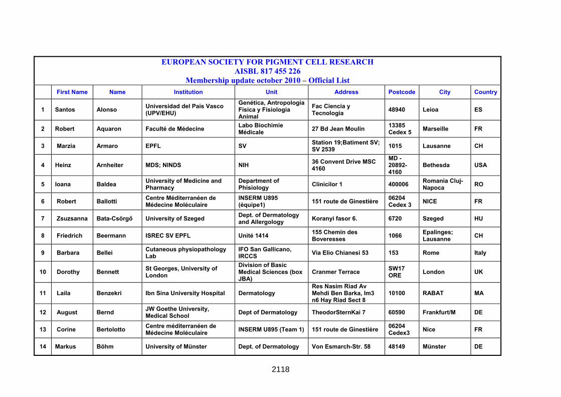
EUROPEAN SOCIETY FOR PIGMENT CELL RESEARCH AISBL 817 455 226
Membership update october 2010 – Official List First Name Name Institution Unit Address Postcode City Country
1 Santos Alonso Universidad del País Vasco (UPV/EHU)
Genética, Antropología Física y Fisiología Animal
Fac Ciencia y Tecnología 48940 Leioa ES
2 Robert Aquaron Faculté de Médecine Labo Biochimie Médicale 27 Bd Jean Moulin 13385
Cedex 5 Marseille FR
3 Marzia Armaro EPFL SV Station 19;Batiment SV; SV 2539 1015 Lausanne CH
4 Heinz Arnheiter MDS; NINDS NIH 36 Convent Drive MSC 4160
MD - 20892-4160
Bethesda USA
5 Ioana Baldea University of Medicine and Pharmacy
Department of Phisiology Clinicilor 1 400006 Romania Cluj-
Napoca RO
6 Robert Ballotti Centre Méditerranéen de Médecine Moléculaire
INSERM U895 (équipe1) 151 route de Ginestière 06204
Cedex 3 NICE FR
7 Zsuzsanna Bata-Csörgő University of Szeged Dept. of Dermatology and Allergology Koranyi fasor 6. 6720 Szeged HU
8 Friedrich Beermann ISREC SV EPFL Unité 1414 155 Chemin des Boveresses 1066 Epalinges;
Lausanne CH
9 Barbara Bellei Cutaneous physiopathology Lab
IFO San Gallicano, IRCCS Via Elio Chianesi 53 153 Rome Italy
10 Dorothy Bennett St Georges, University of London
Division of Basic Medical Sciences (box JBA)
Cranmer Terrace SW17 ORE London UK
11 Laila Benzekri Ibn Sina University Hospital Dermatology Res Nasim Riad Av Mehdi Ben Barka, Im3 n6 Hay Riad Sect 8
10100 RABAT MA
12 August Bernd JW Goethe University, Medical School Dept of Dermatology TheodorSternKai 7 60590 Frankfurt/M DE
13 Corine Bertolotto Centre méditerranéen de Médecine Moléculaire INSERM U895 (Team 1) 151 route de Ginestière 06204
Cedex3 Nice FR
14 Markus Böhm University of Münster Dept. of Dermatology Von Esmarch-Str. 58 48149 Münster DE
2118
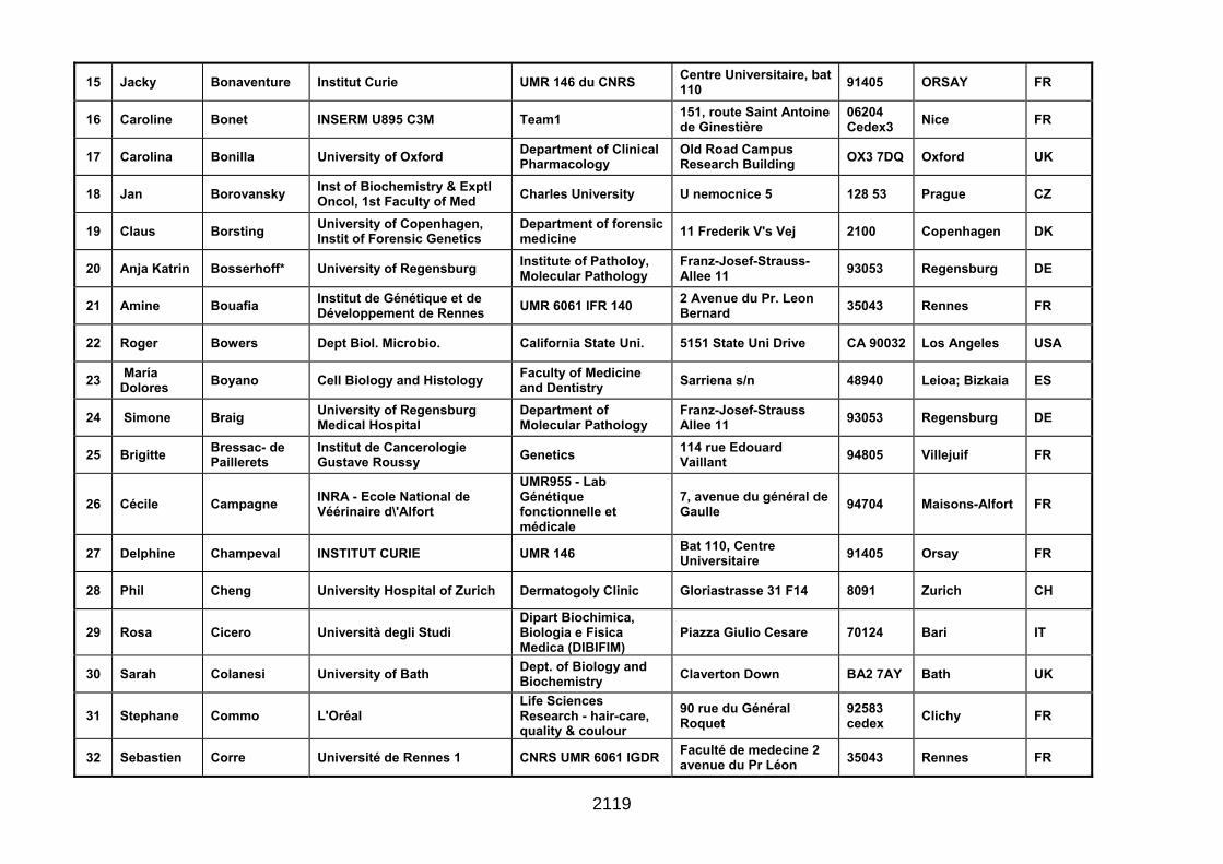
15 Jacky Bonaventure Institut Curie UMR 146 du CNRS Centre Universitaire, bat 110 91405 ORSAY FR
16 Caroline Bonet INSERM U895 C3M Team1 151, route Saint Antoine de Ginestière
06204 Cedex3 Nice FR
17 Carolina Bonilla University of Oxford Department of Clinical Pharmacology
Old Road Campus Research Building OX3 7DQ Oxford UK
18 Jan Borovansky Inst of Biochemistry & Exptl Oncol, 1st Faculty of Med Charles University U nemocnice 5 128 53 Prague CZ
19 Claus Borsting University of Copenhagen, Instit of Forensic Genetics
Department of forensic medicine 11 Frederik V's Vej 2100 Copenhagen DK
20 Anja Katrin Bosserhoff* University of Regensburg Institute of Patholoy, Molecular Pathology
Franz-Josef-Strauss-Allee 11 93053 Regensburg DE
21 Amine Bouafia Institut de Génétique et de Développement de Rennes UMR 6061 IFR 140 2 Avenue du Pr. Leon
Bernard 35043 Rennes FR
22 Roger Bowers Dept Biol. Microbio. California State Uni. 5151 State Uni Drive CA 90032 Los Angeles USA
23 María Dolores Boyano Cell Biology and Histology Faculty of Medicine
and Dentistry Sarriena s/n 48940 Leioa; Bizkaia ES
24 Simone Braig University of Regensburg Medical Hospital
Department of Molecular Pathology
Franz-Josef-Strauss Allee 11 93053 Regensburg DE
25 Brigitte Bressac- de Paillerets
Institut de Cancerologie Gustave Roussy Genetics 114 rue Edouard
Vaillant 94805 Villejuif FR
26 Cécile Campagne INRA - Ecole National de Véérinaire d\'Alfort
UMR955 - Lab Génétique fonctionnelle et médicale
7, avenue du général de Gaulle 94704 Maisons-Alfort FR
27 Delphine Champeval INSTITUT CURIE UMR 146 Bat 110, Centre Universitaire 91405 Orsay FR
28 Phil Cheng University Hospital of Zurich Dermatogoly Clinic Gloriastrasse 31 F14 8091 Zurich CH
29 Rosa Cicero Università degli Studi Dipart Biochimica, Biologia e Fisica Medica (DIBIFIM)
Piazza Giulio Cesare 70124 Bari IT
30 Sarah Colanesi University of Bath Dept. of Biology and Biochemistry Claverton Down BA2 7AY Bath UK
31 Stephane Commo L'Oréal Life Sciences Research - hair-care, quality & coulour
90 rue du Général Roquet
92583 cedex Clichy FR
32 Sebastien Corre Université de Rennes 1 CNRS UMR 6061 IGDR Faculté de medecine 2 avenue du Pr Léon 35043 Rennes FR
2119

Bernard
33 Paola Crepaldi University of Milan Department of Animal Science Via Celoria 2 20133 Milano IT
34 Véronique Del Marmol Université Libre de Bruxelles Hôpital Erasme, Dermatology Route de Lennik 808 1070 Brussels B
35 Maria Lucia Dell'Anna San Gallicano Dermatological Institute
Lab Of Cutaneous Physiopathology via Elio Chianesi 53 144 Rome IT
36 Véronique Delmas Institut Curie UMR146 CNRS Batiment 110 91405 cedex Orsay FR
37 Alida Depase Alidadepase rehabilitative cosmetics
Via Brigata LUPI 3 24122 Bergaus IT
38 Sabine Druillennec-Rodiere Institut Curie
UMR CNRS 146 -Institut Curie Recherche
Batiment 110 91405 cedex Orsay FR
39 Marco d\'Ischia University of Naples Federico II
Dept Organic Chemistry and Biochemistry
Via Cinthia 4 80126 Naples IT
40 Giorgia Egidy Maskos Institut Pasteur Unité Génétique fonctionnelle de la Souris
25, rue du Dr Roux 75724 Paris cedex 15 FR
41 Ossia Eichhoff University Hospital of Zürich Dermatology Gloriastrasse 31 8091 Zürich CH
42 Viktoria Eleftheriadou University of Nottingham Centre of Evidence Based Dermatology Kings Meadow Campus NG7 2NR Nottingham UK
43 Alain Eychene Institut Curie UMR CNRS 146 -Institut Curie Recherche
Batiment 110 91405 cedex Orsay FR
44 Khaled Ezzedine Center for rare skin diseases, Univ Hosp St-André Dermatology 1, Rue Jean Burguet 33075 Bordeaux FR
45 Natasha Feeley Curtin University of Technology Biomedical Science Kent Street 6152 Bentley AU
46 Almudena Fernández CNB-CSIC Molecular and Cellular Biology
Campus de Cantoblanco; C/ Darwin 3
28049 Madrid ES
47 Enrica Flori Cutaneous Physiopathology Laboratory IFO San Gallicano Via Elio Chianesi 53 144 Rome IT
48 Marie-Dominique Galibert
CNRS-UMR6061: Genetics & Developt Inst Rennes, Univ Rennes
Transcriptional Regul & Oncogenesis Group (RTO)
Faculté de Médecine; 2 avenue du Prof Léon Bernard
35043 Rennes FR
2120

49 Anna Gallone Università degli Studi di Foggia
Dept Scienze Biomediche-Facolta di Medicina e Chir
via L. Pinto c/o AOU -Ospedali Riuniti- 71100 Foggia IT
50 José Carlos García-Borrón University of Murcia Dept of Biochemistry and molecular Biology campos de Espinardo 30100 Espinardo,
Murcia ES
51 Yvon Gauthier Hospital Saint Andre Service de Dermatologie 1 rue JEAN BURGUET 33695 Bordeaux FR
52 David Gawkrodger ROYAL HALLAMSHIRE HOSPITAL
DEPARTMENT OF DERMATOLOGY GLOSSOP ROAD S10 2JF SHEFFIELD UK
53 Ghanem Ghanem Univ. Libre Bruxelles LOCE - Institut Jules Bordet Rue Héger-Bordet 1 B-1000 Bruxelles B
54 Maja Grabacka University of Agriculture Dept. of Food Biotechnol, Fac of Food Technology
ul. Balicka 122 30-149 Krakow PL
55 Gabriella Guida Università degli Studi di Bari Dip Biochim Med, Biol Med e Fisica Med Sez Biol Med
Policlinico, Piazza Giulio Cesare 70124 Bari IT
56 Anke Hartmann University Hospital of Erlangen Dept. of Dermatology Hartmannstr. 14 91052 Erlangen DE
57 Eugene Healy University of Southampton Dermatopharmacol MP825 lev F South Block Southampton Gen Hosp
Tremona Road SO16 6YD Southampton UK
58 Vincent Hearing N.I.H. Lab Cell Biology Bldg 37, Room 1B25 20892 Bethesda, MD USA
59 Keith Hoek University Hospital of Zürich Department of Dermatology Gloriastr 31 8091 Zürich CH
60 Vratislav Horak Institute of Animal Physiology and Genetics
Department Animal Embryology, Cell and Tissue Differentation
Rumburska 89 27721 Libechov CZ
61 Shosuke Ito Fujita Health Univ. School Health Sciences Toyoake 470-1192 Aichi JP
62 Delphine Javelaud Institut Curie INSERM U1021- CNRS UMR3347 - Equipe Centre Universitaire 91405 Orsay Cedex FR
63 Celia Jimenez-Cervantes Universidad de Murcia Bioch. Mol. Biol. Campus de Espinardo 30100 Espinardo,
Murcia ES
64 Peter Johansen Faculty of Health Sciences, University of Copenhagen
Department of Forensic Medicine; Section of Forensic Genetics
Frederik V´s vej 11 2100 Copenhagen O DK
65 Fabrice Journe Institut Jules Bordet, Université libre de Bruxelles LOCE Rue Héger-Bordet 1 1000 Brussels B
2121
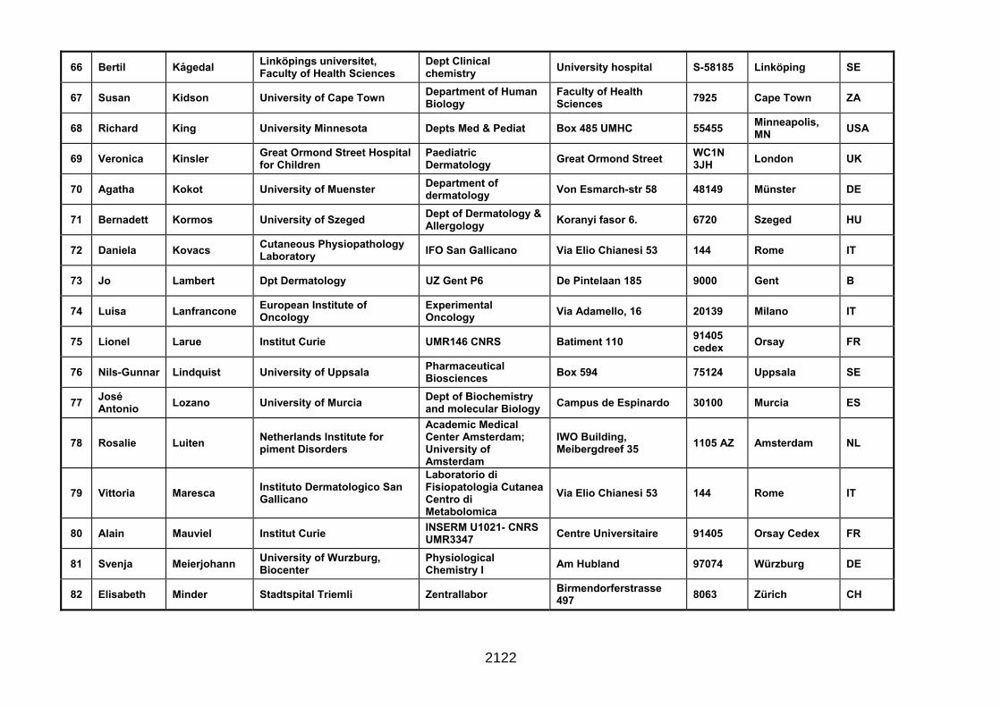
66 Bertil Kågedal Linköpings universitet, Faculty of Health Sciences
Dept Clinical chemistry University hospital S-58185 Linköping SE
67 Susan Kidson University of Cape Town Department of Human Biology
Faculty of Health Sciences 7925 Cape Town ZA
68 Richard King University Minnesota Depts Med & Pediat Box 485 UMHC 55455 Minneapolis, MN USA
69 Veronica Kinsler Great Ormond Street Hospital for Children
Paediatric Dermatology Great Ormond Street WC1N
3JH London UK
70 Agatha Kokot University of Muenster Department of dermatology Von Esmarch-str 58 48149 Münster DE
71 Bernadett Kormos University of Szeged Dept of Dermatology & Allergology Koranyi fasor 6. 6720 Szeged HU
72 Daniela Kovacs Cutaneous Physiopathology Laboratory IFO San Gallicano Via Elio Chianesi 53 144 Rome IT
73 Jo Lambert Dpt Dermatology UZ Gent P6 De Pintelaan 185 9000 Gent B
74 Luisa Lanfrancone European Institute of Oncology
Experimental Oncology Via Adamello, 16 20139 Milano IT
75 Lionel Larue Institut Curie UMR146 CNRS Batiment 110 91405 cedex Orsay FR
76 Nils-Gunnar Lindquist University of Uppsala Pharmaceutical Biosciences Box 594 75124 Uppsala SE
77 José Antonio Lozano University of Murcia Dept of Biochemistry
and molecular Biology Campus de Espinardo 30100 Murcia ES
78 Rosalie Luiten Netherlands Institute for piment Disorders
Academic Medical Center Amsterdam; University of Amsterdam
IWO Building, Meibergdreef 35 1105 AZ Amsterdam NL
79 Vittoria Maresca Instituto Dermatologico San Gallicano
Laboratorio di Fisiopatologia Cutanea Centro di Metabolomica
Via Elio Chianesi 53 144 Rome IT
80 Alain Mauviel Institut Curie INSERM U1021- CNRS UMR3347 Centre Universitaire 91405 Orsay Cedex FR
81 Svenja Meierjohann University of Wurzburg, Biocenter
Physiological Chemistry I Am Hubland 97074 Würzburg DE
82 Elisabeth Minder Stadtspital Triemli Zentrallabor Birmendorferstrasse 497 8063 Zürich CH
2122
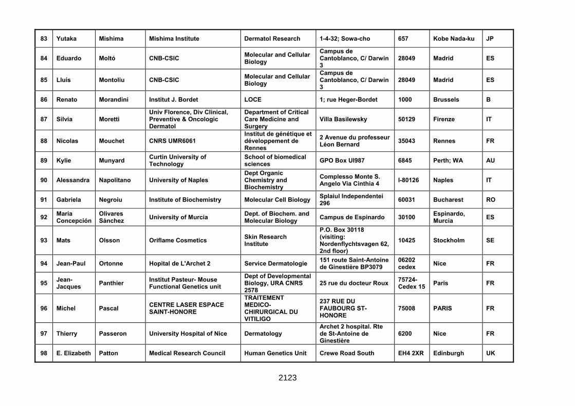
83 Yutaka Mishima Mishima Institute Dermatol Research 1-4-32; Sowa-cho 657 Kobe Nada-ku JP
84 Eduardo Moltó CNB-CSIC Molecular and Cellular Biology
Campus de Cantoblanco, C/ Darwin 3
28049 Madrid ES
85 Lluís Montoliu CNB-CSIC Molecular and Cellular Biology
Campus de Cantoblanco, C/ Darwin 3
28049 Madrid ES
86 Renato Morandini Institut J. Bordet LOCE 1; rue Heger-Bordet 1000 Brussels B
87 Silvia Moretti Univ Florence, Div Clinical, Preventive & Oncologic Dermatol
Department of Critical Care Medicine and Surgery
Villa Basilewsky 50129 Firenze IT
88 Nicolas Mouchet CNRS UMR6061 Institut de génétique et développement de Rennes
2 Avenue du professeur Léon Bernard 35043 Rennes FR
89 Kylie Munyard Curtin University of Technology
School of biomedical sciences GPO Box UI987 6845 Perth; WA AU
90 Alessandra Napolitano University of Naples Dept Organic Chemistry and Biochemistry
Complesso Monte S. Angelo Via Cinthia 4 I-80126 Naples IT
91 Gabriela Negroiu Institute of Biochemistry Molecular Cell Biology Splaiul Independentei 296 60031 Bucharest RO
92 Maria Concepción
Olivares Sánchez University of Murcia Dept. of Biochem. and
Molecular Biology Campus de Espinardo 30100 Espinardo, Murcia ES
93 Mats Olsson Oriflame Cosmetics Skin Research Institute
P.O. Box 30118 (visiting: Nordenflychtsvagen 62, 2nd floor)
10425 Stockholm SE
94 Jean-Paul Ortonne Hopital de L'Archet 2 Service Dermatologie 151 route Saint-Antoine de Ginestière BP3079
06202 cedex Nice FR
95 Jean-Jacques Panthier Institut Pasteur- Mouse
Functional Genetics unit Dept of Developmental Biology, URA CNRS 2578
25 rue du docteur Roux 75724-Cedex 15 Paris FR
96 Michel Pascal CENTRE LASER ESPACE SAINT-HONORE
TRAITEMENT MEDICO-CHIRURGICAL DU VITILIGO
237 RUE DU FAUBOURG ST-HONORE
75008 PARIS FR
97 Thierry Passeron University Hospital of Nice Dermatology Archet 2 hospital. Rte de St-Antoine de Ginestière
6200 Nice FR
98 E. Elizabeth Patton Medical Research Council Human Genetics Unit Crewe Road South EH4 2XR Edinburgh UK
2123
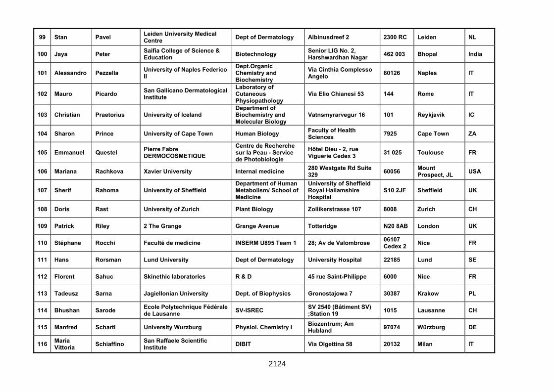
99 Stan Pavel Leiden University Medical Centre Dept of Dermatology Albinusdreef 2 2300 RC Leiden NL
100 Jaya Peter Saifia College of Science & Education Biotechnology Senior LIG No. 2,
Harshwardhan Nagar 462 003 Bhopal India
101 Alessandro Pezzella University of Naples Federico II
Dept.Organic Chemistry and Biochemistry
Via Cinthia Complesso Angelo 80126 Naples IT
102 Mauro Picardo San Gallicano Dermatological Institute
Laboratory of Cutaneous Physiopathology
Via Elio Chianesi 53 144 Rome IT
103 Christian Praetorius University of Iceland Department of Biochemistry and Molecular Biology
Vatnsmyrarvegur 16 101 Reykjavik IC
104 Sharon Prince University of Cape Town Human Biology Faculty of Health Sciences 7925 Cape Town ZA
105 Emmanuel Questel Pierre Fabre DERMOCOSMETIQUE
Centre de Recherche sur la Peau - Service de Photobiologie
Hôtel Dieu - 2, rue Viguerie Cedex 3 31 025 Toulouse FR
106 Mariana Rachkova Xavier University Internal medicine 280 Westgate Rd Suite 329 60056 Mount
Prospect, JL USA
107 Sherif Rahoma University of Sheffield Department of Human Metabolism/ School of Medicine
University of Sheffield Royal Hallamshire Hospital
S10 2JF Sheffield UK
108 Doris Rast University of Zurich Plant Biology Zollikerstrasse 107 8008 Zurich CH
109 Patrick Riley 2 The Grange Grange Avenue Totteridge N20 8AB London UK
110 Stéphane Rocchi Faculté de medicine INSERM U895 Team 1 28; Av de Valombrose 06107 Cedex 2 Nice FR
111 Hans Rorsman Lund University Dept of Dermatology University Hospital 22185 Lund SE
112 Florent Sahuc Skinethic laboratories R & D 45 rue Saint-Philippe 6000 Nice FR
113 Tadeusz Sarna Jagiellonian University Dept. of Biophysics Gronostajowa 7 30387 Krakow PL
114 Bhushan Sarode Ecole Polytechnique Fédérale de Lausanne SV-ISREC SV 2540 (Bâtiment SV)
;Station 19 1015 Lausanne CH
115 Manfred Schartl University Wurzburg Physiol. Chemistry I Biozentrum; Am Hubland 97074 Würzburg DE
116 Maria Vittoria Schiaffino San Raffaele Scientific
Institute DIBIT Via Olgettina 58 20132 Milan IT
2124
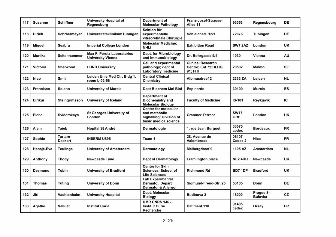
117 Susanne Schiffner University Hospital of Regensburg
Department of Molecular Pathology
Franz-Josef-Strauss-Allee 11 93053 Regensbourg DE
118 Ulrich Schraermeyer UniversitätsklinikumTübingen Sektion für experimentelle vitreoretinale Chirurgie
Schleichstr. 12/1 72076 Tübingen DE
119 Miguel Seabra Imperial College London Molecular Medicine; NHLI Exhibition Road SW7 2AZ London UK
120 Monika Seltenhammer Max F. Perutz Laboratories - University Vienna
Dept. for Microbiology and Immunobiology Dr. Bohrgasse 9/4 1030 Vienna AU
121 Victoria Sherwood LUND University Cell and experimental pathology; dept of Laboratory medicine
Clinical Research Centre; Ent 72;BLDG 91; FI II
20502 Malmö SE
122 Nico Smit Leiden Univ Med Ctr, Bldg 1, room L-02-56
Central Clinical Chemistry Albinusdreef 2 2333 ZA Leiden NL
123 Francisco Solano University of Murcia Dept Biochem Mol Biol Espinardo 30100 Murcia ES
124 Eirikur Steingrimsson University of Iceland Department of Biochemistry and Molecular Biology
Faculty of Medicine IS-101 Reykjavik IC
125 Elena Sviderskaya St Georges University of London
Center for molecular and metabolic signalling; Division of basic medica science
Cranmer Terrace SW17 ORE London UK
126 Alain Taïeb Hopital St André Dermatologie 1, rue Jean Burguet 33575 cedex Bordeaux FR
127 Sophie Tartare-Deckert INSERM U895 Team 1 28, Avenue de
Valombrose 06107 Cedex 2 Nice FR
128 Hansje-Eva Teulings University of Amsterdam Dermatology Meibergdreef 9 1105 AZ Amsterdam NL
129 Anthony Thody Newcastle Tyne Dept of Dermatology Framlington place NE2 4HH Newcastle UK
130 Desmond Tobin University of Bradford Centre for Skin Sciences; School of Life Sciences
Richmond Rd BD7 1DP Bradford UK
131 Thomas Tüting University of Bonn Lab Experimental Dermatol, Depart Dermatol & Allergol
Sigmund-Freud-Str. 25 53105 Bonn DE
132 Jiri Vachtenheim University Hospital Dept. Molecular Biology Budinova 2 18000 Prague 8 -
Bulovka CZ
133 Agathe Valluet Institut Curie UMR CNRS 146 -Institut Curie Recherche
Batiment 110 91405 cedex Orsay FR
2125
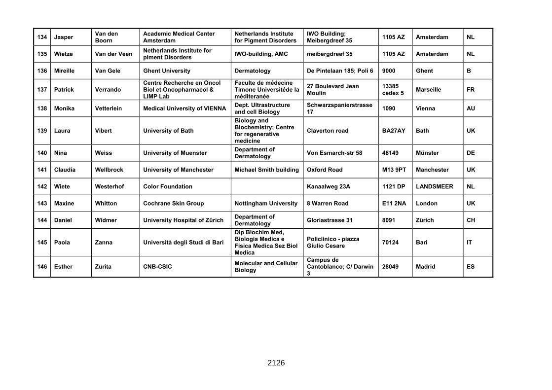
134 Jasper Van den Boorn
Academic Medical Center Amsterdam
Netherlands Institute for Pigment Disorders
IWO Building; Meibergdreef 35 1105 AZ Amsterdam NL
135 Wietze Van der Veen Netherlands Institute for piment Disorders IWO-building, AMC meibergdreef 35 1105 AZ Amsterdam NL
136 Mireille Van Gele Ghent University Dermatology De Pintelaan 185; Poli 6 9000 Ghent B
137 Patrick Verrando Centre Recherche en Oncol Biol et Oncopharmacol & LIMP Lab
Faculte de médecine Timone Universitéde la méditeranée
27 Boulevard Jean Moulin
13385 cedex 5 Marseille FR
138 Monika Vetterlein Medical University of VIENNA Dept. Ultrastructure and cell Biology
Schwarzspanierstrasse 17 1090 Vienna AU
139 Laura Vibert University of Bath Biology and Biochemistry; Centre for regenerative medicine
Claverton road BA27AY Bath UK
140 Nina Weiss University of Muenster Department of Dermatology Von Esmarch-str 58 48149 Münster DE
141 Claudia Wellbrock University of Manchester Michael Smith building Oxford Road M13 9PT Manchester UK
142 Wiete Westerhof Color Foundation Kanaalweg 23A 1121 DP LANDSMEER NL
143 Maxine Whitton Cochrane Skin Group Nottingham University 8 Warren Road E11 2NA London UK
144 Daniel Widmer University Hospital of Zürich Department of Dermatology Gloriastrasse 31 8091 Zürich CH
145 Paola Zanna Università degli Studi di Bari Dip Biochim Med, Biologia Medica e Fisica Medica Sez Biol Medica
Policlinico - piazza Giulio Cesare 70124 Bari IT
146 Esther Zurita CNB-CSIC Molecular and Cellular Biology
Campus de Cantoblanco; C/ Darwin 3
28049 Madrid ES
2126