n-carbamylglutamate and l-arginine improved maternal and
TRANSCRIPT

REPRODUCTION
© 2016 Society for Reproduction and Fertility DOI: 10.1530/REP-16-0067ISSN 1470–1626 (paper) 1741–7899 (online) Online version via www.reproduction-online.org
RESEARCH
N-carbamylglutamate and L-arginine improved maternal and placental development in underfed ewes
Hao Zhang1, Lingwei Sun1, Ziyu Wang1, Mingtian Deng1, Haitao Nie1, Guomin Zhang1, Tiewei Ma1 and Feng Wang1,2
1Jiangsu Engineering Technology Research Center of Mutton Sheep & Goat Industry, Nanjing Agricultural University, Nanjing, People’s Republic of China and 2Jiangsu Livestock Embryo Engineering Laboratory, Nanjing Agricultural University, Nanjing, People’s Republic of China
Correspondence should be addressed to F Wang; Email: [email protected]
Abstract
The objectives of this study were to determine how dietary supplementation of N-carbamylglutamate (NCG) and rumen-protected L-arginine (RP-Arg) in nutrient-restricted pregnant Hu sheep would affect (1) maternal endocrine status; (2) maternal, fetal, and placental antioxidation capability; and (3) placental development. From day 35 to day 110 of gestation, 32 Hu ewes carrying twin fetuses were allocated randomly into four groups: 100% of NRC-recommended nutrient requirements, 50% of NRC recommendations, 50% of NRC recommendations supplemented with 20 g/day RP-Arg, and 50% of NRC recommendations supplemented with 5 g/day NCG product. The results showed that in maternal and fetal plasma and placentomes, the activities of total antioxidant capacity and superoxide dismutase were increased (P < 0.05); however, the activity of glutathione peroxidase and the concentration of maleic dialdehyde were decreased (P < 0.05) in both NCG- and RP-Arg-treated underfed ewes. The mRNA expression of vascular endothelial growth factor and Fms-like tyrosine kinase 1 was increased (P < 0.05) in 50% NRC ewes than in 100% NRC ewes, and had no effect (P > 0.05) in both NCG- and RP-Arg-treated underfed ewes. A supplement of RP-Arg and NCG reduced (P < 0.05) the concentrations of progesterone, cortisol, and estradiol-17β; had no effect on T4/T3; and improved (P < 0.05) the concentrations of leptin, insulin-like growth factor 1, tri-iodothyronine (T3), and thyroxine (T4) in serum from underfed ewes. These results indicate that dietary supplementation of NCG and RP-Arg in underfed ewes could influence maternal endocrine status, improve the maternal–fetal–placental antioxidation capability, and promote fetal and placental development during early-to-late gestation.Reproduction (2016) 151 623–635
10.1530/REP-16-0067
Introduction
Intrauterine growth restriction (IUGR) is a major concern for the livestock industry because fetal growth restriction leads to negative impacts on animal performance later in life (Wu et al. 2006, He et al. 2011). The Hu sheep are noted for their precociousness and prolificacy. During reproduction, insufficient availability and/or delivery of nutrients to the conceptus often occur resulting in IUGR. Surviving infants with IUGR are at an increased risk of neurological, respiratory, intestinal, and circulatory dis-orders (Gluckman & Hanson 2006). To date, there is no therapeutic means for preventing or ameliorating IUGR, the current management being empirical and primarily aimed at selecting a safe time for delivery (Mari & Hanif 2007).
L-arginine (Arg) is a nutritionally important amino acid and plays multiple physiological functions in animals (Mateo et al. 2007, Yao et al. 2008, Tan et al. 2011). As a precursor of polyamines and nitric oxide (NO), Arg enhances placental angiogenes (Raghavan
& Dikshit 2004), fetal–placental blood flow (Tan et al. 2012), and antioxidant ability and ameliorates lipid per-oxidation during gestation (Morris 2009). It has been reported that parenteral administration of Arg in pro-lific ewes improved survival rate of fetal lamb to term (Lassala et al. 2011, McCoard et al. 2013). However, Arg is rapidly degraded in the rumen, and parental administra-tion of Arg to a farm animal is not a practical approach. Therefore, we used rumen-protected L-arginine (RP-Arg) in our study. N-carbamylglutamate (NCG) is a cofac-tor of carbamoyl phosphate synthetase-1, a rate-liming enzyme responsible for both the urea cycle and the Arg synthetic pathway (Wu et al. 2004, 2012a). Dietary supplementation of NCG has the potential to improve pregnancy outcomes (litter size and litter weight) due to its efficiency in increasing the concentrations of the arginine-family AA in maternal circulations and uterine fluids, which could improve early embryonic develop-ment and implantation (Zeng et al. 2012). NCG also exhibits a lower degree of rumen degradation compared with Arg (Chacher et al. 2012). The mechanism of NCG
Q5
Downloaded from Bioscientifica.com at 04/05/2022 01:58:53AMvia free access
624 H Zhang and others
Reproduction (2016) 151 623–635 www.reproduction-online.org
may lie in increasing the endogenous synthesis of Arg (Liu et al. 2012, Wu et al. 2012b). Therefore, NCG is considered as an Arg enhancer.
The placenta plays an important role in the growth and development of the fetus. Placental angiogenesis sup-ports the required blood flow on the fetal side necessary for fetal growth and development. Therefore, vasculo-genesis and angiogenesis are critical for proper placen-tal function and thus for normal embryonic/fetal growth and development (Demir et al. 2007, Arroyo & Winn 2008). L-arginine and NCG supplementation improved litter size, and fetal survival might relate to the mecha-nism of elevating vascular endothelial growth factor (VEGF), placenta growth factor 1 (PlGF1), and endothe-lial NO synthase (eNOS) gene expression in placental surface vessels (Wu et al. 2012a). L-arginine and NCG may regulate angiogenesis and vascular development and functions of umbilical vein and placenta, provid-ing more nutrients and oxygen from mother to fetuses for fetal survival, growth, and development (Liu et al. 2012). Vatnick et al. (1991) first described a morpho-logic classification system for placentomes, in which the placentomes are classified based on their shape. Placen-tome morphology does not affect placental vascularity, expression of angiogenic factors, cell proliferation, or tissue composition (Vonnahme et al. 2008). However, the combined effects of maternal dietary and placental types on placental development are still unknown.
Although the impact of nutrient restriction during gestation in ewes includes reduced fetal growth (Vonnahme et al. 2003), poor wool, and carcass qual-ity (Kelley et al. 1996), it is unknown how dietary sup-plementation of NCG and rumen-protected L- arginine (RP-Arg) could influence maternal endocrine status, antioxidation capability, and placental development in nutrient-restricted Hu sheep. Furthermore, in order to explain how dietary supplementation of NCG and RP-Arg regulates the reproductive performance of Hu sheep under nutrient-restricted condition, we grab our attention from maternal endocrine status to the expression of pla-cental development-associated genes.
Therefore, the objectives of this study are to deter-mine how dietary supplementation of NCG and RP-Arg in nutrient-restricted Hu sheep would affect (1) maternal concentrations of circulating leptin, progesterone, insu-lin-like growth factor 1 (IGF1), cortisol, estradiol-17β, thyroxine (T4), and tri-iodothyronine (T3); (2) maternal, fetal, and placental antioxidation capability; (3) pla-cental growth, differentiation, and cellularity; and (4) placental angiogenic and vasoactive factor abundance.
Materials and methods
Animals
Hu ewes were maintained at the Jiangyan Experimental Station of Taizhou, Jiangsu Province of China. During the
research period, an indoor facility equipped with heating radi-ators was used to keep the mean temperature at 15 ± 0.8°C, and lighting was controlled automatically to mimic the photoperiod of the outdoor environment. For the study, 48 multiparous Hu sheep (body weight = 40.1 ± 1.2 kg) of similar age (18.5 ± 0.5 months) and body condition score (BCS) (2.55 ± 0.18; scale 0 = emaciated to 5 = obese; Russel et al. 1969) were selected. After being drenched with 0.2 mg ivermectin per kilogram of BW against endoparasites, all ewes were synchronized using intravaginal progestogen sponges (30 mg; Pharmp PTY, Herston City, Australia) for 12 days. Estrous behavior was monitored using three vasectomized rams at 0800 and 1600 h following the second day of pessary removal. The ewes were artificially inseminated using fresh semen 48 h after sponge withdrawal (day 0 of gestation) and placed in individual pens (1.05 × 1.60 m) for 35 days. The number of fetuses carried by each ewe was determined by ultrasonography (Asonics Microimager 1000 sector scanning instrument; Ausonics Pty Ltd, Sydney, Australia) at day 35 of gestation. In this study, 32 ewes carrying twin fetuses were utilized. Details of the diets are given in Table 1 to meet 100% of the NRC (1985) nutrient requirements for pregnant sheep. All ewes received 100% of NRC requirements of all nutrients and energy from day 0 to 35. The ewes were fed once daily at 0800 h and had free access to clean water. All trials were conducted in accordance with the procedures approved by the Guide for the Care and Use of Laboratory Animals prepared by the Ethics Committee of Nanjing Agricultural University (SYXK 2011-0036).
Experimental design
On day 35 of pregnancy, ewes were randomly assigned to four groups (n = 8): 100% NRC (1985) recommendations, 50% NRC recommendations, 50% NRC + 20 g/day RP-Arg (Beijing
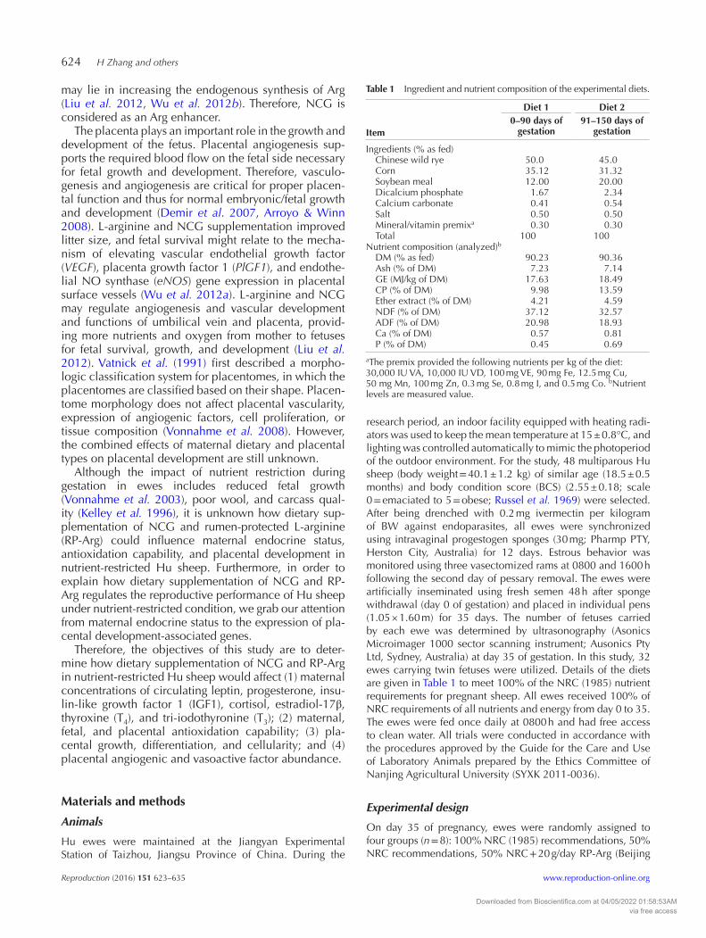
Table 1 Ingredient and nutrient composition of the experimental diets.
Item
Diet 1 Diet 20–90 days of
gestation91–150 days of
gestation
Ingredients (% as fed)Chinese wild rye 50.0 45.0Corn 35.12 31.32Soybean meal 12.00 20.00Dicalcium phosphate 1.67 2.34Calcium carbonate 0.41 0.54Salt 0.50 0.50Mineral/vitamin premixa 0.30 0.30Total 100 100
Nutrient composition (analyzed)bDM (% as fed) 90.23 90.36Ash (% of DM) 7.23 7.14GE (MJ/kg of DM) 17.63 18.49CP (% of DM) 9.98 13.59Ether extract (% of DM) 4.21 4.59NDF (% of DM) 37.12 32.57ADF (% of DM) 20.98 18.93Ca (% of DM) 0.57 0.81P (% of DM) 0.45 0.69
aThe premix provided the following nutrients per kg of the diet: 30,000 IU VA, 10,000 IU VD, 100 mg VE, 90 mg Fe, 12.5 mg Cu, 50 mg Mn, 100 mg Zn, 0.3 mg Se, 0.8 mg I, and 0.5 mg Co. bNutrient levels are measured value.
Downloaded from Bioscientifica.com at 04/05/2022 01:58:53AMvia free access

Arg and NCG supplementation in a sheep IUGR 625
www.reproduction-online.org Reproduction (2016) 151 623–635
Feeding Feed Science Technology Co., Beijing, China), and 50% NRC + 5 g/day NCG (Institute of Subtropical Agriculture, The Chinese Academy of Sciences, Changsha, China). RP-Arg was a 50% Arg product, and NCG was a 50% NCG product. Thus, the actual additional amounts of RP-Arg and NCG was 10 g/day and 2.5 g/day, respectively, which were mixed into the pelleted mixed diet. The protection of RP-Arg in the rumen was ≥85% and the release of RP-Arg in the intestine was ≥90%, which were determined according to the methods in the previous studies (Hervás et al. 2000, Chacher et al. 2012). The RP-Arg was performed from glycerides and phospholipids by spray-drying and spray-congealing processes according to Eldem et al. (1991). The doses of Arg were based on previous studies of pregnant sheep that received parenteral administra-tion of Arg (Wu et al. 2007, Lassala et al. 2010, McCoard et al. 2013, Satterfield et al. 2013). The dose of NCG was based on previous studies on rats, piglets, and dairy cows (Wu et al. 2012b, Zeng et al. 2012, Chacher et al. 2014). Nutrient restric-tion (50% NRC) was achieved by feeding one-half of the total complete diet calculated to meet 100% NRC requirements. Beginning on day 35 of gestation, body weight was analyzed every 10 days and feed intake was adjusted based on the changes in body weight.
Every 20 days from day 50 of pregnancy until killing, 10 mL of maternal blood was collected from all ewes from the jugular vein, using anticoagulant-free, sterile vacuum tubes (Vacutainer; Becton Dickinson, Franklin Lakes, NJ, USA) and 20 gauge × 3.8 cm blood collection needles (Vacutainer; Becton Dickinson, Franklin Lakes, NJ, USA). Blood was drawn in the morning immediately before feeding the ewes. Samples were placed on ice and immediately centrifuged at 3000 g for 15 min at 4°C. Serum was separated and stored at −80°C. Another set of blood samples were collected into heparinized vacutainer tubes (Sigma) and plasma was stored at −80°C. Blood samples were collected for both serum and plasma stored at −80°C until analysis.
Chemical analyses
Feed samples were analyzed for dry matter (DM), ash, crude protein (CP), ether extract (EE), calcium (Ca), and phosphorus (P) (methods 930.15, 942.05, 990.02, 920.39, 968.08, and 965.17, respectively, Association of Official Analytical Chem-ists 1990). The natural detergent fiber (NDF) and acid deter-gent fiber (ADF) concentrations were quantified as described by Van Soest et al. (1991). A bomb calorimeter (C200; IKA Works Inc., Staufen, Germany) was used to measure the gross energy (GE) in dietary ingredients.
Tissue collection and handling following necropsy
At day 110 of gestation, each ewe in the experimental group was stunned with a captive-bolt gun (Supercash Mark 2; Acceles and Shelvoke Ltd., Sutton Coldfield, England) and was exsanguinated. After the tip of the gravid uterine horn was exposed, blood samples were drawn from the fetal umbilical vein into cooled heparinized tubes. The heparinized tubes were immediately centrifuged at 3000 g for 10 min at 4°C to obtain plasma. Plasma was stored at −80°C until analysis.
The placenta was further dissected to isolate all placentomes for the assessment of placentome number, gross morphology, and weight. Morphologic type was based on the classification scheme of Vatnick et al. (1991) and was based on the plac-entome appearance (Braun et al. 2011) as follows: (1) carun-cular tissue completely surrounding the cotyledonary tissue (type A), (2) cotyledonary tissue beginning to grow over the surrounding caruncular tissue (type B), (3) flat placentomes with cotyledonary tissue on one surface and caruncular tissue on the other (type C), and (4) everted placentomes resembling bovine placentomes (type D). All placentomes were pooled to determine total placentomal weight. Furthermore, within each placentome type, total and average weight and average diam-eter were calculated. These placentomes with different types were then separated manually with gentle traction to reveal the individual maternal caruncle and fetal cotyledon compo-nents. Individual caruncle and cotyledon components were separated and weighted from each placentome type, and then snap-frozen in liquid nitrogen-cooled isopentane and stored at −80°C for subsequent antioxidation capability and gene expression studies. Simultaneously, the fetuses were weighted and the values were then recorded.
Biochemical analyses of maternal serum and plasma
Maternal leptin, progesterone, estradiol-17β, and IGF1 con-centrations were measured in duplicate by radioimmunoassay using a commercial kit (Diagnostic Product; Nanjing Jiancheng Bioengineering Institute, Nanjing, China). The sensitivities of the assays were 0.1 ng leptin/mL, 0.2 ng progesterone/mL, 1 pg estradiol-17β/mL, and 6 pmol IGF1/mL. The concentration of each hormone was measured within a single assay, and the intra-assay CV values were all <10%, and the inter-assay CV values were all <15%.
Thyroxine (T4; A02PZB) and tri-iodothyronine (T3; A01PZB) concentrations were measured through an equilibrium-competitive RIA using a commercial kit (Diagnostic Product; Nanjing Jiancheng Bioengineering Institute, Nanjing, China). According to the manufacturer, the sensitivities of the assays were 5 and 0.2 ng/mL respectively. The intra-assay CV were all <10%, and the inter-assay CV were all <15%.
Cortisol concentrations were determined by chemilumi-nescence immunoassay using the Immulite 1000, utilizing components of commercial kits (Diagnostic Product; Nanjing Jiancheng Bioengineering Institute, Nanjing, China). Within the cortisol assay, lesser, medium, and greater cortisol pools were assayed in duplicate. Serum samples were assayed in duplicate for cortisol. The intra- and inter-assay CV were 8.7 and 5.1% respectively.
Detection of T-AOC, GSH-Px, SOD, and MDA concentrations in placental tissue and maternal and fetal plasma
The maleic dialdehyde (MDA), total antioxidant capacity (T-AOC), as well as activities of superoxide dismutase (SOD), and glutathione peroxidase (GSH-Px) were determined by using commercial kits (Diagnostic Product; Nanjing Jiancheng Bioengineering Institute, Nanjing, China) following the
Downloaded from Bioscientifica.com at 04/05/2022 01:58:53AMvia free access
626 H Zhang and others
Reproduction (2016) 151 623–635 www.reproduction-online.org
manufacturer’s protocols. In maternal and fetal plasma, con-centrations of GSH-Px, SOD, and MDA were analyzed using commercial kits (Diagnostic Product; Nanjing Jiancheng Bio-engineering Institute, Nanjing, China). Concentrations were determined using colorimetric methods with a spectrophotom-eter (WFJ 2100; UNIC Instrument Co. Ltd., Shanghai, China) according to the procedures of Paglia and Valentine (1967), Panchenko et al. (1975), and Placer et al. (1966) respectively. Total antioxidant capacity was examined by commercial kit (Diagnostic Product; Nanjing Jiancheng Bioengineering Institute, Nanjing, China), and a spectrometric method was applied to evaluate T-AOC. In the reaction mixture, ferric ion was reduced by antioxidant-reducing agents and blue com-plex Fe2+-2,4,6-tri(2-pyridyl)-s-triazine (TPTZ) was produced; absorbance was measured at 520 nm. One unit of T-AOC was defined as the amount that increased the absorbance by 0.01 at 37°C and was expressed as units per milliliter in the plasma.
Before the assays, the caruncular and cotyledonary samples from each placentome type were homogenized in 1 mL of 0.15 M NaCl solution and centrifuged at 1510 g for 10 min at 4°C. Then, the supernatant was used for the analysis. The MDA concentration was quantified using the thiobarbituric acid method (Buege & Aust 1978), which is based on the reaction of MDA with thiobarbituric acid to form a pink chromogen. Data were normalized to protein content and expressed as nmol/mg protein. T-AOC, enzyme-specific activities of GSH-Px, and SOD were expressed as units/mg of protein. Total pro-tein concentration was determined using the Bradford method and was expressed as mg/mL.
Cellularity estimates
Freshly thawed tissue samples (0.5 g) were homogenized in 0.05 M Tris aminomethane, 2.0 M sodium chloride, and 2 mM EDTA buffer (pH 7.4) using a Polytron with a PT-10 s probe (Brinkmann, Westbury, NY, USA). The caruncular and coty-ledonary samples from each placentome type were analyzed for DNA, RNA, and protein concentration. The DNA and RNA analyses were performed using the diphenylamine and orcinol procedures respectively (Johnson et al. 1997). Protein in tissue homogenates was determined using Coomassie brilliant blue G (Bradford 1976) with bovine serum albumin (FractionV; Sigma-Aldrich) as the standard (Johnson et al. 1997). The prepared sam-ples were analyzed with a spectrophotometer (Beckman DU 640; Beckman Coulter Inc., Brea, CA, USA) and were assessed against concentration curves of known standards. The concentration of DNA was used as an index of hyperplasia, and protein:DNA and RNA:DNA ratios were used as indexes of hypertrophy and potential cellular activity respectively (Scheaffer et al. 2004).
Quantification of angiogenic factors
Several angiogenic factors that have been shown to affect placental angiogenesis were analyzed using quantitative real-time PCR. These include the following: vascular endo-thelial growth factor (VEGF), Fms-like tyrosine kinase 1 (FLT1) and kinase insert domain containing receptor (KDR) (VEGF receptors), placental growth factor (PGF), NO syn-thase 3 (NOS3), guanylate cyclase 1, soluble, beta 3 (NO
receptor, GUCY1B3) (Table 2). Caruncular and cotyledon-ary RNA from each placentome type was extracted using Tri-Reagent (Molecular Research Center, Cincinnati, OH, USA), and total RNA concentration was determined by capillary electrophoresis with an Agilent 2100 Bioanalyzer (Agilent Technologies). Real-time reverse transcriptase polymerase chain reaction (RT-PCR) analysis was used to quantify the amounts of mRNA for VEGF, FLT1, KDR, PGF, NOS3, and GUCY1B3 using methods described (Borow-icz et al. 2007, Redmer et al. 2012), with the modification that a multiplex reaction was performed after 18S mRNA was added to each well to serve as an internal control (Vonnahme et al. 2008). The ratio of the gene of interest to the 18S RNA was used for quantifying the gene expression.
Statistical analysis
Statistical comparisons between the four groups of ewes killed on day 110 of gestation or four types of placentomes were completed using a one-way ANOVA. Fetal sex was included in the original model and was found to be nonsignificant (P > 0.05) and was therefore removed in the final model, which only contained maternal nutritional treatment. Maternal and fetal antioxidant capacity in plasma was subjected to least-squares analysis of variance using the general linear model procedures of the Statistical Analysis System (SAS Institute Inc., Cary, NC, USA) with pair-wise comparisons. Data on the concentrations of metabolites and hormones in serum on day
Table 2 Sequence of TaqMan primers and probes for ovine angiogenic factors and receptors.
Probe or primer§ Nucleotide sequence
GenBank accession
No.
VEGF FP 5′-GGA TGT CTA CCA GCG CAG C-3′ X89506VEGF RP 5′-TCT GGG TAC TCC TGG AAG ATG
TC-3′VEGF probe 5′ (6FAM)-TCT GCC GTC CCA TTG AGA
CCC TG-(TAMRA)3′FLT1 FP 5′-TGG ATT TCA GGT GAG CTT GGA-3′ AF488351FLT1 RP 5′-TCA CCG TGC AAG ACA GCT TC-3′FLT1 probe 5′(6FAM)-AAA ATG CCT GCG GAA GGA
GAG GAC C-(TAMRA)3′KDR FP 5′-CTT CCA GTG GGC TGA TGA CC-3′ AF233076KDR RP 5′-GCA ACA AAC GGC TTT TCA TGT-3′KDR probe 5′(6FAM)-AGA AGA ACA GCA CGT TCG
TCC GGG-(TAMRA)3′NOS3 FP 5′-CAG CGG CTG GTA CAT GAG C-3′ AF201926NOS3 RP 5′-TTG TAG CGG TGA GGG TCA CA-3′NOS3 probe 5′(6FAM)-CGG AGA TTG GCA CGC
GGA ACC-(TAMRA)3′GUCY1B3 FP 5′-CCG AGC CGT GCA TCC A-3′ AF486295GUCY1B3 RP 5′-ATC TCC ATC ATG TCC AAG GCC-3′GUCY1B3
probe5′(6FAM)-CAT GCA CGG TCC ATC TGC
CAC C-(TAMRA)3′PGF FP 5′-CCC TGG AGA CAG CCA ACG T-3′ AY157708PGF RP 5′-GGC TGG TCC AGA GAG TGG TAC T-3′PGF probe 5′(6FAM)-CCA TGC AGC TCA TG-
(MGBNFQ)3′§FP, forward primer; RP, reverse primer; VEGF, vascular endothelial growth factor; PGF, placental growth factor; FLT1, Fms-like tyrosine kinase 1, VEGF and PGF receptor; KDR, kinase insert domain-containing receptor, VEGF receptor; NOS3, nitric oxide synthase 3 (endothelial cell); GUCY1B3, guanylate cyclase 1, soluble, beta 3.
Downloaded from Bioscientifica.com at 04/05/2022 01:58:53AMvia free access

Arg and NCG supplementation in a sheep IUGR 627
www.reproduction-online.org Reproduction (2016) 151 623–635
50, 70, 90, and 110 of pregnancy in ewes were analyzed by two-way ANOVA for repeated measures using the MIXED pro-cedures of SAS to determine the effects of day of pregnancy, group, and day of pregnancy × group interactions. Data on the antioxidant capacity, estimates of cellularity, and mRNA concentration of select angiogenic and vasoactive factors and their receptors among type A, B, C, and D of caruncle and cot-yledon in the four groups were analyzed by two-way ANOVA to determine the effects of types of placentome, group, and types of placentome × group interactions. When interactions were significant (P ≤ 0.10), means were separated using the least significant difference procedure. Data are presented as the least-squares mean (LSM) with standard error of the mean (s.e.m.). In one-way or two-way ANOVA, differences between treatment means were determined by the Student–Newman–Keuls multiple comparison test. Log transformation of variables was performed when variance of data were not homogenous among treatment groups, as assessed by the Levene’s test. All analyses were performed using the statistical package SAS (ver-sion 9.1; SAS Institute Inc). Differences in means were consid-ered to be statistically significant when a P value was ≤0.05.
Results
Fetal weight, average placentome weight, total plascentome weight, and placentome number
There was no difference (P > 0.05) in the weights of the total placentome, mean placentome, caruncular, cotyledonary, and the numbers of placentome among treatments (Table 3). The fetal weights were reduced (P < 0.05) in
50% NRC ewes compared with 100% NRC ewes, and the fetal weights were increased (P < 0.05) in both NCG- and RP-Arg-treated underfed ewes compared with the 50% NRC ewes. For all types of placentome, there was no dif-ference (P > 0.05) in the weights of average placentome and the length of average diameter among treatments. For type A placentome, the number of placentome and the weight of the total placentome were reduced (P < 0.05) in 50% NRC ewes compared with 100% NRC ewes and were increased (P < 0.05) in both NCG- and RP-Arg-treated underfed ewes. For the type B, C, and D placentomes, the number of placentome and the weight of the total placen-tome were increased (P < 0.05) in 50% NRC ewes com-pared with 100% NRC ewes and were reduced (P < 0.05) in both NCG- and RP-Arg-treated underfed ewes.
The average weight and diameter of each placentome category increased (P < 0.05) with chorioallantoic ever-sion from A to D types at day 110 of gestation in Hu sheep fed control diet (Fig. 1A and D). The total placen-tome weight and number of each placentome category decreased (P < 0.05) with chorioallantoic eversion from A to D types at day 110 of gestation in Hu sheep fed control diet, except for the total placentome weight from C to D types (P > 0.05).
The antioxidant capacity of maternal and fetal plasma
For maternal and fetal plasma, the activities of T-AOC and SOD were decreased in the 50% NRC group compared with those of the 100% NRC group (P < 0.05),
Table 3 Effects of dietary supplementation of N-carbamylglutamate (NCG) and rumen-protected L-arginine (RP-Arg) on fetal weight, average placentome weight, total plascentome weight, and placentome number on type A, B, C, and D placentomes in nutrient-restricted Hu sheep on day 110 of gestation.
Diet treatment 100% NRC 50% NRC 50% NRC + RP-Arg 50% NRC + NCG s.e.m. P
Fetal weight (kg) 1.88a 1.41c 1.66b 1.68b 0.53 0.031Average placentome weight (g) 8.62 8.05 8.13 8.06 1.03 0.080Total placentome weight (g) 562 546 569 572 19.71 0.081Placentome number 65.0 68.0 70.0 71.0 8.97 0.063Caruncular (g) 90.2 81.2 85.3 88.3 7.23 0.113Cotyledonary (g) 318 289 296 304 20.23 0.091Type A placentomes
Average placentome weight (g) 8.12 7.98 8.03 8.31 1.41 0.061Total placentome weight (g) 312.3a 202.54c 255.12b 263.41b 12.32 0.012Placentome number 38.7a 25.38c 30.03b 32.29b 1.53 0.009Average diameter (cm) 2.34 2.18 2.20 2.31 0.12 0.089
Type B placentomesAverage placentome weight (g) 10.15 9.67 9.98 10.34 1.02 0.069Total placentome weight (g) 217.81c 312.38a 254.62b 268.38b 13.21 0.029Placentome number 21.2c 32.28a 25.51b 26.04b 3.09 0.005Average diameter (cm) 2.66 2.58 2.67 2.69 0.09 0.091
Type C placentomesAverage placentome weight (g) 15.27 14.99 15.03 15.32 1.01 0.098Total placentome weight (g) 134.23c 331.28a 246.32b 224.89b 11.31 0.016Placentome number 9.5c 22.1a 16.21b 14.30b 0.98 0.003Average diameter (cm) 3.48 3.32 3.39 3.46 1.01 0.062
Type D placentomesAverage placentome weight (g) 19.89 19.01 19.51 20.03 2.01 0.062Total placentome weight (g) 127.65c 332.09a 256.84b 240.35b 14.21 0.024Placentome number 6.3c 17.45a 13.21b 11.56b 0.88 0.001Average diameter (cm) 4.25 4.09 4.24 4.19 0.96 0.073
Data are mean values with pooled s.e.m. Within each row, values with different alphabets differ significantly (P < 0.05).
Downloaded from Bioscientifica.com at 04/05/2022 01:58:53AMvia free access

628 H Zhang and others
Reproduction (2016) 151 623–635 www.reproduction-online.org
and were increased (P < 0.05) in both NCG- and RP-Arg-treated underfed ewes (Table 4); the activity of GSH-Px and the concentration of MDA were increased in the 50% NRC group compared with that of the 100% NRC group (P < 0.05) and were decreased (P < 0.05) in both NCG- and RP-Arg-treated underfed ewes.
The antioxidant capacity of placentomes
For caruncle and cotyledon, the activities of T-AOC and SOD were decreased (P < 0.05) in 50% NRC ewes compared with 100% NRC ewes, and were increased (P < 0.05) in both NCG- and RP-Arg-treated underfed ewes, except for the activity of SOD in the cotyledon tis-
sue of RP-Arg-treated underfed ewes (P > 0.05) (Table 5). The activities of T-AOC and SOD of each placentome category decreased (P < 0.05) with chorioallantoic eversion from A to D types. By contrast, the activity of GSH-Px and the concentration of MDA were increased (P < 0.05) in 50% NRC ewes compared with 100% NRC ewes and were decreased (P < 0.05) in both NCG- and RP-Arg-treated underfed ewes; the activity of GSH-Px and the concentration of MDA of each placentome cat-egory increased (P < 0.05) with chorioallantoic eversion from A to D types. Of all the antioxidation capability measured, only the activity of T-AOC was affected in caruncle and cotyledon by a group × type interaction (P < 0.05).
Placental cellularity
There were no placental types × diet treatment inter-actions (P > 0.05) on placental cellularity (Table 6). In caruncular tissue, there was no effect (P > 0.05) of diet or placental type on DNA, RNA, RNA:DNA ratio, protein:DNA ratio, or protein concentrations. In cotyle-donary tissue, neither diet nor placental type affected (P > 0.05) DNA, RNA concentration, or RNA:DNA ratio. The protein concentration and protein:DNA ratio were decreased (P < 0.05) in 50% NRC ewes compared with 100% NRC ewes, and were increased (P < 0.05) in both NCG- and RP-Arg-treated underfed ewes. The protein concentration and protein:DNA of each placentome category decreased (P < 0.05) with chorioallantoic ever-sion from A to D types.
Placental angiogenic gene expression
There were no placental type × diet treatment interactions (P > 0.05) on mRNA concentration of select angiogenic and vasoactive factors and their receptors (Table 7). In caruncular tissue, there was no effect (P > 0.05) of diet or placental type on mRNA concentration of select angiogenic and vasoactive factors and their receptors. In cotyledonary tissue, neither diets nor placental types
Table 4 Effects of dietary supplementation of N-carbamylglutamate (NCG) and rumen-protected L-arginine (RP-Arg) on the antioxidant capacity in nutrient-restricted maternal and fetal plasma on day 110 of gestation.
Item§ 100% NRC 50% NRC 50% NRC + RP-Arg 50% NRC + NCG s.e.m. P
Maternaln 8 8 8 8T-AOC (U/mL) 7.35a 4.98c 6.01b 6.42b 1.02 0.003SOD (U/mL) 76.09a 52.08c 65.89b 71.23a,b 3.81 0.012GSH-Px (μmol/L) 100.21c 124.20a 113.31b 109.89b 3.09 0.001MDA (nmol/mL) 3.21c 5.02a 4.13b 4.32b 0.72 0.007
Fetaln 16 16 16 16T-AOC (U/mL) 2.83a 1.42c 2.07b 2.12b 0.67 0.004SOD (U/mL) 98.3a 87.03c 90.23b,c 92.62b 3.06 0.012GSH-Px (μmol/L) 82.31c 103.24a 90.34b 92.61b 4.09 0.009MDA (nmol/mL) 2.63c 3.98a 3.12b 3.20b 0.37 0.003
§T-AOC, total antioxidant capacity; SOD, superoxide dismutase; GSH-Px, glutathione peroxidase; MDA, maleic dialdehyde. Data are mean values with pooled s.e.m. Within each row, values with different alphabets differ significantly (P < 0.05).
dc
b
a
0
5
10
15
20
25
A B C D
Type of placentomes
A
a
b
cc
0
100
200
300
400
A B C DT
otal
pla
cent
ome
wei
ght
(g)
Ave
rage
pla
cent
ome
wei
ght
(g)
Type of placentomes
B
a
b
cd
0
10
20
30
40
50
A B C D
Pla
cent
ome
num
ber
Type of placentome
C
cc
ba
0
1
2
3
4
5
A B C D
Ave
rage
dia
met
er (
cm)
Type of placentome
D
Figure 1 Effects of type A, B, C, and D placentomes on average placentome weight (A), total plascentome weight (B), placentome number (C), and average diameter (D) in Hu sheep fed control diet on day 110 of gestation.
Downloaded from Bioscientifica.com at 04/05/2022 01:58:53AMvia free access

Arg and NCG supplementation in a sheep IUGR 629
www.reproduction-online.org Reproduction (2016) 151 623–635
affected (P > 0.05) on mRNA expression of KDR, NOS3, and PGF. The mRNA expression of VEGF and FLT1 were increased (P < 0.05) in 50% NRC ewes compared with 100% NRC ewes, and were no effect (P > 0.05) in both NCG- and RP-Arg-treated underfed ewes. There was no effect (P > 0.05) of placental type on the mRNA expres-sion of VEGF and FLT1. There was no effect (P > 0.05) of diets on the mRNA expression of GUCY1B3. The mRNA expression of GUCY1B3 of each placentome category increased (P < 0.05) with chorioallantoic eversion from A to D types.
Serum concentrations of metabolites and hormones
Of all the metabolites and hormones measured, they were not affected by a treatment × day interaction (P > 0.05) (Table 8). A supplement of RP-Arg and NCG reduced (P < 0.05) the concentrations of progesterone, cortisol, and estradiol-17β in serum from underfed ewes but had no effect on T4/T3. A supplement of RP-Arg and NCG improved (P < 0.05) concentrations of leptin, IGF1,
T3, and T4 in serum from underfed ewes. Concentra-tions of leptin, progesterone, T4/T3, and estradiol-17β increased (P < 0.01), whereas concentrations of IGF1, T3, T4, and cortisol decreased (P < 0.05) in ewes between day 50 and 110 of gestation.
Discussion
Vonnahme et al. (2006) reported that Baggs ewes or their conceptuses, which were adapted to both harsh environments and limited nutrition, initiated conversion of type A placentomes to other placentomal types when subjected to an early-to-mid gestational nutrient restric-tion. Similarly, in this study of Hu sheep during preg-nancy, nutrient restriction resulted in the morphologic conversion of placentomes from type A to D (Table 3). It was suggested that the shift to more advanced types of placentomes was an attempt to rescue the fetus, perhaps because of increased placentome vascularity or blood flow; however, it seemed that the rescue was failed,
Table 5 Effects of dietary supplementation of N-carbamylglutamate (NCG) and rumen-protected L-arginine (RP-Arg) on the antioxidant capacity among type A, B, C, and D placentomes in nutrient-restricted Hu sheep on day 110 of gestation.
Item§
Groups (G) Types (T)
s.e.m.
P-value
100% 50% 50% NRC 50% NRCA B C D G T G × TNRC NRC +RP-Arg +NCG
Caruncle T-AOC (U/mg prot) 2.62a 1.39c 1.99b 2.03b 2.98a 2.86a 1.98b 1.21c 0.31 0.023 0.017 0.033 SOD (U/mg prot) 156.82a 133.23c 145.12b 148.29b 160.12a 147.23b 136.32b,c 129.09c 3.67 0.004 0.008 0.089 GSH-Px (U/mg prot) 53.52c 70.13a 60.12b 62.15b 49.09d 55.76c 63.21b 74.32a 2.31 0.001 0.004 0.092 MDA (nmol/mg prot) 1.98c 2.96a 2.34b 2.42b 1.18c 1.25b,c 1.41b 3.14a 0.25 0.024 0.001 0.089
Cotyledon T-AOC (U/mg prot) 3.04a 1.97c 2.35b 2.41b 3.21a 3.12a 2.45b 1.89c 0.35 0.003 0.012 0.026 SOD (U/mg prot) 187.23a 162.45c 168.89c 175.23b 192.31a 178.21b 165.32c 157.23d 4.21 0.009 0.045 0.067 GSH-Px (U/mg prot) 67.03c 85.32a 76.12b 72.12b 59.17d 67.27c 74.78b 90.12a 5.01 0.009 0.003 0.087 MDA (nmol/mg prot) 2.69c 4.01a 3.41b 3.20b 1.52d 2.06c 2.98b 4.16a 0.51 0.007 0.003 0.057
§T-AOC, total antioxidant capacity; SOD, superoxide dismutase; GSH-Px, glutathione peroxidase; MDA, maleic dialdehyde. Data are mean values with pooled s.e.m. Within each row, means without a common superscript letter differ significantly (P < 0.05) with regard to effects of group (G), type (T), or group × type (G × T) interaction.
Table 6 Effects of dietary supplementation of N-carbamylglutamate (NCG) and rumen-protected L-arginine (RP-Arg) on estimates of cellularity among type A, B, C, and D placentomes in nutrient-restricted Hu sheep on day 110 of gestation.
Item
Groups (G) Types (T)
s.e.m.
P-value
100% 50% 50% NRC 50% NRCA B C D G T G × TNRC NRC +RP-Arg +NCG
CaruncleDNA (mg/g) 1.92 2.11 2.01 2.09 1.92 2.13 2.01 2.11 0.19 0.067 0.079 0.134RNA (mg/g) 2.65 2.98 2.67 2.72 3.01 2.66 2.78 2.93 0.25 0.134 0.203 0.098RNA:DNA 1.38 1.41 1.33 1.30 1.27 1.40 1.38 1.35 0.11 0.213 0.078 0.218Protein (mg/g) 47.89 45.62 45.97 46.71 44.36 47.09 48.02 46.81 4.09 0.251 0.106 0.312Protein:DNA 24.95 22.76 23.09 22.19 25.12 22.02 23.16 24.04 3.05 0.312 0.425 0.375
CotyledonDNA (mg/g) 3.32 3.56 3.53 3.51 3.21 3.45 3.60 3.51 0.32 0.098 0.068 0.138RNA (mg/g) 4.78 4.42 4.67 4.51 4.80 4.44 4.56 4.69 0.41 0.102 0.137 0.097RNA:DNA 1.41 1.37 1.33 1.35 1.40 1.38 1.35 1.36 0.19 0.231 0.061 0.210Protein (mg/g) 58.98a 50.23c 54.98b 55.39b 62.09a 60.09a 56.98b 52.87c 4.12 0.003 0.021 0.109Protein:DNA 17.77a 13.98c 15.43b 15.72b 18.79a 17.13a 15.83b 15.06b 1.89 0.018 0.039 0.118
Data are mean values with pooled s.e.m. Within each row, means without a common superscript letter differ significantly (P < 0.05) with regard to the effects of group (G), type (T), or group × type (G × T) interaction.
Downloaded from Bioscientifica.com at 04/05/2022 01:58:53AMvia free access

630 H Zhang and others
Reproduction (2016) 151 623–635 www.reproduction-online.org
which showed that the fetal weight was reduced in 50% NRC group than in 100% NRC group. For the type B, C, and D of placentome, the number of placentome was reduced in both NCG- and RP-Arg-treated under-fed ewes (Table 3), which showed that NCG and RP-Arg could ameliorate the eversion of placentomal types from A to D, and further promote the fetal growth. The results in Fig. 1 showed that the average weight and diameter of each placentome category increased, and the total placentome weight and number of each placentome category decreased with chorioallantoic eversion from A to D types in Hu sheep fed control diet, except for the total placentome weight from C to D types.
Oxidative stress (OS) can be induced by various fac-tors during animal growth and development (Yin et al. 2015b), including physical (weaning, housing, trans-port, and novel handling) (Yin et al. 2013a), social (relocation with unfamiliar penmates), and pathologi-
cal environments (Yin et al. 2015a). Oxidative stress is also caused by an imbalance between pro-oxidants and antioxidants (Al-Gubory et al. 2010). This ratio can be altered by increased levels of reactive oxygen spe-cies (ROS) and/or reactive nitrogen species (RNS), or a decrease in antioxidant defense mechanisms (Ruder et al. 2009). Maleic dialdehyde (MDA), as one of the metabolic products of lipid peroxides (Zhan et al. 2007), is an indicator of ROS-induced oxidative stress. In this study, with the decrease of the maternal dietary intake level during pregnancy, although the concentra-tion of GSH-Px was increased to prevent more products of lipid peroxides, the higher concentration of MDA in ovine maternal and fetal blood and placentomes indicated that oxidative stress was generated in 50% NRC group (Tables 4 and 5). This finding was in accor-dance with the results reported by Gao et al. (2012, 2013). In our study, we found that the concentration of
Table 7 Effects of dietary supplementation of N-carbamylglutamate (NCG) and rumen-protected L-arginine (RP-Arg) on mRNA concentration of select angiogenic and vasoactive factors and their receptors among type A, B, C, and D placentomes in nutrient-restricted Hu sheep on day 110 of gestation.
Item§
Groups (G) Types (T)
s.e.m.
P-value
100% 50% 50% NRC 50%NRCA B C D G T G × TNRC NRC +RP-Arg +NCG
CaruncleVEGF 16.21 17.31 18.56 17.98 18.09 17.98 18.67 16.92 2.74 0.077 0.085 0.462FLT1 9.86 10.83 9.02 9.72 9.95 10.35 9.98 10.83 1.05 0.134 0.203 0.098KDR 11.67 12.09 12.63 11.83 10.99 12.70 11.22 12.09 2.09 0.308 0.086 0.142NOS3 20.37 22.01 20.89 21.98 20.01 22.41 20.98 21.31 3.23 0.092 0.141 0.241GUCY1B3 16.32 15.99 16.01 15.76 15.61 16.09 16.59 15.72 2.91 0.671 0.201 0.074PGF 10.23 9.89 10.72 9.57 9.09 10.53 9.66 10.01 2.76 0.141 0.091 0.219
CotyledonVEGF 19.08b 26.16a 27.13a 26.97a 22.51 20.99 21.83 22.07 1.873 0.031 0.108 0.219FLT1 11.42b 16.09a 15.99a 16.71a 12.73 12.09 13.01 12.77 2.09 0.026 0.103 0.088KDR 9.56 9.02 8.99 9.45 9.89 9.01 9.34 10.00 1.92 0.112 0.078 0.098NOS3 24.67 25.01 24.09 24.62 23.98 24.07 25.09 24.81 3.08 0.401 0.301 0.162GUCY1B3 12.01 11.88 11.92 12.13 11.21b 11.42b 12.98a,b 13.67a 1.04 0.301 0.038 0.202PGF 13.09 12.99 13.25 13.17 12.84 13.41 13.09 13.21 2.81 0.081 0.167 0.391
§VEGF, vascular endothelial growth factor; PGF, placental growth factor; FLT1, Fms-like tyrosine kinase 1, VEGF and PGF receptor; KDR, kinase insert domain-containing receptor, VEGF receptor; NOS3, nitric oxide synthase 3 (endothelial cell); GUCY1B3, guanylate cyclase 1, soluble, beta 3. Data are mean values with pooled s.e.m. Within each row, means without a common superscript letter differ significantly (P < 0.05) with regard to the effects of group (G), type (T), or group × type (G × T) interaction.
Table 8 Effects of dietary supplementation of N-carbamylglutamate (NCG) and rumen-protected L-arginine (RP-Arg) on serum concentrations of metabolites and hormones in nutrient-restricted Hu sheep on day 50, 70, 90, and 110 of gestation.
Item§
Groups (G) Day (D) P-value
100% NRC
50% NRC
50% NRC + RP-Arg
50% NRC + NCG 50 70 90 110 s.e.m. T D T × D
Leptin (ng/mL) 3.07a 1.50c 2.03b 2.11b 1.64c 2.03b 2.34b 3.19a 1.01 0.021 0.008 0.302Progesterone (ng/mL) 7.32c 15.25a 10.65b 11.78b 7.23c 7.65c 11.27b 16.53a 2.56 0.038 0.005 0.352IGF1(ng/mL) 149.83a 74.21c 105.62b 110.91b 145.65a 118.23b 105.21b 79.32c 20.78 0.002 0.03 0.312T3 (ng/mL) 1.96a 1.02c 1.42b 1.47b 2.01a 1.35b 0.78c 0.71c 1.04 0.042 0.021 0.351T4 (ng/mL) 88.92a 45.35c 68.21b 64.25b 83.56a 60.34b 52.81b,c 45.27c 7.43 0.034 0.001 0.421T4/T3 45.37 44.96 48.18 43.93 41.57b 44.71b 67.71a 63.76a 5.61 0.09 0.008 0.103Cortisol (ng/mL) 14.25c 19.21a 16.67b 16.31b 17.68a 15.31b 14.36b 10.12c 3.21 0.006 0.021 0.292Estradiol-17β (ng/mL) 9.78c 16.76a 12.93b 13.28b 9.89c 9.31c 14.89b 17.21a 2.10 0.0315 0.001 0.318
§IGF1, insulin-like growth factor 1; T4, thyroxine; T3, tri-iodothyronine. Data are mean values with pooled s.e.m. Within each row, means without a common superscript letter differ significantly (P < 0.05) with regard to the effects of group (G), day (D), or group × day (G × D) interaction. n = 32 observations/day (8 ewes × 4 groups) and n = 32 observations/group (8 ewes × 4 days).
Downloaded from Bioscientifica.com at 04/05/2022 01:58:53AMvia free access
Arg and NCG supplementation in a sheep IUGR 631
www.reproduction-online.org Reproduction (2016) 151 623–635
GSH-Px and MDA was reduced and the concentration of T-AOC and SOD was increased in the maternal and fetal blood samples and placental tissues (caruncle and cotyledon) in both NCG- and RP-Arg-treated underfed ewes (Tables 4 and 5), which showed that NCG and RP-Arg could reduce the stress reaction and improve the antioxidative capacity. We also found that supple-mentation with NCG or RP-Arg significantly increased the fetal body weight compared with 50% NRC group, suggesting a potentially important role for NCG or RP-Arg in mitigating the adverse effects of oxidative stress on fetus. The present data also indicated that NCG- or RP-Arg-alleviated feed restricted-induced oxidative stress via enhancing SOD and T-AOC levels and inhib-iting lipid oxidation subsequent with MDA generation. Glutathione homeostasis in body is an important cellu-lar defense against oxidative stress and involves in the cellular redox state and in the detoxification process (Yin et al. 2013b). Thus, we speculated that NCG- or RP-Arg-alleviated feed restricted-induced oxidative stress in fetus through increasing glutathione synthe-sis mechanism. However, further data about the serum and hepatic glutathione synthesis function are needed to validate this explanation. The results of our study also showed that the antioxidative capacity of each placentome category (caruncle and cotyledon) was decreased with chorioallantoic eversion from A to D types (Table 5).
The concentration of DNA was used as an index of hyperplasia, and the protein:DNA and RNA:DNA ratios were used as indexes of hypertrophy and potential cellular protein synthetic activity, respec-tively (Scheaffer et al. 2004). In caruncular tissue, there was no effect of diet on DNA, RNA, or pro-tein concentrations, and the RNA:DNA ratio and the protein:DNA ratio were not affected by treat-ment (Lekatz et al. 2010a,b). These reports were in accordance with the results of our study (Table 6). In cotyledonary tissue, the amount of nutrition did not affect DNA concentration. However, nutrition-restricted ewes had lower protein concentration compared with the control ewes, and the con-trol ewes had a greater cell size as indicated by a greater protein:DNA ratio compared with the nutrition-restricted ewes (Lekatz et al. 2010b), which was in accordance with the results in our study. We also found that RP-Arg and NCG could increase the cell size by improving the ratio of protein:DNA. The protein:DNA of each placen-tome category decreased with chorioallantoic eversion from A to D types (Table 6). By contrast, there were no differences in DNA, RNA, protein, or the ratios of RNA:DNA (potential cellular activity) or protein:DNA (potential hypertrophy) based on placentome type in caruncular or cotyledonary tissue (Vonnahme et al. 2008). The reason for the difference will need to be further studied.
The VEGF proteins are mostly known to regulate the processes of vasculogenesis and angiogenesis (Demir et al. 2007, Arroyo & Winn 2008). VEGF, as a potent endo-thelial survival factor, induces vasodilation and facili-tates blood flow by increasing NO production, and also a potent promoter of endothelial permeability the role of placental growth factor1 (PlGF1) (Valdes & Corthorn 2011). The VEGF appears to be nutritionally sensitive, as we have previously shown increased placental VEGF mRNA expression in the cotyledon following a switch from a normal to an underfed intake, which was associ-ated with an increase in maternal progesterone (Luther et al. 2007). This was in accordance with our finding in this study (Tables 7 and 8). This suggests that the increased VEGF mRNA expression in the cotyledon of under-nourished pregnant Hu ewes may have been directly mediated by progesterone. We also found that maternal progesterone concentration was decreased by RP-Arg or NCG supplement in nutrient-restricted ewes compared with nutrient-restricted ewes; however, the VEGF mRNA expression did not differ by RP-Arg or NCG supplement in nutrient-restricted ewes (Table 7) because L-arginine and N-carbamyl glutamic acid metabolism results in the NO production. Nitric oxide (NO), a product of arginine catabolism, plays a crucial role in regulating placental angiogenesis (Satterfield et al. 2012). In the cotyledon, nutrient restriction causes an increase in FLT1 mRNA expression (Lekatz et al. 2010b), which agrees with our findings in ewes (Table 7). The expression of FLT1 mRNA in cotyledonary tissue was in accordance with the VEGF mRNA expression in our study (Table 7). This may be due to FLT1, which is the receptor for VEGF (Lekatz et al. 2010b). There was a tendency for more advanced placentomes (from A to D) to have greater cotyledon-ary GUCY1B3 mRNA levels (Vonnahme et al. 2008), which was in accordance with the results in our study (Table 7). There was also no effect of placentome type on the expression of select angiogenic and vasoactive factors and their receptors, except for GUCY1B3 mRNA levels (Table 7). By contrast, Jensen et al. (2007) reported an increase in VEGF mRNA levels in type B placentome compared with all other types. These differences might be related in part to breed and environmental conditions.
Leptin, a product primarily of white adipose tissue, links metabolic state with fertility (Wathes 2012). It has been reported that as body fat percentage and leptin concentrations increase, fertility may become impaired in several species (Zieba et al. 2005). How-ever, physiological leptin concentration is critical for normal reproductive function in mammals (Zieba et al. 2005). The concentrations of leptin in plasma have been shown to be a direct reflection of the amount of body fat (Dagogo-Jack et al.1996, McGregor et al. 1996), and reproductive function is substantially affected by bodyweight and nutritional status (Martin et al. 1994). Results from our study reported that 50% NRC ewes had approximately two-fold less serum
Downloaded from Bioscientifica.com at 04/05/2022 01:58:53AMvia free access

632 H Zhang and others
Reproduction (2016) 151 623–635 www.reproduction-online.org
leptin compared with 100% NRC ewes, whereas the RP-Arg or NCG supplement in nutrient-restricted ewes had 1.35-fold more leptin than the 50% NRC ewes (Table 8). These results showed that leptin was reduced by feed restriction, which may cause signifi-cant decrease in body weight due to a reduced fat mass. Leptin levels were improved to promote fetal development by RP-Arg or NCG supplementation in nutrient-restricted ewes. In our study, leptin was increased as gestation advanced (Table 8). These were in accordance with the results reported by Linnemann et al. (2000), who also found that leptin levels were markedly increased in maternal dietary intake level during pregnancy. These may be due to an increase in maternal body weight and the amount of body fat as gestation advanced.
IGF1 regulates endocrine and reproductive func-tions in ruminants (Monget & Martin 1997). It has been reported that nutrition directly relates to circulating IGF1 concentrations in ruminants (Caldeira et al. 2007). Thyroid hormones have been shown to be involved in the regulation of IGF1 (Thissen et al. 1995). Nutrient restriction during ges-tation reduces maternal IGF1, T3, and T4 concentra-tions, and maternal serum T3 and T4 concentrations declined with advancing day of gestation (Ward et al. 2008). T4:T3 ratios were unaffected by source or con-centration of Se treatment, and T4:T3 ratio increased as gestation progressed (Ward et al. 2008). These were in accordance with the results of our study. In this study, IGF1, T3, and T4 concentrations were reduced by feed restriction and were improved to promote fetal and placental developments, although T4:T3 ratios were unaffected, by RP-Arg or NCG supplementation in nutrient-restricted ewes (Table 8).
The activation of mechanisms in response to stress is coordinated by the hypothalamus–pituitary–adre-nal (HPA) axis (Munhoz et al. 2008). The HPA axis is a major part of the neuroendocrine system and its main role is to subserve the body’s response to a stressor, physical, or emotional condition, which disrupts the homeostatic balance of the organism (Habib et al. 2001). Synthesis and secretion of cortisol are regulated by the pituitary hormone adrenocorticotropin, which in turn is regulated by hypothalamic corticotropin-releasing hormone with the synergistic action of argi-nine vasopressin (Papadimitriou & Priftis 2009). In this study, nutrient restriction induced a stress response increasing maternal cortisol concentration. Further we report that this maternal cortisol concentration was reduced by RP-Arg or NCG supplementation in nutrient-restricted ewes (Table 8) because RP-Arg or NCG supplements could reduce the stress reaction to suppress the activity of the HPA axis to produce less cortisol. In addition, maternal cortisol concentration declined with advancing day of gestation from 50 to 110. This was in accordance with the result reported by
Lemley et al. (2014) because the reaction of the HPA axis to stress is different over the course of pregnancy and the function of the HPA axis is influenced by preg-nancy (Obel et al. 2005).
During the last third of gestation, both progesterone and estradiol-17β concentrations were increased in nutrient-restricted ewes compared with controls and were increased as gestation advanced (Vonnahme et al. 2013, Lemley et al. 2014). These were in accordance with the results of our study. In addition, in our study, maternal progesterone and estradiol-17β concentra-tions were decreased in both NCG-treated underfed ewes and RP-Arg–treated underfed ewes compared with the underfed ewes. The decrease in circulat-ing estradiol-17β and progesterone concentrations could be attributed to either a reduction in the pro-duction of the steroids from the placenta or an increase in the catabolism by the liver (Sangsritavong et al. 2002), as increased NO, a product of arginine catabolism, results in increased liver blood flow, and increased steroid catabolism. However, Satterfield et al. (2013) reported that L-arginine administration did not affect maternal progesterone concentration in nutrient-restricted ewes, which was different from the result in our study. This difference may be due to litter size, the length of fetal nutrient deprivation, as well as the dose, duration, and timing of L-arginine administration.
In conclusion, the fetal weights were increased in both NCG- and RP-Arg-treated underfed ewes than in the 50% NRC ewes. Dietary supplementation of NCG and RP-Arg to underfed ewes could influence mater-nal endocrine status, improve the maternal–fetal– placental antioxidation capability, and promote fetal and placental development during early-to-late gesta-tion. Future studies are needed to investigate the pres-ence of other metabolic regulators in the placenta. We will also study the new framework of molecular mechanisms responsible for the beneficial effects of NCG and RP-Arg in regulating conceptus growth and development.
Declaration of interest
The authors declare that there is no conflict of interest that could be perceived as prejudicing the impartiality of the research reported.
Funding
The project was supported by the earmarked fund for China Agriculture Research System (No. CARS-39), the Key Research Program of Jiangsu Province (BE2015362), and the National Science and Technology Support Program (2015BAD03B05-06).
Downloaded from Bioscientifica.com at 04/05/2022 01:58:53AMvia free access

Arg and NCG supplementation in a sheep IUGR 633
www.reproduction-online.org Reproduction (2016) 151 623–635
Acknowledgments
The authors thank all the members of the F Wang’s laboratory who contributed to sample determination.
ReferencesAl-Gubory KH, Fowler PA & Garrel C 2010 The roles of cellular reactive
oxygen species, oxidative stress and antioxidants in pregnancy outcomes. International Journal of Biochemistry & Cell Biology 42 1634–1650. (doi:10.1016/j.biocel.2010.06.001)
Arroyo JA & Winn VD 2008 Vasculogenesis and angiogenesis in the IUGR placenta. Seminars in Perinatology 32 172–177. (doi:10.1053/ j.semperi.2008.02.006)
Association of Official Analytical Chemists (AOAC) 1990 Official Methods of Analysis, 15th edn. Arlington, VA, USA: The Association.
Borowicz PP, Arnold DR, Johnson ML, Grazul-Bilska AT, Redmer DA & Reynolds LP 2007 Placental growth throughout the last two thirds of pregnancy in sheep: vascular development and angiogenic factor expression. Biology of Reproduction 76 259–267. (doi:10.1095/biolreprod.106.054684)
Bradford MM 1976 A rapid and sensitive method for the quantification of microgram quantities of protein utilizing the principle of protein–dye binding. Analytical Biochemistry 72 248–254. (doi:10.1016/0003-2697(76)90527-3)
Braun T, Li S, Moss TJM, Connor KL, Doherty DA, Nitsos I & Sloboda DM 2011 Differential appearance of placentomes and expression of prostaglandin H synthase type 2 in placentome subtypes after betamethasone treatment of sheep late in gestation. Placenta 32 295–303. (doi:10.1016/j.placenta.2011.01.012)
Buege JA & Aust SD 1978 Microsomal lipid peroxidation. Methods in Enzymology 52 302–310. (doi:10.1016/S0076-6879(78)52032-6)
Caldeira RM, Belo AT, Santos CC, Vazques MI & Portugal AV 2007 The effect of body condition score on blood metabolites and hormonal profiles in ewes. Small Ruminant Research 68 233–241. (doi:10.1016/ j.smallrumres.2005.08.027)
Chacher B, Wang DM, Liu HY & Liu JX 2012 Degradation of l-arginine and N-carbamoyl glutamate and their effect on rumen fermentation in vitro. Italian Journal of Animal Science 11 374–377. (doi:10.4081/ijas.2012.e68)
Chacher B, Zhu W, Ye JA, Wang DM & Liu JX 2014 Effect of dietary N-carbamoylglutamate on milk production and nitrogen utilization in high-yielding dairy cows. Journal of Dairy Science 97 2338–2345. (doi:10.3168/jds.2013-7330)
Dagogo-Jack S, Fanelli C, Paramore D, Brothers J & Landt M 1996 Plasma leptin and insulin relationships in obese and nonobese humans. Diabetes 45 695–698. (doi:10.2337/diab.45.5.695)
Demir R, Seval Y & Huppertz B 2007 Vasculogenesis and angiogenesis in the early human placenta. Acta Histochemica 109 257–265. (doi:10.1016/j.acthis.2007.02.008)
Eldem T, Speiser P & Hincal A 1991 Optimization of spray-dried and-congealed lipid micropellets and characterization of their surface morphology by scanning electron microscopy. Pharmaceutical Research 8 47–54. (doi:10.1023/A:1015874121860)
Gao F, Liu YC, Zhang ZH, Zhang CZ, Su HW & Li SL 2012 Effect of prepartum maternal energy density on the growth performance, immunity, and antioxidation capability of neonatal calves. Journal of Dairy Science 95 4510–4518. (doi:10.3168/jds.2011-5087)
Gao F, Liu Y, Zhang C, Zhang Z & Song S 2013 Effect of intrauterine growth restriction during late pregnancy on the growth performance, blood components, immunity and anti-oxidation capability of ovine fetus. Livestock Science 155 435–441. (doi:10.1016/j.livsci.2013.04.016)
Gluckman PD & Hanson MA 2006 The consequences of being born small–an adaptive perspective. Hormone Research in Paediatrics 65 5–14. (doi:10.1159/000091500)
Habib KE, Gold PW & Chrousos GP 2001 Neuroendocrinology of stress. Endocrinology and Metabolism Clinics of North America 30 695–728. (doi:10.1016/S0889-8529(05)70208-5)
He QH, Ren PP, Kong XF, Xu WX, Tang HR, Yin YL & Wang YL 2011 Intrauterine growth restriction alters the metabonome of the serum and jejunum in piglets. Molecular BioSystems 7 2147–2155. (doi:10.1039/c1mb05024a)
Hervás G, Frutos P, Serrano E, Mantecón AR & Giráldez FJ 2000 Effect of tannic acid on rumen degradation and intestinal digestion of treated soya bean meals in sheep. Journal of Agricultural Science 135 305–310. (doi:10.1017/S0021859699008151)
Jensen E, Wood CE & Keller-Wood M 2007 Reduction of maternal adrenal steroids in increased VEGF protein without increased eNOS in the ovine placenta. Placenta 28 658–667. (doi:10.1016/j.placenta. 2006.09.005)
Johnson ML, Redmer DA & Reynolds LP 1997 Uterine growth, cell proliferation, and c-fos proto-oncogene expression throughout the estrous cycle in ewes. Biology of Reproduction 56 393–401. (doi:10.1095/biolreprod56.2.393)
Kelley KM, Oh Y, Grossly SE, Gucev Z, Matsumoto T, Hwa V, Ng L, Simpson DM & Rosenfeld RG 1996 Insulin-like growth factor binding proteins (IGFBPs) and their regulatory dynamics. International Journal of Biochemistry & Cell Biology 28 619–637. (doi:10.1016/1357-2725(96)00005-2)
Lassala A, Bazer FW, Cud TA, Datta S, Keisler DH, Satterfield MC & Wu G 2010 Parenteral administration of L-arginine prevents fetal growth restriction in undernourished ewes. Journal of Nutrition 140 1242–1248. (doi:10.3945/jn.110.125658)
Lassala A, Bazer FW, Cudd TA, Datta S, Keisler DH, Satterfield MC & Wu G 2011 Parenteral administration of L-arginine enhances fetal survival and growth in sheep carrying multiple fetuses. Journal of Nutrition 141 849–855. (doi:10.3945/jn.111.138172)
Lekatz LA, Caton JS, Taylor JB, Reynolds LP, Redmer DA & Vonnahme KA 2010a Maternal selenium supplementation and timing of nutrient restriction in pregnant sheep: effects on maternal endocrine status and placental characteristics. Journal of Animal Science 88 55–971. (doi:10.2527/jas.2009-2152)
Lekatz LA, Ward MA, Borowicz PP, Taylor JB, Redmer DA, Grazul-Bilska AT & Vonnahme KA 2010b Cotyledonary responses to maternal selenium and dietary restriction may influence alterations in fetal weight and fetal liver glycogen in sheep. Animal Reproduction Science 117 216–225. (doi:10.1016/j.anireprosci.2009.05.009)
Lemley CO, Meyer AM, Neville TL, Hallford DM, Camacho LE, Maddock-Carlin KR & Vonnahme KA 2014 Dietary selenium and nutritional plane alter specific aspects of maternal endocrine status during pregnancy and lactation. Domestic Animal Endocrinology 46 1–11. (doi:10.1016/ j.domaniend. 2013.09.006)
Linnemann K, Malek A, Sager R, Blum WF, Schneider H & Fusch C 2000 Leptin production and release in the dually in vitro perfused human placenta 1. Journal of Clinical Endocrinology & Metabolism 85 4298–4301. (doi:10.1210/jcem.85.11.6933)
Liu XD, Wu X, Yin YL, Liu YQ, Geng MM, Yang HS & Wu GY 2012 Effects of dietary L-arginine or N-carbamylglutamate supplementation during late gestation of sows on the miR-15b/16, miR-221/222, VEGFA and eNOS expression in umbilical vein. Amino Acids 42 2111–2119. (doi:10.1007/s00726-011-0948-5)
Luther JS, Aitken RP, Milne JS, Matsuzaki M, Reynolds LP & Redmer DA 2007 Placental growth, angiogenic gene expression and vascular development in undernourished adolescent sheep. Biology of Reproduction 77 351–357. (doi:10.1095/biolreprod.107.061457)
Mari G & Hanif F 2007 Intrauterine growth restriction: how to manage and when to deliver. Clinical Obstetrics and Gynecology 50 497–509. (doi:10.1097/GRF.0b013e31804c96a9)
Martin GB, Walkden-Brown SW, Boukhliq RACHID, Tjondronegoro SOEDJIHARTI, Miller DW, Fisher JS & Adams NR 1994 Non-photoperiodic inputs into seasonal breeding in male ruminants. In Perspectives in Comparative Endocrinology, pp 574–585. Eds KG Davey, RE Peter & SS Tobe. Ottawa, Canada: National Research Council of Canada.
Mateo RD, Wu G, Bazer FW, Park JC, Shinzato I & Kim SW 2007 Dietary L-arginine supplementation enhances the reproductive performance of gilts. Journal of Nutrition 137 652–656.
McCoard S, Sales F, Wards N, Sciascia Q, Oliver M, Koolaard J & van der Linden D 2013 Parenteral administration of twin-bearing ewes with L-arginine enhances the birth weight and brown fat stores in sheep. SpringerPlus 2 684. (doi:10.1186/2193-1801-2-684)
McGregor GP, Desaga JF, Ehlenz K, Fischer A, Heese F, Hegele A & Lang RE 1996 Radiommunological measurement of leptin in plasma of obese and diabetic human subjects. Endocrinology 137 1501–1504. (doi:10.1210/endo.137.4.8625930)
Downloaded from Bioscientifica.com at 04/05/2022 01:58:53AMvia free access

634 H Zhang and others
Reproduction (2016) 151 623–635 www.reproduction-online.org
Monget P & Martin GB 1997 Involvement of insulin-like growth factors in the interactions between nutrition and reproduction in female mammals. Human Reproduction 12 (Supplement 1) 33–52. (doi:10.1093/humrep/12.suppl_1.33)
Morris SM Jr 2009 Recent advance in arginine metabolism: roles and regulation of arginases. British Journal of Pharmacology 157 922–930. (doi:10.1111/j.1476-5381.2009.00278.x)
Munhoz CD, Garcia-Bueno B, Madrigal JLM, Lepsch LB, Scavone C & Leza JC 2008 Stress-induced neuroinflammation: mechanisms and new pharmacological targets. Brazilian Journal of Medical and Biological Research 41 1037–1046. (doi:10.1590/S0100-879X2008001200001)
National Research Council (NRC) 1985 Nutrient Requirements of Sheep, 6th edn. Washington, DC, USA: National Academy Press.
Obel C, Hedegaard M, Henriksen TB, Secher NJ, Olsen J & Levine S 2005 Stress and salivary cortisol during pregnancy. Psychoneuroendocrinology 30 647–656. (doi:10.1016/j.psyneuen.2004.11.006)
Paglia DE & Valentine WN 1967 Studies on the quantitative and qualitative characterization of erythrocytes glutathione peroxidase. Journal of Laboratory and Clinical Medicine 70 158–169.
Panchenko LF, Brusov OS, Gerasimov AM & Loktaeva AE 1975 Intramitochondrial localization and release of rat liver superoxide dismutase. FEBS Letters 55 84–87. (doi:10.1016/0014-5793(75)80964-1)
Papadimitriou A & Priftis KN 2009 Regulation of the hypothalamic-pituitary-adrenal axis. Neuroimmunomodulation 16 265–271. (doi:10.1159/000216184)
Placer ZA, Cushman LL & Johnson BC 1966 Estimation of product of lipid peroxidation (malondialdehyde) in biochemical systems. Analytical Biochemistry 16 359–364. (doi:10.1016/0003-2697(66)90167-9)
Raghavan SAV & Dikshit M 2004 Vascular regulation by the L-arginine metabolites, nitric oxide and agmatine. Pharmacological Research 49 397–414. (doi:10.1016/j.phrs.2003.10.008)
Redmer DA, Milne JS, Aitken RP, Johnson ML, Borowicz PP, Reynolds LP & Wallace JM 2012 Decreasing maternal nutrient intake during the final third of pregnancy in previously overnourished adolescent sheep: effects on maternal nutrient partitioning and feto-placental development. Placenta 33 114–121. (doi:10.1016/ j.placenta.2011.11.023)
Ruder EH, Hartman TJ & Goldman MB 2009 Impact of oxidative stress on female fertility. Current Opinion in Obstetrics & Gynecology 21 219–222.
Russel AJF, Doney JM & Gunn RG 1969 Subjective assessment of body fat in live sheep. Journal of Agricultural Science 72 451–454. (doi:10.1017/S0021859600024874)
Sangsritavong S, Combs DK, Sartori R, Armentano LE & Wiltbank MC 2002 High feed intake increases liver blood flow and metabolism of progesterone and estradiol-17β in dairy cattle. Journal of Dairy Science 85 2831–2842. (doi:10.3168/jds.S0022-0302(02)74370-1)
Satterfield MC, Dunlap KA & Keisler DH 2012 Arginine nutrition and fetal brown adipose tissue development in diet-induced obese sheep. Amino Acids 43 1593–1603. (doi:10.1007/s00726-012-1235-9)
Satterfield MC, Dunlap KA, Keisler DH, Bazer FW & Wu G 2013 Arginine nutrition and fetal brown adipose tissue development in nutrient-restricted sheep. Amino Acids 45 489–499. (doi:10.1007/s00726-011-1168-8)
Scheaffer AN, Caton JS, Redmer DA, Arnold DR & Reynolds LP 2004 Effect of dietary restriction, pregnancy, and fetal type on intestinal cellularity and vascularity in Columbia and Romanov ewes. Journal of Animal Science 82 3024–3033. (doi:/2004.82103024x)
Tan BE, Yin YL, Liu ZQ, Tang WJ, Xu HJ, Konga XF, Li XG, Yao K, Gu WT, Smith SB et al. 2011 Dietary L-arginine supplementation differentially regulates expression of fat-metabolic genes in porcine adipose tissue and skeletal muscle. Journal of Nutritional Biochemistry 22 441–445. (doi:10.1016/j.jnutbio.2010.03.012)
Tan BE, Li XG, Wu GY & Yin YL 2012 Dynamic changes in blood flow and oxygen consumption in the portal-drained viscera of growing pigs receiving acute administration of L-arginine. Amino Acids 43 2481–2489. (doi:10.1007/s00726-012-1328-5)
Thissen JP, Maiter D & Maes M 1995 Nutritional regulation of insulin-like growth factor-I. Metabolism 44 50–57. (doi:10.1016/0026-0495(95)90221-X)
Valdes G & Corthorn J 2011 Review: the angiogenic and vasodilatory utero-placental network. Placenta 32 S170–S175. (doi:10.1016/ j.placenta.2011.01.008)
Van Soest PJ, Robertson JB & Lewis BA 1991 Methods for dietary fiber, neutral detergent fiber, and nonstarch polysaccharides in relation to animal nutrition. Journal of Dairy Science 74 3583–3597. (doi:10.3168/jds.S0022-0302(91)78551-2)
Vatnick I, Schoknecht PA, Darrigrand R & Bell AW 1991 Growth and metabolism of the placenta after unilateral fetectomy in twin pregnant ewes. Journal of Developmental Physiology 15 351–356.
Vonnahme KA, Hess BW, Nijland MJ, Skinner DC, Nathanielsz PW & Ford SP 2003 Maternal undernutrition from early- to midgestation leads to growth retardation, cardiac ventricular hypertrophy, and increased liver weight in the fetal sheep. Biology of Reproduction 69 133–140. (doi:10.1095/biolreprod.102.012120)
Vonnahme KA, Hess BW, Nijland MJ, Nathanielsz PW & Ford SP 2006 Placentomal differentiation may compensate for maternal nutrient restriction in ewes adapted to harsh range conditions. Journal of Animal Science 84 3451–3459. (doi:10.2527/jas.2006-132)
Vonnahme KA, Arndt WJ, Johnson ML, Borowicz PP & Reynolds LP 2008 Effect of morphology on placentome size, vascularity, and vasoreactivity in late pregnant sheep. Biology of Reproduction 79 976–982. (doi:10.1095/biolreprod.108.070748)
Vonnahme KA, Neville TL, Perry GA, Redmer DA, Reynolds LP & Caton JS 2013 Maternal dietary intake alters organ mass and endocrine and metabolic profiles in pregnant ewe lambs. Animal Reproduction Science 141 131–141. (doi:10.1016/j.anireprosci.2013.07.010)
Ward MA, Neville TL, Reed JJ, Taylor JB, Hallford DM, Soto-Navarro SA & Caton JS 2008 Effects of selenium supply and dietary restriction on maternal and fetal metabolic hormones in pregnant ewe lambs. Journal of Animal Science 86 1254–1262. (doi:10.2527/jas.2007-0509)
Wathes DC 2012 Mechanisms linking metabolic status and disease with reproductive outcome in the dairy cow. Reproduction in Domestic Animals 47 (Supplement 4) 304–312. (doi:10.1111/j.1439-0531.2012.02090.x)
Wu G, Knabe DA & Kim SW 2004 Arginine nutrition in neonatal pigs. Journal of Nutrition 134 2783S–2790S.
Wu G, Bazer FW, Wallace JM & Spencer TE 2006 Board-invited review: intrauterine growth retardation: implications for the animal sciences. Journal of Animal Science 84 2316–2337. (doi:10.2527/jas.2006-156)
Wu G, Bazer FW, Cudd TA, Jobgen WS, Kim SW, Lassala A & Spencer TE 2007 Pharmacokinetics and safety of arginine supplementation in animals. Journal of Nutrition 137 1673S–1680S.
Wu X, Yin YL, Liu YQ, Liu XD, Liu ZQ, Li TJ & Deng ZY 2012a Effect of dietary arginine and N-carbamoylglutamate supplementation on reproduction and gene expression of eNOS, VEGFA and PlGF1 in placenta in late pregnancy of sows. Animal Reproduction Science 132 187–192. (doi:10.1016/j.anireprosci.2012.05.002)
Wu X, Zhang Y, Liu Z, Li TJ & Yin YL 2012b Effects of oral supplementation with glutamate or combination of glutamate and N-carbamylglutamate on intestinal mucosa morphology and epithelium cell proliferation in weanling piglets. Journal of Animal Science 90 337–339. (doi:10.2527/jas.53752)
Yao K, Yin YL, Chu WY, Liu ZQ, Dun D, Li TJ, Huang RL, Zhang JS, Tan B, Wang WC et al. 2008 Dietary arginine supplementation increases mTOR signaling activity in skeletal muscle of neonatal pigs. Journal of Nutrition 138 867–872.
Yin J, Ren WK, Liu G, Duan JL, Yang G, Wu L, Li TJ & Yin YL 2013a Birth oxidative stress and the development of an antioxidant system in newborn piglets. Free Radical Research 47 1027–1035. (doi:10.3109/ 10715762.2013.848277)
Yin J, Ren WK, Wu XS, Yang G, Wang J, Li TJ & Su DD 2013b Oxidative stress-mediated signaling pathways: a review. Journal of Food Agriculture and Environment 11 132–139.
Yin J, Duan JL, Cui ZJ, Ren WK, Li TJ & Yin YL 2015a Hydrogen peroxide-induced oxidative stress activates NF-kB and Nrf2/Keap1 signals and triggers autophagy in piglets. Rsc Advances 5 15479–15486. (doi:10.1039/C4RA13557A)
Yin J, Liu MF, Ren WK, Duan JL, Yang G, Zhao YR, Fang RJ, Chen LX, Li TJ & Yin YL 2015b Effects of dietary supplementation with glutamate and aspartate on diquat-induced oxidative stress in piglets. PLoS ONE 10 e0122893. (doi:10.1371/journal.pone.0122893)
Downloaded from Bioscientifica.com at 04/05/2022 01:58:53AMvia free access
Arg and NCG supplementation in a sheep IUGR 635
www.reproduction-online.org Reproduction (2016) 151 623–635
Zeng X, Huang Z, Mao X, Wang J, Wu G & Qiao S 2012 N-carbamylglutamate enhances pregnancy outcome in rats through activation of the PI3K/PKB/mTOR signaling pathway. PLoS ONE 7 e41192. (doi:10.1371/journal.pone.0041192)
Zhan XA, Wang M, Zhao RQ, Li WF & Xu ZR 2007 Effects of different selenium source on selenium distribution, loin quality and antioxidant status in finishing pigs. Animal Feed Science and Technology 132 202–211. (doi:10.016/j.anifeedsci. 2006.03.020)
Zieba DA, Amstalden M & Williams GL 2005 Regulatory roles of leptin in reproduction and metabolism: a comparative review.
Domestic Animal Endocrinology 29 166–185. (doi:10.1016/ j.domaniend.2005.02.019)
Received 19 October 2015First decision 23 November 2015Revised manuscript received 6 March 2016Accepted 15 March 2016
Downloaded from Bioscientifica.com at 04/05/2022 01:58:53AMvia free access