nad(p)h-dependent enzymes for reductive amination: active
TRANSCRIPT
HAL Id: hal-02945508https://hal.archives-ouvertes.fr/hal-02945508
Submitted on 22 Sep 2020
HAL is a multi-disciplinary open accessarchive for the deposit and dissemination of sci-entific research documents, whether they are pub-lished or not. The documents may come fromteaching and research institutions in France orabroad, or from public or private research centers.
L’archive ouverte pluridisciplinaire HAL, estdestinée au dépôt et à la diffusion de documentsscientifiques de niveau recherche, publiés ou non,émanant des établissements d’enseignement et derecherche français ou étrangers, des laboratoirespublics ou privés.
NAD(P)H-Dependent Enzymes for ReductiveAmination: Active Site Description and
Carbonyl-Containing Compound SpectrumLaurine Ducrot, Megan Bennett, Gideon Grogan, Carine Vergne-Vaxelaire
To cite this version:Laurine Ducrot, Megan Bennett, Gideon Grogan, Carine Vergne-Vaxelaire. NAD(P)H-Dependent En-zymes for Reductive Amination: Active Site Description and Carbonyl-Containing Compound Spec-trum. Advanced Synthesis and Catalysis, Wiley-VCH Verlag, In press, �10.1002/adsc.202000870�.�hal-02945508�
1
REVIEW
DOI: 10.1002/adsc.201((will be filled in by the editorial staff))
NAD(P)H-Dependent Enzymes for Reductive Amination: Active Site Description and Carbonyl-Containing Compound Spectrum.
Laurine Ducrot,a Megan Bennett,b Gideon Groganb and Carine Vergne-Vaxelairea*
a Génomique Métabolique, Genoscope, Institut François Jacob, CEA, CNRS, Univ Evry, Université Paris-
Saclay, 91057 Evry, France b York Structural Biology Laboratory, Department of Chemistry, University of York, Heslington, York, YO10
5DD, UK.
Received: ((will be filled in by the editorial staff))
Abstract. The biocatalytic asymmetric synthesis of amines from carbonyl compounds and amine precursors presents an important advance in sustainable synthetic chemistry. Oxidoreductases (ORs) that catalyze the NAD(P)H-dependent reductive amination of carbonyl compounds directly to amines using amine donors present advantages complementary to those of amine transaminases (ATAs) with respect to selectivity, stability and substrate scope. Indeed some ORs accept alkyl and aryl amines as reaction partners enabling access to chiral secondary amine products that are not directly accessible using ATAs. Moreover, superior atom economy can usually be achieved as no sacrificial amines are required as with ATAs. In recent years a number of ORs that apparently catalyze both imine formation and imine reduction in the reductive amination of carbonyls has been identified using structure informed protein engineering, sequence analysis from natural biodiversity and increasingly a mixture of both.
In this review we summarize the development of such enzymes from the engineering of amino acid dehydrogenases (AADHs) and opine dehydrogenases (OpDHs) to become amine dehydrogenases (AmDHs), which are active toward ketones devoid of any requisite carboxylate and/or amine functions, through to the discovery of native AmDHs and reductive aminases (RedAms), and the engineering of all of these scaffolds for improved or altered activity. Structural and mechanistic studies have revealed similarities, but also differences in the determinants of substrate binding and mechanism in the enzymes. The survey reveals that a complementary approach to enzyme discovery that utilizes both natural genetic resources and engineering can be combined to deliver biocatalysts that have significant potential for the industrial synthesis of chiral amines.
Keywords: Amines; reductive amination; amine dehydrogenases; oxidoreductases; imine reductases
1 Introduction
For environmental and societal reasons, there is increasing pressure to utilize green processes and renewable materials in industry. In the context of chemical processes, enzyme-mediated transformations, i.e. biocatalysis, are now considered as credible alternatives to conventional synthesis as, in general, they operate under milder conditions and offer shorter synthetic routes to desired products.[1] Efforts to study biocatalytic alternatives are mainly directed toward functions present in bulk chemicals produced in high tonnage or high value-added products such as pharmaceuticals.[2] Among these, aliphatic primary amines[3] and chiral secondary amines are some of the most important compounds, and they have therefore
been the focus of research by both academic and industrial groups. One main advantage of biocatalysis is that enzymes are intrinsically chiral, and they are therefore often able to differentiate between enantiomers of a racemic substrate and to impart high stereoselectivity to a transformation. Given the importance of chiral amine moieties in Active Pharmaceutical Ingredients (APIs) and agrochemicals,[4] classes of enzymes able to access this functionality with high process efficiencies suitable for industry, have emerged in the last decades. A majority of amines made in industry are synthesized using reductive amination, namely the reaction of a carbonyl-containing compound, i.e. ketone or aldehyde, with ammonia or amine in the presence of a reductant, to form the corresponding amine with one or more substitutions. In the case of chiral amines,
2
either enantioselective synthesis, or additional steps of resolution (enzyme-catalyzed by lipases or not) are employed. Asymmetric reductive amination mainly consists of organometallic catalysis, with the use of unsustainable transition metals and hydrogen gas, and organocatalytic approaches.[5] In a recent review, Afanasyev et al. describe all the pharmaceutical drugs, classified by therapeutic targets, that are formed using reductive amination reactions.[6] The equivalent enzymatic process was defined as one of the major challenges for biocatalysis in the pharmaceutical industry.[7] In recent years, a number of different enzymes that enable the asymmetric reductive amination of carbonyl substrates have emerged. In addition to ω-transaminases, which carry out the formal reductive amination of carbonyls using PLP/PMP cofactors and an amine donor,[8] NAD(P)H-dependent oxidoreductases[9] performing reductive aminations have become increasingly established as important biocatalysts for these reactions.[10] One major advantage of these enzymes is the access to various efficient methods available to recycle their nicotinamide cofactor,[11] including self-sufficient hydride transfer processes.[12] The availability of such enzymes for chemists has increased significantly in recent years and includes amino acid dehydrogenases
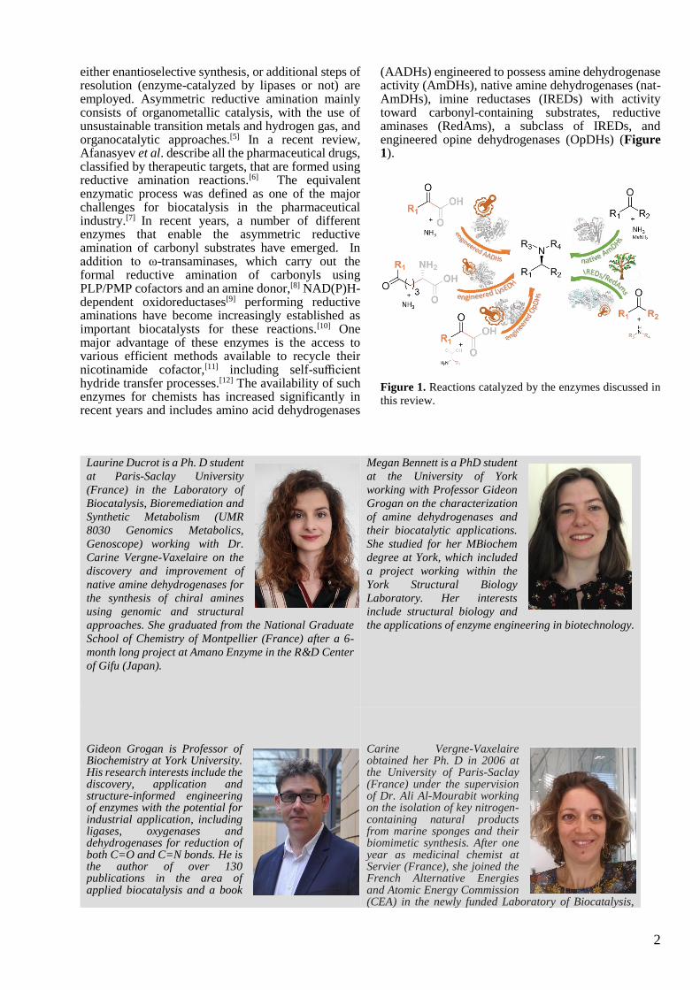
(AADHs) engineered to possess amine dehydrogenase activity (AmDHs), native amine dehydrogenases (nat-AmDHs), imine reductases (IREDs) with activity toward carbonyl-containing substrates, reductive aminases (RedAms), a subclass of IREDs, and engineered opine dehydrogenases (OpDHs) (Figure 1).
Figure 1. Reactions catalyzed by the enzymes discussed in
this review.
Laurine Ducrot is a Ph. D student
at Paris-Saclay University
(France) in the Laboratory of
Biocatalysis, Bioremediation and
Synthetic Metabolism (UMR
8030 Genomics Metabolics,
Genoscope) working with Dr.
Carine Vergne-Vaxelaire on the
discovery and improvement of
native amine dehydrogenases for
the synthesis of chiral amines
using genomic and structural
approaches. She graduated from the National Graduate
School of Chemistry of Montpellier (France) after a 6-
month long project at Amano Enzyme in the R&D Center
of Gifu (Japan).
Megan Bennett is a PhD student
at the University of York
working with Professor Gideon
Grogan on the characterization
of amine dehydrogenases and
their biocatalytic applications.
She studied for her MBiochem
degree at York, which included
a project working within the
York Structural Biology
Laboratory. Her interests
include structural biology and
the applications of enzyme engineering in biotechnology.
Gideon Grogan is Professor of Biochemistry at York University. His research interests include the discovery, application and structure-informed engineering of enzymes with the potential for industrial application, including ligases, oxygenases and dehydrogenases for reduction of both C=O and C=N bonds. He is the author of over 130 publications in the area of applied biocatalysis and a book
Carine Vergne-Vaxelaire obtained her Ph. D in 2006 at the University of Paris-Saclay (France) under the supervision of Dr. Ali Al-Mourabit working on the isolation of key nitrogen-containing natural products from marine sponges and their biomimetic synthesis. After one year as medicinal chemist at Servier (France), she joined the French Alternative Energies and Atomic Energy Commission (CEA) in the newly funded Laboratory of Biocatalysis,
((Author Portrait))
((Author
Portrait))
3
‘Practical Biotransformations’, a guide to enzyme technology for organic chemists.
Bioremediation and Synthetic Metabolism of the Genoscope Unit UMR 8030 Genomics Metabolics. Her research interests are in the area of biocatalysis with particular emphasis on the discovery of new biocatalysts among biodiversity by genome-mining approaches mastered in the unit, and their applications as green tools for the synthesis of key building blocks. Her current main project is focused upon discovering native amine dehydrogenases for the production of amines and undertaking structural and biochemical studies thereof.
In this review we focus on the application of
NAD(P)H-dependent enzymes that have been used to either catalyze, or enable, the asymmetric reductive amination of a carbonyl group (excluding α-ketoacids) with an amine donor, and therefore we do not include, for example, enzymes strictly confined to the reduction/oxidation of imine/amine substrates such as Imine Reductases (IREDs)[13], monoamine oxidases (MAOs) [10b, 14] or amine transaminases (ATAs).[8] All the enzymes included possess a nucleotide binding domain that allows them to recruit the NAD(P)H/NAD(P)+ redox cofactor essential for their activity using a common Rossman fold. But despite this common feature, their catalytic domains and the way the cofactor binding domains are connected to them, differ from one another.[9] This structural diversity results in closely related but different mechanisms, using various amino acid residues, as well as different substrate specificities, cofactor preference and stereoselectivities within the selected group of enzymes. To highlight this variety, we intend to provide readers with an illustrated summary of active sites of the various native enzymes and/or mutants and their corresponding substrate spectra. Selected examples of conversions and yields are given to exemplify the biocatalytic potential of these enzymes, but this survey does not cover all their reported applications in synthesis or cascade reactions including strategies to regenerate the cofactor.
2 From Amino Acid Dehydrogenases (AADHs) to Amine Dehydrogenases (AmDHs)
While there was a pressing demand for enzymes capable of catalyzing the reductive amination of non keto-acid carbonyl substrates for synthetic purposes, there were few examples of such enzymes in the available biodiversity. In 2012 the group of Bommarius designed the first enzyme capable of catalyzing the reductive amination of simple ketones by using AADHs (EC: 1.4.1), and more precisely leucine (LeuDH) and phenylalanine dehydrogenases (PheDH), as a platform for engineering.[15] These well-described wild-type enzymes catalyze the reductive amination of keto-acid substrates 1 and 2 into amino acid products 1a and 2a respectively, using NAD(P)H as the hydride donor (Scheme 1).
Scheme 1. General reactions catalyzed by AADHs and
engineered AADHs into AmDHs.
4
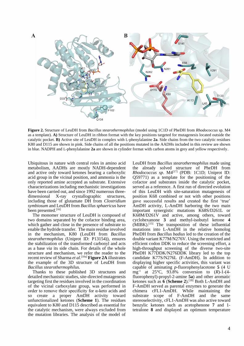
Figure 2. Structure of LeuDH from Bacillus stearothermophilus (model using 1C1D of PheDH from Rhodococcus sp. M4
as a template). A) Structure of LeuDH in ribbon format with the key positions targeted for mutagenesis located outside the
catalytic pocket. B) Active site of LeuDH in complex with L-phenylalanine 2a. Side chains from the two catalytic residues
K80 and D115 are shown in pink. Side chains of all the positions mutated in the AADHs included in this review are shown
in blue. NADPH and L-phenylalanine 2a are shown in cylinder format with carbon atoms in grey and yellow respectively.
Ubiquitous in nature with central roles in amino acid metabolism, AADHs are mostly NADH-dependent and active only toward ketones bearing a carboxylic acid group in the vicinal position, and ammonia is the only reported amine accepted as substrate. Extensive characterizations including mechanistic investigations have been carried out, and since 1992 numerous three-dimensional X-ray crystallographic structures, including those of glutamate DH from Clostridium symbiosum and LeuDH from Bacillus sphaericus have been presented.[16]
The monomer structure of LeuDH is composed of two domains separated by the cofactor binding area, which gather and close in the presence of substrate to enable the hydride transfer. The main residue involved in the mechanism, K80 (LeuDH from Bacillus stearothermophilus (Uniprot ID: P13154)), ensures the stabilization of the transformed carbonyl and acts as a base via its side chain. For details of the whole structure and mechanism, we refer the reader to the recent review of Sharma et al.[10a] Figure 2A illustrates the example of the 3D structure of LeuDH from Bacillus stearothermophilus.
Thanks to these published 3D structures and detailed mechanistic studies, site-directed mutagenesis targeting first the residues involved in the coordination of the vicinal carboxylate group, was performed in order to remove their specificity for α-keto acids and so create a proper AmDH activity toward unfunctionalized ketones (Scheme 1). The residues equivalent to K80 and D115 described as essential for the catalytic mechanism, were always excluded from the mutation libraries. The analysis of the model of
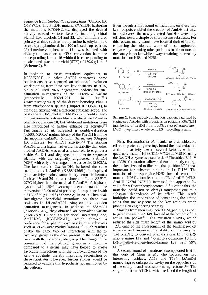
LeuDH from Bacillus stearothermophilus made using the already solved structure of PheDH from Rhodococcus sp. M4[17] (PDB: 1C1D; Uniprot ID: Q59771) as a template for the positioning of the cofactor and substrates inside the catalytic pocket, served as a reference. A first run of directed evolution of this LeuDH with site-saturation mutagenesis of position K68 combined or not with other positions gave successful results and created the first ‘true’ AmDH activity, L-AmDH harboring the two main important synergistic mutations K68S/D261L or K68M/D261V and active, among others, toward cyclohexanone 3 and methyl-isobutyl ketone 4 (MIBK).[15] The transposition of these influential mutations into L-AmDH in the relative homolog PheDH from Bacillus badius led to the creation of the double variant K77M/N276V. Using the restricted and efficient codon DDK to reduce the screening effort, a high-throughput screening of the diverse two-site PheDH K77DDK/N276DDK library led to the top candidate K77S/N276L (F-AmDH). In addition to displaying higher specific activities, this variant was capable of aminating p-fluorophenylacetone 5 (4 U mg-1 at 25°C, 93.8% conversion to (R)-1-(4-fluorophenyl)-propyl-2-amine 5a) and other aromatic ketones such as 6 (Scheme 2).[18] Both L-AmDH and F-AmDH served as parental enzymes to generate the chimera cFL1-AmDH. While maintaining the substrate scope of F-AmDH and the same stereoselectivity, cFL1-AmDH was also active toward benzylic ketones such as acetophenone 7 and 1-tetralone 8 and displayed an optimum temperature
5
(>70°C) more than 20°C higher than that of F-AmDH.[18a, 19]
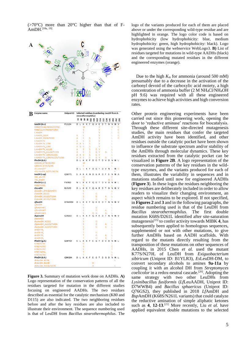
Figure 3. Summary of mutation work done on AADHs. A)
Logo representation of the conservation patterns of all the
residues targeted for mutation in the different studies
focusing on engineered AADHs. The two residues
described as essential for the catalytic mechanism (K80 and
D115) are also indicated. The two neighboring residues
before and after the key residues are also included to
illustrate their environment. The sequence numbering used
is that of LeuDH from Bacillus stearothermophilus. The
logo of the variants produced for each of them are placed
above or under the corresponding wild-type residue and are
highlighted in orange. The logo color code is based on
hydrophobicity (low hydrophobicity: blue, medium
hydrophobicity: green, high hydrophobicity: black). Logo
was generated using the webservice WebLogo3. B) List of
residues targeted for mutations in wild-type AADHs (black)
and the corresponding mutated residues in the different
engineered enzymes (orange).
Due to the high Km for ammonia (around 500 mM) presumably due to a decrease in the activation of the carbonyl devoid of the carboxylic acid moiety, a high concentration of ammonia buffer (2 M NH4Cl/NH4OH pH 9.6) was required with all these engineered enzymes to achieve high activities and high conversion rates. Other protein engineering experiments have been carried out since this pioneering work, opening the door to ‘reductive aminase’ reactions for biocatalysis. Through these different site-directed mutagenesis studies, the main residues that confer the targeted AmDH activity have been identified, and other residues outside the catalytic pocket have been shown to influence the substrate spectrum and/or stability of the AmDHs through molecular dynamics. These key residues extracted from the catalytic pocket can be visualized in Figure 2B. A logo representation of the conservation patterns of the key residues in the wild-type enzymes, and the variants produced for each of them, illustrates the variability in sequences and in mutations studied until now for engineered AADHs (Figure 3). In these logos the residues neighboring the key residues are deliberately included in order to allow readers to visualize their changing environment, an aspect which remains to be explored. If not specified, in Figures 2 and 3 and in the following paragraphs, the residue numbering used is that of the LeuDH from Bacillus stearothermophilus. The first double mutation K68S/D261L identified after site-saturation mutagenesis[15] to confer activity towards MIBK 4, has subsequently been applied to homologous sequences, supplemented or not with other mutations, to give further AmDHs based on AADH scaffolds. With regard to the mutants directly resulting from the transposition of these mutations on other sequences of AADHs, in 2015 Chen et al. used the mutant K77S/N270L of LeuDH from Exiguobacterium sibiricum (Uniprot ID: B1YLR3), EsLeuDH-DM, to convert secondary alcohols to amines 9a-11a by coupling it with an alcohol DH from Streptomyces coelicolor in a redox-neutral cascade.[20]. Adopting the same strategy with two other LeuDHs from Lysinibacillus fusiformis (LfLeuAADH, Uniprot ID: D7WWB4) and Bacillus sphaericus (Uniprot ID: Q76GS2), they published in 2018 LfAmDH and BspAmDH (K68S/N261L variants) that could catalyze the reductive amination of simple aliphatic ketones such as 4, 12-13.[21] More recently, Liu et al. have applied equivalent double mutations to the selected
6
sequence from Geobacillus kaustophilus (Uniprot ID: Q5KYC0). The PheDH mutant, GkAmDH harboring the mutations K78S/N276L, displayed the desired activity toward various ketones including chiral vicinal keto alcohols 14 and 15, with ammonia a or primary amines such as methylamine b, ethylamine c or cyclopropylamine d. In a 100 mL scale up reaction, (R)-4-methoxyamphetamine 16a was isolated with 43% yield based on a >99% conversion from the corresponding ketone 16 within 6 h, corresponding to a calculated space–time yield (STY) of 130.9 g L−1 d−1
(Scheme 2).[22] In addition to these mutations equivalent to K68S/N261L in other AADH sequences, some publications have reported an additional evolution work starting from these two key positions. In 2015, Ye et al. used NKK degenerate codons for site-saturation mutagenesis of the K66/N262 variant (respectively K68/D261 in Bacillus stearothermophilus) of the distant homolog PheDH from Rhodococcus sp. M4 (Uniprot ID: Q59771), to create an enzyme with a different substrate scope. The best variant, DM_pheDH K66Q/N262L, could already convert aromatic ketones like phenylacetone 17 and 4-phenyl-2-butanone 18, but additional mutations were also introduced to further enhance its activity.[23] Pushpanath et al. screened a double-saturation (K68X/N266X) mutant library of the PheDH from the thermophile Caldalkalibacillus thermarum (Uniprot ID: F5L9G2) for AmDH activity.[24] The starting AADH, with a higher native thermostability than other studied AADHs, was used as a scaffold to engineer a stable AmDH and displayed a moderate sequence identity with the originally engineered F-AmDH (63%) with only one change in the active site (S303A). The best variant, Cal-AmDH, harbored the same mutations as L-AmDH (K68S/N266L). It displayed good activity against some bulky aromatic ketones such as 19 and 20 but also showed a Tm of 83.5°C, 27°C higher than the original F-AmDH. A biphasic system with 25% iso-amyl acetate enabled the conversion of 400 mM of phenoxy-2-propanone 6 with a STY of 60 g L−1 d−1 (Scheme 2). In 2019, Chen et al. investigated beneficial mutations on these two positions in LfLeuAADH using on this occasion saturation mutagenesis. In addition to LfAmDH (K68S/N261L), they obtained an equivalent variant (K68C/N261L) and an additional interesting one, AmDH-M0 (K68T/N261L), which showed a preference for aliphatic α-hydroxy ketones substrates such as 21-23 over methyl ketones.[25] Such residues enable the same type of interactions with the α-hydroxyl group as the ones present in the wild type make with the α-carboxylate group. The change in the orientation of the hydroxyl group in a threonine compared to a serine may have helped to create favorable interactions with the hydroxyl group of the ketone substrate, thereby improving recognition of these substrates. However, further studies would be required to validate this hypothesis not mentioned by the authors.
Even though a first round of mutations on these two key hotspots enabled the creation of AmDH activity, in most cases, the newly created AmDHs were only efficient toward simple or short ketone substrates. For this reason, many teams have focused their efforts on enhancing the substrate scope of these engineered enzymes by mutating other positions inside or outside the catalytic pocket while always retaining the two key mutations on K68 and N261.
Scheme 2. Some reductive amination reactions catalyzed by
engineered AADHs with mutations on positions K68/N261
(numbering of LeuDH from Bacillus stearothermophilus).
LWC = lyophilized whole cells. RS = recycling system.
First, Bommarius et al., thanks to a considerable effort in protein engineering, found the best reductive amination activity toward several ketones with the quadruple mutant K68S/E114V/N261L/V291C using the LeuDH enzyme as a scaffold.[15] The added E114V and V291C mutations allowed them to directly enlarge the pocket size and to illustrate that position V291 was important for substrate binding in LeuDH.[26] The mutation of the asparagine N262, located next to the mutated N261L, into leucine in cFL1-AmDH (cFL2-AmDH N270L/N271L) increased the apparent kcat
value for p-fluorophenylacetone 5.[19] Despite this, the mutation could not be always transposed due to a substrate dependence of its effect. This result highlights the importance of considering the amino acids that are adjacent to the key residues when planning an engineering strategy.
Starting from their engineered DM_pheDH, Li et al. targeted the residue S149, located at the bottom of the active site pocket.[23] The mutation S149G, which reduced the side chain length of the amino acid by ~2Å, enabled the enlargement of the binding pocket entrance and improved the ability of the enzyme, TM_pheDH, to convert phenylacetone 17 into (R)-amphetamine 17a and 4-phenyl-2-butanone 18 into (R)-1-methyl-3-phenylpropylamine 18a with 99% ee.[18a, 23]
A second round of mutations also appeared first in the work of Chen et al., who focused on two interesting residues, A113 and T134 (LfAmDH numbering) to enlarge the cavity on the opposite side of the catalytic and substrate-binding residues.[21] The single mutation A113G, which reduced the length of
7
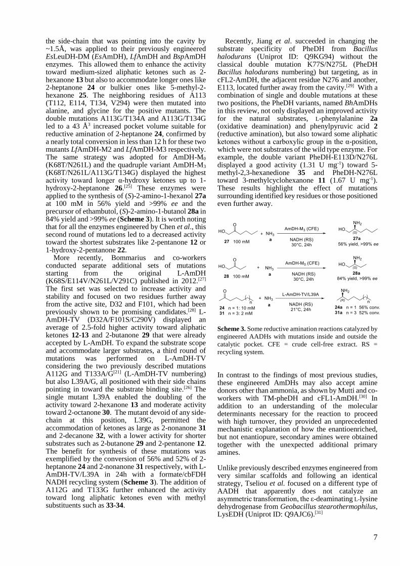
the side-chain that was pointing into the cavity by ~1.5Å, was applied to their previously engineered EsLeuDH-DM (EsAmDH), LfAmDH and BspAmDH enzymes. This allowed them to enhance the activity toward medium-sized aliphatic ketones such as 2-hexanone 13 but also to accommodate longer ones like 2-heptanone 24 or bulkier ones like 5-methyl-2-hexanone 25. The neighboring residues of A113 (T112, E114, T134, V294) were then mutated into alanine, and glycine for the positive mutants. The double mutations A113G/T134A and A113G/T134G led to a 43 Å3 increased pocket volume suitable for reductive amination of 2-heptanone 24, confirmed by a nearly total conversion in less than 12 h for these two mutants LfAmDH-M2 and LfAmDH-M3 respectively. The same strategy was adopted for AmDH-M0
(K68T/N261L) and the quadruple variant AmDH-M3 (K68T/N261L/A113G/T134G) displayed the highest activity toward longer α-hydroxy ketones up to 1-hydroxy-2-heptanone 26.[25] These enzymes were applied to the synthesis of (S)-2-amino-1-hexanol 27a at 100 mM in 56% yield and >99% ee and the precursor of ethambutol, (S)-2-amino-1-butanol 28a in 84% yield and >99% ee (Scheme 3). It is worth noting that for all the enzymes engineered by Chen et al., this second round of mutations led to a decreased activity toward the shortest substrates like 2-pentanone 12 or 1-hydroxy-2-pentanone 22.
More recently, Bommarius and co-workers conducted separate additional sets of mutations starting from the original L-AmDH (K68S/E114V/N261L/V291C) published in 2012.[27] The first set was selected to increase activity and stability and focused on two residues further away from the active site, D32 and F101, which had been previously shown to be promising candidates.[28] L-AmDH-TV (D32A/F101S/C290V) displayed an average of 2.5-fold higher activity toward aliphatic ketones 12-13 and 2-butanone 29 that were already accepted by L-AmDH. To expand the substrate scope and accommodate larger substrates, a third round of mutations was performed on L-AmDH-TV considering the two previously described mutations A112G and T133A/G[21] (L-AmDH-TV numbering) but also L39A/G, all positioned with their side chains pointing in toward the substrate binding site.[26] The single mutant L39A enabled the doubling of the activity toward 2-hexanone 13 and moderate activity toward 2-octanone 30. The mutant devoid of any side-chain at this position, L39G, permitted the accommodation of ketones as large as 2-nonanone 31 and 2-decanone 32, with a lower activity for shorter substrates such as 2-butanone 29 and 2-pentanone 12. The benefit for synthesis of these mutations was exemplified by the conversion of 56% and 52% of 2-heptanone 24 and 2-nonanone 31 respectively, with L-AmDH-TV/L39A in 24h with a formate/cbFDH NADH recycling system (Scheme 3). The addition of A112G and T133G further enhanced the activity toward long aliphatic ketones even with methyl substituents such as 33-34.
Recently, Jiang et al. succeeded in changing the substrate specificity of PheDH from Bacillus halodurans (Uniprot ID: Q9KG94) without the classical double mutation K77S/N275L (PheDH Bacillus halodurans numbering) but targeting, as in cFL2-AmDH, the adjacent residue N276 and another, E113, located further away from the cavity.[29] With a combination of single and double mutations at these two positions, the PheDH variants, named BhAmDHs in this review, not only displayed an improved activity for the natural substrates, L-phenylalanine 2a (oxidative deamination) and phenylpyruvic acid 2 (reductive amination), but also toward some aliphatic ketones without a carboxylic group in the α-position, which were not substrates of the wild type enzyme. For example, the double variant PheDH-E113D/N276L displayed a good activity (1.31 U mg-1) toward 5-methyl-2,3-hexanedione 35 and PheDH-N276L toward 3-methylcyclohexanone 11 (1.67 U mg-1). These results highlight the effect of mutations surrounding identified key residues or those positioned even further away.
Scheme 3. Some reductive amination reactions catalyzed by
engineered AADHs with mutations inside and outside the
catalytic pocket. CFE = crude cell-free extract. RS =
recycling system.
In contrast to the findings of most previous studies, these engineered AmDHs may also accept amine donors other than ammonia, as shown by Mutti and co-workers with TM-pheDH and cFL1-AmDH.[30] In addition to an understanding of the molecular determinants necessary for the reaction to proceed with high turnover, they provided an unprecedented mechanistic explanation of how the enantioenriched, but not enantiopure, secondary amines were obtained together with the unexpected additional primary amines.
Unlike previously described enzymes engineered from very similar scaffolds and following an identical strategy, Tseliou et al. focused on a different type of AADH that apparently does not catalyze an asymmetric transformation, the ε-deaminating L-lysine dehydrogenase from Geobacillus stearothermophilus, LysEDH (Uniprot ID: Q9AJC6).[31]

8
Scheme 4. General reaction catalyzed by LysEDH and the
engineered LysEDH.
In this case, the reaction occurs at the terminal amino group of L-lysine 36a instead of the α-amine moiety, which was shown to be non-essential for the activity in this wild type enzyme. The objectives of this study were to remove the essential nature of the α-carboxyl group for recognition by the wild type enzyme, which prevents any activity toward substrates devoid of this function, such as m-fluoroacetophenone 37, 2-heptanone 24, acetophenone 7 or α-chromanone 38 (Scheme 4).
Scheme 5. Example of reductive amination reaction
catalyzed by the engineered ε-deaminating L-lysine
dehydrogenase.
Based on computational studies of a LysEDH homology model, created using different templates with both NADH cofactor and ligand within the active site, seven residues, shown in Figure 4, were identified as potential targets for mutagenesis. Among them, only the mutation of F173 enabled the conversion of medium-sized aliphatic ketones and, above all, the targeted bulky aromatic ketones such as acetophenone 7 or 1-tetralone 8. The bulky residue F173 is located at the opposite side of the hydrophilic cavity to that which accommodates the α-amino moiety of L-lysine 36a and serves to provide both the hydrophobic property of the cavity and the orientation of the side chain of the substrate. Its mutation into alanine enabled the enlargement of the hydrophobic binding pocket by reducing the size of the residue by 4.3Å, without substantially increasing the flexibility of the active site, which was shown to be detrimental for the activity by measuring the activity of mutant F173G. Apart from the variants harboring the additonal mutations V130A/G to F173A, which also displayed interesting activity toward aromatic ketones, all the other mutated residues (H181, Y238, T240 and V172) gave unsatisfactory results, including R242, also involved in the binding of the α-carboxylic group of L-lysine 36. This surprising result accords with that of Jiang et al. with BhPheDH, who obtained active variants without any mutation of the residue binding the α-carboxylic group of the native substrate.[29] Remarkably, LE-AmDH-v1 (F173A) displayed a high stability (Tm: 69°C) and a reduced product inhibition compared to other engineered AADHs, thus leading to a high biocatalytic performance.
Figure 4. Model of the ε-deaminating L-lysine dehydrogenase from Geobacillus stearothermophilus (model using 1E5Q of
saccharopine dehydrogenase from Magnaporthe oryzae). A) Structure of LysEDH in ribbon format B) Detail of key
positions described in this survey, F173 is highlighted in orange as the target for successful mutagenesis. Side chains of the
key positions detailed in the paper are shown with carbon atoms in blue. NADPH is shown in cylinder format with carbon
atoms in grey.

9
Figure 5. Substrate specificity of all the engineered AADHs described in this review. The substrates associated to each
variant were selected as the ones displaying the highest activities or considered as “new” substrates compared to the substrate
scope of the whole family. In the case of enzymes without activity data, the substrates showing the highest conversion rates
for one experimental condition were selected. If not mentioned, the amine substrate is ammonia. The orange and blue lines
correspond to a set of mutations respectively inside or outside the catalytic pocket. The grey dashed lines correspond to the
creation of a chimera from the two parental enzymes L-AmDH and F-AmDH. The specific activities indicated in this Figure
are given as information but not to be compared as all the assays were not done with the exact same conditions. The following
ones correspond to a range of typical conditions: 10-40 mM carbonyl substrate, 0.1-0.2 mM NAD(P)H cofactor, 1-6 M
ammonium-based buffer pH 8.8-9.6, 25-30°C. [a] Activity assays done with lower ammonia concentrations 0.225-0.5 M that
could not allow to reach the saturation of ammonia. [b] Activity assays done at 60°C, conversion tests at 30°C. The mutations
written in bold correspond to the L-AmDH or F-AmDH numbering while the other mutations refer to the numbering of the
targeted enzyme.

10
The reductive amination of 5 mM acetophenone 7 and propiophenone 39 was performed on a 600 mg scale to give respectively 82 and 65% isolate yield of the (R)-amine products 7a and 39a with excellent ee >99.9% (Scheme 5). The stereochemistry of the amines obtained was (R), except for 2-aminoheptanoic acid 40a, which was produced with an enantiomerically pure (S)-configuration as with the wild type enzyme. Figure 5 summarizes all the type of ketones described as substrates of the various mutants obtained from AADHs and ε-deaminating L-lysine dehydrogenase, and the engineering schemes followed to obtain them. Since the first engineering of an AADH for AmDH activity in 2012, a considerable amount of engineering work has been carried out by many groups. As illustrated in Figure 3, there is still scope for more mutagenesis, especially in the vicinity of the already mutated residues and outside the catalytic pocket for which examples have only been published very recently.
3 Engineered Opine dehydrogenases (OpDHs)
Opine dehydrogenases (OpDHs) are a class of oxidoreductases that catalyze the reductive amination of α-keto acids, not with ammonia like AADHs, but with α-amino acids, using NADH as cofactor (Scheme 6). The resulting products, opines, have very different physiological roles, depending on whether they are formed in invertebrates or in plants.[32] In the former, their formation ensures the consumption of pyruvate and NADH keeping glycolysis running upon anaerobic exercise; in the latter they ensure the growth of the parasitic Agrobacteria responsible for crown
gall disease of plants after genomic insertion of the OpDH coding region into the host plant.[33]
Scheme 6. General reaction catalyzed by OpDHs and
engineered CENDH.
The OpDH from Arthrobacter sp. strain 1C (Uniprot ID: Q44297), named CENDH, is one of the most characterized OpDHs,[34] together with that of the great scallop Pecten maximus.[35] In addition to NAD+-dependent oxidation of opines of plant origin, such as methiopine, reductive aminations are also performed by these enzymes, with the (R)-configuration at the newly generated stereogenic center of the opine products. They preferentially bind pyruvate 41 as electrophile and, to a lesser extent, α-ketobutyrate 42, α-ketovalerate 43 and oxaloacetate 44. In addition to its activity toward (S)-methionine and (S)-phenylalanine, ODH was found to be even more active toward L-norvaline, hence its name N-(1-D-carboxyethyl)-L-norvaline dehydrogenase (CENDH) (Scheme 6).
Figure 6. Global structure of CENDH (PDB: 1BG6). A) Structure of CENDH in ribbon format B) Detail of the key positions
targeted for mutagenesis by Codexis, shown with carbon atoms in blue. The position of NADPH and pyruvate, respectively
in grey and yellow, were obtained by 3D-alignment with the structure of OpDH from Pecten maximus (PDB: 3C7D).
11
The resolution of its structure (PDB: 1BG6 apo structure) combined with a mechanistic study of the already resolved OpDH from Pecten maximus (PDB: 3C7C holo structure with L-arginine; 3C7D holo structure with pyruvate 41) provided preliminary insights into its mechanism (Figure 6). Like other AADHs, these enzymes possess a two-domain structure with a cleft at the interface holding the cofactor. Despite the identification of key residues binding pyruvate 41 (Q118, H212 and E142 in the case of OpDH from Pecten maximus) and some NMR evidence of sequential order of substrate binding,[36] no further description of the mechanism has been reported. Considering its relaxed substrate scope, Codexis used CENDH as a starting template to evolve it into a catalyst able to synthesize tertiary amines.[37]
The variants, obtained after an extensive effort on directed evolution experiments using the protein engineering technology CodeEvolver®, were not only more active toward the two native substrates of CENDH, pyruvate 41 and L-norvaline, but were also able to catalyze the coupling of a range of ketone substrates, such as cyclohexanone 3, cyclopentanone 45 or 5-methoxy-tetralone 46 (Scheme 6). A variety of amine partners was accepted, such as butylamine or pyrrolidinol, and transformed to products with high enantiomeric excess. The mutations that could positively impact the activity toward a wide range of substrates mostly targeted residues located in the active site cleft. For example, the multiple variant A111M/K156T/N198H/Y259M/Y280L/R292V/Y293H was ranked first for the conversion of cyclohexanone 3 and butylamine into N-butyl-N-cyclohexylamine. To our knowledge, no other work regarding the engineering of these enzymes for reductive amination has been published. More structural studies are required to expand their biocatalytic potential, bearing in mind their native activity toward various type of amines and the actual mechanistic knowledge of AADHs. Other types of OpDHs, such as saccharopine DHs, may also be valuable templates for engineering. In view of the significant differences between their native substrates and the targeted substrates, the evolution of OpDHs constitutes a considerable piece of enzyme engineering.
4 Native Amine Dehydrogenases
To meet the demand in synthetically useful AmDHs, an exploration of protein biodiversity was considered a good strategy to find wild-type AmDHs with various substrate scopes and new scaffolds to provide templates for protein engineering (Scheme 7). Some examples of this activity occurring in nature were published at the beginning of the 2000s by Itoh and coworkers, who reported an amine dehydrogenase from Streptomyces virginiae that was active toward a
broad range of ketone and aldehyde substrates but displayed low enantioselectivity for most of these substrates.[38] Later on, Wang et al. also identified an activity using whole cells of Pseudomonas kilonensis.[39] The absence of gene identification in these studies prevented any further characterization of these enzymes, but these detected native activities demonstrated the usefulness of exploring biodiversity in addition to protein engineering for the identification of novel AmDH activities in addition to carrying out engineering work with other families.
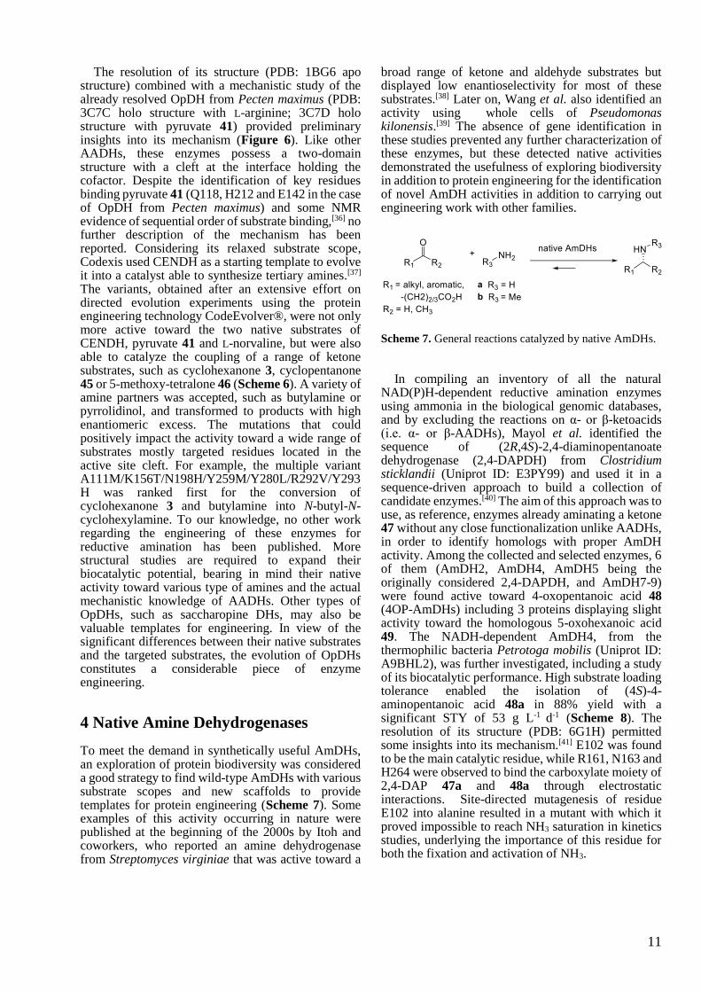
Scheme 7. General reactions catalyzed by native AmDHs.
In compiling an inventory of all the natural NAD(P)H-dependent reductive amination enzymes using ammonia in the biological genomic databases, and by excluding the reactions on α- or β-ketoacids (i.e. α- or β-AADHs), Mayol et al. identified the sequence of (2R,4S)-2,4-diaminopentanoate dehydrogenase (2,4-DAPDH) from Clostridium sticklandii (Uniprot ID: E3PY99) and used it in a sequence-driven approach to build a collection of candidate enzymes.[40] The aim of this approach was to use, as reference, enzymes already aminating a ketone 47 without any close functionalization unlike AADHs, in order to identify homologs with proper AmDH activity. Among the collected and selected enzymes, 6 of them (AmDH2, AmDH4, AmDH5 being the originally considered 2,4-DAPDH, and AmDH7-9) were found active toward 4-oxopentanoic acid 48 (4OP-AmDHs) including 3 proteins displaying slight activity toward the homologous 5-oxohexanoic acid 49. The NADH-dependent AmDH4, from the thermophilic bacteria Petrotoga mobilis (Uniprot ID: A9BHL2), was further investigated, including a study of its biocatalytic performance. High substrate loading tolerance enabled the isolation of (4S)-4-aminopentanoic acid 48a in 88% yield with a significant STY of 53 g L-1 d-1 (Scheme 8). The resolution of its structure (PDB: 6G1H) permitted some insights into its mechanism.[41] E102 was found to be the main catalytic residue, while R161, N163 and H264 were observed to bind the carboxylate moiety of 2,4-DAP 47a and 48a through electrostatic interactions. Site-directed mutagenesis of residue E102 into alanine resulted in a mutant with which it proved impossible to reach NH3 saturation in kinetics studies, underlying the importance of this residue for both the fixation and activation of NH3.

12
Figure 7. Structure of CfusAmDH (PDB: 6IAU) A) Structure of CfusAmDH in ribbon format B) Active site of CfusAmDH
with a focus on the P1-P20 positions (blue), shown in cylinder format, defining the cavity with NADPH (grey),
cyclohexanone (yellow) and ammonia. For clarity, P1-2-4-10-13-14-15-18-20 are omitted.
To alter the substrate specificity and enable the conversion of ketone substrates devoid of the carboxylic moiety, these polar residues were mutated into non-polar ones. Among the 8 variants produced, N135V/N163V/R161M/H264L mutant, named here AmDH4_M1, displayed the highest activity with 105 mU mg-1 toward 2-pentanone 12. The main mutation responsible for this switch of substrate scope seemed to be R161M but as yet no further mutation work has been done to further explore this site. Interestingly, 4OP-AmDHs do not share significant sequence homology with the previously described engineered AADHs nor RedAms, demonstrating that enzymes catalyzing reductive amination are distributed among very varied families. To further explore AmDH biodiversity and discover distant homologs with activity toward unfunctionalized ketones, 4OP-AmDHs were in turn used in a second sequence-driven approach by the same group. This iterative approach successfully led to the identification and characterization of six AmDHs (MsmeAmDH Uniprot ID: A0A0D6I8P6, CfusAmDH Uniprot ID: S9Q235, MicroAmDH Uniprot ID: C3UMY1, ApauAmDH Uniprot ID: E3CZE3, MycoAmDH Uniprot ID: A0A101AWU7 and MvacAmDH Uniprot ID: K0UKT5) with similar substrate spectra, with a preference for cycloalkanones such as 3 and 50, and aliphatic aldehydes such as 51-52. MsmeAmDH displayed also significant activity toward α-hydroxy ketone 53. No activity toward the initial substrates 47 and 48 was detected with these enzymes. With low sequence identity (<30%) with 2,4-DAPDH and 4OP-AmDHs, and no apparent common biological role, these enzymes are members
of the first family of native AmDHs (nat-AmDHs).[41] As indicated by biochemical characterization and substrate spectrum, these enzymes are slightly more efficient with NADPH than NADH even if the substrate seemed to influence this cofactor specificity.
As for engineered AADHs, the high Km of ammonia required the use of a high concentration of ammonia buffer, usually in the range of 1 – 2 M, at pH 8.0 – 9.5.
Scheme 8. Examples of reductive amination reactions
catalyzed by native 4OP-AmDH and nat-AmDHs. RS =
recycling system.

13
Biocatalytic synthesis performed on 100 mg scale afforded (S)-2-aminopentane 12a or (1S, 2R)-2-methylcyclohexylamine 50a from the equivalent carbonyl compounds in 39% and 35% yield respectively, corresponding to a STY of 2.4-2.6 g L-1
d-1 (Scheme 8). The structural resolution of CfusAmDH (PDB: 6IAU) and MsmeAmDH (PDB: 6IAQ) showed that their whole structure is highly similar to that of AmDH4. Their dimeric structures are rather similar to meso-diaminopimelate dehydrogenases, with a well-conserved Rossman fold in the N-terminal domain, but significant differences in the C-terminal β-sheet domain. (Figure 7A) The binding of the substrate in the catalytic pocket significantly changes the nature of the active site, bringing it into close proximity with the nicotinamide cofactor. The catalytic pockets are also rather similar to that of AmDH4, with the same conserved key residue E102, but with the R161, N163 and H264 carboxylate binding residues (AmDH4 numbering) absent, as would be expected (Figure 7B). Similarly to AmDH4, the hypothetical mechanism involved the conserved glutamate activating ammonia, which in turn would attack the electrophilic carbon of the carbonyl secured by the conserved hydrogen bond donors tyrosine/tryptophan within the active site (II). The resulting carbinolamine (III) would be dehydrated to lead to the amine product (V) after reduction by the hydride of the nicotinamide cofactor (IV) on the re-face in the case of prochiral substrate (Scheme 9).
Scheme 9. Hypothetical mechanism of nat-AmDHs. The
proposed mechanism is showed for CfusAmDH and
cyclohexanone as carbonyl substrate.
Figure 8. Pocket diversity found in the nat-AmDHs family and mutation work done on AmDH4 A) Logo representation of
the conservation patterns of the main residues defining the catalytic pocket of characterized native AmDHs (AmDH2,
AmDH4-5, AmDH7-9, MsmeAmDH, CfusAmDH, MicroAmDH, ApauAmDH, MycoAmDH, MvacAmDH, ChatAmDH,
SgorAmDH, AcolAmDH, IGCAmDH1, IGCAmDH5 and MATOUAmDH2). For clarity, positions P13 and P14 have been
removed from this Figure. The two neighboring residues before and after these residues are included as part of the logos to
illustrate their environment. The mutated amino acids in AmDH4 engineering are placed above or under the corresponding
wild-type residue and are highlighted in orange. The sequence numbering used is that of CfusAmDH. The color code is
based on hydrophobicity (low hydrophobicity: blue, medium hydrophobicity: green, high hydrophobicity: black). Logos
were generated using the webservice WebLogo3. B) List of main residues defining the catalytic pocket in wild-type nat-
AmDHs (black) and the corresponding mutated residues in the engineered enzyme (orange).
14
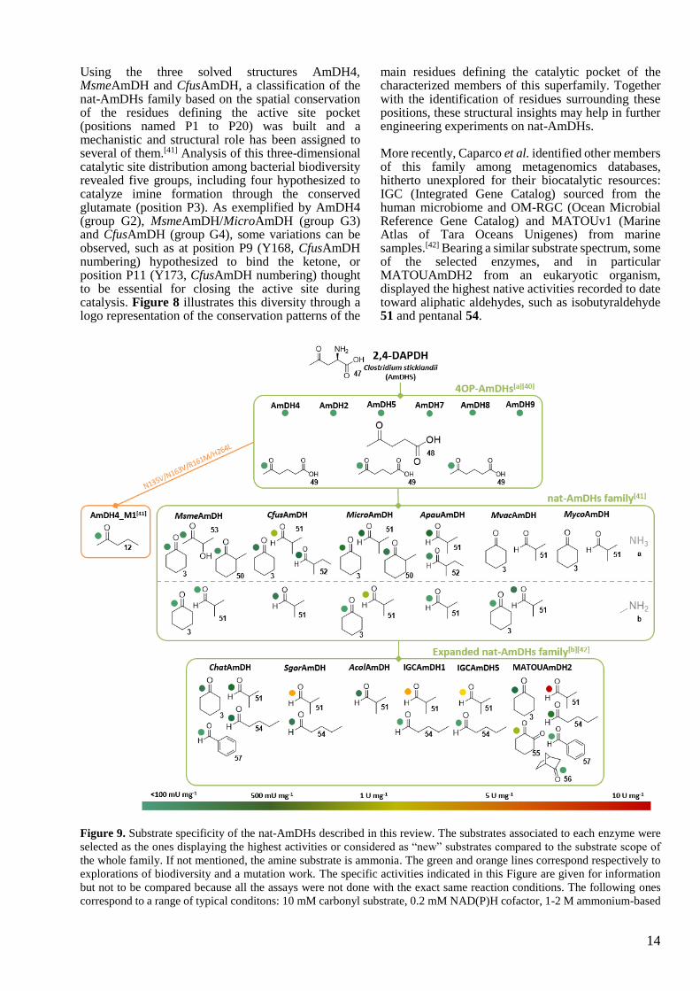
Using the three solved structures AmDH4, MsmeAmDH and CfusAmDH, a classification of the nat-AmDHs family based on the spatial conservation of the residues defining the active site pocket (positions named P1 to P20) was built and a mechanistic and structural role has been assigned to several of them.[41] Analysis of this three-dimensional catalytic site distribution among bacterial biodiversity revealed five groups, including four hypothesized to catalyze imine formation through the conserved glutamate (position P3). As exemplified by AmDH4 (group G2), MsmeAmDH/MicroAmDH (group G3) and CfusAmDH (group G4), some variations can be observed, such as at position P9 (Y168, CfusAmDH numbering) hypothesized to bind the ketone, or position P11 (Y173, CfusAmDH numbering) thought to be essential for closing the active site during catalysis. Figure 8 illustrates this diversity through a logo representation of the conservation patterns of the
main residues defining the catalytic pocket of the characterized members of this superfamily. Together with the identification of residues surrounding these positions, these structural insights may help in further engineering experiments on nat-AmDHs.
More recently, Caparco et al. identified other members of this family among metagenomics databases, hitherto unexplored for their biocatalytic resources: IGC (Integrated Gene Catalog) sourced from the human microbiome and OM-RGC (Ocean Microbial Reference Gene Catalog) and MATOUv1 (Marine Atlas of Tara Oceans Unigenes) from marine samples.[42] Bearing a similar substrate spectrum, some of the selected enzymes, and in particular MATOUAmDH2 from an eukaryotic organism, displayed the highest native activities recorded to date toward aliphatic aldehydes, such as isobutyraldehyde 51 and pentanal 54.
Figure 9. Substrate specificity of the nat-AmDHs described in this review. The substrates associated to each enzyme were
selected as the ones displaying the highest activities or considered as “new” substrates compared to the substrate scope of
the whole family. If not mentioned, the amine substrate is ammonia. The green and orange lines correspond respectively to
explorations of biodiversity and a mutation work. The specific activities indicated in this Figure are given for information
but not to be compared because all the assays were not done with the exact same reaction conditions. The following ones
correspond to a range of typical conditons: 10 mM carbonyl substrate, 0.2 mM NAD(P)H cofactor, 1-2 M ammonium-based

15
buffer pH 8-9.5, 20-25°C. [a] Activity assays done with 100 mM carbonyl substrate and 0.3 M ammonium-based buffer [b]
Activity assays performed at 50°C.
Above all, this more distant homolog also showed high activity toward 1,2-cyclohexadione 55, but lower activity with bulkier substrates like norcamphor 56 or benzaldehyde 57, not substrates of previously identified nat-AmDHs. The noticeable differences, in terms of P1-P20 positions, of this eukaryote-sourced enzyme but also of some of the other modelled ones, can explain their different cofactor specificities (NADH-dependent enzymes, excepting MATOUAmDH2) and slightly different substrate scope compared to previously reported nat-AmDHs. Further studies including resolution of some 3D-structures and protein engineering are required to better link structure / activity of these recently discovered enzymes which have a synthetic potential, as exemplified by the 85% conversion of 10 mM isobutyraldehyde 51 into isobutyrylamine 51a by IGCAmDH1 at 30°C or even 50°C (Scheme 8). Figure 9 highlights the main substrates of native AmDHs discovered throughout the different (meta)genomic approaches and the only preliminary engineering done on AmDH4. Reliable engineering work can now be undertaken on these recently discovered enzymes. Further scope for beneficial mutagenesis work is possible, either on the well-conserved residues (such as P1, P4, P9, P10, P11, P12, P20) or the less conserved ones (such as P2, P6, P7, P8, P17), including all the neighboring positions.
For all these native AmDHs, the (S)-amines were preferentially formed as for 4OP-AmDHs and 2,4-DAPDHs, in contrast to engineered AADHs and LE-AmDH-v1, each of which giving the (R)-enantiomer for the reaction with ammonia a (following Cahn-Ingold-Prelog priority rules in the case of unfunctionalized carbonyl substrates). The latter is the amine source highly preferentially accepted by these native enzymes, but secondary activity toward methylamine b was also recorded, particularly for MATOUAmDH2, which is promising for widening the application of AmDHs to the synthesis of secondary amines.
5 Imine Reductases (IREDs) active toward carbonyl-containing substrates and Reductive Aminases (RedAms)
Imine reductases (IREDs) are NADPH-dependent oxidoreductases that have been shown to catalyze the asymmetric reduction of pre-formed prochiral imines.[13b, 43] IREDs present a good starting point for biocatalytic reductive amination as the imine reduction step constitutes the second half of the reductive amination reaction (Scheme 10) and a subset of these enzymes, RedAms, have been shown to also catalyze the imine formation reaction.
Scheme 10. IREDs (red) catalyze the reduction of imines
preformed in solution to give optically active amines.
RedAms (blue) catalyze the formation of the imine from a
carbonyl substrate and amine donor and also the reduction
of the imine.
Stereocomplementary IREDs from Streptomyces sp. GF3587 and GF3546, with (R)- and (S)-selectivity respectively for the reduction of 2-methylpyrroline, were the first examples to be identified.[44] In 2014, Huber and co-workers showed for the first time that, in the presence of a large excess of an amine donor, in that case a 212 mM methylammonium buffer at pH 9.5, low conversions (up to 8.8 ± 1.6%) of a ketone, 4-phenyl-2-butanone 18, to the secondary amine (S)-2‐(methylamino)‐4‐phenylbutane 18b with 76% ee, could be achieved in the presence of high loadings of the (S)-IRED from Streptomyces sp. GF3546 (Uniprot ID: M4ZS15) (Scheme 11).[45] The excess of amine donor and high pH were thought to favor the formation of an intermediate imine that would be recruited from solution and reduced from one prochiral face by the enzyme.
Scheme 11. Example of reaction catalyzed by (S)-IRED
from Streptomyces sp. GF3546.
The discovery of new IREDs and the exploration of their substrate scope yielded further observations of reductive amination reactions. (R)-IRED-Sr (Uniprot ID: D2B7Z8), from Streptosporangium roseum, was screened for carbonyl reaction scope as well as activity towards different amine donors in reductive amination reactions. In the presence of (R)-IRED-Sr, benzaldehyde 57 was transformed to amine products using either ammonia a or methylamine b, with conversions of up to 73% for the reaction with methylamine b, when 50 equivalents of the amine were employed.[46] Lower conversions were observed with aniline e. Moreover, incubation of acetophenone 7 with methylamine b gave 39% conversion to the (R)-

16
amine product N-methyl-1-phenylethylamine 7b with 87% ee using 50 equivalents of amine and at a pH of 9.0, each of which was thought to favor the formation of the intermediate imine in solution. Indeed, NMR studies failed to show accumulation of an imine intermediate, suggesting that (R)-IRED-Sr, was remarkably efficient in withdrawing the imine from solution for reduction.
The application of IREDs to the asymmetric reductive amination of ketones was expanded by Wetzl and co-workers at Roche, who screened a library of 28 IREDs (named here Roche_IRXX) for activity with various aromatic, aliphatic and cyclic ketones, for the production of either secondary or primary amines.[47] It was found that all of the IREDs displayed activity towards at least one ketone, with higher conversions typically achieved with methylamine b as the amine donor, when supplied at 12.5 molar equivalents and at pH 9.3. The best examples were carried out on 100 mg scale, with the IRED Roche_IR11 (Uniprot ID: F4F8G5) converting substrate 11 and methylamine b to the (1S, 3R)-11b product in 71% yield with 98% de, and 2-hexanone 13 to the (R)- amine product 13b in 55% yield with 96% ee (Scheme 12). Again, NMR studies did not reveal significant formation of the imine intermediate in solution. Höhne and co-workers subsequently screened the Roche IRED enzymes and others for additional reductive amination reactions using cyclohexanone 3 and indanone 58, among other ketone substrates, and short alkyl amines, including secondary amines such a pyrrolidine f, as donors.[48] The enzymes Roche_IR14 (Uniprot ID: H6RB67) and IR-Sip (Uniprot ID: L1KNB7) were applied to the synthesis of the (R)- and (S)- enantiomers respectively of the anti-Parkinson’s agent rasagiline 58g from indanone 58 and propargylamine g, the latter supplied at 40 molar equivalents at pH 9.5 (Scheme 12). In this way, (R)- and (S)- rasagiline 58g were produced in 58% and 81% yields with 90% and 83% ee respectively. Additionally, Roche_IR14 enabled the reductive amination of cyclohexanone 3 with 3-methylamino-1-propyne h to form the tertiary amine 3h in 59% yield (Scheme 12).
Scheme 12. Some reductive amination of ketones using the
IRED collection at Roche. RS = recycling system.
These advances were significant in establishing the utility of IREDs in asymmetric reductive aminations, however, the reactions were still disadvantaged by their dependence upon a large molar excess of amine, presumably because of the need to favor imine formation in solution and so creating the substrate for IRED-catalyzed reduction. In 2017, an oxidoreductase of the IRED family from the fungus Aspergillus oryzae, AspRedAm (Uniprot ID: Q2TW47), was reported to be the first enzyme capable of enabling the reductive amination of ketones with amine partners supplied at, or near to, molar equivalence and at neutral pH.[49] This suggested that this enzyme was both catalyzing imine formation, as well as imine reduction within the active site and therefore constituted a true enzymatic reductive amination reaction. AspRedAm catalyzed the reductive amination of alicyclic and aliphatic carbonyls of various chain lengths using small amine donors with typically higher conversion rates achieved when compared to previously studied IREDs. In some cases, notably with cyclohexanone 3 and small amines such as allylamine i and propargylamine g, up to 94% conversion to the amine product was achieved even at equimolar concentrations of the amine nucleophile (Scheme 13). The reductive amination of 50 mM cyclohexanone 3 with two equivalents of methylamine b was performed on a 100 mg scale using 0.1 mg mL-
1 AspRedAm to give 75% isolated yield of the N-methylcyclohexylamine product 3b.

17
Scheme 13. Some reductive amination reactions catalyzed
by AspRedAm from Aspergillus oryzae using low amine:
ketone ratios. RS = recycling system.
In addition to this remarkable property, it was also shown that, in contrast to other IREDs, the activity of AspRedAm was largely unaffected by pH between values of 7.0 and 9.0, indicating that catalysis was not dependent upon the ambient concentration of imine formed in solution by abiotic means.
Kinetic studies using cyclohexanone 3 and methylamine b suggested an ordered sequential Ter bi mechanism in which, following the binding of reduced cofactor, ketone and amine were bound sequentially, as had previously been demonstrated for the N-methyl-L-amino acid dehydrogenase from Pseudomonas putida.[50] A structure of AspRedAm in complex with
the amine product (R)-rasagiline 58g revealed that the enzymes displayed an overall dimer fold similar to that of known IREDs (Figure 10A).
Each monomer in the dimer features an N-terminal Rossmann fold domain connected to a C-terminal helical bundle through a long inter-domain helix. A reciprocal domain sharing results in the active site being formed at the interface between the N-terminal domain of one monomer and the C-terminal domain of its neighbor. The structure in complex with (R)-rasagiline 58g, revealed active site residues in close contact with the ligand (Figure 10B), and these observations suggested mutations that might alter the activity of the enzyme. Site-directed mutagenesis highlighted the importance of Y177 and D169 in the reductive amination abilities of AspRedAm. Y177A and D169A/D169N mutants displayed significantly reduced reductive aminase activity by ~30 and ~200 fold respectively. The mutation of another active site residue, W210, to alanine, resulted in a mutant W210A that displayed a switch in enantioselectivity for the reductive amination of 4-phenyl-2-butanone 18 with allylamine i from 30% ee (R)- for the wild-type to 90% (S)-.
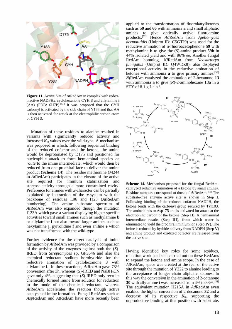
Further mechanistic insight into the reductive amination catalyzed by fungal RedAms was provided by structural and mutational studies on the closely related enzyme AtRedAm from Aspergillus terreus (Uniprot ID: Q0CCT3).[51] A crystal complex of AtRedAm with cyclohexanone 3 and allylamine i, achieved through co-crystallization with the redox inactive cofactor analog NADPH4, suggested recognition of the carbonyl group by Y183 through a water molecule, and the amine nitrogen by an aspartate D175 (Figure 11).
Figure 10. Structure of AspRedAm. A) Structure of AspRedAm with monomers A and B shown in ribbon format in brown
and blue respectively. (PDB: 56GS)[49] B) Active site of AspRedAm in complex with (R)-rasagiline 58g. Side chains from
monomers A and B are shown with carbon atoms in brown and blue respectively. NADPH and (R)-rasagiline 58g are shown
in cylinder format with carbon atoms in grey and yellow respectively.
18
Figure 11. Active Site of AtRedAm in complex with redox-
inactive NADPH4, cyclohexanone CYH 3 and allylamine i
(AA) (PDB: 6H7P).[51] It was proposed that the CYH
carbonyl is activated by the side chain of Y183 and that AA
is then activated for attack at the electrophilic carbon atom
of CYH 3.
Mutation of these residues to alanine resulted in variants with significantly reduced activity and increased Km values over the wild-type. A mechanism was proposed in which, following sequential binding of the reduced cofactor and the ketone, the amine would be deprotonated by D175 and positioned for nucleophile attack to form hemiaminal species en route to the imine intermediate, which would then be reduced from one prochiral face to deliver the amine product (Scheme 14). The residue methionine (M244 in AtRedAm) participates in the closure of the active site required for iminium stabilization and stereoselectivity through a more constrained cavity. Preference for amines with π-character can be partially explained by interaction of the π-system with the backbone of residues L96 and I123 (AtRedAm numbering). The amine substrate spectrum of AtRedAm was also expanded though the mutation I123A which gave a variant displaying higher specific activities toward small amines such as methylamine b or allylamine i but also toward larger amines such as hexylamine j, pyrrolidine f and even aniline e which was not transformed with the wild-type.
Further evidence for the direct catalysis of imine formation by AtRedAm was provided by a comparison of the activity of the enzymes against both the (S)-IRED from Streptomyces sp. GF3546 and also the chemical reductant sodium borohydride for the reductive amination of cyclohexanone 3 with allylamine i. In these reactions, AtRedAm gave 73% conversion after 3h, whereas (S)-IRED and NaBH3CN gave only 4%, suggesting that (S)-IRED only recruits chemically formed imine from solution for reduction in the mode of the chemical reductant, whereas AtRedAm accelerates the reaction though active catalysis of imine formation. Fungal RedAms such as AspRedAm and AtRedAm have more recently been
applied to the transformation of fluorokarylketones such as 59 and 60 with ammonia a and small aliphatic amines to give optically active fluoroamine products.[52] Hence AdRedAm from Ajellomyces dermatitidis (Uniprot ID: C5GTJ9) was used for the reductive amination of α-fluoroacetophenone 59 with methylamine b to give the (S)-amine product 59b in 44% isolated yield and with 96% ee. Another fungal RedAm homolog, NfRedAm from Neosartorya fumigatus (Uniprot ID: Q4WDZ8), also displayed exceptional activity in the reductive amination of ketones with ammonia a to give primary amines.[53] NfRedAm catalyzed the amination of 2-hexanone 13 with ammonia a to give (R)-2-aminohexane 13a in a STY of 8.1 g L-1 h-1.
Scheme 14. Mechanism proposed for the fungal RedAm-
catalyzed reductive amination of a ketone by small amines.
Residue numbers correspond to those of AtRedAm.[51] The
substrate-free enzyme active site is shown in Step I.
Following binding of the reduced cofactor NADPH, the
ketone binds with the carbonyl group secured by Tyr183.
The amine binds to Asp175 and is activated for attack at the
electrophilic carbon of the ketone (Step II). A hemiaminal
intermediate results (Step III), from which water is
eliminated to yield the prochiral iminium ion (Step IV). The
imine is reduced by hydride delivery from NADPH (Step V)
and amine product and oxidized cofactor are released from
the active site.
Having identified key roles for some residues, mutation work has been carried out on these RedAms to expand the ketone and amine scope. In the case of AtRedAm, space was created at the rear of the active site through the mutation of Y222 to alanine leading to the acceptance of longer chain aliphatic ketones. In this way the conversion in the amination of 2-octanone 30 with allylamine i was increased from 4% to 53%.[51] The equivalent mutation H215A in AdRedAm even enabled the higher conversion of 2-decanone 32 and a decrease of its respective Km, supporting the unproductive binding at this position with substrate.

19
Activity toward the ester derivative 61 was also detected with this mutant. The identified position I123 in AtRedAm proved to be a key position to extend amine range, as evidenced by the increased specific activities of the mutant I123A for both small (ammonia a, methylamine b) and bulkier amines (hexylamine j, pyrrolidine f, aniline e).[51] The description of reductive aminase activity in fungal enzymes such as AspRedAm and AtRedAm has prompted further screening of enzyme libraries for similar activity, in some cases by industrial groups. Roiban and co-workers at GSK screened 85 IRED homologs (named here GSK_IRXX) for reductive amination activity at pH 7.0 using amine:ketone ratios of 1:1 and including especially arylamines as substrates.[54] Several candidate enzymes (particularly GSK_IR01 and GSK_IR49) catalyzed the coupling of cyclohexanone 3 with both aniline e and benzylamine k with conversions up to 99% and 74% respectively, extending the known substrate range of enzymatic reductive amination reactions. The amination of cyclohexanone 3 with a range of substituted anilines showed that those with electron donating substituents
were preferred, suggesting that amine nucleophilicity was a factor in efficient amine formation.
A further screen of a library of 48 IRED-related sequences (corresponding enzymes named here Pfizer_IRXX) from bacteria was performed by France and colleagues at Pfizer, for enzymes that enable the reductive amination reaction at low amine:ketone ratios.[55] Interestingly, several homologs were described that catalyzed the coupling of cyclohexanone 3 with pyrrolidine f to form the tertiary amine product 3f in up to 99% conversion. The enzymes proved amenable to the catalysis of scaled-up reactions, with Pfizer_IR44 (Uniprot ID: X6HB51) catalyzing the coupling of cyclohexanone 3 with pyrrolidine i on a 625 mg scale to give the product 3f in 71% isolated yield. Notably, none of these IREDs gave significant conversions with ammonia a, although Pfizer_IR66 afforded 20% conversion to product.
Structural analysis, based on homology models and sequence alignments with AspRedAm as reference, highlight some conserved residues among top performing enzymes (Pfizer_IR44, IR47, IR48, IR50, IR56, IR58, IR61, IR66, IR77, IR91) such as aspartate D169.
Figure 12. Summary of key residues and mutation work done on selected IREDs with reductive aminase activity i.e. the
ability to catalyze secondary amine formation from equimolar or low equivalents of ketone and amine (AspRedAm,
AtRedAm, AdRedAm, NfRedAm, NfisRedAm, JM_IR23/48, GSK_IR01/46/49, Pfizer_IR44). A) Logo representation of the
conservation patterns of main residues of these enzymes defining the catalytic pocket and targeted for mutations inside or
outside the active site. The two neighboring residues before and after these residues are included as part of the logos to
illustrate their environment. The logo of the variants produced for extending substrate scope are placed above or under the
corresponding wild-type residue and are highlighted in orange. Mutated residues of the variants M1-M3 of GSK_IR46 used
by Schober et al. have not been included for clarity. The sequence numbering used is that of AspRedAm. The color code is
based on hydrophobicity (low hydrophobicity: blue, medium hydrophobicity: green, high hydrophobicity: black). Logo were
generated using the webservice WebLogo3. B) List of main residues defining the catalytic pocket and targeted for mutations
inside or outside the active site in wild-type selected IREDs/RedAms (black) and the corresponding mutated residues in the
engineered enzymes (orange).
20
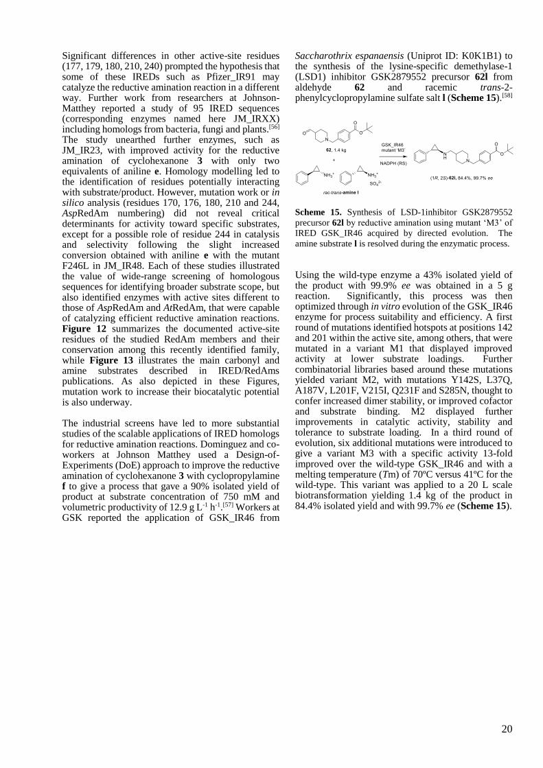
Significant differences in other active-site residues (177, 179, 180, 210, 240) prompted the hypothesis that some of these IREDs such as Pfizer_IR91 may catalyze the reductive amination reaction in a different way. Further work from researchers at Johnson-Matthey reported a study of 95 IRED sequences (corresponding enzymes named here JM_IRXX) including homologs from bacteria, fungi and plants.[56] The study unearthed further enzymes, such as JM_IR23, with improved activity for the reductive amination of cyclohexanone 3 with only two equivalents of aniline e. Homology modelling led to the identification of residues potentially interacting with substrate/product. However, mutation work or in silico analysis (residues 170, 176, 180, 210 and 244, AspRedAm numbering) did not reveal critical determinants for activity toward specific substrates, except for a possible role of residue 244 in catalysis and selectivity following the slight increased conversion obtained with aniline e with the mutant F246L in JM_IR48. Each of these studies illustrated the value of wide-range screening of homologous sequences for identifying broader substrate scope, but also identified enzymes with active sites different to those of AspRedAm and AtRedAm, that were capable of catalyzing efficient reductive amination reactions. Figure 12 summarizes the documented active-site residues of the studied RedAm members and their conservation among this recently identified family, while Figure 13 illustrates the main carbonyl and amine substrates described in IRED/RedAms publications. As also depicted in these Figures, mutation work to increase their biocatalytic potential is also underway. The industrial screens have led to more substantial studies of the scalable applications of IRED homologs for reductive amination reactions. Dominguez and co-workers at Johnson Matthey used a Design-of-Experiments (DoE) approach to improve the reductive amination of cyclohexanone 3 with cyclopropylamine f to give a process that gave a 90% isolated yield of product at substrate concentration of 750 mM and volumetric productivity of 12.9 g L-1 h-1.[57] Workers at GSK reported the application of GSK_IR46 from
Saccharothrix espanaensis (Uniprot ID: K0K1B1) to the synthesis of the lysine-specific demethylase-1 (LSD1) inhibitor GSK2879552 precursor 62l from aldehyde 62 and racemic trans-2-phenylcyclopropylamine sulfate salt l (Scheme 15).[58]
Scheme 15. Synthesis of LSD-1inhibitor GSK2879552
precursor 62l by reductive amination using mutant ‘M3’ of
IRED GSK_IR46 acquired by directed evolution. The
amine substrate l is resolved during the enzymatic process.
Using the wild-type enzyme a 43% isolated yield of the product with 99.9% ee was obtained in a 5 g reaction. Significantly, this process was then optimized through in vitro evolution of the GSK_IR46 enzyme for process suitability and efficiency. A first round of mutations identified hotspots at positions 142 and 201 within the active site, among others, that were mutated in a variant M1 that displayed improved activity at lower substrate loadings. Further combinatorial libraries based around these mutations yielded variant M2, with mutations Y142S, L37Q, A187V, L201F, V215I, Q231F and S285N, thought to confer increased dimer stability, or improved cofactor and substrate binding. M2 displayed further improvements in catalytic activity, stability and tolerance to substrate loading. In a third round of evolution, six additional mutations were introduced to give a variant M3 with a specific activity 13-fold improved over the wild-type GSK_IR46 and with a melting temperature (Tm) of 70ºC versus 41ºC for the wild-type. This variant was applied to a 20 L scale biotransformation yielding 1.4 kg of the product in 84.4% isolated yield and with 99.7% ee (Scheme 15).
21
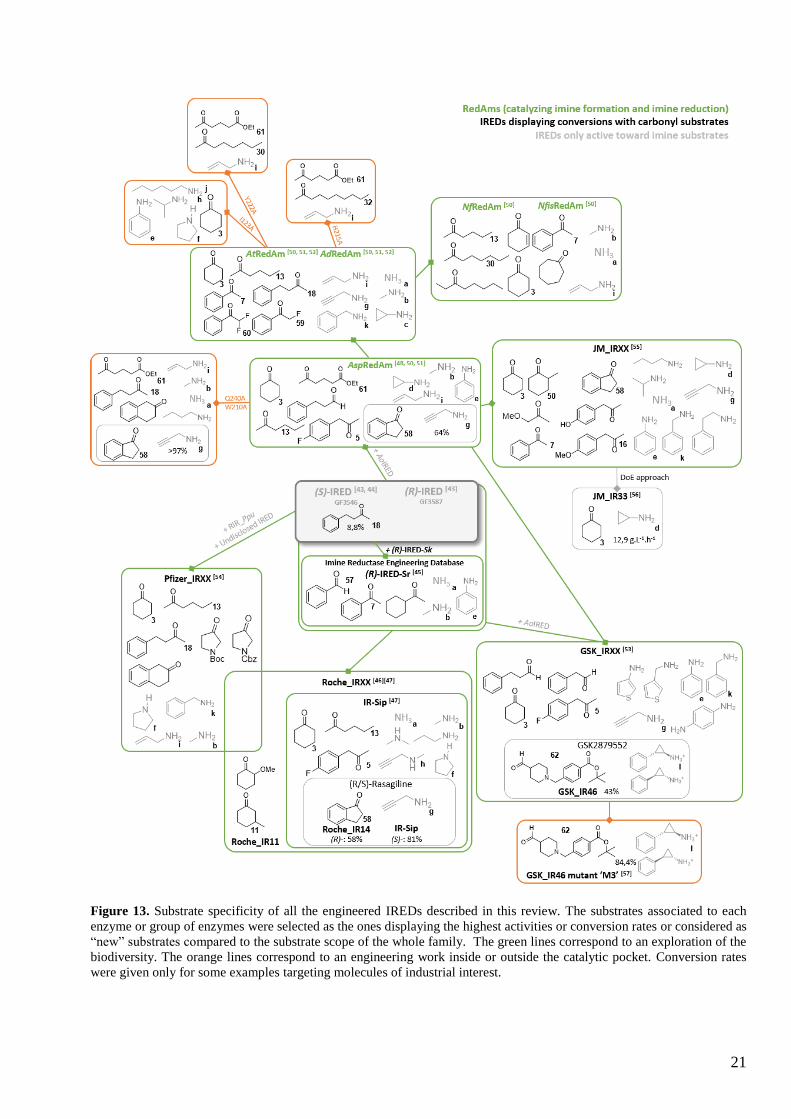
Figure 13. Substrate specificity of all the engineered IREDs described in this review. The substrates associated to each
enzyme or group of enzymes were selected as the ones displaying the highest activities or conversion rates or considered as
“new” substrates compared to the substrate scope of the whole family. The green lines correspond to an exploration of the
biodiversity. The orange lines correspond to an engineering work inside or outside the catalytic pocket. Conversion rates
were given only for some examples targeting molecules of industrial interest.
22
6 Conclusion
The prevalence of amine functions in APIs, but also in bulk chemicals, have stimulated the study of enzymatic alternatives for amine synthesis. The discovery of enzymes competent for the asymmetric reductive amination of carbonyl compounds has therefore been a major advance in biocatalytic technology. The rapid progress of research has meant that activities that were only discovered within the last ten years are already seeing application at considerable scale in an industrial context. While a number of enzymes have been shown to reduce preformed imines from solution that have arisen from favorable equilibria at high amine:ketone ratios, enzymes that enable or catalyze reductive aminations at low amine:ketone ratios offer important benefits with respect to the atom economy and sustainability of these processes. In the case of ammonia as amine donor, the poor affinity of the enzymes for this small molecule dictates its use at high concentration, however this has been addressed by the use of simple ammonia-based buffers, an approach that has already been validated in synthesis.
In some cases experimental evidence for these enzymes catalyzing both the formation of imines and their reduction within the active site has been presented. These include the acceleration of catalysis at low amine:ketone ratios in comparison to both other enzymes and abiotic reagents when the amine is not ammonia, but also structural and kinetic data alongside mutational studies. In the case of AADHs engineered to be AmDHs, there is a range of evidence to show that both carbonyl group and amine donor (ammonia being the only well studied one) are bound and activated within the active site by residues lysine and aspartate respectively; in native AmDHs these mechanistic aspects are conserved, with amine activated by carboxylate side chain of glutamate for attack at a carbonyl held by tyrosine or tryptophan. There is also evidence to suggest that in fungal RedAms, a carboxylate side chain (Asp) and a proton donor residue (Tyr) have roles in amine and carbonyl activation respectively. It is interesting that these determinants of intermolecular imine formation are also reflected in the active site of the imine forming enzyme norcoclaurine synthase described by Keep and co-workers.[59] However, it is important to note that wider screens of enzyme families for reductive amination catalysis have and will continue to reveal alternate active site determinants of the essential mechanistic requirements of the reductive amination reaction. For many of these enzymes, detailed examination of these mechanisms through mutation, kinetic and structural analysis remains to be explored.
The diversity in sequence and structure of reductive amination enzymes means that organic chemists now have access to several types of biocatalysts identified either directly from natural biodiversity or from applying protein engineering, with a range of
properties and substrate scopes. The enzymatic possibilities now presented by engineered AADHs, native AmDHs, engineered and native IREDs and RedAms, in addition to engineered OpDH and LysEDH should be considered as complementary, leading to synthetic strategies that can be adapted according to the relative merits of these enzymes. All the enzyme types that are able to perform reductive aminations are at a different stage of investigation, however the studies on improving one family can be used to inform the engineering of the others. The work done to obtain enzymes active toward substrates devoid of carboxylic and/or other amine functions, either by engineering for LeuDHs/PheDHs and LysEDH or by genome mining for nat-AmDHs from DAPDH, is a very good example of this. Based on the preliminary recently published studies, one of the next challenges to overcome is to master the amine substrate spectra attainable with all these classes of enzymes, to fully extend amination to the synthesis of secondary and tertiary amines, mainly with engineered AADHs and nat-AmDHs, and to primary amines with RedAms. Another significant challenge that remains is to advance the understanding of the selectivity of these enzymes for imines over alcohols, and whether promiscuous alcohol dehydrogenase may be obtained in these enzymes or their mutants. Moreover, research efforts must focus on increasing the stability of these enzymes in their reaction medium, which is often basic and highly-loaded with amines, and on factors that would enable synthesis at higher substrate concentrations, in particular for primary amine synthesis. The combination of biodiversity screening and structure guided and random mutagenesis, and also in silico modelling will all make important contributions to the further understanding of these enzymes, and to their application of these important activities in the future.
Acknowledgements
The author would like to thanks Pr. Anne Zaparucha for her contribution to this Viewpoint.
References
[1] a) B. Hauer, ACS Catal. 2020, 8418-
8427; b) J. M. Woodley, N. J. Turner, in
Handbook of Green Chemistry, Green
Chemical Engineering, Vol. 12 (Eds.:
P.T. Anastas, A. A. Lapkin), 2019; c) N.
J. Turner, R. Kumar, Curr. Opin. Chem.
Biol. 2018, 43, A1-A3; d) R. A. Sheldon,
D. Brady, Chem. Commun. 2018, 54
6088-6104; e) U. T. Bornscheuer, Phil.
23
Trans. R. Soc. A, 2017 376: 20170063.; f)
R. A. Sheldon, J. M. Woodley, Chem.
Rev 2018, 118, 801–838.
[2] a) B. Wiltschi, T. Cernava, A. Dennig, M.
Galindo, M. Geier, S. Gruber, M.
Haberbauer, P. Heidinger, E. H. Acero,
R. Kratzer, C. Luley-Goedl, C. A. Müller,
J. Pitzer, D. Ribitsch, M. Sauer, K.
Schmölzer, W. Schnitzhofer, C. W.
Sensen, J. Soh, K. Steiner, C. K. Winkler,
M. Winkler, T. Wriessnegger,
Biotechnol. Adv. 2020, 107520; b) J. P.
Adams, M. J. B. Brown, A. Diaz-
Rodriguez, R. C. Lloyd, G.-D. Roiban,
Adv. Synth. Catal. 2019, 361, 2421-2432;
c) K. Rosenthal, S. Lütz, Curr. Opin.
Green Sustainable Chem. 2018, 11, 58-
64; d) M. C. Bryan, P. J. Dunn, D.
Entwistle, F. Gallou, S. G. Koenig, J. D.
Hayler, M. R. Hickey, S. Hughes, M. E.
Kopach, G. Moine, P. Richardson, F.
Roschangar, A. Steven, F. J. Weiberth,
Green Chem. 2018, 20, 5082-5103; e) J.
L. Porter, R. A. Rusli, D. L. Ollis,
ChemBioChem 2016, 17, 197-203.
[3] H. Groger, Appl. Microbiol. Biotechnol.
2019, 103, 83-95.
[4] a) B. G. de la Torre, F. Albericio,
Molecules 2019, 24, 809; b) P. Das, M.
D. Delost, M. H. Qureshi, D. T. Smith, J.
T. Njardarson, J. Med. Chem. 2019, 62,
9, 4265–4311; c) S. D. Roughley, A. M.
Jordan, J. Med. Chem. 2011, 54, 10,
3451–3479.
[5] a) T. C. Nugent, M. El‐Shazly, Adv.
Synth. Catal. 2010, 352, 753-819; b) C.
Wang, J. Xiao, in Stereoselective
Formation of Amines, Vol. 343 (Eds.: W.
Li, X. Zhang), Springer, Berlin,
Heidelberg, 2014, pp. 261-282.
[6] O. I. Afanasyev, E. Kuchuk, D. L.
Usanov, D. Chusov, Chem. Rev. 2019,
119, 11857–11911.
[7] D. J. Constable, P. J. Dunn, J. D. Hayler,
G. R. Humphrey, J. L. Leazer Jr, R. J.
Linderman, K. Lorenz, J. Manley, B. A.
Pearlman, A. Wells, Green Chem. 2007,
9, 411-420.
[8] M. D. Patil, G. Grogan, A. Bommarius,
H. Yun, Catalysts 2018, 8, 254.
[9] L. S. Vidal, C. L. Kelly, P. M. Mordaka,
J. T. Heap, Biochim. Biophys. Acta –
Proteins Proteomics 2018, 1866, 327-
347.
[10] a) M. Sharma, J. Mangas-Sanchez, G.
Grogan, Adv. Synth. Catal. 2017, 359,
2011-2025; b) G. Grogan, Curr. Opin.
Chem. Biol. 2018, 43, 15-22.
[11] a) M. Yuan, M. Kummer, R. D. Milton,
T. Quah, S. D. Minteer, ACS Catal. 2019,
9, 5486–5495; b) C. E. Paul, F.
Hollmann, App.l Microbiol. Biotechnol.
2016, 100, 4773-4778; c) R. Wichmann,
D. Vasic-Racki, in Advances in
Biochemical Engineering, Vol. 92:
Technology Transfer in Biotechnology
(Ed.: U. Kragl), Springer, Berlin,
Heidelberg, 2005, pp. 225-260; d) H.
Zhao, W. A. van der Donk, Curr. Opin.
Biotechnol. 2003, 14, 583-589.
[12] E. Tassano, M. Hall, Chem. Soc. Rev.
2019, 48, 5596-5615.
[13] a) M. Lenz, J. Meisner, L. Quertinmont,
S. Lutz, J. Kästner, B. M. Nestl,
ChemBioChem 2017, 18, 253-256; b) J.
Mangas-Sanchez, S. P. France, S. L.
Montgomery, G. A. Aleku, H. Man, M.
Sharma, J. I. Ramsden, G. Grogan, N. J.
Turner, Curr. Opin. Chem. Biol. 2017,
37, 19-25; c) K. Mitsukura, T. Yoshida,
in Future Directions in Biocatalysis, 2nd
ed. (Ed.: T. Matsuda), Elsevier,
Amsterdam, 2017, pp. 97-117; d) J. H.
Schrittwieser, S. Velikogne, W. Kroutil,
Adv. Synth. Catal. 2015, 357, 1655-1685.
[14] M. D. Patil, G. Grogan, A. S.
Bommarius, H. Yun, ACS Catal. 2018, 8,
10985–11015.
[15] M. J. Abrahamson, E. Vazquez-Figueroa,
N. B. Woodall, J. C. Moore, A. S.
Bommarius, Angew. Chem., Int. Ed.
2012, 51, 3969-3972.
[16] J. L. Vanhooke, J. B. Thoden, N. M. W.
Brunhuber, J. S. Blanchard, H. M.
Holden, Biochemistry 1999, 38, 2326-
2339.
[17] P. J. Baker, A. P. Turnbull, S. E.
Sedelnikova, T. J. Stillman, D. W. Rice,
Structure 1995, 3, 693-705.
[18] a) T. Knaus, W. Bohmer, F. G. Mutti,
Green Chem. 2017, 19, 453-463; b) M. J.
Abrahamson, J. W. Wong, A. S.

24
Bommarius, Adv. Synth. Catal. 2013,
355, 1780-1786; c) S. Bommarius
Andreas, J. Abrahamson Michael, B.
Bommarius, 2014, Engineered amine
dehydrogenases and methods of use
thereof. US patent 2013309734.
[19] B. R. Bommarius, M. Schurmann, A. S.
Bommarius, Chem. Comm.2014, 50,
14953-14955.
[20] F.-F. Chen, Y.-Y. Liu, G.-W. Zheng, J.-
H. Xu, ChemCatChem 2015, 7, 3838-
3841.
[21] F. Chen, G.-W. Zheng, L. Liu, H. Li, Q.
Chen, f. li, C.-X. Li, J.-H. Xu, ACS Catal.
2018, 8, 2622-2628.
[22] L. Liu, D.-H. Wang, F.-F. Chen, Z.-J.
Zhang, Q. Chen, J.-H. Xu, Z.-L. Wang,
G.-W. Zheng, Catal. Sci. Technol. 2020,
10, 2353-2358
[23] L. J. Ye, H. H. Toh, Y. Yang, J. P.
Adams, R. Snajdrova, Z. Li, ACS Catal.
2015, 5, 1119-1122.
[24] A. Pushpanath, E. Siirola, A. Bornadel,
D. Woodlock, U. Schell, ACS Catal.
2017, 3204-3209.
[25] F. Chen, S. C. Cosgrove, W. R.
Birmingham, J. Mangas-Sanchez, J.
Citoler, M. Thompson, G.-W. Zheng, J.-
H. Xu, N. J. Turner, ACS Catal. 2019.
[26] K. Kataoka, K. Tanizawa, J.Mol. Catal.
B: Enzym. 2003, 23, 299-309.
[27] R. D. Franklin, C. J. Mount, B. R.
Bommarius, A. S. Bommarius,
ChemCatChem 2020, 12, 2436-2439.
[28] R. J. David, H. Ling, M. Martin, N. Scott,
2004, Mutants of enzymes and methods
for their use. US Patent 20040115691.
[29] W. Jiang, Y. Wang, J. Microbiol.
Biotechnol. 2020, 30, 146-154.
[30] V. Tseliou, M. F. Masman, W. Böhmer,
T. Knaus, F. G. Mutti, ChemBioChem
2019, 20, 800-812.
[31] V. Tseliou, T. Knaus, M. F. Masman, M.
L. Corrado, F. G. Mutti, Nat. Commun.
2019, 10, 3717.
[32] Springer Handbook of Enzymes, in d-
Octopine dehydrogenase, Vol. 23 (Eds.:
Schomburg D., Schomburg I.), Springer,
Berlin, Heidelberg, 2005, pp. 108-121.
[33] S. Watanabe, R. Sueda, F. Fukumori, Y.
Watanabe, PloS ONE 2015, 10,
e0138434.
[34] Y. Asano, K. Yamaguchi, K. Kondo, J.
Bacteriol. 1989, 171, 4466-4471.
[35] S.H.J. Smits, A. Mueller, L. Schmitt, M.
K. Grieshaber, J. Mol. Biol. 2008, 381,
200-211.
[36] S. H. J. Smits, T. Meyer, A. Müller, N.
van Os, M. Stoldt, D. Willbold, L.
Schmitt, M. K. Grieshaber, PloS ONE
2010, 5, e12312.
[37] H. Chen, J. Moore, S.J. Collier, D.Smith,
J. Nazor, G. Hughes, J. Janey, G.
Huisman, S. Novick, N.Agard, O. Alvizo,
G. Cope, W-L. Yeo, J. Sukumaran, S.
Ng, 2013, Engineered imine reductases
and methods for the reductive amination
of ketone and amine compounds. US
Patent Application 20130302859.
[38] N. Itoh, C. Yachi, T. Kudome, J. Mol.
Catal. B: Enzym. 2000, 10, 281-290.
[39] a) S. Wang, B. Fang, Method for
preparing chiral amine through
asymmetric reduction under catalysis of
marine strain OTI, 2013, Chinese Patent
103224963; b) S. Wang, Marine strain
and its preparation method of chiral
amines amine dehydrogenase. 2016,
Chinese Patent 105567756.
[40] O. Mayol, S. David, E. Darii, A. Debard,
A. Mariage, V. Pellouin, J.-L. Petit, M.
Salanoubat, V. de Berardinis, A.
Zaparucha, C. Vergne-Vaxelaire, Catal.
Sci Technol. 2016, 6, 7421-7428.
[41] O. Mayol, K. Bastard, L. Beloti, A. Frese,
J. P. Turkenburg, J.-L. Petit, A. Mariage,
A. Debard, V. Pellouin, A. Perret, V. de
Berardinis, A. Zaparucha, G. Grogan, C.
Vergne-Vaxelaire, Nat. Catal. 2019, 2,
324-333.
[42] A. A. Caparco, E. Pelletier, J. L. Petit, A.
Jouenne, B. R. Bommarius, V. de
Berardinis, A. Zaparucha, J. A.
Champion, A. S. Bommarius, C. Vergne-
Vaxelaire, Adv. Synth. Catal. 2020, 362,
2427-2436
[43] G. Grogan, N. J. Turner, Chem. Eur. J.
2016, 22, 1900-1907.
[44] a) K. Mitsukura, T. Kuramoto, T.
Yoshida, N. Kimoto, H. Yamamoto, T.
Nagasawa, Appl. Microbiol. Biotechnol.
2013, 97, 8079-8086; b) K. Mitsukura,
M. Suzuki, S. Shinoda, T. Kuramoto, T.
Yoshida, T. Nagasawa, Biosci.,

25
Biotechnol. Biochem. 2011, 75, 1778-
1782.
[45] T. Huber, L. Schneider, A. Präg, S.
Gerhardt, O. Einsle, M. Müller,
ChemCatChem 2014, 6, 2248-2252.
[46] P. N. Scheller, M. Lenz, S. C. Hammer,
B. Hauer, B. M. Nestl, ChemCatChem
2015, 7, 3239-3242.
[47] D. Wetzl, M. Gand, A. Ross, H. Müller,
P. Matzel, S. P. Hanlon, M. Müller, B.
Wirz, M. Höhne, H. Iding,
ChemCatChem 2016, 8, 2023-2026.
[48] P. Matzel, M. Gand, M. Höhne, Green
Chem. 2017, 19, 385-389.
[49] G. A. Aleku, S. P. France, H. Man, J.
Mangas-Sanchez, S. L. Montgomery, M.
Sharma, F. Leipold, S. Hussain, G.
Grogan, N. J. Turner, Nat. Chem. 2017,
9, 961-969.
[50] H. Mihara, H. Muramatsu, R. Kakutani,
M. Yasuda, M. Ueda, T. Kurihara, N.
Esaki, FEBS J. 2005, 272, 1117-1123.
[51] M. Sharma, J. Mangas-Sanchez, S. P.
France, G. A. Aleku, S. L. Montgomery,
J. I. Ramsden, N. J. Turner, G. Grogan,
ACS Catal. 2018, 8, 11534-11541.
[52] D. González-Martínez, A. Cuetos, M.
Sharma, M. García-Ramos, I. Lavandera,
V. Gotor-Fernandez, G. Grogan,
ChemCatChem 2020, 12, 2421-2425.
[53] J. Mangas-Sanchez, M. Sharma, S. C.
Cosgrove, J. I. Ramsden, J. R. Marshall,
T. W. Thorpe, R. B. Palmer, G. Grogan,
N. J. Turner, Chem. Sci. 2020, 11, 5052-
5057.
[54] G.-D. Roiban, M. Kern, Z. Liu, J. Hyslop,
P. L. Tey, M. S. Levine, L. S. Jordan, K.
K. Brown, T. Hadi, L. A. F. Ihnken, M. J.
B. Brown, ChemCatChem 2017, 9, 4475-
4479.
[55] S. P. France, R. M. Howard, J. Steflik, N.
J. Weise, J. Mangas-Sanchez, S. L.
Montgomery, R. Crook, R. Kumar, N. J.
Turner, ChemCatChem 2018, 10, 1-6.
[56] S. L. Montgomery, A. Pushpanath, R. S.
Heath, J. R. Marshall, U. Klemstein, J. L.
Galman, D. Woodlock, S. Bisagni, C. J.
Taylor, J. Mangas-Sanchez, J. I.
Ramsden, B. Dominguez, N. J. Turner,
Sci. Adv. 2020, 6, eaay9320.
[57] A. Bornadel, S. Bisagni, A. Pushpanath,
S. L. Montgomery, N. J. Turner, B.
Dominguez, Org. Process Res. Dev.
2019, 23, 1262–1268.
[58] M. Schober, C. MacDermaid, A. A. Ollis,
S. Chang, D. Khan, J. Hosford, J.
Latham, L. A. F. Ihnken, M. J. B. Brown,
D. Fuerst, M. J. Sanganee, G.-D. Roiban,
Nat. Catal. 2019, 2, 909-915.
[59] B. R. Lichman, A. Sula, T. Pesnot, H. C.
Hailes, J. M. Ward, N. H. Keep,
Biochemistry 2017, 56, 5274-5277.
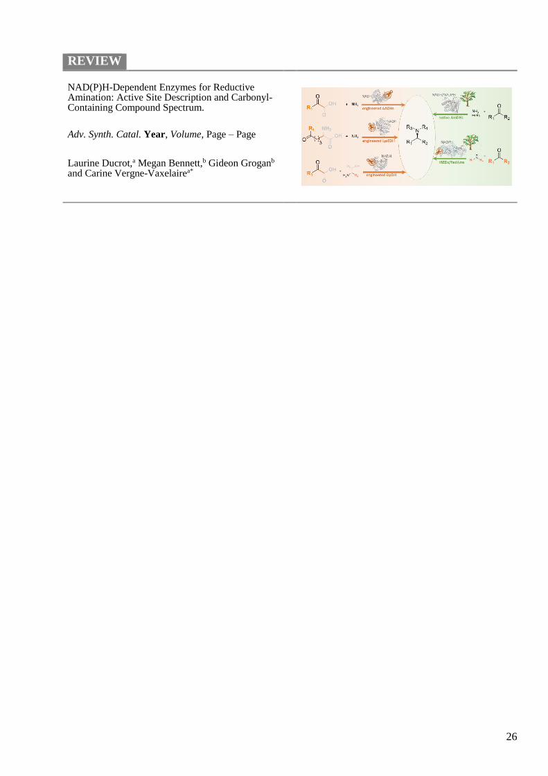
26
REVIEW
NAD(P)H-Dependent Enzymes for Reductive Amination: Active Site Description and Carbonyl-Containing Compound Spectrum.
Adv. Synth. Catal. Year, Volume, Page – Page
Laurine Ducrot,a Megan Bennett,b Gideon Groganb and Carine Vergne-Vaxelairea*