nanoparticles: properties, applications and toxicities · review nanoparticles: properties,...
TRANSCRIPT

Arabian Journal of Chemistry (2019) 12, 908–931
King Saud University
Arabian Journal of Chemistry
www.ksu.edu.sawww.sciencedirect.com
REVIEW
Nanoparticles: Properties, applications and
toxicities
* Corresponding author.
E-mail address: [email protected] (I. Khan).
Peer review under responsibility of King Saud University.
Production and hosting by Elsevier
http://dx.doi.org/10.1016/j.arabjc.2017.05.0111878-5352 � 2017 The Authors. Production and hosting by Elsevier B.V. on behalf of King Saud University.This is an open access article under the CC BY-NC-ND license (http://creativecommons.org/licenses/by-nc-nd/4.0/).
Ibrahim Khan a,*, Khalid Saeed b, Idrees Khan c
aCenter of Research Excellence in Nanotechnology (CENT), King Fahd University of Petroleum and Minerals (KFUPM),Saudi ArabiabDepartment of Chemistry, Bacha Khan University, Charsadda, PakistancDepartment of Chemistry, University of Malakand, Chakdara, Pakistan
Received 18 March 2017; accepted 10 May 2017Available online 18 May 2017
KEYWORDS
Nanoparticles;
Fullerenes;
Optical;
Plasmonic;
Toxicity
Abstract This review is provided a detailed overview of the synthesis, properties and applications
of nanoparticles (NPs) exist in different forms. NPs are tiny materials having size ranges from 1 to
100 nm. They can be classified into different classes based on their properties, shapes or sizes. The
different groups include fullerenes, metal NPs, ceramic NPs, and polymeric NPs. NPs possess
unique physical and chemical properties due to their high surface area and nanoscale size. Their
optical properties are reported to be dependent on the size, which imparts different colors due to
absorption in the visible region. Their reactivity, toughness and other properties are also dependent
on their unique size, shape and structure. Due to these characteristics, they are suitable candidates
for various commercial and domestic applications, which include catalysis, imaging, medical appli-
cations, energy-based research, and environmental applications. Heavy metal NPs of lead, mercury
and tin are reported to be so rigid and stable that their degradation is not easily achievable, which
can lead to many environmental toxicities.� 2017 The Authors. Production and hosting by Elsevier B.V. on behalf of King Saud University. This is
an open access article under theCCBY-NC-ND license (http://creativecommons.org/licenses/by-nc-nd/4.0/).
Contents
1. Introduction . . . . . . . . . . . . . . . . . . . . . . . . . . . . . . . . . . . . . . . . . . . . . . . . . . . . . . . . . . . . . . . . . . . . . . . . . . . 9092. Classification of NPs . . . . . . . . . . . . . . . . . . . . . . . . . . . . . . . . . . . . . . . . . . . . . . . . . . . . . . . . . . . . . . . . . . . . . 909
2.1. Carbon-based NPs . . . . . . . . . . . . . . . . . . . . . . . . . . . . . . . . . . . . . . . . . . . . . . . . . . . . . . . . . . . . . . . . . . . 909
Nanoparticles 909
2.2. Metal NPs . . . . . . . . . . . . . . . . . . . . . . . . . . . . . . . . . . . . . . . . . . . . . . . . . . . . . . . . . . . . . . . . . . . . . . . . . 910
2.3. Ceramics NPs. . . . . . . . . . . . . . . . . . . . . . . . . . . . . . . . . . . . . . . . . . . . . . . . . . . . . . . . . . . . . . . . . . . . . . . 9102.4. Semiconductor NPs. . . . . . . . . . . . . . . . . . . . . . . . . . . . . . . . . . . . . . . . . . . . . . . . . . . . . . . . . . . . . . . . . . . 9102.5. Polymeric NPs . . . . . . . . . . . . . . . . . . . . . . . . . . . . . . . . . . . . . . . . . . . . . . . . . . . . . . . . . . . . . . . . . . . . . . 910
2.6. Lipid-based NPs . . . . . . . . . . . . . . . . . . . . . . . . . . . . . . . . . . . . . . . . . . . . . . . . . . . . . . . . . . . . . . . . . . . . . 9113. Synthesis of nanoparticles . . . . . . . . . . . . . . . . . . . . . . . . . . . . . . . . . . . . . . . . . . . . . . . . . . . . . . . . . . . . . . . . . 912
3.1. Top-down syntheses . . . . . . . . . . . . . . . . . . . . . . . . . . . . . . . . . . . . . . . . . . . . . . . . . . . . . . . . . . . . . . . . . . 9123.2. Bottom-up syntheses . . . . . . . . . . . . . . . . . . . . . . . . . . . . . . . . . . . . . . . . . . . . . . . . . . . . . . . . . . . . . . . . . . 913
4. Characterization of NPs. . . . . . . . . . . . . . . . . . . . . . . . . . . . . . . . . . . . . . . . . . . . . . . . . . . . . . . . . . . . . . . . . . . 9154.1. Morphological characterizations. . . . . . . . . . . . . . . . . . . . . . . . . . . . . . . . . . . . . . . . . . . . . . . . . . . . . . . . . . 9154.2. Structural characterizations . . . . . . . . . . . . . . . . . . . . . . . . . . . . . . . . . . . . . . . . . . . . . . . . . . . . . . . . . . . . . 916
4.3. Particle size and surface area characterization . . . . . . . . . . . . . . . . . . . . . . . . . . . . . . . . . . . . . . . . . . . . . . . . 9184.4. Optical characterizations . . . . . . . . . . . . . . . . . . . . . . . . . . . . . . . . . . . . . . . . . . . . . . . . . . . . . . . . . . . . . . . 919
5. Physicochemical properties of NPs . . . . . . . . . . . . . . . . . . . . . . . . . . . . . . . . . . . . . . . . . . . . . . . . . . . . . . . . . . . 921
5.1. Electronic and optical properties . . . . . . . . . . . . . . . . . . . . . . . . . . . . . . . . . . . . . . . . . . . . . . . . . . . . . . . . . 9215.2. Magnetic properties . . . . . . . . . . . . . . . . . . . . . . . . . . . . . . . . . . . . . . . . . . . . . . . . . . . . . . . . . . . . . . . . . . 9225.3. Mechanical properties . . . . . . . . . . . . . . . . . . . . . . . . . . . . . . . . . . . . . . . . . . . . . . . . . . . . . . . . . . . . . . . . . 9225.4. Thermal properties . . . . . . . . . . . . . . . . . . . . . . . . . . . . . . . . . . . . . . . . . . . . . . . . . . . . . . . . . . . . . . . . . . . 923
6. Applications of NPs . . . . . . . . . . . . . . . . . . . . . . . . . . . . . . . . . . . . . . . . . . . . . . . . . . . . . . . . . . . . . . . . . . . . . 9246.1. Applications in drugs and medications . . . . . . . . . . . . . . . . . . . . . . . . . . . . . . . . . . . . . . . . . . . . . . . . . . . . . 9246.2. Applications in manufacturing and materials. . . . . . . . . . . . . . . . . . . . . . . . . . . . . . . . . . . . . . . . . . . . . . . . . 924
6.3. Applications in the environment. . . . . . . . . . . . . . . . . . . . . . . . . . . . . . . . . . . . . . . . . . . . . . . . . . . . . . . . . . 9256.4. Applications in electronics . . . . . . . . . . . . . . . . . . . . . . . . . . . . . . . . . . . . . . . . . . . . . . . . . . . . . . . . . . . . . . 9256.5. Applications in energy harvesting . . . . . . . . . . . . . . . . . . . . . . . . . . . . . . . . . . . . . . . . . . . . . . . . . . . . . . . . . 925
6.6. Applications in mechanical industries . . . . . . . . . . . . . . . . . . . . . . . . . . . . . . . . . . . . . . . . . . . . . . . . . . . . . . 9267. Toxicity of NP . . . . . . . . . . . . . . . . . . . . . . . . . . . . . . . . . . . . . . . . . . . . . . . . . . . . . . . . . . . . . . . . . . . . . . . . . 9268. Conclusion . . . . . . . . . . . . . . . . . . . . . . . . . . . . . . . . . . . . . . . . . . . . . . . . . . . . . . . . . . . . . . . . . . . . . . . . . . . . 927
9. Recommendations . . . . . . . . . . . . . . . . . . . . . . . . . . . . . . . . . . . . . . . . . . . . . . . . . . . . . . . . . . . . . . . . . . . . . . . 927References . . . . . . . . . . . . . . . . . . . . . . . . . . . . . . . . . . . . . . . . . . . . . . . . . . . . . . . . . . . . . . . . . . . . . . . . . . . . 927
1. Introduction
Nanotechnology is a known field of research since last century. Since
‘‘nanotechnology” was presented by Nobel laureate Richard P. Feyn-
man during his well famous 1959 lecture ‘‘There’s Plenty of Room at
the Bottom” (Feynman, 1960), there have been made various revolu-
tionary developments in the field of nanotechnology. Nanotechnology
produced materials of various types at nanoscale level. Nanoparticles
(NPs) are wide class of materials that include particulate substances,
which have one dimension less than 100 nm at least (Laurent et al.,
2010). Depending on the overall shape these materials can be 0D,
1D, 2D or 3D (Tiwari et al., 2012). The importance of these materials
realized when researchers found that size can influence the physio-
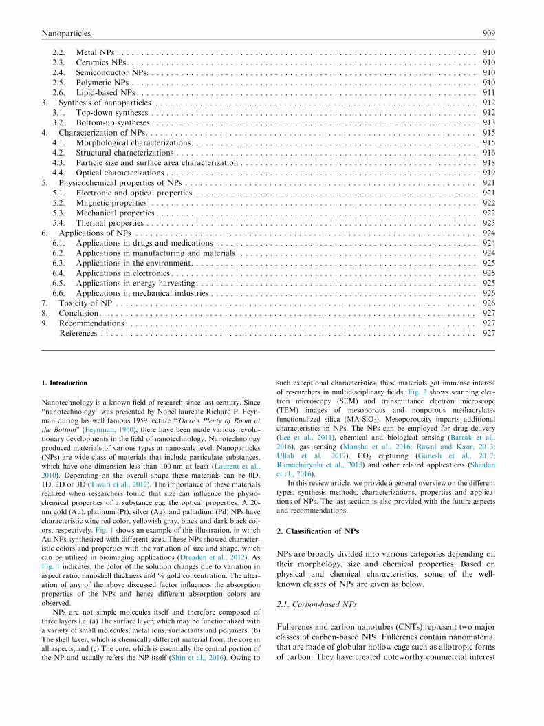
chemical properties of a substance e.g. the optical properties. A 20-
nm gold (Au), platinum (Pt), silver (Ag), and palladium (Pd) NPs have
characteristic wine red color, yellowish gray, black and dark black col-
ors, respectively. Fig. 1 shows an example of this illustration, in which
Au NPs synthesized with different sizes. These NPs showed character-
istic colors and properties with the variation of size and shape, which
can be utilized in bioimaging applications (Dreaden et al., 2012). As
Fig. 1 indicates, the color of the solution changes due to variation in
aspect ratio, nanoshell thickness and % gold concentration. The alter-
ation of any of the above discussed factor influences the absorption
properties of the NPs and hence different absorption colors are
observed.
NPs are not simple molecules itself and therefore composed of
three layers i.e. (a) The surface layer, which may be functionalized with
a variety of small molecules, metal ions, surfactants and polymers. (b)
The shell layer, which is chemically different material from the core in
all aspects, and (c) The core, which is essentially the central portion of
the NP and usually refers the NP itself (Shin et al., 2016). Owing to
such exceptional characteristics, these materials got immense interest
of researchers in multidisciplinary fields. Fig. 2 shows scanning elec-
tron microscopy (SEM) and transmittance electron microscope
(TEM) images of mesoporous and nonporous methacrylate-
functionalized silica (MA-SiO2). Mesoporousity imparts additional
characteristics in NPs. The NPs can be employed for drug delivery
(Lee et al., 2011), chemical and biological sensing (Barrak et al.,
2016), gas sensing (Mansha et al., 2016; Rawal and Kaur, 2013;
Ullah et al., 2017), CO2 capturing (Ganesh et al., 2017;
Ramacharyulu et al., 2015) and other related applications (Shaalan
et al., 2016).
In this review article, we provide a general overview on the different
types, synthesis methods, characterizations, properties and applica-
tions of NPs. The last section is also provided with the future aspects
and recommendations.
2. Classification of NPs
NPs are broadly divided into various categories depending ontheir morphology, size and chemical properties. Based onphysical and chemical characteristics, some of the well-
known classes of NPs are given as below.
2.1. Carbon-based NPs
Fullerenes and carbon nanotubes (CNTs) represent two major
classes of carbon-based NPs. Fullerenes contain nanomaterialthat are made of globular hollow cage such as allotropic formsof carbon. They have created noteworthy commercial interest
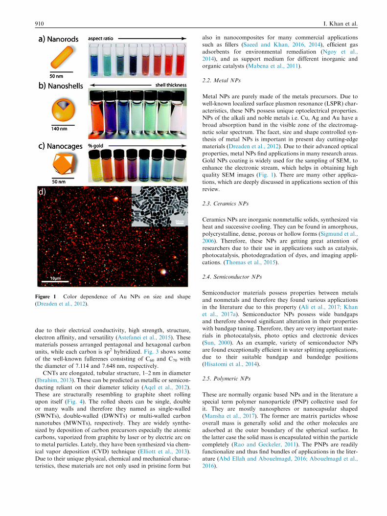
Figure 1 Color dependence of Au NPs on size and shape
(Dreaden et al., 2012).
910 I. Khan et al.
due to their electrical conductivity, high strength, structure,electron affinity, and versatility (Astefanei et al., 2015). Thesematerials possess arranged pentagonal and hexagonal carbon
units, while each carbon is sp2 hybridized. Fig. 3 shows someof the well-known fullerenes consisting of C60 and C70 withthe diameter of 7.114 and 7.648 nm, respectively.
CNTs are elongated, tubular structure, 1–2 nm in diameter(Ibrahim, 2013). These can be predicted as metallic or semicon-ducting reliant on their diameter telicity (Aqel et al., 2012).These are structurally resembling to graphite sheet rolling
upon itself (Fig. 4). The rolled sheets can be single, doubleor many walls and therefore they named as single-walled(SWNTs), double-walled (DWNTs) or multi-walled carbon
nanotubes (MWNTs), respectively. They are widely synthe-sized by deposition of carbon precursors especially the atomiccarbons, vaporized from graphite by laser or by electric arc on
to metal particles. Lately, they have been synthesized via chem-ical vapor deposition (CVD) technique (Elliott et al., 2013).Due to their unique physical, chemical and mechanical charac-
teristics, these materials are not only used in pristine form but
also in nanocomposites for many commercial applicationssuch as fillers (Saeed and Khan, 2016, 2014), efficient gasadsorbents for environmental remediation (Ngoy et al.,
2014), and as support medium for different inorganic andorganic catalysts (Mabena et al., 2011).
2.2. Metal NPs
Metal NPs are purely made of the metals precursors. Due towell-known localized surface plasmon resonance (LSPR) char-
acteristics, these NPs possess unique optoelectrical properties.NPs of the alkali and noble metals i.e. Cu, Ag and Au have abroad absorption band in the visible zone of the electromag-
netic solar spectrum. The facet, size and shape controlled syn-thesis of metal NPs is important in present day cutting-edgematerials (Dreaden et al., 2012). Due to their advanced opticalproperties, metal NPs find applications in many research areas.
Gold NPs coating is widely used for the sampling of SEM, toenhance the electronic stream, which helps in obtaining highquality SEM images (Fig. 1). There are many other applica-
tions, which are deeply discussed in applications section of thisreview.
2.3. Ceramics NPs
Ceramics NPs are inorganic nonmetallic solids, synthesized viaheat and successive cooling. They can be found in amorphous,polycrystalline, dense, porous or hollow forms (Sigmund et al.,
2006). Therefore, these NPs are getting great attention ofresearchers due to their use in applications such as catalysis,photocatalysis, photodegradation of dyes, and imaging appli-
cations. (Thomas et al., 2015).
2.4. Semiconductor NPs
Semiconductor materials possess properties between metalsand nonmetals and therefore they found various applicationsin the literature due to this property (Ali et al., 2017; Khan
et al., 2017a). Semiconductor NPs possess wide bandgapsand therefore showed significant alteration in their propertieswith bandgap tuning. Therefore, they are very important mate-rials in photocatalysis, photo optics and electronic devices
(Sun, 2000). As an example, variety of semiconductor NPsare found exceptionally efficient in water splitting applications,due to their suitable bandgap and bandedge positions
(Hisatomi et al., 2014).
2.5. Polymeric NPs
These are normally organic based NPs and in the literature aspecial term polymer nanoparticle (PNP) collective used forit. They are mostly nanospheres or nanocapsular shaped
(Mansha et al., 2017). The former are matrix particles whoseoverall mass is generally solid and the other molecules areadsorbed at the outer boundary of the spherical surface. Inthe latter case the solid mass is encapsulated within the particle
completely (Rao and Geckeler, 2011). The PNPs are readilyfunctionalize and thus find bundles of applications in the liter-ature (Abd Ellah and Abouelmagd, 2016; Abouelmagd et al.,
2016).

Figure 2 FE-SEM micrographs of (a) nonporous MA-SiO2 NPs, (b) mesoporous MA-SiO2 NPs. TEM images of (c) nonporous MA-
SiO2 NPs and (d) mesoporous MA-SiO2 NPs (Lee et al., 2011).
Figure 3 Different form of Fullerenes/buck balls (A) C60 and (B) C70.
Nanoparticles 911
2.6. Lipid-based NPs
These NPs contain lipid moieties and effectively using in manybiomedical applications. Generally, a lipid NP is characteristi-
cally spherical with diameter ranging from 10 to 1000 nm. Likepolymeric NPs, lipid NPs possess a solid core made of lipid
and a matrix contains soluble lipophilic molecules. Surfactantsor emulsifiers stabilized the external core of these NPs (Rawat
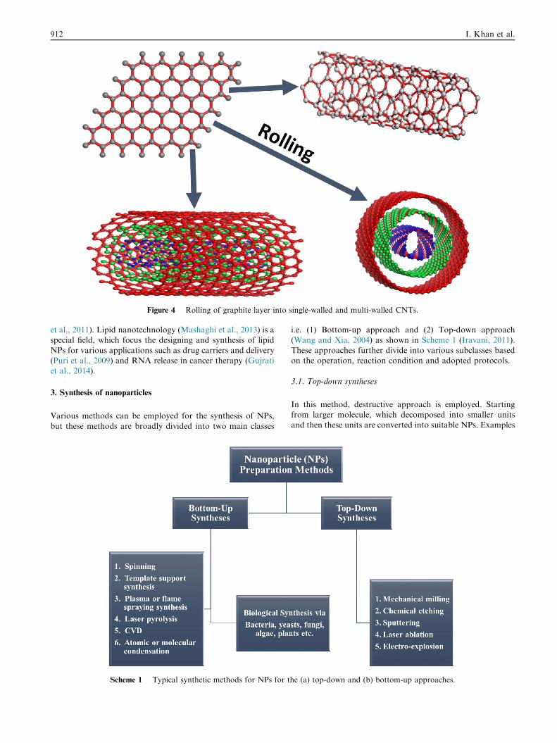
Figure 4 Rolling of graphite layer into single-walled and multi-walled CNTs.
912 I. Khan et al.
et al., 2011). Lipid nanotechnology (Mashaghi et al., 2013) is aspecial field, which focus the designing and synthesis of lipid
NPs for various applications such as drug carriers and delivery(Puri et al., 2009) and RNA release in cancer therapy (Gujratiet al., 2014).
3. Synthesis of nanoparticles
Various methods can be employed for the synthesis of NPs,
but these methods are broadly divided into two main classes
Scheme 1 Typical synthetic methods for NPs for t
i.e. (1) Bottom-up approach and (2) Top-down approach(Wang and Xia, 2004) as shown in Scheme 1 (Iravani, 2011).
These approaches further divide into various subclasses basedon the operation, reaction condition and adopted protocols.
3.1. Top-down syntheses
In this method, destructive approach is employed. Startingfrom larger molecule, which decomposed into smaller unitsand then these units are converted into suitable NPs. Examples
he (a) top-down and (b) bottom-up approaches.
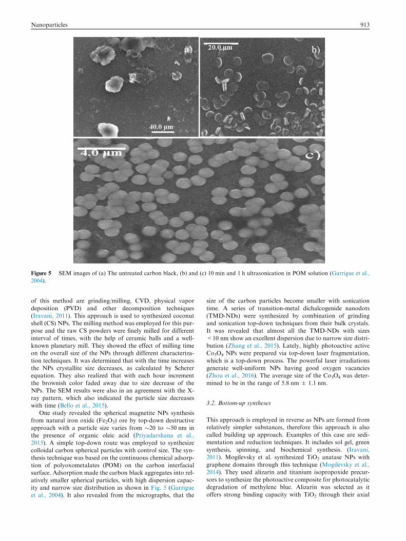
Figure 5 SEM images of (a) The untreated carbon black, (b) and (c) 10 min and 1 h ultrasonication in POM solution (Garrigue et al.,
2004).
Nanoparticles 913
of this method are grinding/milling, CVD, physical vapor
deposition (PVD) and other decomposition techniques(Iravani, 2011). This approach is used to synthesized coconutshell (CS) NPs. The milling method was employed for this pur-pose and the raw CS powders were finely milled for different
interval of times, with the help of ceramic balls and a well-known planetary mill. They showed the effect of milling timeon the overall size of the NPs through different characteriza-
tion techniques. It was determined that with the time increasesthe NPs crystallite size decreases, as calculated by Schererequation. They also realized that with each hour increment
the brownish color faded away due to size decrease of theNPs. The SEM results were also in an agreement with the X-ray pattern, which also indicated the particle size decreases
with time (Bello et al., 2015).One study revealed the spherical magnetite NPs synthesis
from natural iron oxide (Fe2O3) ore by top-down destructiveapproach with a particle size varies from �20 to �50 nm in
the presence of organic oleic acid (Priyadarshana et al.,2015). A simple top-down route was employed to synthesizecolloidal carbon spherical particles with control size. The syn-
thesis technique was based on the continuous chemical adsorp-tion of polyoxometalates (POM) on the carbon interfacialsurface. Adsorption made the carbon black aggregates into rel-
atively smaller spherical particles, with high dispersion capac-ity and narrow size distribution as shown in Fig. 5 (Garrigueet al., 2004). It also revealed from the micrographs, that the
size of the carbon particles become smaller with sonication
time. A series of transition-metal dichalcogenide nanodots(TMD-NDs) were synthesized by combination of grindingand sonication top-down techniques from their bulk crystals.It was revealed that almost all the TMD-NDs with sizes
<10 nm show an excellent dispersion due to narrow size distri-bution (Zhang et al., 2015). Lately, highly photoactive activeCo3O4 NPs were prepared via top-down laser fragmentation,
which is a top-down process. The powerful laser irradiationsgenerate well-uniform NPs having good oxygen vacancies(Zhou et al., 2016). The average size of the Co3O4 was deter-
mined to be in the range of 5.8 nm ± 1.1 nm.
3.2. Bottom-up syntheses
This approach is employed in reverse as NPs are formed fromrelatively simpler substances, therefore this approach is alsocalled building up approach. Examples of this case are sedi-mentation and reduction techniques. It includes sol gel, green
synthesis, spinning, and biochemical synthesis. (Iravani,2011). Mogilevsky et al. synthesized TiO2 anatase NPs withgraphene domains through this technique (Mogilevsky et al.,
2014). They used alizarin and titanium isopropoxide precur-sors to synthesize the photoactive composite for photocatalyticdegradation of methylene blue. Alizarin was selected as it
offers strong binding capacity with TiO2 through their axial

Scheme 2 Synthesis of TiO2 via bottom-up technique. SEM images showing the TiO2 NPs (Mogilevsky et al., 2014).
914 I. Khan et al.
hydroxyl terminal groups. The anatase form was confirmed byXRD pattern. The SEM images taken for different sampleswith reaction scheme are provided in scheme 2. SEM indicates
that with temperature elevation, the size of NPs also increases(Mogilevsky et al., 2014).
Well-uniform spherical shaped Au nanospheres with
monocrystalline have been synthesized via laser irradiationtop-down technique (Liu et al., 2015a, 2015b). Liu et al. selec-tively transform the octahedra morphology to spherical shape
by controlling the laser treatment time and other reactionparameters. Fig. 6 provides the SEM and TEM of the preparedAu nanospheres, which showed average diameter of 75
± 2.6 nm of Au nanospheres (red column Fig. 6e) and 72± 3.1 in edge length of Au octahedra per particle (blue columnFig. 6f).
More recently, solvent-exchange method is used to achievelimit sized low density lipoprotein (LDL) NPs for medical can-cer drug delivery purpose by Needham et al. In this method
nucleation is the bottom approach followed by growth whichis the up approach. The LDL NPs were obtained without usingphospholipid and possessed high hydrophobicity, which is
essential for drug delivery applications (Needham et al., 2016).The monodispersed spherical bismuth (Bi) NPs were syn-
thesized by both top-down and bottom-up approaches
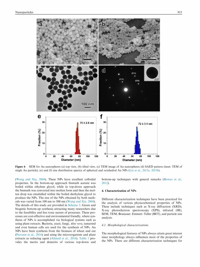
Figure 6 SEM for Au nanospheres (a) top view, (b) tilted view, (c) TEM image of Au nanospheres (d) SAED pattern (inset: TEM of
single Au particle), (e) and (f) size distribution spectra of spherical and octahedral Au NPs (Liu et al., 2015a, 2015b).
Nanoparticles 915
(Wang and Xia, 2004). These NPs have excellent colloidal
properties. In the bottom-up approach bismuth acetate wasboiled within ethylene glycol, while in top-down approachthe bismuth was converted into molten form and then the mol-
ten drop was emulsified within the boiled diethylene glycol toproduce the NPs. The size of the NPs obtained by both meth-ods was varied from 100 nm to 500 nm (Wang and Xia, 2004).
The details of this study are provided in Scheme 3. Green andbiogenic bottom-up synthesis attracting many researchers dueto the feasibility and less toxic nature of processes. These pro-cesses are cost-effective and environmental friendly, where syn-
thesis of NPs is accomplished via biological systems such asusing plant extracts. Bacteria, yeast, fungi, Aloe vera, tamarindand even human cells are used for the synthesis of NPs. Au
NPs have been synthesis from the biomass of wheat and oat(Parveen et al., 2016) and using the microorganism and plantextracts as reducing agent (Ahmed et al., 2016). Table 1 pro-
vides the merits and demerits of various top-down and
bottom-up techniques with general remarks (Biswas et al.,
2012).
4. Characterization of NPs
Different characterization techniques have been practiced forthe analysis of various physicochemical properties of NPs.These include techniques such as X-ray diffraction (XRD),
X-ray photoelectron spectroscopy (XPS), infrared (IR),SEM, TEM, Brunauer–Emmett–Teller (BET), and particle sizeanalysis.
4.1. Morphological characterizations
The morphological features of NPs always attain great interest
since morphology always influences most of the properties ofthe NPs. There are different characterization techniques for

Scheme 3 (A) Bottom-up approach: A molecular precursor is disintegrated to simpler metal atoms that grow into colloids. (B) Top-
down approach: Large drops of a metal broken into smaller drops (Wang and Xia, 2004).
916 I. Khan et al.
morphological studies, but microscopic techniques such aspolarized optical microscopy (POM), SEM and TEM are themost important of these.
SEM technique is based on electron scanning principle, andit provides all available information about the NPs at nanos-cale level. Wide literature is available, where people used this
technique to study not only the morphology of their nanoma-terials, but also the dispersion of NPs in the bulk or matrix.The dispersion of SWNTs in the polymer matrix poly(buty-lene) terephthalate (PBT) and nylon-6 revealed through this
technique (Saeed and Khan, 2016, 2014). The same group alsoprovides POM study of their materials, which showed star-likespherulites of the formed materials, whose size was decreased
with the incremental filling of SWNTs. The morphological fea-tures of ZnO modified metal organic frameworks (MOFs) werestudied through SEM technique, which indicates the ZnO NPs
dispersion and morphologies of MOFs at different reactionconditions (Fig. 7) (Mirzadeh and Akhbari, 2016).
Similarly, TEM is based on electron transmittance princi-
ple, so it can provide information of the bulk material fromvery low to higher magnification. The different morphologiesof gold NPs are studied via this technique. Fig. 8 providessome TEM micrographs showing various morphologies of
gold NPs, prepared via different methods (Khlebtsov andDykman, 2011, 2010a, 2010b). TEM also provides essentialinformation about two or more layer materials, such as the
quadrupolar hollow shell structure of Co3O4 NPs observedthrough TEM. These NPs founded to be exceptionally activeas anode in Li-ion batteries (Fig. 9). Porous multishell struc-
ture induces shorter Li+ diffusion path length with adequateannulled space to buffer the volume expansion, good cyclingperformance, greater rate capacity, and specific capacity as
well (Wang et al., 2013).
4.2. Structural characterizations
The structural characteristics are of the primary importance to
study the composition and nature of bonding materials. It pro-vides diverse information about the bulk properties of the sub-
ject material. XRD, energy dispersive X-ray (EDX), XPS, IR,Raman, BET, and Zieta size analyzer are the common tech-niques used to study structural properties of NPs.
XRD is one of the most important characterization tech-niques to reveal the structural properties of NPs. It givesenough information about the crystallinity and phase of
NPs. It also provides rough idea about the particle sizethrough Debye Scherer formula (Khan et al., 2017b, 2017c;Ullah et al., 2017). This technique worked well in both singleand multiphase NPs identification (Emery et al., 2016). Never-
theless, in the case of smaller NPs having size less than hun-dreds of atoms, the acquisition and correct measurement ofstructural and other parameters may be difficult. Moreover,
NPs having more amorphous characteristics with varied interatomic lengths can influence the XRD diffractogram. In thatcase, proper comparison of the diffractograms of bimetallic
NPs with those of the corresponding monometallic NPs andtheir physical mixtures is required to obtain accurate informa-tion. Comparison of computer simulated structural model of
bimetallic NPs with observed XRD spectra is the best wayto get good contrast (Ingham, 2015). EDX, which is normallyfixed with field emission scanning electron miscopy (FE-SEM)or TEM device is widely used to know about the elemental
composition with a rough idea of % wt. The electron beamfocused over a single NP by SEM or TEM through the pro-gram functions, to acquire the insight information from the
NP under observation. NP comprises of constituent elementsand each of them emits characteristics energy X-rays by elec-tron beam irradiation. The intensity of specific X-ray is directly
proportional to the concentration of the explicit element in theparticle. This technique is widely used by researchers to givesupport to SEM and other techniques for the confirmation
of their elements in prepared materials (Avasare et al., 2015;Iqbal et al., 2016). The EDX technique used to determinethe elemental composition of ultra-sonochemically synthesizedpseudo-flower shaped BiVO4 NPs (Khan et al., 2017b). Simi-
larly, by utilizing similar technique the elemental confirmationand graphene impregnation of In2O3/graphene heterostructureNPs was carried out, which showed C, In and O as contribut-
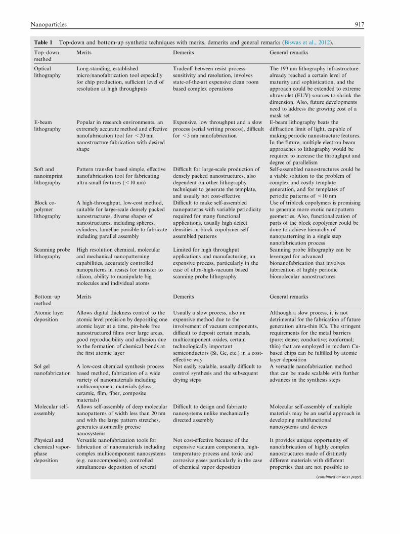
Table 1 Top-down and bottom-up synthetic techniques with merits, demerits and general remarks (Biswas et al., 2012).
Top–down
method
Merits Demerits General remarks
Optical
lithography
Long-standing, established
micro/nanofabrication tool especially
for chip production, sufficient level of
resolution at high throughputs
Tradeoff between resist process
sensitivity and resolution, involves
state-of-the-art expensive clean room
based complex operations
The 193 nm lithography infrastructure
already reached a certain level of
maturity and sophistication, and the
approach could be extended to extreme
ultraviolet (EUV) sources to shrink the
dimension. Also, future developments
need to address the growing cost of a
mask set
E-beam
lithography
Popular in research environments, an
extremely accurate method and effective
nanofabrication tool for <20 nm
nanostructure fabrication with desired
shape
Expensive, low throughput and a slow
process (serial writing process), difficult
for <5 nm nanofabrication
E-beam lithography beats the
diffraction limit of light, capable of
making periodic nanostructure features.
In the future, multiple electron beam
approaches to lithography would be
required to increase the throughput and
degree of parallelism
Soft and
nanoimprint
lithography
Pattern transfer based simple, effective
nanofabrication tool for fabricating
ultra-small features (<10 nm)
Difficult for large-scale production of
densely packed nanostructures, also
dependent on other lithography
techniques to generate the template,
and usually not cost-effective
Self-assembled nanostructures could be
a viable solution to the problem of
complex and costly template
generation, and for templates of
periodic patterns of <10 nm
Block co-
polymer
lithography
A high-throughput, low-cost method,
suitable for large-scale densely packed
nanostructures, diverse shapes of
nanostructures, including spheres,
cylinders, lamellae possible to fabricate
including parallel assembly
Difficult to make self-assembled
nanopatterns with variable periodicity
required for many functional
applications, usually high defect
densities in block copolymer self-
assembled patterns
Use of triblock copolymers is promising
to generate more exotic nanopattern
geometries. Also, functionalization of
parts of the block copolymer could be
done to achieve hierarchy of
nanopatterning in a single step
nanofabrication process
Scanning probe
lithography
High resolution chemical, molecular
and mechanical nanopatterning
capabilities, accurately controlled
nanopatterns in resists for transfer to
silicon, ability to manipulate big
molecules and individual atoms
Limited for high throughput
applications and manufacturing, an
expensive process, particularly in the
case of ultra-high-vacuum based
scanning probe lithography
Scanning probe lithography can be
leveraged for advanced
bionanofabrication that involves
fabrication of highly periodic
biomolecular nanostructures
Bottom–up
method
Merits Demerits General remarks
Atomic layer
deposition
Allows digital thickness control to the
atomic level precision by depositing one
atomic layer at a time, pin-hole free
nanostructured films over large areas,
good reproducibility and adhesion due
to the formation of chemical bonds at
the first atomic layer
Usually a slow process, also an
expensive method due to the
involvement of vacuum components,
difficult to deposit certain metals,
multicomponent oxides, certain
technologically important
semiconductors (Si, Ge, etc.) in a cost-
effective way
Although a slow process, it is not
detrimental for the fabrication of future
generation ultra-thin ICs. The stringent
requirements for the metal barriers
(pure; dense; conductive; conformal;
thin) that are employed in modern Cu-
based chips can be fulfilled by atomic
layer deposition
Sol gel
nanofabrication
A low-cost chemical synthesis process
based method, fabrication of a wide
variety of nanomaterials including
multicomponent materials (glass,
ceramic, film, fiber, composite
materials)
Not easily scalable, usually difficult to
control synthesis and the subsequent
drying steps
A versatile nanofabrication method
that can be made scalable with further
advances in the synthesis steps
Molecular self-
assembly
Allows self-assembly of deep molecular
nanopatterns of width less than 20 nm
and with the large pattern stretches,
generates atomically precise
nanosystems
Difficult to design and fabricate
nanosystems unlike mechanically
directed assembly
Molecular self-assembly of multiple
materials may be an useful approach in
developing multifunctional
nanosystems and devices
Physical and
chemical vapor-
phase
deposition
Versatile nanofabrication tools for
fabrication of nanomaterials including
complex multicomponent nanosystems
(e.g. nanocomposites), controlled
simultaneous deposition of several
Not cost-effective because of the
expensive vacuum components, high-
temperature process and toxic and
corrosive gases particularly in the case
of chemical vapor deposition
It provides unique opportunity of
nanofabrication of highly complex
nanostructures made of distinctly
different materials with different
properties that are not possible to
(continued on next page)
Nanoparticles 917
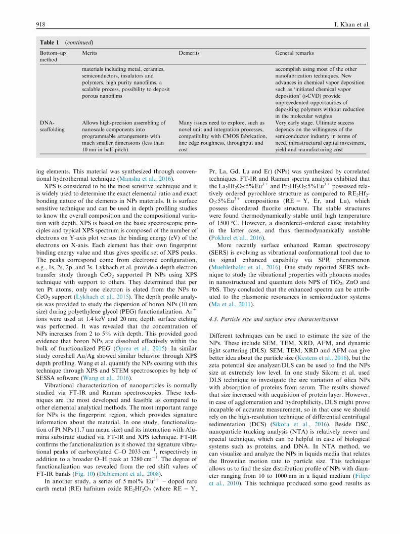
Table 1 (continued)
Bottom–up
method
Merits Demerits General remarks
materials including metal, ceramics,
semiconductors, insulators and
polymers, high purity nanofilms, a
scalable process, possibility to deposit
porous nanofilms
accomplish using most of the other
nanofabrication techniques. New
advances in chemical vapor deposition
such as ‘initiated chemical vapor
deposition’ (i-CVD) provide
unprecedented opportunities of
depositing polymers without reduction
in the molecular weights
DNA-
scaffolding
Allows high-precision assembling of
nanoscale components into
programmable arrangements with
much smaller dimensions (less than
10 nm in half-pitch)
Many issues need to explore, such as
novel unit and integration processes,
compatibility with CMOS fabrication,
line edge roughness, throughput and
cost
Very early stage. Ultimate success
depends on the willingness of the
semiconductor industry in terms of
need, infrastructural capital investment,
yield and manufacturing cost
918 I. Khan et al.
ing elements. This material was synthesized through conven-tional hydrothermal technique (Mansha et al., 2016).
XPS is considered to be the most sensitive technique and itis widely used to determine the exact elemental ratio and exactbonding nature of the elements in NPs materials. It is surface
sensitive technique and can be used in depth profiling studiesto know the overall composition and the compositional varia-tion with depth. XPS is based on the basic spectroscopic prin-
ciples and typical XPS spectrum is composed of the number ofelectrons on Y-axis plot versus the binding energy (eV) of theelectrons on X-axis. Each element has their own fingerprintbinding energy value and thus gives specific set of XPS peaks.
The peaks correspond come from electronic configuration,e.g., 1s, 2s, 2p, and 3s. Lykhach et al. provide a depth electrontransfer study through CeO2 supported Pt NPs using XPS
technique with support to others. They determined that perten Pt atoms, only one electron is elated from the NPs toCeO2 support (Lykhach et al., 2015). The depth profile analy-
sis was provided to study the dispersion of boron NPs (10 nmsize) during polyethylene glycol (PEG) functionalization. Ar+
ions were used at 1.4 keV and 20 nm; depth surface etchingwas performed. It was revealed that the concentration of
NPs increases from 2 to 5% with depth. This provided goodevidence that boron NPs are dissolved effectively within thebulk of functionalized PEG (Oprea et al., 2015). In similar
study coreshell Au/Ag showed similar behavior through XPSdepth profiling. Wang et al. quantify the NPs coating with thistechnique through XPS and STEM spectroscopies by help of
SESSA software (Wang et al., 2016).Vibrational characterization of nanoparticles is normally
studied via FT-IR and Raman spectroscopies. These tech-
niques are the most developed and feasible as compared toother elemental analytical methods. The most important rangefor NPs is the fingerprint region, which provides signatureinformation about the material. In one study, functionaliza-
tion of Pt NPs (1.7 nm mean size) and its interaction with Alu-mina substrate studied via FT-IR and XPS technique. FT-IRconfirms the functionalization as it showed the signature vibra-
tional peaks of carboxylated C–O 2033 cm�1, respectively inaddition to a broader O–H peak at 3280 cm�1. The degree offunctionalization was revealed from the red shift values of
FT-IR bands (Fig. 10) (Dablemont et al., 2008).In another study, a series of 5 mol% Eu3+ – doped rare
earth metal (RE) hafnium oxide RE2Hf2O7 (where RE = Y,
Pr, La, Gd, Lu and Er) (NPs) was synthesized by correlatedtechniques. FT-IR and Raman spectra analysis exhibited that
the La2Hf2O7:5%Eu3+ and Pr2Hf2O7:5%Eu3+ possessed rela-tively ordered pyrochlore structure as compared to RE2Hf2-O7:5%Eu3+ compositions (RE = Y, Er, and Lu), which
possess disordered fluorite structure. The stable structureswere found thermodynamically stable until high temperatureof 1500 �C. However, a disordered–ordered cause instability
in the latter case, and thus thermodynamically unstable(Pokhrel et al., 2016).
More recently surface enhanced Raman spectroscopy(SERS) is evolving as vibrational conformational tool due to
its signal enhanced capability via SPR phenomenon(Muehlethaler et al., 2016). One study reported SERS tech-nique to study the vibrational properties with phonons modes
in nanostructured and quantum dots NPS of TiO2, ZnO andPbS. They concluded that the enhanced spectra can be attrib-uted to the plasmonic resonances in semiconductor systems
(Ma et al., 2011).
4.3. Particle size and surface area characterization
Different techniques can be used to estimate the size of theNPs. These include SEM, TEM, XRD, AFM, and dynamiclight scattering (DLS). SEM, TEM, XRD and AFM can givebetter idea about the particle size (Kestens et al., 2016), but the
zeta potential size analyzer/DLS can be used to find the NPssize at extremely low level. In one study Sikora et al. usedDLS technique to investigate the size variation of silica NPs
with absorption of proteins from serum. The results showedthat size increased with acquisition of protein layer. However,in case of agglomeration and hydrophilicity, DLS might prove
incapable of accurate measurement, so in that case we shouldrely on the high-resolution technique of differential centrifugalsedimentation (DCS) (Sikora et al., 2016). Beside DSC,nanoparticle tracking analysis (NTA) is relatively newer and
special technique, which can be helpful in case of biologicalsystems such as proteins, and DNA. In NTA method, wecan visualize and analyze the NPs in liquids media that relates
the Brownian motion rate to particle size. This techniqueallows us to find the size distribution profile of NPs with diam-eter ranging from 10 to 1000 nm in a liquid medium (Filipe
et al., 2010). This technique produced some good results as

Figure 7 SEM images of ZnO modified MOFs at different temperatures (Mirzadeh and Akhbari, 2016).
Nanoparticles 919
compared to DLS and found to be very precise for sizingmonodisperse as well as polydisperse samples, with substan-tially better peak resolution. Gross et al. detected the particle
size and concentration of different sized NPs in suspensionsof polymer and protein samples and provided an overviewon the effect of experimental and data evaluation parameters
(Gross et al., 2016).Large surface area of nanomaterials offers great room for
various applications and BET is the best technique to deter-
mine the surface area of NPs materials. This technique is basedon adsorption and desorption principle and Brunauer–Emmett–Teller (BET) theorem. Normally nitrogen gas is used for
this purpose. BET produces four types of isotherm specifically,which are labeled as Type-I, Type-II, Type-III and Type-IV(Fagerlund, 1973). The fresh 7Cu3Ce/ZSM-5 showed typicalfeatures of Type-I isotherm obtain from nitrogen adsorption/
desorption. It was discovered that N2 adsorption volume isprogressively increased with relative pressure until certain limitsignifying the availability of pores. The BET specific surface
area for this material was 133–144 m2/g, while the total porevolume was 0.112–0.185 cm3/g. But after sulphidation process,the BET surface area reduced to 110 m2/mg and the pore vol-
ume decreased to 0.096 cm3/g, respectively (Liu et al., 2016).
4.4. Optical characterizations
Optical properties are of great concerned in photocatalyticapplications and therefore, photo-chemists acquired goodknowledge of this technique to reveal the mechanism of their
photochemical processes. These characterizations are basedon the famous beer-lambert law and basic light principles
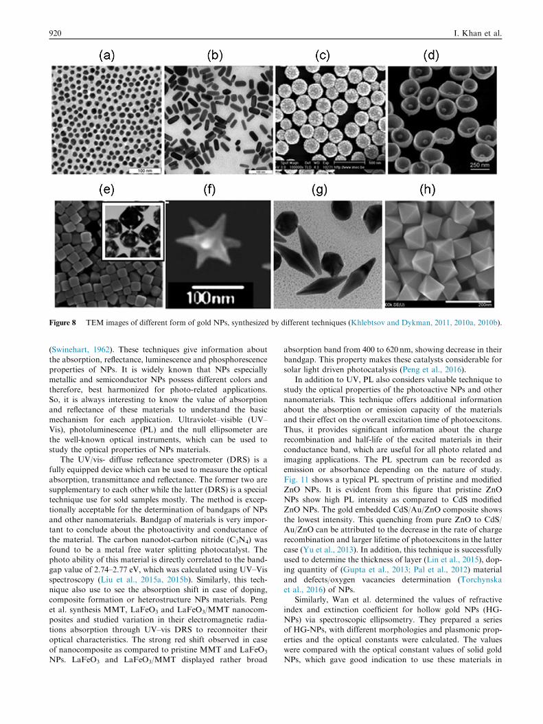
Figure 8 TEM images of different form of gold NPs, synthesized by different techniques (Khlebtsov and Dykman, 2011, 2010a, 2010b).
920 I. Khan et al.
(Swinehart, 1962). These techniques give information aboutthe absorption, reflectance, luminescence and phosphorescenceproperties of NPs. It is widely known that NPs especially
metallic and semiconductor NPs possess different colors andtherefore, best harmonized for photo-related applications.So, it is always interesting to know the value of absorption
and reflectance of these materials to understand the basicmechanism for each application. Ultraviolet–visible (UV–Vis), photoluminescence (PL) and the null ellipsometer arethe well-known optical instruments, which can be used to
study the optical properties of NPs materials.The UV/vis- diffuse reflectance spectrometer (DRS) is a
fully equipped device which can be used to measure the optical
absorption, transmittance and reflectance. The former two aresupplementary to each other while the latter (DRS) is a specialtechnique use for sold samples mostly. The method is excep-
tionally acceptable for the determination of bandgaps of NPsand other nanomaterials. Bandgap of materials is very impor-tant to conclude about the photoactivity and conductance ofthe material. The carbon nanodot-carbon nitride (C3N4) was
found to be a metal free water splitting photocatalyst. Thephoto ability of this material is directly correlated to the band-gap value of 2.74–2.77 eV, which was calculated using UV–Vis
spectroscopy (Liu et al., 2015a, 2015b). Similarly, this tech-nique also use to see the absorption shift in case of doping,composite formation or heterostructure NPs materials. Peng
et al. synthesis MMT, LaFeO3 and LaFeO3/MMT nanocom-posites and studied variation in their electromagnetic radia-tions absorption through UV–vis DRS to reconnoiter their
optical characteristics. The strong red shift observed in caseof nanocomposite as compared to pristine MMT and LaFeO3
NPs. LaFeO3 and LaFeO3/MMT displayed rather broad
absorption band from 400 to 620 nm, showing decrease in theirbandgap. This property makes these catalysts considerable forsolar light driven photocatalysis (Peng et al., 2016).
In addition to UV, PL also considers valuable technique tostudy the optical properties of the photoactive NPs and othernanomaterials. This technique offers additional information
about the absorption or emission capacity of the materialsand their effect on the overall excitation time of photoexcitons.Thus, it provides significant information about the chargerecombination and half-life of the excited materials in their
conductance band, which are useful for all photo related andimaging applications. The PL spectrum can be recorded asemission or absorbance depending on the nature of study.
Fig. 11 shows a typical PL spectrum of pristine and modifiedZnO NPs. It is evident from this figure that pristine ZnONPs show high PL intensity as compared to CdS modified
ZnO NPs. The gold embedded CdS/Au/ZnO composite showsthe lowest intensity. This quenching from pure ZnO to CdS/Au/ZnO can be attributed to the decrease in the rate of chargerecombination and larger lifetime of photoexcitons in the latter
case (Yu et al., 2013). In addition, this technique is successfullyused to determine the thickness of layer (Lin et al., 2015), dop-ing quantity of (Gupta et al., 2013; Pal et al., 2012) material
and defects/oxygen vacancies determination (Torchynskaet al., 2016) of NPs.
Similarly, Wan et al. determined the values of refractive
index and extinction coefficient for hollow gold NPs (HG-NPs) via spectroscopic ellipsometry. They prepared a seriesof HG-NPs, with different morphologies and plasmonic prop-
erties and the optical constants were calculated. The valueswere compared with the optical constant values of solid goldNPs, which gave good indication to use these materials in
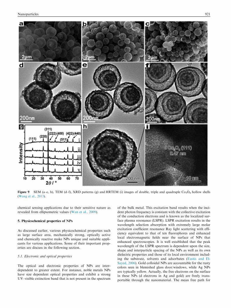
Figure 9 SEM (a–c, h), TEM (d–f), XRD patterns (g) and HRTEM (i) images of double, triple and quadruple Co3O4 hollow shells
(Wang et al., 2013).
Nanoparticles 921
chemical sensing applications due to their sensitive nature asrevealed from ellipsometric values (Wan et al., 2009).
5. Physicochemical properties of NPs
As discussed earlier, various physicochemical properties suchas large surface area, mechanically strong, optically activeand chemically reactive make NPs unique and suitable appli-
cants for various applications. Some of their important prop-erties are discuss in the following section.
5.1. Electronic and optical properties
The optical and electronic properties of NPs are inter-dependent to greater extent. For instance, noble metals NPs
have size dependent optical properties and exhibit a strongUV–visible extinction band that is not present in the spectrum
of the bulk metal. This excitation band results when the inci-dent photon frequency is constant with the collective excitationof the conduction electrons and is known as the localized sur-face plasma resonance (LSPR). LSPR excitation results in the
wavelength selection absorption with extremely large molarexcitation coefficient resonance Ray light scattering with effi-ciency equivalent to that of ten fluorophores and enhanced
local electromagnetic fields near the surface of NPs thatenhanced spectroscopies. It is well established that the peakwavelength of the LSPR spectrum is dependent upon the size,
shape and interparticle spacing of the NPs as well as its owndielectric properties and those of its local environment includ-ing the substrate, solvents and adsorbates (Eustis and El-
Sayed, 2006). Gold colloidal NPs are accountable for the rustycolors seen in blemished glass door/windows, while Ag NPsare typically yellow. Actually, the free electrons on the surfacein these NPs (d electrons in Ag and gold) are freely trans-
portable through the nanomaterial. The mean free path for

Figure 10 FTIR spectra of platinum (1.7 nm) (a) extracted from polyol, (b) dodecanethiol coated Pt, and (c) MUDA coated Pt
(Dablemont et al., 2008).
Figure 11 Photoluminescence (PL) spectra of pristine ZnO,
CdS/ZnO, and CdS/Au/ZnO measured with 270 nm excitation
wavelength at normal temperature (Yu et al., 2013).
922 I. Khan et al.
Ag and gold is �50 nm, which is more than the NPs size ofthese materials. Thus, no scattering is expected from the bulk,
upon light interaction, instead they set into a standing reso-nance conditions, which is responsible for LSPR in theseNPs (Fig. 12) (Khlebtsov and Dykman, 2010a, 2010b).
5.2. Magnetic properties
Magnetic NPs are of great curiosity for investigators from an
eclectic range of disciplines, which include heterogenous andhomogenous catalysis, biomedicine, magnetic fluids, data stor-age magnetic resonance imaging (MRI), and environmental
remediation such as water decontamination. The literaturerevealed that NPs perform best when the size is <critical valuei.e. 10–20 nm (Reiss and Hutten, 2005). At such low scale themagnetic properties of NPs dominated effectively, which make
these particle priceless and can be used in different applications(Faivre and Bennet, 2016; Priyadarshana et al., 2015; Reissand Hutten, 2005; Zhu et al., 1994). The uneven electronic dis-
tribution in NPs leads to magnetic property. These propertiesare also dependent on the synthetic protocol and various syn-thetic methods such as solvothermal (Qi et al., 2016), co-precipitation, micro-emulsion, thermal decomposition, and
flame spray synthesis can be used for their preparation (Wuet al., 2008).
5.3. Mechanical properties
The distinct mechanical properties of NPs allow researchers tolook for novel applications in many important fields such as
tribology, surface engineering, nanofabrication and nanoman-ufacturing. Different mechanical parameters such as elasticmodulus, hardness, stress and strain, adhesion and friction
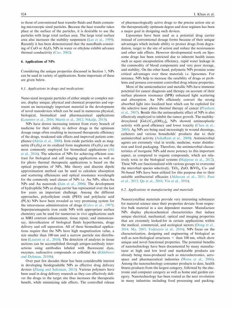
can be surveyed to know the exact mechanical nature ofNPs. Beside these parameters surface coating, coagulation,and lubrication also aid to mechanical properties of NPs(Guo et al., 2014) (see Scheme 4). NPs show dissimilar
mechanical properties as compared to microparticles and theirbulk materials. Moreover, in a lubricated or greased contact,the contrast in the stiffness between NPs and the contacting
external surface controls whether the NPs are indented intothe plan surface or deformed when the pressure at contact issignificantly large. This important information could divulge
how the NPs perform in the contact situation. Decent controlsover mechanical features of NPs and their interactions withany kind of surface are vital for enlightening the surface qual-
ity and elevating material removal. Fruitful outcomes in thesefields generally need a deep insight into the basics of themechanical properties of NPs, such as elastic modulus andhardness, movement law, friction and interfacial adhesion
and their size dependent characteristics (Guo et al., 2014).
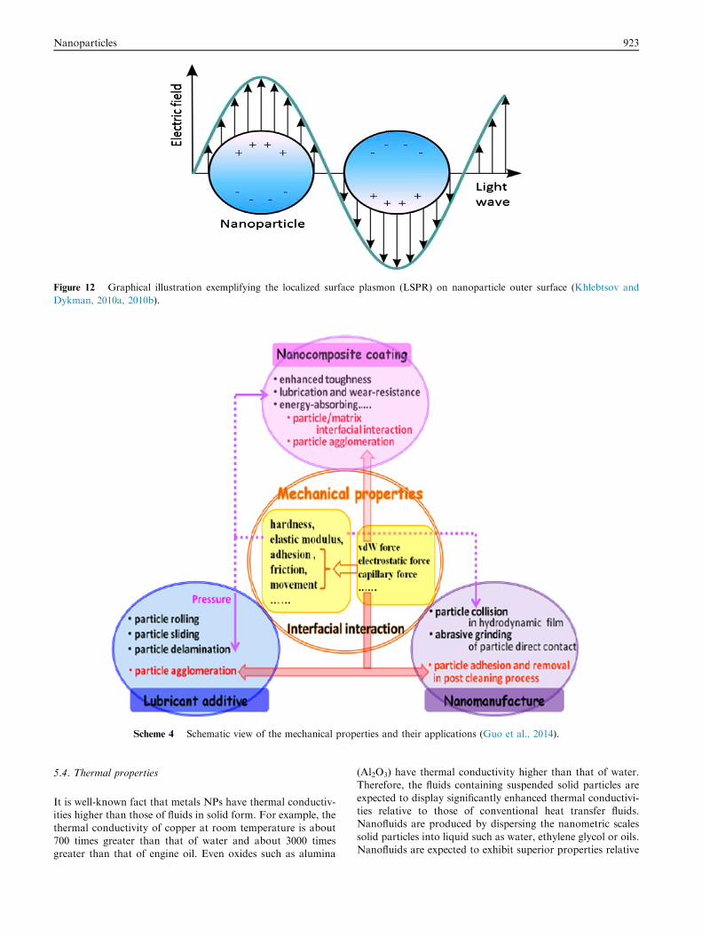
Figure 12 Graphical illustration exemplifying the localized surface plasmon (LSPR) on nanoparticle outer surface (Khlebtsov and
Dykman, 2010a, 2010b).
Scheme 4 Schematic view of the mechanical properties and their applications (Guo et al., 2014).
Nanoparticles 923
5.4. Thermal properties
It is well-known fact that metals NPs have thermal conductiv-ities higher than those of fluids in solid form. For example, thethermal conductivity of copper at room temperature is about
700 times greater than that of water and about 3000 timesgreater than that of engine oil. Even oxides such as alumina
(Al2O3) have thermal conductivity higher than that of water.Therefore, the fluids containing suspended solid particles are
expected to display significantly enhanced thermal conductivi-ties relative to those of conventional heat transfer fluids.Nanofluids are produced by dispersing the nanometric scales
solid particles into liquid such as water, ethylene glycol or oils.Nanofluids are expected to exhibit superior properties relative
924 I. Khan et al.
to those of conventional heat transfer fluids and fluids contain-ing microscopic sized particles. Because the heat transfer takesplace at the surface of the particles, it is desirable to use the
particles with large total surface area. The large total surfacearea also increases the stability suspension (Lee et al., 1999).Recently it has been demonstrated that the nanofluids consist-
ing of CuO or Al2O3 NPs in water or ethylene exhibit advancethermal conductivity (Cao, 2002).
6. Applications of NPs
Considering the unique properties discussed in Section 5, NPscan be used in variety of applications. Some important of these
are given below.
6.1. Applications in drugs and medications
Nano-sized inorganic particles of either simple or complex nat-ure, display unique, physical and chemical properties and rep-resent an increasingly important material in the developmentof novel nanodevices which can be used in numerous physical,
biological, biomedical and pharmaceutical applications(Loureiro et al., 2016; Martis et al., 2012; Nikalje, 2015).
NPs have drawn increasing interest from every branch of
medicine for their ability to deliver drugs in the optimumdosage range often resulting in increased therapeutic efficiencyof the drugs, weakened side effects and improved patient com-
pliance (Alexis et al., 2008). Iron oxide particles such as mag-netite (Fe3O4) or its oxidized form maghemite (Fe2O3) are themost commonly employed for biomedical applications (Aliet al., 2016). The selection of NPs for achieving efficient con-
trast for biological and cell imaging applications as well asfor photo thermal therapeutic applications is based on theoptical properties of NPs. Mie theory and discrete dipole
approximation method can be used to calculate absorptionand scattering efficiencies and optical resonance wavelengthfor the commonly used classes of NPs i.e. Au NPs, silica-Au
NPs and Au nanorods (Jain et al., 2006). The developmentof hydrophilic NPs as drug carrier has represented over the lastfew years an important challenge. Among the different
approaches, polyethylene oxide (PEO) and polylactic acid(PLA) NPs have been revealed as very promising system forthe intravenous administration of drugs (Calvo et al., 1997).Superparamagnetic iron oxide NPs with appropriate surface
chemistry can be used for numerous in vivo applications suchas MRI contrast enhancement, tissue repair, and immunoas-say, detoxification of biological fluids hyperthermia, drugs
delivery and cell separation. All of these biomedical applica-tions require that the NPs have high magnetization value, asize smaller than 100 nm and a narrow particle size distribu-
tion (Laurent et al., 2010). The detection of analytes in tissuesections can be accomplished through antigen-antibody inter-actions using antibodies labeled with fluorescent dyes,
enzymes, radioactive compounds or colloidal Au (Khlebtsovand Dykman, 2010b).
Over past few decades these has been considerable interestin developing biodegradable NPs as effective drug delivery
devices (Zhang and Saltzman, 2013). Various polymers havebeen used in drug delivery research as they can effectively deli-ver the drugs to the target site thus increases the therapeutic
benefit, while minimizing side effects. The controlled release
of pharmacologically active drugs to the precise action site atthe therapeutically optimum degree and dose regimen has beena major goal in designing such devices.
Liposomes have been used as a potential drug carrierinstead of conventional dosage forms because of their uniqueadvantages which include ability to protect drugs from degra-
dation, target to the site of action and reduce the noxiousnessand other side effects. However developmental work on lipo-some drugs has been restricted due to inherent health issues
such as squat encapsulation efficiency, rapid water leakage inthe commodity of blood components and very poor storage,and stability. On the other hand, polymeric NPs promise somecritical advantages over these materials i.e. liposomes. For
instance, NPs help to increase the ratability of drugs or prob-lems and possess convenient controlled drug release properties.
Most of the semiconductor and metallic NPs have immense
potential for cancer diagnosis and therapy on account of theirsurface plasmon resonance (SPR) enhanced light scatteringand absorption. Au NPs efficiently convert the strong
absorbed light into localized heat which can be exploited forthe selective laser photo thermal therapy of cancer (Prashantet al., 2007). Beside this the antineoplastic effect of NPs is also
effectively employed to inhibit the tumor growth. The multihy-droxylated [Gd@C82(OH)22]n NPs showed antineoplasticactivity with good efficiency and lower toxicity (Chen et al.,2005). Ag NPs are being used increasingly in wound dressings,
catheters and various households’ products due to theirantimicrobial activity (AshaRani et al., 2009). Antimicrobialagents are extremely vital in textile, medicine, water disinfec-
tion and food packaging. Therefore, the antimicrobial charac-teristics of inorganic NPs add more potency to this importantaspect, as compared to organic compounds, which are rela-
tively toxic to the biological systems (Hajipour et al., 2012).These NPs are functionalized with various groups to overcomethe microbial species selectively. TiO2, ZnO, BiVO4, Cu- and
Ni-based NPs have been utilized for this purpose due to theirsuitable antibacterial efficacies (Akhavan et al., 2011; Pantet al., 2013; Qu et al., 2016; Yin et al., 2016).
6.2. Applications in manufacturing and materials
Nanocrystalline materials provide very interesting substancesfor material science since their properties deviate from respec-
tive bulk material in a size dependent manner. ManufactureNPs display physicochemical characteristics that induceunique electrical, mechanical, optical and imaging properties
that are extremely looked-for in certain applications withinthe medical, commercial, and ecological sectors (Dong et al.,2014; Ma, 2003; Todescato et al., 2016). NPs focus on thecharacterization, designing and engineering of biological as
well as non-biological structures < than 100 nm, which showunique and novel functional properties. The potential benefitsof nanotechnology have been documented by many manufac-
turer at high and low level and marketable products arealready being mass-produced such as microelectronics, aero-space and pharmaceutical industries (Weiss et al., 2006).
Among the nanotechnology consumer products to date, healthfitness products from the largest category, followed by the elec-tronic and computer category as well as home and garden cat-
egory. Nanotechnology has been touted as the next revolutionin many industries including food processing and packing.
Nanoparticles 925
Resonant energy transfer (RET) system consisting of organicdye molecules and noble metals NPs have recently gamed con-siderable interest in bio photonics as well as in material science
(Lei et al., 2015). The presence of NPs in commercially avail-able products is becoming more common.
Metals NPs such as noble metals, including Au and Ag
have many colors in the visible region based on plasmon reso-nance, which is due to collective oscillations of the electrons atthe surface of NPs (Khlebtsov and Dykman, 2010a, 2010b;
Unser et al., 2015). The resonance wavelength strong dependson size and shape of NPs, the interparticle distance, and thedielectric property of the surrounding medium. The uniqueplasmon absorbance features of these noble metals NPs have
been exploited for a wide variety of applications includingchemical sensors and biosensors (Unser et al., 2015).
6.3. Applications in the environment
The increasing area of engineered NPs in industrial and house-hold applications leads to the release of such materials into the
environment. Assessing the risk of these NPs in the environ-ment requires on understanding of their mobility, reactivity,Eco toxicity and persistency (Ripp and Henry, 2011; Zhuang
and Gentry, 2011). The engineering material applications canincrease the concentration of NPs in groundwater and soilwhich presents the most significant exposure avenues forassessing environmental risks (Golobic et al., 2012;
Masciangioli and Zhang, 2003). Due to high surface to massratio natural NPs play an important role in the solid/waterpartitioning of contaminants can be absorbed to the surface
of NPs, co-precipitated during the formation of natural NPsor trapped by aggregation of NPs which had contaminantsadsorbed to their surface. The interaction of contaminants
with NPs is dependent on the NPs characteristics, such as size,composition, morphology, porosity, aggregation/disaggrega-tion and aggregate structure. The luminophores are not safe
in the environment and are protected from the environmentaloxygen when they are doped inside the silica network(Swadeshmukul et al., 2001).
Most of environmental applications of nanotechnology fall
into three categories:
1. Environmentally benign sustainable products (e.g. green
chemistry or pollution prevention).2. Remediation of materials contaminated with hazardous
substances and
3. Sensors for environmental stages (Tratnyek and Johnson,2006).
The removal of heavy metals such as mercury, lead, thal-
lium, cadmium and arsenic from natural water has attractedconsiderable attention because of their adverse effects on envi-ronmental and human health. Superparamagnetic iron oxide
NPs are an effective sorbent material for this toxic soft mate-rial. So, for no measurements of engineered NPs in the envi-ronment have been available due to the absence of analytical
methods, able to quantify trace concentration of NPs(Mueller and Nowack, 2008). Photodegradation by NPs is alsovery common practice and many nanomaterials are utilized for
this purpose. Rogozea et al. used NiO/ZnO NPs modified sil-ica in the tandem fashion for photodegradation purpose. The
high surface area of NPs due to very small size (<10 nm),facilitated the efficient photodegradation reaction (Rogozeaet al., 2017). The same group has reported the synthesis of
variety of NPs and reported their optical, florescence anddegradation applications (Olteanu et al., 2016a, 2016b;Rogozea et al., 2016).
6.4. Applications in electronics
There has been growing interest in the development of printed
electronics in last few years because printed electronics offerattractive to traditional silicon techniques and the potentialfor low cost, large area electronics for flexible displays, sensors.
Printed electronics with various functional inks containingNPs such as metallic NPs, organic electronic molecules, CNTsand ceramics NPs have been expected to flow rapidly as a massproduction process for new types of electronic equipment
(Kosmala et al., 2011).Unique structural, optical and electrical properties of one
dimensional semiconductor and metals make them the key
structural block for a new generation of electronic, sensorsand photonic materials (Holzinger et al., 2014; Millstoneet al., 2010; Shaalan et al., 2016).
The good example of the synergism between scientific dis-covery and technological development is the electronic indus-try, where discoveries of new semiconducting materialsresulted in the revolution from vacuumed tubes to diodes
and transistors, and eventually to miniature chips (Cushinget al., 2004).
The important characteristics of NPs are facile manipula-
tion and reversible assembly which allow for the possibilityof incorporation of NPs in electric, electronic or opticaldevices such as ‘‘bottom up” or ‘‘self-assembly” approaches
are the bench mark of nanotechnology (O’Brien et al., 2001).
6.5. Applications in energy harvesting
Recent studies warned us about the limitations and scarcityof fossil fuels in coming years due to their nonrenewable nat-ure. Therefore, scientists shifting their research strategies togenerate renewable energies from easily available resources
at cheap cost. They found that NPs are the best candidatefor this purpose due to their, large surface area, opticalbehavior and catalytic nature. Especially in photocatalytic
applications, NPs are widely used to generate energy fromphotoelectrochemical (PEC) and electrochemical water split-ting (Avasare et al., 2015; Mueller and Nowack, 2008; Ning
et al., 2016). Beside water splitting, electrochemical CO2
reduction to fuels precursors, solar cells and piezoelectric gen-erators also offered advance options to generate energy (Fang
et al., 2013; Gawande et al., 2016; Lei et al., 2015; Li et al.,2016; Nagarajan et al., 2014; Sagadevan, 2015; Young et al.,2012; Zhou et al., 2016). NPs also use in energy storageapplications to reserve the energy into different forms at
nanoscale level (Greeley and Markovic, 2012; Liu et al.,2015a, 2015b; Sagadevan, 2015; Wang and Su, 2014).Recently, nanogenerators are created, which can convert the
mechanical energy into electricity using piezoelectric, whichis an unconventional approach to generate energy (Wanget al., 2015). Fig. 13 shows some energy generating devices,
and uses NPs.

Figure 13 Energy generation approaches from (A) Piezoelectrics actuators (B) Water splitting (C) CO2 reduction.
926 I. Khan et al.
6.6. Applications in mechanical industries
As revealed from their mechanical properties through excellentyoung modulus, stress and strain properties, NPs can offermany applications in mechanical industries especially in coat-ing, lubricants and adhesive applications. Besides, this prop-
erty can be useful to achieve mechanically strongernanodevices for various purposes. Tribological propertiescan be controlled at nanoscale level by embedding NPs in
the metal and polymer matrix to increase their mechanicalstrengths. It is because, the rolling mode of NPs in the lubri-cated contact area could provide very low friction and wear.
In addition, NPs offer good sliding and delamination proper-ties, which could also effect in low friction and wear, and henceincrease lubrication effect (Guo et al., 2014). Coating can lead
to various mechanically strong characteristics, as it improvestoughness and wear resistance. Alumina, Titania and carbonbased NPs successfully demonstrated to get the desirable
mechanical properties in coatings (Kot et al., 2016;Mallakpour and Sirous, 2015; Shao et al., 2012).
7. Toxicity of NP
Beside many industrial and medical applications, there are cer-tain toxicities which are associated with NPs and other nano-
materials (Bahadar et al., 2016; Ibrahim, 2013; Khlebtsov andDykman, 2011, 2010b) and basic knowledge is required forthese toxic effects to encounter them properly. NPs surrepti-
tiously enter the environment through water, soil, and air dur-ing various human activities. However, the application of NPs
Nanoparticles 927
for environmental treatment deliberately injects or dumpsengineered NPs into the soil or aquatic systems. This has resul-tantly attracted increasing concern from all stakeholders. The
advantages of magnetic NPs such as their small size, high reac-tivity and great capacity, could become potential lethal factorsby inducing adverse cellular toxic and harmful effects, unusual
in micron-sized counter parts. Studies also illustrated that NPscan enter organisms during ingestion or inhalation and cantranslocate within the body to various organs and tissues
where the NPs have the possibility to exert the reactivity beingtoxicology effects. Although some studies have also addressedthe toxicological effects of NPs on animal cells and plant cellsthe toxicological studies with magnetic NPs on plants to date
are still limited. The uses of Ag NPs in numerous consumerproducts lead them to their release to the aquatic environmentand become a source of dissolved Ag and thus exert toxic
effects on aquatic organisms including bacteria, algae, fishand daphnia (Navarro et al., 2008). The respiratory systemrepresents an unique target for the potential toxicity of NPs
due to the fact that in addition to being the portal of entryfor inhaled particles, it also receives the entire cardiac output(Ferreira et al., 2013). NPs are used in bio applications widely
but despite the rapid progress and early acceptance ofnanobiotechnology the potential for adverse health effectsdue to prolong exposure at various concentrations levels inhuman in the environment has not yet been established. How-
ever, the environmental impact of NPs is expected to increasein the future. One of the NPs toxicity is the ability to organizearound the protein concentration that depends on particles
size, curvature, shape and surface characteristics charge, func-tionalized groups, and free energy. Due to this binding, someparticles generate adverse biological outcomes through protein
unfolding, fibrillation, thiol crosslinking, and loss of enzymaticactivity. Another paradigm is the release of toxic ions when thethermodynamic properties of materials favor particles dissolu-
tion in a suspending medium or biological environment (Xiaet al., 2008).
NPs tend to aggregate in hard water and seawater and aregreatly influenced by the specific type of organic matter or
other natural particles (colloids) present in fresh water. Thestate of dispersion will alter the ecotoxicity, but many abioticfactors that influence this, such as pH, salinity, and the pres-
ence of organic matters remain to be systematically investi-gated as part of ecotoxicological studies (Handy et al., 2008).
8. Conclusion
In this review, we presented a detail overview about NPs, their types,
synthesis, characterizations, physiochemical properties and applica-
tions. Through different characterization techniques such as SEM,
TEM and XRD, it was revealed that NPs have size ranges from few
nanometer to 500 nm. While the morphology is also controllable.
Due to their tiny size, NPs have large surface area, which make them
suitable candidate for various applications. Beside this, the optical
properties are also dominant at that size, which further increase the
importance of these materials in photocatalytic applications. Synthetic
techniques can be useful to control the specific morphology, size and
magnetic properties of NPs. Though NPs are useful for many applica-
tions, but still there are some health hazard concerns due to their
uncontrollable use and discharge to natural environment, which
should be consider for make the use of NPs more convenient and envi-
ronmental friendly.
9. Recommendations
Our recommendation for future work is that different reactionparameters such as temperature, pressure, time, and pH. can
play important role in controlling the shape and morphologyof the NPs materials, so that should be optimize for achievingspecific characteristic product. Beside this for good implica-
tions and properties study specific characterization techniquesshould be used. More importantly environmental issuedshould be taken into account before using these materials forany applications, especially in case of heavy metals, which
are prone to environmental hazards and can also affect the liv-ings as well.
References
Abd Ellah, N.H., Abouelmagd, S.A., 2016. Surface functionalization
of polymeric nanoparticles for tumor drug delivery: approaches
and challenges. Expert Opin. Drug Deliv. 1–14. http://dx.doi.org/
10.1080/17425247.2016.1213238.
Abouelmagd, S.A., Meng, F., Kim, B.-K., Hyun, H., Yeo, Y., 2016.
Tannic acid-mediated surface functionalization of polymeric
nanoparticles. ACS Biomater. Sci. Eng., 6b00497 http://dx.doi.
org/10.1021/acsbiomaterials.6b004.
Ahmed, S., Annu, S., Yudha, S.S., 2016. Biosynthesis of gold
nanoparticles: a green approach. J. Photochem. Photobiol. B: Biol.
161, 141–153. http://dx.doi.org/10.1016/j.jphotobiol.2016.04.034.
Akhavan, O., Azimirad, R., Safa, S., Hasani, E., 2011. CuO/Cu(OH)2hierarchical nanostructures as bactericidal photocatalysts. J.
Mater. Chem. 21, 9634. http://dx.doi.org/10.1039/c0jm04364h.
Alexis, F., Pridgen, E., Molnar, L.K., Farokhzad, O.C., 2008.
Factors affecting the clearance and biodistribution of polymeric
nanoparticles. Mol. Pharm. 5, 505–515. http://dx.doi.org/10.1021/
mp800051m.
Ali, A., Zafar, H., Zia, M., Ul Haq, I., Phull, A.R., Ali, J.S., Hussain,
A., 2016. Synthesis, characterization, applications, and challenges
of iron oxide nanoparticles. Nanotechnol. Sci. Appl. 9, 49–67.
http://dx.doi.org/10.2147/NSA.S99986.
Ali, S., Khan, I., Khan, S.A., Sohail, M., Ahmed, R., Rehman, A.,
Ur Ansari, M.S., Morsy, M.A., 2017. Electrocatalytic performance
of Ni@Pt core–shell nanoparticles supported on carbon nanotubes
for methanol oxidation reaction. J. Electroanal. Chem. 795, 17–25.
http://dx.doi.org/10.1016/j.jelechem.2017.04.040.
Aqel, A., El-Nour, K.M.M.A., Ammar, R.A.A., Al-Warthan, A.,
2012. Carbon nanotubes, science and technology part (I) structure,
synthesis and characterisation. Arab. J. Chem. 5, 1–23. http://dx.
doi.org/10.1016/j.arabjc.2010.08.022.
AshaRani, P.V., Low Kah Mun, G., Hande, M.P., Valiyaveettil, S.,
2009. Cytotoxicity and genotoxicity of silver nanoparticles in
human cells. ACS Nano 3, 279–290. http://dx.doi.org/10.1021/
nn800596w.
Astefanei, A., Nunez, O., Galceran, M.T., 2015. Characterisation and
determination of fullerenes: a critical review. Anal. Chim. Acta 882,
1–21. http://dx.doi.org/10.1016/j.aca.2015.03.025.
Avasare, V., Zhang, Z., Avasare, D., Khan, I., Qurashi, A., 2015.
Room-temperature synthesis of TiO2 nanospheres and their solar
driven photoelectrochemical hydrogen production. Int. J. Energy
Res. 39, 1714–1719. http://dx.doi.org/10.1002/er.3372.
Bahadar, H., Maqbool, F., Niaz, K., Abdollahi, M., 2016. Toxicity of
nanoparticles and an overview of current experimental models.
Iran. Biomed. J. 20, 1–11. http://dx.doi.org/10.7508/
ibj.2016.01.001.
Barrak, H., Saied, T., Chevallier, P., Laroche, G., M’nif, A.,
Hamzaoui, A.H., 2016. Synthesis, characterization, and function-
alization of ZnO nanoparticles by N-(trimethoxysilylpropyl)

928 I. Khan et al.
ethylenediamine triacetic acid (TMSEDTA): investigation of the
interactions between phloroglucinol and ZnO@TMSEDTA. Arab.
J. Chem. http://dx.doi.org/10.1016/j.arabjc.2016.04.019.
Bello, S.A., Agunsoye, J.O., Hassan, S.B., 2015. Synthesis of coconut
shell nanoparticles via a top down approach: assessment of milling
duration on the particle sizes and morphologies of coconut shell
nanoparticles. Mater. Lett. http://dx.doi.org/10.1016/
j.matlet.2015.07.063.
Biswas, A., Bayer, I.S., Biris, A.S., Wang, T., Dervishi, E., Faupel, F.,
2012. Advances in top–down and bottom–up surface nanofabrica-
tion: techniques, applications & future prospects. Adv. Coll.
Interface. Sci. 170, 2–27. http://dx.doi.org/10.1016/
j.cis.2011.11.001.
Calvo, P., Remuoon-Lopez, C., Vila-Jato, J.L., Alonso, M.J., 1997.
Novel hydrophilic chitosan-polyethylene oxide nanoparticles as
protein carriers. J. Appl. Polym. Sci. 63, 125–132. http://dx.doi.org/
10.1002/(SICI)1097-4628(19970103)63:1*125::AID-APP13*3.0.
CO;2-4.
Cao, Y.C., 2002. Nanoparticles with Raman spectroscopic finger-
prints for DNA and RNA detection. Science 80 (297), 1536–1540.
http://dx.doi.org/10.1126/science.297.5586.1536.
Chen, C., Xing, G., Wang, J., Zhao, Y., Li, B., Tang, J., Jia, G.,
Wang, T., Sun, J., Xing, L., Yuan, H., Gao, Y., Meng, H., Chen,
Z., Zhao, F., Chai, Z., Fang, X., 2005. Multihydroxylated [Gd@C
82 (OH) 22 ] n nanoparticles: antineoplastic activity of high
efficiency and low toxicity. Nano Lett. 5, 2050–2057. http://dx.doi.
org/10.1021/nl051624b.
Cushing, B.L., Kolesnichenko, V.L., O’Connor, C.J., 2004. Recent
advances in the liquid-phase syntheses of inorganic nanoparticles.
Chem. Rev. 104, 3893–3946. http://dx.doi.org/10.1021/cr030027b.
Dablemont, C., Lang, P., Mangeney, C., Piquemal, J.-Y., Petkov, V.,
Herbst, F., Viau, G., 2008. FTIR and XPS study of Pt nanoparticle
functionalization and interaction with alumina. Langmuir 24,
5832–5841. http://dx.doi.org/10.1021/la7028643.
Dong, H., Wen, B., Melnik, R., 2014. Relative importance of grain
boundaries and size effects in thermal conductivity of nanocrys-
talline materials. Sci. Rep. 4, 7037. http://dx.doi.org/10.1038/
srep07037.
Dreaden, E.C., Alkilany, A.M., Huang, X., Murphy, C.J., El-Sayed,
M.A., 2012. The golden age: gold nanoparticles for biomedicine.
Chem. Soc. Rev. 41, 2740–2779. http://dx.doi.org/10.1039/
C1CS15237H.
Elliott, J.A., Shibuta, Y., Amara, H., Bichara, C., Neyts, E.C., 2013.
Atomistic modelling of CVD synthesis of carbon nanotubes and
graphene. Nanoscale 5, 6662. http://dx.doi.org/10.1039/c3nr01925j.
Emery, A.A., Saal, J.E., Kirklin, S., Hegde, V.I., Wolverton, C.,
2016. High-throughput computational screening of perovskites for
thermochemical water splitting applications. Chem. Mater. 28.
http://dx.doi.org/10.1021/acs.chemmater.6b01182.
Eustis, S., El-Sayed, M.A., 2006. Why gold nanoparticles are more
precious than pretty gold: noble metal surface plasmon resonance
and its enhancement of the radiative and nonradiative properties of
nanocrystals of different shapes. Chem. Soc. Rev. 35, 209–217.
http://dx.doi.org/10.1039/B514191E.
Fagerlund, G., 1973. Determination of specific surface by the BET
method. Materiaux Constr. 6, 239–245. http://dx.doi.org/10.1007/
BF02479039.
Faivre, D., Bennet, M., 2016. Materials science: magnetic nanopar-
ticles line up. Nature 535, 235–236. http://dx.doi.org/10.1038/
535235a.
Fang, X.-Q., Liu, J.-X., Gupta, V., 2013. Fundamental formulations
and recent achievements in piezoelectric nano-structures: a review.
Nanoscale 5, 1716. http://dx.doi.org/10.1039/c2nr33531j.
Ferreira, A.J., Cemlyn-Jones, J., Robalo Cordeiro, C., 2013.
Nanoparticles, nanotechnology and pulmonary nanotoxicology.
Rev. Port. Pneumol. 19, 28–37. http://dx.doi.org/10.1016/j.
rppneu.2012.09.003.
Filipe, V., Hawe, A., Jiskoot, W., 2010. Critical evaluation of
nanoparticle tracking analysis (NTA) by nanosight for the
measurement of nanoparticles and protein aggregates. Pharm.
Res. 27, 796–810. http://dx.doi.org/10.1007/s11095-010-0073-2.
Ganesh, M., Hemalatha, P., Peng, M.M., Jang, H.T., 2017. One pot
synthesized Li, Zr doped porous silica nanoparticle for low
temperature CO2 adsorption. Arab. J. Chem. 10, S1501–S1505.
Garrigue, P., Delville, M.-H., Labrugere, C., Cloutet, E., Kulesza, P.
J., Morand, J.P., Kuhn, A., 2004. Top–down approach for the
preparation of colloidal carbon nanoparticles. Chem. Mater. 16,
2984–2986. http://dx.doi.org/10.1021/cm049685i.
Gawande, M.B., Goswami, A., Felpin, F.-X., Asefa, T., Huang, X.,
Silva, R., Zou, X., Zboril, R., Varma, R.S., 2016. Cu and
Cu-based nanoparticles: synthesis and applications in catalysis.
Chem. Rev. 116, 3722–3811. http://dx.doi.org/10.1021/acs.
chemrev.5b00482.
Golobic, M., Jemec, A., Drobne, D., Romih, T., Kasemets, K.,
Kahru, A., 2012. Upon exposure to Cu nanoparticles, accumula-
tion of copper in the isopod Porcellio scaber is due to the dissolved
cu ions inside the digestive tract. Environ. Sci. Technol. 46, 12112–
12119. http://dx.doi.org/10.1021/es3022182.
Greeley, J., Markovic, N.M., 2012. The road from animal electricity
to green energy: combining experiment and theory in electrocatal-
ysis. Energy Environ. Sci. 5. http://dx.doi.org/10.1039/c2ee21754f.
Gross, J., Sayle, S., Karow, A.R., Bakowsky, U., Garidel, P., 2016.
Nanoparticle tracking analysis of particle size and concentration
detection in suspensions of polymer and protein samples: Influence
of experimental and data evaluation parameters. Eur. J. Pharm.
Biopharm. 104, 30–41. http://dx.doi.org/10.1016/j.
ejpb.2016.04.013.
Gujrati, M., Malamas, A., Shin, T., Jin, E., Sun, Y., Lu, Z.-R., 2014.
Multifunctional cationic lipid-based nanoparticles facilitate endo-
somal escape and reduction-triggered cytosolic siRNA release.
Mol. Pharm. 11, 2734–2744. http://dx.doi.org/10.1021/mp400787s.
Guo, D., Xie, G., Luo, J., 2014. Mechanical properties of nanopar-
ticles: basics and applications. J. Phys. D Appl. Phys. 47, 13001.
http://dx.doi.org/10.1088/0022-3727/47/1/013001.
Gupta, K., Singh, R.P., Pandey, A., Pandey, A., 2013. Photocatalytic
antibacterial performance of TiO2 and Ag-doped TiO2 against S.
aureus. P. aeruginosa and E. coli. Beilstein J. Nanotechnol. 4, 345–
351. http://dx.doi.org/10.3762/bjnano.4.40.
Hajipour, M.J., Fromm, K.M., Ashkarran, A. Akbar, de Aberasturi,
D. Jimenez, de Larramendi, I.R., Rojo, T., Serpooshan, V., Parak,
W.J., Mahmoudi, M., 2012. Antibacterial properties of nanopar-
ticles. Trends Biotechnol. 30, 499–511. http://dx.doi.org/10.1016/j.
tibtech.2012.06.004.
Handy, R.D., von der Kammer, F., Lead, J.R., Hassellov, M., Owen,
R., Crane, M., 2008. The ecotoxicology and chemistry of manu-
factured nanoparticles. Ecotoxicology 17, 287–314. http://dx.doi.
org/10.1007/s10646-008-0199-8.
Hisatomi, T., Kubota, J., Domen, K., 2014. Recent advances in
semiconductors for photocatalytic and photoelectrochemical water
splitting. Chem. Soc. Rev. 43, 7520–7535. http://dx.doi.org/
10.1039/C3CS60378D.
Holzinger, M., Le Goff, A., Cosnier, S., 2014. Nanomaterials for
biosensing applications: a review. Front. Chem. 2, 63. http://dx.doi.
org/10.3389/fchem.2014.00063.
Ibrahim, K.S., 2013. Carbon nanotubes-properties and applications:
a review. Carbon Lett. 14, 131–144. http://dx.doi.org/10.5714/
CL.2013.14.3.131.
Ingham, B., 2015. X-ray scattering characterisation of nanoparticles.
Crystallogr. Rev. 21, 229–303. http://dx.doi.org/10.1080/
0889311X.2015.1024114.
Iqbal, N., Khan, I., Yamani, Z.H., Qurashi, A., 2016. Sonochemical
assisted solvothermal synthesis of gallium oxynitride nanosheets
and their solar-driven photoelectrochemical water-splitting appli-
cations. Sci. Rep. 6, 32319. http://dx.doi.org/10.1038/srep32319.
Nanoparticles 929
Iravani, S., 2011. Green synthesis of metal nanoparticles using plants.
Green Chem. 13, 2638. http://dx.doi.org/10.1039/c1gc15386b.
Jain, P.K., Lee, K.S., El-Sayed, I.H., El-Sayed, M.A., 2006. Calcu-
lated absorption and scattering properties of gold nanoparticles of
different size, shape, and composition: applications in biological
imaging and biomedicine. J. Phys. Chem. B 110, 7238–7248. http://
dx.doi.org/10.1021/jp057170o.
Kestens, V., Roebben, G., Herrmann, J., Jamting, A., Coleman, V.,
Minelli, C., Clifford, C., De Temmerman, P.-J., Mast, J., Junjie, L.,
Babick, F., Colfen, H., Emons, H., 2016. Challenges in the size
analysis of a silica nanoparticle mixture as candidate certified
reference material. J. Nanopart. Res. 18, 171. http://dx.doi.org/
10.1007/s11051-016-3474-2.
Khan, I., Abdalla, A., Qurashi, A., 2017a. Synthesis of hierarchical
WO3 and Bi2O3/WO3 nanocomposite for solar-driven water
splitting applications. Int. J. Hydrogen Energy 42, 3431–3439.
http://dx.doi.org/10.1016/j.ijhydene.2016.11.105.
Khan, I., Ali, S., Mansha, M., Qurashi, A., 2017b. Sonochemical
assisted hydrothermal synthesis of pseudo-flower shaped Bismuth
vanadate (BiVO4) and their solar-driven water splitting application.
Ultrason. Sonochem. 36, 386–392. http://dx.doi.org/10.1016/j.
ultsonch.2016.12.014.
Khan, I., Ibrahim, A.A.M., Sohail, M., Qurashi, A., 2017c. Sono-
chemical assisted synthesis of RGO/ZnO nanowire arrays for
photoelectrochemical water splitting. Ultrason. Sonochem. 37,
669–675. http://dx.doi.org/10.1016/j.ultsonch.2017.02.029.
Khlebtsov, N., Dykman, L., 2011. Biodistribution and toxicity of
engineered gold nanoparticles: a review of in vitro and in vivo
studies. Chem. Soc. Rev. 40, 1647–1671. http://dx.doi.org/10.1039/
C0CS00018C.
Khlebtsov, N., Dykman, L., 2010. Plasmonic nanoparticles. pp. 37–
85. http://dx.doi.org/10.1201/9781439806296-c2.
Khlebtsov, N.G., Dykman, L.A., 2010b. Optical properties and
biomedical applications of plasmonic nanoparticles. J. Quant.
Spectrosc. Radiat. Transf. 111, 1–35. http://dx.doi.org/10.1016/j.
jqsrt.2009.07.012.
Kosmala, A., Wright, R., Zhang, Q., Kirby, P., 2011. Synthesis of
silver nano particles and fabrication of aqueous Ag inks for inkjet
printing. Mater. Chem. Phys. 129, 1075–1080. http://dx.doi.org/
10.1016/j.matchemphys.2011.05.064.
Kot, M., Major, Ł., Lackner, J.M., Chronowska-Przywara, K.,
Janusz, M., Rakowski, W., 2016. Mechanical and tribological
properties of carbon-based graded coatings. J. Nanomater. 2016,
1–14. http://dx.doi.org/10.1155/2016/8306345.
Laurent, S., Forge, D., Port, M., Roch, A., Robic, C., Vander Elst,
L., Muller, R.N., 2010. Magnetic iron oxide nanoparticles:
synthesis, stabilization, vectorization, physicochemical characteri-
zations, and biological applications. Chem. Rev. 110. http://dx.doi.
org/10.1021/cr900197g, pp. 2574–2574.
Lee, J.E., Lee, N., Kim, T., Kim, J., Hyeon, T., 2011. Multifunctional
mesoporous silica nanocomposite nanoparticles for theranostic
applications. Acc. Chem. Res. 44, 893–902. http://dx.doi.org/
10.1021/ar2000259.
Lee, S., Choi, S.U.-S., Li, S., Eastman, J.A., 1999. Measuring thermal
conductivity of fluids containing oxide nanoparticles. J. Heat
Transfer 121, 280–285. http://dx.doi.org/10.1115/1.2825978.
Lei, Y.-M., Huang, W.-X., Zhao, M., Chai, Y.-Q., Yuan, R., Zhuo,
Y., 2015. Electrochemiluminescence resonance energy transfer
system: mechanism and application in ratiometric aptasensor for
lead ion. Anal. Chem. 87, 7787–7794. http://dx.doi.org/10.1021/
acs.analchem.5b01445.
Li, D., Baydoun, H., Verani, C.N., Brock, S.L., 2016. Efficient water
oxidation using CoMnP nanoparticles. J. Am. Chem. Soc. 138,
4006–4009. http://dx.doi.org/10.1021/jacs.6b01543.
Lin, G., Zhang, Q., Lin, X., Zhao, D., Jia, R., Gao, N., Zuo, Z., Xu,
X., Liu, D., 2015. Enhanced photoluminescence of gallium
phosphide by surface plasmon resonances of metallic nanoparticles.
RSC Adv. 5, 48275–48280. http://dx.doi.org/10.1039/
C5RA07368E.
Liu, D., Li, C., Zhou, F., Zhang, T., Zhang, H., Li, X., Duan, G.,
Cai, W., Li, Y., 2015a. Rapid synthesis of monodisperse Au
nanospheres through a laser irradiation -induced shape conversion,
self-assembly and their electromagnetic coupling SERS enhance-
ment. Sci. Rep. 5, 7686. http://dx.doi.org/10.1038/srep07686.
Liu, D., Zhou, W., Wu, J., 2016. CuO-CeO2/ZSM-5 composites for
reactive adsorption of hydrogen sulphide at high temperature. Can.
J. Chem. Eng. 94, 2276–2281. http://dx.doi.org/10.1002/cjce.22613.
Liu, J., Liu, Y., Liu, N., Han, Y., Zhang, X., Huang, H., Lifshitz, Y.,
Lee, S.-T., Zhong, J., Kang, Z., 2015b. Metal-free efficient
photocatalyst for stable visible water splitting via a two-electron
pathway. Science 80 (347), 970–974. http://dx.doi.org/
10.1126/science.aaa3145.
Loureiro, A., Azoia, N.G., Gomes, A.C., Cavaco-Paulo, A., 2016.
Albumin-based nanodevices as drug carriers. Curr. Pharm. Des. 22,
1371–1390.
Lykhach, Y., Kozlov, S.M., Skala, T., Tovt, A., Stetsovych, V., Tsud,
N., Dvorak, F., Johanek, V., Neitzel, A., Myslivecek, J., Fabris, S.,
Matolın, V., Neyman, K.M., Libuda, J., 2015. Counting electrons
on supported nanoparticles. Nat. Mater. http://dx.doi.org/10.1038/
nmat4500.
Ma, E., 2003. Nanocrystalline materials: controlling plastic instabil-
ity. Nat. Mater. 2, 7–8. http://dx.doi.org/10.1038/nmat797.
Ma, S., Livingstone, R., Zhao, B., Lombardi, J.R., 2011. Enhanced
Raman spectroscopy of nanostructured semiconductor phonon
modes. J. Phys. Chem. Lett. 2, 671–674. http://dx.doi.org/10.1021/
jz2001562.
Mabena, L.F., Sinha Ray, S., Mhlanga, S.D., Coville, N.J., 2011.
Nitrogen-doped carbon nanotubes as a metal catalyst support.
Appl. Nanosci. 1, 67–77. http://dx.doi.org/10.1007/s13204-011-
0013-4.
Mallakpour, S., Sirous, F., 2015. Surface coating of a-Al2O3
nanoparticles with poly(vinyl alcohol) as biocompatible coupling
agent for improving properties of bio-active poly(amide-imide)
based nanocomposites having l-phenylalanine linkages. Prog. Org.
Coat. 85, 138–145. http://dx.doi.org/10.1016/
j.porgcoat.2015.03.021.
Mansha, M., Khan, I., Ullah, N., Qurashi, A., 2017. Synthesis,
characterization and visible-light-driven photoelectrochemical
hydrogen evolution reaction of carbazole-containing conjugated
polymers. Int. J. Hydrogen Energy. http://dx.doi.org/10.1016/j.
ijhydene.2017.02.053.
Mansha, M., Qurashi, A., Ullah, N., Bakare, F.O., Khan, I., Yamani,
Z.H., 2016. Synthesis of In2O3/graphene heterostructure and their
hydrogen gas sensing properties. Ceram. Int. 42, 11490–11495.
http://dx.doi.org/10.1016/j.ceramint.2016.04.035.
Martis, E., Badve, R., Degwekar, M., 2012. Nanotechnology based
devices and applications in medicine: an overview. Chron. Young
Sci. 3, 68. http://dx.doi.org/10.4103/2229-5186.94320.
Masciangioli, T., Zhang, W.-X., 2003. Peer reviewed: environmental
technologies at the nanoscale. Environ. Sci. Technol. 37, 102A–
108A. http://dx.doi.org/10.1021/es0323998.
Mashaghi, S., Jadidi, T., Koenderink, G., Mashaghi, A., 2013. Lipid
nanotechnology. Int. J. Mol. Sci. 14, 4242–4282. http://dx.doi.org/
10.3390/ijms14024242.
Millstone, J.E., Kavulak, D.F.J., Woo, C.H., Holcombe, T.W.,
Westling, E.J., Briseno, A.L., Toney, M.F., Frechet, J.M.J., 2010.
Synthesis, properties, and electronic applications of size-controlled
poly(3-hexylthiophene) nanoparticles. Langmuir 26, 13056–13061.
http://dx.doi.org/10.1021/la1022938.
Mirzadeh, E., Akhbari, K., 2016. Synthesis of nanomaterials with
desirable morphologies from metal–organic frameworks for vari-
ous applications. CrystEngComm 18, 7410–7424. http://dx.doi.org/
10.1039/C6CE01076H.
Mogilevsky, G., Hartman, O., Emmons, E.D., Balboa, A., DeCoste,
J.B., Schindler, B.J., Iordanov, I., Karwacki, C.J., 2014. Bottom-up
930 I. Khan et al.
synthesis of anatase nanoparticles with graphene domains. ACS
Appl. Mater. Interfaces. 6, 10638–10648. http://dx.doi.org/10.1021/
am502322y.
Muehlethaler, C., Leona, M., Lombardi, J.R., 2016. Review of
surface enhanced Raman scattering applications in forensic science.
Anal. Chem. 88, 152–169. http://dx.doi.org/10.1021/acs.
analchem.5b04131.
Mueller, N.C., Nowack, B., 2008. Exposure modeling of engineered
nanoparticles in the environment. Environ. Sci. Technol. 42, 4447–
4453. http://dx.doi.org/10.1021/es7029637.
Nagarajan, P.K., Subramani, J., Suyambazhahan, S., Sathyamurthy,
R., 2014. Nanofluids for solar collector applications: a review.
Energy Procedia 61, 2416–2434. http://dx.doi.org/10.1016/
j.egypro.2014.12.017.
Navarro, E., Piccapietra, F., Wagner, B., Marconi, F., Kaegi, R.,
Odzak, N., Sigg, L., Behra, R., 2008. Toxicity of silver nanopar-
ticles to Chlamydomonas reinhardtii. Environ. Sci. Technol. 42,
8959–8964. http://dx.doi.org/10.1021/es801785m.
Needham, D., Arslanagic, A., Glud, K., Hervella, P., Karimi, L.,
Høeilund-Carlsen, P.-F., Kinoshita, K., Mollenhauer, J., Parra, E.,
Utoft, A., Walke, P., 2016. Bottom up design of nanoparticles for
anti-cancer diapeutics: ‘‘put the drug in the cancer’s food”. J. Drug
Target. 1–21. http://dx.doi.org/10.1080/1061186X.2016.1238092.
Ngoy, J.M., Wagner, N., Riboldi, L., Bolland, O., 2014. A CO2
capture technology using multi-walled carbon nanotubes with
polyaspartamide surfactant. Energy Procedia 63, 2230–2248. http://
dx.doi.org/10.1016/j.egypro.2014.11.242.
Nikalje, A.P., 2015. Nanotechnology and its applications in medicine.
Med Chem 5. http://dx.doi.org/10.4172/2161-0444.1000247.
Ning, F., Shao, M., Xu, S., Fu, Y., Zhang, R., Wei, M., Evans, D.G.,
Duan, X., 2016. TiO2/graphene/NiFe-layered double hydroxide
nanorod array photoanodes for efficient photoelectrochemical
water splitting. Energy Environ. Sci. 9, 2633–2643. http://dx.doi.
org/10.1039/C6EE01092J.
O’Brien, S., Brus, L., Murray, C.B., 2001. Synthesis of monodisperse
nanoparticles of barium titanate: toward a generalized strategy of
oxide nanoparticle synthesis. J. Am. Chem. Soc. 123, 12085–12086.
http://dx.doi.org/10.1021/ja011414a.
Olteanu, N.L., Lazar, C.A., Petcu, A.R., Meghea, A., Rogozea, E.A.,
Mihaly, M., 2016a. ‘‘One-pot” synthesis of fluorescent Au@SiO2
and SiO2@Au nanoparticles. Arab. J. Chem. 9, 854–864. http://dx.
doi.org/10.1016/j.arabjc.2015.12.014.
Olteanu, N.L., Rogozea, E.A., Popescu, S.A., Petcu, A.R., Lazar, C.
A., Meghea, A., Mihaly, M., 2016b. ‘‘One-pot” synthesis of Au–
ZnO–SiO2 nanostructures for sunlight photodegradation. J. Mol.
Catal. A: Chem. 414, 148–159. http://dx.doi.org/10.1016/
j.molcata.2016.01.007.
Oprea, B., Martınez, L., Roman, E., Vanea, E., Simon, S., Huttel, Y.,
2015. Dispersion and functionalization of nanoparticles synthesized
by gas aggregation source: opening new routes toward the
fabrication of nanoparticles for biomedicine. Langmuir 31,
13813–13820. http://dx.doi.org/10.1021/acs.langmuir.5b03399.
Pal, M., Pal, U., Jimenez, J.M.G.Y., Perez-Rodrıguez, F., 2012.
Effects of crystallization and dopant concentration on the emission
behavior of TiO2: Eu nanophosphors. Nanoscale Res. Lett. 7, 1.
http://dx.doi.org/10.1186/1556-276X-7-1.
Pant, H.R., Pant, B., Sharma, R.K., Amarjargal, A., Kim, H.J., Park,
C.H., Tijing, L.D., Kim, C.S., 2013. Antibacterial and photocat-
alytic properties of Ag/TiO2/ZnO nano-flowers prepared by facile
one-pot hydrothermal process. Ceram. Int. 39, 1503–1510. http://
dx.doi.org/10.1016/j.ceramint.2012.07.097.
Parveen, K., Banse, V., Ledwani, L., 2016. Green synthesis of
nanoparticles: their advantages and disadvantages. Acta Nat.,
20048 http://dx.doi.org/10.1063/1.4945168.
Peng, K., Fu, L., Yang, H., Ouyang, J., 2016. Perovskite LaFeO3/-
montmorillonite nanocomposites: synthesis, interface characteris-
tics and enhanced photocatalytic activity. Sci. Rep. 6, 19723. http://
dx.doi.org/10.1038/srep19723.
Pokhrel, M., Wahid, K., Mao, Y., 2016. Systematic studies on RE2-
Hf2O7:5%Eu3+ (RE = Y, La, Pr, Gd, Er, and Lu) nanoparticles:
effects of the A-site RE3+ cation and calcination on structure and
photoluminescence. J. Phys. Chem. C 120, 14828–14839. http://dx.
doi.org/10.1021/acs.jpcc.6b04798.
Prashant, K.J., Ivan, H.S., 2007. Au NPs target cancer. Nanotoday 2,
19–29.
Priyadarshana, G., Kottegoda, N., Senaratne, A., de Alwis, A.,
Karunaratne, V., Priyadarshana, G., Kottegoda, N., Senaratne, A.,
de Alwis, A., Karunaratne, V., 2015. Synthesis of magnetite
nanoparticles by top-down approach from a high purity ore. J.
Nanomater. 2015, 1–8. http://dx.doi.org/10.1155/2015/317312.
Puri, A., Loomis, K., Smith, B., Lee, J.-H., Yavlovich, A., Heldman,
E., Blumenthal, R., 2009. Lipid-based nanoparticles as pharma-
ceutical drug carriers: from concepts to clinic. Crit. Rev. Ther.
Drug Carrier Syst. 26, 523–580.
Qi, M., Zhang, K., Li, S., Wu, J., Pham-Huy, C., Diao, X., Xiao, D.,
He, H., 2016. Superparamagnetic Fe3O4 nanoparticles: synthesis by
a solvothermal process and functionalization for a magnetic
targeted curcumin delivery system. New J. Chem. 4480, 4480–
4491. http://dx.doi.org/10.1039/c5nj02441b.
Qu, Z., Liu, P., Yang, X., Wang, F., Zhang, W., Fei, C., 2016.
Microstructure and characteristic of BiVO4 prepared under differ-
ent pH values: photocatalytic efficiency and antibacterial activity.
Materials 9, 129. http://dx.doi.org/10.3390/ma9030129.
Ramacharyulu, P.V.R.K., Muhammad, R., Praveen Kumar, J.,
Prasad, G.K., Mohanty, P., 2015. Iron phthalocyanine modified
mesoporous titania nanoparticles for photocatalytic activity and
CO2 capture applications. Phys. Chem. Chem. Phys. 17, 26456–
26462. http://dx.doi.org/10.1039/C5CP03576G.
Rao, J.P., Geckeler, K.E., 2011. Polymer nanoparticles: preparation
techniques and size-control parameters. Prog. Polym. Sci. 36, 887–
913. http://dx.doi.org/10.1016/j.progpolymsci.2011.01.001.
Rawal, I., Kaur, A., 2013. Synthesis of mesoporous polypyrrole
nanowires/nanoparticles for ammonia gas sensing application.
Sens. Actuators A Phys. 203, 92–102. http://dx.doi.org/10.1016/j.
sna.2013.08.023.
Rawat, M.K., Jain, A., Singh, S., Mehnert, W., Thunemann, A.F.,
Souto, E.B., Mehta, A., Vyas, S.P., 2011. Studies on binary lipid
matrix based solid lipid nanoparticles of repaglinide: in vitro and
in vivo evaluation. J. Pharm. Sci. 100, 2366–2378. http://dx.doi.org/
10.1002/jps.22435.
Reiss, G., Hutten, A., 2005. Magnetic nanoparticles: applications
beyond data storage. Nat. Mater. 4, 725–726. http://dx.doi.org/
10.1038/nmat1494.
Feynman, Richard P., 1960. There’s plenty of room at the bottom.
Eng. Sci. 22, 22–36.
Ripp, S., Henry, T.B. (Eds.), 2011. Biotechnology and Nanotechnol-
ogy Risk Assessment: Minding and Managing the Potential
Threats around Us, ACS Symposium Series. American Chemical
Society, Washington, DC, DC. http://dx.doi.org/10.1021/bk-2011-
1079.
Rogozea, E.A., Olteanu, N.L., Petcu, A.R., Lazar, C.A., Meghea, A.,
Mihaly, M., 2016. Extension of optical properties of ZnO/SiO2
materials induced by incorporation of Au or NiO nanoparticles.
Opt. Mater. 56, 45–48. http://dx.doi.org/10.1016/j.
optmat.2015.12.020.
Rogozea, E.A., Petcu, A.R., Olteanu, N.L., Lazar, C.A., Cadar, D.,
Mihaly, M., 2017. Tandem adsorption-photodegradation activity
induced by light on NiO-ZnO p–n couple modified silica nanoma-
terials. Mater. Sci. Semicond. Process. 57, 1–11. http://dx.doi.org/
10.1016/j.mssp.2016.10.006.
Saeed, K., Khan, I., 2016. Preparation and characterization of single-
walled carbon nanotube/nylon 6,6 nanocomposites. Instrum Sci.
Technol. 44, 435–444. http://dx.doi.org/10.1080/
10739149.2015.1127256.
Saeed, K., Khan, I., 2014. Preparation and properties of single-walled
carbon nanotubes/poly(butylene terephthalate) nanocomposites.

Nanoparticles 931
Iran. Polym. J. 23, 53–58. http://dx.doi.org/10.1007/s13726-013-
0199-2.
Sagadevan, S., 2015. A review on role of nanofluids for solar energy
applications, vol. 3, p. 53. <http://www.sciencepublishing-
group.com>, http://dx.doi.org/10.11648/J.NANO.20150303.14.
Shaalan, M., Saleh, M., El-Mahdy, M., El-Matbouli, M., 2016.
Recent progress in applications of nanoparticles in fish medicine: a
review. Nanomed. Nanotechnol. Biol. Med. 12, 701–710. http://dx.
doi.org/10.1016/j.nano.2015.11.005.
Shao, W., Nabb, D., Renevier, N., Sherrington, I., Luo, J.K., 2012.
Mechanical and corrosion resistance properties of TiO2 nanopar-
ticles reinforced Ni coating by electrodeposition. IOP Conf. Ser.
Mater. Sci. Eng. 40, 12043. http://dx.doi.org/10.1088/1757-899X/
40/1/012043.
Shin, W.-K., Cho, J., Kannan, A.G., Lee, Y.-S., Kim, D.-W., 2016.
Cross-linked composite gel polymer electrolyte using mesoporous
methacrylate-functionalized SiO2 nanoparticles for lithium-ion
polymer batteries. Sci. Rep. 6, 26332. http://dx.doi.org/10.1038/
srep26332.
Sigmund, W., Yuh, J., Park, H., Maneeratana, V., Pyrgiotakis, G.,
Daga, A., Taylor, J., Nino, J.C., 2006. Processing and structure
relationships in electrospinning of ceramic fiber systems. J. Am.
Ceram. Soc. 89, 395–407. http://dx.doi.org/10.1111/j.1551-
2916.2005.00807.x.
Sikora, A., Shard, A.G., Minelli, C., 2016. Size and f-potentialmeasurement of silica nanoparticles in serum using tunable resistive
pulse sensing. Langmuir 32, 2216–2224. http://dx.doi.org/10.1021/
acs.langmuir.5b04160.
Sun, S., 2000. Monodisperse FePt nanoparticles and ferromagnetic
FePt nanocrystal superlattices. Science 80 (287), 1989–1992. http://
dx.doi.org/10.1126/science.287.5460.1989.
Swadeshmukul, S., Peng, Z., Kemin, W., Rovelyn, T., Weihong, T.,
2001. Conjugation of biomolecules with luminophore-doped silica
nanoparticles for photostable biomarkers. Anal. Chem. 73, 4988–
4993. http://dx.doi.org/10.1021/AC010406+.
Swinehart, D.F., 1962. The Beer-Lambert law. J. Chem. Educ. 39,
333. http://dx.doi.org/10.1021/ed039p333.
Thomas, S., Harshita, B.S.P., Mishra, P., Talegaonkar, S., 2015.
Ceramic nanoparticles: fabrication methods and applications in
drug delivery. Curr. Pharm. Des. 21, 6165–6188. http://dx.doi.org/
10.2174/1381612821666151027153246.
Tiwari, J.N., Tiwari, R.N., Kim, K.S., 2012. Zero-dimensional, one-
dimensional, two-dimensional and three-dimensional nanostruc-
tured materials for advanced electrochemical energy devices. Prog.
Mater Sci. 57, 724–803. http://dx.doi.org/10.1016/j.
pmatsci.2011.08.003.
Todescato, F., Fortunati, I., Minotto, A., Signorini, R., Jasieniak, J.,
Bozio, R., 2016. Engineering of semiconductor nanocrystals for
light emitting applications. Materials 9, 672. http://dx.doi.org/
10.3390/ma9080672.
Torchynska, T.V., El Filali, B., Ballardo Rodrıguez, I.C., Shcher-
byna, L., 2016. Defect related emission of ZnO and ZnO Cu
nanocrystals prepared by electrochemical method. Phys. Status
Solidi 13, 594–597. http://dx.doi.org/10.1002/pssc.201510277.
Tratnyek, P.G., Johnson, R.L., 2006. Nanotechnologies for environ-
mental cleanup. Nano Today 1, 44–48. http://dx.doi.org/10.1016/
S1748-0132(06)70048-2.
Ullah, H., Khan, I., Yamani, Z.H., Qurashi, A., 2017. Sonochemical-
driven ultrafast facile synthesis of SnO2 nanoparticles: growth
mechanism structural electrical and hydrogen gas sensing proper-
ties. Ultrason. Sonochem. 34, 484–490. http://dx.doi.org/10.1016/j.
ultsonch.2016.06.025.
Unser, S., Bruzas, I., He, J., Sagle, L., 2015. Localized surface
plasmon resonance biosensing: current challenges and approaches.
Sensors 15, 15684–15716. http://dx.doi.org/10.3390/s150715684.
Wan, D., Chen, H.-L., Lin, Y.-S., Chuang, S.-Y., Shieh, J., Chen, S.-
H., 2009. Using spectroscopic ellipsometry to characterize and
apply the optical constants of hollow gold nanoparticles. ACS
Nano 3, 960–970. http://dx.doi.org/10.1021/nn8009008.
Wang, D.-W., Su, D., 2014. Heterogeneous nanocarbon materials for
oxygen reduction reaction. Energy Environ. Sci. 7, 576. http://dx.
doi.org/10.1039/c3ee43463j.
Wang, J., Yang, N., Tang, H., Dong, Z., Jin, Q., Yang, M., Kisailus,
D., Zhao, H., Tang, Z., Wang, D., 2013. Accurate control of
multishelled Co3O4 hollow microspheres as high-performance
anode materials in lithium-ion batteries. Angew. Chemie Int. Ed.
52, 6417–6420. http://dx.doi.org/10.1002/anie.201301622.
Wang, Y.-C., Engelhard, M.H., Baer, D.R., Castner, D.G., 2016.
Quantifying the impact of nanoparticle coatings and nonuniformi-
ties on XPS analysis: gold/silver core-shell nanoparticles. Anal.
Chem. 88, 3917–3925. http://dx.doi.org/10.1021/acs.
analchem.6b00100.
Wang, Y., Xia, Y., 2004. Bottom-up and top-down approaches to the
synthesis of monodispersed spherical colloids of low melting-point
metals.NanoLett. 4, 2047–2050. http://dx.doi.org/10.1021/nl048689j.
Wang, Z., Pan, X., He, Y., Hu, Y., Gu, H., Wang, Y., Wang, Z., Pan,
X., He, Y., Hu, Y., Gu, H., Wang, Y., 2015. Piezoelectric
nanowires in energy harvesting applications. Adv. Mater. Sci.
Eng. 2015, 1–21. http://dx.doi.org/10.1155/2015/165631.
Weiss, J., Takhistov, P., McClements, D.J., 2006. Functional
materials in food nanotechnology. J. Food Sci. 71, R107–R116.
http://dx.doi.org/10.1111/j.1750-3841.2006.00195.x.
Wu, W., He, Q., Jiang, C., 2008. Magnetic iron oxide nanoparticles:
synthesis and surface functionalization strategies. Nanoscale Res.
Lett. 3, 397–415. http://dx.doi.org/10.1007/s11671-008-9174-9.
Xia, T., Kovochich, M., Liong, M., Madler, L., Gilbert, B., Shi, H.,
Yeh, J.I., Zink, J.I., Nel, A.E., 2008. Comparison of the mechanism
of toxicity of zinc oxide and cerium oxide nanoparticles based on
dissolution and oxidative stress properties. ACS Nano 2, 2121–
2134. http://dx.doi.org/10.1021/nn800511k.
Yin, Q., Wu, W., Qiao, R., Ke, X., Hu, Y., Li, Z., 2016. Glucose-
assisted transformation of Ni-doped-ZnO@carbon to a Ni-doped-
ZnO@void@SiO2 core–shell nanocomposite photocatalyst. RSC
Adv. 6, 38653–38661. http://dx.doi.org/10.1039/C5RA26631A.
Young, K.J., Martini, L.A., Milot, R.L., Snoeberger, R.C., Batista,
V.S., Schmuttenmaer, C.A., Crabtree, R.H., Brudvig, G.W., 2012.
Light-driven water oxidation for solar fuels. Coord. Chem. Rev.
http://dx.doi.org/10.1016/j.ccr.2012.03.031.
Yu, Z.B., Xie, Y.P., Liu, G., Lu, G.Q., Ma, X.L., Cheng, H.-M.,
2013. Self-assembled CdS/Au/ZnO heterostructure induced by
surface polar charges for efficient photocatalytic hydrogen evolu-
tion. J. Mater. Chem. A 1, 2773. http://dx.doi.org/10.1039/
c3ta01476b.
Zhang, J., Saltzman, M., 2013. Engineering biodegradable nanopar-
ticles for drug and gene delivery. Chem. Eng. Prog. 109, 25–30.
Zhang, X., Lai, Z., Liu, Z., Tan, C., Huang, Y., Li, B., Zhao, M., Xie,
L., Huang, W., Zhang, H., 2015. A facile and universal top-down
method for preparation of monodisperse transition-metal dichalco-
genide nanodots. Angew. Chemie Int. Ed. 54, 5425–5428. http://dx.
doi.org/10.1002/anie.201501071.
Zhou, Y., Dong, C.-K., Han, L., Yang, J., Du, X.-W., 2016. Top-
down preparation of active cobalt oxide catalyst. ACS Catal. 6,
6699–6703. http://dx.doi.org/10.1021/acscatal.6b02416.
Zhu, Y., Goodridge, a G., Stapleton, S.R., 1994. Zinc, vanadate and
selenate inhibit the tri-iodothyronine-induced expression of fatty
acid synthase and malic enzyme in chick-embryo hepatocytes in
culture. Biochem. J. 303, 213–216.
Zhuang, J., Gentry, R.W., 2011. Environmental application and risks
of nanotechnology: a balanced view. pp. 41–67. http://dx.doi.org/
10.1021/bk-2011-1079.ch003.