nanotechnology in biosensors - mcmaster universityibruce/courses/ee3ba3_2007/ee3ba3... · the use...
TRANSCRIPT
1
Nanotechnology in Biosensors
By: Hamzah Qureshi & Kundan Thind

2
Presentation Outline
IntroductionBrief Overview/Review of BiosensorsIntroduction to Nanotechnology 3 Case Examples of Nanotechnology use in BiosensorsConclusion
3
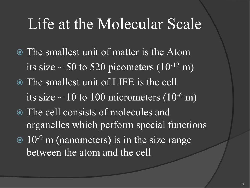
Life at the Molecular ScaleThe smallest unit of matter is the Atomits size ~ 50 to 520 picometers
(10-12
m)
The smallest unit of LIFE is the cellits size ~ 10 to 100 micrometers (10-6
m)
The cell consists of molecules and organelles which perform special functions 10-9 m (nanometers) is in the size range between the atom and the cell

4
Cell Video

5
Brief Overview of BiosensorsDefinition
“a device that utilises biological components e.g. enzymes to indicate the amount of a biomaterial”Eg: blood glucose monitor, pregnancy test

6
Brief History
Leland C. Clark
7
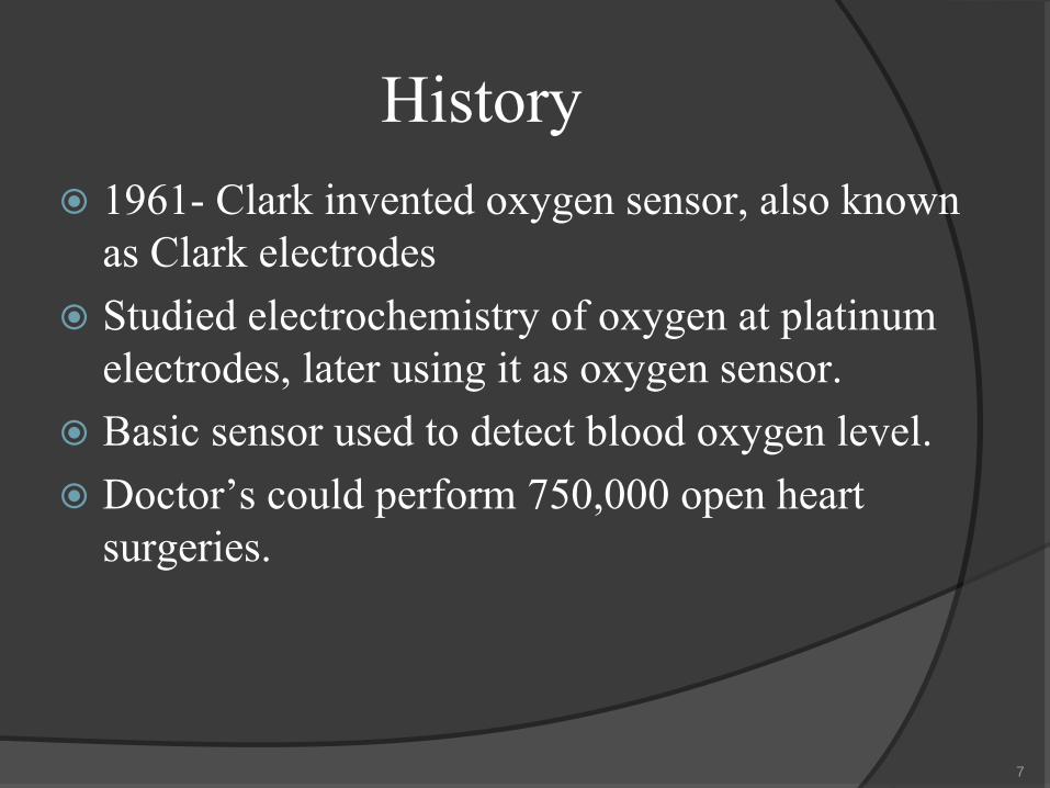
History1961- Clark invented oxygen sensor, also known as Clark electrodesStudied electrochemistry of oxygen at platinum electrodes, later using it as oxygen sensor.Basic sensor used to detect blood oxygen level.Doctor’s could perform 750,000 open heart surgeries.

8
Basic Biosensor
Bio-recognition element Transducer
Signal Output
Enzymes, antibodies, receptors, whole cells
Electrochemical, Optical

9
ApplicationsIndustry- Process monitor and control, food and drink in particularMedicine- Diagnostics, metabolism and hormonesMilitary- battlefield monitoring of poisonous gases, nerve agent and peopleDomestic- Home monitoring of non acute conditions

10
Clark Electrodes

11
Clark ElectrodesThe sample is brought into contact with a membrane (usually polypropylene or Teflon) through which oxygen diffuses into a measurement chamber containing potassium chloride solution. In the chamber are two electrodes; one is a reference silver/silver chloride electrode and the other is a platinum electrode coated with glass to expose only a tiny area of platinum (e.g. 20 mm diameter). The electric current flow between the two electrodes when polarized with a potential of -600 mV (vs. Ag/AgCl) determines the oxygen concentration in the solution

12
Blood glucose biosensor
•Two parallel plates•Small gap•Electrodes•Reagents (Glucose oxidase, Ferricyanide)
13
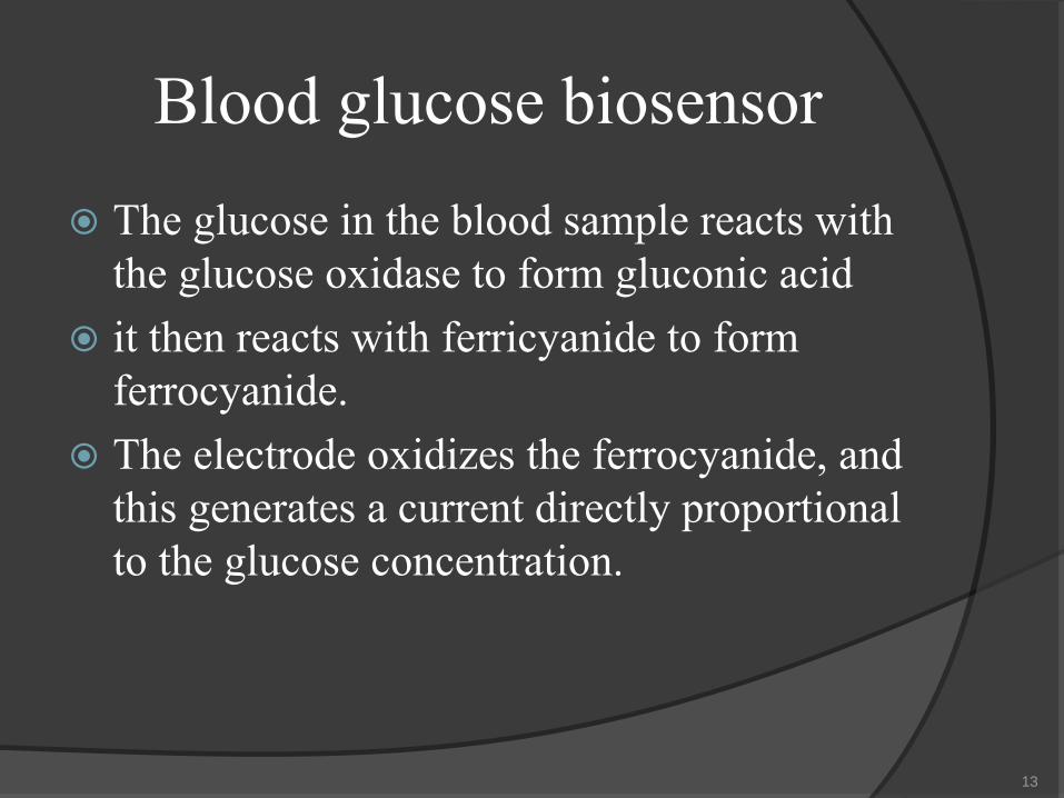
Blood glucose biosensorThe glucose in the blood sample reacts with the glucose oxidase to form gluconic acidit then reacts with ferricyanide to form ferrocyanide. The electrode oxidizes the ferrocyanide, and this generates a current directly proportional to the glucose concentration.

14
Introduction to Nanotechnology
Here we will be introducing the basics of nanotechnology and asking the following questions:
What nanotechnology really isThe emergence of nanotechnologyThe different types of nanotechnology and its usesThe use of nanotechnology in biosensors (as it pertains to Biomedical Engineering)

15
What Nanotechnology Really is
Nanotechnology is the term given to technology at the nanometer scale (Duh!)Unifying theme: control of matter from the atomic and molecular scales and fabrication of devices within this rangeDynamic field, which involves many different Engineering fields

16
What Nanotechnology Really is
Nano is such a buzz word, we need to actually quantify it, to give it significance 10-9 m is 1 millionth of a millimeter!
Imagine dividing 1 millimeter into 1 million partsAn typical bond length is on the order of 10-10 m only a 10-1 less than a nmStory time….☺ (short)

17
The Emergence of Nanotechnology
Physicists theorized concepts of nanotechnology, and how it may one day lead to changes of atomic/molecular featuresIn the early 1980’s is when nanotechnology really emerged Lead by two major developments: 1) Cluster Science 2) Scanning tunneling microscope

18
Use of Nanotechnology The further development of this hot topic has lead to current research in:Nanomaterials- The study of materials that have unique properties arising from their nanoscaledimensions
Bottom-Up Approaches- Arrangement of smaller components into complex assemblies example: DNA nanotechnology

19
Use of NanotechnologyTop-down Approaches- Create smaller devices using larger ones to examples: nanoelectronics
and
nanoelectromechanical
systems (NEMS)Functional Approaches- Develop components of desired functionality without regard to how they may be assembled examples: Molecular electronics

20
Examples of Nanotechnology in Biosensors
We will now explore 3 case examples of how Nanotechnology is used in the field of BiosensorsThe 3 examples are:Nanoelectromechanical Systems NEMSHYPER-CEST- Molecular imagingCancer Cell Nanotechnology
21
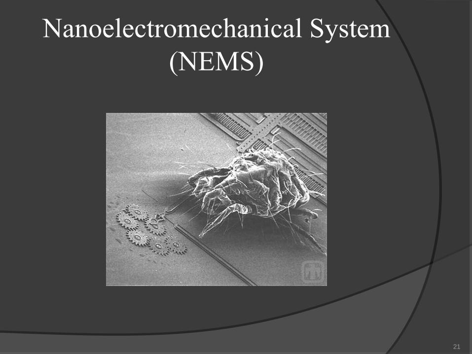
Nanoelectromechanical
System (NEMS)

22
Nanoelectromechanical
Systems (NEMS)
What is a NEMS?An NEMS can be thought of as a “mini-machine” at the nano-scale
How are they formed?Usually formed by the Top-down approach
Where are they usually used?In the field of signal processing VHF, UHF, and microwave band

23
NEMS Use in BiosensorsHow can this be used for Biosensors?
BioNEMS are NEMS systems that work in a biological environment to detect the forces of interaction between biological molecules

24
NEMS Use in BiosensorsAdvances in AFM (atomic force microscopy) and “optical tweezers” have helped in this fieldAFM has been very useful in measuring the extremely weak forcesOptical tweezers also can measure very weak forces, they use an optical beam which focuses on the diffraction limit

25
NEMS Use in BiosensorsAdvantages of using BioNEMS:1) Scaleable2) Can interact with a highly controlled, extremely
reduced population3) Strong sensitivity 4) Fast response times (μs-scale)
26
NEMS Use in BiosensorsThese advantages offer an alternative to fluorescent labeling and optical detection that are the basis of biochemical assays

27
HYPER-CEST- Molecular imaging
HYPER-CEST -for hyperpolarized xenon chemical exchange saturation transferdetection of signals from molecules present at 10,000 times lower concentrations than conventional MRIPines and Wemmer team at University of California

28
HYPER-CEST- How MRI works
property of atomic nuclei with an unpaired proton or neutron called “spin”
Such nuclei spin on an axis like miniature tops, giving rise to a magnetic moment, which means the nuclei act as if they were bar magnets with a north and south pole

29
HYPER-CESTWhen exposed to an external magnetic field, these spinning "bar magnets" attempt to align their axes along the lines of magnetic force
the alignment is not exact, the result is a wobbling rotation, or “precession,” that’s unique to each type of atom

30
HYPER-CESTwhile exposed to the magnetic field, the precessing nuclei are also hit with a radiofrequency (rf) pulse, they will absorb and re-emit energy at specific frequencies according to their rate of precession. When the rf pulse is combined with magnetic field gradients, a spatially encoded signal is produced that can be detected and translated into images.

31
HYPER-CESTObtaining a spatially encoded MRI signal from a sample depends upon the spins of its precessingnuclei being polarized so that an excess points in one direction, either “up” or “down.”Because the natural excess of up versus down spins for any typical population of atomic nuclei is only about one in 100,000, conventional MRI techniques are designed to detect nuclei that are highly abundant in tissue, usually the protons in water.

32
HYPER-CESTzapping rubidium vapor with a beam of polarized laser light creates a "hyperpolarized" effect that can be transferred to nuclei of xenon, an inert gas whose nuclei naturally feature a tiny degree of spin polarizationcalled “optical-pumping,” vastly increases the proportion of spin-up nuclei, producing a population of xenon atoms with nearly 50 percent of their nuclei in the up state

33
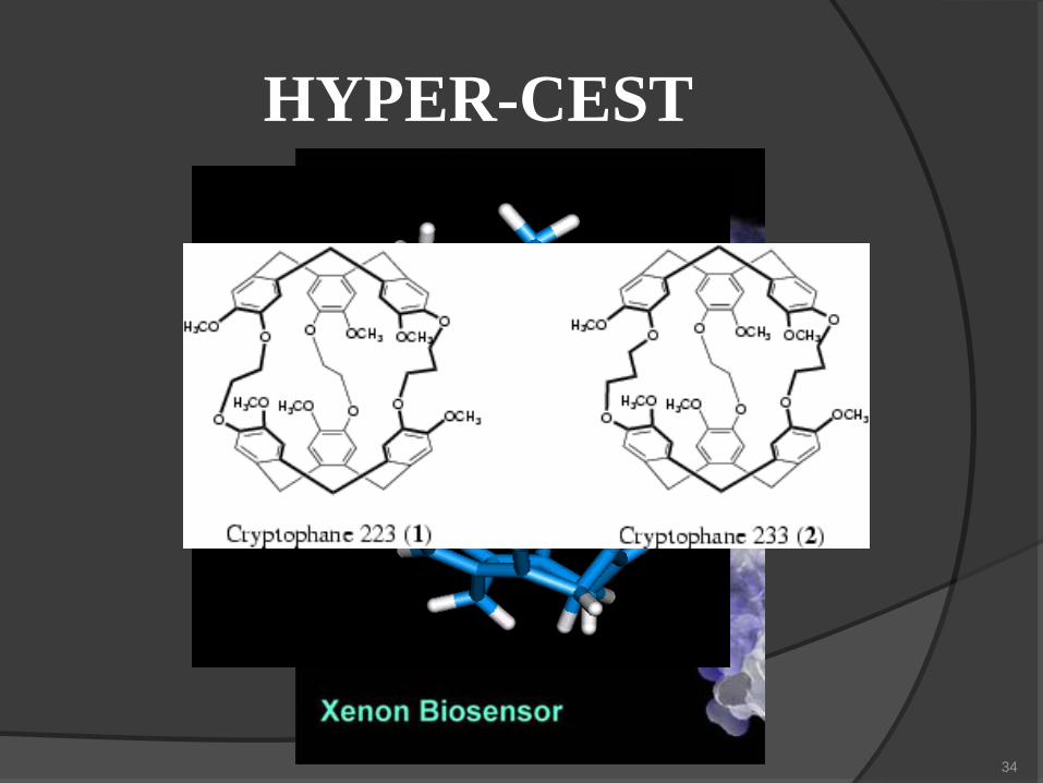
HYPER-CESTused a nanoscale molecular cage, called a cryptophane, that they adapted to hold hyperpolarized xenon atoms.
34
HYPER-CEST

35
HYPER-CESTAddition of a biochemical “linker” that makes the nanocage soluble in water, they created a agent that binds to a specific target molecule and associates the hyperpolarized xenon with it. Hyperpolarized xenon has a much longer relaxation time than protons, which means that the enhanced MRI signal is not only stronger, but lasts much longer.

36
Cancer Cell NanotechnologyWhat is Cancer cell nanotechnology?
Cancer cell nanotechnology are nanotech devices which are used in the diagnosis and treatment of cancer
Examples of Cancer cell nanotechnology are:injectable drug deliverynanosized magnetic resonance imaging (MRI) contrast agentsNanoparticle methods for detection of DNA and protein

37
Cancer Cell NanotechnologyNano-devices have revolutionized the field of cancer researchDevices such as nanovectors have allowed for targeted drug delivery of anti-cancer drugs to cancer cellsNanowires and nanocantilever arrays are among the approaches under development for the early detection of precancerous and malignant lesions from biological fluids
38
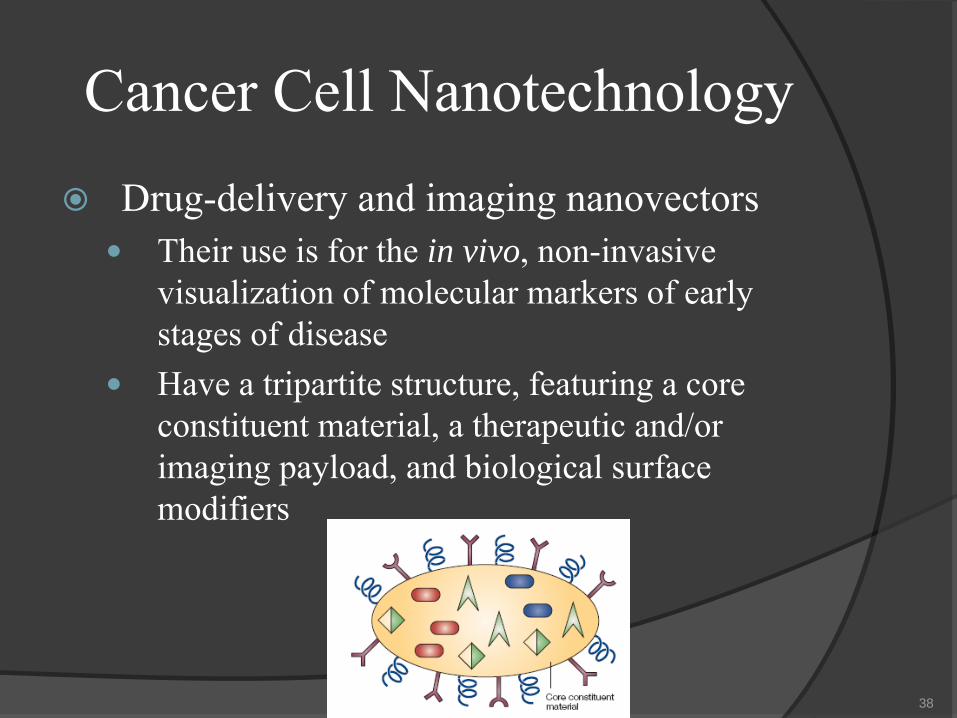
Cancer Cell NanotechnologyDrug-delivery and imaging nanovectors
Their use is for the in vivo, non-invasive visualization of molecular markers of early stages of diseaseHave a tripartite structure, featuring a core constituent material, a therapeutic and/or imaging payload, and biological surface modifiers

39
Cancer Cell NanotechnologyCancer nanotechnology: the challenges1) Developing approaches for the in vivo detection and monitoring of cancer markers2) Refining technology platforms for early detection of cancer biomarkers ex vivo3) Improving the targeting efficacy of therapeutic or imaging agents to cancer lesions and their microenvironment

40
Cancer Cell NanotechnologyCancer nanotechnology: the challenges (cont)4) Engineering nanoparticles
to avoid
biological and biophysical barriers

41
Questions/Answers?????????????????????????????

42
REFERENCEShttp://en.wikipedia.org/wiki/Nanosensorhttp://www.life.uiuc.edu/crofts/bioph354/lect13.htmlhttp://nano.cancer.gov/news_center/2007/oct/nanotech_news_2007-10-31a.asphttp://chem.ch.huji.ac.il/history/clark_leland.htmhttp://people.clarkson.edu/~ekatz/scientists/clark_leland.htmhttp://graingerchallenge.org/NAE/awardscom.nsf/weblinks/LRAO-69LMKN?OpenDocumenthttp://www.chem13news.uwaterloo.ca/issues/343/343_dec_2006_pages_5-6.pdfhttp://www.e-mri.org/nmr/net-magnetization.htmlhttp://www.e-mri.org/nmr/precession.htmlhttp://www.e-mri.org/nmr/nuclear-spin.htmlhttp://nano.cancer.gov/news_center/nanotech_news_2006-11-06e.asphttp://en.wikipedia.org/wiki/Ligand_(biochemistry)http://www.in-pharmatechnologist.com/news/ng.asp?n=58523-nanotechnology-to-revolutionisehttp://en.wikipedia.org/wiki/Biosensorhttp://www.lsbu.ac.uk/biology/enztech/biosensors.htmlhttp://media.wiley.com/product_data/excerpt/43/04718991/0471899143.pdfhttp://www.gwent.org/cd/Gwent%20Group%20Presentations/biointro.pdfhttp://www.ens-lyon.fr/CHIMIE/Fr/Groupes/page_groupe_supra/crypto_complex.htmlhttp://en.wikipedia.org/wiki/Nanotechnologyhttp://www.nano2life.com/download/nano_cancer_ferrari_natbiotec.pdfhttp://www.e-drexler.com/d/06/00/Nanosystems/toc.htmlhttp://en.wikipedia.org/wiki/Nanoelectromechanical_systems