nasa technical. note nasa tn d-7209 · nasa technical. note nasa tn d-7209 cas c microstructural...
TRANSCRIPT

NASA TECHNICAL. NOTE NASA TN D-7209
CASC
MICROSTRUCTURAL STUDY OF
THE NICKEL-BASE ALLOY WAZ-20
USING QUALITATIVE AND QUANTITATIVE
ELECTRON OPTICAL TECHNIQUES
by Stanley G, Young
Lewis Research Center
Cleveland, Ohio 44135
NATIONAL AERONAUTICS AND SPACE ADMINISTRATION • WASHINGTON, D. C. • MARCH 1973
https://ntrs.nasa.gov/search.jsp?R=19730009814 2018-06-30T18:29:59+00:00Z

1. Report No.
NASA TN D-7209
2. Government Accession No.
4. Title and Subtitle MICROSTRUCTURAL STUDY OF THE NICKEL-
BASE ALLOY WAZ-20 USING QUALITATIVE AND QUANTITA-
TIVE ELECTRON OPTICAL TECHNIQUES
7. Author(s)
Stanley G. Young
9. Performing Organization Name and AddressLewis Research Center
National Aeronautics and Space
Cleveland, Ohio 44135
Administration
12. Sponsoring Agency Name and AddressNational Aeronautics and Space Administration
Washington, D. C. 20546
3. Recipient's Catalog No.
5, Report DateMarch 1973
6. Performing Organization Code
8. Performing Organization Report No.E-7250
10. Work Unit No.
501-011 1 . Contract or Grant No.
13. Type of Report and Period Covered
Technical Note14. Sponsoring Agency Code >
15. Supplementary Notes
16. Abstract
The NASA nickel-base alloy WAZ-20 was analyzed by advanced metallographic techniques toqualitatively and quantitatively characterize its phases and stability. The as-cast alloy con-tained primary gamma-prime, a coarse gamma-gamma prime eutectic, a gamma-fine gammaprime matrix, and MC carbides. A specimen aged at 870° C for 1000 hours containedthese same constituents and a few widely scattered high W particles. No detrimental phases(such as sigma or mu) were observed. Scanning electron microscope, light metallography,and replica electron microscope methods are compared. The value of quantitative electronmicroprobe techniques such as spot and area analysis is demonstrated.
17. Key Words (Suggested by Author(s))
Microstructure; Alloy phases; Nickel-basealloys; WAZ-20; Analytical techniques;Characterization; Scanning electron micro-scope; Electron microprobe; X-ray diffrac-tion analysis; Metallography; Stability
19. Security Classif. (of this report)
Unclassified
18. Distribution Statement
Unclassified - unlimited
20. Security Classif. (of this page)
Unclassified21. No. of Pages
28
22. Price*
$3.00
*For sale by the National Technical Information Service, Springfield, Virginia 22151

MICROSTRUCTURAL STUDY OF THE NICKEL-BASE ALLOY
WAZ-20 USING QUALITATIVE AND QUANTITATIVE
ELECTRON OPTICAL TECHNIQUES
by Stanley C. Young
Lewis Research Center
SUMMARY
The constituents of the NASA nickel-base alloy, WAZ-20, were studied bylight metallography, scanning electron microscopy (SEM), and electron microprobe •analysis (EMXA) . The alloy was analyzed both in the as-cast condition and afteraging for 1000 hours at 870° C to determine its stability. The composition of theconstituents of the alloy were analyzed quantitatively using computer methods tocorrect the raw EMXA output for background, absorption, fluorescence, etc.
The as-cast alloy contained primary gamma prime, a gamma-fine gammaprime matrix, a coarse gamma-gamma prime constituent believed to be a eutecticand metal carbides (MC). The aged specimen contained all the constituents foundin the as-cast specimen. Also a few high W particles were observed that appearedto have a M_C carbide chemistry. All the constituents except the carbides showedapproximately the same chemistry after aging as observed in the as-cast condition.No detrimental phases were observed that could be expected to weaken the alloyor indicate lack of stability at the time and temperature exposures of this study.
Two observations were made regarding metallographic techniques. First,SEM photomicrographs at X1700, when compared with light photomicrographs atthe same magnification, showed adequate resolution, so that features were visiblethat would normally have to be observed by the use of more expensive replicaelectron microscope techniques at equivalent magnifications. Second, an EMXAanalysis of a single phase (gamma prime) obtained with a broad area scan yieldedan analysis nearly identical to a point analysis of that phase. This suggests that, ifthe composition and area or volume percent of one phase of a two-phase region isknown, the composition of the other phase can be calculated from an EMXA broadarea analysis, even though it may be too small for direct EMXA quantitative analysis.

INTRODUCTION
The lack of adequate materials for advanced turbine engines has limited oper-ating temperatures and engine efficiency. The high-temperature limits are beingextended, however, with the development of improved nickel (Ni) and cobalt (Co)based superalloys, and by the use of new processing techniques. The NASA Ni-base alloy, WAZ-20, was developed to permit higher engine temperatures and hasbeen cast with both random and directional grain orientations (ref. 1). Both formsof WAZ-20 showed substantially higher tensile strength at 1205° C than most otherknown cast Ni-base alloys. Certain strength properties, such as intermediate-temperature tensile strength, ductility, and stress-rupture life, of the directionalpolycrystalline form exceeded those of random WAZ-20. The 100-hour use temper-atures at 103.4 meganewtons per square meter (15 000 psi) are 1063 C for thedirectional polycrystalline material, and 1038 C for the random polycrystallinematerial. In addition, WAZ-20 has excellent room-temperature Charpy impactstrengths both as-cast and after aging.
The WAZ-20 alloy also was shown to possess limited workability potential. A0.33-centimeter chill cast slab of the random crystalline form was hot rolled to athickness of 0.05 centimeter in a conventional rolling mill.
WAZ-20 has a melting point higher than most presently known Ni-base systemsbecause of its high tungsten (W) content and because it contains relatively fewalloying constituents as contrasted to most other nickel base alloys. Other alloyingconstituents include aluminum (Al) , carbon (C) and zirconium (Zr) in the mostfavorable combination consistent with maintaining a high melting point and achievingsound castings with relatively little segregation (ref. 1). The nominal chemistryrange and chemical analyses are presented in table I.
Previous metallographic studies on this alloy were limited primarily to lightmicroscopy metallographic evaluation of the structure. The object of this work was,therefore, to more completely characterize the constituent phases, by the applica-tion of electron microscopy and electron microprobe analysis techniques, and at thesame time to demonstrate and expand the capabilities of the advanced metallographictechniques used. Electron optical techniques have recently been applied to thestudy of many Ni and Co alloys and alloy-coating systems (refs. 2 to 6). Thedirectionally solidified form of the WAZ-20 alloy was used for this study in both the

as-cast condition and after aging for 1000 hours at 870 C to determine its micro-structural and chemical stability.
The alloy was examined by means of the scanning electron microscope (SEM);the resulting photomicrographs were compared with both light photomicrographsand replica electron photomicrographs. The electron microprobe X-ray analyzer(EMXA) was used to show the chemical composition of the phases present on aqualitative basis by exhibiting X-ray raster scans of the elements present and on aquantitative basis by analyzing each individual phase. Recently developed computercorrection methods were applied to yield atom percent and weight percent compo-sitions from the raw EMXA data. The structures of extracted carbide phases were ,identified by X-ray diffraction analysis. The results of the studies are presentedwith comparisons between the as-cast and aged materials.
MATERIALS AND TECHNIQUES
Materials
The chemical composition of WAZ-20 is given in table I (ref. 1). The nominalcomposition is included with chemical analyses made by an independent laboratoryof typical heats of directionally solidified polycrystalline WAZ-20. Argon inductionmelted ingots were remelted and again cast into ingots under vacuum. They werethen remelted under vacuum and cast in the directional solidification apparatusdescribed in reference 7. The apparatus consisted of a three-zone resistance moldheater that surrounded a refractory shell mold. A copper chill block extended intothe lower portion of the shell mold to establish grain growth directions. Ruptureand tensile bars were cast with a gage length of 3.05 centimeters and a gage diameterof 6.35 millimeters. The tungsten and aluminum are near the low sides of thespecified nominal values because of small losses during each of the three melting "processes. Zirconium increased to the high side of the specified nominal valuesbecause this element and other trace elements were picked up from crucibles duringinduction melting. The same WAZ-20 material was used for studies of the effectsof aging at 870° C (1600° F) for 1000 hours.
Metal lographic Preparation
Specimens used for the metallographic studies were cut perpendicular to the

growth direction. Previous work (ref. 1) , showed that sections cut both paralleland perpendicular to the growth direction showed the same general features. Spec-imens were etched in a solution that was 92 parts hydrochloric acid, three parts,nitric acid, and five parts sulfuric acid. Photomicrographs were made by lightmicroscopy, SEM (secondary and backscatter modes) , and EMXA (backscatter andX-ray modes). The specimens were then repolished to prepare them for the quanti-tative EMXA studies. Specimens were often repolished to remove carbonaceouscontamination that was deposited by the EMXA.
Scanning Electron Microscopy
Scanning electron microscope (SEM) microphotographs in backscatter-electron•and secondary-electron modes and light photomicrographs were made at the samemagnifications of the same sample areas. Secondary electrons are emitted fromnear the surface and result in a photomicrograph revealing predominantly surfacetopography. The backscatter electrons are released from deeper within the speci-men and result in a photomicrograph that is semiqualitative: the heavier elementsemit more electrons than the lighter elements and thus phases containing the highatomic number elements appear lighter than the phases containing lower atomicnumber elements. Higher magnification SEM photomicrographs were compared withreplica electron photomicrographs from the transmission electron microscope (TEM)to determine similarities. The three techniques for examining structures at highmagnification differ in several ways; however, only their approximate resolutionlimitations are indicated here. For light microscopy, this may be considered to be0.5 micrometer, for the SEM 0.03 micrometer, and for the replica TEM 0.005V
micrometer.
«Electron Microprobe X-ray Analysis
The electron microprobe X-ray analyzer (EMXA) was used to provide back-scatter photomicrographs and X-ray raster photomicrographs of each element in thespecimens. The X-ray raster photomicrographs are rectangular (TV) rasterpatterns, in which counts of the desired X-rays were accumulated. This techniqueprovides an average analysis over the area (raster) swept by the electron beam. Inthe EMXA spot analysis, the electron beam is narrowed as finely as possible,

usually to a spot approximately 1 to 2 micrometers in diameter. The EMXA was alsoused for quantitative determination of the composition of each phase or constituentobserved in the specimens.
To obtain a quantitative analysis of the phases, a Colby microprobe analysisgeneral intensity correction (MAGIC) computer program (ref. 8) was used to correctthe raw EMXA data. The program was modified for use with a time sharing computersystem, which allowed input from a remote typewriter terminal. Briefly, input tothe program consisted of the chemical abbreviations of all elements in the specimen,characteristic X-ray lines analyzed, background and X-ray counts for high-puritystandards of each element, and background and X-ray counts for each element injeach microconstituent analyzed.
In this program the X-ray counts of each unknown in the specimen were comparedwith X-ray counts of high purity standards of each element (X-ray intensity ratio) .Input data were corrected for background, counter dead time, absorption, fluo-rescence , backscatter, and ionization penetration. The MAGIC program oftenallowed overcorrection of the amount of absorption of X-rays from lighter elementssuch as carbon. As a result these lighter elements would be multiplied by a factorin the original MAGIC program that would increase them by unreasonably largeamounts. Therefore, we established an empirical factor that would limit the amountof correction for absorption using analyses of known carbides and a well-characterized nickel aluminide (NiAl) reference material as standards. This factornever allowed the theoretical fraction of radiation emerging from the specimen to bereduced by more than 46 percent for any element during the iterative process of thecomputer program. Carbon was the only element influenced to any extent by thisempirical factor.
The estimated error for our analyses using the modified MAGIC program variedinversely with the atomic number and amount of element present. For example, Niwas considered to be accurate within about 2 percent of the amount present, but theanalyses of the lighter elements (C, Al, Si) are not considered to be more accuratethan 10 percent of the amounts present. Detailed descriptions of electron beammicroanalysis and correction procedures can be found in references 8 and 9.
Electrolytic Extraction
The extraction procedure used for the carbides was essentially the same as that
5

suggested by the ASTM Committee E-4 and that used in references 10 and 11. Theapparatus used for the extraction consisted of a direct-current power supply, whichproduced a current of approximately 200 milliamps between electrodes in a 10 per-cent solution of hydrochloric acid in methanol. A 1- by 1- by 0.25-centimeter speci-men attached to a platinum wire was used as the anode, and a cylindrical platinumscreen served as the cathode. The solution was contained in a 250-milliliter glassbeaker. The total carbide phases that were separated were weighed and submittedfor X-ray analysis. Procedures were essentially the same for both the as-cast andthe aged materials.
f
RESULTS AND DISCUSSION
Microstructure of As-Cast Directionally Solidified WAZ-20
Metallography. - Light photomicrographs of the as-cast WAZ-20 specimens areshown in figure 1, at magnifications of X100, X250, and X500 and in figure 2(a) atX1700. These different magnifications were included to show relationships withelectron optical photomicrographs at the same magnifications shown later inthis section. In figure l(a) at X100, microhardness indentations can be seen. Thesewere used as locators for the specific areas to be examined by the electron opticalmethods. General features of the microstructure include: a gamma matrix containinga gamma prime precipitate and large primary gamma prime nodules with smallangular carbides scattered throughout (ref. 1). The gamma-gamma prime structureis present in two forms: fine structured regions (gamma-fine gamma prime matrix) ,and coarser eutectic regions associated with or near the boundaries of the largeprimary gamma prime particles. Carbides also seem to be generally associated withthe eutectic regions rather than occurring isolated within gamma-fine gamma primematrix regions.
The X500 photomicrograph of figure l(c) shows a single primary gamma primenodule, the surrounding eutectic region, and several carbide particles. All ofthese are surrounded by the gamma-fine gamma prime matrix structure. This areaof the sample, at this magnification, will be compared with backscatter-electron andX-ray raster photomicrographs taken with the EMXA (shown in the next section) .
Figure 2 shows X1700 photomicrographs of part of the same area as shown infigure 1. Figure 2 (a) was taken with an oil immersion objective lens at X1700 on a

light microscope, and figure 2(b) was taken on the SEM in the secondary-electronmode. Figure 2(b) shows that photomicrographs may be made with the SEM withmuch greater resolution than is possible with light optics because of the inherentlyhigher resolution and the much greater depth of field of the SEM. The same metal-lographic mount was used, and there were no differences in surface preparationbetween these two photomicrographs.
The SEM photomicrograph was taken with the specimen at a 20° tilt, allowinghighlighting of raised areas. The carbide particle in the upper left of the picture infigure 2(b) is raised above the surface. The gamma phase is also raised above thegamma prime phase because the gamma prime phase was more susceptible to attackby the etchant. The bright area in the lower part of the photomicrograph (fig. 2(b))is a small pit that is clearly evident in the SEM photomicrograph but is unresolvedin the corresponding light photomicrograph. The bright contrast is due to excessiveelectron scattering in this rough area, which contains thin discontinuous features.The increased resolution and depth of field in the SEM are responsible for the de-tails revealed inside of the pit. Also note the clear resolution of the gamma-finegamma prime region.
The secondary-electron mode on the SEM emphasizes surface topography. TheSEM can also be operated in the backscatter electron (BSE) mode. In this mode theheavier elements emit more electrons than lighter elements and phases containingheavy elements show up lighter in the photomicrographs than those containing thelighter elements; thus (BSE) photomicrographs allow a semiqualitative first lookat the specimen chemistry.
Figure 3 shows the same area as displayed in figure 2, taken in the BSE modeat a 20 tilt. Detail in the "holes" is not as clearly defined by this technique becauseof its strong oblique illumination effect; thus the pit shows up dark. The carbideshows up very light and must therefore contain a higher percentage of heavy ele-ments than any other phase present. The gamma phase is lighter than the gammaprime. Although this could be partly a topographic effect (the specimen wasetched) , it is believed that the gamma contains a greater percentage of heavierelements than the gamma prime. A light band appeared across the gamma primein the center of this photomicrograph. It may be a submerged phase, slightly belowthe sample surface, of higher average atomic number than the gamma prime(possibly gamma).

Figure 4 shows X5000 SEM photomicrographs of the same area of the specimen.Figure 4(a) shows a carbide, and 40)) shows the interface between the coarsegamma-gamma prime eutectic and the fine structure. These photomicrographs canbe compared with figure 5, which is a X6000 TEM photomicrograph of a replica froman area similar to that shown in figures 1 to 4. At this magnification very littleadditional information was obtained by the considerably more expensive method ofreplica electron microscopy than was obtained from the SEM photomicrograph. How-ever, the value of the replica technique is apparent in figure 6, which shows areplica TEM photomicrograph at X17000 of the gamma-fine gamma prime matrix. Aneven finer precipitate (presumably also gamma prime) was observed in the gammaphase.
Electron microprobe X-ray analysis. - Qualitative and quantitative EMXAstudies were made on the specimens. The qualitative studies consisted of spectralscans to identify the elements present and of BSE and X-ray raster photomicrographsof the areas previously studied by light microscopy to show the relative concentra-tions of the elements in the phases. Quantitative studies consisted of both spotanalysis and raster area analysis of constituents of the specimen and correction ofthe intensity ratio results by means of the modified Colby computer program describedpreviously.
Figure 7 shows a backscatter photomicrograph and five X-ray raster photo-micrographs at X500 of the specimen area shown in the light photomicrograph offigure l(c). X-ray raster photomicrographs for C, W, Zr, Ni, and Al are shownfor this area in figure 7. (Any lighter area in the BSE photomicrograph indicateshigher concentration of heavy elements.) In X-ray raster photomicrographs for aparticular element, lighter areas indicate locally higher concentrations of that ele-ment . From the X-ray raster photomicrographs, carbides were noted to be high inW and Zr, and noticably low in Ni and Al. Carbon differences did not show upclearly in the X-ray photomicrographs, but did show up in the more sensitivequantitative results reported later. Carbon is difficult to separate from the back-ground because it has a very low, broad peak (X-ray intensity against wavelength),and also because carbonaceous contamination deposits form where the beam impingeson the specimen. These deposits can increase the carbon X-ray count rate and canmask microstructural differences.
The X-ray raster photomicrographs did not clearly indicate differences between
8

gamma and gamma prime in individual element concentration, but the BSE photo-micrograph (fig. 7(a)) showed the primary gamma prime to be slightly lower inheavy elements than the gamma phase. This was further substantiated by thequantitative measurements. A light-to-dark change was seen through the gammaprime region of the X500 photomicrograph of figure 7(a) (indicated) , which seemedto separate the primary gamma prime from the gamma prime associated with theeutectic. This was noted in about the same location of the possible submergedgamma phase (indicated by the band) mentioned earlier (see fig. 3 at X1700) .
Quantitative studies were made to determine element concentrations in eachphase or region of the alloy. The raw EMXA results were computer corrected forthe various effects mentioned previously.
Figure 8 shows light photomicrographs of the as-cast specimen. The areasexamined by the microprobe are indicated in the etched specimen (fig. 8(a)) . Car-bonaceous contamination deposits delineating the regions scanned in EMXA studycan be seen on the unetched polished specimen in figure 8(b) .
Table II shows the analysis of the constituents of the as-cast WAZ-20 shown infigure 8. Table II (a) shows the analysis in terms of weight percent and table II (b)in terms of atomic percent. The final results shown in both tables were normalizedto 100 percent in an attempt to make up for small errors in the original analysis andcorrection procedures. Also, percentages were rounded to the nearest 0.1 percent.In all cases for the as-cast WAZ-20, the total element content analyzed before nor-malizing was approximately 93 to 95 percent except for the carbide which resultedin about 85 percent total element content. Three general reasons may contribute tothis incomplete accountability of all the elements present. First, a limiting factor(as previously noted) was intentionally placed on the absorption correction in thecomputer program to prevent it from overcorrecting small amounts of light elementsby unreasonably large numbers. Second, with the instrument used, only threeelements could be analyzed at a time, and the operating conditions between groupsof three can be slightly different because of instrumental drift or other potentialvariables in the raw X-ray count analysis by the EMXA. Finally, if long timeanalyses are made, carbonaceous contamination builds up on the surface during theanalysis. This causes an overall reduction in X-ray count rates from the specimen(for all elements except carbon) that is very difficult to correct. The carbon percentwill increase and the other element percents will decrease by this process. Examples

of the carbonaceous contamination can be seen in the polished, unetched specimenof figure 8(b) .
From table II several observations can be made. The carbide was observed tobe a very high Zr, high W, carbide. X-ray diffraction of the extracted carbides in-dicated that they were a MC type.
Analysis of the gamma prime was done on both a spot (2- to 3-fim diam) and onan area basis (30- by 26-^m area) , with all the EMXA conditions held constant. Inboth cases the analysis was essentially the same. This suggests that a two-phase region could be area scanned by EMXA to obtain its average analysis.Then, if the composition of one phase and the volume percent of the phases presentwere known or estimated by other methods, the composition of the unknown phasecould be calculated, even though it might be too small for direct EMXA quantitativeanalysis. The known phase must, of course, be free from major chemical gradientsor changes throughout the area analyzed.
The gamma-fine gamma prime matrix was analyzed by area scanning and foundto be lower in Al and Zr, about the same in Ni and Si, and higher in W than thegamma prime. Carbon was noticeably higher in the gamma prime than in the gammaplus gamma prime regions but this was believed to be due to carbonaceous con-tamination buildup during analysis or to carbides submerged beneath the gamma-prime area being scanned. The eutectic region had an overall chemistry betweenthe primary gamma prime and the gamma-fine gamma prime matrix.
Microstructure of Aged Directional ly Solidified WAZ-20
Metallography. - Light photomicrographs of the aged directionally solidifiedWAZ-20 specimens are shown in figure 9 at X100, X250, and X500. This specimenwas aged for 1000 hours at 870° C. In figure 9 (a) at X100, a microhardness in-dentation can be seen, which, as in the case of the as-cast specimen, was used as alocator for the specific area to be examined by electron optical methods. At X250the general features were similar to those of the as-cast specimen. No embrittlingphases such as sigma or mu were observed after aging. Coarsening of the primarygamma prime was evident, and the regions adjacent to the primary gamma prime inthe gamma-fine gamma prime matrix appear to have become somewhat enriched ingamma prime. Less of the eutectic was observed after aging. A new type of angularparticle was observed in addition to the MC carbides. One large example of this new
10

type of particle can be seen in the lower left of the photomicrograph (fig. 903)) •The smaller MC carbides are scattered around the edges of the gamma prime phase.
Figure 9(c) at X500 shows the large angular particle imbedded in a primarygamma prime nodule. Smaller carbides can be seen at the edge of the primarygamma prime particle, and are often associated with voids or etching pits. Thesurrounding gamma-fine gamma prime matrix comprises the remaining area shownin this photomicrograph. This region at this magnification was used for laterelectron optical studies of each constituent indicated.
Electron microprobe X-ray analysis. - The aged specimen was analyzed inmuch the same manner as described previously for the as-cast specimen. Qualita-tive and quantitative studies were made on the specimen. Qualitative analysisconsisted of spectral scans to identify elements present. Quantitative studies con- -sisted of both spot and area analysis of regions within the specimen and computercorrection of the X-ray intensity ratio results.
Table III shows the results of the quantitative analysis of the aged WAZ-20 basedon computer-corrected and normalized EMXA data. Table III (a) shows the resultsin terms of weight percent, and table HI(b) in terms of atomic percent. As in thecase for the as-cast WAZ-20, the total element contents of each constituent in theaged specimen, except for the carbide, were greater than 92 percent before nor-malizing to 100 percent. The results for the carbide in this case are consideredquestionable because only 66 percent of the element content was accounted for. Thespectral scans did not indicate any other elements present. Therefore, it must beassumed that the most likely cause for this discrepency is the presence of voidsassociated with the small carbide particles. The EMXA analysis depends on depthmuch more than on area. If only a thin layer of carbide were present here, otherphases and/or subsurface voids could interfere with the analysis. Many otherpossibilities could contribute to this error such as slight misalinement of the spec-trometers, and changing EMXA operating conditions. However, as previouslymentioned, all other constituents of the as-cast and aged specimens were in goodagreement with regard to the total element content.
From table III the following observations were made: The analysis for primarygamma prime and the gamma-fine gamma prime matrix were strikingly similiar inthe as-cast and aged specimens, except that the gamma-fine gamma prime matrixadjacent to the primary gamma prime has become slightly (less than 4 wt.%) enriched
11

in W. The eutectic was not analyzed in the aged specimen for two reasons : First,the eutectic regions were smaller, and an area scan analysis was not able to take inas representative an area as in the case of the as-cast specimen. Second, thesimilarity of both analyses of the primary gamma prime and the gamma-fine gammaprime matrix in the aged and as-cast specimens led to the further assumption thatthe eutectic would not change significantly (the eutectic had a chemistry generallybetween the above two mentioned constituents in the as-cast specimen). The largeangular particle, found in the aged specimen, contained nearly 75 percent W andapproximately 22 percent Ni. This particle appeared to be a MgC precipitate on astochiometric basis, but X-ray diffraction of the extracted carbides indicated onlyMC carbides, as in the case of the as-cast specimen. Perhaps there was too littleof this type of carbide, or perhaps it is not a carbide but rather an intermetallicphase that was dissolved in the extraction process. In any case, no phases such assigma or mu were observed that would be expected to weaken the alloy or indicatea detrimental lack of stability after the 870° C, 1000-hour exposure.
SUMMARY OF RESULTS
The constituent phases of the NASA Ni-base alloy, WAZ-20, have been analyzedby light metallography, scanning electron microscopy, and electron microprobeX-ray analysis (EMXA). The alloy was analyzed in both the as-cast condition andafter aging for 1000 hours at 870 C. The following results were obtained:
1. The as-cast alloy consisted of primary gamma prime; a gamma-fine gammaprime matrix, which was higher in W, lower in Zr and aluminum (Al) , and about thesame in nickel (Ni) and silicon (Si) as the primary gamma prime; coarse gamma-gamma prime eutectic with a chemistry generally between that of the primary gammaprime and the gamma-fine gamma prime matrix; and metal carbides (MC) , high inzirconium (Zr) and tungsten (W).
2. The aged specimen also contained the same phases as the as-cast specimen.The gamma prime and the gamma-fine gamma prime matrix showed approximatelythe same chemistry after aging as observed in the as-cast condition. Less of theeutectic phase was observed after aging. In addition to the MC carbide particles,a few angular particles of very high W content were noted. These appeared to be
a MgC carbide, but, most likely because of their scarcity, they were not observedby X-ray diffraction in the extraction studies.
12

3. No detrimental phases such as sigma or mu, which would be expected toweaken the alloy or indicate lack of stability after extended high-temperature ex-posure , were observed with the time and exposure temperature of this study.
4. Scanning electron photomicrographs at X1700 were compared with lightphotomicrographs. The resolution by the SEM was adequate so that features werevisible that would normally have to be observed by use of the more expensivereplica electron microscope techniques at equivalent or somewhat higher magnifi-cations .
5. An analysis of an area 30 by 26 micrometers obtained by raster scanning ofa single phase (gamma prime) was found to yield a nearly identical analysis to asingle point analysis of that phase (all other EMXA conditions held constant) .
CONCLUDING REMARKS
WAZ-20 can generally be classified as a gamma prime strengthened alloy withsolid solution of W in the matrix for additional strengthening. The constituents ofas-cast WAZ-20 were: primary gamma prime, a coarse gamma-gamma prime (be-lieved to be a eutectic) , a gamma-fine gamma prime matrix, and scattered MCcarbides.
Although aging the alloy for 1000 hours at 870° C did not cause the precipitationof any phases such as sigma or mu that are generally detrimental to alloy strength,the analysis did indicate that aging may result in the formation of a small amount ofM f iC. Further aging studies at other temperatures, under stress and for longertimes, should be undertaken to completely verify its stability.
Part of the emphasis of this work was to expand and demonstrate the capabilitiesof the advanced metallographic techniques used. The results of scanning electronmicroscopy (SEM) were compared directly with light microscopy at X1700 andreplica transmission electron microscopy (TEM) at higher magnifications. Theresolution of the SEM photomicrographs was shown to be adequate so that featureswere visible that would normally have to be observed by the use of more expensivereplica TEM techniques.
The results of the electron microprobe X-ray analyzer were computer correctedby a Colby MAGIC program with additional empirical corrections. Other MAGICprogram versions have since been written by Colby. Several other programs havealso been written by other investigators. Each will give slightly different quanti-
13

tative results, which are dependent on the various equations, assumptions, andconstants used. No standard computer correction program exists as yet, and theoryis currently being updated and improved in attempts to make the EMXA results moreexact.
A method has been proposed herein that appears to make possible the quantita-tive analysis of phases too small to be analyzed by EMXA. An X-ray analysis usinga broad area scan of a single phase (gamma prime) yielded a nearly identicalanalysis as a single point analysis of that phase (all other EMXA conditions heldconstant). The significance of this result is as follows: When a two-phase regionis analyzed by EMXA using area (raster) electron beam scanning and when thecomposition and area or volume percent of one phase is known or estimated, thecomposition of the other phase can be calculated, even though it may be too smallfor direct EMXA quantitative analysis. Of course, the known phase must havenearly homogeneous composition, and the unknown region should be free from gen-eral concentration gradients. This method might also have application to analysismethods with wider beam spots, such as the ion beam mass spectrometer, or the ionscattering spectrometer.
Lewis Research Center,National Aeronautics and Space Administration,
Cleveland, Ohio, December 8, 1972,501-01.
REFERENCES
1. Waters, William J.; and Freche, John C.: A Nickel Base Alloy, WAZ-20, with
Improved Strength in the 2000° to 2200° F Range. NASA TN D-5352, 1969.
2. Calhoun, C. D.: Development of Superalloys by Powder Metallurgy for use at
1000-1400 F . Rep. 71AEG248, General Electric Co. (NASA CR-72968) , Nov.
1971.
3. Kent, William B.: Development Study of Compositions for Advanced Wrought
Nickel-Base Superalloys. Rep. U-C-R-1055, Cyclops Corp. (NASA CR-
120934), Jan. 1972.
4. Caves, Robert M.; and Grisaffe, Salvatore J.: Electron and Ion Microprobes
Applied to Characterize and Aluminide Coating on IN-100. NASA TN D-6317,
1971.
14

5. Johnston, James R.; and Young, Stanley G.: Oxidation and Thermal Fatigue ofCoated and Uncoated NX-188 Nickel-Base Alloy in a High-Velocity Gas Stream.
NASA TN D-6795, 1972.6. Radavich, John F.; and Gouts, Wilford H., Jr.: Metallography of the Super-
Alloys. Rev. High-Temp. Mat., vol. 1, no. 1, Aug. 1971, pp. 55-96.7. Freche, John C.; Waters, William J.; and Ashbrook, Richard L.: Application of
Directional Solidification to a NASA Nickel-Base Alloy (TAZ-8B) . NASA TND-4390, 1968.
8. Colby, J. W.: Quantitative Microprobe Analysis of Thin Insulating Films.Advances in X-Ray Analysis. Vol. 11. Plenum Press, 1968, pp. 287-303.
9. Beaman, D. R.; and Isasi, J. A.: Electron Beam Microanalysis. Part 1: The
Fundamentals and Applications. Mat. Res. Standards, vol. 11, no. 11, Nov.1971, p. 8; Part 2: Experimental "Considerations and Qualitative Analysis.Mat. Res. Standards, vol. 11, no. 12, Dec. 1971. pp. 12-31, 51-56; alsoElectron Beam Microanalysis. Spec. Tech. Publ. No. 506, ASTM, 1972.
10. Kriege, Owen H.; and Sullivan, C. P.: The Separation of Gamma Prime fromUdimet 700. Trans. ASM, vol. 61, no. 2, June 1968, pp. 278-282.
11. Kriege, Owen H.; and Baris, J. M.: The Chemical Partitioning of Elementsin Gamma Prime Separated from Precipitation-Hardened, High-TemperatureNickel-Base Alloys. Trans. ASM, vol. 62, no. 1, Mar. 1969, pp. 195-200.
15

TABLE I. - ALLOY COMPOSITION*1
Alloy
WAZ-20 (nominal range)
Typical heats ofdirectionalpolycrystallineWAZ-20b
Composition, wt. %
W
17 - 20
16.9717.1117.90
Al
6 - 7 . 0
5.935.955.85
Zr
1.4 - 1.6
1.721.521.70
C
0.10 - 0.20
0.113.113.113
Ni
Bal
75.0675.0874.21
Si
0.10.10.11
B
0.0003.0003.0003
*A11 data from ref. 1.Argon melted, followed by double vacuum remelt.
TABLE H. - ANALYSIS OF THE CONSTITUENTS OF AS CAST WAZ -20"
(a) Weight percent (b) Atomic percent
Element
CAlSiNiZrW
Composition, wt.%
Carbide
5.6.2
<. 15.2
55.034.0
Primarygamma prime
Spot
0.7b
7.3.1
81.5.8
9.9
Area
1.3b
7.1<. 1
80.7.9
10.0
Gamma plus gammaprime
Eutectic
<. 16.2.1
81.5.9
11,3
Matrix
0.15.5<. 1
80.9.2
13.3
Element
CAlSiNiZrW
Composition, at. %
Carbide
34.4.6
<. 16.5
44.713.7
Primarygamma prime
Spot
3.1b
14.7.2
78.4.5
3.0
Area
6.1b
14.4.2
75.9.5
3.0
Gamma plus gammaprime
Eutectic
<. 113.6
.281.9
.63.6
Matrix
0.412.2
.182.9
.24.3
Based on computer corrected and normalized EMXA data.This value may be high due to possible submerged carbides or carbon buildup during analysis.
16

rtoCM
ISI
o
o
H
|
8Was
o23
S
5w
s£3Co
•4-*
aB8
cu
cuw
*£jj
.2inoo.EoO
o>E
* -3•6 r.K'" rta
aSg xttfl £
3 p"3. cu
E "CE a
a0
aE
ac g
t< °Crt a.
cu
1
h30. OJ
^ rt— -M
bo
as
rtE
« 1o| aiC4 E£ 's-5ont£
& %>. .5LI •-,CS O.
Ei_
ACU
s
6
in o> •* m t- oCO CO Oi CMi— t
CM CO CM t> CM OJ
CO O O •*i-C 00
CM CO CO CO CD OJ
^ ^ t; CM
C- •*»• co co t- t-
OO CD CD t—CO ^
0 < in 2 N ^
CD O »H TT CD V
T-I I-H CM •*CM t-
CO OJ i-« »-l CO O
o ^ V o* ""
oj OJ -H co o m
O CD i— I i-H OJCO
t-) CM i-l CD CO CD
c- m in »-iCD CM
u S3 53 £ N £
•oCJ
I—•OJ4JC
goo
kc0)£
"o
3O
o"e
oo
i
oo
0)
•53Hi
toCD
Co
&
•o Scu
•fr! «
S -SS CIj
o
•_, o0> J=
£ «*Ho in o
S O.
•8inPQ H
rt XI
oOJ
h0)
s
17

' §
s,1
-g.IXJ
Io
oo
ac
18

S
I IB Q.
~IXI<
I sI a.1 I— O
19
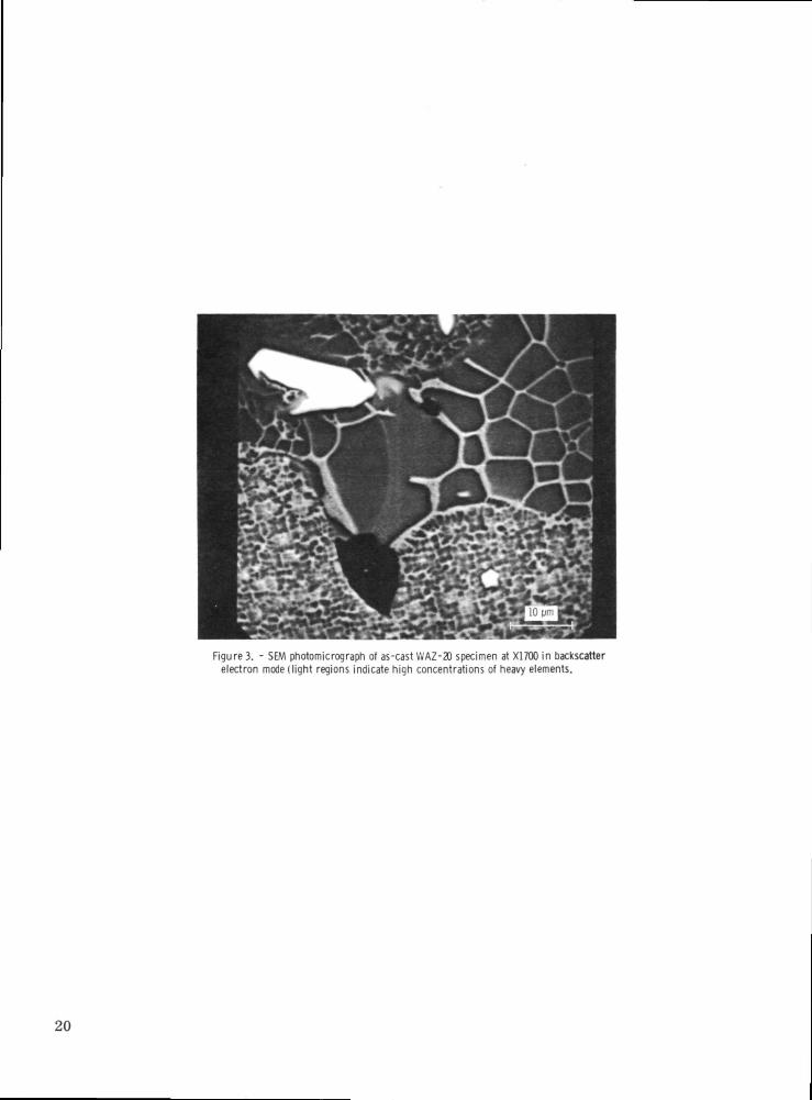
Figure 3. - SEM photomicrograph of as-cast WAZ-20 specimen at X1700 in backscatterelectron mode (light regions indicate high concentrations of heavy elements.
20

21

Figure 5. - Electron microscope photomicrograph of a replica from as-cast WAZ-20 specimen showing carbide, eutectic, and gamma-fine gammaprime structure at X6000.
22

Figure 6. - Electron microscope photomicrograph of replica from the as-cast WAZ-20 specimen showing the gamma-fine gamma prime structure atXI7000.
23

s
. .
I .1
24

25

|
<u .3
I 1g-s"S °
"S
E'oI00
ra
„ i*f*-*t

ra
|
£
"g
. a
o<s\
fi
NASA-Langley, 1973 „ 17 E~7250 27

NATIONAL AERONAUTICS AND SPACE ADMINISTRATION.
WASHINGTON. D.C. 2O546
OFFICIAL BUSINESS
PENALTY FOR PRIVATE USE $3OO SPECIAL FOURTH-CLASS RATEBOOK
NATIONAL AERONAUTICS ANDSPACE ADMINISTRATION
431
POSTMASTER : If Undeliverable (Section 158Postal Manual) Do Not Return
"The aeronautical and space activities of the United States shall beconducted, so as to contribute . . . to the expansion of human knowl-edge of phenomena in the atmosphere and space. The Administrationshall pro-vide for the widest practicable and appropriate disseminationof information concerning its activities and the results thereof."
—NATIONAL AERONAUTICS AND SPACE ACT OF 1958
NASA SCIENTIFIC AND TECHNICAL PUBLICATIONSTECHNICAL REPORTS: Scientific andtechnical information considered important,complete, and a lasting contribution to existingknowledge.
TECHNICAL NOTES: Information less broadin scope but nevertheless of importance as acontribution to existing knowledge.
TECHNICAL MEMORANDUMS:Information receiving limited distributionbecause of preliminary data, security classifica-tion, or other reasons. Also includes conferenceproceedings with either limited or unlimiteddistribution.
CONTRACTOR REPORTS: Scientific andtechnical information generated under a NASAcontract or grant and considered an importantcontribution to existing knowledge.
TECHNICAL TRANSLATIONS: Informationpublished in a foreign language consideredto merit NASA distribution in English.
SPECIAL PUBLICATIONS: Informationderived from or of value to NASA activities.Publications include final reports of majorprojects, monographs, data compilations,handbooks, sourcebooks, and specialbibliographies.
TECHNOLOGY UTILIZATIONPUBLICATIONS: Information on technologyused by NASA that may be of particularinterest in commercial and other non-aerospaceapplications. Publications include Tech Briefs,Technology Utilization Reports andTechnology Surveys.
Details on the avaifabiiify of fhese publications may be obtained from:
SCIENTIFIC AND TECHNICAL INFORMATION OFFICE
N A T I O N A L A E R O N A U T I C S A N D S P A C E A D M I N I S T R A T I O NWashington, D.C. 20546