nasal cavity and paranasal sinus cancers-et final 2016 · malignant tumors of the nose and...
TRANSCRIPT

ACR Appropriateness Criteria® Nasal Cavity and Paranasal Sinus Cancers
EVIDENCE TABLE
* See Last Page for Key 2016 Original Siddiqui/Smith Page 1
Reference Study Type Patients/ Events
Study Objective (Purpose of Study) Study Results Study
Quality 1. Muir CS, Nectoux J. Descriptive
epidemiology of malignant neoplasms of nose, nasal cavities, middle ear and accessory sinuses. Clin Otolaryngol Allied Sci. 1980;5(3):195-211.
Review/Other-Tx
N/A To review international incidence and mortality data for ICD rubric 160 (nose and nasal cavities, middle ear and accessory sinuses). The relative frequency data for cancer for each of the constituent anatomical locations are presented and the histological types of neoplasms encountered tabulated to determine if geographical differences exist which might be worth further investigation.
Relatively high rates for this generally rare disease were found in Asian and African populations, the highest age-adjusted rates, between 2.6 and 2.5 per 100,000 per annum, occurring in Japanese males. Independent of the higher rates, the extremely low proportion of cancers of the nose and nasal cavities together with the very high proportion of cancer of the maxillary sinus in Japan are in contrast with a much higher relative frequency of nose and nasal cavity cancer in other countries.
4
2. Roush GC. Epidemiology of cancer of the nose and paranasal sinuses: current concepts. Head Neck Surg. 1979;2(1):3-11.
Review/Other-Tx
N/A To review epidemiology of cancer of the nose and paranasal sinuses.
Study of the epidemiology of the nose and paranasal sinuses may identify unrecognized carcinogens and occupational groups at increased cancer risk.
4
3. Ansa B, Goodman M, Ward K, et al. Paranasal sinus squamous cell carcinoma incidence and survival based on Surveillance, Epidemiology, and End Results data, 1973 to 2009. Cancer. 2013;119(14):2602-2610.
Review/Other-Tx
2,553 patients
To analyze the incidence trend and survival outcomes for patients diagnosed with paranasal sinus SCC in the Surveillance, Epidemiology, and End Results (SEER) database.
In total, 2,553 patients with paranasal sinus SCC were identified. While incidence of paranasal sinus SCC showed a gradual decline, survival remained largely unchanged. The proportion of patients with advanced disease decreased from 14.7% during the period from 1983 to 1992 to 12.4% during 1993-2002 and to 9.5% during 2003-2009. Compared with whites, incidence was higher among African Americans (relative risk 1.63; 95% CI, 1.39, 1.90) and among all other racial groups (relative risk, 1.78; 95% CI: 1.53–2.07). After adjusting for age, sex, disease stage, tumor site, and treatment, mortality among African American patients also was increased (HR, 1.22; 95% CI, 1.04–1.43). Among patients with localized disease, the relation between race and mortality was no longer evident once the results were controlled for tumor classification.
4
4. Goldenberg D, Golz A, Fradis M, Martu D, Netzer A, Joachims HZ. Malignant tumors of the nose and paranasal sinuses: a retrospective review of 291 cases. Ear Nose Throat J. 2001;80(4):272-277.
Review/Other-Tx
291 cases To present a retrospective study of cases of malignant tumors of the nose and paranasal sinuses that were diagnosed in a northern Romanian population over a period of 35 years. The authors reviewed the etiology, diagnosis, prognosis, and treatment of these tumors.
No results stated in abstract. 4

ACR Appropriateness Criteria® Nasal Cavity and Paranasal Sinus Cancers
EVIDENCE TABLE
* See Last Page for Key 2016 Original Siddiqui/Smith Page 2
Reference Study Type Patients/ Events
Study Objective (Purpose of Study) Study Results Study
Quality 5. Jiang GL, Ang KK, Peters LJ, Wendt CD,
Oswald MJ, Goepfert H. Maxillary sinus carcinomas: natural history and results of postoperative radiotherapy. Radiother Oncol. 1991;21(3):193-200.
Observational-Tx
73 patients To analyze the results of postoperative RT for carcinomas of the maxillary sinus in patients treated over a span of 16 years.
The 5-year relapse-free survival for the whole group was 51%. The overall LC rate was 78%. Patients with larger tumors, particularly if they also had histological signs of nerve invasion, had a higher recurrence rate than others. The overall nodal recurrence rate without elective neck treatment was 38% for squamous and undifferentiated carcinoma, and only 5% for adenoid cystic carcinomas.
3
ACR Appropriateness Criteria® Nasal Cavity and Paranasal Sinus Cancers
EVIDENCE TABLE
* See Last Page for Key 2016 Original Siddiqui/Smith Page 3
Reference Study Type Patients/ Events
Study Objective (Purpose of Study) Study Results Study
Quality 6. Le QT, Fu KK, Kaplan MJ, Terris DJ, Fee
WE, Goffinet DR. Lymph node metastasis in maxillary sinus carcinoma. Int J Radiat Oncol Biol Phys. 2000;46(3):541-549.
Observational-Tx
97 patients To evaluate the incidence and prognostic significance of lymph node metastasis in maxillary sinus carcinoma.
The median survival for all patients was 22 months (range: 2.4–356 months). The 5- and 10-year actuarial survivals were 34% and 31%, respectively. 10 patients relapsed in the neck, with a 5-year actuarial risk of nodal relapse of 12%. The 5-year risk of neck relapse was 14% for SCC, 25% for adenocarcinoma, and 7% for both undifferentiated carcinoma and adenoid cystic carcinoma. The overall risk of nodal involvement at either diagnosis or on follow-up was 28% for SCC, 25% for adenocarcinoma, 12% for undifferentiated carcinoma, and 10% for adenoid cystic carcinoma. All patients with nodal involvement had T3-4, and none had T2 tumors. ENI effectively prevented nodal relapse in patients with SCC and N0 neck; the 5-year actuarial risk of nodal relapse was 20% for patients without ENI and 0% for those with elective neck therapy. There was no correlation between neck relapse and primary tumor control or tumor extension into areas containing a rich lymphatic network. The most common sites of nodal relapse were in the ipsilateral level 1–2 nodal regions (11/13). Patients with nodal relapse had a significantly higher risk of distant metastasis on both univariate (P=0.02) and multivariate analysis (HR = 4.5, P=0.006). The 5-year actuarial risk of distant relapse was 29% for patients with neck control vs 81% for patients with neck failure. There was also a trend for decreased survival with nodal relapse. The 5-year actuarial survival was 37% for patients with neck control and 0% for patients with neck relapse.
2
7. Allen MW, Schwartz DL, Rana V, et al. Long-term radiotherapy outcomes for nasal cavity and septal cancers. Int J Radiat Oncol Biol Phys. 2008;71(2):401-406.
Observational-Tx
68 patients To update the long-term outcomes for RT of nasal cavity and septal cancer at the University of Texas M.D. Anderson Cancer Center.
Of the 68 patients, 19 (28%) developed a locoregional relapse, 14 (21%) locally and 5 (7%) regionally. The LC rate at 5 and 10 years was 86% and 76%, respectively. The DSS rate was 86% and 78%, and the OS rate was 82% and 62% at 5 and 10 years, respectively.
2

ACR Appropriateness Criteria® Nasal Cavity and Paranasal Sinus Cancers
EVIDENCE TABLE
* See Last Page for Key 2016 Original Siddiqui/Smith Page 4
Reference Study Type Patients/ Events
Study Objective (Purpose of Study) Study Results Study
Quality 8. Ang KK, Jiang GL, Frankenthaler RA, et
al. Carcinomas of the nasal cavity. Radiother Oncol. 1992;24(3):163-168.
Observational-Tx
31 patients To review the data on patients with epithelial tumors of the nasal fossa properly treated at The University of Texas M. D. Anderson Cancer Center.
The median length of follow-up was 11 years (range: 2.8–16.8 years). 36 patients had no evidence of disease at the last follow-up visit. All 14 patients with carcinoma of the nasal septum had the disease controlled. 9/31 patients with lesions of the lateral wall and floor died of the disease, 5 of uncontrolled local disease, 2 of distant metastases, and 2 of both. The DSS rates at 5 and 10 years were 83% and 80%, respectively, and the corresponding OS rates were 75% and 60%, respectively. Blindness occurred in 4 patients, 2 due to orbital exenteration and 2 to radiation injury to the cornea and optic pathway. Other infrequent side effects were bone necrosis, dental decay, nasal stenosis, and septal perforation.
2
9. Dulguerov P, Jacobsen MS, Allal AS, Lehmann W, Calcaterra T. Nasal and paranasal sinus carcinoma: are we making progress? A series of 220 patients and a systematic review. Cancer. 2001;92(12):3012-3029.
Meta-analysis 220 patients Authors reviewed treatment results in patients with nasal and paranasal sinus carcinoma from a large retrospective cohort and conducted a systematic literature review.
The 5-year survival rate was 40%, and the LC rate was 59%. The 5-year actuarial survival rate was 63%, and the LC rate was 57%. Factors that were associated statistically with a worse prognosis, with results expressed as 5-year actuarial specific survival rates, included the following: 1) histology, with rates of 79% for patients with glandular carcinoma, 78% for patients with adenocarcinoma, 60% for patients with SCC, and 40% for patients with undifferentiated carcinoma; 2) T classification, with rates of 91%, 64%, 72%, and 49% for patients with T1, T2, T3, and T4 tumors, respectively; 3) localization, with rates of 77% for patients with tumors of the nasal cavity, 62% for patients with tumors of the maxillary sinus, and 48% for patients with tumors of the ethmoid sinus; 4) treatment, with rates of 79% for patients who underwent surgery alone, 66% for patients who were treated with a combination of surgery and RT, and 57% for patients who were treated exclusively with RT.
M

ACR Appropriateness Criteria® Nasal Cavity and Paranasal Sinus Cancers
EVIDENCE TABLE
* See Last Page for Key 2016 Original Siddiqui/Smith Page 5
Reference Study Type Patients/ Events
Study Objective (Purpose of Study) Study Results Study
Quality 10. Duprez F, Madani I, Morbee L, et al.
IMRT for sinonasal tumors minimizes severe late ocular toxicity and preserves disease control and survival. Int J Radiat Oncol Biol Phys. 2012;83(1):252-259.
Observational-Tx
130 patients To report late ocular (primary endpoint) and other toxicity, disease control, and survival (secondary endpoints) after IMRT for sinonasal tumors.
Median follow-up was 52, range 15–121 months. There was no radiation-induced blindness in 86 patients available for late toxicity assessment (≥6 month follow-up). We observed late Grade 3 tearing in 10 patients, which reduced to Grade 1-2 in 5 patients and Grade 3 visual impairment because of radiation-induced ipsilateral retinopathy and neovascular glaucoma in 1 patient. There was no severe dry eye syndrome. The worst grade of late ocular toxicity was Grade 3 (n = 11), Grade 2 (n = 31), Grade 1 (n = 33), and Grade 0 (n = 11). Brain necrosis and osteoradionecrosis occurred in 6 and 1 patients, respectively. Actuarial 5-year LC and OS were 59% and 52%, respectively. On multivariate analysis, LC was negatively affected by cribriform plate and brain invasion (P=0.044 and 0.029, respectively) and absence of surgery (P=0.009); OS was negatively affected by cribriform plate and orbit invasion (P=0.04 and <0.001, respectively) and absence of surgery (P=0.001).
2

ACR Appropriateness Criteria® Nasal Cavity and Paranasal Sinus Cancers
EVIDENCE TABLE
* See Last Page for Key 2016 Original Siddiqui/Smith Page 6
Reference Study Type Patients/ Events
Study Objective (Purpose of Study) Study Results Study
Quality 11. Guntinas-Lichius O, Kreppel MP, Stuetzer
H, Semrau R, Eckel HE, Mueller RP. Single modality and multimodality treatment of nasal and paranasal sinuses cancer: a single institution experience of 229 patients. Eur J Surg Oncol. 2007;33(2):222-228.
Observational-Tx
229 patients To assess the single and multimodal treatment results and prognostic factors for sinonasal carcinoma.
32% of the patients were operated only, 47% underwent multimodal therapy, and 20% were treated without operation. The 5-year OS rate was 41%, and the DSS rate was 51%. The LC rate was 64%, and the DFS rate was 34%. Prognostic for DSS were M status (P<0.001), UICC stage (P<0.001), T classification (P=0.001), N status (P=0.002), intracranial tumor infiltration (P=0.008), infiltration of the pterygopalatine fossa (P=0.02), infiltration of the skull base (P=0.021), infiltration of the orbital (P=0.041), and the type of therapy (P<0.001): The 5-year DSS rate was 63% for patients operated only, 56% for all operated patients, 46% for patients undergoing surgery and RT, but only 21% for patients treated with RT +/- chemotherapy. Multivariate analysis revealed that T classification (P=0.042), N classification (P=0.035), M classification (P=0.007), UICC stage (P=0.038), and type of therapy (P=0.038) were independent prognostic factors for DSS.
2

ACR Appropriateness Criteria® Nasal Cavity and Paranasal Sinus Cancers
EVIDENCE TABLE
* See Last Page for Key 2016 Original Siddiqui/Smith Page 7
Reference Study Type Patients/ Events
Study Objective (Purpose of Study) Study Results Study
Quality 12. Wiegner EA, Daly ME, Murphy JD, et al.
Intensity-modulated radiotherapy for tumors of the nasal cavity and paranasal sinuses: clinical outcomes and patterns of failure. Int J Radiat Oncol Biol Phys. 2012;83(1):243-251.
Observational-Tx
52 patients To report outcomes in patients treated with IMRT for tumors of the paranasal sinuses and nasal cavity.
18 patients (35%) developed local-regional failure at median time of 7.2 months. 13 local failures (25%) were observed, 12 in-field and 1 marginal. 6 regional failures were observed, 2 in-field and 4 out-of-field. No patients treated with elective nodal radiation had nodal regional failure. 2-year local-regional control, in-field local-regional control, freedom from distant metastasis, and OS were 64%, 74%, 71%, and 66% among all patients, respectively, and 43%, 61%, 61%, and 53% among patients with SCC, respectively. On multivariate analysis, SCC and >1 subsite involved had worse local-regional control (P=0.0004 and P=0.046, respectively) and OS (P=0.003 and P=0.046, respectively). Cribriform plate invasion (P=0.005) and residual disease (P=0.047) also had worse local-regional control. Acute toxicities included Grade ≥3 mucositis in 19 patients (37%) and Grade 3 dermatitis in 8 patients (15%). 6 patients had Grade ≥3 late toxicity including 1 optic toxicity.
2
13. Ganly I, Patel SG, Singh B, et al. Craniofacial resection for malignant paranasal sinus tumors: Report of an International Collaborative Study. Head Neck. 2005;27(7):575-584.
Observational-Tx
334 patients To examine a large cohort of patients accumulated from multiple institutions experienced in craniofacial surgery, with the aim of reporting benchmark figures for outcomes and identifying patient-related and tumor-related predictors of prognosis after craniofacial resection.
Postoperative mortality occurred in 15 patients (4.5%). Postoperative complications occurred in 110 patients (32.9%). The 5-year OS, DSS, and recurrence-free survival rates were 48.3%, 53.3%, and 45.8%, respectively. The status of surgical margins, histologic findings of the primary tumor, and intracranial extent were independent predictors of OS, DSS, and recurrence-free survival on multivariate analysis.
2
ACR Appropriateness Criteria® Nasal Cavity and Paranasal Sinus Cancers
EVIDENCE TABLE
* See Last Page for Key 2016 Original Siddiqui/Smith Page 8
Reference Study Type Patients/ Events
Study Objective (Purpose of Study) Study Results Study
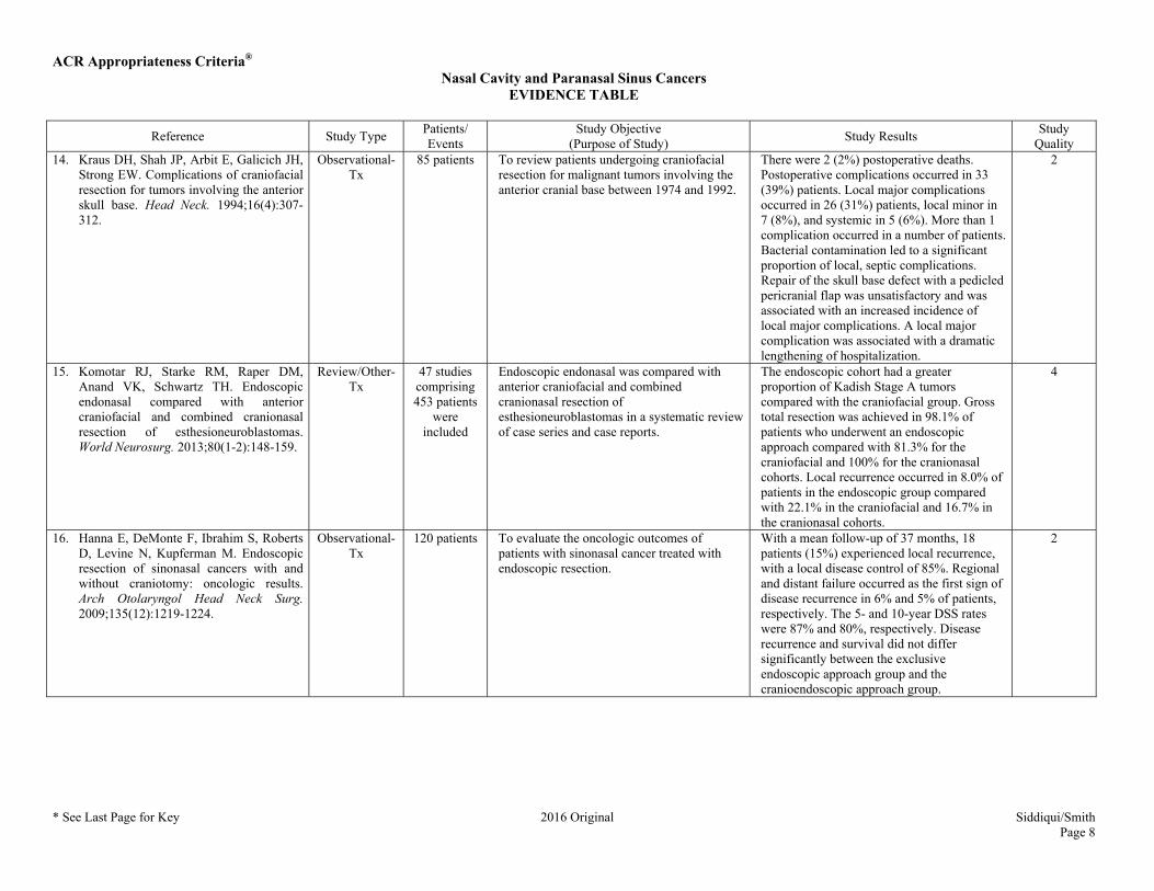
Quality 14. Kraus DH, Shah JP, Arbit E, Galicich JH,
Strong EW. Complications of craniofacial resection for tumors involving the anterior skull base. Head Neck. 1994;16(4):307-312.
Observational-Tx
85 patients To review patients undergoing craniofacial resection for malignant tumors involving the anterior cranial base between 1974 and 1992.
There were 2 (2%) postoperative deaths. Postoperative complications occurred in 33 (39%) patients. Local major complications occurred in 26 (31%) patients, local minor in 7 (8%), and systemic in 5 (6%). More than 1 complication occurred in a number of patients. Bacterial contamination led to a significant proportion of local, septic complications. Repair of the skull base defect with a pedicled pericranial flap was unsatisfactory and was associated with an increased incidence of local major complications. A local major complication was associated with a dramatic lengthening of hospitalization.
2
15. Komotar RJ, Starke RM, Raper DM, Anand VK, Schwartz TH. Endoscopic endonasal compared with anterior craniofacial and combined cranionasal resection of esthesioneuroblastomas. World Neurosurg. 2013;80(1-2):148-159.
Review/Other-Tx
47 studies comprising 453 patients
were included
Endoscopic endonasal was compared with anterior craniofacial and combined cranionasal resection of esthesioneuroblastomas in a systematic review of case series and case reports.
The endoscopic cohort had a greater proportion of Kadish Stage A tumors compared with the craniofacial group. Gross total resection was achieved in 98.1% of patients who underwent an endoscopic approach compared with 81.3% for the craniofacial and 100% for the cranionasal cohorts. Local recurrence occurred in 8.0% of patients in the endoscopic group compared with 22.1% in the craniofacial and 16.7% in the cranionasal cohorts.
4
16. Hanna E, DeMonte F, Ibrahim S, Roberts D, Levine N, Kupferman M. Endoscopic resection of sinonasal cancers with and without craniotomy: oncologic results. Arch Otolaryngol Head Neck Surg. 2009;135(12):1219-1224.
Observational-Tx
120 patients To evaluate the oncologic outcomes of patients with sinonasal cancer treated with endoscopic resection.
With a mean follow-up of 37 months, 18 patients (15%) experienced local recurrence, with a local disease control of 85%. Regional and distant failure occurred as the first sign of disease recurrence in 6% and 5% of patients, respectively. The 5- and 10-year DSS rates were 87% and 80%, respectively. Disease recurrence and survival did not differ significantly between the exclusive endoscopic approach group and the cranioendoscopic approach group.
2

ACR Appropriateness Criteria® Nasal Cavity and Paranasal Sinus Cancers
EVIDENCE TABLE
* See Last Page for Key 2016 Original Siddiqui/Smith Page 9
Reference Study Type Patients/ Events
Study Objective (Purpose of Study) Study Results Study
Quality 17. Nicolai P, Battaglia P, Bignami M, et al.
Endoscopic surgery for malignant tumors of the sinonasal tract and adjacent skull base: a 10-year experience. Am J Rhinol. 2008;22(3):308-316.
Observational-Tx
184 patients The authors report their experience with the endoscopic management of nasoethmoidal malignancies possibly involving the adjacent skull base.
An exclusive endoscopic approach was performed in 134 patients and the remaining 50 patients underwent the cranioendoscopic approach. The most frequent histotypes encountered were adenocarcinoma (37%), SCC (13.6%), olfactory neuroblastoma (12%), mucosal melanoma (9.2%), and adenoid cystic carcinoma (7.1%). Overall, 86 (46.7%) patients received some form of adjuvant treatment. The patients were followed up for a mean of 34.1 months (range, 2–123 months). The 5-year DSS was 91.4% +/- 3.9% and 58.8% +/- 8.6% (P=0.0004) for the exclusive endoscopic approach and cranioendoscopic approach group, respectively.
2

ACR Appropriateness Criteria® Nasal Cavity and Paranasal Sinus Cancers
EVIDENCE TABLE
* See Last Page for Key 2016 Original Siddiqui/Smith Page 10
Reference Study Type Patients/ Events
Study Objective (Purpose of Study) Study Results Study
Quality 18. Jansen EP, Keus RB, Hilgers FJ, Haas
RL, Tan IB, Bartelink H. Does the combination of radiotherapy and debulking surgery favor survival in paranasal sinus carcinoma? Int J Radiat Oncol Biol Phys. 2000;48(1):27-35.
Observational-Tx
73 patients To determine the contribution of debulking surgery on LC and survival in paranasal sinus tumors.
5-year LC was 65% with combination of RT and debulking surgery in comparison with 47% with RT alone, but this difference was not statistically significant (P=0.58). However, combination treatment gave significantly better 5-year OS (60% vs 9%; P=0.001) and 5-year DFS (53% vs 6%; P<0. 0001). Cox-regression analysis showed that pathologic N status (P=0.04), palliative intention of treatment (P=0.018), clinical orbital invasion (P=0.003), and orbital wall invasion (P=0.003) were parameters significantly associated with poor LC. Total RT dose >65 Gy (P=0.05) and treatment consisting of RT alone (P=0.002) were associated with worse OS; for DFS clinical orbital invasion (P=0.0005), age >65 years (P=0.013) and pathologic T4 classification (P=0.002) were significant factors for an unfavorable outcome. In 19/73 patients, 26 serious (mainly ophthalmological) complications were reported; in the majority of these, the visual tract was (partly) included in the treatment fields because of tumor extension. To analyze on which basis patients were selected for the combination therapy, a logistic regression was performed, concluding that clinical T4 classification (P=0.05), radiological evidence of skull base invasion (P=0.005), age >65 years (P=0.026), radiological evidence of nasopharynx invasion (P=0.02), clinical suspicion of palate invasion (P=0.02), and radiological evidence of skin invasion (P=0.009) were associated with choosing RT alone.
2

ACR Appropriateness Criteria® Nasal Cavity and Paranasal Sinus Cancers
EVIDENCE TABLE
* See Last Page for Key 2016 Original Siddiqui/Smith Page 11
Reference Study Type Patients/ Events
Study Objective (Purpose of Study) Study Results Study
Quality 19. Mendenhall WM, Amdur RJ, Morris CG,
et al. Carcinoma of the nasal cavity and paranasal sinuses. Laryngoscope. 2009;119(5):899-906.
Observational-Tx
109 patients To determine the outcomes after RT alone or combined with surgery at the University of Florida for patients with carcinomas of the nasal cavity and paranasal sinuses.
The 5-year LC rates were: T1-T3, 82%; T4, 50%; and overall, 63%. LC at 5 years was 43% after definitive RT vs 84% after surgery and adjuvant RT (P<.0001). Multivariate analysis of LC revealed that both overall stage and treatment group (definitive RT vs surgery and adjuvant RT) significantly impacted this endpoint. Cause-specific survival rates were: stages I to III, 81%; stage IV, 54%; and overall, 62%. Multivariate analysis revealed that T-stage, N-stage, and treatment group significantly influenced this endpoint. 31 (20%) of 109 patients sustained severe complications; 17/56 patients (16%) after definitive RT and 14/53 patients (25%) after surgery and adjuvant RT.
2
20. Resto VA, Chan AW, Deschler DG, Lin DT. Extent of surgery in the management of locally advanced sinonasal malignancies. Head Neck. 2008;30(2):222-229.
Observational-Tx
102 patients To determine the relative importance of surgery within multimodality regimens commonly used to treat advanced sinonasal malignancies.
Extent of surgery correlated with DFS and OS rates. LC rate, however, was independent of the degree of surgical resection achieved. Overall, treatment failure most commonly resulted from distant metastases, which occurred in 30% of patients and also correlated with extent of surgical resection. Tumor type-specific outcomes reveal differences associated with the extent of surgery achieved.
2
21. Blanco AI, Chao KS, Ozyigit G, et al. Carcinoma of paranasal sinuses: long-term outcomes with radiotherapy. Int J Radiat Oncol Biol Phys. 2004;59(1):51-58.
Observational-Tx
106 patients To assess the clinical features, prognostic factors, results, and complications of treatment of carcinomas of the paranasal sinus.
Follow-up ranged from 1.7 months to 24 years (median 5 years). The 5-year local tumor control, locoregional tumor control, DFS, and OS rate was 58%, 39%, 33%, and 27%, respectively. A statistically significant improvement in DFS was noted with the addition of surgical resection to RT (35% vs 29%, P=0.05). Nodal status at presentation emerged as a statistically significant predictor for locoregional tumor control and DFS in multivariate analysis. Distant metastases occurred in 29% of patients.
2
ACR Appropriateness Criteria® Nasal Cavity and Paranasal Sinus Cancers
EVIDENCE TABLE
* See Last Page for Key 2016 Original Siddiqui/Smith Page 12
Reference Study Type Patients/ Events
Study Objective (Purpose of Study) Study Results Study
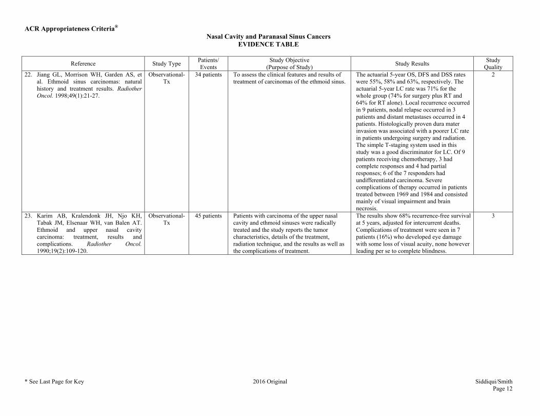
Quality 22. Jiang GL, Morrison WH, Garden AS, et
al. Ethmoid sinus carcinomas: natural history and treatment results. Radiother Oncol. 1998;49(1):21-27.
Observational-Tx
34 patients To assess the clinical features and results of treatment of carcinomas of the ethmoid sinus.
The actuarial 5-year OS, DFS and DSS rates were 55%, 58% and 63%, respectively. The actuarial 5-year LC rate was 71% for the whole group (74% for surgery plus RT and 64% for RT alone). Local recurrence occurred in 9 patients, nodal relapse occurred in 3 patients and distant metastases occurred in 4 patients. Histologically proven dura mater invasion was associated with a poorer LC rate in patients undergoing surgery and radiation. The simple T-staging system used in this study was a good discriminator for LC. Of 9 patients receiving chemotherapy, 3 had complete responses and 4 had partial responses; 6 of the 7 responders had undifferentiated carcinoma. Severe complications of therapy occurred in patients treated between 1969 and 1984 and consisted mainly of visual impairment and brain necrosis.
2
23. Karim AB, Kralendonk JH, Njo KH, Tabak JM, Elsenaar WH, van Balen AT. Ethmoid and upper nasal cavity carcinoma: treatment, results and complications. Radiother Oncol. 1990;19(2):109-120.
Observational-Tx
45 patients Patients with carcinoma of the upper nasal cavity and ethmoid sinuses were radically treated and the study reports the tumor characteristics, details of the treatment, radiation technique, and the results as well as the complications of treatment.
The results show 68% recurrence-free survival at 5 years, adjusted for intercurrent deaths. Complications of treatment were seen in 7 patients (16%) who developed eye damage with some loss of visual acuity, none however leading per se to complete blindness.
3

ACR Appropriateness Criteria® Nasal Cavity and Paranasal Sinus Cancers
EVIDENCE TABLE
* See Last Page for Key 2016 Original Siddiqui/Smith Page 13
Reference Study Type Patients/ Events
Study Objective (Purpose of Study) Study Results Study
Quality 24. Katz TS, Mendenhall WM, Morris CG,
Amdur RJ, Hinerman RW, Villaret DB. Malignant tumors of the nasal cavity and paranasal sinuses. Head Neck. 2002;24(9):821-829.
Observational-Tx
78 patients To evaluate the role of RT in patients with nasal cavity and paranasal sinus tumors.
The 5-year actuarial LC rate for stage I (limited to the site of origin; 22 patients) was 86%; for stage II (extension to adjacent sites (eg, adjacent sinuses, orbit, pterygomaxillary fossa, nasopharynx; 21 patients) was 65%; and for stage III (destruction of skull base or pterygoid plates, or intracranial extension; 35 patients) was 34%. The 5-year actuarial LC rate for patients receiving postoperative RT was 79% and for patients receiving RT alone was 49% (P=.05). The 5-, 10-, 15-, and 20-year ultimate LC rates for all 78 patients were 60%, 56%, 48%, and 48%, respectively. The 5-, 10-, 15-, and 20-year cause-specific survival rates for all 78 patients were 56%, 45%, 39%, and 39%, respectively. The 5-, 10-, 15-, and 20-year absolute survival rates for all 78 patients were 50%, 31%, 21%, and 16%, respectively. Of the 67 (86%) patients who were initially seen with node-negative disease, 39 (58%) received no elective neck treatment, and 28 (42%) received ENI. Of the 39 patients who received no elective neck treatment, 33 (85%) did not experience recurrence in the neck compared with 25 (89%) of 28 patients who received ENI. Most patients who received ENI (57%) had stage III disease. 21 (27%) of 78 patients had unilateral blindness develop secondary to radiation retinopathy or optic neuropathy; the complication was anticipated in most of these patients, because the ipsilateral eye was irradiated to a high dose. 4 patients (5%) unexpectedly had bilateral blindness develop because of optic neuropathy. All 4 of these patients received irradiation alone.
2
ACR Appropriateness Criteria® Nasal Cavity and Paranasal Sinus Cancers
EVIDENCE TABLE
* See Last Page for Key 2016 Original Siddiqui/Smith Page 14
Reference Study Type Patients/ Events
Study Objective (Purpose of Study) Study Results Study
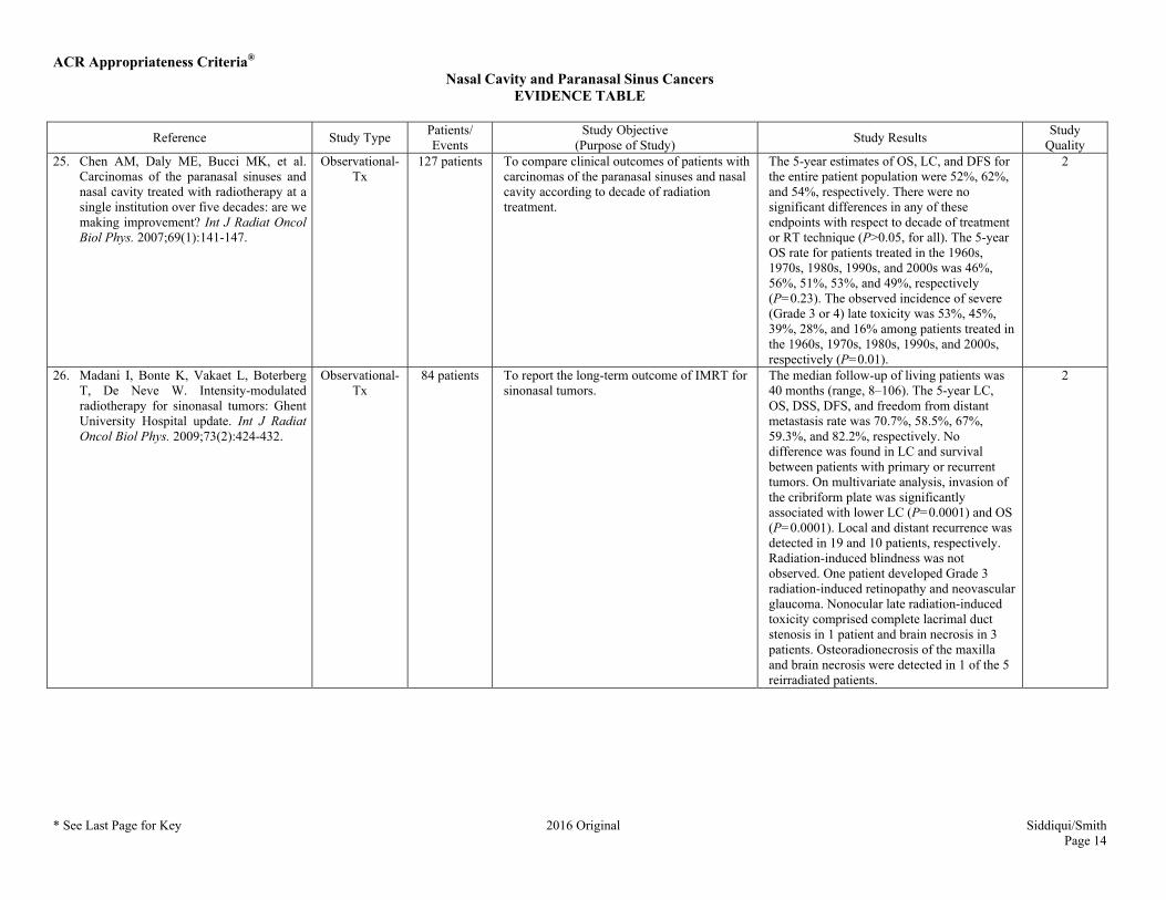
Quality 25. Chen AM, Daly ME, Bucci MK, et al.
Carcinomas of the paranasal sinuses and nasal cavity treated with radiotherapy at a single institution over five decades: are we making improvement? Int J Radiat Oncol Biol Phys. 2007;69(1):141-147.
Observational-Tx
127 patients To compare clinical outcomes of patients with carcinomas of the paranasal sinuses and nasal cavity according to decade of radiation treatment.
The 5-year estimates of OS, LC, and DFS for the entire patient population were 52%, 62%, and 54%, respectively. There were no significant differences in any of these endpoints with respect to decade of treatment or RT technique (P>0.05, for all). The 5-year OS rate for patients treated in the 1960s, 1970s, 1980s, 1990s, and 2000s was 46%, 56%, 51%, 53%, and 49%, respectively (P=0.23). The observed incidence of severe (Grade 3 or 4) late toxicity was 53%, 45%, 39%, 28%, and 16% among patients treated in the 1960s, 1970s, 1980s, 1990s, and 2000s, respectively (P=0.01).
2
26. Madani I, Bonte K, Vakaet L, Boterberg T, De Neve W. Intensity-modulated radiotherapy for sinonasal tumors: Ghent University Hospital update. Int J Radiat Oncol Biol Phys. 2009;73(2):424-432.
Observational-Tx
84 patients To report the long-term outcome of IMRT for sinonasal tumors.
The median follow-up of living patients was 40 months (range, 8–106). The 5-year LC, OS, DSS, DFS, and freedom from distant metastasis rate was 70.7%, 58.5%, 67%, 59.3%, and 82.2%, respectively. No difference was found in LC and survival between patients with primary or recurrent tumors. On multivariate analysis, invasion of the cribriform plate was significantly associated with lower LC (P=0.0001) and OS (P=0.0001). Local and distant recurrence was detected in 19 and 10 patients, respectively. Radiation-induced blindness was not observed. One patient developed Grade 3 radiation-induced retinopathy and neovascular glaucoma. Nonocular late radiation-induced toxicity comprised complete lacrimal duct stenosis in 1 patient and brain necrosis in 3 patients. Osteoradionecrosis of the maxilla and brain necrosis were detected in 1 of the 5 reirradiated patients.
2

ACR Appropriateness Criteria® Nasal Cavity and Paranasal Sinus Cancers
EVIDENCE TABLE
* See Last Page for Key 2016 Original Siddiqui/Smith Page 15
Reference Study Type Patients/ Events
Study Objective (Purpose of Study) Study Results Study
Quality 27. Truong MT, Kamat UR, Liebsch NJ, et al.
Proton radiation therapy for primary sphenoid sinus malignancies: treatment outcome and prognostic factors. Head Neck. 2009;31(10):1297-1308.
Observational-Tx
20 patients To determine treatment outcome and prognostic factors in patients with locally advanced primary sphenoid sinus malignancy treated with proton RT.
With a median follow-up of 27 months, the 2-year local, regional, and freedom from distant metastasis rates were 86%, 86%, and 50%, respectively. The DFS and OS rates at 2 years were 31% and 53%, respectively. In multivariate analysis, oropharyngeal involvement (P=.005) and anterior cranial fossa invasion (P=.02) were predictive for poor DFS rate. Brain invasion was predictive for decreased OS rate (P=.05).
2
28. Zenda S, Kohno R, Kawashima M, et al. Proton beam therapy for unresectable malignancies of the nasal cavity and paranasal sinuses. Int J Radiat Oncol Biol Phys. 2011;81(5):1473-1478.
Observational-Tx
39 patients To retrospectively analyze the clinical profile of proton beam therapy for unresectable malignancies of the nasal cavity and paranasal sinuses.
Median patient age was 57 years (range, 22–84 years); 22 of the patients were men and 17 were women. The most frequent primary site was the nasal cavity (n=26, 67%). The LC rates at 6 months and 1 year were 84.6% and 77.0%, respectively. With a median active follow-up of 45.4 months, 3-year PFS and OS were 49.1% and 59.3%, respectively. The most common acute toxicities were mild dermatitis (Grade 2, 33.3%), but no severe toxicity was observed (Grade 3 or greater, 0%). 5 patients (12.8%) experienced Grade 3 to 5 late toxicities, and 1 treatment-related death was reported, caused by cerebrospinal fluid leakage Grade 5 (2.6%).
2
29. Fukumitsu N, Okumura T, Mizumoto M, et al. Outcome of T4 (International Union Against Cancer Staging System, 7th edition) or recurrent nasal cavity and paranasal sinus carcinoma treated with proton beam. Int J Radiat Oncol Biol Phys. 2012;83(2):704-711.
Observational-Tx
17 patients To investigate the clinical features, prognostic factors, and toxicity of treatment for unresectable carcinomas of the nasal cavity and paranasal sinus treated with proton beam therapy.
The OS rate was 47.1% at 2 years and 15.7% at 5 years, and the LC rate was 35.0% at 2 years and 17.5% at 5 years. Invasion of the frontal or sphenoid sinus was a prognostic factor for OS or LC. Late toxicity of more than Grade 3 was found in 2 patients (brain necrosis in 1 and ipsilateral blindness in 1); however, no mortal adverse effects were observed.
3

ACR Appropriateness Criteria® Nasal Cavity and Paranasal Sinus Cancers
EVIDENCE TABLE
* See Last Page for Key 2016 Original Siddiqui/Smith Page 16
Reference Study Type Patients/ Events
Study Objective (Purpose of Study) Study Results Study
Quality 30. Choussy O, Ferron C, Vedrine PO, et al.
Adenocarcinoma of Ethmoid: a GETTEC retrospective multicenter study of 418 cases. Laryngoscope. 2008;118(3):437-443.
Observational-Tx
418 patients To determine risk factors and evaluate the treatment of ethmoid adenocarcinoma. Epidemiologic data were recorded and compared with the literature.
The gender ratio was 2.8 men per 1 woman. Toxic exposure was classic for this lesion, exposure to wood and leather for most cases. The mean age was 63 years (range 31–91). Symptoms were nonspecific and based on clinical rhinologic signs. Nasal endoscopy after mucosal retraction was found useful to evaluate the extension of the lesion and to perform biopsies. Computed tomography scan and magnetic resonance imagery must be carried out prior to treatment to define extra nasal extension. The survival rate was significantly influenced by the size of the lesion (T4, N+) and extension to brain or dura. Surgery with postoperative RT remains the treatment of choice. Total excision must be a major priority, as confirmed in our series.
2
31. Vedrine PO, Thariat J, Merrot O, et al. Primary cancer of the sphenoid sinus--a GETTEC study. Head Neck. 2009;31(3):388-397.
Observational-Tx
23 patients To describe the symptoms associated with primary tumors of the sphenoid sinus and patient management and to identify some prognostic factors for locoregional control and survival.
Cranial neuropathies were present in 12 patients. Pathologic findings included adenoid cystic carcinoma, adenocarcinoma, lymphoma, SCC, sarcoma, neuroendocrine carcinoma, melanoma, and malignant hemangiopericytoma. All but 2 patients had stages III to IV cancer. RT was performed in 18 patients and chemotherapy in 12. Of 10 patients undergoing surgery, total excision with grossly negative margins was achieved in 4 patients and subtotal resection in 6. Median locoregional control and OS were 12 and 41 months, respectively. On multivariate analysis, cranial neuropathy was associated with worse locoregional control and survival. Surgery was rarely complete because of advanced stages at presentation, but it yielded better outcomes than other treatments without surgery in nonlymphoma-cases.
2

ACR Appropriateness Criteria® Nasal Cavity and Paranasal Sinus Cancers
EVIDENCE TABLE
* See Last Page for Key 2016 Original Siddiqui/Smith Page 17
Reference Study Type Patients/ Events
Study Objective (Purpose of Study) Study Results Study
Quality 32. DeMonte F, Ginsberg LE, Clayman GL.
Primary malignant tumors of the sphenoidal sinus. Neurosurgery. 2000;46(5):1084-1091; discussion 1091-1082.
Observational-Tx
27 patients To ascertain the frequency and pathological nature of primary tumors of the sphenoidal sinus, to describe the symptoms and signs associated with tumors in this location, and to describe patient management and outcome.
A malignant pathological process was found in 26 of the 27 patients. The most common diseases were SCC (n = 9), adenoid cystic carcinoma (n = 4), chondrosarcoma (n = 3), and neuroendocrine carcinoma (n = 3). Treatments included surgery (n = 1), surgery and RT (n = 6), surgery and chemotherapy (n = 3), surgery, RT, and chemotherapy (n = 4), chemotherapy (n = 5), chemotherapy and RT (n = 3), and RT (n = 2). The mean follow-up period from initial evaluation was 41 months (range, 2-199 mo). At the last follow-up, 12 patients (48%) were still alive and 13 (52%) had died. The 2-year survival rate for patients with SCC was 44%.
2
33. Bernier J, Domenge C, Ozsahin M, et al. Postoperative irradiation with or without concomitant chemotherapy for locally advanced head and neck cancer. N Engl J Med. 2004;350(19):1945-1952.
Experimental-Tx
334 patients Randomized study to compare concomitant cisplatin and irradiation with RT alone as adjuvant treatment for stage III or IV head and neck cancer.
After a median follow-up of 60 months, the rate of PFS was significantly higher in the combined-therapy group than in the group given RT alone, with 5-year Kaplan-Meier estimates of PFS of 47% and 36%, respectively. The OS rate was also significantly higher in the combined-therapy group than in the RT group, with 5-year Kaplan-Meier estimates of OS of 53% and 40%, respectively.
1
ACR Appropriateness Criteria® Nasal Cavity and Paranasal Sinus Cancers
EVIDENCE TABLE
* See Last Page for Key 2016 Original Siddiqui/Smith Page 18
Reference Study Type Patients/ Events
Study Objective (Purpose of Study) Study Results Study
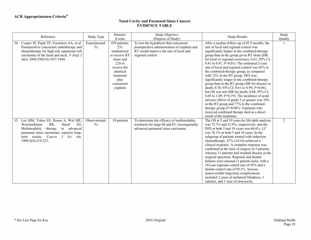
Quality 34. Cooper JS, Pajak TF, Forastiere AA, et al.
Postoperative concurrent radiotherapy and chemotherapy for high-risk squamous-cell carcinoma of the head and neck. N Engl J Med. 2004;350(19):1937-1944.
Experimental-Tx
459 patients; 231
randomized to receive RT
alone and 228 to
receive the identical treatment
plus concurrent cisplatin
To test the hypothesis that concurrent postoperative administration of cisplatin and RT would improve the rate of local and regional control.
After a median follow-up of 45.9 months, the rate of local and regional control was significantly higher in the combined-therapy group than in the group given RT alone (HR for local or regional recurrence, 0.61; 95% CI, 0.41 to 0.91; P=0.01). The estimated 2-year rate of local and regional control was 82% in the combined-therapy group, as compared with 72% in the RT group. DFS was significantly longer in the combined-therapy group than in the RT group (HR for disease or death, 0.78; 95% CI, 0.61 to 0.99; P=0.04), but OS was not (HR for death, 0.84; 95% CI, 0.65 to 1.09; P=0.19). The incidence of acute adverse effects of grade 3 or greater was 34% in the RT group and 77% in the combined-therapy group (P<0.001). 4 patients who received combined therapy died as a direct result of the treatment.
1
35. Lee MM, Vokes EE, Rosen A, Witt ME, Weichselbaum RR, Haraf DJ. Multimodality therapy in advanced paranasal sinus carcinoma: superior long-term results. Cancer J Sci Am. 1999;5(4):219-223.
Observational-Tx
19 patients To determine the efficacy of multimodality treatment for stage III and IV, locoregionally advanced paranasal sinus carcinoma.
The OS at 5 and 10 years by life table analysis was 72.7% and 53.9%, respectively, and the DFS at both 5 and 10 years was 66.6%. LC was 76.1% at both 5 and 10 years. In the subgroup of patients treated with induction chemotherapy, 87% (14/16) achieved a clinical response. A complete response was confirmed at the time of surgery in 5 patients, whereas 11 patients had residual disease in the surgical specimen. Regional and distant failures were unusual (1 patient each), with a 10-year regional control rate of 93% and a distant control rate of 95.5%. Serious, nonreversible long-term complications included 2 cases of unilateral blindness, 1 cataract, and 1 case of ototoxicity.
2

ACR Appropriateness Criteria® Nasal Cavity and Paranasal Sinus Cancers
EVIDENCE TABLE
* See Last Page for Key 2016 Original Siddiqui/Smith Page 19
Reference Study Type Patients/ Events
Study Objective (Purpose of Study) Study Results Study
Quality 36. Hanna EY, Cardenas AD, DeMonte F, et
al. Induction chemotherapy for advanced squamous cell carcinoma of the paranasal sinuses. Arch Otolaryngol Head Neck Surg. 2011;137(1):78-81.
Observational-Tx
46 patients To review the oncologic outcomes in patients with advanced (stage III-IV) SCC of the paranasal sinuses treated with induction chemotherapy prior to definitive local therapy.
Of the 46 patients (median age, 59 years), the tumor epicenter was in the maxillary sinus in 31 (67%), ethmoid sinus in 9 (20%), nasal cavity in 4 (9%), and sphenoid sinus in 2 (4%). All patients had T3 or T4 tumors, and 12 (26%) patients had clinical evidence of nodal metastasis, with an overall stage of III (20%) or IV (80%). Induction chemotherapy regimens consisted of a combination of a taxane and platinum in 80% of patients, by themselves (14 patients) or in combination with a third agent, such as ifosfamide (14 patients) or 5-fluorouracil (9 patients). The combination of a taxane and 5-fluorouracil was used in the remaining 9 patients. More than two-thirds (67%) of the patients achieved at least a partial response to induction chemotherapy, 24% had progressive disease, and 9% had stable disease. Subsequent treatment after induction chemotherapy consisted of surgery, usually followed by radiation or chemoradiation or by definitive radiation or chemoradiation with surgical salvage of any residual disease. Overall, surgical resection was performed in only 24 of 46 patients (52%) treated with induction chemotherapy. The 2-year survival for patients with at least a partial response or stable disease after induction chemotherapy was 77% in contrast to only 36% for patients with progressive disease.
2

ACR Appropriateness Criteria® Nasal Cavity and Paranasal Sinus Cancers
EVIDENCE TABLE
* See Last Page for Key 2016 Original Siddiqui/Smith Page 20
Reference Study Type Patients/ Events
Study Objective (Purpose of Study) Study Results Study
Quality 37. Licitra L, Locati LD, Cavina R, et al.
Primary chemotherapy followed by anterior craniofacial resection and radiotherapy for paranasal cancer. Ann Oncol. 2003;14(3):367-372.
Experimental-Tx
49 patients To study prospectively the activity of primary chemotherapy with cisplatin, fluorouracil and leucovorin in patients with paranasal cancer receiving surgery and postoperative RT.
32 patients (65%) completed 3 or more chemotherapy courses. 2 deaths from thrombotic events were observed after the first cycle. 8 cardiac toxicities were recorded during chemotherapy causing treatment discontinuation. Objective response to cisplatin, fluorouracil and leucovorin was observed in 21 patients [43%; 95% CI, 29% to 58%], including 4 complete responses (8%; 95% CI, 2% to 20%) and 17 partial responses (35%). Pathological complete remission was achieved in 8 of 49 patients (16%). At 3 years, OS was 69% and event-free survival 57%. OS and event-free survival in patients achieving pathological complete remission is 100%.
2
38. Hoppe BS, Nelson CJ, Gomez DR, et al. Unresectable carcinoma of the paranasal sinuses: outcomes and toxicities. Int J Radiat Oncol Biol Phys. 2008;72(3):763-769.
Observational-Tx
39 patients To evaluate long-term outcomes and toxicity in patients with unresectable paranasal sinus carcinoma treated with RT, with or without chemotherapy.
With a median follow-up of 90 months, the 5-year local PFS, regional PFS, distant metastasis-free survival, DFS, and OS were 21%, 61%, 51%, 14%, and 15%, respectively. Patients primarily experienced local relapse (n = 25, 64%), mostly within the irradiated field (n = 22). 9 patients developed neck relapses; however none of the 4 patients receiving ENI had a nodal relapse. In 13 patients acute Grade 3 mucositis developed. Severe late toxicities occurred in 2 patients with radionecrosis and 1 patient with unilateral blindness 7 years after IMRT (77 Gy to the optic nerve). The only significant factor for improved local PFS and OS was a biologically equivalent dose of radiation ≥65 Gy.
2
ACR Appropriateness Criteria® Nasal Cavity and Paranasal Sinus Cancers
EVIDENCE TABLE
* See Last Page for Key 2016 Original Siddiqui/Smith Page 21
Reference Study Type Patients/ Events
Study Objective (Purpose of Study) Study Results Study
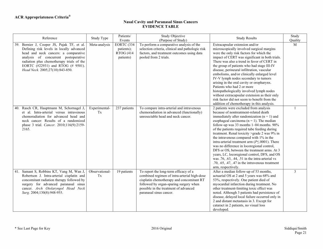
Quality 39. Bernier J, Cooper JS, Pajak TF, et al.
Defining risk levels in locally advanced head and neck cancers: a comparative analysis of concurrent postoperative radiation plus chemotherapy trials of the EORTC (#22931) and RTOG (# 9501). Head Neck. 2005;27(10):843-850.
Meta-analysis EORTC (334 patients);
RTOG (414 patients)
To perform a comparative analysis of the selection criteria, clinical and pathologic risk factors, and treatment outcomes using data pooled from 2 trials.
Extracapsular extension and/or microscopically involved surgical margins were the only risk factors for which the impact of CERT was significant in both trials. There was also a trend in favor of CERT in the group of patients who had stage III-IV disease, perineural infiltration, vascular embolisms, and/or clinically enlarged level IV-V lymph nodes secondary to tumors arising in the oral cavity or oropharynx. Patients who had 2 or more histopathologically involved lymph nodes without extracapsular extension as their only risk factor did not seem to benefit from the addition of chemotherapy in this analysis.
M
40. Rasch CR, Hauptmann M, Schornagel J, et al. Intra-arterial versus intravenous chemoradiation for advanced head and neck cancer: Results of a randomized phase 3 trial. Cancer. 2010;116(9):2159-2165.
Experimental-Tx
237 patients To compare intra-arterial and intravenous chemoradiation in advanced (functionally) unresectable head and neck cancer.
2 patients were excluded from analysis because of nontreatment-related death immediately after randomization (n = 1) and esophageal carcinoma (n = 1). The median follow-up was 33 months 1–04 months. 90% of the patients required tube feeding during treatment. Renal toxicity >grade 2 was 9% in the intravenous compared with 1% in the intra-arterial treatment arm (P≤.0001). There was no difference in locoregional control, DFS or OS, between the treatment arms. At 3 years, LC, locoregional control, DFS, and OS was .76, .63, .44, .51 in the intra-arterial vs .70, .65, .47, .47 in the intravenous treatment arm, respectively.
1
41. Samant S, Robbins KT, Vang M, Wan J, Robertson J. Intra-arterial cisplatin and concomitant radiation therapy followed by surgery for advanced paranasal sinus cancer. Arch Otolaryngol Head Neck Surg. 2004;130(8):948-955.
Observational-Tx
19 patients To report the long-term efficacy of a combined regimen of intra-arterial high-dose cisplatin chemotherapy and concomitant RT followed by organ-sparing surgery when possible in the treatment of advanced paranasal sinus cancer.
After a median follow-up of 53 months, actuarial OS at 2 and 5 years was 68% and 53%, respectively. One patient died of myocardial infarction during treatment. No other treatment-limiting toxic effect was noted. Although 3 patients had persistence of disease, delayed local failure occurred only in 2 and distant metastasis in 3. Except for cataract in 2 patients, no visual loss developed.
3

ACR Appropriateness Criteria® Nasal Cavity and Paranasal Sinus Cancers
EVIDENCE TABLE
* See Last Page for Key 2016 Original Siddiqui/Smith Page 22
Reference Study Type Patients/ Events
Study Objective (Purpose of Study) Study Results Study
Quality 42. Homma A, Oridate N, Suzuki F, et al.
Superselective high-dose cisplatin infusion with concomitant radiotherapy in patients with advanced cancer of the nasal cavity and paranasal sinuses: a single institution experience. Cancer. 2009;115(20):4705-4714.
Experimental-Tx
47 patients To evaluate the efficacy of superselective high-dose cisplatin infusion with concomitant RT for previously untreated patients with advanced cancer of the nasal cavity and paranasal sinuses.
There were 7 patients (14.9%) diagnosed with T3, 22 (46.8%) with T4a, and 18 (38.3%) with T4b disease. During the median follow-up period of 4.6 years, the 5-year local PFS rate was 78.4% for all patients (n=47), 69.0% for patients with T4b disease (n=18), and 83.2% for patients with <T4b disease (n=29). The 5-year OS rate was 69.3% for all patients, 61.1% for patients with T4b disease, and 71.1% for patients with <T4b disease. Superselective high-dose cisplatin infusion with concomitant RT was feasible in 45 patients (95.7%). No patient died as a result of treatment toxicity or had a cerebrovascular accident. Osteonecrosis (n=7), brain necrosis (n=2), and ocular/visual problems (n=16) were observed as late adverse reactions.
2
43. Dirix P, Nuyts S, Geussens Y, et al. Malignancies of the nasal cavity and paranasal sinuses: long-term outcome with conventional or three-dimensional conformal radiotherapy. Int J Radiat Oncol Biol Phys. 2007;69(4):1042-1050.
Observational-Tx
127 patients To evaluate the long-term clinical outcome and toxicity of conventional and 3D-CRT for malignancies of the nasal cavity and paranasal sinuses.
Median follow-up was 5.6 years (range, 3–307 months) for all patients, and 7.3 years (range, 47–307 months) for patients still alive at the close-out date. The actuarial 5-year LC, OS, and DFS rates were 53%, 54%, and 37%, respectively. Only 6 (5%) of all 127 patients and 4 (3%) of 122 originally N0 patients developed a regional failure in the neck. Distant metastasis occurred in 20% of patients. Both primary tumor extent and lymph node involvement were the most important prognostic factors, together with SCC histology.
2

ACR Appropriateness Criteria® Nasal Cavity and Paranasal Sinus Cancers
EVIDENCE TABLE
* See Last Page for Key 2016 Original Siddiqui/Smith Page 23
Reference Study Type Patients/ Events
Study Objective (Purpose of Study) Study Results Study
Quality 44. Guan X, Wang X, Liu Y, Hu C, Zhu G.
Lymph node metastasis in sinonasal squamous cell carcinoma treated with IMRT/3D-CRT. Oral Oncol. 2013;49(1):60-65.
Observational-Tx
59 patients To analyze the patterns of neck and retropharyngeal lymph nodes metastases with magnetic resonance imaging in patients with sinonasal SCC, and to explore the patterns of treatment failure treated with IMRT or 3D-CRT. The authors also attempt to discuss the role of ENI in the treatment of cervical negative patients.
The estimated 3-year local-regional control rate, distant-metastasis free survival rate, DFS rate, and OS rate were 63.3%, 81.9%, 60.1%, and 68.9%, respectively. On multivariate analysis, old age (>60 years) significantly influenced the OS rate (HR=9.428, P=0.000). As for the pattern of treatment failures developed in 26 patients in the follow-up time, local failure, neck recurrence, and distant metastases were seen in 18, 7, and 9 patients, respectively. Level Ib and level IIa were the most common sites of cervical nodal recurrence. None of the 11 patients who received ENI developed failure in the neck.
2
45. Bristol IJ, Ahamad A, Garden AS, et al. Postoperative radiotherapy for maxillary sinus cancer: long-term outcomes and toxicities of treatment. Int J Radiat Oncol Biol Phys. 2007;68(3):719-730.
Observational-Tx
146 patients To determine the effects of 3 changes in RT technique on the outcomes for patients irradiated postoperatively for maxillary sinus cancer.
No differences were found in the 5-year OS, recurrence-free survival, LC, nodal control, or distant metastasis rates between the 2 groups (51% vs 62%, 51% vs 57%, 76% vs 70%, 82% vs 83%, and 28% vs 17% for Groups 1 and 2, respectively). The 3 changes were to increase the portals to cover the base of the skull in patients with perineural invasion, reducing their risk of local recurrence; the addition of ENI in patients with squamous or undifferentiated histologic features, improving the nodal control, distant metastasis, and recurrence-free survival rates (64% vs 93%, 20% vs 3%, and 45% vs 67%, respectively; P<0.05 for all comparisons); and improving the dose distributions within the target volume, reducing the late Grade 3-4 complication rates (34% in Group 1 vs 8% in Group 2, P=0.014). Multivariate analysis revealed advancing age, the need for enucleation, and positive margins as independent predictors of worse OS. The need for enucleation also predicted for worse LC.
2
ACR Appropriateness Criteria® Nasal Cavity and Paranasal Sinus Cancers
EVIDENCE TABLE
* See Last Page for Key 2016 Original Siddiqui/Smith Page 24
Reference Study Type Patients/ Events
Study Objective (Purpose of Study) Study Results Study
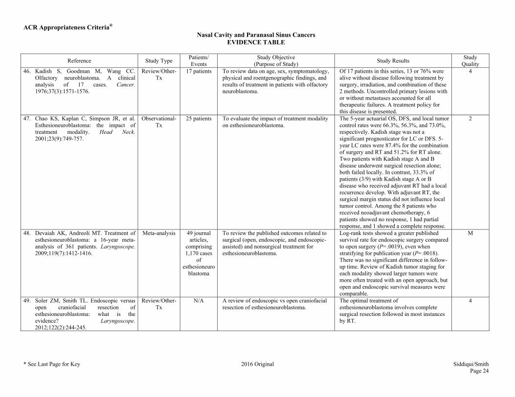
Quality 46. Kadish S, Goodman M, Wang CC.
Olfactory neuroblastoma. A clinical analysis of 17 cases. Cancer. 1976;37(3):1571-1576.
Review/Other-Tx
17 patients To review data on age, sex, symptomatology, physical and roentgenographic findings, and results of treatment in patients with olfactory neuroblastoma.
Of 17 patients in this series, 13 or 76% were alive without disease following treatment by surgery, irradiation, and combination of these 2 methods. Uncontrolled primary lesions with or without metastases accounted for all therapeutic failures. A treatment policy for this disease is presented.
4
47. Chao KS, Kaplan C, Simpson JR, et al. Esthesioneuroblastoma: the impact of treatment modality. Head Neck. 2001;23(9):749-757.
Observational-Tx
25 patients To evaluate the impact of treatment modality on esthesioneuroblastoma.
The 5-year actuarial OS, DFS, and local tumor control rates were 66.3%, 56.3%, and 73.0%, respectively. Kadish stage was not a significant prognosticator for LC or DFS. 5-year LC rates were 87.4% for the combination of surgery and RT and 51.2% for RT alone. Two patients with Kadish stage A and B disease underwent surgical resection alone; both failed locally. In contrast, 33.3% of patients (3/9) with Kadish stage A or B disease who received adjuvant RT had a local recurrence develop. With adjuvant RT, the surgical margin status did not influence local tumor control. Among the 8 patients who received neoadjuvant chemotherapy, 6 patients showed no response, 1 had partial response, and 1 showed a complete response.
2
48. Devaiah AK, Andreoli MT. Treatment of esthesioneuroblastoma: a 16-year meta-analysis of 361 patients. Laryngoscope. 2009;119(7):1412-1416.
Meta-analysis 49 journal articles,
comprising 1,170 cases
of esthesioneuro
blastoma
To review the published outcomes related to surgical (open, endoscopic, and endoscopic-assisted) and nonsurgical treatment for esthesioneuroblastoma.
Log-rank tests showed a greater published survival rate for endoscopic surgery compared to open surgery (P=.0019), even when stratifying for publication year (P=.0018). There was no significant difference in follow-up time. Review of Kadish tumor staging for each modality showed larger tumors were more often treated with an open approach, but open and endoscopic survival measures were comparable.
M
49. Soler ZM, Smith TL. Endoscopic versus open craniofacial resection of esthesioneuroblastoma: what is the evidence? Laryngoscope. 2012;122(2):244-245.
Review/Other-Tx
N/A A review of endoscopic vs open craniofacial resection of esthesioneuroblastoma.
The optimal treatment of esthesioneuroblastoma involves complete surgical resection followed in most instances by RT.
4
ACR Appropriateness Criteria® Nasal Cavity and Paranasal Sinus Cancers
EVIDENCE TABLE
* See Last Page for Key 2016 Original Siddiqui/Smith Page 25
Reference Study Type Patients/ Events
Study Objective (Purpose of Study) Study Results Study
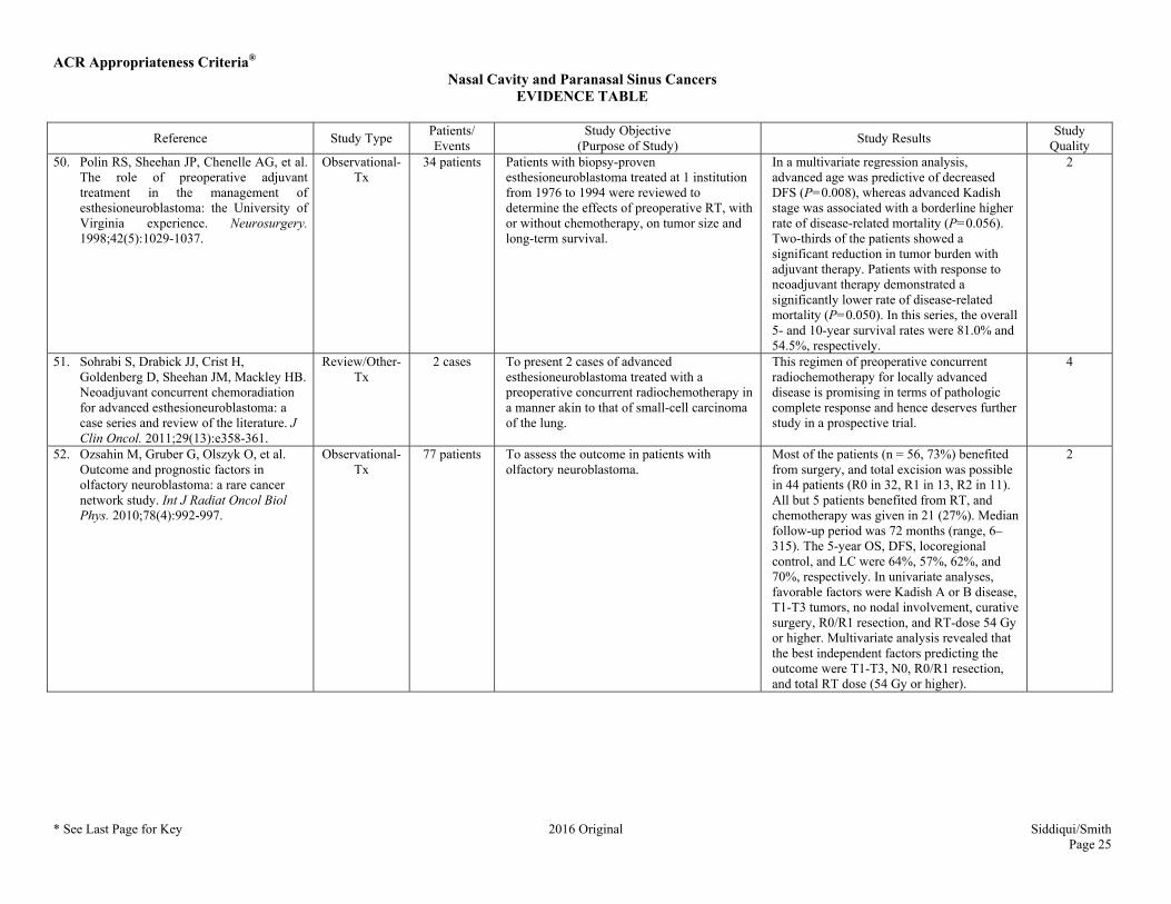
Quality 50. Polin RS, Sheehan JP, Chenelle AG, et al.
The role of preoperative adjuvant treatment in the management of esthesioneuroblastoma: the University of Virginia experience. Neurosurgery. 1998;42(5):1029-1037.
Observational-Tx
34 patients Patients with biopsy-proven esthesioneuroblastoma treated at 1 institution from 1976 to 1994 were reviewed to determine the effects of preoperative RT, with or without chemotherapy, on tumor size and long-term survival.
In a multivariate regression analysis, advanced age was predictive of decreased DFS (P=0.008), whereas advanced Kadish stage was associated with a borderline higher rate of disease-related mortality (P=0.056). Two-thirds of the patients showed a significant reduction in tumor burden with adjuvant therapy. Patients with response to neoadjuvant therapy demonstrated a significantly lower rate of disease-related mortality (P=0.050). In this series, the overall 5- and 10-year survival rates were 81.0% and 54.5%, respectively.
2
51. Sohrabi S, Drabick JJ, Crist H, Goldenberg D, Sheehan JM, Mackley HB. Neoadjuvant concurrent chemoradiation for advanced esthesioneuroblastoma: a case series and review of the literature. J Clin Oncol. 2011;29(13):e358-361.
Review/Other-Tx
2 cases To present 2 cases of advanced esthesioneuroblastoma treated with a preoperative concurrent radiochemotherapy in a manner akin to that of small-cell carcinoma of the lung.
This regimen of preoperative concurrent radiochemotherapy for locally advanced disease is promising in terms of pathologic complete response and hence deserves further study in a prospective trial.
4
52. Ozsahin M, Gruber G, Olszyk O, et al. Outcome and prognostic factors in olfactory neuroblastoma: a rare cancer network study. Int J Radiat Oncol Biol Phys. 2010;78(4):992-997.
Observational-Tx
77 patients To assess the outcome in patients with olfactory neuroblastoma.
Most of the patients (n = 56, 73%) benefited from surgery, and total excision was possible in 44 patients (R0 in 32, R1 in 13, R2 in 11). All but 5 patients benefited from RT, and chemotherapy was given in 21 (27%). Median follow-up period was 72 months (range, 6–315). The 5-year OS, DFS, locoregional control, and LC were 64%, 57%, 62%, and 70%, respectively. In univariate analyses, favorable factors were Kadish A or B disease, T1-T3 tumors, no nodal involvement, curative surgery, R0/R1 resection, and RT-dose 54 Gy or higher. Multivariate analysis revealed that the best independent factors predicting the outcome were T1-T3, N0, R0/R1 resection, and total RT dose (54 Gy or higher).
2

ACR Appropriateness Criteria® Nasal Cavity and Paranasal Sinus Cancers
EVIDENCE TABLE
* See Last Page for Key 2016 Original Siddiqui/Smith Page 26
Reference Study Type Patients/ Events
Study Objective (Purpose of Study) Study Results Study
Quality 53. Monroe AT, Hinerman RW, Amdur RJ,
Morris CG, Mendenhall WM. Radiation therapy for esthesioneuroblastoma: rationale for elective neck irradiation. Head Neck. 2003;25(7):529-534.
Observational-Tx
22 patients To describe the University of Florida experience using RT in the treatment of esthesioneuroblastoma, particularly the use of elective nodal irradiation.
Rates of LC, cause-specific survival, and absolute survival at 5 years were 59%, 54%, and 48%, respectively. The cause-specific survival rate at 5 years was lower after primary RT (17%) than after craniofacial resection and postoperative RT (56%). Cervical metastases occurred in 6/22 patients (27%). No neck recurrences occurred in 11 patients treated with ENI compared with 4 neck recurrences in 9 patients (44%) not receiving ENI (P=.02).
2
54. Zanation AM, Ferlito A, Rinaldo A, et al. When, how and why to treat the neck in patients with esthesioneuroblastoma: a review. Eur Arch Otorhinolaryngol. 2010;267(11):1667-1671.
Review/Other-Tx
N/A To provide a comprehensive review of the incidence of N+ disease at presentation, make recommendations about the optimal treatment strategy of patients with N+ disease, explain the role of elective neck treatment in patients with N0 disease, and comment on treatment of patients with late cervical metastases that require salvage therapy, using the literature review of the incidence and treatment of neck disease in patients with esthesioneuroblastoma.
The review revealed an approximately 5%–8% incidence of cervical nodal metastasis at the time of presentation. Combined modality therapy with surgery and RT is recommended to treat the N+ neck at the time of diagnosis and later. Chemotherapy may have a role combined with radiation treatment, but there are little data to support this. There is limited evidence to substantiate the use of elective neck dissection or elective RT in the clinically and radiologically N0 neck. Patients who have late cervical metastases have a clear survival advantage (59% vs 14%) when treated with combined surgery and RT relative to single modality methods alone. The results indicate that the management of the neck in esthesioneuroblastoma continues to be a significant challenge in the treatment algorithm of these complex patients.
4
55. Resto VA, Eisele DW, Forastiere A, Zahurak M, Lee DJ, Westra WH. Esthesioneuroblastoma: the Johns Hopkins experience. Head Neck. 2000;22(6):550-558.
Observational-Tx
27 patients To examine authors' experience with esthesioneuroblastoma at The Johns Hopkins Hospital. Patient demographics, prognostic factors, and treatment regimen outcomes are reported.
85% of patients had surgical resection as part of their disease management. Complete surgical resection was achieved in 62% of patients who had a craniofacial resection. 80% of patients with negative surgical margins remain with no evidence of disease, with a median follow-up of 5.6 years. Adjuvant RT was beneficial to 62% of patients with positive surgical margins. Clinical responses were observed with cisplatin- and etoposide-containing chemotherapy regimens in patients with advanced disease. A revised staging system based on our experience is proposed.
3

ACR Appropriateness Criteria® Nasal Cavity and Paranasal Sinus Cancers
EVIDENCE TABLE
* See Last Page for Key 2016 Original Siddiqui/Smith Page 27
Reference Study Type Patients/ Events
Study Objective (Purpose of Study) Study Results Study
Quality 56. Frierson HF, Jr., Mills SE, Fechner RE,
Taxy JB, Levine PA. Sinonasal undifferentiated carcinoma. An aggressive neoplasm derived from schneiderian epithelium and distinct from olfactory neuroblastoma. Am J Surg Pathol. 1986;10(11):771-779.
Review/Other-Tx
8 cases To describe cases of a highly aggressive undifferentiated carcinoma of the nasal cavity and paranasal sinuses.
6 tumors extended into the orbital bones, and 5 penetrated the cranial cavity. 5 patients died of disease from 1 to 41 months after diagnosis (median: 4 months), and 3 are alive with tumor <1 year following diagnosis. Microscopically, the neoplasms formed nests, trabeculae, and sheets containing medium-sized cells with small to moderate amounts of eosinophilic cytoplasm. A high mitotic rate, tumor necrosis, and prominent vascular permeation were characteristic. 7 neoplasms were immunoreactive for cytokeratin, 5 for epithelial membrane antigen, and 4 for neuron-specific enolase. Ultrastructurally, occasional small desmosomes and rare membrane-bound, dense-core granules were observed.
4
57. Ejaz A, Wenig BM. Sinonasal undifferentiated carcinoma: clinical and pathologic features and a discussion on classification, cellular differentiation, and differential diagnosis. Adv Anat Pathol. 2005;12(3):134-143.
Review/Other-Tx
N/A To review clinical and pathologic features of sinonasal undifferentiated carcinoma. Article also discusses classification, cellular differentiation, and differential diagnosis.
No results stated in abstract. 4
58. Chen AM, Daly ME, El-Sayed I, et al. Patterns of failure after combined-modality approaches incorporating radiotherapy for sinonasal undifferentiated carcinoma of the head and neck. Int J Radiat Oncol Biol Phys. 2008;70(2):338-343.
Observational-Tx
21 patients To report the clinical outcome of patients treated with combined-modality approaches for sinonasal undifferentiated carcinoma of the head and neck.
The 2- and 5-year estimates of LC were 60% and 56%, respectively. There was no difference in LC according to initial treatment approach, but among the 19 patients who underwent surgery the 5-year LC rate was 74% for those with gross tumor resection, compared with 24% for those with subtotal tumor resection (P=0.001). The 5-year rates of OS and distant metastasis-free survival were 43% and 64%, respectively. Late complications included cataracts (2 patients), lacrimal stenosis (1 patient), and sino-cutaneous fistula (1 patient).
2

ACR Appropriateness Criteria® Nasal Cavity and Paranasal Sinus Cancers
EVIDENCE TABLE
* See Last Page for Key 2016 Original Siddiqui/Smith Page 28
Reference Study Type Patients/ Events
Study Objective (Purpose of Study) Study Results Study
Quality 59. Reiersen DA, Pahilan ME, Devaiah AK.
Meta-analysis of treatment outcomes for sinonasal undifferentiated carcinoma. Otolaryngol Head Neck Surg. 2012;147(1):7-14.
Meta-analysis 30 studies with 167
cases
To review the published outcomes regarding sinonasal undifferentiated carcinoma since the initial description in 1986. The article attempts to 1) understand and better describe the benefit and survival advantages associated with using RT, chemotherapy, and surgical treatment, and 2) support the recommendations of a treatment regimen with current available data in the literature.
Follow-up range was 1 to 195 months (mean 23.4 months, median 15 months). At last follow-up, 26.3% of patients were alive with no evidence of disease, 21.0% were alive with disease, and 52.7% were dead of disease. The use of surgery was found to be the best single modality, but chemotherapy and RT were important as adjuncts in extensive and aggressive disease. The presence of neck metastases was a poor prognostic sign.
M
60. Tanzler ED, Morris CG, Orlando CA, Werning JW, Mendenhall WM. Management of sinonasal undifferentiated carcinoma. Head Neck. 2008;30(5):595-599.
Observational-Tx
15 patients To report the outcomes of treatment for sinonasal undifferentiated carcinoma.
7 patients (47%) developed a recurrence from 3 to 50 months (median, 9) after treatment. The 3-year outcomes were: LC, 78%; locoregional control, 65%; distant metastasis-free survival, 82%; cause-specific survival, 77%, and survival, 67%. The LC rates vs treatment modality were: surgery, 0/1 (0%); surgery and postoperative RT, 7/7 (100%); preoperative RT and surgery, 2/2 (100%); and definitive RT, 2/5 (40%). One patient (7%) treated with surgery and postoperative RT sustained a fatal complication.
2
ACR Appropriateness Criteria®
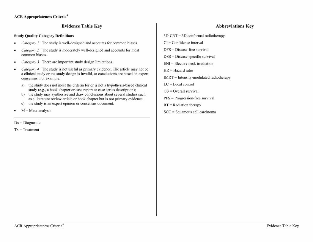
ACR Appropriateness Criteria® Evidence Table Key
Evidence Table Key
Study Quality Category Definitions
Category 1 The study is well-designed and accounts for common biases.
Category 2 The study is moderately well-designed and accounts for most common biases.
Category 3 There are important study design limitations.
Category 4 The study is not useful as primary evidence. The article may not be a clinical study or the study design is invalid, or conclusions are based on expert consensus. For example:
a) the study does not meet the criteria for or is not a hypothesis-based clinical study (e.g., a book chapter or case report or case series description);
b) the study may synthesize and draw conclusions about several studies such as a literature review article or book chapter but is not primary evidence;
c) the study is an expert opinion or consensus document.
M = Meta-analysis
Dx = Diagnostic
Tx = Treatment
Abbreviations Key
3D-CRT = 3D conformal radiotherapy
CI = Confidence interval
DFS = Disease-free survival
DSS = Disease-specific survival
ENI = Elective neck irradiation
HR = Hazard ratio
IMRT = Intensity-modulated radiotherapy
LC = Local control
OS = Overall survival
PFS = Progression-free survival
RT = Radiation therapy
SCC = Squamous cell carcinoma