national case note review booklet
TRANSCRIPT
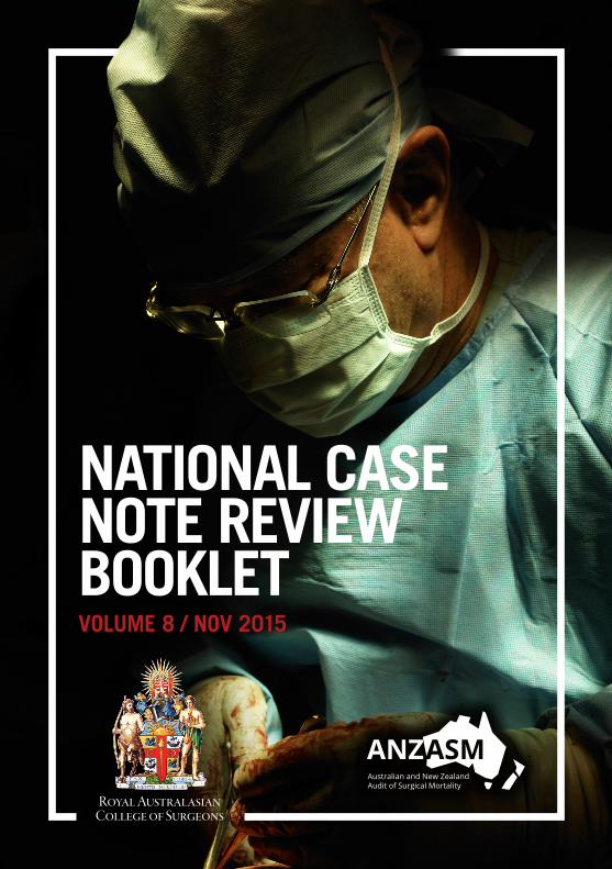
NATIONAL CASE NOTE REVIEW BOOKLETVOLUME 8 / NOV 2015
The information contained in this case note review booklet has been prepared on behalf of the Royal Australasian College of Surgeons, Australian Audit of Surgical Mortality Steering Committee. The Australian and New Zealand Audit of Surgical Mortality, including the Western Australian, Tasmanian, South Australian, Australian Capital Territory, Northern Territory, New South Wales, Victorian and Queensland Audits of Surgical Mortality, has protection under the Commonwealth Qualified Privilege Scheme under Part VC of the Health Insurance Act 1973 (gazetted 23 August 2011).
Royal Australasian College of Surgeons Australian and New Zealand Audit of Surgical Mortality 199 Ward Street North Adelaide SA 5006 Australia
Telephone: +61 8 8219 0900 Facsimile: +61 8 8219 0999 Email: [email protected]
Website: surgeons.org/for-health-professionals/audits-and-surgical-research/anzasm.aspx
ContentsChairman’s Report ......................................................................................4
ANZASM Clinical Editor’s Report ...............................................................5
Recommendations .....................................................................................6
Case Studies ..............................................................................................7
Case study 1: Delay in diagnosis of fractured neck of femur can be fatal .......................... 7
Case study 2: Postoperative deterioration poorly managed by junior staff ........................ 9
Case study 3: Should humeral internal fixation have been performed? ........................... 10
Case study 4: Earlier consultant involvement needed ...................................................... 11
Case study 5: Narcotic analgesia in the elderly ................................................................ 13
Case study 6: Failure to recognise an in-hospital fall as an adverse event....................... 14
Case study 7: Missed acute abdomen following a fall ...................................................... 14
Case study 8: Documentation - a simple insurance policy ............................................... 15
Case study 9: Postoperative low urine output .................................................................. 16
Case study 10: Late at night – not a good idea ................................................................ 18
Case study 11: Beware of ankylosing spondylitis, falls and spinal injury ......................... 19
Case study 12: Preoperative assessment of clotting state not done ................................ 20
Case study 13: Late referral in subdural haematoma ........................................................ 21
Case Study 14: Delay in diagnosis of colonic tumour and inappropriate treatment ......... 23
Case study 15: Delay in diagnosis of pseudomembranous colitis .................................... 25
Case study 16: Elderly and frail patient sustained a fall when self-ambulating ................ 26
Case study 17: Initial underestimation of the seriousness of the problem ........................ 27
Case study 18: Another drunk – or perhaps a head injury? .............................................. 29
Shortened Forms ......................................................................................30
Contact details .........................................................................................30
4 National Case Note Review Booklet / Volume 8 / Nov 2015
Chairman’s ReportHospital, care facility and home falls, and mortality is the theme of this, the 8th National Case Note Review Booklet. The increased focus on “falls” in the hospital environment as a quality measure has been welcomed by those concerned in improving the standards within our health system.
Deaths from falls are largely preventable deaths if an appropriate environment can be delivered and maintained around our patients.
With increasing numbers of single rooms, ageing patients and more interventions in the elderly, falls as an important issue are here to stay. Deaths always gain attention but in the case of falls they represent the tip of an expensive and morbid iceberg of care. This is a topic that requires our attention and engagement.
I trust you find this booklet an educational opportunity and welcome any constructive feedback.
Professor Guy Maddern Chair, Australian and New Zealand Audits of Surgical Mortality (ANZASM)
5National Case Note Review Booklet / Volume 8 / Nov 2015
ANZASM Clinical Editor’s ReportThe eighth booklet includes cases from all states and territories and forms part of the feedback process that is seen as essential in the quality improvement processes of the audits of surgical mortality.
A national booklet is produced to provide a wider readership to cases from various states. It also assists smaller states who do not have enough cases to produce their own booklet and may have difficulty in adequately de-identifying cases. The larger states will continue to publish their own case note review booklets as well as contribute to the national booklet.
The cases in this booklet are focussed on events arising as a consequence of falls – before hospital and within hospital. There is a heavy preponderance of orthopaedic cases as one would expect as falls often produce orthopaedic injuries. However the lessons to be learned are common to all specialties. Some have been edited to focus on a few points in a complex story or to reduce the length of the report. There is variability in the writing style as the text is, in general, written by
assessors and treating surgeons and not by the editor.
There may be cases where readers may not entirely agree with the assessment and comments but if we have stimulated you to think about the case we have succeeded in our aim. Correspondence and questions about specific cases are welcome but ANZASM cannot provide identifying information, however may be able to explain the case in more detail than we have in this booklet.
As the ANZASM office is in the same building as the South Australian Audit of Perioperative Mortality (SAAPM) office, it seemed logical that the final clinical editing process would be done by the Clinical Director of SAAPM on behalf of ANZASM. I must emphasise that I did not write this booklet. The real authors are the treating surgeons, the clinical directors, and the first- and second-line assessors of the various states and territories. To the assessors and the treating surgeons we all owe a debt of gratitude as this publication would not be possible without them. Please learn from these cases.
Glenn McCulloch Clinical Director, SAAPM Clinical Editor, National Case Note Review Booklet, ANZASM
6 National Case Note Review Booklet / Volume 8 / Nov 2015
Recommendations• In complex cases there needs to
be clear, demonstrable leadership in patient management. There should be regular team meetings involving all disciplines to ensure the treatment plan is understood by all.
• Communication is one of the most essential factors in good patient care. This includes communication between surgeons and their junior staff, between disciplines, and between hospitals, particularly in relation to the transfer of critically ill patients.
• All clinicians should provide clear and relevant records. Some of the cases in this report had record keeping deficiencies.
• The surgical case form must contain good and accurate documentation. It should be completed by a team member who was involved in the care of the patient and has sufficient experience to contribute in a useful fashion to the audit process. In instances where the surgical case form is completed by a junior staff member, a consultant should check the completed form or provide advice in advance on salient points that need to be recorded.
Even unpalatable truths should be stated in the form.
• All clinicians should keep in mind that clinical deterioration of a patient, in the absence of a clear cause, may be related to something outside their particular specialty.
• Elderly, frail, confused or very sick patients are at greater risk of falls. There needs to be an increased vigilance of care-givers in this group of patients.
• Proper deep vein thrombosis (DVT) prophylaxis is critical in the care of acute surgical patients. Proper care includes the correct dosage, the correct drug and timely commencement of treatment.
• Consultants should be actively involved in the care of their patients, including in the decision-making process. They have an obligation to make personal entries in the case record of their thoughts that led to the decision. They should also be willing to obtain other opinions if something is not right.
7National Case Note Review Booklet / Volume 8 / Nov 2015
Case StudiesCase study 1: Delay in diagnosis of fractured neck of femur can be fatalCASE SUMMARY
This very elderly patient presented to an emergency department (ED) following an unwitnessed fall. The patient complained of right hip pain and was unable to walk. Plain x-rays of the pelvis and hip did not show a fracture. The patient was discharged back to a high-care nursing home but, due to ongoing pain and immobilisation, returned to hospital one week later. Further x-rays, including a computed tomography (CT) scan, showed a subcapital fracture of the neck of the right femur. The patient was on clopidogrel and there was a delay in being able to operatively treat the fractured neck of femur for five days. It was decided that the fracture would be treated with a Moore’s unipolar hemiarthroplasty. After reaming, the Moore’s prosthesis could not be inserted and the decision was made to change to a cemented Unitrax Exeter femoral stem. In the process of passing a plug down the femoral canal to block the passage of cement distally, the patient developed significant bradycardia and, subsequently,
a cardiac arrest. The patient was resuscitated intraoperatively and the wound closed, leaving the patient with, in effect, an excisional arthroplasty of the hip. The patient’s condition continued to deteriorate and she died soon afterwards.
CLINICAL LESSONS
It is evident that on the initial presentation the patient had a fracture of the neck of the right femur. The casualty resident noted that she had pain when rotating and abducting the hip. A so-called normal initial x-ray does not exclude a fracture of the hip which was clinically confirmed by the ongoing pain and failed to mobilise.
On re-presentation one week later it was commented that there was a subcapital fracture of the neck of the right femur noted on x-rays and CT scanning. In the documentation, however, there was very little evidence in relation to whether this fracture was displaced or undisplaced. There was one comment about impaction of the fracture. The patient was assessed preoperatively from both a medical and anaesthetic point of view. The regular medication of clopidogrel was the major factor associated with the delay in operative treatment of the
8 National Case Note Review Booklet / Volume 8 / Nov 2015
hip fracture. The treating orthopaedic team identified the patient to be of a small build and to have a small femur and pelvis on radiological investigation. It was decided to carry out a Moore’s hemiarthroplasty. This was, presumably, on the basis that the patient walked minimally and had a displaced subcapital fracture of the neck of the right femur.
It is very difficult to template x-rays in relation to the use of a Moore’s prosthesis. The current use of computerised x-ray systems also makes it very difficult to template prostheses. Intraoperatively, the Moore’s prosthesis could not be seated. It was decided to use an Exeter type small stem to circumvent the problem. The equipment required for the Exeter prosthesis was not open and ready for use. It had to be retrieved from the theatre storeroom and opened. Clearly this added time to the length of the surgical procedure, and it is probable, as is often the case after a period of fasting and intraoperative blood loss, that around this time the patient was relatively under-volumed. The procedure of passing a femoral plug down a tight canal is equivalent to passing a nail. It would seem that this procedure resulted in some sort of embolic phenomenon that resulted in cardiovascular collapse. The other possibility in this patient is that a pulmonary embolism occurred,
as the patient had been in bed for 12 days after the fracture. Initial resuscitation was successful but postoperatively the patient quickly deteriorated and died.
IN SUMMARY
1. The decision to discharge the patient on the first attendance without adequately radiologically excluding a hip fracture was incorrect. The casualty resident made the diagnosis clinically. We need to remind ourselves that diagnoses in medicine and surgery are based on history and examination. Radiological investigations may be confirmatory of the clinical diagnosis.
2. The use of an irreversible antiplatelet agent disrupted the appropriate management of the patient. We need to ask whether or not an elderly patient should be on an irreversible antiplatelet agent (versus aspirin etc.). The scientific literature overwhelmingly indicates that patients with fractures of the neck of the femur must be treated operatively as soon as possible in order to minimise complications and the likelihood of death.
3. There was insufficient information to determine whether the subcapital fracture of the neck of the femur was significantly displaced. If there was complete displacement, that is, a Garden grade 3 or Garden grade 4
9National Case Note Review Booklet / Volume 8 / Nov 2015
fracture, then excision of the femoral head and hemiarthroplasty, either via a Moore’s prosthesis or cemented unipolar hemiarthroplasty, was appropriate. If the subcapital fracture was undisplaced or minimally displaced, then consideration could have been given to fixation with a hip screw or cannulated screws. This is a less invasive procedure as it does not open up the hip joint and expose the patient to all the risks of partial or total hip arthroplasty.
Some surgeons would be willing to carry out fixation with a hip screw or cannulated screws soon after the fracture even in the setting of a patient on clopidogrel.
Earlier diagnosis, less operative delay and a wider choice of operative procedures may have led to a better clinical outcome. However, given that the patient was very elderly and had a history of a previous transient ischaemic attack and possible heart disease, the potential for significant complications and even death were high at the time of admission.
Case study 2: Postoperative deterioration poorly managed by junior staffCASE SUMMARY
An elderly patient was admitted to hospital following a fall with a fractured right subtrochanteric neck
of femur. The patient had previously been admitted to several geriatric wards for assessment of increasing confusion, agitation and aggression. The patient suffered an unwitnessed fall the day prior to the admission and underwent surgery the day following admission. During the procedure a right cephalomedullary femoral nail was inserted with additional cable fixation. The patient suffered a relatively rapid postsurgical decline and passed away approximately 30 hours after the procedure.
CLINICAL LESSONS
During the period from surgery to death the patient was seen several times by junior medical staff. There were concerns regarding the patient’s condition, particularly the urine output, low haemoglobin and respiratory function. This case reveals several issues, as outlined below.
• It was unclear from the available information whether the consultant orthopaedic surgeon was present at the time of surgery. Given the difficulty of fixation of subtrochanteric femoral fractures, it is my opinion that these surgeries need to be performed under the direct supervision of a consultant orthopaedic surgeon or a senior registrar with experience in the fixation of these fractures. It was
10 National Case Note Review Booklet / Volume 8 / Nov 2015
unclear from the initial case notes whether this occurred.
• It appears that there was minimal supervision of the junior medical staff. In the period from the surgery to the patient’s death, the patient was reviewed five times by the surgical intern. The medical registrar was spoken to on each of these occasions but did not attend the patient. The patient had two further medical emergency team (MET) calls during this period and it is unclear who attended the patient. There were ongoing issues regarding the patient’s fluid balance and there is no record of any senior doctor addressing this issue. There appears to have been a lack of clinical support for the junior medical staff who were managing a difficult and complex postoperative patient.
• There was a lack of documentation regarding the patient’s not for resuscitation (NFR) status. There was no formal documentation in the case notes of the NFR status, apart from one entry in the admission note stating “not for resuscitation (NFR), For MET call”. Best practice would be for these end-of-life decisions to be well documented on an independent form. Proper
documentation would allow timely, reasonable and rational decisions to be made at times of patient difficulty.
Case study 3: Should humeral internal fixation have been performed?CASE SUMMARY
This patient in her 80s was taken to hospital for treatment of hypoglycaemia and a right neck of humerus fracture sustained in an unwitnessed fall. The patient had type 2 diabetes and the fall was presumed to be secondary to hypoglycaemia. There was evidence that the patient’s blood sugar level was 8.4 mmol/L at the time she went to bed, at approximately 2000. At around 2200 the ambulance team found the patient’s blood sugar level to be 2.0 mmol/L.
The patient received appropriate and prompt medical treatment on arrival and was admitted under an acute medical unit with orthopaedic consultation. The medical treatment throughout was timely and appropriate. In consultation with the family it was decided that the patient was “not for extraordinary measures” in terms of resuscitation.
The initial orthopaedic treatment was for non-operative management, with a hanging cast
11National Case Note Review Booklet / Volume 8 / Nov 2015
and serial x-rays. This was trialled for two weeks, at which time the treating orthopaedic consultant determined that the fracture position was unacceptable and would be unlikely to heal. Despite this, it is repeatedly documented that the patient was comfortable and would likely need placement in a residential care facility.
The patient subsequently underwent an open reduction with internal fixation of the fracture performed by a different, yet very senior and experienced orthopaedic consultant. Surgery was uneventful, with a good immediate recovery period.
Over the next week the patient slowly “went off” medically, developing hepatic encephalopathy and acute on chronic renal failure. In the early hours of the morning the patient vomited and was noted to immediately increase her respiration rate. Coincidentally, the ward call doctor was on the ward and attended within one minute. Appropriate resuscitation techniques were used but were unsuccessful.
CLINICAL LESSONS
The question, in this case, is whether the patient should have been operated on at all. A patient in their 80s, with a large number of seemingly poorly controlled medical problems, no real prospects of living
independently, and who only seemed to have mild pain (based on reading of the chart) would be candidates for ongoing conservative measures.
Case study 4: Earlier consultant involvement neededCASE SUMMARY
An elderly man suffered a fall in a care home on a Saturday. He arrived in a peripheral hospital ED and was found to have a fractured hip. The background included dementia and hypertension and limited walking capacity needing a frame. The admission full blood counts included a haemoglobin 80g/L and white blood cell count of 26,000 cells/uL.
He was transferred to a teaching hospital, arriving on the orthopaedic ward at 1900 on the Sunday. A chest x-ray revealed a left hilar mass and probable left lower lobe infection. The patient was reviewed by the anaesthetic team shortly after arrival and was thought “unlikely to be fit for OT [operating theatre] tomorrow”, “needs medical review” and “needs echo”. The first orthopaedic review appears to have been some 22 hours after the patient’s arrival on the ward. There is no written evidence that a consultant was present. Antibiotics were commenced for the chest infection.
12 National Case Note Review Booklet / Volume 8 / Nov 2015
The next orthopaedic note, made at some 40 hours after admission, was by the intern with no evidence of any input by the consultant. The intern wrote “has been cancelled for theatre again today... hopefully tomorrow”. The echo was done that afternoon.
Almost exactly 48 hours after admission to the orthopaedic ward the patient was seen by the orthogeriatric team. There is a note, for the first and only time in the entire folder, that the patient had known myelodysplasia and was transfusion dependent. Although not precisely stated, this appears to be a previously established diagnosis. The note stated that the mass on the chest x-ray was “not for further investigations due to age - likely neoplasm. Plan - review post-op”.
Some five hours after that review a MET call was made. Cardiopulmonary resuscitation was undertaken and appears to have lasted for some 50 minutes before being terminated.
The case was referred to the coroner due to the fall in the care facility. The postmortem revealed a primary bronchogenic cancer.
CLINICAL LESSONS
The orthopaedic consultant returned the audit proforma marked “terminal care” and did not complete the rest of the proforma. This does not seem
consistent with the care offered to this patient. If the patient was for terminal care then questions need to be asked as to why the patient:
• was clearly being worked up for theatre
• was sent for an echo in anticipation of the surgery
• was commenced on antibiotics for a chest infection
• did not complete an NFR form
• received cardiopulmonary resuscitation for almost one hour.
There was no evidence in the notes that the patient was ever seen by the consultant orthopaedic surgeon. This may be why there appears to be a disconnect between what was written on the audit proforma (terminal care) and the actual care received. Had the consultant reviewed the patient it is likely the patient would have been assessed as highly unlikely to survive any surgery and death inevitable, with terminal care a priority
There was a delay of over 12 hours in transferring the patient from the care home to the peripheral hospital ED. There was a further delay of over 12 hours before the patient arrived in an orthopaedic ward of the teaching hospital. A delay of 24 hours to surgery following a hip fracture increases mortality. Such falls are a
13National Case Note Review Booklet / Volume 8 / Nov 2015
predictable event, and care homes and peripheral hospitals need to review their processes to speed up such referrals. Although this patient’s death was not in any way related to the apparent lack of consultant input, the lack of consultant decision making was not conducive to good terminal care.
Editor’s note: There is often confusion in the reporting surgeons’ minds as to what is meant by the term “admitted for terminal care”. If at the time of admission a decision is made that palliation will be the direction taken then the case is correctly identified as “for terminal care”. If however there are investigations performed or further consultations obtained and then a decision is made to palliate the patient it is NOT classified as “admitted for terminal care”. The logic to this is that a decision-making process has taken place after admission and after gaining new information, and this decision-making process needs to be independently evaluated.
Case study 5: Narcotic analgesia in the elderlyCASE SUMMARY
A patient in her 70s was admitted to hospital A following a fall and was complaining of pain in the thigh. She was taking medications
for hypertension and chronic airway disease. An initial x-ray showed possible undisplaced fractures of the left ilium and pubic bones. Orthopaedic opinion was that the patient should have bed rest and mobilisation when the pain improved. A CT scan of the pelvis, however, was said to show ‘significant fractures’. Skeletal traction was applied.
Pain management was difficult after admission and the patient required large doses of narcotic analgesics. She then received oral oxycodone and was given a total of 30 mg on day two, 40 mg on day three, 50 mg on day four and 35 mg on day five. In the evening of day five, the patient was found to be unrousable, with pinpoint pupils and vomit on the sheets. She was managed in the intensive care unit (ICU) for aspiration pneumonia and septic shock. Treatment was unsuccessful.
CLINICAL LESSONS
Oral oxycodone is approximately twice as potent as oral morphine. This elderly patient received an excessive dose over the four days. Care is required when prescribing narcotic analgesics in general, and particularly in elderly patients. This should be considered when prescribing oxycodone. Alternative strategies to reduce narcotic dosage might include patient-controlled
14 National Case Note Review Booklet / Volume 8 / Nov 2015
analgesia or a multimodal approach, using regular paracetamol and/or non-steroidals, if appropriate.
Skeletal traction effectively immobilises a patient. The risk of inhalation of vomitus is increased, particularly in the elderly. Gastric emptying is inhibited by narcotic analgesia, leading to an increased risk of aspiration1.
Case study 6: Failure to recognise an in-hospital fall as an adverse eventCASE SUMMARY
An elderly patient was admitted with a Garden grade 1 undisplaced fracture of the neck of femur after a fall. The patient had numerous medical comorbidities, as a result of which a decision was made to treat the patient conservatively. The patient made good progress and eight days later had a trial home visit with the occupational therapist.
On the ninth day a further x-ray showed some movement in the fracture, but as the patient was walking with a frame no surgery was planned, and plans were in hand for discharge. On the 10th day the patient fell and a subsequent examination revealed pain in the hip. An entry notes that as a result
1 Liukas A, Kuusniemi K, Aantaa R, Virolainen P, Neuvonen M, Neuvonen PJ, Olkkola KT. Plasma concentrations of oral oxycodone are greatly increased in the elderly. Clin Pharmacol Ther 2008;84(4):462–467
of the fall the fracture had become a displaced Garden grade 4.
In view of this surgery had to be performed. As anticipated (and discussed with the patient and family) the patient had a stormy postoperative course and died from comorbidities some 12 days later.
CLINICAL LESSONS
This death was reported to the coroner. In a letter from the hospital’s Medical Services Department it was implied that the surgery was prompted by the x-ray on the ninth day, rather than the in-hospital fall that caused the fracture to become a grade 4 fracture. This in-hospital fall should have been recorded as an adverse event and included in the information provided to the coroner.
Case study 7: Missed acute abdomen following a fallCASE SUMMARY
An elderly patient suffered knee and apparent rib injuries after a fall. The patient was admitted to an orthopaedic ward. No abdominal signs were apparent on admission. The patient was treated with analgesia and physiotherapy and there were no complaints from the patient.
Several days after being admitted the patient’s general condition caused the nurses to report to the patient’s medical officer, and a physician’s
15National Case Note Review Booklet / Volume 8 / Nov 2015
assessment was arranged. Further investigations suggested some medical problems and intravenous (IV) fluids were initiated.
A general surgeon saw the patient and abdominal signs were obvious. Plain x-ray showed gas under the diaphragm but by this time the patient’s condition was extreme. Resuscitation did not improve the situation and the decision was made not to operate. Death followed rapidly.
CLINICAL LESSONS
This patient should have had daily medical assessments with a full clinical examination and careful history about any symptoms. A possible abdominal viscus perforation would have been suspected much earlier if a reasonable history by a resident or registrar been taken. Failure to obtain an early general surgical consultation almost certainly resulted in this patient’s death.
Having the patient in an orthopaedic ward did not help. It is possible that historical questions about abdominal pain were not forthcoming from the junior medical officer on the orthopaedic team.
This case highlights the difficulty of diagnosis in the elderly, especially where there may be a tendency to stoicism. It also highlights the
necessity for repeated examinations in patients following any trauma.
Case study 8: Documentation - a simple insurance policyCASE SUMMARY
A patient suffering from alcoholism, diabetes, schizophrenia and emphysema was found lying outside a local convenience store with a posterior scalp laceration. It was thought that the patient had fallen, but the patient could not recall any incident.
The ambulance service attended the scene and found the patient to be Glasgow Coma Scale (GCS) 11/15. This deteriorated to GCS 9/15 on arrival at the ED. Shortly after assessment the left pupil was noted to be fixed and dilated. Intubation and ventilation began, and the patient was transferred for a CT scan of the brain.
The CT scan confirmed a large acute on chronic subdural haematoma causing >2 cm midline shift and uncal herniation. A left frontal craniotomy was performed with evacuation of the haematoma and insertion of an intracranial pressure monitor. There were no major difficulties encountered during surgery. There was a slight improvement in the left pupil, with a
16 National Case Note Review Booklet / Volume 8 / Nov 2015
sluggish reaction postoperatively, but the patient remained with GCS 3/15.
The following day a postoperative CT showed significant improvement in the midline shift and uncal herniation, but there was extensive retained haematoma inferiorly with almost complete evacuation superiorly. However, a new problem had arisen - hydrocephalus, mainly supratentorial but also involving the fourth ventricle. Intracranial pressures remained > 20 mmHg.
For reasons that are not documented there was no significant change in management, although the team was clearly notified of intracranial pressures and CT findings. No further surgery was performed and the patient died nearly a week after the operation.
CLINICAL LESSONS
The patient’s postoperative progress was unsatisfactory. Investigations clearly showed significant complications yet there was no documentation regarding the apparent decision to do nothing. The continuation of active treatment in the ICU seems futile and without clear documentation as to why this decision was made. This may have been the correct decision but without documentation that is unknown.
Always document the reasons for the decisions made. It is
impossible, in retrospect, to understand why this treatment course was taken for this patient. Document, document, document - it’s always a great investment.
Case study 9: Postoperative low urine outputCASE SUMMARY
This case involves an elderly patient who was admitted with a displaced subcapital fractured left neck of femur following a mechanical fall on that day. The patient was mentally alert and independent. There was a history of atrial fibrillation medicated with warfarin, and intermittent asthma/bronchitis. Medication included Furosemide and carvedilol for controlled hypertension. The patient was also taking Sinemet for restless legs.
Screening blood tests indicated that the patient had adequate platelets, normal haemoglobin, urea of 10.7 mmol/L and creatinine of 100 µmol/L. The serum potassium was slightly elevated (5.5 mmol/L). The patient was given vitamin K to neutralise the effects of the warfarin and was prepared for theatre on the following day for a bipolar hemiarthroplasty. A small dose of prothrombin was administered just prior to the procedure due to a very slightly
17National Case Note Review Booklet / Volume 8 / Nov 2015
elevated International Normalised Ratio (INR) of 1.5. The patient had a combined spinal and general anaesthetic. There was no mention of blood loss or of any particular problems with the operation or the anaesthetic. On return to the ward the patient was alert and able to have something to eat that evening. The patient was seen at 1850 by the covering surgical intern because of a low urine output, and was ordered a 500 mL bolus of IV fluid. At that time the patient appeared well, was afebrile and had stable observations. There was no mention of wound problems.
The last observations appeared to be taken at 0200 on the first postoperative day. The patient was found deceased later that morning at 0510. There was no mention in the case notes (including the 0200 observations) as to whether the urine output had improved. At that time all observations appeared normal and the patient was alert.
CLINICAL LESSONS
The cause of the sudden and unfortunate death was probably of cardiac origin or possibly a massive stroke. It is doubtful that this death could have been prevented under the circumstances, particularly as the patient appeared to be doing well when seen at 0200.
The assessor was critical, however,
of the failure to mention the urine output following the bolus of fluid given at 1850 the previous evening. It would have been appropriate for this patient to have been reviewed on at least one occasion by the covering medical staff to assess the urinary output problem and general state. This would most likely have happened had the patient been in a high dependency unit (HDU). It is highly doubtful, however, that this patient’s death was the direct result of a poor urine output, unless they became extremely hypotensive between the time of the last observation at 0200 and death at 0510. If this patient had been in an HDU they would have received better monitoring and resuscitation may also have been attempted.
One of the lessons that can be learnt from this case is that the low postoperative urinary output may have predicted ongoing problems that would have justified closer supervision throughout the evening and night. If an HDU bed was not available a special area in the ward for postoperative cases would seem desirable to allow closer observation.
18 National Case Note Review Booklet / Volume 8 / Nov 2015
Case study 10: Late at night – not a good ideaCASE SUMMARY
This elderly lady had multiple medical comorbidities including diabetes, atrial fibrillation, blindness and deafness. She sustained an intertrochanteric fracture of the neck of the femur in a fall.
Surgery was delayed for 24 hours after the patient was admitted to hospital due to hyponatremia. This was a known problem that had been present for many years. General practice advice had been to take salt tablets regularly. The cause for the hyponatremia, however, was never defined. The patient had been on antihypertensive medication for many years and had low-grade renal failure.
The patient underwent open reduction with internal fixation and the operation progressed uneventfully. That evening, following surgery, the patient became hypotensive. An electrocardiogram showed some mild ischaemic changes. Hypotension was attributed to hypovolaemia and she was given several litres of crystalloid in the first 24 hours in an attempt to correct the hypotension.
Urine output was still low and over the next day or two it became obvious that the patient had acute renal failure. A number of medical consultations followed and the ICU team was consulted. It was felt that in view of the extensive comorbidities, advanced age and poor quality of life that neither dialysis nor the use of inotropes were appropriate.
Simple supportive measures were then instituted. This decision was made in close consultation with the family. Despite fluid restriction and non-invasive efforts to treat the renal failure with sodium polystyrene sulfonate (Resonium and multiple doses of Furosemide) the renal function remained poor. The patient’s general condition deteriorated and death from renal failure occurred nearly a week after the surgery.
CLINICAL LESSONS
Early resuscitation following surgery was left to probably the most junior member of the team. It was the first night and the night resident medical officer was responsible for the resuscitation. It is almost certain that this medical officer did not appreciate that postoperative hypotension could be due to factors other than hypovolaemia. A large volume of crystalloid was infused and this was sufficient to cause marked fluid overload over the next few days.
19National Case Note Review Booklet / Volume 8 / Nov 2015
The patient’s renal function remained poor and they were fluid overloaded. It is quite likely that this process hastened the patient’s death, which was ultimately the result of renal failure. Maximum medical efforts did not change the downhill course.
This case illustrates the problems associated with operating on sick people late in the day. The patient returned to the ward following the surgery at 2000. This inevitably meant that the problems surrounding fluid balance and hypovolaemia would be left to the most junior staff member in the late hours of the day or early hours of the morning.
Continuity of care is important, indeed vital, in any clinical setting. Postoperative orders in this instance should have included precise parameters defining haemodynamic and fluid balance stability. A timely telephone call to the medical officer on duty outlining the situation and expectations of treatment, particularly overnight, may have resulted in markedly improved clinical care and possibly a better outcome.
Case study 11: Beware of ankylosing spondylitis, falls and spinal injuryAn elderly patient with a history of recent high alcohol intake, presented to Hospital A following a fall the day before. There was low-back tenderness, with limitation of leg movement apparently due to pain but no sensory impairment. On day 2, he had a fall in the ward and developed respiratory distress. A CT scan of the chest showed a collapse of the right lung and a left haemothorax. The thoracic spine showed extensive paravertebral ossification, with rigid fusion and complete traumatic disruption of the vertebral column at the T8/9 level, with a transverse fracture through the disc space. Although the significance of the instability of the thoracic fracture was recognised, the particular pathology of ankylosing spondylitis was not. The patient was transferred to Hospital B for management of the fracture and the spinal cord injury. In Hospital A, bilateral intercostal catheters were inserted to drain the now bilateral haemothoraces.
A repeat CT scan in Hospital B revealed a complete transection of the vertebral column at T8/9. There were two failed attempts to surgically decompress and stabilise his spine, but he was unable to tolerate the prone position during anaesthesia,
20 National Case Note Review Booklet / Volume 8 / Nov 2015
resulting in a rapid deterioration in his ventilation. Conservative therapy was recommended, but his respiratory function further deteriorated and he was managed with palliation until he died. Although the condition of his thoracic spine was not specifically mentioned in the clinical records in Hospitals A or B, other than the initial x-ray report, the diagnosis of ankylosing spondylitis seems highly likely.
CLINICAL LESSONS
• Management in the referring and tertiary hospitals was entirely appropriate.
• Although an unusual condition, in the setting of trauma, particular care needs to be taken when managing a patient with a spinal injury and ankylosing spondylitis.
• The diagnosis of ankylosing spondylitis was not made in Hospital A. Spinal trauma in ankylosing spondylitis is notorious, not only because fractures may occur with transection of the vertebral column and usually spinal cord compression, but also because of heavy bleeding from the bone, which can cause major blood loss.
An assessment of the risk of falls and their prevention, is of particular importance in patients with ankylosing spondylitis.
Case study 12: Preoperative assessment of clotting state not doneCASE SUMMARY
An elderly man was admitted with a pertrochanteric fracture of the left femoral neck following a fall. Comorbidities included chronic renal failure, hypertension, gastroesophageal reflux, bipolar disease and osteoporosis. The patient was a heavy smoker with a high alcohol intake. The patient’s medications included Alendronate, Asasantin, Astrix, Coversyl and numerous psychotropic drugs.
Initial laboratory investigations indicated that urea and creatinine were elevated, low-normal haemoglobin and low platelets. The INR was reported as 1.1. The patient proceeded to open reduction and internal fixation with a short Gamma nail 10 hours after admission to the ED. Appropriate reduction and positioning were ascertained with guidance from an image intensifier, and thromboprophylaxis was commenced.
Blood loss from the operative site necessitated dressing reinforcement 10 hours after surgery. Two hours later, hypotension and a decreasing level of consciousness with a very low oxygen saturation led to a MET call, intubation and a transfer to
21National Case Note Review Booklet / Volume 8 / Nov 2015
the ICU. Blood was noted in the nasogastric tube and the patient’s haemoglobin was well below normal.
The patient’s renal function and conscious state deteriorated. Treatment was withdrawn following discussions with the family and the patient died four days after admission.
CLINICAL LESSONS
In this case it is evident that the orthopaedic and anaesthetic teams did not undertake adequate preoperative assessment:
• this patient was on platelet inhibitors, with liver and renal disease, and yet did not have their clotting or liver function investigated
• there was no referral made to the medical or renal unit prior to surgery
• provision of postoperative care in an HDU might have led to earlier recognition of complications.
This case also calls into question the appropriateness of the trend towards early streaming of frail emergency admissions into subspecialties like orthopaedics. Medical staff in these units do not always possess the appropriate level of knowledge to manage such patients optimally.
Although it is recognised that surgery
is best performed within 48 hours, careful preoperative assessment and management is essential. This medical care should be continued throughout the postoperative period. All health services involved in the management of the elderly with orthopaedic fractures must have in place a system which allows expert and timely medical care of these patients.
Case study 13: Late referral in subdural haematomaCASE SUMMARY
A man in his mid-40s, presented to the hospital with a grade V acute subarachnoid haemorrhage due to the rupture of a large basilar tip aneurysm. The aneurysm was coiled endovascularly and the patient required a prolonged stay in the ICU.
The medical course was complex including a non-ST elevation myocardial infarction the day following admission with pulmonary oedema and a right cephalic vein thrombosis secondary to a right subclavian central venous catheter. The patient had a percutaneous tracheostomy inserted 10 days after admission and a percutaneous gastrostomy inserted in some weeks later.
The patient eventually recovered
22 National Case Note Review Booklet / Volume 8 / Nov 2015
enough to speak in full sentences, even though confused, and was able to spontaneously move the left arm and left leg. He had a residual right hemiparesis.
He was discharged to another hospital for rehabilitation about 2 months after the initial presentation. He had multiple falls while in the rehabilitation facility and this was associated with increasing headache and confusion.. The patient was found unconscious and was transferred back to the parent hospital, arriving in the ED at 1050. The GCS was 4/15. It was noted that he had been on Clexane 60 mg twice daily. It seems from the notes that the patient was observed in the ED until 1530 when he were transferred to the radiology unit for a CT scan of the brain.
The patient was reviewed by the neurosurgery team at 1600. A CT scan of the head revealed a large right acute subdural haematoma. The patient was taken immediately to the operating room for craniectomy and evacuation of the right subdural haematoma. There was no indication that an intracranial pressure monitor or an external ventricular drain was inserted during the operation. The brain was very swollen according to the operative notes, and the surgeon was unable to close the dura.
The patient was managed in the ICU
after surgery. A postoperative scan of the head performed the following day showed good evacuation of the acute subdural haematoma with a very swollen and oedematous brain, extending markedly through the craniectomy site.
Brain stem reflex testing performed two days after the CT scan confirmed brain death. Life support was ceased and the case was referred to the coroner’s office.
CLINICAL LESSONS
The surgical team reporting on this case indicated that there had been a further admission to the ED about 2 weeks prior to the final admission. These notes were not available. According to the reporting surgical team, a CT scan of the head performed at that time was apparently normal, but there was no consultation with the neurosurgery team. The treating surgical team felt that a neurosurgical referral should have been made due to the patient being on anticoagulation. In addition, they felt that strict and close neurological observations and a follow-up CT scan of the brain would have been recommended, and that this may have led to earlier identification of the acute subdural haematoma.
What is most concerning is that
23National Case Note Review Booklet / Volume 8 / Nov 2015
the patient was admitted to the ED in a coma and with a GCS of 4/15 at 1050, but was not referred to neurosurgery and did not receive a CT scan of the head until 1530. The patient remained deeply comatose during this time with charting by the staff at 1100, 1245, 1340, 1400, 1415, 1445 and 1530. It is very concerning that, despite the charting and recording of the patient’s poor neurological condition, no further action was taken until very late.
The neurosurgical team responded promptly. An urgent craniectomy and evacuation of the acute subdural haematoma was performed. There was no indication that an intracranial pressure monitor or an external ventricular drain was inserted. This may be because the brain was very swollen and the surgeon may have felt it necessary to perform a rapid closure before the brain became too swollen for the dura to be closed.
While it would have been preferable to have an intracranial pressure monitor inserted for postoperative monitoring and management of the patient’s raised intracranial pressure, the assessor did not think that this would have made any difference to the outcome.
Case Study 14: Delay in diagnosis of colonic tumour and inappropriate treatmentCASE SUMMARY
A patient in her mid-70s was admitted following a mechanical fall in which the right wrist was injured. There was a history of diabetes, chronic renal impairment, atrial fibrillation (on warfarin), heart failure and a week-long period of right knee pain. The patient was admitted under orthopaedics with medical and allied health input. Problems arose with tachycardia and bradycardia over 48 hours and cardiology was consulted. Daily reviews occurred.
On day three of admission a pre-MET review was obtained due to tachycardia. The patient was documented to be eating breakfast at the time and there was no record of abdominal pain. A MET was called on day four of admission at 1145. The patient was tachycardic and tachypnoeic with low oxygen saturation, and complained of abdominal pain associated with tenderness on the right with “guarding and peritonism”. Fluid resuscitation was commenced and a CT abdomen ordered which showed a right-sided retroperitoneal collection. The general surgical team was called and a decision made to
24 National Case Note Review Booklet / Volume 8 / Nov 2015
place an image guided percutaneous drain which drained a small amount of faecal fluid.
The patient went to ICU post-drainage but deteriorated. A formal laparotomy and right hemicolectomy for a colonic tumour (ends left inside) was performed just before midnight (11 hours after the MET call). The patient was returned to theatre the following day for a second look (all gut appeared viable) and formation of ileostomy and mucous fistula. Over the next three days the patient’s condition deteriorated, despite ICU care, and the patient died.
CLINICAL LESSONS
Warning signs or symptoms do not appear to have been present before the day of deterioration. The initial admission and examination appears thorough. There was no ‘systems review’ but the patient clearly presented with a mechanical fall with bony injury, and the admitting team can be forgiven for not asking about bowel habits.
Areas of concern: It was a mistake to place a percutaneous drain in an unwell patient who was clearly deteriorating with documented signs of peritonism. The patient should have gone straight to laparotomy but the outcome would have been the same. It would also seem that a large obstructing colonic tumour was
missed on CT scan – this may have been a factor in directing the treating team towards a drain rather than the laparotomy that was needed.
Areas of consideration: There were four consultant surgeons within a 12 hour period making decisions on one patient. It is useful and sometimes beneficial to seek advice or a second opinion, but two of the consultants had not seen the patient and were being guided by an (inaccurate) CT report. The grade of experience of the initial surgeon is unknown.
RECORD KEEPING:
The initial ED clerking, medical history, medications and social history were all documented. There was, however, no documented systems review but the patient didn’t appear to have expressed any abdominal symptoms or bowel problems until the patient deteriorated. Pre-MET and MET records were also very good. Unfortunately, the general surgical notes surrounding the patient’s deterioration, CT results, presumed diagnosis and the decisions to drain and then perform laparotomy are non-existent.
25National Case Note Review Booklet / Volume 8 / Nov 2015
Case study 15: Delay in diagnosis of pseudomembranous colitisCASE SUMMARY
An elderly patient with severe hip osteoarthritis had a past history of hypertension, chronic renal failure, gout and emphysema and was admitted for an elective hip arthroplasty. Biochemistry on admission showed an elevated creatinine (283µmol/L) and hyperkalaemia (5.7mEq/L) An uncomplicated total hip arthroplasty was performed as planned. A stat dose of perioperative antibiotic was continued for four days.
Three days later the patient developed a tachycardia, became confused and had a fall in hospital. Neurological observations were instigated and antibiotics recommenced when it was noted that the patient was febrile. The next day the patient was lethargic, confused and incontinent of faeces. A medical registrar noted abdominal distension. The creatinine had increased to 310 µmol/L. The clindamycin was ceased and MS Contin commenced. The next day the medical registrar noted a distended tympanitic abdomen and peritoneal oedema. Clostridium difficile had been cultured from the stool. The cephazoline was ceased
and the working diagnosis was ‘toxic mega colon’. Vancomycin orally was commenced that evening and a surgical review recommended. The surgical registrar suggested a CT scan if there was any deterioration.
Early the next day the medical registrar again noted hypotension, C. difficile and acute on chronic renal failure. The patient was transferred to the HDU and later to the ICU. A CT scan about midday showed thickening of the colon consistent with pseudomembranous colitis. Various lines were inserted, in the course of which the patient arrested and could not be resuscitated. An autopsy was not performed but the probable cause of death was C. difficile.
CLINICAL LESSONS
This death was not primarily caused by the surgery. The patient exhibited a number of risk factors for C. difficile and toxic megacolon: over 65 years old, hospitalisation, multiple antibiotics and narcotic administration. The first symptoms of infection were present when the patient had diarrhoea and abdominal distension. In spite of C. difficile being cultured, the diagnosis of toxic mega colon was considered but appropriate treatment not instituted. The CT scan confirmed the diagnosis beyond doubt but was undertaken too late to alter the outcome. It is
26 National Case Note Review Booklet / Volume 8 / Nov 2015
unlikely that the minor head injury was significant. It is also unlikely that the procedure being undertaken at the time of arrest was causative of it.
Over the five days prior to being admitted to the ICU the patient was assessed by over 10 doctors, many of whom only assessed the patient on one or two occasions. It is unfortunate that continuity of care cannot be provided in these settings.
Case study 16: Elderly and frail patient sustained a fall when self-ambulatingCASE SUMMARY
This female patient in her early 90’s sustained a fall that caused a fracture of the pubic rami. The side of this injury was not documented. The patient was admitted to hospital for approximately two days and then discharged home under the care of a relative. No further information was provided regarding this admission.
The patient was subsequently brought back to the same hospital by ambulance with a febrile illness. At the time of this admission the patient was documented as having a past history of hypertension and hypercholesterolemia. The diagnosis was Klebsiella sepsis arising from pneumonia. She was admitted under a medical unit and commenced on IV antibiotics, which were subsequently
changed to oral antibiotics. The patient then developed abdominal pain. A diagnosis was made of proctitis associated with C. difficile and further antibiotics were commenced. The patient suffered a prolonged period of diarrhoea, with worsening confusion.
The patient then sustained a fall when self-ambulating on the ward. This fall caused an intertrochanteric fracture of the right femur. The orthopaedic unit was contacted to provide an assessment. The patient underwent internal fixation surgery using a sliding hip screw technique. The surgery was delayed by approximately 24 hours owing to the patient’s severe diarrhoea and general medical condition. The surgery proceeded in a routine fashion under general anaesthetic with an endotracheal tube. The initial postoperative recovery of the patient was routine.
In the early afternoon the patient deteriorated rapidly. There was a worsening of the patient’s conscious state and respiratory condition. After appropriate investigation and initial resuscitation efforts a presumed diagnosis was made of pulmonary embolism. It was not clear whether therapeutic anticoagulation was commenced, but the patient was declared dead on the same day at 1630.
27National Case Note Review Booklet / Volume 8 / Nov 2015
CLINICAL LESSONS
While the majority of the record keeping in this case was adequate, the case notes lacked a thorough account of the initial assessment of the patient by the treating medical team and ED staff. The record keeping is thorough following the time of the patient’s admission, particularly the entries by the orthopaedic unit.
It is important to note that the patient was very elderly and had already suffered a fall with a fracture prior to the fall in hospital that resulted in the hip fracture. One must ask whether it was wise to allow a frail 90 year old to self-ambulate in a weakened, confused state from pneumonia and C. difficile proctitis. I did note that there was satisfactory DVT prophylaxis from the time of her admission to hospital – if the death was due to PE all reasonable preventative measures had been taken.
Case study 17: Initial underestimation of the seriousness of the problemCASE SUMMARY
This patient has a history of steroid dependent rheumatoid arthritis, reflux/gastritis and severe peripheral vascular disease with ischaemic foot ulcers. She was on aspirin. She fell at
a shopping centre onto outstretched arms, fracturing her nose. In hospital her nose was packed with a merocel and rapid rhino.
The patient was referred to the ENT unit and was seen by the ENT registrar. The nasal pack was removed and bleeding from lacerated mucosa was noted. Cautery did not stop the bleeding so the rapid rhino nasal pack was reinflated and bolstered with some Surgicel (oxidised cellulose). The patient was admitted under ENT for observation, antibiotics and aspirin cessation. An initial CT of the cervical spine performed as part of the initial trauma workup showed “a dilated oesophagus ? tumour vs. achalasia”.
The patient was referred to gastroenterology due to the CT neck findings, and because of the vomiting of altered blood and melaena. A hypotensive episode occurred and ENT was asked to review the patient as there was doubt that the haematemesis was simply caused by epistaxis. There was no further epistaxis documented during the admission. She was given her usual dose of prednisolone, 5 mg. The impression of the gastroenterology registrar was that this was not an acute haematemesis but swallowed blood associated with a chronic abdominal problem (the patient had lost 47
28 National Case Note Review Booklet / Volume 8 / Nov 2015
kg). The gastroenterology registrar recommended a CT chest-abdomen-pelvis. The patient was seen acutely by the vascular surgical registrar some time later for acute foot paresthesia. The vascular registrar felt that this was an exacerbation of the already severe vascular disease, and that it was due to hypotension and aspirin cessation. The nurse’s note stated that the patient had another large melaena sometime after the vascular assessment. The patient became hypotensive and a MET call was made.
ICU saw the patient and recognised hypotensive shock but were not sure whether it was hypovolaemic shock. They initiated IV filling but at the next review the patient was found asystolic and beyond saving.
CLINICAL LESSONS
There seems to have been an initial underestimation of the seriousness of the problem by the emergency staff and perhaps by the ENT team. The temptation in this case was to treat the obvious problem (facial/nasal fracture with epistaxis) without due consideration being given to the bigger problem, that of a lady who had fallen in the setting of dramatic weight loss (47kg) and major medical comorbidities (rheumatoid on steroids, severe peripheral vascular disease). The techniques used to control the epistaxis appear to be
appropriate and successful (there was no subsequent epistaxis noted and ongoing active bleeding is generally obvious).
AREAS OF CONCERN:
There seems to have been inadequate medical input, either requested or available, for assessment of the fall and assistance with overall management of the medical comorbidities. One must wonder if there should have been an increase in the prednisolone dosage in view of her chronic usage of 5 mg per day.
There seems to have been a significant delay in the patient undergoing assessment by the gastroenterology team. The patient does not appear to have received the care that would normally be expected for a patient with active haematemesis and melaena, namely, large bore IV access, blood cross matched and early gastroscopy or colonoscopy assessment if hypotensive. The ENT team seems to have recognised that the nasal injury alone would not have accounted for this patient’s haematemesis and melaena. A communication lapse between teams seems likely. While the epistaxis may have initially masked the bigger problem it is very clear that, closer to the arrest, the haematemesis and melaena were a real problem, perhaps causing the
29National Case Note Review Booklet / Volume 8 / Nov 2015
initial fall and almost certainly leading to the arrest. Management of the haematemesis and melaena seems to have been inadequate, although this may not have altered the final outcome. Epistaxis to the extent of causing ongoing haematemesis is usually obvious.
Elderly patients with significant comorbidities who present with falls, regardless of their active problems, should be medically assessed and have ongoing input by a physician.
Case study 18: Another drunk – or perhaps a head injury?CASE SUMMARY
A 60 year old man, well known at the regional hospital for his frequent attendances in a drunk state, was admitted to the regional ED with the history that he had fallen two hours earlier striking the occipital region on the pavement. His friend who accompanied him said he had been drinking but only a little and was not drunk. The patient was assessed as “smelling of alcohol” and was said to be confused. There was no record of his GCS or pupillary size or reactivity.
He was placed on hourly observations (vital signs but not neurological observations). Six hours later when it was expected that he would be sober enough to
send home it was noted that he could not be roused and that his right pupil was dilated. A CT Scan was performed which showed a very large acute right sub-dural haematoma with midline shift and trans-tentorial herniation. Urgent transfer was arranged to the city neurosurgical unit but by the time he arrived there four hours later both pupils were fixed and dilated. A decision was made that he was not retrievable and he died on removal of respiratory support..
CLINICAL LESSONS
Alcohol, a fall and a head injury are a bad combination. Drunks fall more often than sober persons and are often written off as “just drunk again”. In this case the initial assessment and observations were inadequate and probably led to this man’s death.
30 National Case Note Review Booklet / Volume 8 / Nov 2015
Shortened FormsANZASM Australian and New Zealand Audit of Surgical Mortality
CT computed tomography
DVT deep vein thrombosis
ED emergency department
ENT ear, nose and throat
GCS Glasgow Coma Scale
HDU high dependency unit
ICU intensive care unit
INR International Normalised Ratio
IV intravenous
MET medical emergency team
NFR not for resuscitation
SAAPM South Australian Audit of Perioperative Mortality
Contact detailsRoyal Australasian College of Surgeons Australian and New Zealand Audit of Surgical Mortality 199 Ward Street North Adelaide SA 5006 Australia
Telephone: +61 8 8219 0900 Facsimile: +61 8 8219 0999 Email: [email protected]
Website: www.surgeons.org/for-health-professionals/audits-and-surgical-research/anzasm.aspx

31National Case Note Review Booklet / Volume 8 / Nov 2015
Royal Australasian College of Surgeons Australian and New Zealand Audit of Surgical Mortality 199 Ward Street North Adelaide SA 5006 Australia
Telephone: +61 8 8219 0900 Facsimile: +61 8 8219 0999
Email: [email protected]