national plan for eye and vision research · doses of zinc and three antioxidant vitamins (c, e,...
TRANSCRIPT

U.S. Department of Health and Human ServicesNIH Publication No. 04-4288
U.S. DEPARTMENT OF HEALTH AND HUMAN SERVICESNational Institutes of HealthNational Eye Institute

Cover:Confocal micrograph of normal human retina labeled by immunofluorescence, courtesy of
Drs. Ann H. Milam, Anand Swaroop, and the Journal of Biological Chemistry

Over the past 30 years, theNational Eye Institute (NEI) hasengaged in a strategic planningprocess that culminated in thepublication of a series of nationalplans for vision research. Thisprocess actively involved theNational Advisory Eye Council(NAEC), members of the visionresearch community, outside pub-lic groups, and professional andadvocacy groups in assessing progress,identifying needs and opportunities, anddeveloping strategies for research conduct-ed and supported by the NEI. While thesenational plans were not designed to be adetailed blueprint for research, they wereuseful in identifying the most pressingneeds and opportunities in vision research.The highest priority for the funding ofresearch has been and continues to besupporting the highest quality of researchthat will help achieve the goals and objec-tives that emerge from the strategicplanning process.
The NEI strategic planning processresponded to advances in scientific knowl-edge and technology, as well as to theexternal requirements and resources thatdetermined the level of funding availableto support vision research. The develop-ment of this current Strategic Plan was noexception. In an era of unique and excitingscientific opportunity, an innovative newprocess was devised to keep the Plan ascurrent as possible, so that the researchpriorities can be articulated to the scientificcommunity, to the public, within theExecutive Branch of the FederalGovernment, and to the Congress inresponse to requests for this information.This process allows the opportunity to morerapidly identify and bring emerging areas ofscience to bear on vision problems.
This new strategic planningprocess consists of two phases orcomponents. The first phase ofthe process is similar to thatused for the development of pre-ceding strategic plans. Panels ofexperts representing each of theNEI’s programmatic researchareas were asked to assessprogress made since the imple-mentation of the previous plan,
determine the most critical or promisingareas of research need or opportunity, anddevelop goals and objectives that addressthese areas. Information from these panelswas the basis for this current StrategicPlan and is included in the sections thatfollow and on the NEI website athttp://www.nei.nih.gov/strategicplanning.In addition, the National Eye HealthEducation Program (NEHEP) Partnershipmeets every 2 years to review and evaluateprogress, identify new critical areas forapplied research, and make recommenda-tions regarding the NEHEP. The results ofthis ongoing planning process are alsoincluded in this Strategic Plan and on thewebsite and will continue to assist theNEHEP Planning Committee in updatingand refining NEHEP goals and objectives.
The second phase of the strategic plan-ning process is an ongoing effort toconduct workshops, conferences, and sym-posia in critical or emerging areas ofscience to explore how they might beapplied to diseases of the eye and disordersof vision. Reports from such forums arebeing posted on the NEI website athttp://www.nei.nih.gov/strategicplanningand will assist the program planning panelsin periodic evaluation and updating of theneeds and opportunities in vision researchand the refinement of the NEI’s goalsand objectives.
1
IINNTTRROODDUUCCTTIIOONN
EYE AND VISION RESEARCH
NATIONAL PLAN

VVIISSIIOONN SSTTAATTEEMMEENNTT AANNDD MMIISSSSIIOONN
Our eyes and the parts of our brain thatallow us to understand the visual informa-tion we receive from our eyes make up aunique and awe-inspiring sense known assight. Our eyesight provides intimate detailof our daily life in the world around us. Itallows us to recognize the faces of thosewho are important to us and perform com-plex tasks for work or pleasure that wouldotherwise be impossible.
Out of its concern for the eyesight of theAmerican people, the Congress created theNEI in 1968. In recognition of its specialresponsibility to address the visual healthneeds of the Nation, the NEI and theNAEC offer this vision and commitment forthe future:
The National Eye Institute willcontinue to protect and improve thevisual health of the Nation through thesupport and performance of the highestquality laboratory and clinical researchaimed at increasing our understandingof the eye and visual system in healthand disease and developing the mostappropriate and effective means of pre-vention, treatment, and rehabilitation,and through the timely disseminationof research findings and informationthat will promote visual health.
This vision statement is the logical exten-sion of the NEI mission to “conduct andsupport research, training, health informa-tion dissemination, and other programswith respect to blinding eye diseases, visualdisorders, mechanisms of visual function,preservation of sight, and the special healthproblems and requirements of the blind.”
Inherent in this mission is the investiga-tion of normal tissue and normal visualprocesses, so that a more complete under-standing may be gained of the abnormalprocesses that lead to diseases of the eyeand disorders of vision. These investiga-tions are conducted in hundreds ofextramural laboratories and clinics through-out the United States and in the NEI’sintramural facilities in Bethesda, Maryland.
2
EYE AND VISION RESEARCH
NATIONAL PLAN

TTRRAANNSSLLAATTIIOONNAALL RREESSEEAARRCCHH
Also inherent in the NEI mission is theapplication of the knowledge gainedthrough research to benefit those who suf-fer from diseases of the eye or disorders ofvision. This translational research is a crit-ical component of NEI research programs.It is defined as the application of funda-mental scientific discoveries and noveltechnologies to the development and testingof solutions for clinically relevant problemsand thus may be relevant to the prevention,treatment, or diagnosis of eye diseases.
There are several aspects to translationalresearch: (1) It may involve the develop-ment of techniques to diagnose or bettercharacterize disease and therefore may notbe hypothesis-driven; (2) it should be basedon recognized biological principles but maynot lead to new biological insights; (3) it caninvolve knowledge transfer among scientificfields; (4) it may bring together already-established technologies, biological models,and/or conceptual approaches to solve aspecific disease-related problem; and (5) it
may ultimately result in clinical trialsfollowing nonhuman animal experimenta-tion and initial human testing.
Because translational research focuseson preventing disease and developing treat-ments and diagnoses for diseases, thescientific principles on which it is basedmay vary in depth. Thus, while it is desir-able to have a complete understanding ofthe basic biological mechanisms underlyinga specific disease process, this may notalways be possible or necessary to developor achieve a successful treatment for a dis-ease. The collaboration between basicscientists and clinicians is an essentialaspect of translational research, and theNEI will continue to promote and supportthis collaboration to encourage high-quality,innovative translational research. Also, thediverse, nontraditional, and multifactorialnature of translational research should berecognized, as well as the fact that it can dif-fer significantly from the traditional,hypothesis-driven research typically fundedby the National Institutes of Health.
3
EYE AND VISION RESEARCH
NATIONAL PLAN

The retina is a complex tissue in theback of the eye that contains specializedphotoreceptor cells called rods and cones.The photoreceptors connect to a networkof nerve cells for the local processing ofvisual information. This information is sentto the brain for decoding into a visualimage. The adjacent retinal pigment epithe-lium (RPE) supports many of the retina’smetabolic functions.
The retina is susceptible to a varietyof diseases, including age-related maculardegeneration (AMD), diabetic retinopathy(DR), retinitis pigmentosa (RP) and otherinherited retinal degenerations, uveitis,retinal detachment, and eye cancers(ocular melanoma and retinoblastoma).Each of these can lead to visual loss orcomplete blindness.
The leading cause of visual loss amongelderly persons is AMD, which has anincreasingly important social and economicimpact in the United States. As the size ofthe elderly population increases in thiscountry, AMD will become a more prevalentcause of blindness than both DR and glau-coma combined. Although laser treatmenthas been shown to reduce the risk of exten-sive macular scarring from the “wet” orneovascular form of the disease, there arecurrently no effective treatments for thevast majority of patients with AMD.
DR is also a major cause of blindness. Inthe proliferative stage of the disease, newlyformed, abnormal blood vessels can breakthrough the retinal surface and hemorrhageinto the normally transparent, gelatin-likevitreous in the middle of the eye. Scartissue may subsequently form and pull theretina away from the back of the eye, caus-ing a retinal detachment to occur. Lasertreatment (laser photocoagulation) is ahighly effective clinical tool for treatingproliferative retinopathy.
The inherited retinal degenerations, typi-fied by RP, result in the destruction ofphotoreceptor cells and the RPE. This groupof debilitating conditions affects approxi-mately 100,000 people in the United States.
One of the major achievements in biolo-gy has been defining the cellular eventsinvolved in the process of visual transduc-tion—the process that captures light byphotoreceptor cells and initiates the electri-cal signals utilized by the brain inprocessing visual information. This is nowa classic model of how signal processingworks in other systems. Advances in under-standing visual biochemistry have yieldedimportant new insights into the causes ofretinal diseases.
The brain decodes and interprets thevisual images that we perceive when electri-cal impulses generated within the retina aretransmitted by ganglion cells via the opticnerve to the visual cortex of the brain. Thetools of modern neurobiology offer thepotential to understand both light adapta-tion (sensitivity to varying light levels) andinactivation (turning off the sensitivity to
RREETTIINNAALL DDIISSEEAASSEESS PPRROOGGRRAAMM
4
EYE AND VISION RESEARCH
NATIONAL PLAN

light). A central unanswered question inneurobiology is how the complex retinalnetwork enables the formation of imagesand the discrimination of colors.
Program Goals
After a thorough evaluation of the entireProgram, the Retinal Diseases Panel recom-mends the following goals for the next5-year period:
• With regard to macular degeneration,understand the molecular and biochemi-cal bases for its different forms, improveearly diagnosis, characterize environ-mental effects on its etiology, anddevelop new treatments.
• Understand the pathogenesis of DR andother vascular diseases of the retina anddevelop strategies for primary preventionand improved treatment.
• Identify the genes involved in both inher-ited and retinal degenerative diseases(including RP), determine the patho-physiological mechanisms underlyingthese mutations, and determine newpotential therapeutic strategies for treat-ment such as gene transfer, tissue andcell transplantation, growth factor thera-py, and pharmacological intervention.
• Establish the causes and etiology ofuveitis and improve methods for its diag-nosis, treatment, and prevention.
• Use both molecular and physiologicalapproaches to study light adaptation inphotoreceptors, with particular empha-sis on the visual cycle, and identify thepostphotoreceptor neural componentsof adaptation.
• Build on knowledge gained from retinalneuroscience to understand how retinalnetworks process visual images, a cen-tral unanswered question of modernneurobiology.
• Understand the genetic, cellular, andimmunologic changes characterizing eyecancers and develop innovative methodsof diagnosis and treatment.
Highlights of Recent Progress
The Age-Related Eye Disease Study(AREDS), in which nearly 5,000 Americansat high risk for AMD were prescribed highdoses of zinc and three antioxidant vitamins(C, E, and beta-carotene), has revealedencouraging effects. The treatment loweredthe risk of developing advanced AMD by25 percent and reduced the risk of visionloss caused by advanced AMD by 19 per-cent. AREDS is a powerful example of theimportance of large-scale clinical trials anda strong indicator of the possible role ofoxidative stress as a contributory factor inAMD and other ischemic retinopathies.
The identification of receptors for thebinding and ingestion of spent rod outersegments by the RPE has been a long-await-ed finding of considerable importance. Ithas long been suspected that dysfunction inouter segment phagocytosis by the RPEcauses retinal degeneration and blindness.The identified receptors are also utilized forthe uptake of apoptotic cells by otherphagocytes. Therefore, the phagocyticmechanism of the RPE belongs to a groupof related clearance mechanisms that sharecommon elements. This is a major concep-tual advance.
The most important development ofrecent decades in the field of visual trans-duction is the production of a rhodopsincrystal structure. This important achieve-ment should make it possible to understandthe molecular basis of some visual dysfunc-tions at a level enabling the design ofstrategies for cures.
The disruption of a number of enzymesand binding proteins involved in the metab-olism and transport of retinoids was shown
5
EYE AND VISION RESEARCH
NATIONAL PLAN

to cause visual dysfunction. The gene RPE-65 was shown to play a critical role inretinoid metabolism and to be essential forthe production of 11-cis-retinol, the precur-sor for the photopigment 11-cis-retinal.Mutations in this gene were discovered inhumans and in dogs. This then culminatedin a dramatic and successful, adeno-associ-ated virus-vectored gene replacementtherapy. The NEI has capitalized on thisevent by funding additional preclinicalinvestigations intended to take this gene-based therapy into human clinical trials.
It had been assumed that the pathwayfor regeneration of the visual pigment11-cis-retinal was the same in both rods andcones. This has been shown not to be thecase; recent research indicates that rodsand cones use different pathways. Threenew enzymatic activities in cone-dominantretinas were discovered that represent anovel retinoid cycle that is apparentlyrequired, since visual pigment bleaching insunlight greatly exceeds the calculated max-imal rate of conversion of all-trans-retinal to11-cis-retinal in the known retinoid cycle.The newly discovered retinoid cycle takesadvantage of biochemical pathway-sharingbetween Müller cells and cones. This newpathway is 20 times faster than the rod cellpathway and selectively supplies cones with11-cis-retinal, eliminating competition withrod cells for chromophore.
The field of protein kinesis (protein traf-ficking) has undergone dramatic advancesduring the past 5 years. This biologicalprocess is vital for all eukaryotic cells, butunderstanding the process has importantimplications in the retina. In the highlyspecialized and polarized photoreceptors,rhodopsin moves from its site of synthesis tothe outer segment discs of the photorecep-tors, where it is available to transduce lightinto a signal. Along with trafficking, thetranslocation of proteins involved in theprocess of phototransduction in light/darkadaptation and in a circadian rhythm is an
important finding that will help unravel thecomplex regulation of photoreception.
Molecular genetic studies have shownthat ABCR gene mutations are the cause ofrecessive Stargardt disease. A major break-through came with the development of anABCR knockout mouse. This provided aconvenient laboratory model for a systemat-ic study of mechanisms underlying recessiveStargardt disease and resulted in fundamen-tal new insights into the retinoid cycle. Asa result, scientists are gaining insight intonew players and potential new candidatedisease genes.
There have been a number of notableaccomplishments in retinal neurobiologyin recent years. These include detailedbiophysical characterization of the mecha-nisms of synaptic transmission at ribbonsynapses, improved understanding of theprocessing of contrast signals in theretina, and important insights into circadi-an signaling.
A nonvisual imaging pathway in the cen-tral nervous system (CNS) has beenidentified on the basis of the initial discov-ery of a subtype of ganglion cell expressinga photopigment. These photoreceptive gan-glion cells have been demonstrated tomediate entrainment of the circadian clockin the hypothalamus.
An elemental advance in the area of reti-nal development is the discovery that theorder in which cell types are generated isdetermined in large part by molecular pro-grams intrinsic to multipotent retinal stemcells. Significant progress has beenachieved in understanding the interplaybetween these factors and the microenvi-ronment in cellular determination.
Another important advance is the discov-ery that multipotent retinal stem cellspersist in the pigmented ciliary margin.Similarly, Müller glia in postnatal chickenretinas, long thought to be incapable ofregeneration, behave as retinal stem cells,
6
EYE AND VISION RESEARCH
NATIONAL PLAN
generating new retinal neurons after retinaldamage. The possibility that mammalianMüller glia may have a similar ability israised by recent findings that mammalianretinal stem cells and Müller glia share theexpression of many genes.
The development of angiostatic agentsfor the control of blood vessel growth inretinal vascular disease is important in DRand the wet form of AMD. The criticalissue of the underlying vessel loss afterangiostatic therapy has not received similarattention. A breakthrough came with thediscovery of a subset of systemically admin-istered bone marrow-derived hematopoieticstem cells (HSCs) from mice, which canfunction as blood vessel progenitors duringretinal neovascularization. When HSCswere engineered to express an antiangio-genic, angiogenesis was inhibited; thesecells also can rescue and stabilize a vascula-ture destined to degenerate. A positivetrophic effect on photoreceptors resultedfrom HSC injection into mouse eyes, result-ing in their increased survival.
More than 130 genes causing inheritedretinopathies in humans have been identi-fied. This makes it possible to identify thecause of RP in approximately 50 percent ofpatients and the cause of Usher syndromein 75 percent of patients. Sophisticatedanalytical techniques such as serial analysisof gene expression (SAGE) have been usedto identify over 80 genes that are retina spe-cific or are enriched in the human retina.This genomic information will be useful inidentifying candidate genes involved in reti-nal disease.
Genetically dominant eye diseases aregain-of-function mutations and cannot becorrected by adding a corrective gene. Thedefective gene first must be inactivated.Progress has been made in this arena byusing a mutation-specific, ribozyme-basedtherapeutic strategy with longstanding res-cue in two different lines of transgenic rats,each with its own rhodopsin mutation.
Two classes, hammerhead and hairpinribozymes, were found to be effective.
Evidence implicating an immune compo-nent in the etiology of AMD has been addedto genetic evidence as the underlying causeof this disease. Research shows that theRPE is replete with the ability to synthesizemolecules involved in the immuneresponse. Drusen, pathological depositsthat form between the RPE and Bruch’smembrane, are a significant risk factor forAMD development. Immune componentsinclude dendritic cells and antigen-present-ing cells, and local inflammatory responseshave been shown to be closely associatedwith drusen development. Proteomics hasbeen used to confirm the notion that oxida-tive mechanisms also contribute to drusenformation. Emerging and evolving lines ofevidence are shored up by powerful newtools such as deoxyribonucleic acid (DNA)microarray analysis, microscopic imaging,and proteomics.
Progress in a number of new technolo-gies, methodologies, analyses, and resourceshave facilitated the advancement ofresearch, including the following:
• High-throughput analysis of retinalexpression including microarrays andSAGE are powerful methods to generatecomplete profiles of specific cell types inthe adult retina. These techniques showgreat promise for revealing new candi-date genes that underlie retinal stem cellprograms, cell type specification, andretinal disease states.
• High-throughput methodologies for pro-teomics.
• Genome-wide mutagenesis and pheno-type-based screening.
• Large-scale generation, at national andinternational levels, of resources forgenetic analyses.
• Bioinformatics.
7
EYE AND VISION RESEARCH
NATIONAL PLAN

• Animal models, including mouse,Xenopus, and zebrafish for visual systemdevelopment, visual behavior, and reti-nal degeneration.
• Transgenic fish and mice expressinggreen fluorescent protein (GFP) in spe-cific retinal cell types. Transgenicanimals that express GFP specifically inretinal ganglion cells, bipolar cells, rods,or cones have been made in the pastfew years.
• Electroporation methods to introduceplasmid expression vectors encodinggenes of interest or small inhibitoryribonucleic acids (siRNAs) into visualpathway cells in vivo, particularlyretinal ganglion cells, recently have beendeveloped.
• New methods for achieving loss and gainof function as part of experimentaldesign include the use of morpholinos,conditional knockouts, inducible trans-genic animals, and siRNAs.
Program Objectives
After carefully considering the researchadvances that have been made in thisProgram and on the basis of a careful analy-sis of current research needs andopportunities, the Retinal Diseases Panelrecommends the following laboratory andclinical research objectives:
• Understand the process and control ofcircadian shedding of photoreceptorouter segments and their phagocytosis bythe RPE.
• Analyze the mechanisms underlyinglight adaptation and recovery followingphototransduction and understand thechanges in neural coding in light/darkadaptation.
• Continue to reveal the presence andeffects of a circadian clock in photorecep-
tors. Several metabolic functions and thetrafficking of proteins may be regulated orinfluenced by the circadian clock. Theseinfluences may present risk factors forAMD and other retinal disorders.
• Understand the cell biology of cones,including outer segment renewal andshedding, the phototransduction cas-cade, retinoid metabolism, opsintrafficking, and the regulation of geneexpression for cone pigments.
• Understand the basic biology of synapto-genesis. The cellular, molecular, andbiophysical determinants of synapse for-mation have important and far-reachingimplications for all aspects of vision,including synaptic remodeling, trans-plantation, and the development ofreceptive fields.
• Explore the topographical and regionaldifferences in the organization of theretina and the relationship of this topog-raphy to disease progression.
• Continue to develop and apply noninva-sive technologies such as functionalmagnetic resonance imaging (fMRI),ocular coherence tomography, adaptiveoptics, and confocal imaging to betterunderstand retinal function and changesin disease states.
• Develop strategies to enhance retinalganglion cell regeneration.
• Understand the role of synchronousactivity in sensory coding and theanatomic, functional, and genomic com-ponents that regulate neural coding (e.g.,finding of synchronicity of firing of reti-nal ganglion cells).
• Understand the pathogenesis of inherit-ed retinal diseases, DR and othervascular diseases, uveitis, and eye can-cers and explore new therapeuticstrategies for their treatment.
8
EYE AND VISION RESEARCH
NATIONAL PLAN

• Explore the role of glia in the mainte-nance of retinal neuron function,including photoreceptor rescue and neu-ral remodeling.
• Understand the causes and etiology ofuveitis and immune modulation in retinaldisorders. Identify the factors that dic-tate the unique properties of intraocularimmunity and inflammation and thatalter systemic immunity to intraocularantigens. Investigate possible therapeu-tic approaches, including gene therapy.
• Study the interacting roles of the envi-ronment and genetics in risk factors forretinal disease.
• Examine the genetic component of pro-liferative vitreoretinopathy and retinaldetachment, determine the immunemechanisms involved, and developantiproliferative drugs.
• Explore the pathophysiological hetero-geneity of AMD to hasten developmentof the tools needed for improved diagno-sis, prevention, and therapy.
• Further develop and critically evaluatetherapies involving gene delivery, growthfactors, and transplantation for the treat-ment of retinal disease.
The Retinal Diseases Panel recognizesthe following additional opportunities/resources that will advance retinal research:
• Standardization of the definitions andcharacteristics of retinal phenotypes inmacular disease. This will allow moreprecise disease definitions based ongenotype-phenotype-environment corre-lations for the study of diseaseprogression and response to therapy.
• Continued funding of large-scale clinicalstudies and the comparison of diversegroups and the development of standardsfor reading centers to compare acrossstudies.
• Expansion of the research resourcesmade available through the NEIBank.
• Continued development of animal mod-els and a coordinated system to shareanimal model data and resources in thevision community.
• Development of diagnostic methods andtherapeutic approaches to distinguishamong infectious, immunopathogenic,and autoimmune posterior segmentintraocular inflammation.
9
EYE AND VISION RESEARCH
NATIONAL PLAN

The cornea is the clear tissue at the frontof the eye that serves two specialized func-tions: It forms a protective barrier thatshields the eye from the external environ-ment and serves as the main refractiveelement of the visual system, directingincoming light through the lens for precisefocusing on the retina. Vision depends onthe cornea acquiring transparency duringdevelopment and maintaining claritythroughout adult life. In the United States,corneal diseases and injuries representsome of the most painful ocular disordersand are the leading cause of visits to eyecare clinicians. In addition, 60 percent ofthe U.S. population have refractive errorsthat need correction for sharper vision.Worldwide, corneal infectious diseases havecompromised the vision of more than250 million people and have blinded over6 million of them.
Program Goals
After a thorough evaluation of the entireProgram, the Corneal Diseases Panel rec-ommends the following goals for theProgram for the next 5-year period:
• Explore the biology of the ocular surfaceas a physiological system, consisting ofthe tear film, cornea, conjunctiva,lacrimal and meibomian glands, eyelids,and their innervations, to gain a betterunderstanding of the interaction andregulation of these components undernormal and pathological conditions.
• Investigate corneal infectious andinflammatory processes and immunolog-ical responses to develop treatments toreduce keratitis and prevent blindness.
• Apply the knowledge acquired from dis-coveries in the basic science of thecornea and other tissues of the ocular
surface to the diagnosis, prevention, andtreatment of ocular injury and disease.
Highlights of Recent Progress
Recent NEI-funded research has led tosignificant progress in defining the compo-sition and function of the tear film and therole of its components in maintaining thehealth of the ocular surface. It is now rec-ognized that the tear film is a complex,dynamically regulated fluid. Scientists haveidentified over 500 components in tears.These include innate defense molecules,such as antimicrobial peptides, growth fac-tors, proteases and their inhibitors, and avariety of cytokines. Also found in tears arenovel molecules whose functions are begin-ning to unfold—including lipocalin, whichregulates the outermost lipid layer of thetear, and lacritin, a modulator of tight junc-tion permeability. While many of thesesubstances are derived from lacrimal ormeibomian gland secretions, the cornea andconjunctiva also contribute a variety ofcomponents to the tear film, such asmucins, which are essential for innatedefense, surface hydration, and refraction.The actions of these compounds do notoccur in isolation and may well be modulat-ed by association with other tearcomponents. New molecular and spectro-scopic techniques have been developed andare beginning to unravel the protein-proteinand protein-lipid interactions that occur inthe tear film.
Recent studies of the causes and mecha-nisms involved in tear deficiency have ledto the suggestion that dry eye syndromesmay involve inflammatory processes.Translational research has led to the devel-opment of therapeutic strategies that targetocular inflammatory responses and increasetear production as a means of managing dryeye diseases. The importance of hormonal
10
EYE AND VISION RESEARCH
NATIONAL PLAN
CCOORRNNEEAALL DDIISSEEAASSEESS PPRROOGGRRAAMM

influences in maintaining lacrimal and mei-bomian gland function is emerging. Studiesof altered protein trafficking in the lacrimalgland suggest that the dry eye in Sjogrensyndrome may involve autoantigens misdi-rected to the plasma membrane fromintracellular sites where they are attackedby regulatory lymphocytes.
Significant progress has been made inunderstanding the pathophysiology of blind-ing corneal infections. The moleculargenetic characteristics of herpes simplexvirus type 1 (HSV-1) and the role of the hosttissue during the acute and latent periods ofinfection are being vigorously explored. TheNEI-sponsored Herpetic Eye Disease Studydemonstrated the efficacy of oral acyclovir inreducing HSV-1 recurrences by 40 percent.River blindness, or onchocerciasis, is a para-sitic infection transmitted by black flies. Inthis immune-mediated disease, Th2 lympho-cytes respond not only to the parasite butalso to symbiotic bacteria that are released inthe cornea from larvae of the parasitic micro-filaria. This knowledge likely will lead tomore precise diagnoses and more effectivetreatments for this globally blinding disease.Studies also continue on the immunologicalresponse to trachoma, a blinding chlamydialdisease prevalent in the tropics. Antibiotictreatment of entire villages over the past 5years has been proven to be an effective tra-choma eradication strategy.
Knowledge about inherited corneal dis-eases has increased considerably over thepast 5 years and will lead to better diagnosisand therapy. The NEI-funded CollaborativeLongitudinal Evaluation of KeratoconusStudy established that keratoconus is aslowly progressing, asymmetric disease, butwhen patients are treated with rigid contactlenses, surprisingly good vision is achieved.Along with the discovery of new cornealdystrophies, molecular genetic studies haveidentified gene loci for more than 30 ofthese disorders, and a variety of gene muta-tions are associated with distinctive clinicaland histopathological characteristics. For
example, more than 20 mutations of theTGFBI (BIGH3) gene have been found in14 clinically distinct disorders, includingvarious types of granular and lattice cornealdystrophies. Similarly, a number of pheno-types of macular corneal dystrophy havebeen attributed to over 70 distinct muta-tions in the CHST6 gene. Defects in thissulfotransferase gene alter the processing ofproteoglycans in the stroma, such as lumi-can and keratocan, which are essential foroptical clarity.
Substantial progress has been made inusing a variety of gene knockout modelsto examine the role of specific stromalmolecular components, such as small,leucine-rich proteoglycans, in corneal trans-parency. Transgenic and knockout studieshave established the essential role of thePax-6 gene during corneal and lacrimalgland development. Proper development ofthe lens and its signals is required for nor-mal corneal development. Ongoing studiesof gene expression changes during develop-ment will expand our knowledge of thespecific contribution of these molecules indeveloping and maintaining transparency.
Corneal trauma, chemical burns, andrefractive surgery induce changes in epithe-lial and stromal cells to repair the wound.Considerable basic knowledge has beenacquired regarding regulation of cell-celland cell-matrix adhesion properties, celldeath, signaling molecules invoked duringepithelial and stromal cell migration andproliferation, and production of new extra-
11
EYE AND VISION RESEARCH
NATIONAL PLAN

cellular matrix by activated stromal cells.The epithelial cells on the corneal surfaceare continuously replaced by new cellsderived from stem cells located in the cor-neoscleral limbus (corneal periphery).Efforts to identify molecular markers andmorphological characteristics of these cellsare ongoing. Recent evidence suggests thatstem cells exist in a variety of adult tissues,and current studies are examining ocularsurface tissues for this possibility. Epithelialstem cells grown on membranes outside thebody have been successfully transplanted torestore transparency to the ocular surfacedamaged by diseases such as Stevens-Johnson syndrome and chemical burns.Unlike most other cells, human cornealendothelial cells generally do not prolifer-ate. However, recent studies of endothelialcell cycle regulation suggest that it may bepossible to coax these cells to divide, thusopening a promising avenue for repair ofdiseased or injured endothelial tissue.
NEI-funded research has contributed tothe development of technological innova-tions that have had a significant impact onclinical care. Silicone hydrogel contactlenses, which allow physiological levels ofoxygen to reach the ocular surface, haveimproved the safety of continuous-wearcontact lenses. Progress continues in cor-recting refractive error by laser-assisted insitu keratomileusis and in understandingthe biological consequences of this proce-dure. Ongoing development of tools tomeasure higher order aberrations will leadto better vision after refractive surgery.Recent development of the femtosecondlaser has encouraged researchers to studypartial-thickness keratoplasty, a less invasivesurgical procedure that will likely reducethe need for corneal transplantation.Advances in imaging the cornea in vivo,including wavefront imaging, optical coher-ence tomography, and confocal microscopy,have provided new methods for studyingpostsurgical and pathological changes inthe optical and biomechanical properties ofthe cornea.
Corneal transplantation has a successrate greater than 90 percent for first-timegrafts and remains the most widespread andsuccessful form of solid-tissue transplanta-tion. However, immune rejection remainsthe leading cause of corneal graft failure.Studies over the past 5 years have revealedthe importance of both donor and host anti-gen-presenting cells and the critical role ofminor histocompatibility antigens in pro-voking corneal graft rejection. Donor agerequirements and tissue quality limit theavailability of donor corneas. The NEI-funded Cornea Donor Study is a prospectivestudy to determine the graft failure rateover a 5-year followup period by comparingtissues obtained from donors older than65 years of age with those of youngerdonors. The NEI also supports tissue bio-engineering studies to generate cornealreplacements in vitro, thereby supplantingthe need for donor eye tissue.
Program Objectives
After carefully considering researchprogress and current research needs andopportunities, the Corneal Diseases Panelrecommends the following objectives toincrease knowledge of the ocular surfacesystem and translate this knowledge intoclinical practice for the prevention andtreatment of corneal disorders:
With respect to the goal of understand-ing the biology of the ocular surface system:
• Characterize the genes and proteinsexpressed in tissues of the ocular surfacesystem; determine the functional conse-quences of changes in expression andmolecular interactions; and determinethe epigenetic, hormonal, neural, andenvironmental influences under bothnormal and pathological conditions.
• Describe intercellular signaling betweenlayers of the cornea as well as betweenvarious tissues of the ocular surface sys-
12
EYE AND VISION RESEARCH
NATIONAL PLAN

tem to understand its function as a uni-fied physiological system.
• Elucidate the intracellular pathways anddiverse effector mechanisms activated byextracellular inputs such as neurotrans-mitters, hormones, cytokines, andgrowth factors.
• Probe changes of gene and proteinexpression in the developing and maturecornea, including the changes inresponse to signals derived from the lensand other sources.
• Identify and characterize the stem cells ofeach tissue that make up the ocular sur-face system, define the ocular stem cellniches, and determine the molecular andstructural attributes that maintain stemcells and promote their differentiation.
With respect to the goal of understandinginfectious, inflammatory, and immunologi-cal processes affecting the cornea:
• Elucidate the pathophysiology of theocular surface tissues in response toinfection, investigate the defensivemechanisms invoked by infectious dis-eases, and determine the factors thatcompromise these mechanisms.
• Investigate interactions of the innate andadaptive immune systems with the tis-
sues of the ocular surface system andcharacterize the impairment of thesesystems in autoimmune and othercorneal diseases.
• Explore, characterize, and analyze thecauses of corneal graft rejection and neo-vascularization and determine the signalsthat disrupt the immunoregulation of theanterior chamber of the eye.
With respect to the goal of translatingdiscoveries into the prevention and treat-ment of ocular surface disorders:
• Gain an understanding of the epidemiol-ogy of and risk factors for infectious andinflammatory corneal and ocular surfacediseases to develop preventive strategies.
• Develop vaccines and other novel thera-peutic interventions for blinding viral,bacterial, fungal, and parasitic cornealand ocular surface diseases.
• Develop new technologies for drug deliv-ery, gene therapy, surgery, and tissuebioengineering for treating disorders ofthe ocular surface system.
• Address the consequences of interven-tions such as wound healing afterrefractive surgery and tear film functionafter drug treatment.
13
EYE AND VISION RESEARCH
NATIONAL PLAN
In contrast to the cellular and molecularcomplexities present in most other tissues,the lens is a relatively simple system, com-posed of a single layer of metabolicallyactive epithelial cells that differentiate intoquiescent, but structurally highly differenti-ated, fiber cells. The ease of obtaining lensepithelial and fiber cells and the relativemolecular simplicity of the fully differenti-ated fiber cells make the lens one of the besttissues to use in the study of events thatcontrol aging. Likewise, the lens providesan accessible system for studying the funda-mental aspects of embryonic induction.
Nonetheless, it is the transparent proper-ties of the lens and its ability to focus lightthat present some of the most clinically rel-evant challenges in eye research. Cataractis an opacity in the normally clear lens thatinterferes with vision and is by far the mostserious problem associated with the lens.The World Health Organization citescataracts as the leading cause of blindnessworldwide. In the United States, cataractsaffect an estimated 20.5 million or aboutone in six Americans older than age40 years. By the age of 80 years, over one-half of all Americans have cataracts. Inspite of readily available effective cataractsurgery, cataracts account for a significantamount of vision impairment, particularlyamong older Americans with limited finan-cial resources. The goals of early detection,prevention, and universal treatment ofcataract are central to the Lens andCataract Program, and advancing thesegoals requires increased understanding ofthe basic molecular processes occurring inthe normal and cataractous lens.
Most people in midlife face anotherproblem associated with the lens—presby-opia, the loss of the ability of the lens tofocus on near objects (known as accommo-dation). By understanding changes in thephysical properties of the normal lens and
its surrounding support structures as afunction of age, it may be possible to devel-op treatments that delay or preventpresbyopia.
Developmental defects in the lens are amajor cause of blindness and visual impair-ment among children. Since many of thepathways required for formation of thelens are also important for lens mainte-nance, a detailed understanding of lensdevelopment will provide a rational basisfor the treatment of childhood cataract andcould shed light on the diseases of agingassociated with the lens. Since lens forma-tion is critical to eye development, thesestudies will help explain the etiology ofcommon congenital eye malformationssuch as microphthalmia.
The Lens and Cataract Program objec-tives listed in this Strategic Plan have beenselected with the understanding that basiclens physiology will provide the frameworkfor learning more about the mechanismsinvolved in presbyopia and cataract, as wellas developmental anomalies associated withthe lens, thereby allowing researchers todevelop more effective treatments.
Program Goals
• Understand the physiological, biochemi-cal, and biophysical bases of lens
14
EYE AND VISION RESEARCH
NATIONAL PLAN
LLEENNSS AANNDD CCAATTAARRAACCTT PPRROOGGRRAAMM
transparency on the cellular and molecu-lar levels.
• Determine the causes and mechanismsof age-related changes in the lens thatlead to cataracts and presbyopia.
• Understand lens development and thediseases associated with defects inthis process.
Highlights of Recent Progress
One of the major highlights in cataractresearch in the past 5 years was the defini-tive establishment of an associationbetween smoking and cataract formation.Recent studies also confirm that cataractdevelopment may be delayed by protectionfrom ultraviolet ray exposure, for example,by wearing sunglasses and a hat with a brim.
In the area of molecular genetics, a num-ber of new genes for hereditary cataractshave been mapped in humans and laborato-ry mice. These include some expectedgenes, such as those encoding lens crys-tallins, and unexpected genes, such as thoseencoding membrane proteins and cytoskele-tal components. This accomplishment wassignificantly driven by expanded publicgenomic DNA and mouse resources, includ-ing sequence and mapping reagents andmurine cataract alleles recovered from eth-ylnitrosourea mutagenesis screens.
This work has provided insight intounderlying molecular mechanisms leadingto opacification. Knowledge that the func-tional inactivation of genes—whose activityis mainly required during early stages of lensformation—also leads to cataract formationdue to their downstream effects in structur-al lens fiber genes such as crystallins hasbrought researchers closer to a mechanisticunderstanding of certain types of cataract.In addition, new genes have been identifiedfor hereditary malformations of the anteriorchamber, including Rieger’s anomaly, anteri-or segment mesodermal dysgenesis, andanophthalmia/microphthalmia.
Pioneering studies using molecular, cel-lular, and whole-animal approaches haveresulted in significant progress in definingthe contribution of the crystallins to thefunction of the lens and to the long-termmaintenance of its optical properties.Consistent with molecular and cellularstudies demonstrating a critical role for α-crystallin in maintaining lens transparency,mutants of human αA and αB crystallinshave been genetically linked to autosomaldominant cataract. Laboratory mice inwhich the αA crystallin gene has been dis-rupted have smaller lenses and developopacity shortly after birth. The nonlenticu-lar role of the crystallins has beenhighlighted by the identification of mutantαB crystallin associated with desmin-relatedmyopathy, the implication of the involve-ment of the ß-crystallins in development,and the discovery of the expression of thecrystallin in the retina.
Investigations into the molecular, struc-tural, and functional properties ofα-crystallin have confirmed its role as amolecular chaperone, or “sensor,” of proteinstability that recognizes early events in pro-tein unfolding, such as those brought aboutby age-related damage. Many types ofcataract have been traced to protein aggre-gation and failures of the chaperonemachinery. In this regard, they share molec-ular characteristics with leading aging
15
EYE AND VISION RESEARCH
NATIONAL PLAN

pathologies, such as Parkinson’s disease,Alzheimer’s disease, and amyotrophic lateralsclerosis. Advances in the understanding ofcataracts and the development of therapeu-tic strategies will likely have a far-reachingimpact that transcends the lens.
Key to understanding lens function is anunderstanding of the controls of lensepithelial cell proliferation and differentia-tion into fiber cells, a process that beginsduring development and continues through-out life. Advances in the past 5 yearsdemonstrate that control of lens epithelialcell proliferation and differentiation intofiber cells is complex and requires the coor-dination of multiple growth factor signalingpathways, including those for fibroblastgrowth factor (FGF), bone morphogeneticprotein, and transforming growth factor-beta (TGFß). These growth factor signalingpathways regulate the activity of cell cyclecontrol proteins (including Rb, E2F, and thecyclin-dependent kinases and theirinhibitors), which in turn controls epithelialcell proliferation and fiber cell differentia-tion. Their importance in understandinglens defects is emphasized by the involve-ment of FGF and TGFß signaling in varioustypes of cataracts.
The availability of genetically alteredmouse models has contributed greatly tounderstanding lens cell cycle regulation andthe differentiation of epithelial cells to fibercells. The foundation of these studiescomes from advances in identifying the pro-moter elements within genes expressed inthe lens. Studies of the α and γ crystallinpromoters, as well as those from develop-mentally expressed genes such as Pax-6,have provided critical insights into thestructure of promoter elements, which inturn has led to the construction of promot-ers that allow for targeted tissue andcellular expression of genes that regulatecritical functions in the lens.
Optical clarity not only must be achievedby the unique differentiation of lens cells
but also must be maintained for decadesafter the differentiation process is complete.Identification of mutations in human lensfiber cell cytoskeletal genes, combined withinformation from studies on geneticallyengineered mice, establishes that some ele-ments of the lens cytoskeleton are notrequired to achieve optical clarity but arerequired to maintain it. These studies sug-gest that the lens has evolved specificmechanisms with which to resist cataracto-genic pressures associated with aging inthese postmitotic cells.
Because the lens is avascular, cell-to-cellcommunication via gap junctions is essen-tial for the maintenance of homeostasis.This is revealed by mutations in human lensfiber connexins, the protein components ofgap junctions, resulting in congenitalcataracts. Supporting these findings inhumans is the demonstration that disrup-tion of connexin genes in mice also resultsin cataracts, providing experimental modelsfor study. Surprisingly, deletion of one ofthe fiber connexins, Cx50, results in small-er lenses in the mouse and anaccompanying microphthalmia due to aslowing of the cell division rate. Deletion ofthe other fiber connexin, Cx46, does nothave this effect on lens cell growth.Replacement of Cx50 with Cx46 results inrescue of the cataract phenotype, but notslower growth, demonstrating the uniquefunctions of these different gap junctionstructural proteins. This finding is uniquein the area of gap junction study anddemonstrates a selectivity provided by thediversity of connexin intercellular channels.
Maintenance of transparency requiresthat, at the time of differentiation, fibercells lose their nucleus along with otherorganelles needed to carry out metabolicprocesses. Over the past 5 years, theprocess by which organelles are lost—denu-cleation—has become better understoodbecause of the recognition that degradationis synchronized and biochemically overlapswith the cell death (apoptotic) cascade.
16
EYE AND VISION RESEARCH
NATIONAL PLAN

Defects in development of the lens are amajor cause of blindness and visual impair-ment. Since many of the pathways requiredfor formation of a lens are also importantfor lens maintenance, a detailed under-standing of lens development will provide asound basis for the treatment of cataracts inboth children and adults. In the past5 years, exceptional progress has been madein defining a gene network critical for earlylens development (lens induction) and sub-sequent lens function. Important findingshave confirmed that a single evolutionarilyconserved gene, Pax-6, can initiate all of theevents required for lens and eye develop-ment and demonstrate that the lens iscritical for normal development and mainte-nance of the retina and cornea. The roles ofother critical genes in this network were sig-nificantly defined, including Prox-1, Sox-2,Maf, Pitx-2, and Pitx-3. These findingswere possible because of major break-throughs in developing new transgenic andconditional knockout lines in both mam-malian and nonmammalian model systems,such as those of mouse, zebrafish, andXenopus. In the past 5 years, this basicdevelopmental framework has converged toa remarkable extent with the clinical genet-ic discoveries noted above, creating acritical synergy between human geneticanalysis and developmental analysis indefining the gene networks required for lensdevelopment.
Program Objectives
• Determine the extent of visual impair-ment due to cataract, especially inminority and other selected populations,in the United States and worldwide.
• Map, identify, and characterize genes inpeople and laboratory mouse modelsthat, when mutated, cause congenital orage-related cataract. Apply populationgenetics to identify gene-environmentinteractions that may confer susceptibil-ity to age-related cataract.
• Identify genes, pathways, and cis generegulatory elements that control eyedevelopment, especially those critical forlens formation, cell fate determination,and cell differentiation. Define in molec-ular terms the classical stages of lenscompetence, bias, and induction. Exploitnew sequence- and phenotype-drivenstrategies. Investigate the mechanisms bywhich the lens influences other tissues ofthe eye, especially during development.
• Characterize the control of the cell cyclein lens epithelial cells by identifying cellcycle regulators, growth factors, recep-tors, and signal transduction pathways.
• Understand the basis of lens accommo-dation and presbyopia at the molecularand mechanistic levels.
• Develop a comprehensive bioinformaticsinfrastructure that will integrate andcoordinate all genetic, genomic, and pro-teomic databases as well as data onanimal models that are important for thedevelopment of the eye and visual func-tions. A website with these databaseswill be the primary vehicle for rapidlydisseminating information and makingthese tools and reagents available to thelens and cataract research community.
17
EYE AND VISION RESEARCH
NATIONAL PLAN
Glaucoma is a group ofdisorders that shares a dis-tinct type of optic nervedamage that leads to lossof visual function. Thedisease is manifested as aprogressive optic neuropa-thy that, if left untreated,leads to blindness. It isestimated that as many as2.2 million Americans have glaucoma, and asimilar number may have the disease with-out knowing it. Of these, as many as120,000 are blind as a result. Furthermore,glaucoma is the number one cause of blind-ness in African Americans. Its mostprevalent form, primary open-angle glauco-ma (POAG), can be insidious.Unfortunately, since quality of life is not sig-nificantly affected until the later stages ofthe disease process, a significant proportionof individuals remain either undiagnosed orundertreated. Glaucoma usually begins inmidlife and progresses slowly but relentless-ly. If detected early, disease progression canfrequently be arrested or slowed with drugand/or surgical treatment. A greater aware-ness of the deterioration of the optic nervein the absence of elevation in the intraocu-lar pressure or even functional field loss hasreaffirmed the importance of recognizingthe disease in its earliest stages.
Continued laboratory and clinicalresearch has provided greater understand-ing of the normal functions of the oculartissues involved in glaucoma and other opticneuropathies. Past studies have led to a bet-ter understanding of glaucoma etiology andpathophysiology. The results have been theintroduction of new glaucoma drugs withstill more on the horizon, identification ofa number of molecules and genes that playa role—either causative or secondary—inthe disease mechanism, development ofnew diagnostic tools, clarification of therole of intraocular pressure (IOP), and
introduction of new experimen-tal disease models. However,there is still a significant need toimprove methods of detectingstructural and functionalchanges and develop new thera-peutic options.
More recently, a conceptualchange has taken place within
the glaucoma research community.Researchers now recognize that to under-stand glaucoma they need to understand thetotal neurodegenerative process, includingthe insults that initiate the neurodegenera-tive process, the mechanisms by whichretinal ganglion cells die, and how theseprocesses relate to end-stage optic nervedamage. In this regard, the workshop“Pathophysiology of Ganglion Cell Deathand Optic Nerve Degeneration” was spon-sored by the NEI to explore the state ofknowledge of neurodegeneration as it appliesto glaucoma. The full report Investigate thePathophysiology of Ganglion Cell Death andOptic Nerve Degeneration can be accessed athttp://www.nei.nih.gov/strategicplanning.
On the basis of the recommendation fromboth this workshop and the NAEC that glau-coma be viewed as an optic neuropathy andbe studied within that context, the Programwill be expanded to include research on alloptic neuropathies. The current overallemphasis for research in this Program is onidentifying the biological mechanismsresponsible for the various forms of glauco-ma and other optic neuropathies so thatimproved treatment can be developed. Thegoals and objectives outlined in this StrategicPlan reflect these changes.
Program Goals
• Develop diagnostic methodologies andtreatment regimens that lead toimproved patient outcomes and prevent
18
EYE AND VISION RESEARCH
NATIONAL PLAN
GGLLAAUUCCOOMMAA AANNDD OOPPTTIICC NNEEUURROOPPAATTHHIIEESS PPRROOGGRRAAMM

vision loss from glaucoma and otheroptic neuropathies.
• Understand the basic biology underlyingthe pathophysiology of glaucoma andother optic neuropathies.
Highlights of Recent Progress
Over the past 5 years, results from threemajor clinical trials confirmed the value ofreducing IOP in patients with ocular hyper-tension or glaucoma to prevent the onset ofglaucoma in the former case and the pro-gression of disease in the latter. The OcularHypertension Treatment Study (OHTS)noted that lowering IOP at least 20 percentproduced a 50 percent protective benefitover baseline among those individuals whohad elevated IOP without optic disc or visu-al field deterioration. The Early ManifestGlaucoma Trial determined that patientswith newly diagnosed glaucoma progressedless often than untreated patients whenIOP was reduced at least 20 percent com-pared with baseline. The CollaborativeInitial Glaucoma Treatment Study demon-strated that patients with glaucoma whoundergo either medical or surgical therapywere equally likely to avoid progression ofdisease after 5 years of followup.
Analyses of key baseline, clinicallyimportant factors among ocular hyperten-sive patients enrolled in the OHTS
uncovered or affirmed a number of risk fac-tors for the development of glaucomatousdamage, including IOP, large cup-to-discratio, age, and central corneal thickness.
Significant advances in identifyingglaucoma-causing or associated geneshave been made with the mapping ofmore than a dozen glaucoma loci and thecloning of more than a half dozen glaucomagenes. New studies involving genome-wide screening are beginning to identify alle-les that may play a combinatorial role incomplex POAG. Identification of trabecularmeshwork glucocorticoid response/myocilin,optineurin, cytochrome P450 1B1 (CYP1B1),and other genes that play a less prominentrole in disease causation promises a betterunderstanding of normal eye developmentand of the molecular pathophysiology ofglaucoma in general. The application ofgenomic technologies has provided an ever-enlarging database of genes/proteinsexpressed in various anterior segment tissues.Having a rich supply of candidate genes willspeed the search for new genes involved inglaucoma pathogenesis.
The introduction of a number of rodentmodels, both genetic mouse and inducedocular hypertension rat models, has expand-ed the ability to investigate mechanisms atthe molecular and systems levels. Oneexciting new development in the field ofcongenital glaucoma is the recent reportthat tyrosinase modifies the glaucoma phe-notype in the CYP1B1 knockout mouse.Significantly, this work supports findingsthat modifier genes play a role in the etiolo-gy of congenital glaucoma in children.Other models have allowed the evaluationof pharmacological approaches that targetneurodegenerative processes.
Over the past 5 years, candidates formolecular mediators of the pathophysiologyof glaucoma have been identified. This listincludes a number of second messengers,stress response proteins, immunologic pro-teins, and transcription factors. Myocilin
19
EYE AND VISION RESEARCH
NATIONAL PLAN

was one of the first proteins to be associat-ed with glaucoma. Much progress has beenmade on the characterization of myocilin,including an improved understanding of thedifferences in biochemical and cell biologi-cal characteristics between disease-causingand benign forms of the protein.
New applications of technologies, suchas the flash and multifocal electroretino-gram and multifocal visual evoked potential,allow objective assessment of inner retinafunctioning in nonhuman animal modelsas well as in humans. Outcomes of bothfunctional and histologic studies point to apathological process that does not discrimi-nate between subsets of ganglion cells andinvolves changes in the glial cells andretinal ganglion cell axons within the opticnerve head, as well as in the dendritesand somas of the retinal ganglion cells inthe retina.
Development of new and existing instru-mentation for quantification of the retina,retinal nerve fiber layer, and optic nerve headsurface has allowed substantial improvementin the clinical detection of structural dam-age. Algorithms for sensitive and specificscreening (detecting glaucomatous damage)and change detection (monitoring glaucoma-tous progression) are approaching clinicalusefulness for several of these instruments.
Program Objectives
• Elucidate the prevalence, pathophysiolo-gy, natural history, and history ofintervention results of optic neu-ropathies such as glaucoma and opticneuritis over the full time course of thesediseases and within ethnic subgroups.
• Develop improved diagnostic measuresto detect optic nerve disease onset, pro-gression, and treatment effectiveness,including development and validation ofpredictive genetic testing.
• Develop novel therapeutic approaches tooptic neuropathies, including stem celltherapy, gene therapy, vaccination, andother neuroprotective strategies, anddevelop safe and effective surgical proce-dures, pharmaceuticals, and deliverysystems for treatment.
• Continue mapping genetic loci con-tributing to diseases of the optic nerve,with expanded efforts to enlarge the baseof well-characterized patient collections.Exploit new paradigms to explore gene-environment interactions.
• Identify and characterize the genes,gene products, developmental regulatorysignals, and pathways responsible fordisease of the optic nerve; modulationof their phenotypes; and variable treat-ment responses using gene mapping,genotype/phenotype studies, animalmodels, gene expression profiling, andproteomics.
• Determine the mechanisms of opticnerve damage and retinal ganglioncell loss and survival in optic nerve dis-eases such as glaucoma, as well as theirfunctional correlates. Characterize glau-comatous neurodegeneration and otheroptic neuropathies within the entirevisual pathway at the cellular, structural,and functional levels.
• Develop transgenic and other geneticanimal models of optic nerve disease,with emphasis on the principal clinicalsubtypes of glaucoma (especially open-angle glaucoma), that display key diseasefeatures such as optic nerve head cup-ping and ischemia, retinal ganglion cellloss, and increased IOP.
• Enhance understanding of the structureand function of the aqueous humor out-flow pathways at the cellular andmolecular levels.
• Exploit genomics and proteomics, as wellas cellular and molecular methodologies,
20
EYE AND VISION RESEARCH
NATIONAL PLAN

to further characterize the response ofthe optic nerve and optic nerve head to avariety of physiologic and pathophysio-logic perturbations.
• Develop a comprehensive bioinformaticsinfrastructure that will integrate andcoordinate all genetic, genomic, and pro-
teomic databases, as well as data on ani-mal models that are important for thedevelopment of the eye and visual func-tions. A website with these databaseswill be the primary vehicle for rapidlydisseminating information and makingthese tools and reagents available to theresearch community.
21
EYE AND VISION RESEARCH
NATIONAL PLAN

The Strabismus, Amblyopia, and VisualProcessing Program supports clinical andlaboratory research on visual development,neural processing, eye movement, and otherdisorders involving output of the retina andother portions of the brain that serve vision.Knowledge of the normal visual system pro-vides a foundation for understanding thecauses of impaired vision and developingcorrective measures.
Over the past several decades, visualneuroscience funded by the NEI hasexerted a substantial influence on otherfields of neuroscience. This is especiallytrue of developmental and functional stud-ies of the central visual pathways, whichcontinue to yield results that can be gener-alized to the brain as a whole. During thepast 5 years, improvements in noninvasivebrain imaging techniques, visual testing,and probability-based models of visual per-ception and new discoveries in axonalguidance and related developmental eventshave enhanced the understanding of visualfunction and factors that influence thedevelopment, maintenance, and regenera-tion of the visual system.
Future vision research that employspromising new technologies and collabora-tions with new disciplines, such asbioengineering, holds great promise forunderstanding the development and func-
tion of the visual and oculomotor systems.Progress in the diagnosis and treatment ofclinical disorders that impair vision, such asamblyopia, strabismus, myopia, and oculo-motor disorders, depends on cutting-edgeresearch. The future promise and closelink between clinical practice and researchare reflected in the overarching Programgoals below.
Program Goals
After evaluating the Program, theStrabismus, Amblyopia, and VisualProcessing Panel recommends the follow-ing goals for the next 5-year period:
• Determine the etiology of myopia inhumans, identify the risk factors associ-ated with myopia and other refractiveerrors, and identify the biochemicalpathways associated with the control ofeye growth.
• Understand how the visual system devel-ops, its capacity for plasticity, and waysto promote its regeneration.
• Investigate the development of visualfunction in children at high risk foramblyopia and strabismus, determineunderlying mechanisms, and developand disseminate information aboutdetection methods and therapeutic inter-ventions for restoring normal vision.
• Analyze visual performance in normaland dysfunctional states and developclinically useful diagnostic tests forassessing visual performance, particular-ly in infants and young children.
• Understand the neural circuitry andmuscular mechanisms that control gazeunder environmental conditions and dis-cover the mechanisms that provideplasticity to the oculomotor system.
22
EYE AND VISION RESEARCH
NATIONAL PLAN
SSTTRRAABBIISSMMUUSS,, AAMMBBLLYYOOPPIIAA,, AANNDD VVIISSUUAALL PPRROOCCEESSSSIINNGG PPRROOGGRRAAMM

• Understand how the brain processesvisual information, how neural activity isrelated to visual perception, and howvisual processing interacts with otherbrain systems underlying cognition.
Highlights of Recent Progress
Advances over the past 5 years have hadan important influence on understandingthe development of the visual system andbrain. The visual system has long been anideal model system for understanding howdevelopment sculpts brain organization andfor studying CNS regeneration. It is idealbecause of its accessibility and high degreeof organization in a variety of animal models.
A key advance has been in identifyingand describing the molecules in developingvisual pathways that guide retinal axonaloutgrowth to appropriate targets in thedeveloping nervous system.
An important series of advances havebeen made in understanding the molecularcontrol of synaptic specificity and topo-graphical mapping. Genetic model systemshave begun to reveal the genes necessary forsynapse formation and the molecular mech-anism by which topographical mapping inthe brain is achieved.
There has also been progress in under-standing regenerative failure in the CNS.Glial cell molecules that inhibit regenera-tion of axons have been identified, and thereis evidence that they can be neutralized toenhance regeneration.
Another step forward has been a clearerunderstanding of the mechanisms provid-ing plasticity to the developing visualsystem, with discoveries of the roles ofactivity and sleep and of the molecular andcellular mechanisms underlying criticalperiods of development. Appropriate neu-rotrophic peptide receptors and electricalactivity have been shown to affect cell sur-
vival, rate of dendritic and axonal growth,and the establishment of neural connec-tions. The molecular mechanism by whichactivity regulates specificity and synaptoge-nesis in the visual system has been thesubject of intensive study over the pastseveral years, illuminating a number ofmolecular mechanisms.
Progress also has been made over thepast 5 years in understanding amblyopiaand strabismus. Evidence has emergedshowing that patching and penalizationtreatment regimens can successfully man-age amblyopia in children. Recent studiesof large populations with amblyopia showdistinctive and systematic variations in thepatterns of functional loss in amblyopia andthe effect of binocularity in these condi-tions. A better understanding of risk factorsand neural mechanisms related to ambly-opia and strabismus, plus earlier screeningof children, has improved treatment andclinical outcomes.
Progress continues to be made in myopiaresearch, including a refinement in theunderstanding of visual and biochemicalcues and genes involved in the regulation ofeye growth and refractive states.Experimental studies have shown, for exam-ple, that quality, quantity, and timing ofvisual stimuli can affect ocular growth,which could influence the development ofnew treatments. Discovery of the role ofcircadian rhythms and of the molecularchanges in the sclera driven by signals fromthe retina has increased understanding ofeye growth. Clinical studies are examiningwhether certain optical or pharmacologicaltreatments can slow the progression ofmyopia in children, while epidemiologicalstudies are identifying risk factors, bothenvironmental and genetic, and are deter-mining the incidence of myopia in theUnited States and elsewhere.
Also significant is progress in under-standing the influence of the oculomotortissues on eye movements, motor neurons,
23
EYE AND VISION RESEARCH
NATIONAL PLAN

extraocular muscles, and connective tissueswithin the orbit. The orbit containsmechanical relationships that support func-tions previously thought to require complexneural inputs. The emergence of the con-cept of pulley arrangements will affectdiagnostic, clinical, and surgical approachesto the treatment of oculomotor disorders.
Researchers have acquired new detailsabout the neural mechanisms that controlthe initiation, direction, and duration of eyemovements, including the brainstem mech-anisms responsible for saccadic eyemovement and the cortical mechanismsresponsible for control of gaze. Progress isbeing made in understanding the relation-ship between eye movements and theposture or movement of the limbs and head.
New techniques for obtaining intracellu-lar recordings from the intact brain ofmammals are increasing the understandingof the circuitry of the visual system.Descriptions of the connectivity betweenvisual areas of the cortex and subcorticalafferents continue to be refined and are lead-ing to better descriptions of hierarchicalrelationships in the brain. Evidence isemerging suggesting that the pattern of visu-al inputs may play a role in refiningdeveloping neural circuits. Parallels betweenand limits to the presumed homology of thehuman visual system and that of nonhumanprimates also are emerging. It is importantto recognize that research on nonhuman pri-mates will continue to play an essential rolein understanding the human visual system inhealth and disease.
A number of studies have shown thatcontextual cues markedly alter the perceptof a given retinal image feature. This hasled to the recognition that context plays afundamental role in the formation of per-ceptual or “scene-based” representations inthe visual system. This transformation isaccomplished at relatively early stages invisual processing. A view of perceptual con-stancy has emerged in which different
retinal stimuli with common environmentalcauses elicit the same percept. These stud-ies have revealed neuronal responses thatvary over time, reflecting the pattern per-ceived rather than the retinal stimulus.
Studies of visual processing have identi-fied neuronal activity that is correlated withthe decision to execute a particular action inresponse to a particular visual stimulus,rather than simply correlated with either thestimulus or the action alone. These studieshave led to promising theories and haveidentified neuronal substrates in whichvisual information is mapped to action.
A better understanding of the cell typesand their connections in the visual corticalareas has given rise to the idea that theremay be fundamental circuits that underlievisual function. This information enhancescomputational and theoretical approaches tounderstanding how these circuits function.
Among the advances of the past 5 yearshas been the refinement of noninvasiveneuroimaging technologies. Research usingthis approach has provided new insightsabout how the human visual cortex is organ-ized. It has confirmed understanding of thehuman visual system previously establishedby anatomical and physiological studies inanimals. It has advanced knowledge of thebrain processes involved in visual motorfunction and attention and is providing newinsights into neural deficits in abnormalitiessuch as amblyopia and brain reorganizationfollowing injury and in oculomotor func-tion. The development and application ofimaging technologies at the cellular levelhave provided important insights into devel-opmental processes, such as axonalguidance, and have led to the discovery ofdendritic spine motility underlying bothplasticity and development.
24
EYE AND VISION RESEARCH
NATIONAL PLAN

Program Objectives
After considering the research advancesthat have been made in this Program andon the basis of an analysis of researchneeds and opportunities, the Strabismus,Amblyopia, and Visual Processing Panelrecommends the following laboratory andclinical research objectives:
• Determine how stem cells differentiatein the development of the visual systemand how they can be used to understandthe molecular logic of cell-type-specificidentity in the visual system. Develop aclearer understanding of the molecularand cellular signals influencing thegrowth of axons and the axonalcytoskeleton. Understand how and whyaxons stop growing once they reach theirtargets and the mechanisms that controldevelopment and maintenance ofsynapses. Gain an understanding of themechanisms that control arealizationand specification in the visual cortex.
• Elucidate the mechanism of the criticalperiod to determine how experiencealters connectivity in the developingvisual system. Develop an understand-ing of how environment and visioninfluence neural activity in developingcircuits and how these interactions altergene expression and the molecularchanges that alter circuit properties.
• Further study the mechanisms that leadto degeneration and regeneration of thecentral visual pathways, including gan-glion cell death and optic nerveregeneration. Determine the basis forCNS regenerative failure. Develop aclearer understanding of the molecularand cellular interactions within the CNSin the context of both normal functionand neurodegenerative disorders of theeye and the central visual pathways.
• Develop and apply new recordingapproaches, electrical or optical, todetermine how synaptic input and out-
put give rise to properties such asreceptive fields at any level in thevisual pathways. Study the function, cir-cuitry, and development of higher ordervisual areas to determine the effect ofattention and top-down influences onvisual processing.
• Expand the knowledge of myopia by fur-ther characterizing the visual signals thatgovern eye growth. Identify the genesand gene products associated with thesesignaling mechanisms. Identify thehuman risk factors, environmental andgenetic, for myopia and abnormal eyegrowth. Evaluate the efficacy of poten-tial treatments, such as pharmacologicalapproaches, special spectacles, and con-tact lenses, for slowing the progressionof myopia.
• Develop and use innovative approachesto detect and treat strabismus andamblyopia. Develop imaging technolo-gies to further understand the neuralbasis of amblyopia and related visualdeficits. Identify the underlying geneticcomponents related to strabismus andoculomotor disorders. Translate knowl-edge into tests for reliable screening andearly diagnosis.
• Within each processing stage in the visu-al system, determine the relationshipsamong neuronal cell morphology, lami-nar position, input patterns, outputtargets, and encoded sensory informa-tion. Understand how the patterns ofcircuitry within and among visual areasaccount for the influence of stimuluscontext on neuronal responsivity.Identify the role that inhibitory circuitsplay in higher visual processing.
• Determine the cellular mechanisms thatgive rise to changes in visual sensitivityassociated with attention and perceptuallearning. Discover the larger role of neu-ronal plasticity in the formation of visualassociative memories and imagery.
25
EYE AND VISION RESEARCH
NATIONAL PLAN
Develop a mechanistic understanding ofthe origin of the signals that controlattention and how they alter the respons-es of neurons in visual processing andsensorimotor transformations.
• Bridge the knowledge of what happens atthe cellular level in the visual systemwith the knowledge of visual psy-chophysics. Develop molecular/cellularapproaches and imaging technologies togain an understanding of the role of thecell activity that underlies behavior.Exploit the knowledge of the functionalorganization of the visual system in ani-mal models with noninvasive imagingstudies in humans.
• Attain a clearer understanding of howsignals are processed within cortical cir-cuits for voluntary eye movements andcharacterize the signals that pass fromcortical motor circuits to subcortical andcerebellar circuits. Develop a better
understanding of the cellular mecha-nisms underlying plasticity in theoculomotor system that ensure accurategaze shift and alignment of the eyes.
• Develop a better understanding of theneural control, biomechanical properties,and anatomical relationships of the tis-sues around the eye muscles and the rolesthey play in guiding eye movements.
• Encourage the development and applica-tion of emerging technologies that willaffect the progress of vision research inthe future. Examples include geneticengineering, proteomics, imaging tech-nologies at the cellular and systemslevels, development of reagents that sig-nal neural function, and bioinformatics.Encourage collaboration among visionresearchers, clinicians, computationalscientists, and bioengineers to fullyexploit these emerging technologies.
26
EYE AND VISION RESEARCH
NATIONAL PLAN

Visual impairment canbe defined as any chronicvisual deficit that impairseveryday functioning andis not correctable by ordi-nary eyeglasses or contactlenses. Visual impairmentcan be mild or moderatebut also includes totalblindness or functionalblindness where no usefulvision remains. Although there have beenimportant strides over the past few decadesin the treatment and prevention of eye dis-eases that cause visual impairment, thereare still many causes of vision loss for whichthere is no cure. Even with the best med-ical treatment, many Americans live withimpaired vision. In the United States,where normal vision is 20/20, legal blind-ness is defined as visual acuity with bestcorrection in the better eye worse than orequal to 20/200 or a visual field extent ofless than 20 degrees in diameter. In manyStates, a visual acuity of less than 20/40 dis-qualifies a person from obtaining a driver’slicense, as do some visual field deficits.Research in visual impairment and blind-ness is aimed at developing and assessingnew methods for the rehabilitation of visu-ally impaired individuals through assistivetechnologies, training, and rehabilitationservices and education.
Current estimates of the number of peo-ple who are visually impaired vary greatly bysource and method of measurement, as wellas by the inclusion criteria applied.Conservative estimates suggest that thereare at least 3.5 to 5 million Americans whoare visually impaired, and more than 1 mil-lion of these are legally blind. Because ofthe narrowly defined definitions of visualimpairment, these figures undoubtedlyunderestimate the problem. However,many people experience functional limita-tions due to vision loss even though they do
not meet the criteria forlegal blindness. Even rela-tively mild impairment ofvision can affect the per-formance of everyday taskssuch as driving, reading,and walking. When morebroadly defined as visualproblems that hamper theperformance and enjoy-ment of everyday activities,
other recent estimates indicate that almost14 million Americans suffer from visualimpairment. Older adults represent themajority of the visually impaired population.Visual impairment is included among the10 most prevalent causes of disability in theUnited States.
The leading causes of visual impairmentare diseases that are common in elderly per-sons, including AMD, glaucoma, DR,cataract, and optic nerve atrophy. More thantwo-thirds of people with visual impairmentare older than 65 years of age. It is estimat-ed that there were almost 34 millionAmericans older than age 65 years in 1992and that by 2030 this number will more thandouble. Visual impairment in elderly personsdecreases independence, increases the riskof falls and fractures, and often leads to iso-lation and depression.
The leading causes of visual impairmentin infants and children are retinopathy ofprematurity, deficits in the visual centersof the brain, and structural ocular abnor-malities such as cataract and retinalabnormalities. These conditions sometimeshave a severe impact on children’s quality oflife, especially when vision impairmentcoexists with other impairments, and canhave major consequences on education andfuture opportunities for employment.However, children, like adults, benefitsignificantly from coordinated and compre-hensive services to ameliorate disability.
27
EYE AND VISION RESEARCH
NATIONAL PLAN
LLOOWW VVIISSIIOONN AANNDD BBLLIINNDDNNEESSSS RREEHHAABBIILLIITTAATTIIOONN

The next 5 years of research on visualimpairment and blindness and their rehabil-itation can lead to great strides in improvingthe quality of life for visually disabled peo-ple. These accomplishments can be realizedif the existing research infrastructure isenhanced and if there is a broad-based pro-gram to educate clinicians, neuroscientists,and engineers from a variety of backgroundsabout the research opportunities in lowvision and blindness rehabilitation.
The points below are not meant to reflectall opportunities for ameliorating visualimpairment. However, they represent asample of the many opportunities that existfor enriching the base of knowledge in theresearch and clinical communities and forassisting people with low vision and blind-ness in improving the quality of their lives.
Program Goals
After a thorough evaluation of the entireProgram, the Low Vision and BlindnessRehabilitation Panel recommends the fol-lowing goals for the Program for the next5-year period:
• Improve understanding of CNS struc-ture and function, neural plasticity, andthe performance of everyday tasks so thatthe perceptual processing capabilities ofpeople with low vision or blindness canbe optimized.
• Develop assistive devices, environmentalmodifications, and rehabilitation strate-gies to minimize the impact of visualimpairment in everyday life and reducedisability and societal limitations in visu-ally impaired persons.
• Determine which interventions are mosteffective and develop research tools sothat the approaches can be scientificallyevaluated, leading to improved clinicaland rehabilitative care for the visuallyimpaired population.
• Establish the scope of impaired visionand blindness in U.S. society and itsramifications for everyday life, identify-ing the prevalence of visual impairment,its functional limitations, and the riskfactors for visual disability. These effortswill permit interventions to be targetedto high-risk and underserved subpopula-tions of individuals with visualimpairments and blindness.
• Create a network for the disseminationof research findings to rehabilitationspecialists, educators, clinicians, andtechnology developers. Establish themeans and avenues for adaptingresearch for use in clinical and rehabili-tation settings.
• Build an infrastructure for developingfuture generations of researchers whoare concerned with a broad range of top-ics in visual impairment andrehabilitation. Encourage the multidis-ciplinary collaboration and training ofresearchers from other areas aboutissues pertinent to visual impairment.
Highlights of Recent Progress
Important advances have been made inthe past 5 years that influence visualimpairment and rehabilitation.
Psychometric measures for characteriz-ing disability and assessing quality of life
28
EYE AND VISION RESEARCH
NATIONAL PLAN

issues have improved. This has helpedresearchers recognize the importance ofpsychosocial factors in vision loss, and thenew measures have been applied to rehabil-itation research for evaluating the outcomesof intervention programs.
Technologies for low vision enhance-ment and wayfinding have continued toimprove, allowing for advances in mobilityresearch on ways people with normal visionand visual impairment get around usingvisual, auditory, and haptic cues from theenvironment. Cognitive factors based onperception of the environment also play animportant navigational role in walkingand driving.
Progress has been made in leveragingmainstream technologies for people withvisual impairments. Accessibility require-ments have been incorporated into WorldWide Web standards, text-to-speech andspeech-to-text software programs haveimproved, and magnified text is availableon most computers. New embossing tech-nologies that produce dynamic tactile cuesgive people who are blind better accessto graphics.
There has been progress in the develop-ment and deployment of assistive aids suchas the global positioning system-based navi-gation system, talking signs, auditory signalsat street crossings, and barcoding schemesfor labeling locations and objects. Theselocation-aware technologies providedetailed information about the environmentand a person’s whereabouts. Portable assis-tive devices that enhance residual visionalso represent important gains. Someprogress has also been made in the develop-ment of assistive intraocular aids such asthe implantable monocular telescope.
fMRI and other imaging technologieshave increased the understanding ofchanges in the brain related to visual deficitsand impairments. Researchers are continu-ing to learn about the organization of the
brain, which may have implications for reha-bilitative training and device development.
Research has identified the incidence ofvisual impairment in certain segments of thepopulation and its effect on mortality.Earlier epidemiological research revealedthat functional impairment and disabilityfrom low vision and blindness were muchmore prevalent than had been thought.More recently, populations have been identi-fied in which visual impairment occurs mostfrequently. It was learned that visual impair-ment occurs mostly among people who areunderserved by the health care system andamong members of minority groups.Additionally, it has been learned that visualimpairment is linked to increased mortalityindependent of comorbid conditions. Thisresearch has direct implications for healthcare planning and public health policy.
There has been progress in raising aware-ness of visual impairment and rehabilitation.Education and outreach programs inschools, community centers, and elsewhereare teaching ways to prevent vision loss andmanage visual impairment. Well-writtenand targeted educational materials, many onthe Web, are helping convey the messagethat progress is being made to understandand overcome vision loss and develop tech-nologies for managing visual impairment.
Program Objectives
On the basis of a review of the researchadvances that have been made in this
29
EYE AND VISION RESEARCH
NATIONAL PLAN

Program and an analysis of the currentresearch needs and opportunities, the LowVision and Blindness Rehabilitation Panelrecommends the following laboratory andclinical research objectives:
• Evaluate the effectiveness of existingrehabilitation strategies and programsand assess their impact on task perform-ance, psychosocial and psychologicalfactors, and quality of life parameters inpeople with visual impairment.
• Develop an understanding of visual andnonvisual requirements for performingeveryday tasks. Develop comprehensivedefinitions of visual disabilities.
• Develop an understanding of perceptualand cognitive factors involved in theperformance of everyday tasks suchas driving, other forms of mobility,and reading.
• Leverage and adapt mainstream tech-nologies such as microelectronics,navigation and location aids, and haptictechnologies for use by people with
visual impairments. Encourage thedevelopment of software for separatingcontent from display format.
• Develop a knowledge base of designrequirements for architectural struc-tures, open spaces, and parks and thedevices necessary for optimizing the exe-cution of navigation and other everydaytasks by people with visual impairments.
• Focus additional resources on the devel-opment of training programs andassistive devices for the rehabilitation ofindividuals with visual impairments.
• Exploit research opportunities providedby new imaging technologies (especiallyfMRI) to better understand sensory sub-stitution and visual system and CNSplasticity.
• Encourage and support the developmentof infrastructure and programs for train-ing researchers in the multidisciplinarycomponents of vision rehabilitationresearch to ensure continued research inthis area.
30
EYE AND VISION RESEARCH
NATIONAL PLAN
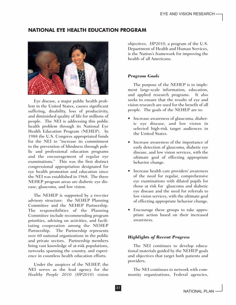
Eye disease, a major public health prob-lem in the United States, causes significantsuffering, disability, loss of productivity,and diminished quality of life for millions ofpeople. The NEI is addressing this publichealth problem through its National EyeHealth Education Program (NEHEP). In1988 the U.S. Congress appropriated fundsfor the NEI to “increase its commitmentto the prevention of blindness through pub-lic and professional education programsand the encouragement of regular eyeexaminations.” This was the first distinctcongressional appropriation designated foreye health promotion and education sincethe NEI was established in 1968. The threeNEHEP program areas are diabetic eye dis-ease, glaucoma, and low vision.
The NEHEP is supported by a two-tieradvisory structure: the NEHEP PlanningCommittee and the NEHEP Partnership.The responsibilities of the PlanningCommittee include recommending programpriorities, advising on activities, and facili-tating cooperation among the NEHEPPartnership. The Partnership representsover 60 national organizations in the publicand private sectors. Partnership membersbring vast knowledge of at-risk populations,networks spanning the country, and experi-ence in countless health education efforts.
Under the auspices of the NEHEP, theNEI serves as the lead agency for theHealthy People 2010 (HP2010) vision
objectives. HP2010, a program of the U.S.Department of Health and Human Services,is the Nation’s framework for improving thehealth of all Americans.
Program Goals
The purpose of the NEHEP is to imple-ment large-scale information, education,and applied research programs. It alsoseeks to ensure that the results of eye andvision research are used for the benefit of allpeople. The goals of the NEHEP are to:
• Increase awareness of glaucoma, diabet-ic eye disease, and low vision inselected high-risk target audiences inthe United States.
• Increase awareness of the importance ofearly detection of glaucoma, diabetic eyedisease, and low vision services, with theultimate goal of effecting appropriatebehavior change.
• Increase health care providers’ awarenessof the need for regular, comprehensiveeye examinations with dilated pupils forthose at risk for glaucoma and diabeticeye disease and the need for referrals tolow vision services, with the ultimate goalof effecting appropriate behavior change.
• Encourage these groups to take appro-priate action based on their increasedawareness.
Highlights of Recent Progress
The NEI continues to develop educa-tional materials guided by the NEHEP goalsand objectives that target both patients andproviders.
The NEI continues to network with com-munity organizations, Federal agencies,
31
EYE AND VISION RESEARCH
NATIONAL PLAN
NNAATTIIOONNAALL EEYYEE HHEEAALLTTHH EEDDUUCCAATTIIOONN PPRROOGGRRAAMM

allied health care organizations, health careprofessional groups, and organizations thatassist older adults. The NEI maintains thatthe community (patients/consumers) andthose who support the community(providers, social service advocates, andFederal agencies) are key in educating thepublic. The NEI uses a multidisciplinaryapproach to reach both primary and sec-ondary audiences. In an effort to make surethat the general public is reached, the NEIhas incorporated the use of conferences,teleconferences, standard and online focusgroups, print materials (brochures and pam-phlets), TV/radio/print public serviceannouncements, websites, audiovisualmaterials, interactive multimedia programs,traveling exhibits, and posters.
The NEI strives to provide culturally spe-cific and appropriate messages for all of theNEHEP content areas and materials. Theglaucoma materials are being translatedinto Spanish. The primary low vision book-let has already been translated into Spanish.The NEI is reviewing the diabetes educationmaterials to translate, update, and performnecessary rewrites to reflect the currentstate of scientific knowledge. Once modi-fied, all diabetes education materials alsowill be available in both English andSpanish. The NEI also seeks the advice ofthe Hispanic/Latino community throughfocus groups and feedback mechanisms toensure that the educational tools and mate-rials developed are appropriate for theintended audience.
The NEI has begun developing outreachand communication strategies for theAmerican Indian and Alaska Native popula-tion. The NEHEP conducted an environ-mental scan of the vision-related programsand services provided to American Indiansand Alaska Natives and identified gaps ineye health information, program services,and materials targeted to these groups.
The NEI maintains an up-to-date web-site that posts best practices and lessonslearned. The NEI encourages NEHEPPartnership members to submit pertinentinformation or links to websites to assistother NEHEP Partnership members inraising awareness about eye health preven-tion, education, and treatment. The NEIsupports the further development of itswebsite to encompass useful patient-focused information, cutting-edge clinicalpractices, and lessons-learned approachesto all eye diseases. The NEI has establisheda Community Awards Program that providesseed money to local communities in estab-lishing eye health initiatives.
It is the NEI’s desire to increase therepresentation of racial and ethnic popula-tions and organizations in the NEHEPPartnership. Having a diverse racial andethnic representation and diversity ofthought and perspective are key to thesuccess of the NEHEP Partnership effort.The NEI encourages NEHEP Partners toshare the NEHEP mission, vision, and pur-pose with their respective peers to promotethe inclusion of more racial and ethnicgroups. Where necessary, the NEI will con-tinue to identify and invite prospectiveNEHEP Partners as the program continuesto expand.
The NEI is working on a project to edu-cate eye health care professionals about theissues of vision rehabilitation. The NEI isdeveloping a pilot program to enhancereferrals of individuals with low vision tovision rehabilitation services. The primarypurpose of the program is to increase
32
EYE AND VISION RESEARCH
NATIONAL PLAN

patient referrals from eye care professionalsto qualified vision rehabilitation services.This program will enable the NEI to devel-op, test, and evaluate measurable strategies;identify opportunities and barriers; and pro-vide a cadre of health care and eye careprofessionals and their patients with accessto useful sources of information and servic-es. The program also will assist eye careand vision rehabilitation professionals inreaching and providing services to those inneed. Once the pilot program has beencompleted, the NEI will assess its successand utility to eye care professionals andtheir patients and decide whether to contin-ue the effort. The NEI welcomes anythoughts, suggestions, and/or participationfrom the NEHEP Partners.
One of the NEI’s primary purposes forinitiating the NEHEP Partnership was toencourage and enhance collaboration withother Federal agencies. This increasedcommunication with other Federal agencieshelps avoid duplication and expands thereach of eye health information.
The HP2010 Vision Working Group con-tinues discussions centered aroundstrategies to raise awareness about nationalHP2010 efforts and how local communitiesand agencies can become involved. TheNEI has provided an HP2010 vision websitelink to all Partnership members through theNEI website.
The NEI suggested to all NEHEPPartners that they join in a Memorandum ofUnderstanding with the NEI to work onHP2010 efforts together. The NEI encour-ages this type of collaborative effort with allof its NEHEP Partners. This collaborationwill provide a forum for the development ofa common HP2010 theme and message forthe vision community.
The NEI is developing HP2010 materialsfor dissemination to the general public, eyehealth care professionals, and policymakers.These materials include the Healthy Vision
2010 Community Action Guide, HealthyVision 2010 Toolkit, and the Healthy Vision2010 Companion Document.
Program Objectives
The NEHEP Partnership met in April2002 to develop recommendations for thenext 5 years. Those recommendations are:
• Ensure that NEHEP goals and objectivesare targeted to both patients and providersin providing information on eye healthcare and vision rehabilitation services.
• Use a multidisciplinary approach toreach secondary target audiences, (alliedhealth professionals, community organi-zations, etc.).
• Provide culturally specific and appropri-ate messages for all NEHEP contentareas and materials.
• Develop and maintain a website to includebest practices and lessons learned.
• Increase the representation of racial andethnic populations and organizations inthe NEHEP Partnership and continue tofacilitate the national dialog among cur-rent and future NEHEP Partnershipmembers.
• Educate and encourage eye health careprofessionals about the issues of visionrehabilitation.
• Increase the NEHEP Partnership’s col-laboration with other Federal agencies.
• Assist Partnership members in reachingout to the vision community to raiseawareness about national HP2010efforts.
• Provide an HP2010 vision website link toall Partnership members.
• Develop a common HP2010 theme andmessage for the vision community.
33
EYE AND VISION RESEARCH
NATIONAL PLAN

• Develop, disseminate, and shareHP2010 materials with the generalpublic, eye health care professionals, andpolicymakers.
In addition, two new program areas willbe reviewed for consideration based on acareful analysis of the scientific research:
• Develop and implement an AMD publiceducation program targeted to providersand patients. This program will focus ondisease etiology, risk factors, prevention,intervention, and treatment strategies.
• Coordinate and enhance diverse effortsin children’s vision education and out-reach. The NEI recognizes theopportunities, national health care man-dates, and research conducted tounderstand the importance of children’seye health care.
34
EYE AND VISION RESEARCH
NATIONAL PLAN

Retinal Diseases Panel
*Gary Abrams, M.D.Kresge Eye InstituteDetroit, Michigan
*Sally Atherton, Ph.D.Medical College of GeorgiaAugusta, Georgia
*Joseph Besharse, Ph.D.Medical College of WisconsinMilwaukee, Wisconsin
Leo Chalupa, Ph.D.University of CaliforniaDavis, California
Martin Friedlander, M.D., Ph.D.The Scripps Research InstituteLa Jolla, California
Bernard Godley, M.D., Ph.D.Retina Foundation of the SouthwestDallas, Texas
Carla Green, Ph.D.University of VirginiaCharlottesville, Virginia
Leslie Hyman, Ph.D.State University of New YorkStony Brook, New York
Stuart Mangel, Ph.D.University of AlabamaBirmingham, Alabama
Patsy Nishina, Ph.D.The Jackson LaboratoryBar Harbor, Maine
Krzysztof Palczewski, Ph.D.University of WashingtonSeattle, Washington
Donald Zack, M.D., Ph.D.Johns Hopkins UniversityBaltimore, Maryland
NEI Program Directors:Hemin Chen, Ph.D.Peter Dudley, Ph.D.Chyren Hunter, Ph.D.Andrew Mariani, Ph.D.Grace Shen, Ph.D.
35
EYE AND VISION RESEARCH
NATIONAL PLAN
AACCKKNNOOWWLLEEDDGGMMEENNTTSS
The National Eye Institute and the National Advisory Eye Council wish to thankthose who contributed suggestions, comments, and recommendations during thedevelopment of this Strategic Plan. In particular, we wish to acknowledge the con-tributions of the members of the program planning panels, including the NEI staff,who so freely gave of their time and expertise and the various organizations that pro-vided advice and assistance in reviewing various drafts of the Plan. The visionresearch community is indebted to these people for their generous efforts on behalfof those who suffer from visual impairment or blindness due to eye diseases.
*Trustee of the Association for Research in Vision and Ophthalmology (ARVO)

Corneal Diseases Panel
Dimitri Azar, M.D.Harvard UniversityBoston, Massachusetts
Edouard Cantin, Ph.D.City of Hope Medical CenterDuarte, California
Suzanne Fleiszig, O.D., Ph.D.University of California Berkeley, California
Brigid L.M. Hogan, Ph.D.Duke University Medical CenterDurham, North Carolina
Gordon Klintworth, M.D., Ph.D.Duke UniversityDurham, North Carolina
Sandra Masur, Ph.D.Mount Sinai School of MedicineNew York, New York
Austin Mircheff, Ph.D.University of Southern CaliforniaLos Angeles, California
Jerry Niederkorn, Ph.D. University of Texas Dallas, Texas
Steven Sugrue, Ph.D.University of FloridaGainesville, Florida
Kirk Wilhelmus, M.D., M.P.H.Baylor College of MedicineHouston, Texas
*Steven Wilson, M.D.University of WashingtonSeattle, Washington
NEI Program Director:Richard Fisher, Ph.D.
Lens and Cataract Panel
Steven Bassnett, Ph.D.Washington UniversitySt. Louis, Missouri
Velia Fowler, Ph.D.The Scripps Research InstituteLa Jolla, California
Thomas Glaser, M.D., Ph.D.University of MichiganAnn Arbor, Michigan
Daniel Goodenough, Ph.D.Harvard Medical SchoolBoston, Massachusetts
Joseph Horowitz, Ph.D.University of CaliforniaLos Angeles, California
M. Cristina Leske, M.D., M.P.H.State University of New York Stony Brook, New York
Richard Mathias, Ph.D.State University of New YorkStony Brook, New York
Hassane Mchaourab, Ph.D.Vanderbilt UniversityNashville, Tennessee
Guillermo Oliver, Ph.D.St. Jude Children’s Research HospitalMemphis, Tennessee
*Larry Takemoto, Ph.D.Kansas State UniversityManhattan, Kansas
NEI Program Director:Ellen Liberman, Ph.D.
36
EYE AND VISION RESEARCH
NATIONAL PLAN
*ARVO Trustee

Glaucoma and OpticNeuropathies Panel
Bassem Bejjani, M.D.Washington State UniversitySpokane, Washington
Claude Burgoyne, M.D.Louisiana State UniversityNew Orleans, Louisiana
Laura Frishman, Ph.D.University of HoustonHouston, Texas
Eve Higginbotham, M.D.University of MarylandBaltimore, Maryland
Robert Nickells, Ph.D.University of WisconsinMadison, Wisconsin
Julia Richards, Ph.D.University of MichiganAnn Arbor, Michigan
William Swanson, Ph.D.State College of OptometryNew York, New York
Douglas Vollrath, M.D., Ph.D.Stanford UniversityStanford, California
*Thomas Yorio, Ph.D.University of North TexasFort Worth, Texas
Beatrice Yue, Ph.D.University of IllinoisChicago, Illinois
NEI Program Director:Ellen Liberman, Ph.D.
Strabismus, Amblyopia, andVisual Processing Panel
Thomas Albright, Ph.D.Salk Institute for Biological StudiesSan Diego, California
Ben Barres, M.D., Ph.D.Stanford University School of MedicineStanford, California
William Good, M.D.Smith-Kettlewell Eye Research InstituteSan Francisco, California
Dennis Levi, O.D., Ph.D.University of California Berkeley, California
Steve Lisberger, Ph.D.University of CaliforniaSan Francisco, California
Jeffrey Schall, Ph.D.Vanderbilt UniversityNashville, Tennessee
Carla Shatz, Ph.D.Harvard Medical SchoolBoston, Massachusetts
Michael Shadlen, M.D., Ph.D.University of WashingtonSeattle, Washington
David Trolio, Ph.D.New England College of OptometryBoston, Massachusetts
Brian Wandell, Ph.D.Stanford UniversityStanford, California
Rafael Yuste, M.D., Ph.D.Columbia UniversityNew York, New York
NEI Program Directors:Chyren Hunter, Ph.D.Michael Oberdorfer, Ph.D.
37
EYE AND VISION RESEARCH
NATIONAL PLAN
*ARVO Trustee

Low Vision and BlindnessRehabilitation Panel
Aries Arditi, Ph.D.Lighthouse InternationalNew York, New York
Susana Chung, Ph.D.University of Houston College of
OptometryHouston, Texas
Richard Long, Ph.D.Western Michigan UniversityKalamazoo, Michigan
Jack Loomis, Ph.D.University of CaliforniaSanta Barbara, California
Cynthia Owsley, Ph.D.University of AlabamaBirmingham, Alabama
Eli Peli, O.D.Schepens Eye Research InstituteBoston, Massachusetts
Noa Rensing, Ph.D.MicroOptical Engineering CorporationWestwood, Massachusetts
Kathleen Turano, Ph.D.Johns Hopkins University School of
MedicineBaltimore, Maryland
NEI Program Director:Michael Oberdorfer, Ph.D.
National Eye Health EducationProgram Planning Committee
Dave Baldridge National Indian Council on AgingAlbuquerque, New Mexico
W. Lee Ball, Jr., O.D. Beth Israel Deaconess Medical CenterBoston, Massachusetts
Charles E. Basch, Ph.D.Columbia UniversityNew York, New York
Norma K. Bowyer, O.D., M.P.H. Morgantown, West Virginia
Michael R. Duenas, O.D. Chattahoochee, Florida
David L. Epstein, M.D. Duke University Medical CenterDurham, North Carolina
George E. Foster, O.D.Northeastern State UniversityTahlequah, Oklahoma
Charles A. Garcia, M.D. University of Texas Medical School Houston, Texas
Eve Higginbotham, M.D. University of Maryland Medical CenterBaltimore, Maryland
Robert E. Kalina, M.D. University of WashingtonSeattle, Washington
Andrew G. Lee, M.D. University of Iowa Hospital and Clinics Iowa City, Iowa
Jose S. Pulido, M.D., M.S.University of IllinoisChicago, Illinois
(continued on next page)
38
EYE AND VISION RESEARCH
NATIONAL PLAN
National Eye Health EducationProgram Planning Committee(continued)
Gwen Sterns, M.D. Rochester General HospitalRochester, New York
R. Tracy Williams, O.D. Deicke Center for Visual RehabilitationWheaton, Illinois
Satya Verma, O.D. Pennsylvania College of OptometryElkins Park, Pennsylvania
NEI Staff:Rosemary JaniszewskiKaren Silver
39
EYE AND VISION RESEARCH
NATIONAL PLAN

Administration on Aging, U.S. Departmentof Health and Human Services (DHHS)
AMD Alliance InternationalAmerican Academy of OphthalmologyAmerican Academy of OptometryAmerican Academy of Physician
AssistantsAmerican Association of Diabetes
EducatorsAmerican College of Occupational
and Environmental MedicineAmerican Diabetes AssociationAmerican Foundation for the BlindAmerican Medical AssociationAmerican Optometric AssociationAmerican Pharmaceutical AssociationAmerican Society of Health-System
PharmacistsAmerican Society of Ophthalmic
Registered NursesAssociation for Education and
Rehabilitation of the Blind andVisually Impaired
Association for Research in Visionand Ophthalmology
Association of Schools and Colleges of Optometry
Association of University Professors of Ophthalmology
Centers for Disease Control andPrevention (CDC), DHHS
Chi Eta Phi Sorority, Inc.Council of Citizens of Low Vision
InternationalDelta Gamma FoundationThe Glaucoma FoundationGlaucoma Research FoundationHelen Keller International Illinois Society for the Prevention of
BlindnessInFOCUSJoint Commission on Allied Health
Personnel in OphthalmologyJuvenile Diabetes Research
Foundation InternationalLighthouse InternationalThe Links, Incorporated
Lions Clubs InternationalLions Eye Health Program (Lions
Clubs International Foundation)Low Vision Council Macular Degeneration PartnershipMaryland Society for SightNational Alliance for Hispanic HealthNational Association for Parents of
Children with Visual Impairments, Inc.National Association for Visually
HandicappedNational Association of Area Agencies
on Aging National Association of Hispanic
Elderly (Asociación Nacional PorPersonas Mayores)
National Association of Hispanic NursesNational Association of Vision ProfessionalsNational Black Nurses AssociationThe National Caucus and Center on
Black Aged, Inc.National Community Pharmacists
AssociationNational Council of La RazaNational Council on Patient
Information and EducationNational Council on the Aging, Inc.National Diabetes Education Program,
National Institute of Diabetes andDigestive and Kidney Diseases (NIDDK),National Institutes of Health(NIH)/CDC
National Hispanic Medical AssociationNational Institute on Aging, NIHNational Medical AssociationNational Optometric AssociationNIDDK, NIHOffice of Disease Prevention and
Health Promotion, DHHSOffice of Minority Health, DHHSPrevent Blindness AmericaPrevention of Blindness Society of the
Metropolitan AreaResearch to Prevent BlindnessVeterans Health Administration, U.S.
Department of Veterans AffairsVision Council of America
40
EYE AND VISION RESEARCH
NATIONAL PLAN
RReevviieewweerrss aanndd NNaattiioonnaall EEyyee HHeeaalltthh EEdduuccaattiioonn PPrrooggrraamm PPaarrttnneerrss

U.S. Department of Health and Human ServicesNIH Publication No. 04-4288
U.S. DEPARTMENT OF HEALTH AND HUMAN SERVICESNational Institutes of HealthNational Eye Institute