natural product reports
TRANSCRIPT

Natural ProductReports
REVIEW
Ope
n A
cces
s A
rtic
le. P
ublis
hed
on 2
0 A
ugus
t 202
1. D
ownl
oade
d on
12/
5/20
21 2
:45:
21 P
M.
Thi
s ar
ticle
is li
cens
ed u
nder
a C
reat
ive
Com
mon
s A
ttrib
utio
n-N
onC
omm
erci
al 3
.0 U
npor
ted
Lic
ence
.
View Article OnlineView Journal
Chain release me
Leibniz Institute for Natural Product Resear
E-mail: [email protected]
Cite this: DOI: 10.1039/d1np00035g
Received 31st May 2021
DOI: 10.1039/d1np00035g
rsc.li/npr
This journal is © The Royal Society
chanisms in polyketide and non-ribosomal peptide biosynthesis
Rory F. Little and Christian Hertweck *
Review covering up to mid-2021
The structure of polyketide and non-ribosomal peptide natural products is strongly influenced by how they
are released from their biosynthetic enzymes. As such, Nature has evolved a diverse range of release
mechanisms, leading to the formation of bioactive chemical scaffolds such as lactones, lactams,
diketopiperazines, and tetronates. Here, we review the enzymes and mechanisms used for chain release
in polyketide and non-ribosomal peptide biosynthesis, how these mechanisms affect natural product
structure, and how they could be utilised to introduce structural diversity into the products of
engineered biosynthetic pathways.
1 Introduction2 PKS and NRPS enzymes3 Thioesterase domains (a/b hydrolase fold)3.1 Intermolecular nucleophiles3.1.1 Water (hydrolysis)3.1.2 Alcohols (transesterication)3.1.3 Thiols (transthioesterication)3.1.4 Amines (amidation)3.2 Intramolecular nucleophiles3.2.1 Hydroxyl groups (lactonisation)3.2.2 Amine/amide groups (lactamisation)3.2.3 Carbanions (Dieckmann condensation)3.2.4 Thiol groups (thiolactonisation)4 Chain release catalysed by type II thioesterases (a/
b hydrolase fold)5 Chain release catalysed by hot-dog fold thioesterases6 Chain release catalysed by metallo-b-lactamase (MbL)
thioesterases7 Chain release catalysed by penicillin binding protein
(PBP)-like enzymes8 Chain release catalysed by reductase (R) domains8.1 Structure and mechanism of R domains8.2 PK and NRP pathways that use R domains8.3 Products of two-electron reductase domains8.4 Products of four-electron reductase domains8.5 R* domains9 Chain release catalysed by aldo-keto reductases10 Chain release catalysed by oxygenases11 Chain release catalysed by FabH-like enzymes
ch and Infection Biology, HKI, Germany.
of Chemistry 2021
12 Chain release catalysed by condensation-like (CT)domains
13 Chain release catalysed by acyltransferase-like enzymes14 Chain release catalysed by AfsA/butenolide synthase
(PBS) domains15 Chain release catalysed by standalone Dieckmann
cyclases16 Chain release catalysed by ketosynthase domains17 Chain release catalysed by PLP-dependent enzymes18 Final remarks19 Conicts of interest20 Acknowledgements21 References
1 Introduction
Polyketides (PKs) and the non-ribosomal peptides (NRPs) aretwo of the largest natural product families.1,2 Widely producedby bacteria and fungi, members of each family have been turnedinto important medicines, such as the antibiotics erythromycin(PK) and vancomycin (NRP), the immunosuppressants rapa-mycin (PK) and cyclosporin (NRP), and the anticancer agentsepothilone (PK) and bleomycin (NRP).3–7 One reason for suchsuccess is the remarkable diversity of PK and NRP chemicalscaffolds.2,8,9 The diversity exhibited by PKs and NRPs is all themore impressive because of the relatively simple chemical unitsused to construct them. PKs and NRPs are both polymers (oencalled “chains”) constructed from simple monomers—alsoreferred to as “building blocks” or extension units.1,2 Polyketidesynthase (PKS) enzymes condense small carboxylic acids,primarily acetate and propionate, to form PKs, while non-ribosomal peptide synthetases (NRPSs) condense amino acids(and sometimes other organic acids) to form NRPs.1,2 However,
Nat. Prod. Rep.

Natural Product Reports Review
Ope
n A
cces
s A
rtic
le. P
ublis
hed
on 2
0 A
ugus
t 202
1. D
ownl
oade
d on
12/
5/20
21 2
:45:
21 P
M.
Thi
s ar
ticle
is li
cens
ed u
nder
a C
reat
ive
Com
mon
s A
ttrib
utio
n-N
onC
omm
erci
al 3
.0 U
npor
ted
Lic
ence
.View Article Online
these humble beginnings give rise to numerous medicinallyimportant chemical scaffolds including macrolides, polyethers,enediynes, and b-lactams.2,8
The mechanisms used for diversifying PK/NRP chains arewide ranging. For example, arsenals of tailoring enzymes canmodify the PK/NRP chain aer it has been fully processed bya PKS/NRPS enzyme.2,8 However, one of the most importantdiversication steps oen occurs earlier. During its biosyn-thesis, a PK/NRP chain is covalently tethered to the PKS/NRPSvia a 40-phosphopantetheine (Ppant) group (derived fromcoenzyme A).10 The free thiol group of the Ppant group formsa thioester bond with the terminal carboxyl group of thegrowing PK/NRP chain.1 However, this covalent linkage must bebroken to release the PK/NRP chain from the PKS/NRPS. Arelease step is critical both for allowing the product to enter intothe cytosol, where it may be modied by additional enzymesand/or exported from the cell, and to enable continuoussubstrate processing by the PKS/NRPS.1 Nature has not onlysolved this problem, but also keenly recognised it as anopportunity to profoundly modify the structure of the PK/NRPproduct.11 For instance, the PK/NRP chain can be released viaintra/intermolecular cyclisations, reductions, and fusion toother chemical units, leading to structural diversicationsranging from simple primary alcohols, aldehydes, and carbox-ylic acids, to more complex tetronates, macrolactones/lactams,and oligomers.11,12 The functionalities created as a result ofchain release may themselves undergo subsequent chemicaltransformations, leading to the creation of complex scaffoldssuch as spirotetronates and iminopeptides.12,13 In addition,given the impact that the mechanism of chain release can haveon PK/NRP structure, an appreciation and understanding ofthese mechanisms will aid efforts for creating engineered PKSand NRPS enzymes that produce new and diverse products.Here, we review the enzymes and mechanisms used for chainrelease in PK and NRP biosynthesis pathways, how thesemechanisms directly and indirectly affect natural productstructure, and their potential to be utilised by synthetic biology
Rory Little received a Bachelor(Hons) (2013) and Master ofScience (2015) degree fromVictoria University of Wellington(Te Herenga Waka), New Zea-land. He was awarded a WoolfFisher scholarship to completea Ph.D. (2019) at the Universityof Cambridge in the group ofProfessor Peter Leadlay. He iscurrently an Alexander vonHumboldt postdoctoral fellow inthe group of Professor Christian
Hertweck at the Leibniz HKI. His research interests are microbialnatural product discovery, function, and biosynthesis.
Nat. Prod. Rep.
to produce structurally diverse products from engineered PKS/NRPS enzymes. It is important to note that, while many of themechanisms discussed in this review are experimentally wellcharacterised, some lack direct experimental evidence so areonly proposed mechanisms. In some cases, obtaining suchevidence is stymied by the chain release enzyme acting ona complex, difficult to source, and possibly unstable biosyn-thetic intermediate. Nevertheless, instances where a given chainrelease mechanism requires conrming experimental evidenceare explicitly stated.
2 PKS and NRPS enzymes
Before discussing chain release mechanisms (sometimes alsocalled “offloading”mechanisms), it is worth briey covering thebiochemistry of PKS and NRPS enzymes. There are multipleclasses of both enzymes, differing from one another in char-acteristics such as whether they are modular, act iteratively, orare composed of multiple standalone proteins.1,2 The mostdiscussed classes in this review are the modular cis-acyl-transferase (AT) type I PKSs and Type A NRPSs. In these classes,each protein module is responsible for the incorporation ofa single extension unit into the growing PK/NRP chain. Eachmodule of a PKS/NRPS enzyme is itself comprised of discretecatalytic centres called domains, which catalyse the necessaryreactions for the chain extension to occur.1,2 In contrast, type IIPKSs consist of iteratively acting standalone proteins.14 Theminimal type II PKS biosynthesis pathway consists of a stand-alone ACP and two proteins that resemble KS domains: a KSaand KSb.14 These two KS proteins are highly similar to oneanother and together catalyse the necessary chain initiation andextension events required for polyketide biosynthesis.14 Type IIIPKSs consist of numerous standalone enzymes to catalyse chainelongation without the use a Ppant tether.15 Analogous to PKSs,NRPSs may also act iteratively or be composed of multiplestandalone proteins, as will be discussed later.2
Christian Hertweck is head ofthe Biomolecular ChemistryDepartment at the Leibniz Insti-tute for Natural ProductResearch and Infection Biology(HKI) and holds a Chair at theFriedrich Schiller UniversityJena. Aer his Ph.D. studies atUniversity of Bonn and the MPIfor Chemical Ecology (W.Boland) he was a Feodor Lynenpostdoctoral fellow at theUniversity of Washington, Seat-
tle (H. G. Floss and B. S. Moore). His research focuses on microbialnatural products, their biosynthesis, and their role in microbialinteractions. He is an elected member of the National Academy ofSciences (Leopoldina) and recipient of the Leibniz Award.
This journal is © The Royal Society of Chemistry 2021

Review Natural Product Reports
Ope
n A
cces
s A
rtic
le. P
ublis
hed
on 2
0 A
ugus
t 202
1. D
ownl
oade
d on
12/
5/20
21 2
:45:
21 P
M.
Thi
s ar
ticle
is li
cens
ed u
nder
a C
reat
ive
Com
mon
s A
ttrib
utio
n-N
onC
omm
erci
al 3
.0 U
npor
ted
Lic
ence
.View Article Online
The number of domains present within a type I PKS or type ANRPS module differs from enzyme to enzyme, except for severalessential “core” domains.1,2 To be catalytically active, eachmodule of cis-AT type I PKS must contain an acyltransferase(AT), acyl carrier protein (ACP), and a ketosynthase (KS)domain.1 The ACP of each module serves as an attachmentpoint for a Ppant moiety. Each AT domain selects an extensionunit and transfers it to the Ppant group of the adjacent ACP.10
The most common extension units used in PK biosynthesis areacetate (two carbon) and propionate (three carbon) units, typi-cally delivered in their activated forms of malonyl-CoA and (2S)-methylmalonyl-CoA, respectively.1 The KS domain then catal-yses C–C bond formation via a decarboxylative Claisencondensation between the nascent PK chain and the extensionunit bound to the ACP of the downstream module.10 The PKchain uses the exible Ppant groups to swing between thedifferent PKS modules of the biosynthetic pathway, eachmodule increasing the chain size by one extension unit.10 Thedownstream module can be part of the same enzyme as theupstream module, or be part of a separate PKS enzyme alto-gether.10 Following the Claisen condensation, accessorydomains such as ketoreductase (KR), dehydratase (DH), andenoylreductase (ER) domains may reduce the b-keto group.10
Type A NRPS modules contain a different set of domainsfrom type I PKS modules. At a minimum, each module in anNRPS must contain an adenylation (A) domain, a peptidylcarrier protein (PCP, also sometimes called a thiolation (T)domain), and a condensation (C) domain to be functional.2 Adomains are responsible for selecting an extension unit, anal-ogous to AT domains in PKS enzymes.2 To achieve this, Adomains catalyse an ATP-dependent adenylation reaction of anamino acid, oen with a high degree of selectivity.2 The ade-nylated amino acid is a high energy species with a strong leavinggroup (AMP), facilitating nucleophilic attack by the thiol of thePCP-linked Ppant group with elimination of AMP.2 The functionof C domains is analogous to KS domains, though they catalyseC-N (peptide) bond formation rather than C–C bond formation.2
The PCP-bound NRP chain enters the active site of the Cdomains, where it is attacked by the a-amino group on theamino acid tethered to the PCP domain of the downstream PCPdomain.2
Aer the nal chain extension, the mature chain is letethered to the ACP/PCP of the terminal module and must bereleased. Numerous mechanisms exist, utilising both enzymaticdomains integrated into the PKS/NRPS or dedicated standalonechain release enzymes. The rst class of chain release enzymesto be explored are the a/b hydrolase fold thioesterases.
Fig. 1 The structure of an a/b hydrolase fold thioesterase domain. (A)The crystal structure of the thioesterase domain from the polyketidepikromycin biosynthesis pathway (PDB: 1MN6). (B) The Ser–Asp–Hiscatalytic triad of the TE domain from pikromycin biosynthesis.
3 Thioesterase domains (a/b hydrolase fold)
The a/b hydrolase fold thioesterases either catalyse chainrelease as a discrete domain within a PKS/NRPS (type I thio-esterases), or as standalone proteins (type II thioesterases). Theuse of type I thioesterase (TE) domains to catalyse chain releaseis common in PK/NRP biosynthesis, to the extent that it is oen
This journal is © The Royal Society of Chemistry 2021
considered the canonical method.11 TE domains typicallycatalyse release either by hydrolysis or macrocyclisation, thoughother mechanisms are also possible, as will be discussed. In thepathways that use them, the TE domain is almost always foundon the C-terminus of the nal PKS/NRPS module.11 TE domainsare between 240–290 amino acid residues in size and possess ana/b hydrolase fold, a conformation consisting of seven to eightparallel b-sheets connected by a-helices (Fig. 1).10 a/b hydrolasefolds are commonly found in other enzymes with hydrolyticactivity, such as lipases and proteases.16 Between b-sheets sixand seven is the “lid” region—a dynamic ca. 40 amino acidelement that lines the substrate channel.17 Crystal structures ofexcised TE domains have revealed that the lid region is either inan apparent “open” state, allowing ready access to the bindingpocket, or “closed” state, restricting substrate entry.17
TE domains use a two-step mechanism to catalyse chainrelease, the rst step being a transesterication.17 A the hydroxylof a catalytic serine residue, typically located at the C-terminusof b-sheet ve, attacks the electrophilic carbonyl of the PK/NRPthioester, forming an oxoester.17 The catalytic serine attacks thesubstrate thioester as it is activated via deprotonation bya conserved histidine (Fig. 2).17 Together, these three residuesmake up the Ser–Asp–His catalytic triad that is highly conservedin thioesterase domains and other a/b hydrolases.10 The cata-lytic serine is identiable by its location in a GxSxGmotif (wherex is any amino acid).10 In some cases, a cysteine is presentinstead of a serine residue—in effect using a sulphur nucleo-phile rather than oxygen.18–20 Why some thioesterase domainsselect for a catalytic cysteine rather than serine is poorlyunderstood.19 However, the presence of a catalytic cysteine canbe an indicator that the TE domain has an unusual activity, aswill be discussed in Section 3.2.1.
The second step of the mechanism is the release of the PK/NRP intermediate from the TE domain itself.17 It is herewhere the TE domain exerts the greatest inuence over thestructure of the nal product.11,17 During this step, a nucleo-phile attacks and cleaves the oxoester bond (or thioester bond,in the case of a TE domain with a catalytic cysteine) connectingthe PK/NRP chain to the TE domain.17 A tetrahedral oxyanionintermediate forms following nucleophilic attack that is
Nat. Prod. Rep.
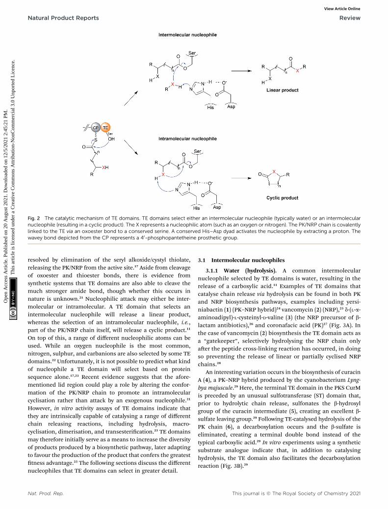
Fig. 2 The catalytic mechanism of TE domains. TE domains select either an intermolecular nucleophile (typically water) or an intermolecularnucleophile (resulting in a cyclic product). The X represents a nucleophilic atom (such as an oxygen or nitrogen). The PK/NRP chain is covalentlylinked to the TE via an oxoester bond to a conserved serine. A conserved His–Asp dyad activates the nucleophile by extracting a proton. Thewavey bond depicted from the CP represents a 40-phosphopantetheine prosthetic group.
Natural Product Reports Review
Ope
n A
cces
s A
rtic
le. P
ublis
hed
on 2
0 A
ugus
t 202
1. D
ownl
oade
d on
12/
5/20
21 2
:45:
21 P
M.
Thi
s ar
ticle
is li
cens
ed u
nder
a C
reat
ive
Com
mon
s A
ttrib
utio
n-N
onC
omm
erci
al 3
.0 U
npor
ted
Lic
ence
.View Article Online
resolved by elimination of the seryl alkoxide/cystyl thiolate,releasing the PK/NRP from the active site.17 Aside from cleavageof oxoester and thioester bonds, there is evidence fromsynthetic systems that TE domains are also able to cleave themuch stronger amide bond, though whether this occurs innature is unknown.21 Nucleophilic attack may either be inter-molecular or intramolecular. A TE domain that selects anintermolecular nucleophile will release a linear product,whereas the selection of an intramolecular nucleophile, i.e.,part of the PK/NRP chain itself, will release a cyclic product.11
On top of this, a range of different nucleophilic atoms can beused. While an oxygen nucleophile is the most common,nitrogen, sulphur, and carbanions are also selected by some TEdomains.22 Unfortunately, it is not possible to predict what kindof nucleophile a TE domain will select based on proteinsequence alone.17,23 Recent evidence suggests that the afore-mentioned lid region could play a role by altering the confor-mation of the PK/NRP chain to promote an intramolecularcyclisation rather than attack by an exogenous nucleophile.21
However, in vitro activity assays of TE domains indicate thatthey are intrinsically capable of catalysing a range of differentchain releasing reactions, including hydrolysis, macro-cyclisation, dimerisation, and transesterication.23 TE domainsmay therefore initially serve as a means to increase the diversityof products produced by a biosynthetic pathway, later adaptingto favour the production of the product that confers the greatesttness advantage.23 The following sections discuss the differentnucleophiles that TE domains can select in greater detail.
Nat. Prod. Rep.
3.1 Intermolecular nucleophiles
3.1.1 Water (hydrolysis). A common intermolecularnucleophile selected by TE domains is water, resulting in therelease of a carboxylic acid.11 Examples of TE domains thatcatalyse chain release via hydrolysis can be found in both PKand NRP biosynthesis pathways, examples including yersi-niabactin (1) (PK–NRP hybrid)24 vancomycin (2) (NRP),25 d-(L-a-aminoadipyl)-L-cysteinyl-D-valine (3) (the NRP precursor of b-lactam antibiotics),26 and coronafacic acid (PK)27 (Fig. 3A). Inthe case of vancomycin (2) biosynthesis the TE domain acts asa “gatekeeper”, selectively hydrolysing the NRP chain onlyaer the peptide cross-linking reaction has occurred, in doingso preventing the release of linear or partially cyclised NRPchains.28
An interesting variation occurs in the biosynthesis of curacinA (4), a PK–NRP hybrid produced by the cyanobacterium Lyng-bya majuscule.29 Here, the terminal TE domain in the PKS CurMis preceded by an unusual sulfotransferase (ST) domain that,prior to hydrolytic chain release, sulfonates the b-hydroxylgroup of the curacin intermediate (5), creating an excellent b-sulfate leaving group.29 Following TE-catalysed hydrolysis of thePK chain (6), a decarboxylation occurs and the b-sulfate iseliminated, creating a terminal double bond instead of thetypical carboxylic acid.29 In vitro experiments using a syntheticsubstrate analogue indicate that, in addition to catalysinghydrolysis, the TE domain also facilitates the decarboxylationreaction (Fig. 3B).29
This journal is © The Royal Society of Chemistry 2021

Fig. 3 TE-catalysed hydrolytic release. (A) The structures of several natural products released by TE domain-catalysed hydrolysis. (B) Formationmechanism of the terminal double bond in curacin A (4) biosynthesis using an unusual sulphur transferase (ST) domain.
Review Natural Product Reports
Ope
n A
cces
s A
rtic
le. P
ublis
hed
on 2
0 A
ugus
t 202
1. D
ownl
oade
d on
12/
5/20
21 2
:45:
21 P
M.
Thi
s ar
ticle
is li
cens
ed u
nder
a C
reat
ive
Com
mon
s A
ttrib
utio
n-N
onC
omm
erci
al 3
.0 U
npor
ted
Lic
ence
.View Article Online
A TE domain that catalyses an additional reaction besideshydrolysis is also found in the nocardicin A biosynthesispathway. Nocardicin A is a tripeptide b-lactam antibioticproduced by Nocardia uniformis sp. tsuyamanensis and is
This journal is © The Royal Society of Chemistry 2021
comprised of L-para-hydroxyphenylglycine (pHPG), L-serine, andL-arginine.30 In this pathway, the TE domain in the NRPS NocBcatalyses the epimerisation of L-para-hydroxyphenylglycine(pHPG) in addition to NRP chain hydrolysis.30 Cocrystallisation
Nat. Prod. Rep.
Natural Product Reports Review
Ope
n A
cces
s A
rtic
le. P
ublis
hed
on 2
0 A
ugus
t 202
1. D
ownl
oade
d on
12/
5/20
21 2
:45:
21 P
M.
Thi
s ar
ticle
is li
cens
ed u
nder
a C
reat
ive
Com
mon
s A
ttrib
utio
n-N
onC
omm
erci
al 3
.0 U
npor
ted
Lic
ence
.View Article Online
of the excised NocB TE domain with a phosphonate substratemimic indicated that the histidine of the catalytic triad isresponsible for extracting the acidic a-proton from pHPG.30 Theresultant carbanion could be stabilised by electron delocalisa-tion across the pHPG aromatic ring.30 A proton donor (possiblywater) is then proposed to reprotonate the a-carbon of pHPGfrom the opposite side to complete the epimerisation.31
However, epimerisation and product hydrolysis can only occuraer the b-lactam ring has formed,30 again indicating thegatekeeper function TE domains can have (a phenomenonrecently reviewed by Horsman et al.17).
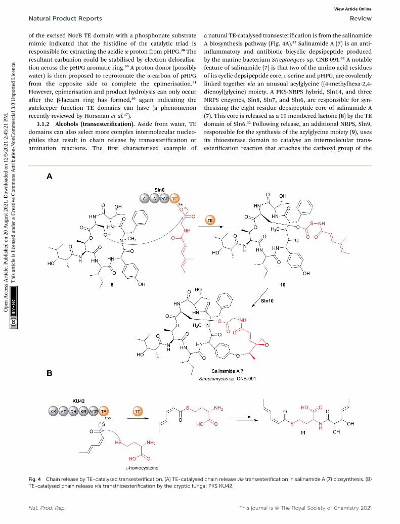
3.1.2 Alcohols (transesterication). Aside from water, TEdomains can also select more complex intermolecular nucleo-philes that result in chain release by transesterication oramination reactions. The rst characterised example of
Fig. 4 Chain release by TE-catalysed transesterification. (A) TE-catalysedTE-catalysed chain release via transthioesterification by the cryptic fung
Nat. Prod. Rep.
a natural TE-catalysed transesterication is from the salinamideA biosynthesis pathway (Fig. 4A).32 Salinamide A (7) is an anti-inammatory and antibiotic bicyclic depsipeptide producedby the marine bacterium Streptomyces sp. CNB-091.32 A notablefeature of salinamide (7) is that two of the amino acid residuesof its cyclic depsipeptide core, L-serine and pHPG, are covalentlylinked together via an unusual acylglycine ((4-methylhexa-2,4-dienoyl)glycine) moiety. A PKS-NRPS hybrid, Sln14, and threeNRPS enzymes, Sln8, Sln7, and Sln6, are responsible for syn-thesising the eight residue depsipeptide core of salinamide A(7). This core is released as a 19 membered lactone (8) by the TEdomain of Sln6.32 Following release, an additional NRPS, Sln9,responsible for the synthesis of the acylglycine moiety (9), usesits thioesterase domain to catalyse an intermolecular trans-esterication reaction that attaches the carboxyl group of the
chain release via transesterification in salinamide A (7) biosynthesis. (B)al PKS KU42.
This journal is © The Royal Society of Chemistry 2021

Review Natural Product Reports
Ope
n A
cces
s A
rtic
le. P
ublis
hed
on 2
0 A
ugus
t 202
1. D
ownl
oade
d on
12/
5/20
21 2
:45:
21 P
M.
Thi
s ar
ticle
is li
cens
ed u
nder
a C
reat
ive
Com
mon
s A
ttrib
utio
n-N
onC
omm
erci
al 3
.0 U
npor
ted
Lic
ence
.View Article Online
acylglycine to the Ser–OH in the depsipeptide core 8, forming anoxoester (10).32 The other end of the acylglycine moiety issubsequently attached to the pHPG residue via an oxidativecyclisation reaction catalysed by the enzyme Sln10, formingsalinamide A (7).32
3.1.3 Thiols (transthioesterication). Another thioesterasedomain capable of catalysing intermolecular trans-esterication, specically a transthioesterication, was identi-ed in the biosynthesis pathway for an aminoacylatedpolyketide (11) in basidiomycete Punctularia strigosozonata.33
Here, the TE domain of the PKS KU42 was shown to catalyserelease of the sorbyl unit using the thiol group of L-cysteine or L-homocysteine as an intermolecular nucleophile (Fig. 4B).33
3.1.4 Amines (amidation). TE domains that catalyse chainrelease via an intermolecular amidation reaction are alsouncommon. A recent example was found in the biosynthesis oflipopeptides in Burkholderia gladioli pv. agaricicola.34 The plantpathogen Burkholderia gladioli is responsible for grain rot, seedrot, and seedling blight in rice.35 Screening culture extracts of B.gladioli pv. agaricicola led to the identication of the lip-opeptides haereogladin A–D and burriogladin A–B (Fig. 5A).34
The NRPS genes responsible for biosynthesis were also identi-ed in the genome.34 The lipopeptides are either free acids thatcorrespond to the predicted size of the polypeptide (haer-eogladins C–D, burriogladin A), indicating hydrolytic release bya TE domain, or have an unusual C-terminal L-threonine tag(haereogladin A, E, burriogladin B).34 The TE domains respon-sible for lipopeptide biosynthesis can therefore either use water(12) or L-threonine (13, 14) as a nucleophile for chain release.34
Interestingly, either the amine or hydroxyl of L-threonine can beused to attack the PCP-tethered NRP chain, resulting in amide(13) or ester (14) formation, respectively.34 In the case of haer-eogladin, having a threonine tag is essential for surfactantproperties, potentially facilitating the colonisation of planthosts by B. gladioli pv. agaricicola.34
Another recent example of a TE domain that catalyses ami-dation is found in the biosynthesis of a small and, as yetunnamed, polyketide (15) produced by the fungus Hydnomer-ulius pinastri.33 The PKS responsible, KU43, produces an ACP-bound octanoate unit that is released using the amino groupof a L-leucine methyl ester as a nucleophile (Fig. 5B).33 Heter-ologous expression of the KU43-TE demonstrated it that itselects L-leucine methyl and catalyses chain release via anamination reaction.33
3.2 Intramolecular nucleophiles
3.2.1 Hydroxyl groups (lactonisation). TE domains thatcatalyse chain release using an intramolecular nucleophileproduce cyclic compounds. When an intramolecular hydroxylgroup is selected a lactone (cyclic ester) is produced.22 The sizeof the lactone formed can vary greatly, ranging from a four-membered b-lactone in obauorin (NRP)18 (16) to a 51-membered macrolactone in stambomycin (PK) (17) (Fig. 6 and7A).22,36
If the lactone contains 12 or more atoms it is classied asa macrolactone/macrolide.37 The formation of lactones is
This journal is © The Royal Society of Chemistry 2021
commonly associated with polyketide biosynthetic pathways,the prototypical example being 6-deoxyerythronolide B (theerythromycin precursor) synthesised by the PKS enzymesDEBS1, DEBS2, and DEBS3.1 Erythromycin A (18), like manymacrolides, is an inhibitor of the bacterial ribosome.38 The TEdomain of DEBS3 is selective for the C13 hydroxyl over the otherhydroxyls in the PK chain, resulting in the exclusive formationof a 14-membered macrolactone.17 TE domains that selecthydroxyl/oxygen nucleophiles may also be responsible for thebiosynthesis of non-lactone rings, such as the pyrone ring in thecercosporin (PK) biosynthesis or the isochromanone ring inajudazol (PK) biosynthesis.39,40
TE domains from NRPSs can also catalyse chain release vialactone formation using the hydroxyl group of an amino acidside chain.18,41 One interesting example is the TE domain usedin the biosynthesis pathway of obauorin (16), a tripeptide b-lactone.18 The b-lactone ring of obauorin (16) is created by anintramolecular cyclisation catalysed by the TE domain of theNRPS ObiF.18 The ObiF-TE domain is unusual for severalreasons. For starters, it catalyses formation of a four memberedring, the smallest ring size produced by TE domains.18,22 Also,unlike most TE domains, it is not the most C-terminal domainof ObiF, instead being located between an upstream PCPdomain and a downstream A domain.18 Non-terminal TEdomains have been identied in only a few other biosyntheticpathways, where their role is unclear or they may catalyse anentirely different reaction (such as cis double bond formation inFR901464 biosynthesis).42–45 Another unusual feature of ObiF-TE is that it has a catalytic cysteine rather than serine.18 Inter-estingly, using mutagenesis to convert this cysteine to a serineabolished production of obauorin (16), highlighting theimportance of a cysteine thioester linkage to the NRP chain.18
The proposed explanation was that the higher ground stateenergy of cysteine thioesters compared to oxoesters, coupledwith the weaker nature of C–S bonds compared to C–O bonds,make forming a strained b-lactone ring more energeticallyfavourable.18,46 The inuence of a catalytic cysteine residue hasalso been investigated in other TE domains. For instance,replacing the active site serine with a cysteine in the TE domainsof the pikromycin (PK) and cilengitide (NRP) biosynthesispathways converted the domain into catalytically more effectivemacrolactonisation catalysts, possibly due to the reasonsproposed above.19,20 In other cases, however, replacing theserine with cysteine signicantly decreased the catalytic activityof the TE domain.47 Why some TE domains select for cysteinewhile others select for serine is still unclear, but it seems likelythere is a tness trade-off occurring between creating an effi-cient catalyst for the target ring size and the stability of the TE-bound PK/NRP intermediate.
Up to this point, the TE domains discussed all catalyse chainrelease via a single lactonisation reaction. A variation of thismechanism, exemplied in the biosynthesis of conglobatin(PK),48 enterobactin (NRP) (19),47 elaiophylin (PK) (20),49 cer-eulide (NRP),50 and valinomycin (NRP)50 is TE-catalysed oligo-merisation of two identical PK/NRP chains (Fig. 8). Twodifferent mechanisms were initially proposed for how theseoligomerisations occur: a “forward transfer” and a “backwards
Nat. Prod. Rep.

Fig. 5 Chain release by TE-catalysed amidation. (A) TE-catalysed chain release in haereogladin biosynthesis. The TE domain of HgdA can selectwater (i), the amine group of L-threonine (ii), or the hydroxyl group of L-threonine (iii) as the intermolecular nucleophile used for chain release. (B)The KU43-TE domain selects the nitrogen of L-leucine methyl ester to form 15.
Nat. Prod. Rep. This journal is © The Royal Society of Chemistry 2021
Natural Product Reports Review
Ope
n A
cces
s A
rtic
le. P
ublis
hed
on 2
0 A
ugus
t 202
1. D
ownl
oade
d on
12/
5/20
21 2
:45:
21 P
M.
Thi
s ar
ticle
is li
cens
ed u
nder
a C
reat
ive
Com
mon
s A
ttrib
utio
n-N
onC
omm
erci
al 3
.0 U
npor
ted
Lic
ence
.View Article Online

Fig. 6 Size range of lactone rings produced by TE domains. Thelactone ring of obafluorin (16) is the smallest known to be produced bya TE domain, while the 51-membered stambomycin (17) is the largest.
Review Natural Product Reports
Ope
n A
cces
s A
rtic
le. P
ublis
hed
on 2
0 A
ugus
t 202
1. D
ownl
oade
d on
12/
5/20
21 2
:45:
21 P
M.
Thi
s ar
ticle
is li
cens
ed u
nder
a C
reat
ive
Com
mon
s A
ttrib
utio
n-N
onC
omm
erci
al 3
.0 U
npor
ted
Lic
ence
.View Article Online
transfer” mechanism.11 In the forward transfer mechanism,a hydroxyl group of a TE-bound PK/NRP chain is proposed toattacks the thioester of an identical PK/NRP chain tethered tothe upstream ACP domain, followed by macrocyclisation/chainrelease.47 In contrast, in the backwards transfer mechanism anACP-bound PK/NRP chain is proposed to attack the thioester ofan identical PK/NRP chain tethered to the downstream TEdomain, sending it “backwards” to the ACP domain.51 Thelinear dimer is then returned to the active site of the TE domainfor macrocyclisation.51 While the forward transfer mechanismwas proposed rst (to account for the biosynthesis of enter-obactin) there is now more evidence that backwards transfer isthe true mechanism for all such oligomerisations(Fig. 9).21,47,48,50,52 The ability of TE domains to catalyse bothdimerisation andmacrolactonisation has been demonstrated invitro for the C2 symmetrical 16-membered dilactones con-globatin and elaiophylin (20).48,52 Puried TE domains from theconglobatin and elaiophylin (20) biosynthesis pathways weredemonstrated to catalyse the dimerisation of two SNAC (N-acetylcysteamine)-substrate analogues,53 forming a linear dimerthat is subsequently cyclised to form a macrodilactone.48,52 A
This journal is © The Royal Society of Chemistry 2021
similar TE-mediated oligomerisation mechanism is likelyoccurring in the biosynthesis of the quinoxaline and quinolinechromodepsipeptide natural products echinomycin (NRP) andsandramycin (NRP), respectively, though direct evidence islacking.54–56
In rare cases, two contiguous TE domains are found on theC-terminus of NPRS proteins. Such “tandem” TE domains arepresent in the biosynthetic pathways for the cyclic lipopeptidesteixobactin,57 arthrofactin,58 malleipeptin,59 syringopeptin,60
massetolide A,61 and the cyclic peptide lysobactin.41 Althoughnot all examples have been biochemically characterised, ingeneral, the rst TE domain of the pair is responsible for lac-tonisation and release. The role of the second TE is less clearand differs from case to case. In the case of teixobactinbiosynthesis, only when the active site serine residues of bothTE domains were mutated was chain release activity fullyabolished, suggesting that the two domain cooperate to releasethe NRP chain.57 In the case of the arthrofactin biosynthesispathway, mutating the active site of the second TE decreasedarthrofactin production by 95%, indicating that it is important,but not essential, for chain release.58 In contrast, mutating theactive site serine in the second TE domain of the lysobactinbiosynthesis pathways had no detectable effect on lysobactinproduction.41 Instead, the second TE domain demonstrateddeacetylase activity, making it more akin to the “proofreading”type II TE enzymes discussed later in Section 4. It was evenproposed that this TE domain may be post-translationallyseparated to act as a standalone type II TE, as the two TEdomains are readily proteolytically cleaved from one another.58
However, direct evidence for this occurring or being relevant invivo is lacking.
3.2.2 Amine/amide groups (lactamisation). If a TE domainselects an intramolecular amine rather than a hydroxy group,a lactam ring is formed (Fig. 7B). Examples of macrolactamsformed using this mechanism include tyrocidine A,62 leinamy-cin (21),63 sulfazecin,64 and vicenistatin.65 The TE domain fromSulM in the sulfazecin biosynthesis pathway produces a b-lac-tam ring, analogous to the b-lactone ring produced by theobauorin (16) ObiF-TE domain (discussed above).18,64,66 Likethe ObiF-TE domain, the SulM TE domain contains a catalyticcysteine rather than a serine. Replacing the cysteine with serineabolished the cyclisation activity of SulM-TE, strengthening thecase that catalytic cysteine residues are better at producingstrained rings than catalytic serine residues are.18,64
TE domains that recognise amine groups are also capable ofcatalysing chain release via oligomerisation reactions. The bestcharacterised example is in the biosynthesis of gramicidin S(NRP), a cyclic dilactam antibiotic produced by Bacillus brevis(Fig. 8).51 The research conducted on gramicidin S (22) biosyn-thesis provided the rst evidence for the “backwards pass”mechanism discussed previously in Section 3.2.1.51
In addition to intramolecular amine groups, the less nucle-ophilic nitrogen atom of amide groups is also selected by someTE domains. Examples include the biosynthesis of the tetramicacids jamaicamide (PK–NRP hybrid)67 and reutericyclin (PK–NRP hybrid).68 In these cases, the nitrogen of an internal
Nat. Prod. Rep.

Fig. 7 Intermolecular nucleophiles selected by thioesterase domains. (A) A hydroxyl group, as in erythromicin A (18) biosynthesis. (B) An amine,as in leinamycin (21) biosynthesis. (C) A carbanion, as in noranthrone (24) (aflatoxin precursor) biosynthesis. (D) A thiol group, as in thiocoraline(26) biosynthesis.
Natural Product Reports Review
Ope
n A
cces
s A
rtic
le. P
ublis
hed
on 2
0 A
ugus
t 202
1. D
ownl
oade
d on
12/
5/20
21 2
:45:
21 P
M.
Thi
s ar
ticle
is li
cens
ed u
nder
a C
reat
ive
Com
mon
s A
ttrib
utio
n-N
onC
omm
erci
al 3
.0 U
npor
ted
Lic
ence
.View Article Online
secondary amide attacks the C1 carbon of the thioester, forminga tetramate ring.67,68
3.2.3 Carbanions (Dieckmann condensation). TE domainscan also catalyse ring chain release/cyclisation using an intra-molecular carbanion (Fig. 7C). Such TE domains are prevalentin fungal PK peptide biosynthesis pathways, where they areoen referred to as Claisen-like cyclase (CLC) domains (theformal name for an intramolecular Claisen reaction is a Die-ckmann condensation).69 Once covalently bound to the TEdomain, the abstraction of an acidic a-proton in a PK chain bya base creates a nucleophilic carbon atom (in the form on anenolate).11 The carbon nucleophile attacks the electrophilic C1atom of TE-oxoester linkage, resulting in C–C bond formationand release of the PK chain.69 The rst characterised example of
Nat. Prod. Rep.
a Dieckmann-catalysing TE was from the naphthopyrone (23)biosynthesis pathway in Aspergillus nidulans.70 (Fig. 10A). In thiscase, the TE domain catalyses a Dieckmann cyclisation to formthe second six membered carbon ring in the tricyclic naphtha-lene core of napthopyrone.70 The formation of the nal, hemi-ketal, ring then occurs non-enzymatically.70 Analogous TEdomains are present in the biosynthetic pathways of otheraromatic fungal polyketides such as phenalenone,71 ster-igmatocystin,72 melanin,73 and noranthrone (24) (the precursorof the carcinogenic aatoxin).74,75 The crystal structure of the TEdomain from aatoxin biosynthesis has been solved to 1.7 Ausing X-ray crystallography.74 The structure conrmed that theSer–Asp–His catalytic triad is intact, with the catalytic His beingproposed as responsible for a-proton abstraction.74 In the
This journal is © The Royal Society of Chemistry 2021
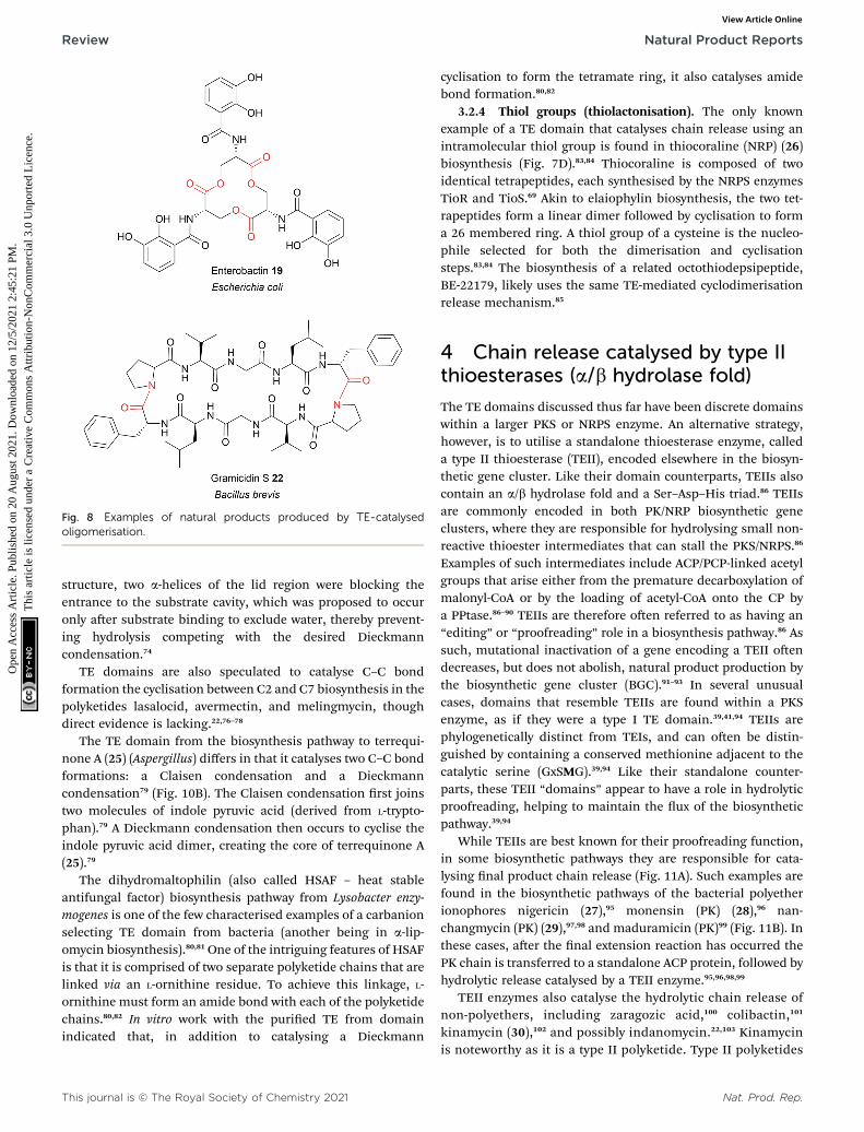
Fig. 8 Examples of natural products produced by TE-catalysedoligomerisation.
Review Natural Product Reports
Ope
n A
cces
s A
rtic
le. P
ublis
hed
on 2
0 A
ugus
t 202
1. D
ownl
oade
d on
12/
5/20
21 2
:45:
21 P
M.
Thi
s ar
ticle
is li
cens
ed u
nder
a C
reat
ive
Com
mon
s A
ttrib
utio
n-N
onC
omm
erci
al 3
.0 U
npor
ted
Lic
ence
.View Article Online
structure, two a-helices of the lid region were blocking theentrance to the substrate cavity, which was proposed to occuronly aer substrate binding to exclude water, thereby prevent-ing hydrolysis competing with the desired Dieckmanncondensation.74
TE domains are also speculated to catalyse C–C bondformation the cyclisation between C2 and C7 biosynthesis in thepolyketides lasalocid, avermectin, and melingmycin, thoughdirect evidence is lacking.22,76–78
The TE domain from the biosynthesis pathway to terrequi-none A (25) (Aspergillus) differs in that it catalyses two C–C bondformations: a Claisen condensation and a Dieckmanncondensation79 (Fig. 10B). The Claisen condensation rst joinstwo molecules of indole pyruvic acid (derived from L-trypto-phan).79 A Dieckmann condensation then occurs to cyclise theindole pyruvic acid dimer, creating the core of terrequinone A(25).79
The dihydromaltophilin (also called HSAF – heat stableantifungal factor) biosynthesis pathway from Lysobacter enzy-mogenes is one of the few characterised examples of a carbanionselecting TE domain from bacteria (another being in a-lip-omycin biosynthesis).80,81One of the intriguing features of HSAFis that it is comprised of two separate polyketide chains that arelinked via an L-ornithine residue. To achieve this linkage, L-ornithine must form an amide bond with each of the polyketidechains.80,82 In vitro work with the puried TE from domainindicated that, in addition to catalysing a Dieckmann
This journal is © The Royal Society of Chemistry 2021
cyclisation to form the tetramate ring, it also catalyses amidebond formation.80,82
3.2.4 Thiol groups (thiolactonisation). The only knownexample of a TE domain that catalyses chain release using anintramolecular thiol group is found in thiocoraline (NRP) (26)biosynthesis (Fig. 7D).83,84 Thiocoraline is composed of twoidentical tetrapeptides, each synthesised by the NRPS enzymesTioR and TioS.69 Akin to elaiophylin biosynthesis, the two tet-rapeptides form a linear dimer followed by cyclisation to forma 26 membered ring. A thiol group of a cysteine is the nucleo-phile selected for both the dimerisation and cyclisationsteps.83,84 The biosynthesis of a related octothiodepsipeptide,BE-22179, likely uses the same TE-mediated cyclodimerisationrelease mechanism.85
4 Chain release catalysed by type IIthioesterases (a/b hydrolase fold)
The TE domains discussed thus far have been discrete domainswithin a larger PKS or NRPS enzyme. An alternative strategy,however, is to utilise a standalone thioesterase enzyme, calleda type II thioesterase (TEII), encoded elsewhere in the biosyn-thetic gene cluster. Like their domain counterparts, TEIIs alsocontain an a/b hydrolase fold and a Ser–Asp–His triad.86 TEIIsare commonly encoded in both PK/NRP biosynthetic geneclusters, where they are responsible for hydrolysing small non-reactive thioester intermediates that can stall the PKS/NRPS.86
Examples of such intermediates include ACP/PCP-linked acetylgroups that arise either from the premature decarboxylation ofmalonyl-CoA or by the loading of acetyl-CoA onto the CP bya PPtase.86–90 TEIIs are therefore oen referred to as having an“editing” or “proofreading” role in a biosynthesis pathway.86 Assuch, mutational inactivation of a gene encoding a TEII oendecreases, but does not abolish, natural product production bythe biosynthetic gene cluster (BGC).91–93 In several unusualcases, domains that resemble TEIIs are found within a PKSenzyme, as if they were a type I TE domain.39,41,94 TEIIs arephylogenetically distinct from TEIs, and can oen be distin-guished by containing a conserved methionine adjacent to thecatalytic serine (GxSMG).39,94 Like their standalone counter-parts, these TEII “domains” appear to have a role in hydrolyticproofreading, helping to maintain the ux of the biosyntheticpathway.39,94
While TEIIs are best known for their proofreading function,in some biosynthetic pathways they are responsible for cata-lysing nal product chain release (Fig. 11A). Such examples arefound in the biosynthetic pathways of the bacterial polyetherionophores nigericin (27),95 monensin (PK) (28),96 nan-changmycin (PK) (29),97,98 and maduramicin (PK)99 (Fig. 11B). Inthese cases, aer the nal extension reaction has occurred thePK chain is transferred to a standalone ACP protein, followed byhydrolytic release catalysed by a TEII enzyme.95,96,98,99
TEII enzymes also catalyse the hydrolytic chain release ofnon-polyethers, including zaragozic acid,100 colibactin,101
kinamycin (30),102 and possibly indanomycin.22,103 Kinamycinis noteworthy as it is a type II polyketide. Type II polyketides
Nat. Prod. Rep.

Fig. 9 TE-catalysed “backwards transfer” dimerisation mechanism in elaiophylin (20) biosynthesis.
Natural Product Reports Review
Ope
n A
cces
s A
rtic
le. P
ublis
hed
on 2
0 A
ugus
t 202
1. D
ownl
oade
d on
12/
5/20
21 2
:45:
21 P
M.
Thi
s ar
ticle
is li
cens
ed u
nder
a C
reat
ive
Com
mon
s A
ttrib
utio
n-N
onC
omm
erci
al 3
.0 U
npor
ted
Lic
ence
.View Article Online
are aromatic natural products produced by type II PKSs(described in Section 2).14 The TEII enzyme AlpS has beenshown to be essential for kinamycin biosynthesis in vivo andits hydrolytic activity with a SNAC-substrate analoguedemonstrated in vitro.102 (Fig. 12A). The chain release mech-anism in type II polyketide biosynthesis pathways are poorlyunderstood, having even been speculated to occur thoughspontaneous hydrolysis/aldol reaction.11,14 The discovery thatsome utilise TEII enzymes is therefore an important discoveryfor the eld.
Chain release catalysed by TEII enzymes is not alwayshydrolytic.104 The TEII enzyme in gregatin A (31) biosynthesiscatalyses chain release by a Claisen condensation reaction.104
The furanone core of gregatin A is produced by the fusion of
Nat. Prod. Rep.
two acyl chains. Intriguingly, in vitro reconstitution experi-ments indicate that the two different chains are bothproduced by the PKS GrgA.104 The thioesterase GrgF thencatalyses fusion of the two PK chains via a Claisen conden-sation, followed by hydrolytic chain release of the lineardimer (Fig. 12B). The linear dimer is then proposed toundergo a spontaneous cyclisation reaction to produce thefuranone-containing gregatin A.104 Aside from gregatin Abiosynthesis, a non-hydrolytic TEII enzyme is also encoded inthe BGC of pyoluteorin, a PK–NRP hybrid rst isolated from P.aeruginosa strains T359 and IFO 3455.105,106 The TEII (PltG) isproposed to catalyse the release of the pyoluteorin interme-diate from the PKS PltC the via a Dieckmann condensation,forming a six-membered ring.106,107
This journal is © The Royal Society of Chemistry 2021

Fig. 10 Chain release by a TE-catalysed Dieckmann condensation. (A) TE-catalysed Dieckmann condensation in the biosynthesis of naph-thopyrone (23). (B) TE-catalysed Claisen condensation and Dieckmann condensation in terrequinone A (25) biosynthesis.
Review Natural Product Reports
Ope
n A
cces
s A
rtic
le. P
ublis
hed
on 2
0 A
ugus
t 202
1. D
ownl
oade
d on
12/
5/20
21 2
:45:
21 P
M.
Thi
s ar
ticle
is li
cens
ed u
nder
a C
reat
ive
Com
mon
s A
ttrib
utio
n-N
onC
omm
erci
al 3
.0 U
npor
ted
Lic
ence
.View Article Online
5 Chain release catalysed by hot-dogfold thioesterases
Enzymes with thioesterase activity are highly diverse, fallinginto at least 23 distinct families.108 Furthermore, those thatcatalyse chain release are not restricted to the a/b hydrolasefold family. One example are the homotetrameric hot-dog foldthioesterases encoded in biosynthetic gene clusters of theenediyne polyketides including calicheamicin (CalE7),109
dynemicin (DynE7),110 and C-1027 (SgcE10) (Fig. 13).111–113
Enediynes are potent DNA-damaging agents synthesised bytype I PKSs.114 A hot-dog folds consists of a 5–6 strand curvedb-sheet “bun” wrapping around a long central a-helix “hot-dog”.115 Aside from their role in enediyne biosynthesis, hot-dog fold thioesterases are found in both prokaryotes and
This journal is © The Royal Society of Chemistry 2021
eukaryotes where they hydrolyse acyl-CoA to release fatty acidsand CoA.116 In enediyne biosynthesis, these thioesterasescatalyse the hydrolytic release of methylketol hexaene (32)and heptaene (33) (Fig. 14A).117,118 While both products wereinitially proposed to be enediyne biosynthetic intermediates,they are now believed to be shunt products.117,118 The role ofthese hot-dog fold thioesterases is therefore akin to theproofreading TEII enzymes discussed in the previoussection.11
Mechanistically, hot-dog fold thioesterases are distinct froma/b-hydrolase fold thioesterases. Unlike the a/b-hydrolase foldthioesterases, the PK chain is never covalently bound to a hot-dog fold thioesterase, instead entering its active site while stilltethered to the adjacent ACP domain.109,110 In terms of catalyticresidues, a conserved arginine was shown viamutagenesis to be
Nat. Prod. Rep.

Fig. 11 Chain release by TEII enzymes in polyether biosynthesis. (A)Mechanism of polyether chain release from a standalone ACP domainby a TEII enzyme. (B) Examples of polyether polyketides where chainhydrolytic chain release is catalysed by a standalone TEII enzyme.
Natural Product Reports Review
Ope
n A
cces
s A
rtic
le. P
ublis
hed
on 2
0 A
ugus
t 202
1. D
ownl
oade
d on
12/
5/20
21 2
:45:
21 P
M.
Thi
s ar
ticle
is li
cens
ed u
nder
a C
reat
ive
Com
mon
s A
ttrib
utio
n-N
onC
omm
erci
al 3
.0 U
npor
ted
Lic
ence
.View Article Online
an essential for activity.109,110 The positively charged guanidi-nium group of the arginine is proposed to stabilise the oxyanionthat forms on the C1 carbonyl following attack by a water
Nat. Prod. Rep.
molecule (Fig. 14B).109,110,119 The tetrahedral intermediate isresolved by loss of the Ppant group and release of the PK chainfrom the active site. Whether the attacking water molecule isactivated via deprotonation (forming an hydroxide ion) isunknown, but could be performed by a conserved glutamic acidor tyrosine residue.110
In contrast to enediyne biosynthesis, in other biosyntheticpathways hot-dog fold thioesterases are responsible for hydro-lytic release of the nal product itself. A notable example isfound in the biosynthesis pathway for the macrolactam cremi-mycin (34) produced by Streptomyces sp. MJ635-86F5.120 Cre-mimycin biosynthesis features the incorporation of an unusualextension unit derived from the b-amino acid 3-amino-nonanoate.120 The biosynthesis of this b-amino fatty acid isperformed by the PKS enzymes CmiP4, CmiP3, and CmiP2. Thethree PKS enzymes produce non-2-enoyl-ACP that is hydrolysedby the standalone hot-dog fold thioesterase CmiS1 (Fig. 14C).120
However, prior to hydrolysis, CmiS1 rst catalyses a Michaeladdition between non-2-enoyl-ACP and glycine, installing whatwill become the b-amino group.120 The crystal structure ofa homologue of CmiS1 from Streptomyces avermitilis MA-4680,SAV606, which catalyses an equivalent reaction has beensolved.121 An in vitro activity assay of SAV606 indicated that italso catalyses the Michael addition with glycine in addition tohydrolytic chain release. Analysis of the SAV606 structure led tothe proposal that a histidine residue (His59) deprotonates theglycine amine group via a water molecule, thereby promoting itsnucleophilic attack on the b-group of the PK chain.121 The sameTE-catalysed mechanism for b-amino fatty acid biosynthesis islikely present in the biosynthesis pathways of the macrolactamsML-449 and BE-14106.122,123
6 Chain release catalysed by metallo-b-lactamase (MbL) thioesterases
A third family of standalone thioesterases that catalyse chainrelease resemble metallo-b-lactamase enzymes (MbL). b-Lacta-mase enzymes are widespread in bacteria where they have animportant role in hydrolysing b-lactam antibiotics.124 MbLenzymes possess an abba-fold and require a metal cofactor,typically up to two Zn2+ ions, to function.125 Fungal genomesalso encode b-lactamases, although their functions are notalways clear and can be involved in processes other thanxenobiotic degradation.124 In select cases, MbLs are thio-esterases (MbL-TEs) that catalyse the hydrolytic chain release offungal polycyclic polyketides. These polycyclic compounds aresynthetised by non-reducing polyketide synthases (nrPKSs).Unlike the modular type I PKSs, nrPKSs act iteratively to syn-thesise a highly reactive poly-b-keto PK chains that undergomultiple aldol condensations to form aromatic polycycliccompounds.126,127 The regioselectivity of the rst aldol conden-sation is controlled by a specialised product template (PT)domain within the nrPKS, which determines the cyclisationpattern for the compound as a whole.128 In addition, nrPKSsalso utilise an N-terminal starter unit acyltransferase (SAT)domain for starter unit selection, which can range from acetyl-
This journal is © The Royal Society of Chemistry 2021
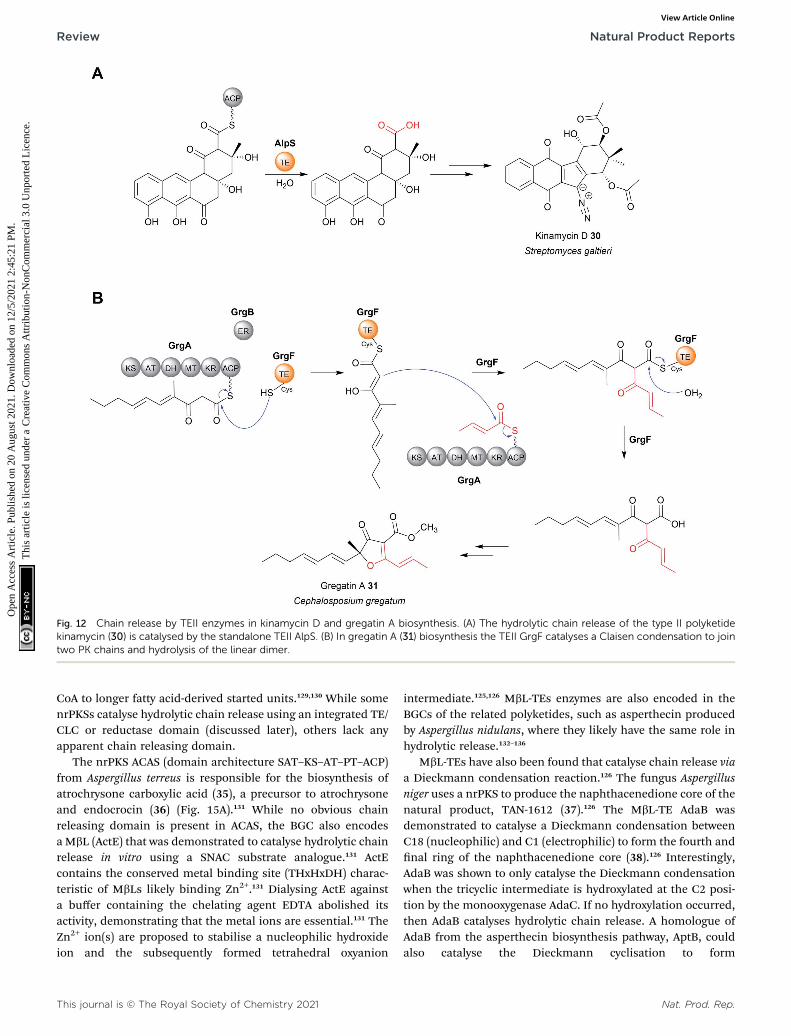
Fig. 12 Chain release by TEII enzymes in kinamycin D and gregatin A biosynthesis. (A) The hydrolytic chain release of the type II polyketidekinamycin (30) is catalysed by the standalone TEII AlpS. (B) In gregatin A (31) biosynthesis the TEII GrgF catalyses a Claisen condensation to jointwo PK chains and hydrolysis of the linear dimer.
Review Natural Product Reports
Ope
n A
cces
s A
rtic
le. P
ublis
hed
on 2
0 A
ugus
t 202
1. D
ownl
oade
d on
12/
5/20
21 2
:45:
21 P
M.
Thi
s ar
ticle
is li
cens
ed u
nder
a C
reat
ive
Com
mon
s A
ttrib
utio
n-N
onC
omm
erci
al 3
.0 U
npor
ted
Lic
ence
.View Article Online
CoA to longer fatty acid-derived started units.129,130 While somenrPKSs catalyse hydrolytic chain release using an integrated TE/CLC or reductase domain (discussed later), others lack anyapparent chain releasing domain.
The nrPKS ACAS (domain architecture SAT–KS–AT–PT–ACP)from Aspergillus terreus is responsible for the biosynthesis ofatrochrysone carboxylic acid (35), a precursor to atrochrysoneand endocrocin (36) (Fig. 15A).131 While no obvious chainreleasing domain is present in ACAS, the BGC also encodesa MbL (ActE) that was demonstrated to catalyse hydrolytic chainrelease in vitro using a SNAC substrate analogue.131 ActEcontains the conserved metal binding site (THxHxDH) charac-teristic of MbLs likely binding Zn2+.131 Dialysing ActE againsta buffer containing the chelating agent EDTA abolished itsactivity, demonstrating that the metal ions are essential.131 TheZn2+ ion(s) are proposed to stabilise a nucleophilic hydroxideion and the subsequently formed tetrahedral oxyanion
This journal is © The Royal Society of Chemistry 2021
intermediate.125,126 MbL-TEs enzymes are also encoded in theBGCs of the related polyketides, such as asperthecin producedby Aspergillus nidulans, where they likely have the same role inhydrolytic release.132–136
MbL-TEs have also been found that catalyse chain release viaa Dieckmann condensation reaction.126 The fungus Aspergillusniger uses a nrPKS to produce the naphthacenedione core of thenatural product, TAN-1612 (37).126 The MbL-TE AdaB wasdemonstrated to catalyse a Dieckmann condensation betweenC18 (nucleophilic) and C1 (electrophilic) to form the fourth andnal ring of the naphthacenedione core (38).126 Interestingly,AdaB was shown to only catalyse the Dieckmann condensationwhen the tricyclic intermediate is hydroxylated at the C2 posi-tion by the monooxygenase AdaC. If no hydroxylation occurred,then AdaB catalyses hydrolytic chain release. A homologue ofAdaB from the asperthecin biosynthesis pathway, AptB, couldalso catalyse the Dieckmann cyclisation to form
Nat. Prod. Rep.

Fig. 13 Crystal structure of the hot-dog fold thioesterase DynE3.Displayed is a single monomer of the tetrameric DynE3, a hot-dog foldthioesterase with a proofreading role found in the biosynthesispathway of the enediyne dynemicin (PDB: 2XEM). The catalytic Arg35residue, proposed to stabilise the quaternary oxyanion polyketideintermediate, is highlighted.
Natural Product Reports Review
Ope
n A
cces
s A
rtic
le. P
ublis
hed
on 2
0 A
ugus
t 202
1. D
ownl
oade
d on
12/
5/20
21 2
:45:
21 P
M.
Thi
s ar
ticle
is li
cens
ed u
nder
a C
reat
ive
Com
mon
s A
ttrib
utio
n-N
onC
omm
erci
al 3
.0 U
npor
ted
Lic
ence
.View Article Online
naphthacenedione.126 Closer biochemical characterisation ofAptB revealed it binds two Mn2+ ions, rather than Zn2+. In theproposed mechanism for AdaB/AptB function, the two Mn2+
ions facilitate substrate binding and may assist in the depro-tonation of the C18 a-proton, though additional experimentalevidence is required (Fig. 15B).126 Given the widespread role ofb-lactamases in catalysing hydrolysis reactions, it is likely thatthe Dieckmann condensation activity is a more recentadaptation.
7 Chain release catalysed by penicillinbinding protein (PBP)-like enzymes
A newly discovered class of chain releasing enzyme are homo-logs of penicillin binding proteins (PBP). The PBPs are pepti-dases that catalyse the nal transpeptidation reaction duringbacterial cell wall biosynthesis.137 The ability of PBP-likeenzymes to catalyse chain release was rst discovered in thebiosynthesis pathway of surugamide A–F, a group of linear andcyclic NRPs produced by Streptomyces sp. JAMM992.138 Sur-ugamide A-E are related cyclic NRPs all produced by the NRPSenzymes SurA and SurD.139 In contrast, surugamide F is anunrelated linear NRP carboxylic acid produced by the NRPSsSurB and SurC, the genes for which are encoded adjacent to surAand surD in the same BGC.139 Interestingly, none of the encodedNRPS enzymes contain a thioesterase or another previouslycharacterised chain release domain.138 Just upstream of surA isa small gene encoding a putative 28 kDa penicillin bindingprotein, SurE.138 The ability of SurE to catalyse a chain releasinglactamisation reaction was demonstrated by incubating anlinear SNAC precyclisation precursor of surugamide B with
Nat. Prod. Rep.
puried SurE, resulting in formation of surugamide B.138
Furthermore, creating an in-frame deletion in surE abolishednot the only production of surugamide A–E, but also sur-ugamide F.140–142 SurE is therefore surprisingly responsible forcatalysing chain release in both pathways.140–142 An in vitro assayusing SNAC-surugamide F revealed that the hydrolysis productis only a minor product, with the major product being a lactam.Surugamide F is therefore proposed to be produced from thislactam by an as yet undiscovered peptidase.142
In regards to the enzymatic mechanism of SurE, it containsthe conserved Ser–Tyr–His–Lys catalytic tetrad of other PBPpeptidases.137,138,140–142 In PBP peptidases the serine acts asa nucleophile while the other catalytic residues are involved inproton transfer/transition state stabilisation.137,141 Mutagenesisof the serine residue in SurE to alanine abolished its activity,consistent with a role in forming an oxoester linkage to thepeptide chain (analogous to the catalytic serine of a/b hydrolasethioesterases). The role of the other residues in catalysing lac-tamisation of the surugamides are still unclear.141 SurE homo-logues are also encoded in the biosynthetic gene clusters ofother NRPs, sometimes even as a dedicated domain withina NRPS enzyme.141 There is therefore still much to be exploredin regards to the function and products of PBP-like chainreleasing enzymes.
8 Chain release catalysed byreductase (R) domains8.1 Structure and mechanism of R domains
Aside from TE domains, another chain releasing domainlocated on the C-termini of some PKS/NRPS enzymes isa reductase (R) domains. R domains catalyse the reductiverelease of PK/NRP chains as either aldehydes (via a two-electronreduction) or primary alcohols (via a four-electron reduction)(Fig. 16, 17 and 18A).13
R domains are mechanistically and structurally distinct froma/b-hydrolase thioesterases, belonging instead to the short-chain dehydrogenases (SDR) family of NAD(P)H dependentoxidoreductases.143 Members of this family all possess an N-terminal Rossmann fold: a sheet of seven parallel b strandsanked by a-helices on either side.144,145 Interestingly, despitetheir differences, the central b-sheets of TE and R domains havea similar spatial arrangement.144,145
An individual R domain (ca. 400 amino acids in size) can besubdivided into two regions: an N-terminal Rossmann foldregion responsible for NAD(P)H binding, and a C-terminalregion responsible for substrate binding.13 In contrast to TEdomains, where the PK/NRP chain is covalently bonded to theTE domain, R domains act directly on CP-linked PK/NRPchains.13 The diphosphate portion of NAD(P)H interacts withthe peptide backbone of a GxxGxxG nucleotide binding motifconserved within the N-terminal region.146 The C-terminalregion consists of 4–6 a-helices and 2 b sheets.144–147 The N-terminal region also contains a mobile “gating loop” thatinteracts with the upstream carrier protein (ACP or PCP) and isproposed to regulate both the binding of NAD(P)H and x the
This journal is © The Royal Society of Chemistry 2021

Fig. 14 Chain release by hot-dog fold thioesterases (A) the release of methylketo hexanene (32) and heptaene (33) by the hot-dog fold thio-esterase DynE8 in dynemicin biosynthesis. These linear products are now believed to be shunt products rather than biosynthetic intermediates.(B) The proposedmechanism of hot-dog fold thioesterases involving an oxyanion-stabilising catalytic arginine. (C) The hot-dog fold thioesteraseCmiS1 is responsible for catalysing hydrolytic chain release in cremimycin (34) biosynthesis. CmiS1 also catalyses the addition of glycine to the b-carbon.
This journal is © The Royal Society of Chemistry 2021 Nat. Prod. Rep.
Review Natural Product Reports
Ope
n A
cces
s A
rtic
le. P
ublis
hed
on 2
0 A
ugus
t 202
1. D
ownl
oade
d on
12/
5/20
21 2
:45:
21 P
M.
Thi
s ar
ticle
is li
cens
ed u
nder
a C
reat
ive
Com
mon
s A
ttrib
utio
n-N
onC
omm
erci
al 3
.0 U
npor
ted
Lic
ence
.View Article Online

Fig. 15 Chain release catalysed by metallo-b-lactamase (MbL) thioesterase. (A) The MbL ActE is responsible for chain release of the endocrocinprecursor from the PKS ACAS. (B) The two possible mechanisms have been proposed for the Mn2+ dependent Dieckmann condensation cat-alysed by AptB/AdaB.
Natural Product Reports Review
Ope
n A
cces
s A
rtic
le. P
ublis
hed
on 2
0 A
ugus
t 202
1. D
ownl
oade
d on
12/
5/20
21 2
:45:
21 P
M.
Thi
s ar
ticle
is li
cens
ed u
nder
a C
reat
ive
Com
mon
s A
ttrib
utio
n-N
onC
omm
erci
al 3
.0 U
npor
ted
Lic
ence
.View Article Online
Ppant arm into a reactive conformation.144,147 Recent work alsoidentied a hydrophobic pocket within the R domain respon-sible for binding the geminal dimethyl group of the Ppantarm.147 Unlike the highly conserved N-terminus region, thesequence identity of the C-terminal region is highly variable,likely reecting the diversity of substrates recognised by Rdomains.144,146,147 Despite this notable sequence diversity, the C-terminal region of R domains are distinguished from other SDRmembers by containing a short helix-turn-helix motif importantfor the interface between the R domain and upstreamCP.144,146,147
R domains contain a Thr–Tyr–Lys catalytic triad character-istic of SDR family members. The catalytic tyrosine and lysineresidues are both critical for binding NAD(P)H, while the Thr
Nat. Prod. Rep.
stabilises the PK/NRP thioester substrate (Fig. 18B).144,146,148
Hydride transfer by NAD(P)H to the C1 carbon of the PK/NRPSthioester generates a tetrahedral thiohemiacetal intermediate.The intermediate is resolved by loss of the Ppant group,generating a free aldehyde. In the case of four-electron Rdomains, NAD(P)+ dissociates and is replaced by second mole-cule of NAD(P)H.13 This second NAD(P)H transfers a hydride tothe electrophilic C1 carbon of the aldehyde, resulting in theformation of primary alcohol.13 For several R domains, thereduction to the alcohol occurs faster than the reduction to thealdehyde.144,146 Aldehyde reduction is accompanied witha notable electronic shi, changing an electrophilic aldehyde toa nucleophilic alcohol.13 How some R domains exclusivelycatalyse a two-electron reduction while others a four-electron
This journal is © The Royal Society of Chemistry 2021

Fig. 16 Examples of natural products synthesised by PKS/NRPS and released/modified using R domains.
Review Natural Product Reports
Ope
n A
cces
s A
rtic
le. P
ublis
hed
on 2
0 A
ugus
t 202
1. D
ownl
oade
d on
12/
5/20
21 2
:45:
21 P
M.
Thi
s ar
ticle
is li
cens
ed u
nder
a C
reat
ive
Com
mon
s A
ttrib
utio
n-N
onC
omm
erci
al 3
.0 U
npor
ted
Lic
ence
.View Article Online
reduction is still poorly understood.147,148 A study on NRPS-related carboxylic acid reductases (CARs), indicated thatconformational changes in the NAD(P)H binding site controlwhether this second reduction occurs.145,149 CARs are multido-main enzymes (A–PCP–R structure) that reduce carboxylic acidsto their corresponding aldehydes. By comparing the structuresof several CAR–R domains, a conformational change in the loopconnecting the N and C terminal regions, particularly in anaspartic acid residue, was identied.145,149 One of the loopconformations appeared to facilitate NAD(P)H binding, whilethe other interfered with it. Based on these observations, it wasproposed that the binding of the PK/NRP chain to the R domainpromotes NAD(P)H binding, resulting in aldehyde forma-tion.145,149 The aldehyde, however, is unable to maintain thefavourable NAD(P)H binding conformation of the R domain,preventing a second molecule of NAD(P)H binding. In supportof this theory, mutating the identied Asp residue to glycineenabled the R domain to form the primary alcohol product.149
This journal is © The Royal Society of Chemistry 2021
There are also other factors at play, however, as some R domainscontain this Asp residue but still perform four-electron reduc-tions.145,150 Furthermore, biophysical studies have also demon-strated that NAD(P)H binding to the R domain is not dependenton the PK/NRP substrate.148 Further complicating matters are Rdomains that produce both two-electron and four-electronproducts, discussed in the following section. The question ofhow R domains control whether a two-electron or four-electronreduction takes place therefore remains open.147,148
8.2 PK and NRP pathways that use R domains
The natural products produced using an R domain have beenextensively covered in an excellent recent review.13 In brief,while the direct products of R domains are either aldehydes orprimary alcohols, both functional groups can undergo addi-tional transformations to further diversify the structure of thenal product (summarised in Fig. 19).
Nat. Prod. Rep.

Fig. 17 Crystal structure of an R domain. (A) The crystal structure ofthe R domain from mycobacterial lipopeptide biosynthesis (PDB:4DQV). The N-terminal domain is responsible for NAD(P)H bindingwhile the C-terminal domain is responsible for substrate binding. (B)The Thr–Lys–Tyr catalytic triad of the R domain located in the N-terminal region.
Fig. 18 Mechanism of reductase (R) domains. (A) R domains can eithercatalyse a two-electron reduction to release the PK/NRP chain as analdehyde or a four-electron reduction to release a primary alcohol. (B)The catalytic mechanism of R domains. The conserved lysine andtyrosine residues bind to NAD(P)H, while the tyrosine binds to the PK/NRP chain.
Natural Product Reports Review
Ope
n A
cces
s A
rtic
le. P
ublis
hed
on 2
0 A
ugus
t 202
1. D
ownl
oade
d on
12/
5/20
21 2
:45:
21 P
M.
Thi
s ar
ticle
is li
cens
ed u
nder
a C
reat
ive
Com
mon
s A
ttrib
utio
n-N
onC
omm
erci
al 3
.0 U
npor
ted
Lic
ence
.View Article Online
8.3 Products of two-electron reductase domains
In some cases, the aldehyde generated by a two-electron Rdomain is retained in the nal product. This tends to beuncommon, however, due to the general instability of alde-hydes. The electrophilicity of aldehydes oen means that thenatural products containing them are oen potent inhibitors ofserine proteases.13 Examples of natural products containingaldehydes produced by two-electron R domains include theNRPs leupeptin (39), linear gramicidin,151 the avopeptins, andthe fellutamides.152–154
Alternatively, the aldehyde group can undergo additionaltransformations to form a range of different functionalgroups.13 Such transformations can range from a simpletransaminations, to more complex reactions that form macro-cyclic peptides or heterocycles. If the aldehyde group undergoesa transamination, an amine is formed.13 The rst characterisedexample of this was from the L-lysine biosynthesis pathway inSaccharomyces cerevisiae (also the rst example of reductiverelease by an R domain).155 The tridomain carboxyl acidreductase Lys2 (A–PCP–R) was identied as responsible forlysine biosynthesis, releasing a-aminoadipate that subse-quently undergoes a transamination to form L-lysine. Otherexamples include the siderophores myxochelin B (40),156,157 thezeamine antibiotics,158 and the lipopeptide antibiotic leucinos-tatin.159 In the case of leucinostatin, the amine of the product isdiversied further by methylation.159 The amine can also act asa nucleophile in an intramolecular cyclisation reaction, asoccurs in the biosynthesis of the alkaloids cyclizidine160 andcoelimycin P1 (41).161
Cyclic iminopeptides, such as the nostocyclopeptides andscytonemide A (42) are formed when the aldehyde generated bya two-electron R domain is spontaneously attacked by anintramolecular amine, followed by a dehydration reaction.162,163
If a dehydration reaction does not occur the nal productcontains a hemiaminal ring, exemplied by the pyrrolobenzo-diazepines anthramycin,164 sibiromycin,165 and tomaymycin.166
Nat. Prod. Rep.
In the case of the cyclic peptide lugdunin (43), a peptide anti-biotic and immune response modulator produced by thehuman microbiome, the imide carbon is subsequently attackedby the thiol group of the adjacent cysteine residue, forminga thioazolidine ring.167,168 In the case of the sorbicillin family ofnatural products, the aldehyde is attacked by a carbanion ina Knoevenagel cyclisation to form a benzene intermediate.169
In the biosynthesis of the fungal indole alkaloid mal-brancheamide, a two-electron R domain releases L-Pro–L-Trpdipeptide as an aldehyde.170 The dipeptide aldehyde undergoesspontaneous cyclisation and dehydration to give a dienamineintermediate that is subsequently prenylated and spontane-ously oxidised. The prenyl group contains a double bond thatacts as a dieneophile in an apparent enzyme catalysed intra-molecular [4 + 2] cycloaddition with the proline-derived pyr-azinone ring.170 Malbrancheamide biosynthesis highlights animpressive case where an aldehyde generated by an R domain isused to generate a substrate for an intramolecular Diels–Alderreaction.
This journal is © The Royal Society of Chemistry 2021
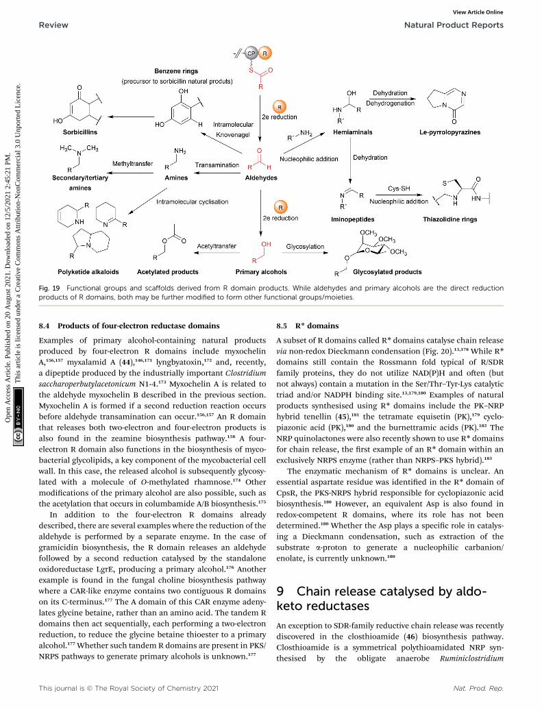
Fig. 19 Functional groups and scaffolds derived from R domain products. While aldehydes and primary alcohols are the direct reductionproducts of R domains, both may be further modified to form other functional groups/moieties.
Review Natural Product Reports
Ope
n A
cces
s A
rtic
le. P
ublis
hed
on 2
0 A
ugus
t 202
1. D
ownl
oade
d on
12/
5/20
21 2
:45:
21 P
M.
Thi
s ar
ticle
is li
cens
ed u
nder
a C
reat
ive
Com
mon
s A
ttrib
utio
n-N
onC
omm
erci
al 3
.0 U
npor
ted
Lic
ence
.View Article Online
8.4 Products of four-electron reductase domains
Examples of primary alcohol-containing natural productsproduced by four-electron R domains include myxochelinA,156,157 myxalamid A (44),146,171 lyngbyatoxin,172 and, recently,a dipeptide produced by the industrially important Clostridiumsaccharoperbutylacetonicum N1-4.173 Myxochelin A is related tothe aldehyde myxochelin B described in the previous section.Myxochelin A is formed if a second reduction reaction occursbefore aldehyde transamination can occur.156,157 An R domainthat releases both two-electron and four-electron products isalso found in the zeamine biosynthesis pathway.158 A four-electron R domain also functions in the biosynthesis of myco-bacterial glycolipids, a key component of the mycobacterial cellwall. In this case, the released alcohol is subsequently glycosy-lated with a molecule of O-methylated rhamnose.174 Othermodications of the primary alcohol are also possible, such asthe acetylation that occurs in columbamide A/B biosynthesis.175
In addition to the four-electron R domains alreadydescribed, there are several examples where the reduction of thealdehyde is performed by a separate enzyme. In the case ofgramicidin biosynthesis, the R domain releases an aldehydefollowed by a second reduction catalysed by the standaloneoxidoreductase LgrE, producing a primary alcohol.176 Anotherexample is found in the fungal choline biosynthesis pathwaywhere a CAR-like enzyme contains two contiguous R domainson its C-terminus.177 The A domain of this CAR enzyme adeny-lates glycine betaine, rather than an amino acid. The tandem Rdomains then act sequentially, each performing a two-electronreduction, to reduce the glycine betaine thioester to a primaryalcohol.177 Whether such tandem R domains are present in PKS/NRPS pathways to generate primary alcohols is unknown.177
This journal is © The Royal Society of Chemistry 2021
8.5 R* domains
A subset of R domains called R* domains catalyse chain releasevia non-redox Dieckmann condensation (Fig. 20).13,178 While R*domains still contain the Rossmann fold typical of R/SDRfamily proteins, they do not utilize NAD(P)H and oen (butnot always) contain a mutation in the Ser/Thr–Tyr-Lys catalytictriad and/or NADPH binding site.13,179,180 Examples of naturalproducts synthesised using R* domains include the PK–NRPhybrid tenellin (45),181 the tetramate equisetin (PK),179 cyclo-piazonic acid (PK),180 and the burnettramic acids (PK).182 TheNRP quinolactones were also recently shown to use R* domainsfor chain release, the rst example of an R* domain within anexclusively NRPS enzyme (rather than NRPS–PKS hybrid).183
The enzymatic mechanism of R* domains is unclear. Anessential aspartate residue was identied in the R* domain ofCpsR, the PKS-NRPS hybrid responsible for cyclopiazonic acidbiosynthesis.180 However, an equivalent Asp is also found inredox-competent R domains, where its role has not beendetermined.180 Whether the Asp plays a specic role in catalys-ing a Dieckmann condensation, such as extraction of thesubstrate a-proton to generate a nucleophilic carbanion/enolate, is currently unknown.180
9 Chain release catalysed by aldo-keto reductases
An exception to SDR-family reductive chain release was recentlydiscovered in the closthioamide (46) biosynthesis pathway.Closthioamide is a symmetrical polythioamidated NRP syn-thesised by the obligate anaerobe Ruminiclostridium
Nat. Prod. Rep.

Fig. 20 R* domains. R* domains are redox incompetent R domains that catalyse chain release by Dieckmann condensation reactions, such as intenellin acid (45) biosynthesis.
Natural Product Reports Review
Ope
n A
cces
s A
rtic
le. P
ublis
hed
on 2
0 A
ugus
t 202
1. D
ownl
oade
d on
12/
5/20
21 2
:45:
21 P
M.
Thi
s ar
ticle
is li
cens
ed u
nder
a C
reat
ive
Com
mon
s A
ttrib
utio
n-N
onC
omm
erci
al 3
.0 U
npor
ted
Lic
ence
.View Article Online
cellulolyticum.184 The biosynthesis of closthioamide (46) hasbeen studied extensively, revealing an unusual thiotemplatedNRPS-independent pathway.185–188 Closthioamide (46) isassembled from L-aspartate and chorismate, with the growingintermediate tethered to the standalone ACP CtaE (Fig. 21).185,187
Once the intermediate 47 has formed it is taken down twodivergent paths, one catalysed by the enzyme CtaJ and the otherby CtaK, which ultimately converge to form closthioamide.185
CtaK catalyses a two-electron reduction, releasing intermediate48 as an aldehyde.185 However, unlike the R domain discussedin the previous section, CtaK belongs to the aldo-keto reductasefamily rather than the SDR family.185 Unlike SDR R domains,aldo-keto reductases lack a Rossmann fold, instead adoptinga (b/a)8 conformation.189 Conserved arginine and lysine resi-dues assist in binding the pyrophosphate backbone of NADH/NADPH, typically favouring the binding of NADPH.189 Whilealdo-keto reductases are known for catalysing a diverse range ofredox reactions in both prokaryotes and eukaryotes, CtaK is therst characterised example of one that catalyses the reductiverelease of a NRP chain.185,189
10 Chain release catalysed byoxygenases
There are several biosynthetic pathways where chain release isproposed to occur through an oxidative mechanism. Examplesinclude the myxothiazole (49),190 melithiazole,190,191 aur-afuron,192 and pederin193 biosynthesis pathways. Myxothiazolesare synthesised by a mixture of PKS and PKS-NRPS hybridenzymes. Myxothiazole A (49) has a terminal amide residue,indicating an unusual chain release mechanism. The nal PKSenzyme in the myxothiazole A biosynthesis pathway, MtaG (C–A–MOx–PCP–TE) contains a monooxygenase-like (MOx) domainembedded within its A domain (Fig. 22A).190 MtaG is proposedto catalyse a condensation reaction between L-glycine and themyxothiazole intermediate, followed by hydroxylation of the a-position of L-glycine by the MOx domain.190 Hydroxylation is
Nat. Prod. Rep.
proposed to lead to spontaneous decomposition of the glycineresidue, releasing myxothiazole A (49). The TE domain is thenpresumed to hydrolyse the residual PCP-bound glyoxylate,190
While plausible, experimental evidence is still required toconrm this mechanism. The related compound melithiazoleappears to use the same mechanism of chain release, thoughthe amide is subsequently converted into a methyl ester.190,191
The aurafurons (50) are synthesised by PKS enzymes, none ofwhich contain a TE domain or any other integrated chainreleasing domain.192 The feeding of isotopically labelledprecursors demonstrated that the aurafuron backbone is syn-thesised from three acetate units and four propionate units.192
Interestingly, the C1 carbon derived from the incorporation ofthe nal propionate unit was absent.192 To account for thismissing carbon, an oxidative release mechanism was proposedthat utilises the putative monooxygenase AufJ encoded in thesame BGC (Fig. 22B).192 AufJ bears signicant sequence simi-larity to the Baeyer–Villiger monooxygenase MtmOIV from themithramycin biosynthesis pathway.192,194 The authors proposedthat AufJ catalyses a Baeyer–Villiger reaction (insertion of anoxygen into the a-position) to generate a carbonic acid diesterPK intermediate.192 Decarboxylation would then result in chainrelease with loss of the C1 carbon (consistent with the isotopelabelling experiment), though experimental evidence isrequired to conrm this theory.192 An AufJ homologue is alsoencoded in the BGCs of the linfuranones, furanones producedby Sphaerimonospora mesophile.192,195 Furanone-containingnatural products have also been isolated from molluscs, butwhether an AufJ homologue is involved in their biosynthesis isunknown.196
The polyketide pederin (51) is synthesised by trans-AT PKSsby unculturable symbionts within Paederus fuscipes beetles.193
Chain release in pederin biosynthesis is proposed, but notexperimentally demonstrated, to occur by oxidative cleavagecatalysed by the FAD-dependent monooxygenase PedG, result-ing in the formation of a terminal primary alcohol (52)(Fig. 22C).193 Alternatively, the pederin intermediate may be
This journal is © The Royal Society of Chemistry 2021
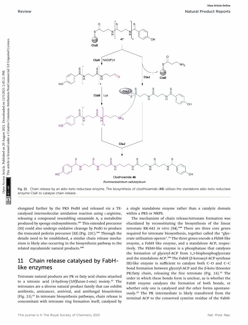
Fig. 21 Chain release by an aldo-keto reductase enzyme. The biosynthesis of closthioamide (46) utilises the standalone aldo-keto reductaseenzyme CtaK to catalyse chain release.
Review Natural Product Reports
Ope
n A
cces
s A
rtic
le. P
ublis
hed
on 2
0 A
ugus
t 202
1. D
ownl
oade
d on
12/
5/20
21 2
:45:
21 P
M.
Thi
s ar
ticle
is li
cens
ed u
nder
a C
reat
ive
Com
mon
s A
ttrib
utio
n-N
onC
omm
erci
al 3
.0 U
npor
ted
Lic
ence
.View Article Online
elongated further by the PKS PedH and released via a TE-catalysed intermolecular amidation reaction using L-arginine,releasing a compound resembling onnamide A, a metaboliteproduced by sponge endosymbionts.197 This extended precursor(53) could also undergo oxidative cleavage by PedG to producethe truncated pederin precursor (52) (Fig. 22C).193 Through thedetails need to be established, a similar chain release mecha-nism is likely also occurring in the biosynthesis pathway to therelated mycalamide natural products.198
11 Chain release catalysed by FabH-like enzymes
Tetronate natural products are PK or fatty acid chains attachedto a tetronic acid (4-hydroxy-[5H]furan-2-one) moiety.12 Thetetronates are a diverse natural product family that can exhibitantibiotic, anticancer, antiviral, and antifungal bioactivities(Fig. 23).12 In tetronate biosynthesis pathways, chain release isconcomitant with tetronate ring formation itself, catalysed by
This journal is © The Royal Society of Chemistry 2021
a single standalone enzyme rather than a catalytic domainwithin a PKS or NRPS.
The mechanism of chain release/tetronate formation waselucidated by reconstituting the biosynthesis of the lineartetronate RK-682 in vitro (54).199 There are three core genesrequired for tetronate biosynthesis, together called the “glyc-erate utilisation operon”.12 The three genes encode a FkbH-likeenzyme, a FabH like enzyme, and a standalone ACP, respec-tively. The FkbH-like enzyme is a phosphatase that catalysesthe formation of glyceryl-ACP from 1,3-bisphosphoglycerateand the standalone ACP.200 The FabH (b-ketoacyl-ACP synthaseIII)-like enzyme is sufficient to catalyse both C–O and C–Cbond formation between glyceryl-ACP and the b-keto thioesterPK/fatty chain, releasing the free tetronate (Fig. 24).12 Theorder in which these bonds form is unclear, as is whether theFabH enzyme catalyses the formation of both bonds, orwhether only one is catalysed and the other forms spontane-ously.22 The PK intermediate is likely transferred from theterminal ACP to the conserved cysteine residue of the FabH-
Nat. Prod. Rep.

Fig. 22 Oxidative chain release. (A) Integrated within the A domain of MtaG is a monooxygenase (MOx) domain proposed to hydroxylate the a-position of the PCP-bound myxothiazole intermediate. Hydroxylation of the a-position is proposed to produce an unstable intermediate thatfragments to release myxothiazole (49) with a terminal amide group. The likely role of the TE domain is remove the PCP-bound glyoxylate,
Nat. Prod. Rep. This journal is © The Royal Society of Chemistry 2021
Natural Product Reports Review
Ope
n A
cces
s A
rtic
le. P
ublis
hed
on 2
0 A
ugus
t 202
1. D
ownl
oade
d on
12/
5/20
21 2
:45:
21 P
M.
Thi
s ar
ticle
is li
cens
ed u
nder
a C
reat
ive
Com
mon
s A
ttrib
utio
n-N
onC
omm
erci
al 3
.0 U
npor
ted
Lic
ence
.View Article Online

Fig. 23 Examples of tetronate natural products.
Fig. 24 The mechanism of chain release by FabH-like enzymes in the biosynthesis of tetronate natural products.
Review Natural Product Reports
Ope
n A
cces
s A
rtic
le. P
ublis
hed
on 2
0 A
ugus
t 202
1. D
ownl
oade
d on
12/
5/20
21 2
:45:
21 P
M.
Thi
s ar
ticle
is li
cens
ed u
nder
a C
reat
ive
Com
mon
s A
ttrib
utio
n-N
onC
omm
erci
al 3
.0 U
npor
ted
Lic
ence
.View Article Online
like enzyme (part of its Cys–His–His catalytic triad).10,22 Thebiosynthesis of the polyether tetronate tetronasin appears tobe a slight exception to the described process, utilising
though experimental evidence is required. (B) Proposed Baeyer–Villigebiosynthesis. Experimental evidence is needed to support this mechanismdifferent routes have been proposed to release the pederin precursor 52.chain release.
This journal is © The Royal Society of Chemistry 2021
glycolyl-ACP rather than glyceryl-ACP.201 It is unknown ifglycolyl-ACP (or derivatives containing additional functionalgroups) are tolerated by the FabH enzymes of tetronate
r monooxygenase (AufJ)-mediated chain release in aurafuron A (50). (C) Oxidative chain release in proposed pederin (51) biosynthesis. TwoExperimental evidence is required to determine the true mechanism of
Nat. Prod. Rep.

Fig. 25 Spirotetronate formation. An exocyclic double bond on thetetronate moiety is formed by an enzyme-catalysed acetylation andelimination reaction. The double bond can then serve as a dieneophilein an intramolecular Diels–Alder reaction ([4 + 2] cycloaddition),forming a spirotetronate.
Natural Product Reports Review
Ope
n A
cces
s A
rtic
le. P
ublis
hed
on 2
0 A
ugus
t 202
1. D
ownl
oade
d on
12/
5/20
21 2
:45:
21 P
M.
Thi
s ar
ticle
is li
cens
ed u
nder
a C
reat
ive
Com
mon
s A
ttrib
utio
n-N
onC
omm
erci
al 3
.0 U
npor
ted
Lic
ence
.View Article Online
pathways that typically accept glyceryl-ACP, if so enabling thecreation of novel tetronate compounds.
Polyketide tetronates such as abyssomicin C (55), tetronasin(56), and versipelostatin A (57) are all synthesised by type I PKSenzymes.12 There are several distinguishing characteristics ofPK tetronate biosynthetic pathways. To begin with, the terminalPKS module of these pathways contains a C-terminal ACPdomain, rather than a C-terminal TE domain or R domain.12
Secondly, in order for chain release to occur, the nal ACP-bound PK requires a b-keto group to acidify the a-position forcarbanion formation.12 As such, the nal PKS module of a pol-yketide tetronate biosynthesis pathway either lacks a KRdomain, or contains one that is catalytically inactive.12,202–205 Itwould be interesting to test if prematurely forming a b-keto PKintermediate, by mutating the KR of an earlier module, wouldresult in the formation of truncated tetronate products due topremature release by the FabH-like enzyme.
Installing a tetronate ring provides additional opportuni-ties for structural diversication. A common modication is
Fig. 26 Dimerisation in myxopyronin biosynthesis. Analogous to tetronrelease/ring formation in myxopyronin A (59) biosynthesis.
Nat. Prod. Rep.
to eliminate the primary alcohol of the tetronate moiety toform an exocyclic double bond.206,207 To achieve this theprimary alcohol is rst acetylated by an acyltransferase tocreate a superior leaving group, followed by elimination bya lyase to create the exocyclic double bond itself.206 Thisexocyclic double bond is an important feature of many tetr-onates, as it can be used as a dieneophile in both intra-molecular or intermolecular [4 + 2] cycloadditions (Diels–Alder reactions) (Fig. 25).208–210 An intramolecular Diels–Alderreaction can produce a spirotetronate moiety, exemplied bycompounds such as abyssomicin C (55)210 and versipelostatinC (57).209 Spirotetronates are characterised as two ringstructures linked together by a spiroatom (a carbon in thecase of the spirotetronates).12 Alternatively, the tetronate maybe halogenated via a yet unknown mechanism, as occurs innonthmicin (58) biosynthesis.211 Exploring the mechanism ofthis halogenation would be valuable, as the identication ofa tetronate halogenase could enable halogens to be enzy-matically added to the tetronate groups of other naturalproducts.
A FabH-like enzyme (MxnB) is also responsible for chainrelease in myxopyronin biosynthesis (59).212,213 Analogous totetronate ring formation, here the central pyrone ring of myx-opyronin is formed by the MxnB-catalysed condensation of PKchains tethered to the PKSs MxnJ and MxnK (Fig. 26).212,213 As intetronate biosynthesis, the order in which the C–C and C–Obonds form is still unclear.206
12 Chain release catalysed bycondensation-like (CT) domains
Condensation (C) domains are one of the core domains foundin NRPSmodules. C domains are responsible for catalysing C–Nbond formation between a PCP-bound NRP chain and an aminoacid tethered to the PCP domain of the downstream module. Cdomains are ca. 50 kDa in size and have a V-shaped pseudo-dimer structure: a single polypeptide forming two subdomains(“monomers”), each of which possesses a chloramphenicolacyltransferase fold.2 At the interface of these two subdomainsis the conserved catalytic HxxxDxxS motif (Fig. 27).2
ate biosynthesis, the FabH-like enzyme MxnB is responsible for chain
This journal is © The Royal Society of Chemistry 2021

Review Natural Product Reports
Ope
n A
cces
s A
rtic
le. P
ublis
hed
on 2
0 A
ugus
t 202
1. D
ownl
oade
d on
12/
5/20
21 2
:45:
21 P
M.
Thi
s ar
ticle
is li
cens
ed u
nder
a C
reat
ive
Com
mon
s A
ttrib
utio
n-N
onC
omm
erci
al 3
.0 U
npor
ted
Lic
ence
.View Article Online
The histidine in this motif was rst proposed to act asa general base to deprotonate the a-amine group of the CP-bound amino acid of the downstream module, promoting itsnucleophilic attack on the upstream CP-linked thioester(Fig. 28A).214,215However, deciphering the role of this histidine isnot straightforward, as in some C domains it is not essential foractivity.216,217 One explanation from recent structural data couldbe that water can serve as an alternative base in the absence ofthe histidine.218 Matters complicated further still by calcula-tions indicating that the histidine is protonated (and thereforeunable to act as a base) under physiological conditions.219
Regardless of their enzymatic mechanism, in some biosyn-thetic pathways use specialised C domains (called CT domains)to catalyse chain release.182,220–223 Like canonical TE domains, CT
domains are located on the C-terminus of an NRPS/PKS enzyme.CT domains are especially prevalent in fungi, where 60–90% ofall encoded NRPS enzymes contain one on their C-terminus.220
Unlike the fungal TE domains, which typically catalyse hydro-lytic chain release, CT domains typically catalyse chain releasevia macrolactamisation, such as during nanangelenin B (60)biosynthesis (Fig. 28B).182,220,221 However, like TE domains, CT
domains can also catalyse chain release by selecting a range ofnucleophiles, both intramolecular and intermolecular. Forexample, CT domains have been found that catalyse chainrelease via amidation224 (Fig. 29A), Dieckmann condensation(Fig. 29B),225 hydrolysis (Fig. 29C),226 transesterication(Fig. 29D),112,227,228 or lactonisation.221 A notable accomplish-ment of the research on CT domains is the clarication of chainrelease in gliotoxin (61) biosynthesis, long proposed to occurspontaneously with diketopiperazine formation.229 However,the second C domain in gliotoxin synthase, GliA (A–PCP–C–A–PCP–CT–PCP), has now been identied as a CT domain, cata-lysing diketopiperazine formation/chain release (Fig. 28C).230
Interestingly, the PCP downstream of the GliA CT domain is alsoessential for chain release, indicating that it serves to tether theNRP intermediate specically for the CT domain.230 The same C-terminal CT–PCP arrangement is found in other NRPSs,including the previously mentioned nanangelenin B synthaseNanA, suggesting a conserved function.182,230
Fig. 27 Structure of a CT domain. The crystal structure of the CT
domain from fumiquinazoline F biosynthesis (PDB: 5DIJ). Theconserved histidine (His178) at the interface between the two pseu-domonomers is depicted.
Fig. 28 The mechanism of CT domains. (A) The proposed catalyticmechanism of CT domains using the conserved histidine residue asa general base. CT domains typically catalyse chain release via lactamformation. (B) CT-catalysed chain release in nanangelenin B (60)biosynthesis. (C) CT-catalysed chain release in gliotoxin (61)biosynthesis.
This journal is © The Royal Society of Chemistry 2021 Nat. Prod. Rep.

Fig. 29 Chain release catalysed by CT domains. (A) CT-domain-catalysed chain release via amidation in wortmanamide B biosynthesis. (B) CT-domain-catalysed chain release via a Dieckmann condensation in malonomycin biosynthesis. (C) CT domain-catalysed chain release viahydrolysis in crocacin B biosynthesis. (D) Chain release via transesterification with (1S,3R,4S)-3,4-dihydroxycyclohexane carboxylic acid (DHCCA)catalysed by the standalone ct enzyme Bamb_5915 in enacyloxin IIa (62) biosynthesis.
Natural Product Reports Review
Ope
n A
cces
s A
rtic
le. P
ublis
hed
on 2
0 A
ugus
t 202
1. D
ownl
oade
d on
12/
5/20
21 2
:45:
21 P
M.
Thi
s ar
ticle
is li
cens
ed u
nder
a C
reat
ive
Com
mon
s A
ttrib
utio
n-N
onC
omm
erci
al 3
.0 U
npor
ted
Lic
ence
.View Article Online
Analogous to the TE enzymes discussed in Section 4, CT
enzymes are not necessarily conned within larger NRPS/PKSenzymes as domains, but can also catalyse chain release as
Nat. Prod. Rep.
freestanding enzymes.112,228,231 A recent example of a free-standing CT is the enzyme Bamb_5915 from the enacyloxin (62)biosynthesis pathway in Burkholderia ambifaria AMMD
This journal is © The Royal Society of Chemistry 2021

Review Natural Product Reports
Ope
n A
cces
s A
rtic
le. P
ublis
hed
on 2
0 A
ugus
t 202
1. D
ownl
oade
d on
12/
5/20
21 2
:45:
21 P
M.
Thi
s ar
ticle
is li
cens
ed u
nder
a C
reat
ive
Com
mon
s A
ttrib
utio
n-N
onC
omm
erci
al 3
.0 U
npor
ted
Lic
ence
.View Article Online
(Fig. 29D).228,231 Bamb_5915 catalyses chain release via a trans-esterication reaction between the ACP-bound polyketide chainand the 3-position hydroxyl of (1S,3R,4S)-3,4-dihydrox-ycyclohexane carboxylic acid (DHCCA).216 Chemical analoguesof DHCCA were also accepted by Bamb_5915, suggesting theenzyme could be used to create novel enacyloxins.216
In regards to the enzymatic mechanism, like their C domaincounterparts, the PK/NRP chain is never covalently bonded tothe CT domain (unlike with TE domains).2 Rather, the PK/NRPchain is inserted into the CT active site while remaining teth-ered to the upstream CP domain (analogous to chain releasecatalysed by R domains).2 The crystal structure of the CT domainand a T–CT didomain from the Penicillium aethiopicum NRPSTqaA has been solved.232 TqaA is an NRPS responsible for thebiosynthesis of the NRP fumiquinazoline F.220 The structure ofthe C-terminal CT domain revealed a highly similar fold (V-shaped pseudodimer) to canonical C domains.232 The exclu-sion of other potential nucleophiles, water in particular, wasproposed to be accomplished by contacts between the a2 helixwith the b11–b13 loop blocking access to the active site, therebyfavouring macrocyclisation.232 Mutating the catalytic histidinein the HxxxDxxS motif abolished CT domain activity, proposedto be due to its ability to activate an amine in the peptidyl chainfor an intramolecular nucleophilic attack on the C1 thioester.220
However, as discussed in the rst paragraph of this section, therole this histidine really plays in C domain catalysis is conten-tious. Additional experimental evidence is therefore required toelucidate the mechanism of CT domains with condence.
A key takeaway from activity studies on CT domains is theimportance of protein–protein interactions between the CT
domain and the upstream PCP domain.220,224 In these studiesthe isolated CT domains are unable to accept SNAC substrateanalogues (which are oen used successfully in activity studieson TE domains).53,220,224 CT domain activity could only bedetected in vitro when a PCP–CT didomain construct was puri-ed and the substrate loaded onto the PCP domain usinga promiscuous PPtase, clearly demonstrating the importance ofcontacts with the upstream PCP domain.220,224 In the case of thestandalone CT domain Bamb_5915, specialised bindingdomains enable its interaction with the its target ACP protein.231
Such protein–protein interactions will likely be critical toreplicate in any engineering efforts where CT domains areexcised and transplanted between different biosynthesispathways.
13 Chain release catalysed byacyltransferase-like enzymes
Acyltransferase (AT) enzymes catalyse the transfer of an acylgroup from a donor to an acceptor. A well-known example ofacyltransferases in natural product biosynthesis are the ATdomains found in type I PKS modules.1 AT domains forma covalent linkage with an acyl group, typically a malonyl or (2S)-methylmalonyl extension unit, via a conserved serine residue(part of the Ser–His catalytic dyad, where the basic His activatesthe serine nucleophile).10 The AT then catalyses the transfer of
This journal is © The Royal Society of Chemistry 2021
the acyl group to the free thiol of the Ppant group attached tothe downstream ACP domain.10 AT domains can also bestandalone enzymes, as in the case of trans-AT polyketidebiosynthesis pathways.233
In select cases, enzymes with homology to characterisedacyltransferases catalyse chain release in PK biosynthesispathways.234–238 An early characterised example was the enzymeLovD from the biosynthesis pathway of lovastatin (63), a poly-ketide produced by Aspergillus terreus.234,235 A key step in thebiosynthesis of lovastatin is the attachment of a methylbutyryldiketide sidechain, synthesised by the PKS LovF, to monacolin Jacid (64) (Fig. 30A). However, LovF lacks a C-terminal TE, R, orCT domain, making its chain release mechanism initiallyunclear. The enzyme LovD was later identied as an acyl-transferase responsible for releasing the LovF-bound methyl-butyryl diketide side chain and transferring it to monacolin Jacid.234,235
Another early example was in the biosynthesis pathway of thepolyketide rifamycin, where an AT-like amide synthase enzyme(RifF) catalyses chain release via a macroamidation reaction.236
Homologues of RifF are encoded in other BGCs, such as thosefor geldanamycin and ansamitocin, and catalyse chain releaseusing their conserved Cys–His–Asp catalytic triad (an analogousmechanism to the a/b hydrolase TE domains).22,239–241
A more recent example of an acyltransferase that catalyseschain release is GdvG from the goadvionin (65) biosynthesispathway. Goadvionin (65) is one of the few known examples ofa polyketide–RiPP (ribosomally synthesised and post-translationally modied peptide) hybrid.237 The acyltransfer-ase GdvG, which resembles members of the GNAT acyl-transferase family, catalyses the transfer of a PK chain tetheredto the polyketide GvdPKS to the N-terminus of the RiPPcomponent (Fig. 30B).237 Homologues of gdvG are present inother polyketide biosynthetic gene clusters, suggesting that thisrelease strategy is used in other biosynthetic pathways to createdifferent PK–RiPP hybrids.237
In addition to catalysing the release of completed PK chains,standalone ATs can also have a proofreading role analogous tothe TEII enzymes discussed in Section 3.3.1.242 The acyl-transferase enzyme PedC was identied in the biosynthesispathway for the polyketide pederin, synthesised by trans-ATPKSs (discussed previously in Section 5).242 PedC was demon-strated to catalyse the hydrolytic release of ACP-bound PKchains but not the malonyl-ACP, indicating that it likely playsa proofreading role, removing aberrant or stalled PK chains.242
The release of aberrant chains by such proofreading enzymes intrans-AT PK pathways can be exploited to study biosyntheticintermediates.243,244 Intentionally stalling the biosyntheticpathways of the trans-AT PKs rhizoxin (Mycetohabitans rhizox-inica) and bacillaene (Bacillus amyloliquefaciens) via mutagen-esis of their respective TE domains led to the release of stalledpathway intermediates by these proofreadingacyltransferases.243,244
Like the thioesterases and CT domains discussed earlier,chain-releasing ATs can either be standalone enzymes, ordiscrete domains within a module. The only known example ofan AT-like domain that catalyses chain release was
Nat. Prod. Rep.
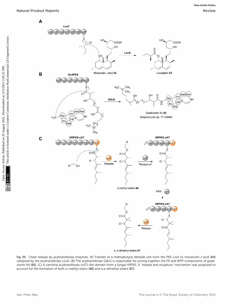
Fig. 30 Chain release by acyltransferase enzymes. (A) Transfer of a methylbutyryl diketide unit from the PKS LovF to monacolin J acid (64)catalysed by the acyltransferase LovD. (B) The acyltransferase GdvG is responsible for joining together the PK and RiPP components of goad-vionin A4 (65). (C) A carnitine acyltransferase (cAT)-like domain from a fungal HRPKS. A “release and recapture” mechanism was proposed toaccount for the formation of both a-methyl esters (66) and a,a-dimethyl esters (67).
Nat. Prod. Rep. This journal is © The Royal Society of Chemistry 2021
Natural Product Reports Review
Ope
n A
cces
s A
rtic
le. P
ublis
hed
on 2
0 A
ugus
t 202
1. D
ownl
oade
d on
12/
5/20
21 2
:45:
21 P
M.
Thi
s ar
ticle
is li
cens
ed u
nder
a C
reat
ive
Com
mon
s A
ttrib
utio
n-N
onC
omm
erci
al 3
.0 U
npor
ted
Lic
ence
.View Article Online

Review Natural Product Reports
Ope
n A
cces
s A
rtic
le. P
ublis
hed
on 2
0 A
ugus
t 202
1. D
ownl
oade
d on
12/
5/20
21 2
:45:
21 P
M.
Thi
s ar
ticle
is li
cens
ed u
nder
a C
reat
ive
Com
mon
s A
ttrib
utio
n-N
onC
omm
erci
al 3
.0 U
npor
ted
Lic
ence
.View Article Online
characterised in the biosynthesis of an as yet-unknown poly-ketide produced by the fungus Trichoderma virens.245 Thegenome of this organism encodes a PKS (HRPKS-cAT) that lacksa TE, CT or R domain, instead containing a C-terminal domainresembling a carnitine acyltransferase (cAT).245 When assayingthe activity of HRPKS-cAT in vitro, a tetraketide product joinedvia an ester linkage to Tris or glycerol, both of which werecomponents of the assay buffer, was identied.245 Neither ofthese esters was produced using a mutated version of HRPKS-cAT where the cAT domain had been deleted, indicating thatthis domain is responsible for catalysing chain release viaa transesterication reaction.245 The nucleophile used in vivoremains unknown.245 The a-position of the tetraketide waseither methylated (66) or dimethylated (67) by the methyl-transferase (MT) domain of HRPKS-cAT.245 Interestingly, themonomethylated tetraketide ester was converted into thegeminal dimethylated form when incubated with HRPKS-cATand S-adenosyl methionine (SAM).245 However, this additionalmethylation did not occur if the cAT domain had beendeleted.245 From these results, the authors proposed anunprecedented “release and recapture” mechanism, wherebythe monomethylated tetraketide ester is transferred back to theterminal ACP of HRPKS-cAT by the cAT domain (Fig. 30C).245
The MT domain then catalyses a second methylation togenerate a geminal dimethyl group, followed by chain release bythe cAT domain to regenerate the free ester.245
The PK/NRP hybrid paenilamicin appears to utilize anunusual BtrH-like acyltransferase to catalyse chain release.246
The enzyme BtrH was characterised in the butirosin biosyn-thetic pathway as the acyltransferase responsible for trans-ferring g-L-Glu-4-amino-2-hydroxybutyrate (AHBA) froma standalone ACP to the aminoglycoside ribostamycin.247 Pae-nilamicin is produced by Paenibacillus larvae, the pathogenresponsible for American Foulbrood in honeybee colonies.248
Interestingly, the C-terminal end of paenilamicin is linked toa molecule of 4,3-spermidine via an amide linkage. Thebiosynthesis pathway of paenilamicin contains four PKS andseven NRPS modules. The nal module in the biosynthesispathway, PamH NRPS module 7, lacks a thioesterase or anotherobvious chain release domain.246 However, the biosyntheticgene cluster also encodes a standalone enzyme, PamI, withhomology to BrtH.2,246 PamI could therefore catalyse chainrelease using 4,3-spermidine as an intermolecular nucleophile,resulting in amide formation.2,246 Further biochemical charac-terisation of BtrH PamI is therefore required to uncover the truemechanism of paenilamicin chain release.2,246
14 Chain release catalysed by AfsA/butenolide synthase (PBS) domains
A recent addition to the stable of domains that catalyse chainrelease are AfsA-like domains.249,250 The monomeric AfsAprotein was rst characterised in the biosynthesis pathway for Afactor (2-isocapryloyl-3R-hydroxymethyl-g-butyrolactone) inStreptomyces griseus.251 AfsA catalyses a condensation reactionbetween dihydroxyacetone phosphate (DHAP) with a b-keto
This journal is © The Royal Society of Chemistry 2021
thioester (derived from fatty acid biosynthesis), followed byspontaneous cyclisation to form the g-butyrolactone ring of Afactor (68) (Fig. 31A).251 A factor (68) itself belongs to the g-butyrolactone family of transcriptional regulators (also referredto as “microbial hormones”) that induce the expression of genesinvolved in natural product biosynthesis and/or cell differenti-ation in Gram-positive bacteria.252 In 2020, two groups inde-pendently reported the discovery of gladiofungin (69) (syngladiostatin), an antifungal glutarimide natural productproduced by Burkholderia gladioli.249,250 In addition to the glu-tarimide moiety, gladiofungin (69) also contains a terminalbutenolide ring. The biosynthetic gene cluster of gladiofungin(69) revealed that its PK backbone is synthesised by the trans-ATPKS enzymes GlaD and GlaE.249 However, instead of a TEdomain, module 10 of GlaE contains the rst example of anAfsA-like domain (syn phosphorylated butenolide synthase(PBS) domain) (Fig. 31B).249,250 Deletion of the AfsA domainabolished production of gladiofungin (69), demonstrating it isessential for biosynthesis.249 By purifying an excised ACP–AfsAdidomain, the condensation between DHAP and a b-keto SNACsubstrate analogue could be reconstituted in vitro, proving therole of the AfsA domain in chain release/butanolide forma-tion.250 Other biosynthetic gene clusters that encode an AfsAdomain could be identied by using its amino acid sequence asa bioinformatic handle, indicating that these pathways alsoproduce butanolide/g-butyrolactone substituted polyketides.249
15 Chain release catalysed bystandalone Dieckmann cyclases
Chain release via a Dieckmann condensation has already beendiscussed in the context of TE, R*, and CT domains. However,other biosynthetic pathways utilize specialised standaloneDieckmann cyclases to catalyse chain release, such as inbiosynthesis pathways of the polyketide tetramates pyrroindo-mycin253 (70) and tirandmycin.254 These examples and more arediscussed in detail in a comprehensive recent review.178 Tobriey cover the topic here, chain release/tetramate ringformation in pyrroindomycin biosynthesis is catalysed by twoenzymes, PyrD3 and PyrD4 (Fig. 32A).253 Surprisingly, bothenzymes are independently capable of releasing/cyclising thePCP-bound N-acetoacetyl-L-alanine substrate. Consistent withthis result, in vivo deletions of either gene decreased pyrroin-domycin production, whereas production was completed abol-ished only in a DpyrD3 DpyrD4 double deletion mutant.253
PyrD3 and PyrD4 are unrelated to one another, with PyrD3resembling a pyruvate dehydrogenase while PyrD4 is predictedto have a a/b hydrolase fold. Whether PyrD3 and PyrD4 actsynergistically is unknown, but how two distinct enzyme foldscatalyse the same reaction is an intriguing question.253 It wouldbe interesting to know if the BGCs of as yet-undiscovered tet-ramates related to pyrroindomcyin (70) also encode homo-logues of both PyrD3 and PyrD4.
Another family of standalone Dieckmann cyclases are enco-ded in the gene clusters coding for the biosynthesis of tetra-mates such as tirandmycin, streptolydigin, and nocamycin
Nat. Prod. Rep.
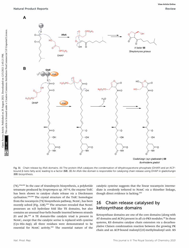
Fig. 31 Chain release by AfsA domains. (A) The protein AfsA catalyses the condensation of dihydroxyacetone phosphate (DHAP) and an ACP-bound b-keto fatty acid, leading to a factor (68). (B) An AfsA-like domain is responsible for catalysing chain release using DHAP in gladiofungin(69) biosynthesis.
Natural Product Reports Review
Ope
n A
cces
s A
rtic
le. P
ublis
hed
on 2
0 A
ugus
t 202
1. D
ownl
oade
d on
12/
5/20
21 2
:45:
21 P
M.
Thi
s ar
ticle
is li
cens
ed u
nder
a C
reat
ive
Com
mon
s A
ttrib
utio
n-N
onC
omm
erci
al 3
.0 U
npor
ted
Lic
ence
.View Article Online
(71).254,255 In the case of tirandmycin biosynthesis, a polyketidetetramate produced by Streptomyces sp. 397-9, the enzyme TrdChas been shown to catalyse chain release via a Dieckmanncyclisation.254,256 The crystal structure of the TrdC homologuefrom the nocamycin (71) biosynthesis pathway, NcmC, has beenrecently solved (Fig. 32B).255 The structure revealed that NcmCpossesses an a/b hydrolase fold like TE domains, but alsocontains an unusual four-helix bundle inserted between strandsb5 and b6.255 A TE domain-like catalytic triad is present inNcmC, except that the catalytic serine is replaced with cysteine(Cys–His–Asp); all three residues were demonstrated to beessential for NcmC activity.255 The essential nature of the
Nat. Prod. Rep.
catalytic cysteine suggests that the linear nocamycin interme-diate is covalently tethered to NcmC via a thioether linkage,though direct evidence is lacking.255
16 Chain release catalysed byketosynthase domains
Ketosynthase domains are one of the core domains (along withAT domains and ACPs) present in all cis-PKSmodules.10 In thesesystems, KS domains catalyse chain extension via a decarbox-ylative Claisen condensation reaction between the growing PKchain and an ACP-bound malonyl/(2S)-methylmalonyl unit. KS
This journal is © The Royal Society of Chemistry 2021

Fig. 32 Chain release catalysed by standalone Dieckmann cyclases. (A) In pyrroindomycin (70) biosynthesis chain release is catalysed viaa Dieckmann condensation. Two enzymes, PyrE3 and PyrE4, are both capable of catalysing this reaction. (B) Formation of the tetramate ring innocamycin I (71) is catalysed by the a/b hydrolase NcmC.
This journal is © The Royal Society of Chemistry 2021 Nat. Prod. Rep.
Review Natural Product Reports
Ope
n A
cces
s A
rtic
le. P
ublis
hed
on 2
0 A
ugus
t 202
1. D
ownl
oade
d on
12/
5/20
21 2
:45:
21 P
M.
Thi
s ar
ticle
is li
cens
ed u
nder
a C
reat
ive
Com
mon
s A
ttrib
utio
n-N
onC
omm
erci
al 3
.0 U
npor
ted
Lic
ence
.View Article Online

Fig. 33 Chain release by a ketosynthase domain. The C-terminal KSdomain in the NRPs Tas1 catalyses a chain release by a Dieckmanncondensation reaction in tenuazonic acid (72) biosynthesis.
Natural Product Reports Review
Ope
n A
cces
s A
rtic
le. P
ublis
hed
on 2
0 A
ugus
t 202
1. D
ownl
oade
d on
12/
5/20
21 2
:45:
21 P
M.
Thi
s ar
ticle
is li
cens
ed u
nder
a C
reat
ive
Com
mon
s A
ttrib
utio
n-N
onC
omm
erci
al 3
.0 U
npor
ted
Lic
ence
.View Article Online
domains themselves are ca. 430 amino acids in size and possessa a/b/a/b/a thiolase fold (alternating layers of a-helices and b-sheets).10 The active site of KS domains contains a Cys–His–Hiscatalytic triad, with the cysteine acting as a nucleophile tocovalently bond the PK chain prior to chain extension. Inaddition to this well-dened role in chain extension, there isevidence from several pathways that KS domains catalyse chainrelease. The rst (and most convincing) example is from thetenuazonic acid (72) biosynthesis pathway in the fungus Alter-naria tenuis.257 The biosynthesis of tenuazonic acid (72) is ach-ieved via the condensation of acetoacetyl-CoA with L-isoleucinefollowed by a Dieckmann condensation, all of which is catalysedby the PKS-NRPS hybrid Tas1.257 Tas1 is a unimodular PKS-NRPS hybrid with an unusual C–A–PCP–KS domain arrange-ment. In vitro experimentation demonstrated that the Cdomain, rather than the terminal KS domain, catalyses thecondensation of SNAC-L-isoleucine with acetoacetyl-CoA toform SNAC-N-acetoactyl-L-isoleucine.257 However, incubatingSNAC-N-acetoactyl-L-isoleucine with a puried form of the KSdomain resulted in tenuazonic acid (72) formation, indicating
Fig. 34 Crystal structure of a ketosynthase domain that catalyses chain rTas1, responsible for catalysing chain release in tenuazonic acid (72) bioconsists of Cys–His–Asn, a modified version of the typical Cys–His–His
Nat. Prod. Rep.
that the KS domain catalyses chain release via a Dieckmanncondensation (Fig. 33).257
The crystal structure of Tas1 KS revealed it was remarkablysimilar to the KS domains from type I PKS pathways.258 Withinits active site was a modied version of the KS Cis–His-Hiscatalytic triad consisting of Cys–His–Asn (Fig. 34).258 Mutagen-esis of the catalytic cysteine residue (Cys 179) abolished catalyticactivity, consistent with the canonical role of this residue tocovalently link the PK chain, while mutagenesis of the catalyticHis residue also decreased the relative activity to only 6% of thewild type.258 Molecular docking simulations with N-acetoactyl-L-ile-Cys179 indicated that the likely role of a conserved His is toabstract the a-proton of the b-keto diketide, leading to carb-anion attack on the C1 thioester to form the tetramate ring andrelease the PK chain.258 Given the similarity between the reac-tion mechanism of ketosynthases and thioesterases (both uti-lising a serine/cysteine nucleophile and a basic residue), it isunsurprising that KS domains can be repurposed to catalysechain release. That a Dieckmann condensation is catalysed byTas1-KS is also fully consistent with the canonical role of KSdomains in catalysing C–C bond formation.
The b-lactam hexaketide ebelactone is synthesised by seventype I cis-PKS enzymes.259 The nal PKS in the pathway, EbeG,contains a C-terminal KS domain, which may be responsible forchain release.259 Chain release is predicted to occur when the b-hydroxyl attacks the C1 thioester carbon, resulting in b-lactonering formation and concomitant chain release.259 However, theformation of the b-lactone ring was shown to occur spontane-ously in aqueous solutions using an SNAC-b-hydroxy-hexaketidesubstrate analogue.259 Additionally, a homologue to the b-lactone synthase OleC (identied in the biosynthesis pathway ofbacterial long-chain olens) is encoded in the BGC of ebe-lactone,260 further making the function of the C-terminal KSdomain unclear.
elease. (A) The crystal structure of C-terminal KS domain from the PKSsynthesis (PDB: 6KOG). (B) The catalytic triad of the Tas1 KS domaincatalytic triad present in KS domains.
This journal is © The Royal Society of Chemistry 2021

Review Natural Product Reports
Ope
n A
cces
s A
rtic
le. P
ublis
hed
on 2
0 A
ugus
t 202
1. D
ownl
oade
d on
12/
5/20
21 2
:45:
21 P
M.
Thi
s ar
ticle
is li
cens
ed u
nder
a C
reat
ive
Com
mon
s A
ttrib
utio
n-N
onC
omm
erci
al 3
.0 U
npor
ted
Lic
ence
.View Article Online
17 Chain release catalysed by PLP-dependent enzymes
Several polyketide biosynthesis pathways use pyridoxal 50-phos-phate (PLP)-dependent enzymes to catalyse chain release. PLP isan enzyme cofactor used for many different reactions, notablytransaminations.261 The rst characterised example of a PLP-dependent chain release mechanism is from the prodigininebiosynthesis pathway in Streptomyces coelicolor.262 The prodigi-nines are a family of red-coloured tripyrrole antibiotics producedby actinobacteria and other eubacteria.263 The biosynthesis ofundecyl prodiginine (73) (a precursor to several of the cyclicprodiginines) is accomplished by the condensation of 4-methoxy-2,20-bipyrrole-5-carbaldehyde (MBC) (74) with 2-undecylpyrrole(2-UP) (75). 2-UP (75) is synthesised from seven acetate units andone glycine unit.263 Homologues of fatty acid biosynthesisenzymes generate dodecanoic acid that is subsequently trans-ferred to the unusual PKS-NRPS hybrid RedL. RedL has thedomain arrangement A–ACP–KS–AT–ACP–OAS, where OAS is anPLP-dependent a-oxoamine synthase domain.263 The ACP-bounddodecanoyl thioester condenses with one molecule of malonyl-ACP to form b-ketomyristoyl-ACP.263 The PLP-dependent OASdomain then catalyses chain release/pyrrole ring formation viaa decarboxylative Claisen condensation with L-glycine (Fig. 35Aand B).264 InMBC (74) biosynthesis, another OAS domain presentin RedN (ACP–ACP–OAS domain arrangement) catalyses a decar-boxylative Claisen condensation reaction between ACP-bound b-keto-b-pyrrolyl-propanoyl and L-serine, releasing MBC (74)precursor.265 Homologues of RedN and RedL are also present inthe biosynthetic gene cluster of the related marineosin naturalproducts,266 where the OAS domains likely have an identical role.
The biosynthesis pathway of saxitoxin (76), a tricyclic alkaloidproduced by cyanobacteria, also uses a OAS domain to catalysechain release.267 Saxitoxin (76) is synthesised by the PKS-likeenzyme SxtA, which possesses a MT–AT–ACP–OAS domainarrangement.267,268 The AT domain is unusual in that it resemblesa GNAT, rather than a canonical PKS AT domain. The AT domainloads a malonyl unit and, together with the methyltransferase(MT) domain, forms propionyl-ACP.268 As in the MBC (74) and 2-UP (75) biosynthesis pathways, the OAS domain then catalysesa decarboxylative Claisen condensation between the acyl-ACPand an amino acid (Fig. 35C). In the case of saxitoxin (76)biosynthesis, L-arginine is selected by the OAS domain to release4-amino-3-oxo-guandidinoheptane, which undergoes severalsubsequent oxidative cyclisations to form saxitoxin (76).268
Aside from OAS domains, standalone PLP-dependentenzymes are also capable of catalysing chain release. The onlycharacterised example is Fum8p from the fumonisin B1 (77)biosynthetic pathway in Fusarium verticillioides.269 The iterativePKS Fum1p (KS–AT–DH–MT–ER–KR–ACP) synthesises a 18-carbon PK chain.269 Fum8p, which, like the OAS domains, bearshomology to a-oxoamine synthases, then catalyses chain releasevia a decarboxylative Claisen condensation between L-alanineand the PK chain (Fig. 35D).269
Aside from the MBC (74), 2-UP (75), and saxitoxin (76)biosynthesis pathway, C-terminal PLP-binding domains have
This journal is © The Royal Society of Chemistry 2021
also been identied in numerous other PKS and NRPS enzymes,suggesting that more yet-to-be-discovered natural products usea PLP-dependent mechanism of chain release.270 In addition tocharacterising the unknown products of these pathways, itwould be interesting to investigate the substrate tolerance ofOAS domains for different amino acids, and whether the OASdomain from one pathway can be transplanted into another tocreate novel natural product analogues.
18 Final remarks
Our knowledge of the enzymes andmechanisms responsible forPK/NRP chain release has made substantial advances in thepast decade. Entirely new enzymatic mechanisms of PK/NRPchain release, including tetronate ring formation, and TE-catalysed transesterication, and AfsA-domain-catalysed butyr-olactone formation have now been described. In addition, ourunderstanding of well-known enzymatic domains including TE,R, and CT domains has deepened as additional crystal struc-tures have been obtained. Despite these discoveries, mysteriesremain. There are still PK/NRP biosynthesis pathways where themechanism of chain release is unclear (for instance in squa-listatin S1, bongkrekic acid and isoquinoline alkaloid biosyn-thesis).271–273 In regards to TE domains, the evidence suggeststhat they are intrinsically able to catalyse a range of differentrelease mechanisms, including hydrolysis, macrocyclization,oligomerisation, and transesterication.23 Given this, how TEdomains ultimately specialise to release only a single product isknown. The evolutionary pressure(s) that selects for some TEdomains to contain a catalytic cysteine rather than serine arealso still poorly understood, though this may be inuenced bythe size of the ring to be formed (for example, smaller, strainedrings favouring cysteine over serine).18–20,46,47
Regarding R domains, while some progress has been made,why some catalyse two-electron reductions and others catalysefour-electron reductions is still unclear. The answer likely lies inconformation changes that either facilitate or prevent a secondmolecule of NAD(P)H binding, though experimental evidence isrequired.13,145 The utility of R domains as synthetic biology toolsto release has also not been explored deeply. For instance, cana TE (or CT) domain can be replaced with an R domain to releasealdehyde/alcohol products? Given the diversity of functionalgroups that can arise from aldehydes and primary alcohols (seeFig. 19), such experiments would be of high importance to theeld of combinatorial biosynthesis. In a recent step towards thisgoal, an aldehyde-producing R domain was successfully fusedto the C-terminus of a protein construct consisting of the rsttwo modules of the NRPS GxpS, resulting in the release ofspontaneously cyclising aldehyde dipeptides.274 However, thegeneral portability and substrate selectivity of R domains innon-native PKS/NRPS enzymes remains to be seen.
Another unsolved mystery is the reason for the largelydivergent role of TE and CT domains in fungi, where TEs oencatalyse hydrolysis while CT domains catalyse macrocyclisation.Regardless, the fact that distinct protein folds of TE, R*, and CT
and KS domains can all catalyse similar/identical chain releasereactions is a remarkable example of convergent evolution.
Nat. Prod. Rep.

Fig. 35 Chain release by PLP-dependent enzymes. (A) The general mechanism of a PLP-dependent Claisen condensation of an amino acid. (B)Chain release catalysed by PLP-dependent OAS domains in the biosynthesis of the prodigiosin precursors 2-UP andMBC (74). (C) In saxitoxin (76)biosynthesis the SxtA OAS domain catalyses chain release by a decarboxylative Claisen condensation reaction with L-arginine. (D) In fumonisin B1(77) biosynthesis the standalone PLP-dependent enzyme Fum8p catalyses chain release via a decarboxylative Claisen condensation with L-alanine.
Nat. Prod. Rep. This journal is © The Royal Society of Chemistry 2021
Natural Product Reports Review
Ope
n A
cces
s A
rtic
le. P
ublis
hed
on 2
0 A
ugus
t 202
1. D
ownl
oade
d on
12/
5/20
21 2
:45:
21 P
M.
Thi
s ar
ticle
is li
cens
ed u
nder
a C
reat
ive
Com
mon
s A
ttrib
utio
n-N
onC
omm
erci
al 3
.0 U
npor
ted
Lic
ence
.View Article Online

Review Natural Product Reports
Ope
n A
cces
s A
rtic
le. P
ublis
hed
on 2
0 A
ugus
t 202
1. D
ownl
oade
d on
12/
5/20
21 2
:45:
21 P
M.
Thi
s ar
ticle
is li
cens
ed u
nder
a C
reat
ive
Com
mon
s A
ttrib
utio
n-N
onC
omm
erci
al 3
.0 U
npor
ted
Lic
ence
.View Article Online
Looking to the future, applying the different chain releasemechanisms discussed here to combinatorial biosynthesissystems has the potential to create highly diverse linear andcyclical products. Towards this goal, a recent study tested theeffect of replacing the native TE domain from a PKS modulewith a TE domain from another biosynthesis pathway, followedby assessing its ability to catalyse macrocyclisation.275 Theresults showed that even when a non-native PKS module wasused, if the TE matched the substrate then effective macro-cyclisation could still occur, highlighting the importance of theTE domain for the effective processing of non-native substratesby engineered PKSs.275 Ensuring that the mechanism of releaseis compatible with the product of the engineered PKS/NRPS willbe essential for the success of these synthetic assembly lines.
Studying chain release mechanisms has benets beyondunderstanding the formation of nal product scaffolds. Asdescribed in Section 8, disrupting chain release can led to therelease of premature PK chains, providing insight into theseearly and difficult-to-study biosynthetic steps.243,244 We shouldthere look to disrupting chain release mechanisms in the futureas a possible means to studying late-stage biosynthetic inter-mediates. Where such genetic disruption experiments are notpossible or unsuccessful, specialised chemical probes havebeen developed that provide a different means to the same end.These probes, such as methyl 6-decanamido-2-uoro-3-oxohexanoate, compete with the CP-bound extension unit toaccept the polyketide acyl chain during a KS/C domain-catalysed extension reaction.276–280 The result is the prematurerelease of PK/NRP pathway intermediates that can then beanalysed by LC-MS. This technique is particularly useful forstudying the timing of tailoring reactions, such as cyclisations,occurring when the PK/NRP chain is still bound to the PKS.205,278
The number of characterised chain release mechanisms willlikely grow as more genomes are sequenced and new biosyn-thesis pathways are discovered. Our job will be to characterisethese new mechanisms and determine what they can teach usabout PK/NRP biosynthesis. The step aer that is the excitingprospect of applying these mechanisms in biosynthetic path-ways of our own design.
19 Conflicts of interest
There are no conicts to declare.
20 Acknowledgements
The authors would like to acknowledge the nancial support oftheir original work by the Deutsche Forschungsgemeinscha(DFG, German Research Foundation)—Project-ID 239748522—SFB 1127 ChemBioSys, Cluster of Excellence, Balance of theMicroverse, and Leibniz Award. RFL is supported by an Alex-ander von Humboldt postdoctoral research fellowship.
21 References
1 J. Staunton and K. J. Weissman, Nat. Prod. Rep., 2001, 18,380–416.
This journal is © The Royal Society of Chemistry 2021
2 R. D. Sussmuth and A. Mainz, Angew. Chem., Int. Ed., 2017,56, 3770–3821.
3 K. H. Altmann, Mini-Rev. Med. Chem., 2003, 3, 149–158.4 U. Galm, M. H. Hager, S. G. Van Lanen, J. Ju, J. S. Thorsonand B. Shen, Chem. Rev., 2005, 105, 739–758.
5 D. P. Levine, Clin. Infect. Dis., 2006, 42, S5–S12.6 J. Li, S. G. Kim and J. Blenis, Cell Metab., 2014, 19, 373–379.7 J. A. Washington II andW. R. Wilson,Mayo Clin. Proc., 1985,60, 189–203.
8 C. Hertweck, Angew. Chem., Int. Ed., 2009, 48, 4688–4716.9 K. J. Weissman and P. F. Leadlay, Nat. Rev. Microbiol., 2005,3, 925–936.
10 A. T. Keatinge-Clay, Nat. Prod. Rep., 2012, 29, 1050–1073.11 L. Du and L. Lou, Nat. Prod. Rep., 2010, 27, 255–278.12 L. Vieweg, S. Reichau, R. Schobert, P. F. Leadlay and
R. D. Sussmuth, Nat. Prod. Rep., 2014, 31, 1554–1584.13 M.W.Mullowney, R. A. McClure, M. T. Robey, N. L. Kelleher
and R. J. Thomson, Nat. Prod. Rep., 2018, 35, 847–878.14 C. Hertweck, A. Luzhetskyy, Y. Rebets and A. Bechthold,
Nat. Prod. Rep., 2007, 24, 162–190.15 D. Yu, F. Xu, J. Zeng and J. Zhan, IUBMB Life, 2012, 64, 285–
295.16 M. Holmquist, Curr. Protein Pept. Sci., 2000, 1, 209–235.17 M. E. Horsman, T. P. A. Hari and C. N. Boddy, Nat. Prod.
Rep., 2016, 33, 183–202.18 J. E. Schaffer, M. R. Reck, N. K. Prasad and T. A. Wencewicz,
Nat. Chem. Biol., 2017, 13, 737–744.19 A. A. Koch, D. A. Hansen, V. V. Shende, L. R. Furan,
K. N. Houk, G. Jimenez-Oses and D. H. Sherman, J. Am.Chem. Soc., 2017, 139, 13456–13465.
20 L. Qiao, J. Fang, P. Zhu, H. Huang, C. Dang, J. Pang, W. Gao,X. Qiu, L. Huang and Y. Li, Protein J., 2019, 38, 658–666.
21 N. Huguenin-Dezot, D. A. Alonzo, G. W. Heberlig,M. Mahesh, D. P. Nguyen, M. H. Dornan, C. N. Boddy,T. M. Schmeing and J. W. Chin, Nature, 2019, 565, 112–117.
22 A. T. Keatinge-Clay, Chem. Rev., 2017, 117, 5334–5366.23 T. P. Hari, P. Labana, M. Boileau and C. N. Boddy,
ChemBioChem, 2014, 15, 2656–2661.24 B. A. Pfeifer, C. C. C. Wang, C. T. Walsh and C. Khosla, Appl.
Environ. Microbiol., 2003, 69, 6698–6702.25 G. Yim, M. N. Thaker, K. Koteva and G. Wright, J. Antibiot.,
2014, 67, 31–41.26 E. A. Felnagle, E. E. Jackson, Y. A. Chan, A. M. Podevels,
A. D. Berti, M. D. McMahon and M. G. Thomas, Mol.Pharm., 2008, 5, 191–211.
27 V. Rangaswamy, S. Jiralerspong, R. Parry and C. L. Bender,Proc. Natl. Acad. Sci. U. S. A., 1998, 95, 15469.
28 M. Peschke, C. Brieke, M. Heimes and M. J. Cryle, ACSChem. Biol., 2018, 13, 110–120.
29 L. Gu, B. Wang, A. Kulkarni, J. J. Gehret, K. R. Lloyd,L. Gerwick, W. H. Gerwick, P. Wipf, K. Hakansson,J. L. Smith and D. H. Sherman, J. Am. Chem. Soc., 2009,131, 16033–16035.
30 N. M. Gaudelli and C. A. Townsend, Nat. Chem. Biol., 2014,10, 251–258.
Nat. Prod. Rep.

Natural Product Reports Review
Ope
n A
cces
s A
rtic
le. P
ublis
hed
on 2
0 A
ugus
t 202
1. D
ownl
oade
d on
12/
5/20
21 2
:45:
21 P
M.
Thi
s ar
ticle
is li
cens
ed u
nder
a C
reat
ive
Com
mon
s A
ttrib
utio
n-N
onC
omm
erci
al 3
.0 U
npor
ted
Lic
ence
.View Article Online
31 K. D. Patel, F. B. d'Andrea, N. M. Gaudelli, A. R. Buller,C. A. Townsend and A. M. Gulick, Nat. Commun., 2019,10, 3868.
32 L. Ray, K. Yamanaka and B. S. Moore, Angew. Chem., Int. Ed.,2016, 55, 364–367.
33 M.-C. Tang, C. R. Fischer, J. V. Chari, D. Tan, S. Suresh,A. Chu, M. Miranda, J. Smith, Z. Zhang, N. K. Garg,R. P. S. Onge and Y. Tang, J. Am. Chem. Soc., 2019, 141,8198–8206.
34 T. Thongkongkaew, W. Ding, E. Bratovanov, E. Oueis,M. a. Garcıa-Altares, N. Zaburannyi, K. Harmrolfs,Y. Zhang, K. Scherlach, R. Muller and C. Hertweck, ACSChem. Biol., 2018, 13, 1370–1379.
35 Y.-S. Seo, J. Y. Lim, J. Park, S. Kim, H.-H. Lee, H. Cheong,S.-M. Kim, J. S. Moon and I. Hwang, BMC Genomics, 2015,16, 349.
36 L. Laureti, L. Song, S. Huang, C. Corre, P. Leblond,G. L. Challis and B. Aigle, Proc. Natl. Acad. Sci. U. S. A.,2011, 108, 6258–6263.
37 G. G. Zhanel, M. Dueck, D. J. Hoban, L. M. Vercaigne,J. M. Embil, A. S. Gin and J. A. Karlowsky, Drugs, 2001, 61,443–498.
38 B. Arsic, J. Barber, A. Cikos, M. Mladenovic, N. Stankovicand P. Novak, Int. J. Antimicrob. Agents, 2018, 51, 283–298.
39 K. Buntin, K. J. Weissman and R. Muller, ChemBioChem,2010, 11, 1137–1146.
40 A. G. Newman, A. L. Vagstad, K. Belecki, J. R. Scheerer andC. A. Townsend, Chem. Commun., 2012, 48, 11772–11774.
41 J. Hou, L. Robbel and M. A. Marahiel, Chem. Biol., 2011, 18,655–664.
42 S. Eys, D. Schwartz, W. Wohlleben and E. Schinko,Antimicrob. Agents Chemother., 2008, 52, 1686–1696.
43 D. Schwartz, N. Grammel, E. Heinzelmann, U. Keller andW. Wohlleben, Antimicrob. Agents Chemother., 2005, 49,4598–4607.
44 F. Trottmann, K. Ishida, J. Franke, A. Stanisic, M. Ishida-Ito,H. Kries, G. Pohnert and C. Hertweck, Angew. Chem., Int.Ed., 2020, 59, 13511–13515.
45 H.-Y. He, M.-C. Tang, F. Zhang and G.-L. Tang, J. Am. Chem.Soc., 2014, 136, 4488–4491.
46 D. A. Hansen, A. A. Koch and D. H. Sherman, J. Am. Chem.Soc., 2017, 139, 13450–13455.
47 C. A. Shaw-Reid, N. L. Kelleher, H. C. Losey, A. M. Gehring,C. Berg and C. T. Walsh, Chem. Biol., 1999, 6, 385–400.
48 Y. Zhou, P. Prediger, L. C. Dias, A. C. Murphy andP. F. Leadlay, Angew. Chem., Int. Ed., 2015, 54, 5232–5235.
49 X. Zhao, Y. Fang, Y. Yang, Y. Qin, P. Wu, T. Wang, H. Lai,L. Meng, D. Wang, Z. Zheng, X. Lu, H. Zhang, Q. Gao,J. Zhou and D. Ma, Autophagy, 2015, 11, 1849–1863.
50 G. W. Heberlig and C. N. Boddy, J. Nat. Prod., 2020, 83,1990–1997.
51 K. M. Hoyer, C. Mahlert and M. A. Marahiel, Chem. Biol.,2007, 14, 13–22.
52 Y. Zhou, A. C. Murphy, M. Samborskyy, P. Prediger,L. C. Dias and P. F. Leadlay, Chem. Biol., 2015, 22, 745–754.
53 J. Franke and C. Hertweck, Cell Chem. Biol., 2016, 23, 1179–1192.
Nat. Prod. Rep.
54 C. Zhang, L. Kong, Q. Liu, X. Lei, T. Zhu, J. Yin, B. Lin,Z. Deng and D. You, PLoS One, 2013, 8, e56772.
55 A. P. Praseuth, C. C. C. Wang, K. Watanabe, K. Hotta,H. Oguri and H. Oikawa, Biotechnol. Prog., 2008, 24, 1226–1231.
56 J. A. Matson, K. L. Colson, G. N. Belofsky and B. B. Bleiberg,J. Antibiot., 1993, 46, 162–166.
57 D. Mandalapu, X. Ji, J. Chen, C. Guo, W.-Q. Liu, W. Ding,J. Zhou and Q. Zhang, J. Org. Chem., 2018, 83, 7271–7275.
58 N. Roongsawang, K. Washio and M. Morikawa,ChemBioChem, 2007, 8, 501–512.
59 J. B. Biggins, H.-S. Kang, M. A. Ternei, D. DeShazer andS. F. Brady, J. Am. Chem. Soc., 2014, 136, 9484–9490.
60 B. K. Scholz-Schroeder, J. D. Soule and D. C. Gross, Mol.Plant-Microbe Interact., 2003, 16, 271–280.
61 I. de Bruijn, M. J. D. de Kock, P. de Waard, T. A. van Beekand J. M. Raaijmakers, J. Bacteriol., 2008, 190, 2777.
62 J. W. Trauger, R. M. Kohli, H. D. Mootz, M. A. Marahiel andC. T. Walsh, Nature, 2000, 407, 215–218.
63 G.-L. Tang, Y.-Q. Cheng and B. Shen, Chem. Biol., 2004, 11,33–45.
64 R. A. Oliver, R. Li and C. A. Townsend, Nat. Chem. Biol.,2018, 14, 5–7.
65 Y. Ogasawara, K. Katayama, A. Minami, M. Otsuka,T. Eguchi and K. Kakinuma, Chem. Biol., 2004, 11, 79–86.
66 R. Li, R. A. Oliver and C. A. Townsend, Cell Chem. Biol.,2017, 24, 24–34.
67 D. J. Edwards, B. L. Marquez, L. M. Nogle, K. McPhail,D. E. Goeger, M. A. Roberts and W. H. Gerwick, Chem.Biol., 2004, 11, 817–833.
68 B. Lin Xiaoxi, T. Lohans Christopher, R. Duar, J. Zheng,C. Vederas John, J. Walter, M. Ganzle and M. A. Elliot,Appl. Environ. Microbiol., 2015, 81, 2032–2041.
69 H. Zhou, Y. Li and Y. Tang, Nat. Prod. Rep., 2010, 27, 839–868.
70 I. Fujii, A. Watanabe, U. Sankawa and Y. Ebizuka, Chem.Biol., 2001, 8, 189–197.
71 S.-S. Gao, A. Duan, W. Xu, P. Yu, L. Hang, K. N. Houk andY. Tang, J. Am. Chem. Soc., 2016, 138, 4249–4259.
72 J. H. Yu and T. J. Leonard, J. Bacteriol., 1995, 177, 4792–4800.
73 A. L. Vagstad, E. A. Hill, J. W. Labonte and C. A. Townsend,Chem. Biol., 2012, 19, 1525–1534.
74 T. P. Korman, J. M. Crawford, J. W. Labonte, A. G. Newman,J. Wong, C. A. Townsend and S. C. Tsai, Proc. Natl. Acad. Sci.U. S. A., 2010, 107, 6246–6251.
75 R. E. Minto and C. A. Townsend, Chem. Rev., 1997, 97, 2537–2556.
76 Y. He, Y. Sun, T. Liu, X. Zhou, L. Bai and Z. Deng, Appl.Environ. Microbiol., 2010, 76, 3283–3292.
77 H. Ikeda, T. Nonomiya, M. Usami, T. Ohta and S. Omura,Proc. Natl. Acad. Sci. U. S. A., 1999, 96, 9509–9514.
78 A. Migita, M. Watanabe, Y. Hirose, K. Watanabe,T. Tokiwano, H. Kinashi and H. Oikawa, Biosci.,Biotechnol., Biochem., 2009, 73, 169–176.
79 C. J. Balibar, A. R. Howard-Jones and C. T. Walsh, Nat.Chem. Biol., 2007, 3, 584–592.
This journal is © The Royal Society of Chemistry 2021

Review Natural Product Reports
Ope
n A
cces
s A
rtic
le. P
ublis
hed
on 2
0 A
ugus
t 202
1. D
ownl
oade
d on
12/
5/20
21 2
:45:
21 P
M.
Thi
s ar
ticle
is li
cens
ed u
nder
a C
reat
ive
Com
mon
s A
ttrib
utio
n-N
onC
omm
erci
al 3
.0 U
npor
ted
Lic
ence
.View Article Online
80 L. Lou, G. Qian, Y. Xie, J. Hang, H. Chen, K. Zaleta-Rivera,Y. Li, Y. Shen, P. H. Dussault, F. Liu and L. Du, J. Am.Chem. Soc., 2011, 133, 643–645.
81 C. Bihlmaier, E. Welle, C. Hofmann, K. Welzel, A. Vente,E. Breitling, M. Muller, S. Glaser and A. Bechthold,Antimicrob. Agents Chemother., 2006, 50, 2113–2121.
82 L. Lou, H. Chen, R. L. Cerny, Y. Li, Y. Shen and L. Du,Biochemistry, 2012, 51, 4–6.
83 F. Romero, F. Espliego, J. Perez Baz, T. Garcıa de Quesada,D. Gravalos, F. de la Calle and J. L. Fernandez-Puentes, J.Antibiot., 1997, 50, 734–737.
84 E. Erba, D. Bergamaschi, S. Ronzoni, M. Faretta, S. Taverna,M. Bonfanti, C. V. Catapano, G. Faircloth, J. Jimeno andM. D'Incalci, Br. J. Cancer, 1999, 80, 971–980.
85 H. Okada, H. Suzuki, T. Yoshinari, H. Arakawa, A. Okura,H. Suda, A. Yamada and D. Uemura, J. Antibiot., 1994, 47,129–135.
86 M. Kotowska and K. Pawlik, Appl. Environ. Microbiol., 2014,98, 7735–7746.
87 E. Yeh, R. M. Kohli, S. D. Bruner and C. T. Walsh,ChemBioChem, 2004, 5, 1290–1293.
88 D. Schwarzer, H. D. Mootz, U. Linne and M. A. Marahiel,Proc. Natl. Acad. Sci. U. S. A., 2002, 99, 14083–14088.
89 M. L. Heathcote, J. Staunton and P. F. Leadlay, Chem. Biol.,2001, 8, 207–220.
90 A. Koglin, F. Lohr, F. Bernhard, V. V. Rogov, D. P. Frueh,E. R. Strieter, M. R. Mod, P. Guntert, G. Wagner,C. T. Walsh, M. A. Marahiel and V. Dotsch, Nature, 2008,454, 907–911.
91 A. R. Butler, N. Bate and E. Cundliffe, Chem. Biol., 1999, 6,287–292.
92 Y. Doi-Katayama, Y. J. Yoon, C. Y. Choi, T. W. Yu, H. G. Flossand C. R. Hutchinson, J. Antibiot., 2000, 53, 484–495.
93 Y. Xue, L. Zhao, H.-w. Liu and D. H. Sherman, Proc. Natl.Acad. Sci. U. S. A., 1998, 95, 12111.
94 H. Nakamura, J. X. Wang and E. P. Balskus, Chem. Sci.,2015, 6, 3816–3822.
95 B. M. Harvey, T. Mironenko, Y. Sun, H. Hong, Z. Deng,P. F. Leadlay, K. J. Weissman and S. F. Haydock, Chem.Biol., 2007, 14, 703–714.
96 B. M. Harvey, H. Hong, M. A. Jones, Z. A. Hughes-Thomas,R. M. Goss, M. L. Heathcote, V. M. Bolanos-Garcia,W. Kroutil, J. Staunton, P. F. Leadlay and J. B. Spencer,ChemBioChem, 2006, 7, 1435–1442.
97 T. Liu, D. You, C. Valenzano, Y. Sun, J. Li, Q. Yu, X. Zhou,D. E. Cane and Z. Deng, Chem. Biol., 2006, 13, 945–955.
98 T. Liu, X. Lin, X. Zhou, Z. Deng and D. E. Cane, Chem. Biol.,2008, 15, 449–458.
99 R. Liu, F. Fang, Z. An, R. Huang, Y. Wang, X. Sun, S. Fu,A. Fu, Z. Deng and T. Liu, J. Ind. Microbiol. Biotechnol.,2020, 47, 275–285.
100 N. Liu, Y.-S. Hung, S.-S. Gao, L. Hang, Y. Zou, Y.-H. Chooiand Y. Tang, Org. Lett., 2017, 19, 3560–3563.
101 N. S. Guntaka, A. R. Healy, J. M. Crawford, S. B. Herzon andS. D. Bruner, ACS Chem. Biol., 2017, 12, 2598–2608.
102 K. Hua, X. Liu, Y. Zhao, Y. Gao, L. Pan, H. Zhang, Z. Dengand M. Jiang, mBio, 2020, 11, e01334.
This journal is © The Royal Society of Chemistry 2021
103 C. Li, K. E. Roege and W. L. Kelly, ChemBioChem, 2009, 10,1064–1072.
104 W.-G. Wang, H. Wang, L.-Q. Du, M. Li, L. Chen, J. Yu,G.-G. Cheng, M.-T. Zhan, Q.-F. Hu, L. Zhang, M. Yao andY. Matsuda, J. Am. Chem. Soc., 2020, 142, 8464–8472.
105 R. Takeda, J. Am. Chem. Soc., 1958, 80, 4749–4750.106 P. C. Dorrestein, E. Yeh, S. Garneau-Tsodikova,
N. L. Kelleher and C. T. Walsh, Proc. Natl. Acad. Sci. U. S.A., 2005, 102, 13843.
107 H. Gross and J. E. Loper, Nat. Prod. Rep., 2009, 26, 1408–1446.
108 D. C. Cantu, Y. Chen and P. J. Reilly, Protein Sci., 2010, 19,1281–1295.
109 M. Kotaka, R. Kong, I. Qureshi, Q. S. Ho, H. Sun, C.W. Liew,L. P. Goh, P. Cheung, Y. Mu, J. Lescar and Z.-X. Liang, J.Biol. Chem., 2009, 284, 15739–15749.
110 C. W. Liew, A. Sharff, M. Kotaka, R. Kong, H. Sun,I. Qureshi, G. Bricogne, Z.-X. Liang and J. Lescar, J. Mol.Biol., 2010, 404, 291–306.
111 T. Annaval, J. D. Rudolf, C.-Y. Chang, J. R. Lohman, Y. Kim,L. Bigelow, R. Jedrzejczak, G. Babnigg, A. Joachimiak,G. N. Phillips and B. Shen, ACS Omega, 2017, 2, 5159–5169.
112 S. Lin, S. G. Van Lanen and B. Shen, Proc. Natl. Acad. Sci. U.S. A., 2009, 106, 4183.
113 X. Yan, H. Ge, T. Huang, Hindra, D. Yang, Q. Teng,I. Crnovcic, X. Li, J. D. Rudolf, J. R. Lohman,Y. Gansemans, X. Zhu, Y. Huang, L.-X. Zhao, Y. Jiang,F. Van Nieuwerburgh, C. Rader, Y. Duan and B. Shen,mBio, 2016, 7, e02104.
114 B. Shen, W. Liu and K. Nonaka, Curr. Med. Chem., 2003, 10,2317–2325.
115 J. W. Labonte and C. A. Townsend, Chem. Rev., 2013, 113,2182–2204.
116 S. C. Dillon and A. Bateman, BMC Bioinf., 2004, 5, 109.117 K. Belecki and C. A. Townsend, J. Am. Chem. Soc., 2013, 135,
14339–14348.118 D. R. Cohen and C. A. Townsend, Nat. Chem., 2018, 10, 231–
236.119 D. C. Cantu, A. Ardevol, C. Rovira and P. J. Reilly, Chem.–
Eur. J., 2014, 20, 9045–9051.120 K. Amagai, R. Takaku, F. Kudo and T. Eguchi,
ChemBioChem, 2013, 14, 1998–2006.121 T. Chisuga, A. Miyanaga, F. Kudo and T. Eguchi, J. Biol.
Chem., 2017, 292, 10926–10937.122 H. Jørgensen, K. F. Degnes, A. Dikiy, E. Fjaervik,
G. Klinkenberg and S. B. Zotchev, Appl. Environ.Microbiol., 2010, 76, 283–293.
123 H. Jørgensen, K. F. Degnes, H. Sletta, E. Fjærvik, A. Dikiy,L. Herndal, P. Bruheim, G. Klinkenberg, H. Bredholt,G. Nygard, S. O. Døskeland, T. E. Ellingsen andS. B. Zotchev, Chem. Biol., 2009, 16, 1109–1121.
124 M. Gao, A. E. Glenn, A. A. Blacutt and S. E. Gold, Front.Microbiol., 2017, 8, 1775.
125 C. Bebrone, Biochem. Pharmacol., 2007, 74, 1686–1701.126 Y. Li, Y.-H. Chooi, Y. Sheng, J. S. Valentine and Y. Tang, J.
Am. Chem. Soc., 2011, 133, 15773–15785.
Nat. Prod. Rep.

Natural Product Reports Review
Ope
n A
cces
s A
rtic
le. P
ublis
hed
on 2
0 A
ugus
t 202
1. D
ownl
oade
d on
12/
5/20
21 2
:45:
21 P
M.
Thi
s ar
ticle
is li
cens
ed u
nder
a C
reat
ive
Com
mon
s A
ttrib
utio
n-N
onC
omm
erci
al 3
.0 U
npor
ted
Lic
ence
.View Article Online
127 Y. Li, W. Xu and Y. Tang, J. Biol. Chem., 2010, 285, 22764–22773.
128 J. M. Crawford, P. M. Thomas, J. R. Scheerer, A. L. Vagstad,N. L. Kelleher and C. A. Townsend, Science, 2008, 320, 243.
129 J. M. Crawford, B. C. R. Dancy, E. A. Hill, D. W. Udwary andC. A. Townsend, Proc. Natl. Acad. Sci. U. S. A., 2006, 103,16728.
130 D. A. Herbst, C. A. Townsend and T. Maier, Nat. Prod. Rep.,2018, 35, 1046–1069.
131 T. Awakawa, K. Yokota, N. Funa, F. Doi, N. Mori,H. Watanabe and S. Horinouchi, Chem. Biol., 2009, 16,613–623.
132 E. Szewczyk, Y.-M. Chiang, C. E. Oakley, A. D. Davidson,C. C. C. Wang and B. R. Oakley, Appl. Environ. Microbiol.,2008, 74, 7607.
133 Y. H. Chooi, R. Cacho and Y. Tang, Chem. Biol., 2010, 17,483–494.
134 L. Liu, Z. Zhang, C.-L. Shao and C.-Y. Wang, Front.Microbiol., 2017, 8, 1685.
135 Y.-M. Chiang, E. Szewczyk, A. D. Davidson, R. Entwistle,N. P. Keller, C. C. C. Wang and B. R. Oakley, Appl.Environ. Microbiol., 2010, 76, 2067.
136 T. Taguchi, T. Awakawa, Y. Nishihara, M. Kawamura,Y. Ohnishi and K. Ichinose, ChemBioChem, 2017, 18, 316–323.
137 R. F. Pratt, Cell. Mol. Life Sci., 2008, 65, 2138–2155.138 T. Kuranaga, K. Matsuda, A. Sano, M. Kobayashi,
A. Ninomiya, K. Takada, S. Matsunaga and T. Wakimoto,Angew. Chem., Int. Ed., 2018, 57, 9447–9451.
139 A. Ninomiya, Y. Katsuyama, T. Kuranaga, M. Miyazaki,Y. Nogi, S. Okada, T. Wakimoto, Y. Ohnishi,S. Matsunaga and K. Takada, ChemBioChem, 2016, 17,1709–1712.
140 D. Thankachan, A. Fazal, D. Francis, L. Song, M. E. Webband R. F. Seipke, ACS Chem. Biol., 2019, 14, 845–849.
141 Y. Zhou, X. Lin, C. Xu, Y. Shen, S.-P. Wang, H. Liao, L. Li,H. Deng and H.-W. Lin, Cell Chem. Biol., 2019, 26, 737–744.
142 K. Matsuda, M. Kobayashi, T. Kuranaga, K. Takada,H. Ikeda, S. Matsunaga and T. Wakimoto, Org. Biomol.Chem., 2019, 17, 1058–1061.
143 H. Jornvall, B. Persson, M. Krook, S. Atrian, R. Gonzalez-Duarte, J. Jeffery and D. Ghosh, Biochemistry, 1995, 34,6003–6013.
144 A. Chhabra, A. S. Haque, R. K. Pal, A. Goyal, R. Rai, S. Joshi,S. Panjikar, S. Pasha, R. Sankaranarayanan andR. S. Gokhale, Proc. Natl. Acad. Sci. U. S. A., 2012, 109,5681–5686.
145 P. Kinatukara, K. D. Patel, A. S. Haque, R. Singh,R. S. Gokhale and R. Sankaranarayananan, J. Struct. Biol.,2016, 194, 368–374.
146 J. F. Barajas, R. M. Phelan, A. J. Schaub, J. T. Kliewer,P. J. Kelly, D. R. Jackson, R. Luo, J. D. Keasling andS.-C. Tsai, Chem. Biol., 2015, 22, 1018–1029.
147 S. Deshpande, E. Altermann, V. Sarojini, J. S. Lott andT. V. Lee, J. Biol. Chem., 2021, 100432, DOI: 10.1016/j.jbc.2021.100432.
Nat. Prod. Rep.
148 A. S. Haque, K. D. Patel, M. V. Deshmukh, A. Chhabra,R. S. Gokhale and R. Sankaranarayanan, J. Struct. Biol.,2014, 187, 207–214.
149 D. Gahloth, M. S. Dunstan, D. Quaglia, E. Klumbys,M. P. Lockhart-Cairns, A. M. Hill, S. R. Derrington,N. S. Scrutton, N. J. Turner and D. Leys, Nat. Chem. Biol.,2017, 13, 975–981.
150 U. R. Awodi, J. L. Ronan, J. Masschelein, E. L. C. de losSantos and G. L. Challis, Chem. Sci., 2017, 8, 411–415.
151 N. Kessler, H. Schuhmann, S. Morneweg, U. Linne andM. A. Marahiel, J. Biol. Chem., 2004, 279, 7413–7419.
152 H. Shigemori, S. Wakuri, K. Yazawa, T. Nakamura, T. Sasakiand J. i. Kobayashi, Tetrahedron, 1991, 47, 8529–8534.
153 J. C. Albright, A. W. Goering, J. R. Doroghazi, W. W. Metcalfand N. L. Kelleher, J. Ind. Microbiol. Biotechnol., 2014, 41,451–459.
154 T. Aoyagi, T. Takeuchi, A. Matsuzaki, K. Kawamura andS. Kondo, J. Antibiot., 1969, 22, 283–286.
155 D. E. Ehmann, A. M. Gehring and C. T. Walsh, Biochemistry,1999, 38, 6171–6177.
156 Y. Li, K. J. Weissman and R. Muller, J. Am. Chem. Soc., 2008,130, 7554–7555.
157 N. Gaitatzis, B. Silakowski, B. Kunze, G. Nordsiek,H. Blocker, G. Hoe and R. Muller, J. Biol. Chem., 2002,277, 13082–13090.
158 J. Masschelein, W. Mattheus, L.-J. Gao, P. Moons, R. VanHoudt, B. Uytterhoeven, C. Lamberigts, E. Lescrinier,J. Rozenski, P. Herdewijn, A. Aertsen, C. Michiels andR. Lavigne, PLoS One, 2013, 8, e54143.
159 G. Wang, Z. Liu, R. Lin, E. Li, Z. Mao, J. Ling, Y. Yang,W.-B. Yin and B. Xie, PLoS Pathog., 2016, 12, e1005685.
160 W. Huang, S. J. Kim, J. Liu and W. Zhang, Org. Lett., 2015,17, 5344–5347.
161 J. P. Gomez-Escribano, L. Song, D. J. Fox, V. Yeo, M. J. Bibband G. L. Challis, Chem. Sci., 2012, 3, 2716–2720.
162 T. Golakoti, W. Y. Yoshida, S. Chaganty and R. E. Moore, J.Nat. Prod., 2001, 64, 54–59.
163 B. S. Evans, I. Ntai, Y. Chen, S. J. Robinson andN. L. Kelleher, J. Am. Chem. Soc., 2011, 133, 7316–7319.
164 Y. Hu, V. Phelan, I. Ntai, C. M. Farnet, E. Zazopoulos andB. O. Bachmann, Chem. Biol., 2007, 14, 691–701.
165 W. Li, A. Khullar, S. Chou, A. Sacramo and B. Gerratana,Appl. Environ. Microbiol., 2009, 75, 2869.
166 W. Li, S. Chou, A. Khullar and B. Gerratana, Appl. Environ.Microbiol., 2009, 75, 2958.
167 K. Bitschar, B. Sauer, J. Focken, H. Dehmer, S. Moos,M. Konnerth, N. A. Schilling, S. Grond, H. Kalbacher,F. C. Kurschus, F. Gotz, B. Krismer, A. Peschel andB. Schittek, Nat. Commun., 2019, 10, 2730.
168 A. Zipperer, M. C. Konnerth, C. Laux, A. Berscheid,D. Janek, C. Weidenmaier, M. Burian, N. A. Schilling,C. Slavetinsky, M. Marschal, M. Willmann, H. Kalbacher,B. Schittek, H. Brotz-Oesterhelt, S. Grond, A. Peschel andB. Krismer, Nature, 2016, 535, 511–516.
169 A. A. Fahad, A. Abood, K. M. Fisch, A. Osipow, J. Davison,M. Avramovic, C. P. Butts, J. Piel, T. J. Simpson andR. J. Cox, Chem. Sci., 2014, 5, 523–527.
This journal is © The Royal Society of Chemistry 2021

Review Natural Product Reports
Ope
n A
cces
s A
rtic
le. P
ublis
hed
on 2
0 A
ugus
t 202
1. D
ownl
oade
d on
12/
5/20
21 2
:45:
21 P
M.
Thi
s ar
ticle
is li
cens
ed u
nder
a C
reat
ive
Com
mon
s A
ttrib
utio
n-N
onC
omm
erci
al 3
.0 U
npor
ted
Lic
ence
.View Article Online
170 Q. Dan, S. A. Newmister, K. R. Klas, A. E. Fraley,T. J. McAfoos, A. D. Somoza, J. D. Sunderhaus, Y. Ye,V. V. Shende, F. Yu, J. N. Sanders, W. C. Brown, L. Zhao,R. S. Paton, K. N. Houk, J. L. Smith, D. H. Sherman andR. M. Williams, Nat. Chem., 2019, 11, 972–980.
171 B. Silakowski, G. Nordsiek, B. Kunze, H. Blocker andR. Muller, Chem. Biol., 2001, 8, 59–69.
172 J. A. Read and C. T. Walsh, J. Am. Chem. Soc., 2007, 129,15762–15763.
173 J. S. Li, C. C. Barber, N. A. Herman, W. Cai, E. Zafrir, Y. Du,X. Zhu, W. Skyrud and W. Zhang, J. Ind. Microbiol.Biotechnol., 2020, 47, 319–328.
174 L. Pang, X. Tian, W. Pan and J. Xie, J. Cell. Biochem., 2013,114, 1705–1713.
175 N. A. Moss, M. J. Bertin, K. Kleigrewe, T. F. Leao, L. Gerwickand W. H. Gerwick, J. Ind. Microbiol. Biotechnol., 2016, 43,313–324.
176 N. Schracke, U. Linne, C. Mahlert and M. A. Marahiel,Biochemistry, 2005, 44, 8507–8513.
177 Y. Hai, A. M. Huang and Y. Tang, Proc. Natl. Acad. Sci. U. S.A., 2019, 116, 10348.
178 X. Mo and T. A. M. Gulder, Nat. Prod. Rep., 2021, DOI:10.1039/D0NP00099J.
179 J. W. Sims and E. W. Schmidt, J. Am. Chem. Soc., 2008, 130,11149–11155.
180 X. Liu and C. T. Walsh, Biochemistry, 2009, 48, 8746–8757.181 L. M. Halo, J. W. Marshall, A. A. Yakasai, Z. Song,
C. P. Butts, M. P. Crump, M. Heneghan, A. M. Bailey,T. J. Simpson, C. M. Lazarus and R. J. Cox, ChemBioChem,2008, 9, 585–594.
182 H. Li, C. L. M. Gilchrist, C.-S. Phan, H. J. Lacey, D. Vuong,S. A. Moggach, E. Lacey, A. M. Piggott and Y.-H. Chooi, J.Am. Chem. Soc., 2020, 142, 7145–7152.
183 F. Zhao, Z. Liu, S. Yang, N. Ding and X. Gao, Angew. Chem.,Int. Ed., 2020, 59, 19108–19114.
184 T. Lincke, S. Behnken, K. Ishida, M. Roth and C. Hertweck,Angew. Chem., Int. Ed., 2010, 49, 2011–2013.
185 K. L. Dunbar, M. Dell, E. M. Molloy, H. Buttner,J. Kumpfmuller and C. Hertweck, Angew. Chem., Int. Ed.,2021, 60, 4104–4109.
186 K. L. Dunbar, M. Dell, E. M. Molloy, F. Kloss andC. Hertweck, Angew. Chem., Int. Ed., 2019, 58, 13014–13018.
187 K. L. Dunbar, H. Buttner, E. M. Molloy, M. Dell,J. Kumpfmuller and C. Hertweck, Angew. Chem., Int. Ed.,2018, 57, 14080–14084.
188 K. L. Dunbar, M. Dell, F. Gude and C. Hertweck, Proc. Natl.Acad. Sci. U. S. A., 2020, 117, 8850.
189 O. A. Barski, S. M. Tipparaju and A. Bhatnagar, Drug Metab.Rev., 2008, 40, 553–624.
190 S. Weinig, H.-J. Hecht, T. Mahmud and R. Muller, Chem.Biol., 2003, 10, 939–952.
191 O. Perlova, J. Fu, S. Kuhlmann, D. Krug, A. F. Stewart,Y. Zhang and R. Muller, Appl. Environ. Microbiol., 2006,72, 7485.
192 B. Frank, S. C. Wenzel, H. B. Bode, M. Scharfe, H. Blockerand R. Muller, J. Mol. Biol., 2007, 374, 24–38.
193 J. Piel, Proc. Natl. Acad. Sci. U. S. A., 2002, 99, 14002–14007.
This journal is © The Royal Society of Chemistry 2021
194 M. Gibson, M. Nur-e-alam, F. Lipata, M. A. Oliveira andJ. Rohr, J. Am. Chem. Soc., 2005, 127, 17594–17595.
195 H. Akiyama, C. Indananda, A. Thamchaipenet,A. Motojima, T. Oikawa, H. Komaki, A. Hosoyama,A. Kimura, N. Oku and Y. Igarashi, J. Nat. Prod., 2018, 81,1561–1569.
196 J. Rovirosa and A. San-Martın, Quim. Nova, 2006, 29, 52–53.197 J. Piel, D. Hui, G. Wen, D. Butzke, M. Platzer, N. Fusetani
and S. Matsunaga, Proc. Natl. Acad. Sci. U. S. A., 2004, 101,16222.
198 R. A. Mosey and P. E. Floreancig, Nat. Prod. Rep., 2012, 29,980–995.
199 Y. Sun, F. Hahn, Y. Demydchuk, J. Chettle, M. Tosin,H. Osada and P. F. Leadlay, Nat. Chem. Biol., 2010, 6, 99–101.
200 Y. Sun, H. Hong, F. Gillies, J. B. Spencer and P. F. Leadlay,ChemBioChem, 2008, 9, 150–156.
201 D. H. Davies, E. W. Snape, P. J. Suter, T. J. King andC. P. Falshaw, J. Chem. Soc., Chem. Commun., 1981, 1073–1074.
202 R. F. Little, M. Samborskyy and P. F. Leadlay, PLoS One,2020, 15, e0239054.
203 E. M. Gottardi, J. M. Krawczyk, H. von Suchodoletz,S. Schadt, A. Muhlenweg, G. C. Uguru, S. Pelzer,H.-P. Fiedler, M. J. Bibb, J. E. M. Stach andR. D. Sussmuth, ChemBioChem, 2011, 12, 1401–1410.
204 Y. Demydchuk, Y. Sun, H. Hong, J. Staunton, J. B. Spencerand P. F. Leadlay, ChemBioChem, 2008, 9, 1136–1145.
205 R. Little, F. C. R. Paiva, R. Jenkins, H. Hong, Y. Sun,Y. Demydchuk, M. Samborskyy, M. Tosin, F. J. Leeper,M. V. B. Dias and P. F. Leadlay, Nat. Catal., 2019, 2, 1045–1054.
206 C. Kanchanabanca, W. Tao, H. Hong, Y. Liu, F. Hahn,M. Samborskyy, Z. Deng, Y. Sun and P. F. Leadlay, Angew.Chem., Int. Ed., 2013, 52, 5785–5788.
207 N. R. Lees, L.-C. Han, M. J. Byrne, J. A. Davies, A. E. Parnell,P. E. J. Moreland, J. E. M. Stach, M. W. van der Kamp,C. L. Willis and P. R. Race, Angew. Chem., Int. Ed., 2019,58, 2305–2309.
208 H.-Y. He, H.-X. Pan, L.-F. Wu, B.-B. Zhang, H.-B. Chai,W. Liu and G.-L. Tang, Chem. Biol., 2012, 19, 1313–1323.
209 T. Hashimoto, J. Hashimoto, K. Teruya, T. Hirano, K. Shin-ya, H. Ikeda, H.-w. Liu, M. Nishiyama and T. Kuzuyama, J.Am. Chem. Soc., 2015, 137, 572–575.
210 M. J. Byrne, N. R. Lees, L.-C. Han, M. W. van der Kamp,A. J. Mulholland, J. E. M. Stach, C. L. Willis andP. R. Race, J. Am. Chem. Soc., 2016, 138, 6095–6098.
211 Y. Igarashi, N. Matsuoka, Y. In, T. Kataura, E. Tashiro,I. Saiki, Y. Sudoh, K. Duangmal and A. Thamchaipenet,Org. Lett., 2017, 19, 1406–1409.
212 H. Sucipto, S. C. Wenzel and R. Muller, ChemBioChem,2013, 14, 1581–1589.
213 H. Sucipto, J. H. Sahner, E. Prusov, S. C. Wenzel,R. W. Hartmann, J. Koehnke and R. Muller, Chem. Sci.,2015, 6, 5076–5085.
214 V. Bergendahl, U. Linne and M. A. Marahiel, Eur. J.Biochem., 2002, 269, 620–629.
Nat. Prod. Rep.

Natural Product Reports Review
Ope
n A
cces
s A
rtic
le. P
ublis
hed
on 2
0 A
ugus
t 202
1. D
ownl
oade
d on
12/
5/20
21 2
:45:
21 P
M.
Thi
s ar
ticle
is li
cens
ed u
nder
a C
reat
ive
Com
mon
s A
ttrib
utio
n-N
onC
omm
erci
al 3
.0 U
npor
ted
Lic
ence
.View Article Online
215 T. Stachelhaus, H. D. Mootz, V. Bergendahl andM. A. Marahiel, J. Biol. Chem., 1998, 273, 22773–22781.
216 C. G. Marshall, N. J. Hillson and C. T. Walsh, Biochemistry,2002, 41, 244–250.
217 T. A. Keating, C. G. Marshall, C. T. Walsh and A. E. Keating,Nat. Struct. Mol. Biol., 2002, 9, 522–526.
218 T. Izore, Y. T. Candace Ho, J. A. Kaczmarski, A. Gavriilidou,K. H. Chow, D. L. Steer, R. J. A. Goode, R. B. Schittenhelm,J. Tailhades, M. Tosin, G. L. Challis, E. H. Krenske,N. Ziemert, C. J. Jackson and M. J. Cryle, Nat. Commun.,2021, 12, 2511.
219 S. A. Samel, G. Schoenanger, T. A. Knappe, M. A. Marahieland L.-O. Essen, Structure, 2007, 15, 781–792.
220 X. Gao, S. W. Haynes, B. D. Ames, P. Wang, L. P. Vien,C. T. Walsh and Y. Tang, Nat. Chem. Biol., 2012, 8, 823–830.
221 Y. Hai, M. Jenner and Y. Tang, Chem. Sci., 2020, 11, 11525–11530.
222 D. H. Scharf, T. Heinekamp, N. Remme, P. Hortschansky,A. A. Brakhage and C. Hertweck, Appl. Microbiol.Biotechnol., 2012, 93, 467–472.
223 K. D. Clevenger, J. W. Bok, R. Ye, G. P. Miley, M. H. Verdan,T. Velk, C. Chen, K. Yang, M. T. Robey, P. Gao,M. Lamprecht, P. M. Thomas, M. N. Islam, J. M. Palmer,C. C. Wu, N. P. Keller and N. L. Kelleher, Nat. Chem. Biol.,2017, 13, 895–901.
224 Y. Hai and Y. Tang, J. Am. Chem. Soc., 2018, 140, 1271–1274.225 B. J. C. Law, Y. Zhuo, M. Winn, D. Francis, Y. Zhang,
M. Samborskyy, A. Murphy, L. Ren, P. F. Leadlay andJ. Mickleeld, Nat. Catal., 2018, 1, 977–984.
226 S. Muller, S. Rachid, T. Hoffmann, F. Surup, C. Volz,N. Zaburannyi and R. Muller, Chem. Biol., 2014, 21, 855–865.
227 K. Zaleta-Rivera, C. Xu, F. Yu, R. A. E. Butchko,R. H. Proctor, M. E. Hidalgo-Lara, A. Raza, P. H. Dussaultand L. Du, Biochemistry, 2006, 45, 2561–2569.
228 J. Masschelein, P. K. Sydor, C. Hobson, R. Howe, C. Jones,D. M. Roberts, Z. Ling Yap, J. Parkhill,E. Mahenthiralingam and G. L. Challis, Nat. Chem., 2019,11, 906–912.
229 C. J. Balibar and C. T. Walsh, Biochemistry, 2006, 45, 15029–15038.
230 J. A. Baccile, H. H. Le, B. T. Pfannenstiel, J. W. Bok,C. Gomez, E. Brandenburger, D. Hoffmeister, N. P. Kellerand F. C. Schroeder, Angew. Chem., Int. Ed., 2019, 58,14589–14593.
231 S. Kosol, A. Gallo, D. Griffiths, T. R. Valentic,J. Masschelein, M. Jenner, E. L. C. de los Santos,L. Manzi, P. K. Sydor, D. Rea, S. Zhou, V. Fulop,N. J. Oldham, S.-C. Tsai, G. L. Challis andJ. R. Lewandowski, Nat. Chem., 2019, 11, 913–923.
232 J. Zhang, N. Liu, R. A. Cacho, Z. Gong, Z. Liu, W. Qin,C. Tang, Y. Tang and J. Zhou, Nat. Chem. Biol., 2016, 12,1001–1003.
233 E. J. N. Helfrich and J. Piel, Nat. Prod. Rep., 2016, 33, 231–316.
234 J. Kennedy, K. Auclair, S. G. Kendrew, C. Park, J. C. Vederasand C. Richard Hutchinson, Science, 1999, 284, 1368.
Nat. Prod. Rep.
235 X. Xie, M. J. Meehan, W. Xu, P. C. Dorrestein and Y. Tang, J.Am. Chem. Soc., 2009, 131, 8388–8389.
236 T.-W. Yu, Y. Shen, Y. Doi-Katayama, L. Tang, C. Park,B. S. Moore, C. Richard Hutchinson and H. G. Floss, Proc.Natl. Acad. Sci. U. S. A., 1999, 96, 9051.
237 R. Kozakai, T. Ono, S. Hoshino, H. Takahashi,Y. Katsuyama, Y. Sugai, T. Ozaki, K. Teramoto,K. Teramoto, K. Tanaka, I. Abe, S. Asamizu and H. Onaka,Nat. Chem., 2020, 12, 869–877.
238 R. Fujii, Y. Matsu, A. Minami, S. Nagamine, I. Takeuchi,K. Gomi and H. Oikawa, Org. Lett., 2015, 17, 5658–5661.
239 J. F. Castro, V. Razmilic, J. P. Gomez-Escribano, B. Andrews,J. A. Asenjo and M. J. Bibb, Appl. Environ. Microbiol., 2015,81, 5820–5831.
240 A. Rascher, Z. Hu, G. O. Buchanan, R. Reid andC. R. Hutchinson, Appl. Environ. Microbiol., 2005, 71,4862–4871.
241 A. Stratmann, C. Toupet, W. Schilling, R. Traber, L. Obererand T. Schupp, Microbiology, 1999, 145(Pt 12), 3365–3375.
242 K. Jensen, H. Niederkruger, K. Zimmermann, A. L. Vagstad,J. Moldenhauer, N. Brendel, S. Frank, P. Poplau,C. Kohlhaas, C. A. Townsend, M. Oldiges, C. Hertweckand J. Piel, Chem. Biol., 2012, 19, 329–339.
243 B. Kusebauch, B. Busch, K. Scherlach, M. Roth andC. Hertweck, Angew. Chem., Int. Ed., 2009, 48, 5001–5004.
244 J. Moldenhauer, X.-H. Chen, R. Borriss and J. Piel, Angew.Chem., Int. Ed., 2007, 46, 8195–8197.
245 L. Hang, M.-C. Tang, C. J. B. Harvey, C. G. Page, J. Li,Y.-S. Hung, N. Liu, M. E. Hillenmeyer and Y. Tang, Angew.Chem., Int. Ed., 2017, 56, 9556–9560.
246 S. Muller, E. Garcia-Gonzalez, A. Mainz, G. Hertlein,N. C. Heid, E. Mosker, H. van den Elst, H. S. Overklee,E. Genersch and R. D. Sussmuth, Angew. Chem., Int. Ed.,2014, 53, 10821–10825.
247 N. M. Llewellyn, Y. Li and J. B. Spencer, Chem. Biol., 2007,14, 379–386.
248 E. Genersch, E. Forsgren, J. Pentikainen, A. Ashiralieva,S. Rauch, J. Kilwinski and I. Fries, Int. J. Syst. Evol.Microbiol., 2006, 56, 501–511.
249 S. P. Niehs, J. Kumpfmuller, B. Dose, R. F. Little, K. Ishida,L. V. Florez, M. Kaltenpoth and C. Hertweck, Angew. Chem.,Int. Ed., 2020, 59, 23122.
250 I. T. Nakou, M. Jenner, Y. Dashti, I. Romero-Canelon,J. Masschelein, E. Mahenthiralingam and G. L. Challis,Angew. Chem., Int. Ed., 2020, 59, 23145–23153.
251 J.-y. Kato, N. Funa, H. Watanabe, Y. Ohnishi andS. Horinouchi, Proc. Natl. Acad. Sci. U. S. A., 2007, 104, 2378.
252 E. Takano, Curr. Opin. Microbiol., 2006, 9, 287–294.253 Q. Wu, Z. Wu, X. Qu and W. Liu, J. Am. Chem. Soc., 2012,
134, 17342–17345.254 C. Gui, Q. Li, X. Mo, X. Qin, J. Ma and J. Ju, Org. Lett., 2015,
17, 628–631.255 D. P. Cogan, J. Ly and S. K. Nair, ACS Chem. Biol., 2020, 15,
2783–2791.256 J. C. Carlson, J. L. Fortman, Y. Anzai, S. Li, D. A. Burr and
D. H. Sherman, ChemBioChem, 2010, 11, 564–572.
This journal is © The Royal Society of Chemistry 2021
Review Natural Product Reports
Ope
n A
cces
s A
rtic
le. P
ublis
hed
on 2
0 A
ugus
t 202
1. D
ownl
oade
d on
12/
5/20
21 2
:45:
21 P
M.
Thi
s ar
ticle
is li
cens
ed u
nder
a C
reat
ive
Com
mon
s A
ttrib
utio
n-N
onC
omm
erci
al 3
.0 U
npor
ted
Lic
ence
.View Article Online
257 C.-S. Yun, T. Motoyama and H. Osada, Nat. Commun., 2015,6, 8758.
258 C.-S. Yun, K. Nishimoto, T. Motoyama, T. Shimizu, T. Hino,N. Dohmae, S. Nagano and H. Osada, J. Biol. Chem., 2020,295, 11602–11612.
259 M. A. Wyatt, Y. Ahilan, P. Argyropoulos, C. N. Boddy,N. A. Magarvey and P. H. M. Harrison, J. Antibiot., 2013,66, 421–430.
260 J. K. Christenson, J. E. Richman, M. R. Jensen, J. Y. Neufeld,C. M. Wilmot and L. P. Wackett, Biochemistry, 2017, 56,348–351.
261 Y.-L. Du and K. S. Ryan, Nat. Prod. Rep., 2019, 36, 430–457.262 A. M. Cerdeno, M. J. Bibb and G. L. Challis, Chem. Biol.,
2001, 8, 817–829.263 N. R. Williamson, P. C. Fineran, F. J. Leeper and
G. P. C. Salmond, Nat. Rev. Microbiol., 2006, 4, 887–899.264 S. Mo, P. K. Sydor, C. Corre, M. M. Alhamadsheh,
A. E. Stanley, S. W. Haynes, L. Song, K. A. Reynolds andG. L. Challis, Chem. Biol., 2008, 15, 137–148.
265 A. E. Stanley, L. J. Walton, M. Kourdi Zerikly, C. Corre andG. L. Challis, Chem. Commun., 2006, 38, 3981–3983.
266 S. M. Salem, P. Kancharla, G. Florova, S. Gupta, W. Lu andK. A. Reynolds, J. Am. Chem. Soc., 2014, 136, 4565–4574.
267 R. Kellmann, T. K. Mihali, Y. J. Jeon, R. Pickford, F. Pomatiand B. A. Neilan, Appl. Environ. Microbiol., 2008, 74, 4044.
268 S. W. Chun, M. E. Hinze, M. A. Skiba and A. R. H. Narayan,J. Am. Chem. Soc., 2018, 140, 2430–2433.
269 R. Gerber, L. Lou and L. Du, J. Am. Chem. Soc., 2009, 131,3148–3149.
This journal is © The Royal Society of Chemistry 2021
270 T. Milano, A. Paiardini, I. Grgurina and S. Pascarella, BMCStruct. Biol., 2013, 13, 26.
271 J. A. Baccile, J. E. Spraker, H. H. Le, E. Brandenburger,C. Gomez, J. W. Bok, J. Macheleidt, A. A. Brakhage,D. Hoffmeister, N. P. Keller and F. C. Schroeder, Nat.Chem. Biol., 2016, 12, 419–424.
272 B. Bonsch, V. Belt, C. Bartel, N. Duensing, M. Koziol,C. M. Lazarus, A. M. Bailey, T. J. Simpson and R. J. Cox,Chem. Commun., 2016, 52, 6777–6780.
273 P. D. Walker, A. N. M. Weir, C. L. Willis and M. P. Crump,Nat. Prod. Rep., 2021, 38, 723–756.
274 A. Tietze, Y.-N. Shi, M. Kronenwerth and H. B. Bode,ChemBioChem, 2020, 21, 2750–2754.
275 A. A. Koch, J. J. Schmidt, A. N. Lowell, D. A. Hansen,K. M. Coburn, J. A. Chemler and D. H. Sherman, Angew.Chem., Int. Ed., 2020, 59, 13575–13580.
276 M. Tosin, D. Spiteller and J. B. Spencer, ChemBioChem,2009, 10, 1714–1723.
277 M. Tosin, L. Betancor, E. Stephens, W. M. Ariel Li,J. B. Spencer and P. F. Leadlay, ChemBioChem, 2010, 11,539–546.
278 M. Tosin, L. Smith and P. F. Leadlay, Angew. Chem., Int. Ed.,2011, 50, 11930–11933.
279 J. S. Parascandolo, J. Havemann, H. K. Potter, F. Huang,E. Riva, J. Connolly, I. Wilkening, L. Song, P. F. Leadlayand M. Tosin, Angew. Chem., Int. Ed., 2016, 55, 3463–3467.
280 Y. T. C. Ho, D. J. Leng, F. Ghiringhelli, I. Wilkening,D. P. Bushell, O. Kostner, E. Riva, J. Havemann,D. Passarella and M. Tosin, Chem. Commun., 2017, 53,7088–7091.
Nat. Prod. Rep.