nature protocols: doi:10.1038/nprot.2017 · in the presence of uv light and a radical...
TRANSCRIPT

Supplementary Figure 1
Rheological characterization of a representative CD-MeHA and Ad-MeHA hydrogel before and after covalent crosslinking (20% modified CD-MeHA mixed with 20% modified Ad-MeHA, stoichiometric ratio 1:1, 3.5% (wt/vol) polymer concentration).
(a) Frequency dependence of GH hydrogels presents an increasing storage modulus (G’) at higher frequencies. (b) Photocrosslinking in the presence of UV light and a radical photoinitiator (0.05% Irgacure 2959) induced mechanical stabilization of the material as shown by an increased G’ and reduced frequency dependence with a lack of re-arrangement (i.e. no cross-over of the G’ and G’’).
Nature Protocols: doi:10.1038/nprot.2017.053

Supplementary Figure 2
Preparation of agarose well-in-well molds for culturing cellular guest-host hydrogels.
(a) Customized plastic mold made of Acrylonitrile-Butadiene-Styrene (ABS). (b) The mold is designed to fit into a 24-well plate with inserts of 8 mm length and 5 mm diameter. (c) After solidification of 2 mL agarose, molds can be used to contain injected guest-host hydrogels for culture.
Nature Protocols: doi:10.1038/nprot.2017.053

Supplementary Figure 3
Reaction setup for synthesis of 6-o-monotosyl-6-deoxy-β-cyclodextrin (CD-Tos) and 6-(6-aminohexyl)amino-6-deoxy-β-cyclodextrin (CD-HDA).
Setup for (a) CD-Tos and (b) CD-HDA synthesis under anhydrous conditions.
Nature Protocols: doi:10.1038/nprot.2017.053

Supplementary Figure 4
Rheological characterization of GH hydrogel replicates (20% modified CD-HA mixed with 20% modified Ad-HA, stoichiometric ratio 1:1, 7.5% (wt/vol) polymer concentration).
Frequency sweeps with G’ (filled symbols) and G’’ (open symbols) show the crossover of G’ and G’’ and the reproducibility of the measurement (n=3).
Nature Protocols: doi:10.1038/nprot.2017.053

Supplementary Figure 5
Erosion and molecule release characteristics of GH hydrogels.
Examples for (a) cumulative hydrogel erosion profile and (b) FITC-BSA (0.1% (wt/vol)) release for 5% (wt/vol) GH hydrogels over 60 days (n=3, mean ± SD).
Nature Protocols: doi:10.1038/nprot.2017.053

Supplementary Figure 6
In vivo fluorescence imaging of GH hydrogels pre- and post injection subcutaneously in the right flank of mice.
Representative overlay of near-infrared and black and white images taken after subcutaneous injection of 25 µl GH hydrogels (3.5%
(wt/vol) and subsequently over 14 days illustrate degradation behavior in vivo (Pearl® Impulse, LI-COR, ex/ em = 785/820 nm).
Nature Protocols: doi:10.1038/nprot.2017.053

Supplementary Figure 7
Confocal microscopy (calcein and ethidium staining) of encapsulated human MSCs.
Human MSCs (5x10⁶/mL) were encapsulated in GH hydrogels (5% (wt/vol)) and high viability (95%) was observed after 24 h, scale bar
200 μm.
Nature Protocols: doi:10.1038/nprot.2017.053

Supplementary Figure 8
1H NMR spectrum of methacrylated hyaluronic acid in D2O.
Methacrylate modification (26% shown) is determined by integration of the vinyl singlets (1H each, shaded green) relative to the sugar ring of hyaluronic acid (HA, 10H, shaded gray).
Nature Protocols: doi:10.1038/nprot.2017.053
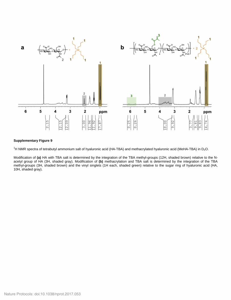
Supplementary Figure 9
1H NMR spectra of tetrabutyl ammonium salt of hyaluronic acid (HA-TBA) and methacrylated hyaluronic acid (MeHA-TBA) in D2O.
Modification of (a) HA with TBA salt is determined by the integration of the TBA methyl-groups (12H, shaded brown) relative to the N-acetyl group of HA (3H, shaded gray). Modification of (b) methacrylation and TBA salt is determined by the integration of the TBA methyl-groups (3H, shaded brown) and the vinyl singlets (1H each, shaded green) relative to the sugar ring of hyaluronic acid (HA, 10H, shaded gray).
Nature Protocols: doi:10.1038/nprot.2017.053

Supplementary Figure 10
1H NMR spectrum of 6-o-monotosyl-6-deoxy-β-cyclodextrin (CD-Tos) in DMSO-d6.
Tosylation of CD is confirmed by integration of the methyl (3H, shaded red) and aromatic hydrogens (2H each, shaded orange), relative
to the secondary hydroxyls of CD (14H, shaded gray). Anticipated minor impurities in the spectra may include trace acetone ( = 2.10),
ammonium chloride ( = 6.98, 7.12, 2.27 (s)), ethyl ether ( = 3.42 (q), 1.13 (t, 3H)), and water (see Supplementary Figure 11).
Nature Protocols: doi:10.1038/nprot.2017.053

Supplementary Figure 11
1H NMR spectrum of 6-o-monotosyl-6-deoxy-β-cyclodextrin (CD-Tos) in DMSO-d6.
The product was not fully dried, which causes combination of the HOD peak with the hydroxyl groups of the CD ring (purple shaded), prohibiting reliable integration of the CD necessary to confirm mono-tosylation.
Nature Protocols: doi:10.1038/nprot.2017.053

Supplementary Figure 12
1H NMR spectrum of 6-(6-aminohexyl)amino-6-deoxy-β-cyclodextrin (CD-HDA) in DMSO-d6.
Modification of CD with HDA is determined by integration of the hexane linker (12H, shaded red) relative to the secondary hydroxyls of
CD (14H, shaded gray). Anticipated minor impurities in the spectra may include trace dimethlyformamide ( = 7.98, 2.92, 2.08), acetone
( = 2.10), and ethyl ether ( = 3.42 (q), 1.13 (t, 3H)).
Nature Protocols: doi:10.1038/nprot.2017.053

Supplementary Figure 13
1H NMR spectra of β-cyclodextrin modification of hyaluronic acid (CD-HA) and methacrylated hyaluronic acid (MeHA-CD) in D2O.
(a) Modification of HA with pendant CD (30.2%) is determined by integration of the hexane linkers (12H, shaded red) relative to the N-acetyl singlet of HA (3H, shaded gray). (b) CD functionalization of MeHA shows an overlap of the spectra of the modifications with the sugar ring and the N-acetyl group of HA. Therefore, to determine the modification of MeHA with CD (30.3%), the hexane linker (12H, shaded red) is integrated relative to the methacrylate modification, which is assumed to be conserved throughout the reaction (25%, 1H each, shaded green).
Nature Protocols: doi:10.1038/nprot.2017.053

Supplementary Figure 14
1H NMR spectra of adamantane functionalization of hyaluronic acid (Ad-HA) and methacrylated adamantane hyaluronic acid (Ad-
MeHA) in D2O.
(a) Modification of HA with pendant Ad (32.5%) is determined by integration of the ethyl multiplet (12H, shaded blue) relative to the sugar ring of HA (10H, shaded gray). (b) Modification of MeHA with pendant Ad (32.8%) is determined by integration of the ethyl multiplet (12H, shaded blue) relative to the sugar ring of HA (10H, shaded gray). Integration of the vinyl singlets (1H each, shaded green) relative to the sugar ring of hyaluronic acid (HA, 10H, shaded gray) confirms the conservation of the methacrylate modification.
Nature Protocols: doi:10.1038/nprot.2017.053

Supplementary Figure 15
Confocal microscopy (calcein and ethidium staining) of rat endothelial progenitor cells (EPCs) encapsulated in GH hydrogels (5% (wt/vol)).
EPCs were not homogenously mixed with the GH hydrogel, which caused poor cell viability (45.5%) after injection through a 25G ¼ needle, scale bar 200 μm.
Nature Protocols: doi:10.1038/nprot.2017.053