nccn overview hodgkin lymphoma · overview hodgkin disease/lymphoma (hd/hl) is an uncom-mon...
TRANSCRIPT

© JNCCN–Journal of the National Comprehensive Cancer Network | Volume 9 Number 9 | September 2011
1020
OverviewHodgkin disease/lymphoma (HD/HL) is an uncom-mon malignancy involving lymph nodes and the lymphatic system. In 2010, an estimated 8490 new diagnoses of HD/HL and 1320 deaths from the dis-ease occurred in the United States.1 Most patients are diagnosed between 15 and 30 years of age, followed by another peak in adults aged 55 years or older.
The past few decades have seen significant prog-ress in the management of HL; it is now curable in at least 80% of patients.2 With the advent of more effec-tive treatment options, national statistics have shown an improvement in the 5-year survival rates of these patients that is unmatched in any other cancer over the past 4 decades. When appropriate treatment is selected, every patient with newly diagnosed HL has
NCCN
Hodgkin LymphomaClinical Practice Guidelines in Oncology
Richard T. Hoppe, MD; Ranjana H. Advani, MD; Weiyun Z. Ai, MD; Richard F. Ambinder, MD, PhD; Celeste M. Bello, MD, MSPH; Philip J. Bierman, MD; Kristie A. Blum, MD; Bouthaina Dabaja, MD; Ysabel Duron; Andres Forero, MD; Leo I. Gordon, MD; Francisco J. Hernandez-Ilizaliturri, MD; Ephraim P. Hochberg, MD; David G. Maloney, MD, PhD David Mansur, MD; Peter M. Mauch, MD; Monika Metzger, MD; Joseph O. Moore, MD; David Morgan, MD; Craig H. Moskowitz, MD; Matthew Poppe, MD; Barbara Pro, MD; Lawrence Weiss, MD; Jane N. Winter, MD; and Joachim Yahalom, MD
NCCN Clinical Practice Guidelines in Oncology for Hodgkin Lymphoma
Key WordsNCCN Clinical Practice Guidelines, NCCN Guidelines, Hodgkin disease, lymphoma, classical Hodgkin disease, lymphocyte predominance, combined modality therapy (JNCCN 2011;9:1020–1058)
NCCN Categories of Evidence and ConsensusCategory 1: Based upon high-level evidence, there is uni-form NCCN consensus that the intervention is appropriate.Category 2A: Based upon lower-level evidence, there is uni-form NCCN consensus that the intervention is appropriate.Category 2B: Based upon lower-level evidence, there is NCCN consensus that the intervention is appropriate.Category 3: Based upon any level of evidence, there is major NCCN disagreement that the intervention is appropriate.
All recommendations are category 2A unless otherwise noted.
Clinical trials: NCCN believes that the best management for any cancer patient is in a clinical trial. Participation in clinical trials is especially encouraged.
Please NoteThe NCCN Clinical Practice Guidelines in Oncology (NCCN GuidelinesTM) are a statement of consensus of the authors regarding their views of currently accepted ap-proaches to treatment. Any clinician seeking to apply or consult the NCCN Guidelines™ is expected to use indepen-dent medical judgment in the context of individual clinical circumstances to determine any patient’s care or treatment. The National Comprehensive Cancer Network® (NCCN®) makes no representation or warranties of any kind regarding their content, use, or application and disclaims any respon-sibility for their applications or use in any way.
© National Comprehensive Cancer Network, Inc. 2011, All rights reserved. The NCCN Guidelines and the illustrations herein may not be reproduced in any form without the express written permission of NCCN.Disclosures for the NCCN Guidelines Panel for Hodgkin Lymphoma
At the beginning of each NCCN Guidelines panel meeting, panel members disclosed any financial support they have received from industry. Through 2008, this information was published in an aggregate statement in JNCCN and online. Furthering NCCN’s commitment to public transparency, this disclosure process has now been expanded by listing all potential conflicts of interest respective to each individual expert panel member.
Individual disclosures for the NCCN Guidelines for Hodgkin Lymphoma panel members can be found on page 1058. (The most recent version of these guidelines and accompanying disclosures, including levels of compensation, are available on the NCCN Web site at www.NCCN.org.)
These guidelines are also available on the Internet. For the latest update, visit www.NCCN.org.

Hodgkin Lymphoma
NCCNGuidelines™
© JNCCN–Journal of the National Comprehensive Cancer Network | Volume 9 Number 9 | September 2011
1021
Journal of the National Comprehensive Cancer Network
Text continues on p. 1038
an overwhelming likelihood of being cured. In fact, cure rates for HL have increased so markedly that the overriding treatment considerations often relate to long-term toxicity, especially for patients with early- or intermediate-stage disease. For advanced disease, clinical trials still emphasize improvement in cure rates, but the potential long-term effects of treatment remain an important consideration.
The WHO classification divides HL into 2 main types: classical and lymphocyte-predominant Hodg-kin lymphoma (CHL and LPHL, respectively).3 CHL is divided into 4 subtypes: nodular sclerosis, mixed cellularity, lymphocyte-depleted, and lymphocyte-rich. In Western countries, LPHL accounts for 5% and CHL for 95% of all HL cases.
CHL is characterized by the presence of Reed-Sternberg cells in an inflammatory background,
whereas LPHL lacks Reed-Sternberg cells but is characterized by the presence of lymphocyte-pre-dominant cells, sometimes termed popcorn cells. LPHL can have a nodular or diffuse pattern. The nodular subtype has lymphocyte-predominant cells embedded in a background predominantly composed of B lymphocytes, whereas the diffuse subtype has a background consisting mainly of T cells.
These NCCN Guidelines discuss the clinical management of CHL and LPHL, focusing exclu-sively on patients from postadolescence through the seventh decade of life who do not have serious intercurrent disease. The guidelines do not address HL in pediatric or elderly patients or those with unusual situations, such as HIV positivity or preg-nancy. Individualized treatment may be necessary for older patients and those with concomitant disease.
NCCN Hodgkin Lymphoma Panel Members*Richard T. Hoppe, MD/Chair§
Stanford Cancer InstituteRanjana H. Advani, MD†
Stanford Cancer InstituteWeiyun Z. Ai, MD‡
UCSF Helen Diller Family Comprehensive Cancer CenterRichard F. Ambinder, MD, PhD†
The Sidney Kimmel Comprehensive Cancer Center at John Hopkins
Celeste M. Bello, MD, MSPH†H. Lee Moffitt Cancer Center & Research Institute
Philip J. Bierman, MD†‡ξUNMC Eppley Cancer Center at The Nebraska Medical Center
Kristie A. Blum, MD‡Arthur G. James Cancer Hospital & Richard J. Solove Research Institute at The Ohio State University
Bouthaina Dabaja, MD§The University of Texas MD Anderson Cancer Center
Ysabel Duron¥Latinas Contra Cancer
Andres Forero, MD†‡University of Alabama at Birmingham Comprehensive Cancer Center
Leo I. Gordon, MD‡Robert H. Lurie Comprehensive Cancer Center of Northwestern University
Francisco J. Hernandez-Ilizaliturri, MD†Roswell Park Cancer Institute
Ephraim P. Hochberg, MD†Massachusetts General Hospital Cancer Center
David G. Maloney, MD, PhD†‡Fred Hutchinson Cancer Research Center/ Seattle Cancer Care Alliance
David Mansur, MD§Siteman Cancer Center at Barnes-Jewish Hospital and Washington University School of Medicine
Peter M. Mauch, MD§Dana-Farber/Brigham and Women’s Cancer Center
Monika Metzger, MD€St. Jude Children’s Research Hospital/ University of Tennessee Cancer Institute
Joseph O. Moore, MD†Duke Cancer Institute
David Morgan, MD†‡ξVanderbilt-Ingram Cancer Center
Craig H. Moskowitz, MD†ÞMemorial Sloan-Kettering Cancer Center
Matthew Poppe, MD§Huntsman Cancer Institute at the University of Utah
Barbara Pro, MD†ÞFox Chase Cancer Center
Lawrence Weiss, MD≠City of Hope Comprehensive Cancer Center
Jane N. Winter, MD‡Robert H. Lurie Comprehensive Cancer Center of Northwestern University
Joachim Yahalom, MD§Memorial Sloan-Kettering Cancer Center
NCCN Staff: Kristina Gregory, RN, MSN, OCN, and Hema Sundar, PhD
KEY:
*Writing Committee Member
Specialties: §Radiation Oncology; †Medical Oncology; ‡Hematology/Hematology Oncology; ξBone Marrow Transplantation; €Pediatric Oncology; ≠Pathology; ÞInternal Medicine; ¥Patient Advocacy

© JNCCN–Journal of the National Comprehensive Cancer Network | Volume 9 Number 9 | September 2011
1022
Hodgkin Lymphoma Version 2:2011
Clinical trials: NCCN believes that the best management of any cancer patient is in a clinical trial. Participation in clinical trials is especially encouraged. All recommendations are category 2A unless otherwise indicated.
DIAGNOSIS WORKUP
•
•
•
•
Excisional biopsy(recommended)Core needle biopsymay be adequate ifdiagnosticFNA alone is generallyinsufficientImmunohistochemistryhighly recommendedfor Hodgkin lymphomaa
aTypical immunophenotype for classical Hodgkin lymphoma: CD30+, CD15+(majority); CD3-, CD45-; CD20+ (<40%). Lymphocyte-predominant Hodgkinlymphoma: CD20+, CD45+; CD3-, CD15-, CD30-. An expanded panel ofmarkers may be required especially if equivocal diagnosis.
cellularity,lymphocyte-depleted, and lymphocyte-rich.
LPHL has a different natural history and response to therapy than classical Hodgkin lymphoma, especially stages I-II. For that reason,
ns for postadolescent Hodgkin lymphoma.
e
See NCCNClinical Practice Guidelines in Oncology [NCCN Guidelines] for Non-Hodgkin’s Lymphomas.*
Classical Hodgkin lymphomas include nodular sclerosis, mixed
separate guidelines are presented for LPHL.NCCN unfavorable factors for stage I-II disease include bulky mediastinal or
> 10-cm disease, B symptoms, ESR > 50, > 3 sites of disease(see Examples of Unfavorable Factors for Stage I-II Hodgkin Disease, page 1033).
Treatment recommendatio
b
c
A separate diagnostic CT is not necessary if it was part of the integratedPET/CT scan.
In cases of PET positivity where sites of disease are inconsistent with usualpresentation of Hodgkin lymphoma, or in the presence of an unusualdisease presentation (i.e., HIV), additional clinical evaluation may berequired to upstage patient. See the staging table, available online,
at www.NCCN.org (ST-1).
d
f
g
CLINICAL STAGINGSee PrimaryTreatment(facingpage)g
See PrimaryTreatment(page 1030)g
ClassicalHodgkinlymphomad
Lymphocyte-predominantHodgkin lymphoma(LPHL)e
Stage IA, IIAfavorablef
Stage I-II
(bulkydisease)
unfavorablef
Stage III-IV
See PrimaryTreatment(page 1025)g
See PrimaryTreatment(page 1028)g
Stage I-II
(nonbulkydisease)
unfavorablef See PrimaryTreatment(page 1027)g
Essential
Useful in selected cases
:H&P including: B symptoms, alcoholintolerance, pruritus, fatigue, performancestatus, and examination of lymphoid regions,spleen, and liverCBC, differential, plateletsErythrocyte sedimentation rate (ESR)LDH, LFT, albuminBUN, creatininePregnancy test: women of childbearing ageChest x-rayDiagnostic chest/abdominal/pelvic CTPET/CT scanAdequate bone marrow biopsy in stage IB, IIBand stage III-IVEvaluation of ejection fraction fordoxorubicin-containing regimensCounseling: fertility, smoking cessation,psychosocial (
:Semen cryopreservation, if chemotherapy orpelvic RT contemplatedIVF or ovarian tissue or oocytecryopreservationNeck CT, if neck RT plannedPulmonary functions tests (PFTs includingDLCO) if ABVD or BEACOPP are being usedPneumococcal, H-flu, meningococcal vaccines,if splenic RT contemplatedHIV, if risk factors, unusual diseasepresentations
•
••••••••••
••••
•
••
bc
see NCCN Clinical PracticeGuidelines in Oncology [NCCN Guidelines] forDistress Management*)
Combined modalitytherapy (ABVD x2-4 cycles orStanford V x 8weeks + involved-field RT [IFRT])(category 1)
ij
kl
or
ABVD alone(category 2B)
PRIMARY TREATMENTh
CLINICAL PRESENTATION:Classical Hodgkin lymphomad
d
f
h
Classical Hodgkin lymphomas include nodular sclerosis, mixedcellularity, lymphocyte-depleted, and lymphocyte-rich.
NCCN unfavorable factors for stage I-II disease include bulkymediastinal or > 10-cm disease, B symptoms, ESR > 50, > 3 sites ofdisease (see Examples of Unfavorable Factors for Stage I-II HodgkinDisease, page 1033).
Individualized treatment may be necessary for older patients andpat
ients with concomitant disease.iSee Principles of Systemic Therapy (page 1034).4 cycles of ABVD unless patient fulfills strict criteria of only 2 sites of
disease and no extralymphatic lesions in which case 2 cycles is
sufficient.See Principles of Radiation Therapy (page 1035).Depending on comorbidities, subtotal lymphoid irradiation (category 1) or
mantle alone may be considered for patients not able to toleratechemotherapy.An integrated PET/CT or a PET with a diagnostic CT is recommended.See Revised Response Criteria for Lymphoma (page 1036).Recommend ABVD x 4 cycles (total) before proceeding to IFRT or biopsy.Biopsy to confirm no change in histology. Clinical circumstances may warrantadditional treatment even in the presence of a negative biopsy.j
k
l
m
n
o
p
See Follow-up (page 1031)
Restage afterchemotherapywith PET/CTm
Completeresponse(CR)n
IFRTk
Stabledisease(SD)orprogressivedisease(PD)n
See ProgressiveDisease (page 1032)
See Follow-up(page 1031)
Partialresponse(PR)n,o
Biopsyp
RestagewithPET/CTm
PET-positive
PET-negative
SeeProgressiveDisease(page 1032)
Observe
IFRTk
Biopsy
or
Positive
Negative
RestagewithPET/CTm
PET-positive
PET-negative
IFRTk
IFRTk
or
See Progressive Disease (page 1032)
Stage IA, IIAfavorable f
See PrimaryTreatment(page 1024)
See Follow-up(page 1031)
*To view the most recent version of these guidelines, visit the NCCN Web site at www.NCCN.org.
in these guidelines,

NCCN Clinical Practice Guidelines in Oncology
© JNCCN–Journal of the National Comprehensive Cancer Network | Volume 9 Number 9 | September 2011
1023
Hodgkin Lymphoma Version 2:2011
Version 2.2011, 05-04-11 ©2011 National Comprehensive Cancer Network, Inc. All rights reserved. The NCCN Guidelines™ and this illustration may not be reproduced in any form without the express written permission of NCCN®.
DIAGNOSIS WORKUP
•
•
•
•
Excisional biopsy(recommended)Core needle biopsymay be adequate ifdiagnosticFNA alone is generallyinsufficientImmunohistochemistryhighly recommendedfor Hodgkin lymphomaa
aTypical immunophenotype for classical Hodgkin lymphoma: CD30+, CD15+(majority); CD3-, CD45-; CD20+ (<40%). Lymphocyte-predominant Hodgkinlymphoma: CD20+, CD45+; CD3-, CD15-, CD30-. An expanded panel ofmarkers may be required especially if equivocal diagnosis.
cellularity,lymphocyte-depleted, and lymphocyte-rich.
LPHL has a different natural history and response to therapy than classical Hodgkin lymphoma, especially stages I-II. For that reason,
ns for postadolescent Hodgkin lymphoma.
e
See NCCNClinical Practice Guidelines in Oncology [NCCN Guidelines] for Non-Hodgkin’s Lymphomas.*
Classical Hodgkin lymphomas include nodular sclerosis, mixed
separate guidelines are presented for LPHL.NCCN unfavorable factors for stage I-II disease include bulky mediastinal or
> 10-cm disease, B symptoms, ESR > 50, > 3 sites of disease(see Examples of Unfavorable Factors for Stage I-II Hodgkin Disease, page 1033).
Treatment recommendatio
b
c
A separate diagnostic CT is not necessary if it was part of the integratedPET/CT scan.
In cases of PET positivity where sites of disease are inconsistent with usualpresentation of Hodgkin lymphoma, or in the presence of an unusualdisease presentation (i.e., HIV), additional clinical evaluation may berequired to upstage patient. See the staging table, available online,
at www.NCCN.org (ST-1).
d
f
g
CLINICAL STAGINGSee PrimaryTreatment(facingpage)g
See PrimaryTreatment(page 1030)g
ClassicalHodgkinlymphomad
Lymphocyte-predominantHodgkin lymphoma(LPHL)e
Stage IA, IIAfavorablef
Stage I-II
(bulkydisease)
unfavorablef
Stage III-IV
See PrimaryTreatment(page 1025)g
See PrimaryTreatment(page 1028)g
Stage I-II
(nonbulkydisease)
unfavorablef See PrimaryTreatment(page 1027)g
Essential
Useful in selected cases
:H&P including: B symptoms, alcoholintolerance, pruritus, fatigue, performancestatus, and examination of lymphoid regions,spleen, and liverCBC, differential, plateletsErythrocyte sedimentation rate (ESR)LDH, LFT, albuminBUN, creatininePregnancy test: women of childbearing ageChest x-rayDiagnostic chest/abdominal/pelvic CTPET/CT scanAdequate bone marrow biopsy in stage IB, IIBand stage III-IVEvaluation of ejection fraction fordoxorubicin-containing regimensCounseling: fertility, smoking cessation,psychosocial (
:Semen cryopreservation, if chemotherapy orpelvic RT contemplatedIVF or ovarian tissue or oocytecryopreservationNeck CT, if neck RT plannedPulmonary functions tests (PFTs includingDLCO) if ABVD or BEACOPP are being usedPneumococcal, H-flu, meningococcal vaccines,if splenic RT contemplatedHIV, if risk factors, unusual diseasepresentations
•
••••••••••
••••
•
••
bc
see NCCN Clinical PracticeGuidelines in Oncology [NCCN Guidelines] forDistress Management*)
Combined modalitytherapy (ABVD x2-4 cycles orStanford V x 8weeks + involved-field RT [IFRT])(category 1)
ij
kl
or
ABVD alone(category 2B)
PRIMARY TREATMENTh
CLINICAL PRESENTATION:Classical Hodgkin lymphomad
d
f
h
Classical Hodgkin lymphomas include nodular sclerosis, mixedcellularity, lymphocyte-depleted, and lymphocyte-rich.
NCCN unfavorable factors for stage I-II disease include bulkymediastinal or > 10-cm disease, B symptoms, ESR > 50, > 3 sites ofdisease (see Examples of Unfavorable Factors for Stage I-II HodgkinDisease, page 1033).
Individualized treatment may be necessary for older patients andpat
ients with concomitant disease.iSee Principles of Systemic Therapy (page 1034).4 cycles of ABVD unless patient fulfills strict criteria of only 2 sites of
disease and no extralymphatic lesions in which case 2 cycles is
sufficient.See Principles of Radiation Therapy (page 1035).Depending on comorbidities, subtotal lymphoid irradiation (category 1) or
mantle alone may be considered for patients not able to toleratechemotherapy.An integrated PET/CT or a PET with a diagnostic CT is recommended.See Revised Response Criteria for Lymphoma (page 1036).Recommend ABVD x 4 cycles (total) before proceeding to IFRT or biopsy.Biopsy to confirm no change in histology. Clinical circumstances may warrantadditional treatment even in the presence of a negative biopsy.j
k
l
m
n
o
p
See Follow-up (page 1031)
Restage afterchemotherapywith PET/CTm
Completeresponse(CR)n
IFRTk
Stabledisease(SD)orprogressivedisease(PD)n
See ProgressiveDisease (page 1032)
See Follow-up(page 1031)
Partialresponse(PR)n,o
Biopsyp
RestagewithPET/CTm
PET-positive
PET-negative
SeeProgressiveDisease(page 1032)
Observe
IFRTk
Biopsy
or
Positive
Negative
RestagewithPET/CTm
PET-positive
PET-negative
IFRTk
IFRTk
or
See Progressive Disease (page 1032)
Stage IA, IIAfavorable f
See PrimaryTreatment(page 1024)
See Follow-up(page 1031)
*To view the most recent version of these guidelines, visit the NCCN Web site at www.NCCN.org.
in these guidelines,

© JNCCN–Journal of the National Comprehensive Cancer Network | Volume 9 Number 9 | September 2011
1024
Hodgkin Lymphoma Version 2:2011
Clinical trials: NCCN believes that the best management of any cancer patient is in a clinical trial. Participation in clinical trials is especially encouraged. All recommendations are category 2A unless otherwise indicated.
ABVD alonex 2 cycles(category 2B)
i RestagewithPET/CTm,q
CR(with CRon CT)
nr
ABVD x 2cycles(total 4)
Observe (See Follow-up, page 1031)
•
•
ABVD x 4cycles(total 6)RepeatPFTs after4 cycles
PR orCR withPR on CT
nn
SDn
PDn
ABVD x 2cycles(total 4)
•
•
RestagewithPET/CTRepeatPFTs
m
PET-positive
PET-negative
Biopsyp
Biopsyp
IFRTorABVD x 2 cycles± IFRT
k
k
See Progressive Disease(page 1032)
See ProgressiveDisease (page 1032)
CLINICAL PRESENTATION:Classical Hodgkin lymphomaStage IA, IIA favorable
d
Observe (See Follow-up,page 1031)
RestagewithPET/CTm
PET-positivePRn
PET-negativeCRn
Observe (monitor PET/CT)
or
Biopsyp SeeProgressiveDisease(page 1032)
SeeFollow-up(page 1031)
Positive
Negative
d
h
i
mn
p
q
r
Classical Hodgkin lymphomas include nodular sclerosis, mixed cellularity,lymphocyte-depleted, and lymphocyte-rich.
Individualized treatment may be necessary for older patients and patientswith concomitant disease.
An integrated PET/CT or a PET with a diagnostic CT is recommended.
Biopsy to confirm no change in histology. Clinical circumstances may warrantadditional treatment even in the presence of a negative biopsy.
The value of interim PET scan after 2-4 cycles is unclear but may have a rolein management and prognosis.
Patients who have no residual evidence of disease on the diagnostic CTs andhave a negative PET.
See Principles of Systemic Therapy (page 1034).See Principles of Radiation Therapy (page 1035).
See Revised Response Criteria for Lymphoma (page 1036).
k
CRn
•
•
ABVD x2 cycles(total 4)RepeatPFTs
PRorSD
n
n
RestagewithPET/CTm
CRn
PDn
PRorSD
n
n
ABVD x2 cycles(total 6)
Biopsy
RestagewithPET/CTm
ABVD x 2cycles (total 6)
IFRTk
or
See Follow-up (page 1031)
See Follow-up (page 1031)
•
•
ABVD x2 cycles(total 4)RepeatPFTs
ABVD x2 cycles
i RestagewithPET/CTm,q
d
f
h
i
kClassical Hodgkin lymphomas include nodular sclerosis, mixed cellularity,lymphocyte-depleted, and lymphocyte-rich.
NCCN unfavorable factors for stage I-II disease include bulky mediastinal or> 10-cm disease, B symptoms, ESR > 50, > 3 sites of disease(see
Individualized treatment may be necessary for older patients and patientswith concomitant disease.
Examples of Unfavorable Factors for Stage I-II Hodgkin Disease,page 1033).
See Principles of Systemic Therapy (page 1034).
See Principles of Radiation Therapy (page 1035).An integrated PET/CT or a PET with a diagnostic CT is recommended.See Revised Response Criteria for Lymphoma (page 1036).Biopsy to confirm no change in histology. Clinical circumstances may warrantadditional treatment even in the presence of a negative biopsy.
The value of interim PET scan after 2-4 cycles is unclear but may have a rolein management and prognosis.
m
n
p
q
IFRTk
IFRTk
Stanford Vi
x 12 weeks
or
PRIMARYTREATMENTh
See PrimaryTreatment (page 1026)
CLINICAL PRESENTATION:Classical Hodgkin lymphomaStage I-II unfavorable (bulky)
df
PDn Biopsyp See Progressive Disease (page 1032)
Biopsyp
Negative
Positive
ABVD x2 cycles(total 6)
orStage I-IIunfavorablef
(bulky)
CRn
PRorSD
n
n
PDn
See Progressive Disease (page 1032)
See Follow-up (page 1031)
Biopsy
or
IFRTk
IFRTk
Negative
Positive
RestagewithPET/CTm
Biopsyp
PD
CR, PR,SD

NCCN Clinical Practice Guidelines in Oncology
© JNCCN–Journal of the National Comprehensive Cancer Network | Volume 9 Number 9 | September 2011
1025
Hodgkin Lymphoma Version 2:2011
Version 2.2011, 05-04-11 ©2011 National Comprehensive Cancer Network, Inc. All rights reserved. The NCCN Guidelines™ and this illustration may not be reproduced in any form without the express written permission of NCCN®.
ABVD alonex 2 cycles(category 2B)
i RestagewithPET/CTm,q
CR(with CRon CT)
nr
ABVD x 2cycles(total 4)
Observe (See Follow-up, page 1031)
•
•
ABVD x 4cycles(total 6)RepeatPFTs after4 cycles
PR orCR withPR on CT
nn
SDn
PDn
ABVD x 2cycles(total 4)
•
•
RestagewithPET/CTRepeatPFTs
m
PET-positive
PET-negative
Biopsyp
Biopsyp
IFRTorABVD x 2 cycles± IFRT
k
k
See Progressive Disease(page 1032)
See ProgressiveDisease (page 1032)
CLINICAL PRESENTATION:Classical Hodgkin lymphomaStage IA, IIA favorable
d
Observe (See Follow-up,page 1031)
RestagewithPET/CTm
PET-positivePRn
PET-negativeCRn
Observe (monitor PET/CT)
or
Biopsyp SeeProgressiveDisease(page 1032)
SeeFollow-up(page 1031)
Positive
Negative
d
h
i
mn
p
q
r
Classical Hodgkin lymphomas include nodular sclerosis, mixed cellularity,lymphocyte-depleted, and lymphocyte-rich.
Individualized treatment may be necessary for older patients and patientswith concomitant disease.
An integrated PET/CT or a PET with a diagnostic CT is recommended.
Biopsy to confirm no change in histology. Clinical circumstances may warrantadditional treatment even in the presence of a negative biopsy.
The value of interim PET scan after 2-4 cycles is unclear but may have a rolein management and prognosis.
Patients who have no residual evidence of disease on the diagnostic CTs andhave a negative PET.
See Principles of Systemic Therapy (page 1034).See Principles of Radiation Therapy (page 1035).
See Revised Response Criteria for Lymphoma (page 1036).
k
CRn
•
•
ABVD x2 cycles(total 4)RepeatPFTs
PRorSD
n
n
RestagewithPET/CTm
CRn
PDn
PRorSD
n
n
ABVD x2 cycles(total 6)
Biopsy
RestagewithPET/CTm
ABVD x 2cycles (total 6)
IFRTk
or
See Follow-up (page 1031)
See Follow-up (page 1031)
•
•
ABVD x2 cycles(total 4)RepeatPFTs
ABVD x2 cycles
i RestagewithPET/CTm,q
d
f
h
i
kClassical Hodgkin lymphomas include nodular sclerosis, mixed cellularity,lymphocyte-depleted, and lymphocyte-rich.
NCCN unfavorable factors for stage I-II disease include bulky mediastinal or> 10-cm disease, B symptoms, ESR > 50, > 3 sites of disease(see
Individualized treatment may be necessary for older patients and patientswith concomitant disease.
Examples of Unfavorable Factors for Stage I-II Hodgkin Disease,page 1033).
See Principles of Systemic Therapy (page 1034).
See Principles of Radiation Therapy (page 1035).An integrated PET/CT or a PET with a diagnostic CT is recommended.See Revised Response Criteria for Lymphoma (page 1036).Biopsy to confirm no change in histology. Clinical circumstances may warrantadditional treatment even in the presence of a negative biopsy.
The value of interim PET scan after 2-4 cycles is unclear but may have a rolein management and prognosis.
m
n
p
q
IFRTk
IFRTk
Stanford Vi
x 12 weeks
or
PRIMARYTREATMENTh
See PrimaryTreatment (page 1026)
CLINICAL PRESENTATION:Classical Hodgkin lymphomaStage I-II unfavorable (bulky)
df
PDn Biopsyp See Progressive Disease (page 1032)
Biopsyp
Negative
Positive
ABVD x2 cycles(total 6)
orStage I-IIunfavorablef
(bulky)
CRn
PRorSD
n
n
PDn
See Progressive Disease (page 1032)
See Follow-up (page 1031)
Biopsy
or
IFRTk
IFRTk
Negative
Positive
RestagewithPET/CTm
Biopsyp
PD
CR, PR,SD

© JNCCN–Journal of the National Comprehensive Cancer Network | Volume 9 Number 9 | September 2011
1026
Hodgkin Lymphoma Version 2:2011
Clinical trials: NCCN believes that the best management of any cancer patient is in a clinical trial. Participation in clinical trials is especially encouraged. All recommendations are category 2A unless otherwise indicated.
Stanford Vi,s
x 12 weeks
RT to initial sites > 5 cmand residual PET-positivesites (36 Gy beginsoptimally within 3 wk)
k Follow-up(see page 1031)Progressive disease(see page 1032)
Biopsyp
CLINICAL PRESENTATION:Classical Hodgkin lymphomaStage I-II unfavorable (bulky or nonbulky)
df
Restage withPET/CTm
Restage with CT(or PET/CT if lastPET scan was stillpositive) after 3 mo
See Progressive Disease (page 1032)
CR or PRn n
SD or PDn n
PRIMARY TREATMENTh
(continued from page 1025)
dClassical Hodgkin lymphomas include nodular sclerosis, mixed cellularity
or > 3 sites are treated according to the Stanford V algorithmon
,lymphocyte-depleted, and lymphocyte-rich.
NCCN unfavorable factors for stage I-II disease include bulky mediastinalor > 10-cm disease, B symptoms, ESR > 50, > 3 sites of disease(see Examples of Unfavorable Factors for Stage I-II Hodgkin Disease,page 1033).
Individualized treatment may be necessary for older patients and patientswith concomitant disease.
See Principles of Systemic Therapy (page 1034).See Principles of Radiation Therapy (page 1035).An integrated PET/CT or a PET with a diagnostic CT is recommended.
See Revised Response Criteria for Lymphoma (page 1036).Biopsy to confirm no change in histology. Clinical circumstances may warrantadditional treatment even in the presence of a negative biopsy.
The Stanford V regimen is used in this fashion for patients with bulkymediastinal disease or > 10-cm disease and/or B symptoms. Patients withelevated ESR,
page 1023.
f
h
i
k
m
n
p
s
CLINICAL PRESENTATION:Classical Hodgkin lymphomad
CRn
•
•
ABVD x2 cycles(total 4)RepeatPFTs
RestagewithPET/CTm
CRn
PDn
PRorSD
n
n
ABVD x2 cycles(total 6)
Biopsy
RestagewithPET/CTm
ABVD x 2cycles (total 6)
IFRTk
or
See Follow-up (page 1031)
See Follow-up (page 1031)
•
•
ABVD x2 cycles(total 4)RepeatPFTs
ABVD x2 cycles
i RestagewithPET/CTm,q
ObserveorIFRTk
Stanford Vi
or
PRIMARYTREATMENTh
See PrimaryTreatment
PDn Biopsyp See Progressive Disease (page 1032)
Biopsyp
Negative
Positive
ABVD x2 cycles(total 6)
orStage I-IIunfavorablef(nonbulky)
ObserveorIFRTk
dClassical Hodgkin lymphomas include nodular sclerosis, mixed cellular
is unclear but may have a rolein management and prognosis.
ity,lymphocyte-depleted, and lymphocyte-rich.
NCCN unfavorable factors for stage I-II disease include bulky mediastinal or> 10-cm disease, B symptoms, ESR > 50, > 3 sites of disease(see Examples of Unfavorable Factors for Stage I-II Hodgkin Disease,page 1033).
Individualized treatment may be necessary for older patients and patientswith concomitant disease.
See Principles of Systemic Therapy (page 1034).
See Principles of Radiation Therapy (page 1035).An integrated PET/CT or a PET with a diagnostic CT is recommended.See Revised Response Criteria for Lymphoma (page 1036).Biopsy to confirm no change in histology. Clinical circumstances may warrantadditional treatment even in the presence of a negative biopsy.
The value of interim PET scan after 2-4 cycles
f
h
i
k
m
n
p
q
PRorSD
n
n
See Follow-up (page 1031)
CRn
PRorSD
n
n
PDn
See Progressive Disease (page 1032)
IFRTk
ObserveorIFRTk
Biopsy
or
Negative
Positive
RestagewithPET/CTm
CR, PR,SD
Biopsyp
PDObserveorIFRTk
(opposite page)

NCCN Clinical Practice Guidelines in Oncology
© JNCCN–Journal of the National Comprehensive Cancer Network | Volume 9 Number 9 | September 2011
1027
Hodgkin Lymphoma Version 2:2011
Version 2.2011, 05-04-11 ©2011 National Comprehensive Cancer Network, Inc. All rights reserved. The NCCN Guidelines™ and this illustration may not be reproduced in any form without the express written permission of NCCN®.
Stanford Vi,s
x 12 weeks
RT to initial sites > 5 cmand residual PET-positivesites (36 Gy beginsoptimally within 3 wk)
k Follow-up(see page 1031)Progressive disease(see page 1032)
Biopsyp
CLINICAL PRESENTATION:Classical Hodgkin lymphomaStage I-II unfavorable (bulky or nonbulky)
df
Restage withPET/CTm
Restage with CT(or PET/CT if lastPET scan was stillpositive) after 3 mo
See Progressive Disease (page 1032)
CR or PRn n
SD or PDn n
PRIMARY TREATMENTh
(continued from page 1025)
dClassical Hodgkin lymphomas include nodular sclerosis, mixed cellularity
or > 3 sites are treated according to the Stanford V algorithmon
,lymphocyte-depleted, and lymphocyte-rich.
NCCN unfavorable factors for stage I-II disease include bulky mediastinalor > 10-cm disease, B symptoms, ESR > 50, > 3 sites of disease(see Examples of Unfavorable Factors for Stage I-II Hodgkin Disease,page 1033).
Individualized treatment may be necessary for older patients and patientswith concomitant disease.
See Principles of Systemic Therapy (page 1034).See Principles of Radiation Therapy (page 1035).An integrated PET/CT or a PET with a diagnostic CT is recommended.
See Revised Response Criteria for Lymphoma (page 1036).Biopsy to confirm no change in histology. Clinical circumstances may warrantadditional treatment even in the presence of a negative biopsy.
The Stanford V regimen is used in this fashion for patients with bulkymediastinal disease or > 10-cm disease and/or B symptoms. Patients withelevated ESR,
page 1023.
f
h
i
k
m
n
p
s
CLINICAL PRESENTATION:Classical Hodgkin lymphomad
CRn
•
•
ABVD x2 cycles(total 4)RepeatPFTs
RestagewithPET/CTm
CRn
PDn
PRorSD
n
n
ABVD x2 cycles(total 6)
Biopsy
RestagewithPET/CTm
ABVD x 2cycles (total 6)
IFRTk
or
See Follow-up (page 1031)
See Follow-up (page 1031)
•
•
ABVD x2 cycles(total 4)RepeatPFTs
ABVD x2 cycles
i RestagewithPET/CTm,q
ObserveorIFRTk
Stanford Vi
or
PRIMARYTREATMENTh
See PrimaryTreatment
PDn Biopsyp See Progressive Disease (page 1032)
Biopsyp
Negative
Positive
ABVD x2 cycles(total 6)
orStage I-IIunfavorablef(nonbulky)
ObserveorIFRTk
dClassical Hodgkin lymphomas include nodular sclerosis, mixed cellular
is unclear but may have a rolein management and prognosis.
ity,lymphocyte-depleted, and lymphocyte-rich.
NCCN unfavorable factors for stage I-II disease include bulky mediastinal or> 10-cm disease, B symptoms, ESR > 50, > 3 sites of disease(see Examples of Unfavorable Factors for Stage I-II Hodgkin Disease,page 1033).
Individualized treatment may be necessary for older patients and patientswith concomitant disease.
See Principles of Systemic Therapy (page 1034).
See Principles of Radiation Therapy (page 1035).An integrated PET/CT or a PET with a diagnostic CT is recommended.See Revised Response Criteria for Lymphoma (page 1036).Biopsy to confirm no change in histology. Clinical circumstances may warrantadditional treatment even in the presence of a negative biopsy.
The value of interim PET scan after 2-4 cycles
f
h
i
k
m
n
p
q
PRorSD
n
n
See Follow-up (page 1031)
CRn
PRorSD
n
n
PDn
See Progressive Disease (page 1032)
IFRTk
ObserveorIFRTk
Biopsy
or
Negative
Positive
RestagewithPET/CTm
CR, PR,SD
Biopsyp
PDObserveorIFRTk
(opposite page)

© JNCCN–Journal of the National Comprehensive Cancer Network | Volume 9 Number 9 | September 2011
1028
Hodgkin Lymphoma Version 2:2011
Clinical trials: NCCN believes that the best management of any cancer patient is in a clinical trial. Participation in clinical trials is especially encouraged. All recommendations are category 2A unless otherwise indicated.
ABVDx 2-4cycles
i
ObserveorRT to initial bulkydisease
ku
Biopsyor
IFRTorObserve in selectedcircumstances(e.g., equivocal PET)
p
k
ObserveorRT to initial bulky orPET-positive sites(especially for initialbulky disease)
k
u
StageIII-IV
Escalated BEACOPP(selected cases if IPS 4)
i
t
CRn
or
CLINICAL PRESENTATION:Classical Hodgkin lymphomad
or
Stanford V x 12 weeks(selected cases if IPS < 3)
it
See facing page
Restage withPET/CTm
RestagewithPET/CT
ABVD x 2-4cycles(total 6)RepeatPFTs after 4total cycles
PDn
PRIMARY TREATMENTh
PRor SD
nn
CRn
PDn
PRor SD
nn
Biopsyp See Progressive Disease (page 1032)
Negative
Positive
SeeProgressiveDisease(page 1032)
Biopsy
or
ABVD x 2-4cycles(total 6)RepeatPFTs after 4total cycles
See page 1026
SeeFollow-up(page 1031)
d
h
Classical Hodgkin lymphomas include nodular sclerosis, mixed cellularity,lymphocyte-depleted, and lymphocyte-rich.
Individualized treatment may
herapy.
be necessary for older patients and patientswith concomitant disease.
See Principles of Systemic Therapy (page 1034).See Principles of Radiation Therapy (page 1035).An integrated PET/ CT or a PET with a diagnostic CT is recommended.
See Revised Response Criteria for Lymphoma (page 1036).Biopsy to confirm no change in histology. Clinical circumstances may warrantadditional treatment even in the presence of a negative biopsy.
See International Prognostic Score (IPS; page 1033).If initial bulky mediastinal disease is seen on CT, consolidative RT to themediastinum is recommended after completion of chemot
i
k
m
n
p
t
u
SeeFollow-up(page 1031)
SeeFollow-up(page 1031)
EscalatedBEACOPP x 4cycles (selectedcases if IPS 4)
i
t
PRIMARY TREATMENTh
(continued from opposite page)
CLINICAL PRESENTATION:Classical Hodgkin lymphomaStage III-IV
d
RestagewithPET/CTm
dClassical Hodgkin lymphomas include nodular sclerosis, mixed cellularity,lymphocyte-depleted, and lymphocyte-rich.
h
i
k
m
n
p
t
Individualized treatment may be necessary for older patients and patientswith concomitant disease.
See Principles of Systemic Therapy (page 1034).See Principles of Radiation Therapy (pag 1035).
An integrated PET/CT or a PET with a diagnostic CT is recommended.See Revised Response Criteria for Lymphoma (page 1036).Biopsy to confirm no change in histology. Clinical circumstances may warrantadditional treatment even in the presence of a negative biopsy.
See International Prognostic Score (IPS; page 1033).
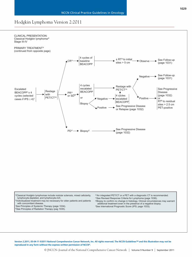
CRn4 cycles ofbaselineBEACOPP
4 cyclesescalatedBEACOPP
or
Biopsy
Negative
Positive
± RT to initialsites > 5 cm
kObserve
Restage withPET/CTm
Negative
Positive
Biopsyp
See Progressive Diseaseor Relapse (page 1032)
4 cyclesescalatedBEACOPP
PDn
PRor SD
nn
See Progressive Disease(page 1032)
See ProgressiveDisease(page 1032)orRT to residualsites > 2.5 cmPET-positive
k
See Follow-up(page 1031)
See Follow-up(page 1031)
NCCN Clinical Practice Guidelines in Oncology
© JNCCN–Journal of the National Comprehensive Cancer Network | Volume 9 Number 9 | September 2011
1029
Hodgkin Lymphoma Version 2:2011
Version 2.2011, 05-04-11 ©2011 National Comprehensive Cancer Network, Inc. All rights reserved. The NCCN Guidelines™ and this illustration may not be reproduced in any form without the express written permission of NCCN®.
ABVDx 2-4cycles
i
ObserveorRT to initial bulkydisease
ku
Biopsyor
IFRTorObserve in selectedcircumstances(e.g., equivocal PET)
p
k
ObserveorRT to initial bulky orPET-positive sites(especially for initialbulky disease)
k
u
StageIII-IV
Escalated BEACOPP(selected cases if IPS 4)
i
t
CRn
or
CLINICAL PRESENTATION:Classical Hodgkin lymphomad
or
Stanford V x 12 weeks(selected cases if IPS < 3)
it
See facing page
Restage withPET/CTm
RestagewithPET/CT
ABVD x 2-4cycles(total 6)RepeatPFTs after 4total cycles
PDn
PRIMARY TREATMENTh
PRor SD
nn
CRn
PDn
PRor SD
nn
Biopsyp See Progressive Disease (page 1032)
Negative
Positive
SeeProgressiveDisease(page 1032)
Biopsy
or
ABVD x 2-4cycles(total 6)RepeatPFTs after 4total cycles
See page 1026
SeeFollow-up(page 1031)
d
h
Classical Hodgkin lymphomas include nodular sclerosis, mixed cellularity,lymphocyte-depleted, and lymphocyte-rich.
Individualized treatment may
herapy.
be necessary for older patients and patientswith concomitant disease.
See Principles of Systemic Therapy (page 1034).See Principles of Radiation Therapy (page 1035).An integrated PET/ CT or a PET with a diagnostic CT is recommended.
See Revised Response Criteria for Lymphoma (page 1036).Biopsy to confirm no change in histology. Clinical circumstances may warrantadditional treatment even in the presence of a negative biopsy.
See International Prognostic Score (IPS; page 1033).If initial bulky mediastinal disease is seen on CT, consolidative RT to themediastinum is recommended after completion of chemot
i
k
m
n
p
t
u
SeeFollow-up(page 1031)
SeeFollow-up(page 1031)
EscalatedBEACOPP x 4cycles (selectedcases if IPS 4)
i
t
PRIMARY TREATMENTh
(continued from opposite page)
CLINICAL PRESENTATION:Classical Hodgkin lymphomaStage III-IV
d
RestagewithPET/CTm
dClassical Hodgkin lymphomas include nodular sclerosis, mixed cellularity,lymphocyte-depleted, and lymphocyte-rich.
h
i
k
m
n
p
t
Individualized treatment may be necessary for older patients and patientswith concomitant disease.
See Principles of Systemic Therapy (page 1034).See Principles of Radiation Therapy (pag 1035).
An integrated PET/CT or a PET with a diagnostic CT is recommended.See Revised Response Criteria for Lymphoma (page 1036).Biopsy to confirm no change in histology. Clinical circumstances may warrantadditional treatment even in the presence of a negative biopsy.
See International Prognostic Score (IPS; page 1033).
CRn4 cycles ofbaselineBEACOPP
4 cyclesescalatedBEACOPP
or
Biopsy
Negative
Positive
± RT to initialsites > 5 cm
kObserve
Restage withPET/CTm
Negative
Positive
Biopsyp
See Progressive Diseaseor Relapse (page 1032)
4 cyclesescalatedBEACOPP
PDn
PRor SD
nn
See Progressive Disease(page 1032)
See ProgressiveDisease(page 1032)orRT to residualsites > 2.5 cmPET-positive
k
See Follow-up(page 1031)
See Follow-up(page 1031)

© JNCCN–Journal of the National Comprehensive Cancer Network | Volume 9 Number 9 | September 2011
1030
Hodgkin Lymphoma Version 2:2011
Clinical trials: NCCN believes that the best management of any cancer patient is in a clinical trial. Participation in clinical trials is especially encouraged. All recommendations are category 2A unless otherwise indicated.
IFRTk
PRIMARY TREATMENTCLINICAL PRESENTATION:Lymphocyte-predominantHodgkin lymphomae
CS IA, IIA
CS IB, IIB
CS IIIA, IVA
CS IIIB, IVB
Chemotherapy ± IFRTorRituximab ±chemotherapy ± IFRT
v k
v k
Chemotherapy ± RTorRituximab ±chemotherapy ± RT
v k
v k
Chemotherapy ± RTv k
orObservation (category 2B)orLocal RT (palliation only)orRituximab ± chemotherapyv
CRn Observe
< CRn
Observe, if asymptomaticorChemotherapy
orRT (if no prior RT)k
(See page 1032)
CRn Observe
< CRn
Observe, if asymptomaticorChemotherapy
orRT (if no prior RT)k
(See page 1032)
Restage
Restage
eLPHL has a different natural history and response to therapy compared withclassical Hodgkin lymphoma, especially stages I-II. Therefore, separateguidelines are presented for LPHL.
See Revised Response Criteria for Lymphoma (page 1036).See Principles of Systemic Therapy (page 1034).
kSee Principles of Radiation Therapy (page 1035).
n
v
FOLLOW-UP AFTER COMPLETION OF TREATMENT AND MONITORING FOR LATE EFFECTS
••
•
It is recommended that the patient be provided with a treatment summary at the completion of therapy.Follow-up with an oncologist is recommended, especially during the first 5-y interval to detect recurrence, then annually because of risk of latecomplications, including second cancers and cardiovascular disease. Late relapse or transformation to large cell lymphoma may occur inLPHL.The frequency and types of tests may vary depending on clinical circumstances, such as age and stage at diagnosis, social habits, andtreatment modality. Few data support specific recommendations; these represent the range of practice at NCCN Member Institutions.
w,x
•
•
•
Interim H&P:Every 2-4 mo for 1-2 y, then every 3-6 mo for next 3-5 y
Annual influenza vaccine➤
➤
➤
Laboratory studies:CBC, platelets, ESR (if elevated at time of initial diagnosis), chemistry profile every 2-4 mo for 1-2 y, then every 3-6 mo for next3-5 yThyroid-stimulating hormone (TSH) at least annually if RT to neck
Chest imaging:Chest radiograph or CT every 6-12 mo during first 2-5 y
wMauch P, Ng A, Aleman B, et al. Report from the Rockefeller Foundation sponsored International Workshop on reducing mortality and improving quality of lifein long-term survivors of Hodgkin's disease: July 9-16, 2003, Bellagio, Italy. Eur J Haematol 2005;75:s66.
x
yAppropriate medical management should be instituted for any abnormalities.Chest imaging optional after 5 y if patient treated with a nonalkylating agent, no RT to the chest, and no other risk factors are present.
Follow-Up After Completion of Treatment
•
•
Interim H&P: Annually
Pneumococcal, m revaccination after 5 y, if patient treated with splenic RT or previous splenectomyAnnual influenza vaccine
➤
➤
➤
➤
➤
➤
➤
Annual blood pressure, aggressive management of cardiovascular risk factorsBaseline stress test/echocardiogram at 10 y
eningococcal, and H-flu
Laboratory studies:CBC, platelets, chemistry profile annuallyTSH at least annually if RT to neckAnnual lipids
••
•••
Annual chest imaging (chest radiograph or chest CT) for patients at increased risk for lung cancerAnnual breast screening:
Initiate 8-10 y posttherapy, or at age 40, whichever comes first, if chest or axillary radiation. The American Cancer Society recommendsbreast MRI in addition to mammography for women who received irradiation to the chest between ages 10 and 30 y.
Counseling:Reproduction, health habits, psychosocial, cardiovascular, breast self-exam, skin cancer risk.
Cardiovascular symptoms may emerge at a young age.Treatment summary and consideration of transfer to primary care practicioner.
y
Monitoring for Late Effects After 5 Yearsw,x
••
Abdominal/pelvic CT (category 2B):➤ Every 6-12 mo for first 2-3 yCounseling:
Reproduction, health habits, psychosocial, cardiovascular, breast self-exam, skin cancer risk, end-of-treatment discussion.Surveillance PET should not be performed routinely because of risk for false-positives. Management decisions should not be based onPET scan alone; clinical or pathologic correlation is needed.
➤
➤
➤
•
➤

NCCN Clinical Practice Guidelines in Oncology
© JNCCN–Journal of the National Comprehensive Cancer Network | Volume 9 Number 9 | September 2011
1031
Hodgkin Lymphoma Version 2:2011
Version 2.2011, 05-04-11 ©2011 National Comprehensive Cancer Network, Inc. All rights reserved. The NCCN Guidelines™ and this illustration may not be reproduced in any form without the express written permission of NCCN®.
IFRTk
PRIMARY TREATMENTCLINICAL PRESENTATION:Lymphocyte-predominantHodgkin lymphomae
CS IA, IIA
CS IB, IIB
CS IIIA, IVA
CS IIIB, IVB
Chemotherapy ± IFRTorRituximab ±chemotherapy ± IFRT
v k
v k
Chemotherapy ± RTorRituximab ±chemotherapy ± RT
v k
v k
Chemotherapy ± RTv k
orObservation (category 2B)orLocal RT (palliation only)orRituximab ± chemotherapyv
CRn Observe
< CRn
Observe, if asymptomaticorChemotherapy
orRT (if no prior RT)k
(See page 1032)
CRn Observe
< CRn
Observe, if asymptomaticorChemotherapy
orRT (if no prior RT)k
(See page 1032)
Restage
Restage
eLPHL has a different natural history and response to therapy compared withclassical Hodgkin lymphoma, especially stages I-II. Therefore, separateguidelines are presented for LPHL.
See Revised Response Criteria for Lymphoma (page 1036).See Principles of Systemic Therapy (page 1034).
kSee Principles of Radiation Therapy (page 1035).
n
v
FOLLOW-UP AFTER COMPLETION OF TREATMENT AND MONITORING FOR LATE EFFECTS
••
•
It is recommended that the patient be provided with a treatment summary at the completion of therapy.Follow-up with an oncologist is recommended, especially during the first 5-y interval to detect recurrence, then annually because of risk of latecomplications, including second cancers and cardiovascular disease. Late relapse or transformation to large cell lymphoma may occur inLPHL.The frequency and types of tests may vary depending on clinical circumstances, such as age and stage at diagnosis, social habits, andtreatment modality. Few data support specific recommendations; these represent the range of practice at NCCN Member Institutions.
w,x
•
•
•
Interim H&P:Every 2-4 mo for 1-2 y, then every 3-6 mo for next 3-5 y
Annual influenza vaccine➤
➤
➤
Laboratory studies:CBC, platelets, ESR (if elevated at time of initial diagnosis), chemistry profile every 2-4 mo for 1-2 y, then every 3-6 mo for next3-5 yThyroid-stimulating hormone (TSH) at least annually if RT to neck
Chest imaging:Chest radiograph or CT every 6-12 mo during first 2-5 y
wMauch P, Ng A, Aleman B, et al. Report from the Rockefeller Foundation sponsored International Workshop on reducing mortality and improving quality of lifein long-term survivors of Hodgkin's disease: July 9-16, 2003, Bellagio, Italy. Eur J Haematol 2005;75:s66.
x
yAppropriate medical management should be instituted for any abnormalities.Chest imaging optional after 5 y if patient treated with a nonalkylating agent, no RT to the chest, and no other risk factors are present.
Follow-Up After Completion of Treatment
•
•
Interim H&P: Annually
Pneumococcal, m revaccination after 5 y, if patient treated with splenic RT or previous splenectomyAnnual influenza vaccine
➤
➤
➤
➤
➤
➤
➤
Annual blood pressure, aggressive management of cardiovascular risk factorsBaseline stress test/echocardiogram at 10 y
eningococcal, and H-flu
Laboratory studies:CBC, platelets, chemistry profile annuallyTSH at least annually if RT to neckAnnual lipids
••
•••
Annual chest imaging (chest radiograph or chest CT) for patients at increased risk for lung cancerAnnual breast screening:
Initiate 8-10 y posttherapy, or at age 40, whichever comes first, if chest or axillary radiation. The American Cancer Society recommendsbreast MRI in addition to mammography for women who received irradiation to the chest between ages 10 and 30 y.
Counseling:Reproduction, health habits, psychosocial, cardiovascular, breast self-exam, skin cancer risk.
Cardiovascular symptoms may emerge at a young age.Treatment summary and consideration of transfer to primary care practicioner.
y
Monitoring for Late Effects After 5 Yearsw,x
••
Abdominal/pelvic CT (category 2B):➤ Every 6-12 mo for first 2-3 yCounseling:
Reproduction, health habits, psychosocial, cardiovascular, breast self-exam, skin cancer risk, end-of-treatment discussion.Surveillance PET should not be performed routinely because of risk for false-positives. Management decisions should not be based onPET scan alone; clinical or pathologic correlation is needed.
➤
➤
➤
•
➤

© JNCCN–Journal of the National Comprehensive Cancer Network | Volume 9 Number 9 | September 2011
1032
Hodgkin Lymphoma Version 2:2011
Clinical trials: NCCN believes that the best management of any cancer patient is in a clinical trial. Participation in clinical trials is especially encouraged. All recommendations are category 2A unless otherwise indicated.
SECOND-LINETHERAPY
If primary therapywas RT alone
If primary therapywas chemotherapyalone orcombinationchemotherapy/RT HDT/ASCR
(category1)± RT
or
chemotherapy
dd,ee,ff,ggk,cc
hhk,cc
Salvage±
RT
If initial stagewas IA-IIA:
No prior RTand failure ininitial sitesonly
•
CLASSICAL HODGKIN LYMPHOMAaa
Suspectedrelapse
•
•
Restaging (same asinitial workup,including bonemarrow biopsy)Consider marrowcytogenetics forMDS markersbefore transplant
All others
Treat as primaryadvanced stageHodgkin lymphoma(See page 1028)
Individualized treatment isrecommended, options include
RTSalvage chemotherapy ± RTHDT/ASCR ± RT
bbk,cc
dd k,ccdd,ee,ff,gg k,cc
•Progressivediseasep
Re-biopsy
Negative
Positive
Observationz
k
hh
l-dose chemotherapy may precede high-dose therapy.Timing of RT may vary.For select patients with a long disease-free interval and other favorablefeatures; selection of chemotherapy should be individualized.
See Principles of Radiation Therapy (page 1035).Biopsy to confirm no change in histology. Clinical circumstances may warrantadditional treatment even in the presence of a negative biopsy.
Clinical circumstances may warrant additional treatment even in the presence of anegative biopsy.Patients with LPHL may be managed according to the same algorithm; however,some patients with LPHL have a chronic indolent course that may not requireaggressive re-treatment. These asymptomatic patients may be observed. Atrelapse, patient should be considered for rebiopsy because of risk fortransformation.No data support a superior outcome with any modalities.
RT recommended when sites have not been previously irradiated. In aradiation-naive patient, TLI may be an appropriate component of HDT.See Principles of Second-Line Chemotherapy (page 1037).Allotransplant is an option in select patients as a category 3.
Biopsy to confirm relapse, especially if plan to treat with high-dosetherapy.Conventiona
p
z
aa
bb
cc
dd
ee
ff
gg
International Prognostic Score (IPS)1 point per factor (advanced disease)1
Albumin < 4 g/dLHemoglobin < 10.5 g/dLMaleAge 45 yStage IV diseaseLeukocytosis (WBC count 15,000/mm )Lymphocytopenia (lymphocyte count < 8% of WBC count, and/or lymphocytecount < 600/mm )
3
3
1Derived from Hasenclever D, Diehl V. A prognostic score for advanced Hodgkin’s disease: International Prognostic Factors Project on Advanced Hodgkin’sDisease. N Engl J Med 1998;339:1506-1514.
Examples of Unfavorable Risk Factors for Stage I-II Hodgkin Disease
Risk Factor GHSG EORTC NCIC NCCN
Age 50 40
Histology MC or LD
ESR and B symptoms > 50 if A; > 30 if B sx > 50 if A; > 30 if B sx > 50 or any B sx > 50 or any B sx
Mediastinal mass MMR > .33 MTR > .35 MMR > .33 or > 10 cm MMR > .33
# Nodal sites > 2 > 3 > 3 > 3
E lesion any
Bulky > 10 cm
Abbreviations: ESR, erythrocyte sedimentation rate; GHSG, German Hodgkin Study Group; LD, lymphocyte-depleted; MC, mixed cellularity; MMR, mediastinal mass ratio, maximum width of mass/maximum intrathoracic diameter; MTR, mediastinal thoracic ratio, maximum width of mediastinal mass/intrathoracic diameter at T5-6; NCIC, National Cancer Institute, Canada; Sx, symptoms.

NCCN Clinical Practice Guidelines in Oncology
© JNCCN–Journal of the National Comprehensive Cancer Network | Volume 9 Number 9 | September 2011
1033
Hodgkin Lymphoma Version 2:2011
Version 2.2011, 05-04-11 ©2011 National Comprehensive Cancer Network, Inc. All rights reserved. The NCCN Guidelines™ and this illustration may not be reproduced in any form without the express written permission of NCCN®.
SECOND-LINETHERAPY
If primary therapywas RT alone
If primary therapywas chemotherapyalone orcombinationchemotherapy/RT HDT/ASCR
(category1)± RTor
chemotherapy
dd,ee,ff,ggk,cc
hhk,cc
Salvage±
RT
If initial stagewas IA-IIA:
No prior RTand failure ininitial sitesonly
•
CLASSICAL HODGKIN LYMPHOMAaa
Suspectedrelapse
•
•
Restaging (same asinitial workup,including bonemarrow biopsy)Consider marrowcytogenetics forMDS markersbefore transplant
All others
Treat as primaryadvanced stageHodgkin lymphoma(See page 1028)
Individualized treatment isrecommended, options include
RTSalvage chemotherapy ± RTHDT/ASCR ± RT
bbk,cc
dd k,ccdd,ee,ff,gg k,cc
•Progressivediseasep
Re-biopsy
Negative
Positive
Observationz
k
hh
l-dose chemotherapy may precede high-dose therapy.Timing of RT may vary.For select patients with a long disease-free interval and other favorablefeatures; selection of chemotherapy should be individualized.
See Principles of Radiation Therapy (page 1035).Biopsy to confirm no change in histology. Clinical circumstances may warrantadditional treatment even in the presence of a negative biopsy.
Clinical circumstances may warrant additional treatment even in the presence of anegative biopsy.Patients with LPHL may be managed according to the same algorithm; however,some patients with LPHL have a chronic indolent course that may not requireaggressive re-treatment. These asymptomatic patients may be observed. Atrelapse, patient should be considered for rebiopsy because of risk fortransformation.No data support a superior outcome with any modalities.
RT recommended when sites have not been previously irradiated. In aradiation-naive patient, TLI may be an appropriate component of HDT.See Principles of Second-Line Chemotherapy (page 1037).Allotransplant is an option in select patients as a category 3.
Biopsy to confirm relapse, especially if plan to treat with high-dosetherapy.Conventiona
p
z
aa
bb
cc
dd
ee
ff
gg
International Prognostic Score (IPS)1 point per factor (advanced disease)1
Albumin < 4 g/dLHemoglobin < 10.5 g/dLMaleAge 45 yStage IV diseaseLeukocytosis (WBC count 15,000/mm )Lymphocytopenia (lymphocyte count < 8% of WBC count, and/or lymphocytecount < 600/mm )
3
3
1Derived from Hasenclever D, Diehl V. A prognostic score for advanced Hodgkin’s disease: International Prognostic Factors Project on Advanced Hodgkin’sDisease. N Engl J Med 1998;339:1506-1514.
Examples of Unfavorable Risk Factors for Stage I-II Hodgkin Disease
Risk Factor GHSG EORTC NCIC NCCN
Age 50 40
Histology MC or LD
ESR and B symptoms > 50 if A; > 30 if B sx > 50 if A; > 30 if B sx > 50 or any B sx > 50 or any B sx
Mediastinal mass MMR > .33 MTR > .35 MMR > .33 or > 10 cm MMR > .33
# Nodal sites > 2 > 3 > 3 > 3
E lesion any
Bulky > 10 cm
Abbreviations: ESR, erythrocyte sedimentation rate; GHSG, German Hodgkin Study Group; LD, lymphocyte-depleted; MC, mixed cellularity; MMR, mediastinal mass ratio, maximum width of mass/maximum intrathoracic diameter; MTR, mediastinal thoracic ratio, maximum width of mediastinal mass/intrathoracic diameter at T5-6; NCIC, National Cancer Institute, Canada; Sx, symptoms.

© JNCCN–Journal of the National Comprehensive Cancer Network | Volume 9 Number 9 | September 2011
1034
Hodgkin Lymphoma Version 2:2011
Clinical trials: NCCN believes that the best management of any cancer patient is in a clinical trial. Participation in clinical trials is especially encouraged. All recommendations are category 2A unless otherwise indicated.
PRINCIPLES OF SYSTEMIC THERAPY
Classical Hodgkin LymphomaThe most common variants of chemotherapy used at NCCN Member Institutions include ABVD and Stanford V. Routine use of growthfactors is not recommended. Leukopenia is not a factor for delay of treatment or reduction of dose intensity (except for escalatedBEACOPP).
•
See Principles of Second-line Chemotherapy, page 1037
•
•
•
ABVD (doxorubicin, bleomycin, vinblastine, and dacarbazine) ± RT
Stanford V (doxorubicin, vinblastine, mechlorethamine, etoposide, vincristine, bleomycin, and prednisone)
BEACOPP (bleomycin, etoposide, doxorubicin, cyclophosphamide, vincristine, procarbazine, and prednisone)
Eich HT, Diehl V, Gorgen H, et al. Intensified chemotherapy and dose-reduced involved-field radiotherapy in patients with early unfavorableHodgkin’s lymphoma: final analysis of the German Hodgkin Study Group HD 11 trial. J Clin Oncol 2010;28:4199-4206.Engert A, Plutschow A, Eich HT, et al. Reduced treatment intensity in patients with early-stage Hodgkin’s lymphoma. N Engl J Med2010;363:640-652.Meyer RM, Gospodarowicz MK, Connors JM, et al. Randomized comparison of ABVD chemotherapy with a strategy that includes radiationtherapy in patients with limited-stage Hodgkin's lymphoma: National Cancer Institute of Canada Clinical Trials Group and the EasternCooperative Oncology Group. J Clin Oncol 2005;23:4634-4642.Bonadonna G, Bonfante V, Viviani S, et al. ABVD plus subtotal nodal versus involved-field radiotherapy in early-stage Hodgkin's disease:long-term results. J Clin Oncol 2004;22:2835-2841.Duggan DB, Petroni GR, Johnson JL, et al. Randomized comparison of ABVD and MOPP/ABV hybrid for the treatment of advancedHodgkin's disease: report of an Intergroup trial. J Clin Oncol 2003;21:607-614.
Advani RH, Hoppe RT, Baer DM, et al. Efficacy of abbreviated Stanford V chemotherapy and involved field radiotherapy in early stageHodgkin’s disease: mature results of the G4 trial [abstract]. Blood 2009;114:Abstract 1670.Edwards-Bennett SM, Jacks LM, Moskowitz CH, et al. Stanford V program for locally extensive and advanced Hodgkin lymphoma: theMemorial Sloan-Kettering Cancer Center experience. Ann Oncol 2010;21:574-581.
Engert A, Diehl V, Franklin J, et al. Escalated-dose BEACOPP in the treatment of patients with advanced-stage Hodgkin’s lymphoma:10 years of follow-up of the GHSG HD9 study. J Clin Oncol 2009;27:4548-4554.
Gordon LI, Hong F, Fisher RI, et al. A randomized phase III trial of ABVD Vs. Stanford V +/- radiation therapy In locally extensive andadvanced stage Hodgkin's lymphoma: an Intergroup study coordinated by the Eastern Cooperative Oncology Group (E2496) [abstract].Blood 2010;116:Abstract 415.
Horning SJ, Hoppe RT, Breslin S, et al. Stanford V and radiotherapy for locally extensive and advanced Hodgkin's disease: mature resultsof a prospective clinical trial. J Clin Oncol 2002;20:630-637.
Regimens and References
Lymphocyte-Predominant Hodgkin LymphomaThe most common chemotherapies used at NCCN Member Institutions for LPHL include:
ABVD (doxorubicin, bleomycin, vinblastine, and dacarbazine) ± rituximabCHOP (cyclophosphamide, doxorubicin, vincristine, and prednisone) ± rituximabCVP (cyclophosphamide, vincristine, and prednisone) ± rituximabEPOCH (cyclophosphamide, doxorubicin, etoposide, vincristine, and prednisone) ± rituximabSingle-agent rituximab
1
2,3
4-6
•➤
➤
➤
➤
➤
1Ongoing clinical trials will help to clarify the role of a watch-and-wait strategy or systemic therapy, including anthracycline (epirubicin or doxorubicin),bleomycin, and vinblastine-based chemotherapy or antibody-based approaches, in the treatment of these patients.
2
3
4
5
6
Savage KJ, Skinnider B, Al Mansour M, et al. Incorporation of ABVD increases cure rates of patients with limited stage nodular lymphocyte predominantHodgkin Lymphoma (NLPHL) [abstract]. Blood 2010;116:Abstract 3887.
Canellos GP, Mauch P. What is the appropriate systemic chemotherapy for lymphocyte-predominant Hodgkin's Lymphoma? J Clin Oncol 2010;28:e8.Ekstrand BC, Lucas JB, Horwitz SM, et al. Rituximab in lymphocyte-predominant Hodgkin disease: results of a phase 2 trial. Blood 2003;101:4285-4289.Schulz H, Rehwald U, Morschhauser F, et al. Rituximab in relapsed lymphocyte-predominant Hodgkin lymphoma: long-term results of a phase 2 trial by theGerman Hodgkin Lymphoma Study Group (GHSG). Blood 2008;111:109-111.
Horning SJ, Bartlett NL, Breslin S, et al. Results of a prospective phase ii trial of limited and extended rituximab treatment in nodular lymphocyte predominantHodgkin's disease (NLPHD) [abstract]. Blood 2007;110:Abstract 644.
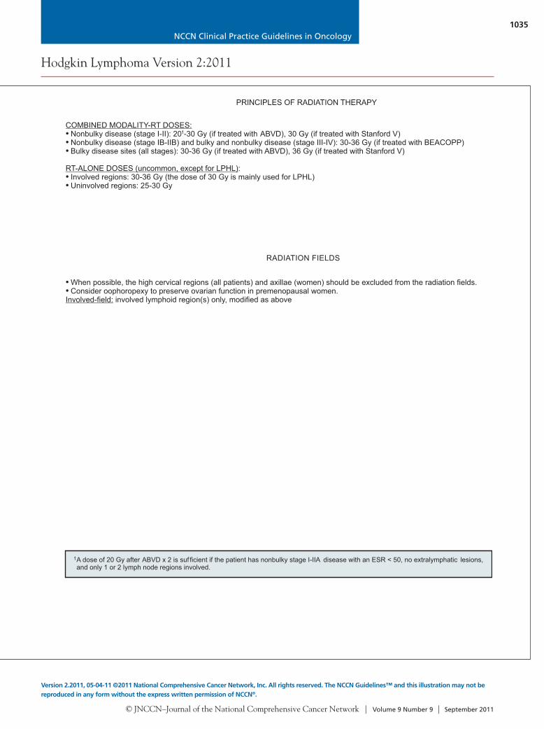
PRINCIPLES OF RADIATION THERAPY
COMBINED MODALITY-RT DOSES:•••
Nonbulky disease (stage I-II): 20 -30 Gy (if treated with ABVD), 30 Gy (if treated with Stanford V)Nonbulky disease (stage IB-IIB) and bulky and nonbulky disease (stage III-IV): 30-36 Gy (if treated with BEACOPP)Bulky disease sites (all stages): 30-36 Gy (if treated with ABVD), 36 Gy (if treated with Stanford V)
RT-ALONE DOSES (uncommon, except for LPHL):Involved regions: 30-36 Gy (the dose of 30 Gy is mainly used for LPHL)Uninvolved regions: 25-30 Gy••
••
When possible, the high cervical regions (all patients) and axillae (women) should be excluded from the radiation fields.Consider oophoropexy to preserve ovarian function in premenopausal women.
Involved-field: involved lymphoid region(s) only, modified as above
RADIATION FIELDS
A dose of 20 Gy after ABVD x 2 is suf ficient if the patient has nonbulky stage I-IIA disease with an ESR < 50, no extralymphatic lesions,and only 1 or 2 lymph node regions involved.
1
1
NCCN Clinical Practice Guidelines in Oncology
© JNCCN–Journal of the National Comprehensive Cancer Network | Volume 9 Number 9 | September 2011
1035
Hodgkin Lymphoma Version 2:2011
Version 2.2011, 05-04-11 ©2011 National Comprehensive Cancer Network, Inc. All rights reserved. The NCCN Guidelines™ and this illustration may not be reproduced in any form without the express written permission of NCCN®.
PRINCIPLES OF SYSTEMIC THERAPY
Classical Hodgkin LymphomaThe most common variants of chemotherapy used at NCCN Member Institutions include ABVD and Stanford V. Routine use of growthfactors is not recommended. Leukopenia is not a factor for delay of treatment or reduction of dose intensity (except for escalatedBEACOPP).
•
See Principles of Second-line Chemotherapy, page 1037
•
•
•
ABVD (doxorubicin, bleomycin, vinblastine, and dacarbazine) ± RT
Stanford V (doxorubicin, vinblastine, mechlorethamine, etoposide, vincristine, bleomycin, and prednisone)
BEACOPP (bleomycin, etoposide, doxorubicin, cyclophosphamide, vincristine, procarbazine, and prednisone)
Eich HT, Diehl V, Gorgen H, et al. Intensified chemotherapy and dose-reduced involved-field radiotherapy in patients with early unfavorableHodgkin’s lymphoma: final analysis of the German Hodgkin Study Group HD 11 trial. J Clin Oncol 2010;28:4199-4206.Engert A, Plutschow A, Eich HT, et al. Reduced treatment intensity in patients with early-stage Hodgkin’s lymphoma. N Engl J Med2010;363:640-652.Meyer RM, Gospodarowicz MK, Connors JM, et al. Randomized comparison of ABVD chemotherapy with a strategy that includes radiationtherapy in patients with limited-stage Hodgkin's lymphoma: National Cancer Institute of Canada Clinical Trials Group and the EasternCooperative Oncology Group. J Clin Oncol 2005;23:4634-4642.Bonadonna G, Bonfante V, Viviani S, et al. ABVD plus subtotal nodal versus involved-field radiotherapy in early-stage Hodgkin's disease:long-term results. J Clin Oncol 2004;22:2835-2841.Duggan DB, Petroni GR, Johnson JL, et al. Randomized comparison of ABVD and MOPP/ABV hybrid for the treatment of advancedHodgkin's disease: report of an Intergroup trial. J Clin Oncol 2003;21:607-614.
Advani RH, Hoppe RT, Baer DM, et al. Efficacy of abbreviated Stanford V chemotherapy and involved field radiotherapy in early stageHodgkin’s disease: mature results of the G4 trial [abstract]. Blood 2009;114:Abstract 1670.Edwards-Bennett SM, Jacks LM, Moskowitz CH, et al. Stanford V program for locally extensive and advanced Hodgkin lymphoma: theMemorial Sloan-Kettering Cancer Center experience. Ann Oncol 2010;21:574-581.
Engert A, Diehl V, Franklin J, et al. Escalated-dose BEACOPP in the treatment of patients with advanced-stage Hodgkin’s lymphoma:10 years of follow-up of the GHSG HD9 study. J Clin Oncol 2009;27:4548-4554.
Gordon LI, Hong F, Fisher RI, et al. A randomized phase III trial of ABVD Vs. Stanford V +/- radiation therapy In locally extensive andadvanced stage Hodgkin's lymphoma: an Intergroup study coordinated by the Eastern Cooperative Oncology Group (E2496) [abstract].Blood 2010;116:Abstract 415.
Horning SJ, Hoppe RT, Breslin S, et al. Stanford V and radiotherapy for locally extensive and advanced Hodgkin's disease: mature resultsof a prospective clinical trial. J Clin Oncol 2002;20:630-637.
Regimens and References
Lymphocyte-Predominant Hodgkin LymphomaThe most common chemotherapies used at NCCN Member Institutions for LPHL include:
ABVD (doxorubicin, bleomycin, vinblastine, and dacarbazine) ± rituximabCHOP (cyclophosphamide, doxorubicin, vincristine, and prednisone) ± rituximabCVP (cyclophosphamide, vincristine, and prednisone) ± rituximabEPOCH (cyclophosphamide, doxorubicin, etoposide, vincristine, and prednisone) ± rituximabSingle-agent rituximab
1
2,3
4-6
•➤
➤
➤
➤
➤
1Ongoing clinical trials will help to clarify the role of a watch-and-wait strategy or systemic therapy, including anthracycline (epirubicin or doxorubicin),bleomycin, and vinblastine-based chemotherapy or antibody-based approaches, in the treatment of these patients.
2
3
4
5
6
Savage KJ, Skinnider B, Al Mansour M, et al. Incorporation of ABVD increases cure rates of patients with limited stage nodular lymphocyte predominantHodgkin Lymphoma (NLPHL) [abstract]. Blood 2010;116:Abstract 3887.
Canellos GP, Mauch P. What is the appropriate systemic chemotherapy for lymphocyte-predominant Hodgkin's Lymphoma? J Clin Oncol 2010;28:e8.Ekstrand BC, Lucas JB, Horwitz SM, et al. Rituximab in lymphocyte-predominant Hodgkin disease: results of a phase 2 trial. Blood 2003;101:4285-4289.Schulz H, Rehwald U, Morschhauser F, et al. Rituximab in relapsed lymphocyte-predominant Hodgkin lymphoma: long-term results of a phase 2 trial by theGerman Hodgkin Lymphoma Study Group (GHSG). Blood 2008;111:109-111.
Horning SJ, Bartlett NL, Breslin S, et al. Results of a prospective phase ii trial of limited and extended rituximab treatment in nodular lymphocyte predominantHodgkin's disease (NLPHD) [abstract]. Blood 2007;110:Abstract 644.
PRINCIPLES OF RADIATION THERAPY
COMBINED MODALITY-RT DOSES:•••
Nonbulky disease (stage I-II): 20 -30 Gy (if treated with ABVD), 30 Gy (if treated with Stanford V)Nonbulky disease (stage IB-IIB) and bulky and nonbulky disease (stage III-IV): 30-36 Gy (if treated with BEACOPP)Bulky disease sites (all stages): 30-36 Gy (if treated with ABVD), 36 Gy (if treated with Stanford V)
RT-ALONE DOSES (uncommon, except for LPHL):Involved regions: 30-36 Gy (the dose of 30 Gy is mainly used for LPHL)Uninvolved regions: 25-30 Gy••
••
When possible, the high cervical regions (all patients) and axillae (women) should be excluded from the radiation fields.Consider oophoropexy to preserve ovarian function in premenopausal women.
Involved-field: involved lymphoid region(s) only, modified as above
RADIATION FIELDS
A dose of 20 Gy after ABVD x 2 is suf ficient if the patient has nonbulky stage I-IIA disease with an ESR < 50, no extralymphatic lesions,and only 1 or 2 lymph node regions involved.
1
1

© JNCCN–Journal of the National Comprehensive Cancer Network | Volume 9 Number 9 | September 2011
1036
Hodgkin Lymphoma Version 2:2011
Clinical trials: NCCN believes that the best management of any cancer patient is in a clinical trial. Participation in clinical trials is especially encouraged. All recommendations are category 2A unless otherwise indicated.
REVISED RESPONSE CRITERIA FOR HODGKIN LYMPHOMA(including PET)
Response Definition
CR
PR
SD
Relapseddisease orprogressivedisease
Nodal Masses Spleen, Liver
Disappearanceof all evidenceof disease
Regression ofmeasurabledisease andno new sites
Failure to attainCR/PR, orprogressivedisease
Any new lesionor increase by
50% ofpreviouslyinvolved sitesfrom nadir
FDG-avid or PET-positivebefore therapy; mass ofany size permitted if PETnegative
50% decrease in SPD of up to 6 largestdominant masses; no increase in size ofother nodesFDG-avid or PET-positive before therapy;one or more PET-positive sites remainpositive
FDG-avid or PET-positivebefore therapy; PET-positive atprior sites of disease and nonew sites on CT or PET
Appearance of a new lesion(s) > 1.5cm in any axis, 50% increase in SPDof more than one node, or 50%increase in longest diameter of apreviously identified node > 1 cm inshort axisLesions PET-positive if FDG-avidlymphoma or PET-positive beforetherapy
Not palpable,nodulesdisappeared
50% decrease inSPD of nodules(for single nodulein greatesttransversediameter); noincrease in size ofliver or spleen
> 50% increase fromnadir in the SPD ofany previous lesions
Source: Adapted from Cheson BD, Pfistner B, Juweid ME, et al. Revised response criteria for malignant lymphoma. J Clin Oncol 2007;25:579-586.Reprinted with permission from the American Society of Clinical Oncology.
Table 2 from
Bone Marrow
Infiltrate cleared onrepeat biopsy; ifindeterminate bymorphology,immunohistochemistryshould be negative
Irrelevant if positivebefore therapy; celltype should bespecified
New or recurrentinvolvement
PRINCIPLES OF SECOND-LINE CHEMOTHERAPY
The selection of second-line chemotherapy regimens depends on the pattern of relapse and the agents previously used.
Mini-BEAM (carmustine, cytarabine, etoposide, and melphalan)MINE (etoposide, ifosfamide, mesna, and mitoxantrone)VIM-D (etoposide, ifosfamide, mitoxantrone, and dexamethasone).GCD (gemcitabine, carboplatin, and dexamethasone)
Some studies have suggested that patients with minimal disease burden at relapse (not refractory) may not need additional treatment prior to high-dosechemotherapy with stem-cell rescue. However, patients tend to have an improved outcome when transplanted in a minimal disease state. Thus,cytoreduction with chemotherapy (see above) before high-dose chemotherapy with stem-cell rescue may be beneficial. In addition, second-line chemotherapyserves as a test for drug sensitivity and to facilitate the harvest of stem cells.
Nitrogen mustard, procarbazine, carmustine, and melphalan may adversely affect both quality and quantity of stem-cell collection.
Examples of second-line chemotherapy prior to transplant include:ICE (ifosfamide, carboplatin, and etoposide)C-MOPP (cyclophosphamide, vincristine, procarbazine, and prednisone)ChlVPP (Chlorambucil, vinblastine, procarbazine, and prednisone)DHAP (dexamethasone, cisplatin, and high-dose cytarabine)ESHAP (etoposide, methylprednisolone, high-dose cytarabine, and cisplatin)GVD (gemcitabine, vinorelbine, and liposomal doxorubicin)IGEV (ifosfamide, gemcitabine, and vinorelbine)
➤
➤
➤
➤
➤
➤
➤
➤
➤
➤
➤
➤
1,23,4
56
7-91011
12,131415
16
17-19 20
1
3
4
18
19
20
Moskowitz CH, Nimer SD, Zelenetz AD, et al. A 2-step comprehensive high-dose chemoradiotherapy second-line program for relapsed and refractory Hodgkindisease: analysis by intent to treat and development of a prognostic model. Blood 2001;97:616-623.
Abali H, Urün Y, Oksüzo lu B, et al. Comparison of ICE (ifosfamide-carboplatin-etoposide) versus DHAP (cytosine arabinoside-cisplatin-dexamethasone) assalvage chemotherapy in patients with relapsed or refractory lymphoma. Cancer Invest 2008;26:401-406.
Takenaka T, Mikuni C, Miura A, et al. Alternating combination chemotherapy C-MOPP (cyclophosphamide, vincristine, procarbazine, prednisone) and ABVD(adriamycin, bleomycin, vinblastine, dacarbazine) in clinical stage II-IV Hodgkin's disease: a multicenter phase II study (JCOG 8905). Jpn J Clin Oncol2000;30:146-152.
Montoto S, Camos M, Lopez-Guillermo A, et al. Hybrid chemotherapy consisting of cyclophosphamide, vincristine, procarbazine, prednisone, doxorubicin,bleomycin, and vinblastine (C-MOPP/ABV) as first-line treatment
Bierman PJ, Anderson JR, Freeman MB, et al. High-dose chemotherapy followed by autologous hematopoietic rescue for Hodgkin's disease patients followingfirst relapse after chemotherapy. .Chopra R, McMillan AK, Linch DC, et al. The place of high-dose BEAM therapy and autologous bone marrow transplantation in poor-risk Hodgkin's disease. Asingle-center eight-year study of 155 patients. .Stewart DA, Guo D, Gluck S, et al. Double high-dose therapy for Hodgkin's disease with dose-intensive cyclophosphamide, etoposide, and cisplatin (DICEP)prior to high-dose melphalan and autologous stem cell transplantation. Bone Marrow Transplant 2000;26:383-388.
ğ
Hodgkin disease. Cancer 2000;88:2142-2148.The International ChlVPP Treatment Group. ChlVPP therapy for Hodgkin's disease: experience of 960 patients. Ann Oncol 1995;6:167-172.Josting A, Rudolph C, Reiser M, et al. Time-intensified dexamethasone/cisplatin/cytarabine: an effective salvage therapy with low toxicity in patients withrelapsed and refractory Hodgkin's disease. Ann Oncol 2002;13:1628-1635.
Aparicio J, Segura A, Garcera S, et al. ESHAP is an active regimen for relapsing Hodgkin's disease. Ann Oncol 1999;10:593-595.Akhtar S, Abdelsalam M, El Weshi A, et al. High-dose chemotherapy and autologous stem cell transplantation for Hodgkin's lymphoma in the kingdom of SaudiArabia: King Faisal specialist hospital and research center experience. Bone Marrow Transplant 2008;42(Suppl 1):S37-40.
Fernández de Larrea C, Martínez C, Gava A, et al. Salvage chemotherapy with alternating MINE-ESHAP regimen in relapsed or refractory Hodgkin's lymphomafollowed by autologous stem cell transplantation. Ann Oncol 2010;21:1211-1216.Bartlett N, Niedzwiecki D, Johnson J, et al. Gemcitabine, vinorelbine, and pegylated liposomal doxorubicin (GVD), a salvage regimen in relapsed Hodgkin'slymphoma: CALGB 59804. Ann Oncol 2007;18:1071-1079.Santoro A, Magagnoli M, Spina M, et al. Ifosfamide, gemcitabine, and vinorelbine: a new induction regimen for refractory and relapsed Hodgkin's lymphoma.Haematologica 2007;92:35-41.Colwill R, Crump M, Couture F, et al. Mini-BEAM as salvage therapy for relapsed or refractory Hodgkin's disease before intensive therapy and autologous bonemarrow transplantation. J Clin Oncol 1995;13:396-402.Martín A, Fernández-Jiménez MC, Caballero MD, et al. Long-term follow-up in patients treated with Mini-BEAM as salvage therapy for relapsed or refractoryHodgkin's disease. Br J Haematol 2001;113:161-171.Rodriguez MA, Cabanillas FC, Hagemeister FB, et al. A phase II trial of mesna/ifosfamide, mitoxantrone and etoposide for refractory lymphoms. Ann Oncol1995;6:609-611.Phillips JK, Spearing RL, Davies JM, et al. VIM-D salvage chemotherapy in Hodgkin's disease. Cancer Chemother Pharmacol 1990;27:161-163.Gopal AK, Press OW, Shustov AR, et al. Efficacy and safety of gemicitabine, carboplatin, dexamethasone, and rituximab in patients with relapsed/refractorylymphoma: a prospective multi-center phase II study by Puget Sound Oncology Consortium. Leuk Lymphoma 2010;51:1523-1529.Sweetenham JW, Taghipour G, Milligan D, et al. High-dose therapy and autologous stem cell rescue for patients with Hodgkin's disease in first relapse afterchemotherapy: results from the EBMT. Lymphoma Working Party of the European Group for Blood and Marrow Transplantation. Bone Marrow Transplant1997;20:745-752.
Ann Oncol 1996;7:151-156
Blood 1993;81:1137-1145
5
6
7
10
11
12
14
15
16
17
2
8
9
13
Abbreviations: CR, complete response; PR, partial response; SD, stable disease; SPD, sum of the product of the diameters.
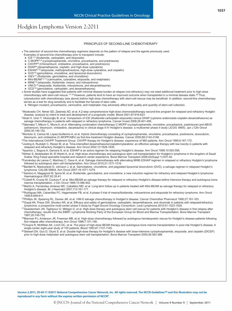
NCCN Clinical Practice Guidelines in Oncology
© JNCCN–Journal of the National Comprehensive Cancer Network | Volume 9 Number 9 | September 2011
1037
Hodgkin Lymphoma Version 2:2011
Version 2.2011, 05-04-11 ©2011 National Comprehensive Cancer Network, Inc. All rights reserved. The NCCN Guidelines™ and this illustration may not be reproduced in any form without the express written permission of NCCN®.
REVISED RESPONSE CRITERIA FOR HODGKIN LYMPHOMA(including PET)
Response Definition
CR
PR
SD
Relapseddisease orprogressivedisease
Nodal Masses Spleen, Liver
Disappearanceof all evidenceof disease
Regression ofmeasurabledisease andno new sites
Failure to attainCR/PR, orprogressivedisease
Any new lesionor increase by
50% ofpreviouslyinvolved sitesfrom nadir
FDG-avid or PET-positivebefore therapy; mass ofany size permitted if PETnegative
50% decrease in SPD of up to 6 largestdominant masses; no increase in size ofother nodesFDG-avid or PET-positive before therapy;one or more PET-positive sites remainpositive
FDG-avid or PET-positivebefore therapy; PET-positive atprior sites of disease and nonew sites on CT or PET
Appearance of a new lesion(s) > 1.5cm in any axis, 50% increase in SPDof more than one node, or 50%increase in longest diameter of apreviously identified node > 1 cm inshort axisLesions PET-positive if FDG-avidlymphoma or PET-positive beforetherapy
Not palpable,nodulesdisappeared
50% decrease inSPD of nodules(for single nodulein greatesttransversediameter); noincrease in size ofliver or spleen
> 50% increase fromnadir in the SPD ofany previous lesions
Source: Adapted from Cheson BD, Pfistner B, Juweid ME, et al. Revised response criteria for malignant lymphoma. J Clin Oncol 2007;25:579-586.Reprinted with permission from the American Society of Clinical Oncology.
Table 2 from
Bone Marrow
Infiltrate cleared onrepeat biopsy; ifindeterminate bymorphology,immunohistochemistryshould be negative
Irrelevant if positivebefore therapy; celltype should bespecified
New or recurrentinvolvement
PRINCIPLES OF SECOND-LINE CHEMOTHERAPY
The selection of second-line chemotherapy regimens depends on the pattern of relapse and the agents previously used.
Mini-BEAM (carmustine, cytarabine, etoposide, and melphalan)MINE (etoposide, ifosfamide, mesna, and mitoxantrone)VIM-D (etoposide, ifosfamide, mitoxantrone, and dexamethasone).GCD (gemcitabine, carboplatin, and dexamethasone)
Some studies have suggested that patients with minimal disease burden at relapse (not refractory) may not need additional treatment prior to high-dosechemotherapy with stem-cell rescue. However, patients tend to have an improved outcome when transplanted in a minimal disease state. Thus,cytoreduction with chemotherapy (see above) before high-dose chemotherapy with stem-cell rescue may be beneficial. In addition, second-line chemotherapyserves as a test for drug sensitivity and to facilitate the harvest of stem cells.
Nitrogen mustard, procarbazine, carmustine, and melphalan may adversely affect both quality and quantity of stem-cell collection.
Examples of second-line chemotherapy prior to transplant include:ICE (ifosfamide, carboplatin, and etoposide)C-MOPP (cyclophosphamide, vincristine, procarbazine, and prednisone)ChlVPP (Chlorambucil, vinblastine, procarbazine, and prednisone)DHAP (dexamethasone, cisplatin, and high-dose cytarabine)ESHAP (etoposide, methylprednisolone, high-dose cytarabine, and cisplatin)GVD (gemcitabine, vinorelbine, and liposomal doxorubicin)IGEV (ifosfamide, gemcitabine, and vinorelbine)
➤
➤
➤
➤
➤
➤
➤
➤
➤
➤
➤
➤
1,23,4
56
7-91011
12,131415
16
17-19 20
1
3
4
18
19
20
Moskowitz CH, Nimer SD, Zelenetz AD, et al. A 2-step comprehensive high-dose chemoradiotherapy second-line program for relapsed and refractory Hodgkindisease: analysis by intent to treat and development of a prognostic model. Blood 2001;97:616-623.
Abali H, Urün Y, Oksüzo lu B, et al. Comparison of ICE (ifosfamide-carboplatin-etoposide) versus DHAP (cytosine arabinoside-cisplatin-dexamethasone) assalvage chemotherapy in patients with relapsed or refractory lymphoma. Cancer Invest 2008;26:401-406.
Takenaka T, Mikuni C, Miura A, et al. Alternating combination chemotherapy C-MOPP (cyclophosphamide, vincristine, procarbazine, prednisone) and ABVD(adriamycin, bleomycin, vinblastine, dacarbazine) in clinical stage II-IV Hodgkin's disease: a multicenter phase II study (JCOG 8905). Jpn J Clin Oncol2000;30:146-152.
Montoto S, Camos M, Lopez-Guillermo A, et al. Hybrid chemotherapy consisting of cyclophosphamide, vincristine, procarbazine, prednisone, doxorubicin,bleomycin, and vinblastine (C-MOPP/ABV) as first-line treatment
Bierman PJ, Anderson JR, Freeman MB, et al. High-dose chemotherapy followed by autologous hematopoietic rescue for Hodgkin's disease patients followingfirst relapse after chemotherapy. .Chopra R, McMillan AK, Linch DC, et al. The place of high-dose BEAM therapy and autologous bone marrow transplantation in poor-risk Hodgkin's disease. Asingle-center eight-year study of 155 patients. .Stewart DA, Guo D, Gluck S, et al. Double high-dose therapy for Hodgkin's disease with dose-intensive cyclophosphamide, etoposide, and cisplatin (DICEP)prior to high-dose melphalan and autologous stem cell transplantation. Bone Marrow Transplant 2000;26:383-388.
ğ
Hodgkin disease. Cancer 2000;88:2142-2148.The International ChlVPP Treatment Group. ChlVPP therapy for Hodgkin's disease: experience of 960 patients. Ann Oncol 1995;6:167-172.Josting A, Rudolph C, Reiser M, et al. Time-intensified dexamethasone/cisplatin/cytarabine: an effective salvage therapy with low toxicity in patients withrelapsed and refractory Hodgkin's disease. Ann Oncol 2002;13:1628-1635.
Aparicio J, Segura A, Garcera S, et al. ESHAP is an active regimen for relapsing Hodgkin's disease. Ann Oncol 1999;10:593-595.Akhtar S, Abdelsalam M, El Weshi A, et al. High-dose chemotherapy and autologous stem cell transplantation for Hodgkin's lymphoma in the kingdom of SaudiArabia: King Faisal specialist hospital and research center experience. Bone Marrow Transplant 2008;42(Suppl 1):S37-40.
Fernández de Larrea C, Martínez C, Gava A, et al. Salvage chemotherapy with alternating MINE-ESHAP regimen in relapsed or refractory Hodgkin's lymphomafollowed by autologous stem cell transplantation. Ann Oncol 2010;21:1211-1216.Bartlett N, Niedzwiecki D, Johnson J, et al. Gemcitabine, vinorelbine, and pegylated liposomal doxorubicin (GVD), a salvage regimen in relapsed Hodgkin'slymphoma: CALGB 59804. Ann Oncol 2007;18:1071-1079.Santoro A, Magagnoli M, Spina M, et al. Ifosfamide, gemcitabine, and vinorelbine: a new induction regimen for refractory and relapsed Hodgkin's lymphoma.Haematologica 2007;92:35-41.Colwill R, Crump M, Couture F, et al. Mini-BEAM as salvage therapy for relapsed or refractory Hodgkin's disease before intensive therapy and autologous bonemarrow transplantation. J Clin Oncol 1995;13:396-402.Martín A, Fernández-Jiménez MC, Caballero MD, et al. Long-term follow-up in patients treated with Mini-BEAM as salvage therapy for relapsed or refractoryHodgkin's disease. Br J Haematol 2001;113:161-171.Rodriguez MA, Cabanillas FC, Hagemeister FB, et al. A phase II trial of mesna/ifosfamide, mitoxantrone and etoposide for refractory lymphoms. Ann Oncol1995;6:609-611.Phillips JK, Spearing RL, Davies JM, et al. VIM-D salvage chemotherapy in Hodgkin's disease. Cancer Chemother Pharmacol 1990;27:161-163.Gopal AK, Press OW, Shustov AR, et al. Efficacy and safety of gemicitabine, carboplatin, dexamethasone, and rituximab in patients with relapsed/refractorylymphoma: a prospective multi-center phase II study by Puget Sound Oncology Consortium. Leuk Lymphoma 2010;51:1523-1529.Sweetenham JW, Taghipour G, Milligan D, et al. High-dose therapy and autologous stem cell rescue for patients with Hodgkin's disease in first relapse afterchemotherapy: results from the EBMT. Lymphoma Working Party of the European Group for Blood and Marrow Transplantation. Bone Marrow Transplant1997;20:745-752.
Ann Oncol 1996;7:151-156
Blood 1993;81:1137-1145
5
6
7
10
11
12
14
15
16
17
2
8
9
13
Abbreviations: CR, complete response; PR, partial response; SD, stable disease; SPD, sum of the product of the diameters.

NCCN Clinical Practice Guidelines in Oncology
Hodgkin Lymphoma
© JNCCN–Journal of the National Comprehensive Cancer Network | Volume 9 Number 9 | September 2011
1038
Text continued from p. 1021
Consistent with NCCN philosophy, participation in clinical trials is always encouraged.
Staging and PrognosisStaging for HL is based on the Ann Arbor staging system (see the staging table, available online, in these guidelines, at www.NCCN.org [ST-1]). Each stage (I–IV) is subdivided into A and B categories. “A” indicates that no systemic symptoms are pres-ent, and “B” is assigned to patients with unexplained weight loss of more than 10% of body weight, unex-plained fevers, or drenching night sweats.4 Patients with HL are usually classified into 3 groups: early-stage favorable (stage I–II with no unfavorable fac-tors), early-stage unfavorable (stage I–II with any unfavorable factors, such as large mediastinal ad-enopathy, B symptoms; numerous sites of disease; or significantly elevated erythrocyte sedimentation rate [ESR]), and advanced-stage disease (stage III–IV).
Various unfavorable prognostic factors have been identified. Mediastinal bulk is an unfavorable prognos-tic factor in patients with early-stage HL. Mediastinal bulk on chest radiograph is measured most commonly using the mediastinal mass ratio (MMR).5 The MMR is the ratio of the maximum width of the mass to the maximum intrathoracic diameter. Any mass with an MMR greater than 0.33 is defined as bulky disease. Another definition of bulk is any single node or nodal mass that is 10 cm or greater in diameter. According to the Cotsworld modification of the Ann Arbor stag-ing system, bulky disease is defined as a mediastinal mass exceeding one-third of the internal transverse diameter of the thorax at the T5–T6 interspace on a posteroanterior chest radiograph.6
Other unfavorable prognostic factors for patients with stage I to II disease include the presence of B symptoms, more than 3 nodal sites of disease, or an ESR of 50 or more. These factors are based largely on the definition of unfavorable prognostic groups from the clinical trials conducted by EORTC, Ger-man Hodgkin Study Group (GHSG), and National Cancer Institute of Canada (NCIC).7,8 NCCN unfa-vorable factors for stage I to II disease include bulky mediastinal disease (MMR > 0.33) or bulky disease greater than 10 cm, B symptoms, ESR greater than 50, and more than 3 nodal sites of disease.
In addition to these unfavorable factors for stage I to II disease, an international collaborative effort
evaluating more than 5000 cases of advanced (stage III–IV) HL identified 7 adverse prognostic factors, each of which reduced survival rates by 7% to 8% per year9:• Age ≥ 45 years• Male gender• Stage IV disease• Albumin level < 4 g/dL• Hemoglobin level < 10.5 g/dL• Leucocytosis (WBC count > 15,000/mm3)• Lymphocytopenia (lymphocyte count < 8% of
the white blood count and/or lymphocyte count < 600/mm3)
The number of unfavorable factors (Interna-tional Prognostic Score [IPS]) helps to determine clinical management and predict prognosis for pa-tients with stage III to IV disease. For instance, if the patient has more than 4 unfavorable factors (IPS ≥ 4) and advanced disease, treatment with a dose- escalated BEACOPP (bleomycin, etoposide, doxo-rubicin, cyclophosphamide, vincristine, procar-bazine, and prednisone) regimen may be a more appropriate option than ABVD (doxorubicin, bleomycin, vinblastine, and dacarbazine) chemo-therapy or Stanford V (mechlorethamine, doxo-rubicin, vinblastine, vincristine, bleomycin, and prednisone).
Response CriteriaClinical management of patients with HL involves initial treatment with chemotherapy or combined modality therapy, followed by restaging at the completion of chemotherapy to assess treatment re-sponse. Assessment of response to initial treatment is essential because the need for additional treatment is based on this.
The International Working Group (IWG) pub-lished guidelines for lymphoma response criteria in 1999.10 These criteria are based on the size reduction of enlarged lymph nodes as measured on CT scan, and the extent of bone marrow involvement deter-mined using bone marrow aspirate and biopsy. The original response criteria included complete response uncertain (CRu), indicating that it was not possible to determine whether residual masses identified on CT scan represented residual HL, scarring, or some other nonmalignant process.

NCCN Clinical Practice Guidelines in Oncology
Hodgkin Lymphoma
© JNCCN–Journal of the National Comprehensive Cancer Network | Volume 9 Number 9 | September 2011
1039
In 2007, the IWG guidelines were revised by the International Harmonization Project to incorporate immunohistochemistry, flow cytometry, and PET scans in the definition of response for lymphoma.11 The revised guidelines eliminated CRu based partly on the ability of PET scan to further characterize re-sidual masses detected with CT. Using the revised system, response is categorized as complete response, partial response, stable disease, relapsed disease, or progressive disease.
Diagnosis Fine needle aspiration alone is generally insufficient for initial diagnosis. Although it is widely used to diagnose malignant neoplasms, its role in diagnos-ing lymphoma is still controversial.12–14 Core needle biopsy may be adequate for diagnosis, but the panel recommends excisional lymph node biopsy.
Immunohistochemistry is recommended but not necessary for CHL. The Reed-Sternberg cells of CHL express CD15 and CD30 in most cases and are usually negative for CD3 and CD45. CD20 may be detectable in fewer than 40% of the cases. Immu-nostaining for CD3, CD15, CD20, CD30, and CD45 is recommended. LPHL cells are usually CD45+ and CD20+, do not express CD15, and rarely express CD30. In addition, LPHL cells also express epitheli-al membrane antigen, which is usually not present in CHL. For LPHL, the guidelines recommend staining for CD3, CD15, CD20, CD21, CD30, and CD57. An expanded panel of markers may be required, es-pecially for equivocal diagnosis.
WorkupWorkup should include a thorough history and physi-cal examination, including determination of B symp-toms, alcohol intolerance, pruritus, fatigue, and per-formance status, and examination of the lymphoid regions, spleen, and liver. Standard laboratory testing should include a CBC, differential, platelets, ESR, se-rum lactate dehydrogenase level, albumin, and liver and renal function tests. Patients with risk factors for HIV or unusual disease presentations should be given an HIV test. A pregnancy test should be performed before women of childbearing age undergo treatment.
Chest radiograph and diagnostic CT scans of the chest, abdomen, and pelvis are appropriate imag-
ing studies. A neck CT scan is also recommended for patients in whom radiotherapy is planned. PET scanning (or more commonly, integrated PET/CT scanning) is an integral part of initial staging. An ad-equate bone marrow biopsy should be performed for patients with B symptoms or stage III to IV disease.
The NCCN Guidelines recommend fertility pres-ervation (semen cryopreservation in men; ovarian tis-sue or oocyte cryopreservation in women) before the initiation of chemotherapy with alkylating agents or pelvic radiotherapy.15,16 Evaluation of ejection fraction is recommended for patients undergoing doxorubicin-based chemotherapy. Pulmonary function tests (PFTs), including the test of the diffusion capacity of the lungs for carbon monoxide, are recommended for patients receiving bleomycin-based chemotherapy. H-flu, pneumococcal, and meningococcal vaccines are rec-ommended if splenic radiotherapy is contemplated.
PET/CT (hereafter referred to as “PET”) scan-ning has been used for initial staging, restaging, and follow-up of patients with lymphoma.17 In a recent meta-analysis, PET showed high positivity and spec-ificity when used to stage and restage patients with lymphoma.18 PET is widely used after completion of therapy to assess response and also during therapy for assessment of response, as reviewed by Juweid.19 PET-positivity at the end of treatment has been shown to be a significant adverse risk factor in patients with early- and advanced-stage disease.20,21
Early interim PET scan after 2 to 4 cycles of standard-dose chemotherapy has also been shown to be a sensitive prognostic indicator in patients with advanced-stage disease.22–24 In prospective studies, the PET scan after 2 cycles of standard ABVD che-motherapy was a strong and independent prognostic factor of progression-free survival in patients with advanced-stage and extranodal disease.25,26 The 2-year progression-free survival was significantly better for patients with negative PET after 2 cycles of ABVD than those with positive PET (95% vs. 13%).25 Advani et al.27 recently showed that in patients treated with the Stanford V regimen, freedom from progression was 96% in those with negative PET scans compared with 33% in those whose scans were positive at the completion of 12 weeks of chemotherapy. Markova et al.28 recently reported that PET scan after 4 cycles of BEACOPP chemotherapy is predictive of treatment outcome in patients with advanced-stage disease. At a median follow-up of 25 months, 2 of 14 patients with

NCCN Clinical Practice Guidelines in Oncology
Hodgkin Lymphoma
© JNCCN–Journal of the National Comprehensive Cancer Network | Volume 9 Number 9 | September 2011
1040
a positive PET after 4 cycles had experienced progres-sion or relapse, whereas no patients with a negative PET experienced progression or relapse. Dann et al.29 from an Israeli Study group reported on the usefulness of interim PET/CT scan after 2 cycles of BEACOPP therapy in standard- and high-risk patients. Relapse or progression occurred in 27% of patients with a posi-tive PET/CT compared with 2.3% of patients with a negative PET/CT. The role of PET in posttherapy surveillance remains controversial, and further studies are needed to determine its role.
The significance of interim PET scan in patients with early-stage disease is unclear, but it may have a role in the management or prognosis. In a study of 73 patients (most of whom had stage I–II disease), the actuarial 2-year failure-free survival rate was 95% for those who were PET-negative at the end of chemo-therapy, and 69% for the PET-positive group.30 How-ever, among the 46 patients who underwent interim PET scan after 2 to 3 cycles of chemotherapy, 20 pa-tients had positive interim scans and 13 of these had negative scans at the completion of chemotherapy. The actuarial 2-year failure-free survival was 92% for patients with interim PET-positive and postchemo-therapy PET-negative disease, and 96% for those with interim PET-negative and postchemotherapy PET-negative disease.
The NCCN PET/CT Task Force recommends using PET scans for initial staging of patients with lymphomas, including HL, and evaluating residual masses at the end of treatment.31 The panel rec-ommends using PET scans to define the extent of disease, especially if the CT scan is equivocal. An integrated PET/CT scan plus a diagnostic CT is recommended, although a separate diagnostic CT is not needed if it was part of the integrated PET/CT scan. However, caution should always be taken and common sense used in applying PET findings to patient management. For example, PET scans are often positive in sites of infection or inflamma-tion, even in the absence of HL. In cases of PET positivity outside of the disease already identified, or if the PET-positive sites are inconsistent with the usual presentation of HL, additional clinical or pathologic evaluation is recommended. PET scans should not be used for routine surveillance because of the risk for false-positives.
Principles of Radiation TherapyInvolved-field radiation therapy (IFRT) refers to treatment of the involved lymphoid regions only. The NCCN Guidelines Panel recommends that high cervical regions in all patients and axillae in women be excluded from radiation fields, if those regions are uninvolved. Oophoropexy should be considered to preserve ovarian function in premenopausal women if pelvic radiotherapy is contemplated.
In combined modality therapy, the panel recom-mends a radiotherapy dose of 30 to 36 Gy when com-bined with ABVD or 36 Gy with Stanford V for pa-tients with bulky disease (all stages). In patients with stage I to II nonbulky disease, the recommended ra-diotherapy dose is 20 to 30 Gy after ABVD and 30 Gy after Stanford V. This recommendation is based on experience and practice across NCCN Member Institutions. The recommended radiotherapy dose with BEACOPP is 30 to 36 Gy.
CHLPatients are divided into the following groups after initial diagnosis and workup:• Stage I–II• Stage III–IV
Patients with stage I to II CHL are further clas-sified into the following subgroups depending on the presence or absence of unfavorable factors:• Stage IA–IIA (favorable)• Stage I–II (unfavorable with bulky disease)• Stage I–II (unfavorable with nonbulky disease)
Stage I to IIRadiotherapy alone was a standard treatment option for patients with favorable early-stage HL for many decades.32 However, the potential long-term toxic-ity of high-dose, large-field irradiation includes an increased risk for heart disease, pulmonary dysfunc-tion, and secondary malignancies.33 Chemotherapy regimens (ABVD and Stanford V) routinely used in advanced disease have also been incorporated into the management of early-stage CHL.34,35
The ABVD regimen was first introduced by Santoro et al.36 as an alternative to MOPP (mech-lorethamine, vincristine, prednisone, and procarba-zine) and is associated with lower rates of sterility and leukemia. The Stanford V regimen is one of the new regimens initially developed by the Stanford
NCCN Clinical Practice Guidelines in Oncology
Hodgkin Lymphoma
© JNCCN–Journal of the National Comprehensive Cancer Network | Volume 9 Number 9 | September 2011
1041
group for patients with early-stage bulky and ad-vanced-stage HL.37,38 Radiotherapy is an integral part of the Stanford V regimen.39 Although the regimen is dose-intensive, the cumulative doses of these drugs are significantly less than those in MOPP, ABVD, alternating MOPP/ABVD, or other hybrid regimens, thereby reducing the risks for chemotherapy-related infertility, secondary neoplasms, and cardiac and pulmonary toxicity.
The ground-breaking study to show the value of combined modality therapy with ABVD and limited radiation was the trial reported by Bonadonna et al.40 Patients with stage I to II disease were treated with 4 cycles of ABVD and were then randomized to treatment with IFRT or extended field radiotherapy (EFRT). No difference was seen in outcome between the radiation arms.
The HD8 trial from the GHSG is the largest that investigated the efficacy of IFRT versus EFRT in early-stage unfavorable HL.41 This trial random-ized 1204 patients to undergo 4 cycles of chemo-therapy (COPP [cyclophosphamide, vincristine, procarbazine, and prednisone) plus ABVD) followed by EFRT or IFRT. At 5 years of follow-up, freedom from treatment failure (85.8% for EFRT and 84.2% for IFRT) and overall survival (90.8% vs. 92.4%) were similar for the groups. In contrast, acute side ef-fects, including leukopenia, thrombocytopenias, and gastrointestinal toxicity, were more frequent in the EFRT group.
The GHSG HD10 trial investigated reduction in the number of ABVD cycles and the IFRT dose in patients with stage I to II disease with no risk factors. Patients were not eligible if they had 3 or more sites of disease, any E-lesions, bulky mediastinal adenop-athy, ESR greater than 50, or ESR greater than 30 in conjunction with B symptoms. In this trial, 1370 patients were randomized to 1 of the 4 treatment groups: 4 cycles of ABVD followed by 30 or 20 Gy of IFRT; 2 cycles of ABVD followed by 30 or 20 Gy of IFRT.42 The final analysis of this trial showed (with a median follow-up of 79–91 months) no significant differences between 4 and 2 cycles of ABVD in terms of 5-year overall survival (97.1% vs. 96.6%), free-dom from treatment failure (93.0% vs. 91.1%), and progression-free survival rates (93.5% vs. 91.2%). With respect to the dose of IFRT, the overall survival
(97.7% vs. 97.5%), freedom from treatment failure (93.4% vs. 92.9%), and progression-free survival rates
(93.7% vs. 93.2%) were also not significantly differ-ent between 30 and 20 Gy of IFRT.42 More impor-tantly, no significant differences were seen in overall survival, progression-free survival, and freedom from treatment failure rates among the 4 treatment arms. Results from the HD10 trial confirm that 2 cycles of ABVD with 20 Gy of IFRT is an effective primary treatment for patients with a very favorable presenta-tion of early-stage disease with no risk factors, there-by minimizing the risk of late effects.
Studies conducted by the Stanford Group showed that the Stanford V regimen with IFRT was also effective in treating early-stage favorable or un-favorable disease. In the G4 study, 87 patients with nonbulky stage IA or IIA disease received 8 weeks (2 cycles) of Stanford V plus 30 Gy of IFRT, and 61 patients with bulky stage I to II disease were treated with 12 weeks of Stanford V plus 36 Gy of IFRT to bulky sites. At the median follow-up of 6 years, the actuarial 8-year freedom from progression was 96% in patients with stage I to II nonbulky disease and 92% for those with stage I to II bulky disease.43 Post-treatment conceptions occurred in 25% of patients. Advani et al.38 recently reported the updated results for the 87 patients with nonbulky stage IA or IIA dis-ease treated in the G4 study. Among the 87 patients, unfavorable risk factors according to GHSG criteria (> 2 nodal sites, ESR > 50, or extranodal involve-ment) were present in 47 patients (54%). At a me-dian follow-up of 9 years, freedom from progression and overall survival rates were 94% and 96%, respec-tively. Freedom from progression was 100% for pa-tients with favorable disease and 89% for those with unfavorable nonbulky disease with no differences in overall survival (96.9% vs. 95.7%). No secondary acute myeloid leukemia or late cardiac or pulmonary
toxicities have been observed. The updated results confirm that Stanford V chemotherapy (8 weeks; 2 cycles) with IFRT (30 Gy) is a safe and highly effec-tive regimen for patients with unfavorable stage I to II disease without bulky or symptomatic disease.
In a randomized Italian study comparing a modified Stanford V regimen with MOPPEBVCAD (mechlorethamine, vincristine, procarbazine, pred-nisone, epidoxorubicin, bleomycin, vinblastine, lo-mustine, doxorubicin, and vindesine) and ABVD in intermediate- and advanced-stage HL, ABVD and MOPPEBVCAD were superior to the Stanford V regimen in response rate, failure-free survival, and

NCCN Clinical Practice Guidelines in Oncology
Hodgkin Lymphoma
© JNCCN–Journal of the National Comprehensive Cancer Network | Volume 9 Number 9 | September 2011
1042
progression-free survival.44 However, interpretation of these results was difficult because the timing of re-sponse evaluation was different among the arms (8 and 12 weeks for Stanford V, 16 weeks for ABVD, and 24 weeks for MOPPEBVCAD). In addition, modifications of the radiotherapy protocol for the Stanford V arm were substantial, including limita-tion of the number of sites irradiated (no more than 2) and a different definition of bulky disease.
Other investigators have confirmed that when radiotherapy is administered according to Stanford guidelines, the Stanford V regimen is highly effective for locally extensive and advanced HL, with a low toxicity profile.45–47 In the Memorial Sloan-Kettering Cancer Center (MSKCC) study, 126 patients with either locally extensive or advanced disease were treated with the 12-week Stanford V chemotherapy regimen followed by 36 Gy of IFRT to bulky sites (≥ 5 cm) and/or to macroscopic splenic disease.46 The 5- and 7-year overall survival rates were 90% and 88%, respectively. Among the patients for whom the Stanford V regimen failed, 58% under-went successful second-line therapy with high-dose therapy and autologous stem cell rescue (HDT/ASCR). Aversa et al.45 from another Italian study group also reported similar findings in patients with bulky or advanced disease. The randomized trial conducted by the United Kingdom National Can-cer Research Institute Lymphoma Group (ISRCTN 64141244) compared Stanford V and ABVD in pa-tients with stage IIB, III, or IV disease or stage I to IIA disease with bulky disease or other adverse features.47 Radiotherapy was administered in both arms to sites of previous bulky sites (> 5 cm) and to splenic deposits. This study showed that the ef-ficacies of Stanford V and ABVD were comparable in terms of overall response rates (91% and 92%, respectively). At the median follow-up of 4.3 years, no difference was seen in the projected 5-year pro-gression-free and overall survival rates (76% and 90%, respectively, for ABVD; 74% and 92%, re-spectively, for Stanford V).
The recently completed phase III Intergroup tri-al (E2496) compared the Stanford V regimen with ABVD plus radiotherapy for the management of pa-tients with stage I to IIA/B and bulky mediastinal dis-ease and those with stage III to IV disease.48 In this study, 812 patients were randomized to ABVD (6–8 cycles) plus 36 Gy of radiotherapy (only for patients
with bulky mediastinal disease) or Stanford V (12 weeks) plus 36 Gy of radiotherapy (for sites > 5 cm or for macroscopic splenic disease). With a median fol-low-up of 5 years, no difference was seen in response
rates between the arms (72% complete response, 7.7% partial response, and 7.9% stable disease for ABVD; 69% complete response, 7% partial response, and 10% stable disease for Stanford V).48 Toxicity was also similar in the groups. No significant differences were seen in either failure-free or overall survival be-tween the treatment groups. The 5-year failure-free and overall survival rates were 73% and 88%, respec-tively, for ABVD, and 71% and 87%, respectively, for Stanford V. In a subset analysis of patients with stage I to II bulky mediastinal disease, the overall response rate was 82% for ABVD and 86% for Stanford V.49 At a median follow-up of 5.5 years, no significant
differences were seen between ABVD and Stanford V in either failure-free survival rates (85% vs. 77%; P = .13) or overall survival rates (95% vs. 92%; P = .31). ABVD (6–8 cycles) plus 36 Gy of radiotherapy remains the standard of care for patients with bulky stage I to II and stage III to IV disease. Stanford V, when given as described with radiotherapy, remains an acceptable alternative for some patients.
The results of the HD11 multicenter trial from the GHSG showed that intensified chemotherapy with BEACOPP did not significantly improve out-come of patients with early-stage unfavorable disease compared with ABVD.50 In this study, 1395 patients were randomized to either ABVD (4 cycles followed by 30 or 20 Gy of IFRT) or baseline BEACOPP (4 cycles followed by 30 or 20 Gy of IFRT). BEACOPP was more effective than ABVD when followed by 20 Gy IFRT (5-year freedom from treatment failure and progression-free survival rates were 86.8% and 87%, respectively, for BEACOPP compared with 81% and 82%, respectively, for ABVD). However, no differ-ences were seen between the regimens when followed by 30 Gy of IFRT (5-year freedom from treatment fail-ure and progression-free survival rates were 87% and 88%, respectively, for BEACOPP compared with 85% and 87%, respectively, for ABVD). Because the toxic-ity of the BEACOPP regimen was greater, ABVD plus 30 Gy of IFRT was considered the better treatment.
Chemotherapy alone has also been investigated as a treatment option for patients with early-stage nonbulky disease (stage I–II or IIIA).51–54 In the multicenter study conducted by the NCIC Clinical
NCCN Clinical Practice Guidelines in Oncology
Hodgkin Lymphoma
© JNCCN–Journal of the National Comprehensive Cancer Network | Volume 9 Number 9 | September 2011
1043
Trials Group and ECOG, patients with stage IA or IIA HL were randomized to receive ABVD (4–6 cycles) or subtotal lymphoid radiation therapy (STLI).52 In patients assigned to radiotherapy, those with any of the adverse prognostic factors (high ESR or ≥ 4 nodal sites) were treated with 2 cycles of ABVD before radiotherapy. At a median follow-up of 4.2 years, patients assigned to ABVD plus radio-therapy or radiotherapy alone had better freedom from progression (93% vs. 87%, respectively) and event-free survival rates (88% vs. 86%, respective-ly) than those treated with ABVD alone, with no significant difference in overall survival rates (94% vs. 96%, respectively). In a subset analysis of pa-tients with unfavorable prognostic factors, freedom from progression was superior for those treated with ABVD plus radiotherapy (95% vs. 88%), but no dif-ferences were seen in 5-year overall or event-free survival rates. In the MSKCC study, no significant differences were seen in complete response duration (91% vs. 87%, respectively), freedom from progres-sion (86% vs. 81%, respectively), and overall sur-vival (97% vs. 90%, respectively; P = .08) among patients treated with ABVD plus radiation and those treated with ABVD alone.54
In a recent retrospective study, Canellos et al.55 reported that 6 cycles of ABVD is an effective and safe treatment for selected patients with limited-stage, nonbulky disease. Most patients (69%) had stage IIA disease; 13% had stage IA and 15% had stage IIB disease. Of 75 patients, 55 (76%) received 6 cycles of ABVD; 2 patients (2.6%) received 4 cycles of ABVD. In 16 (21%) of 75 patients, bleomycin was discontinued after a median of 4 cycles because of concern for pulmonary dysfunction. All patients in-cluded in this series experienced a clinical complete remission with chemotherapy alone. The failure-free survival rate was 92% and the median follow-up was at least 60 months.
Results of these trials suggest that ABVD alone could be a reasonable choice of treatment for young-er patients with favorable presentations of stage I to II nonbulky disease, especially if they experience prompt and complete response to the first 2 cycles of ABVD (as documented with CT scan), to avoid the long-term risks of radiotherapy.
NCCN RecommendationsStage IA to IIA (Favorable Disease): Combined modality therapy (ABVD plus 20–30 Gy of IFRT or
Stanford V chemotherapy plus 30 Gy of IFRT) is the preferred treatment (category 1) for patients with fa-vorable disease. The panel has also included ABVD alone as an alternative treatment option with a cat-egory 2B recommendation.52,54,55 Highly selected pa-tients who are unable to tolerate chemotherapy be-cause of the presence of comorbidities may be treated with radiotherapy alone (category 1 recommendation for STLI and category 2A for mantle field irradiation).
In combined modality therapy, ABVD is gen-erally administered for 2 to 4 cycles with 30 Gy of IFRT (involved lymphoid regions only) and Stan-ford V regimen is administered for 8 weeks (2 cycles) with 30 Gy IFRT. Consolidative radiotherapy is op-timally instituted within 3 weeks. In patients who fulfill the criteria for favorable disease (ESR < 50, no extralymphatic lesions, and only 1 or 2 lymph node regions involved), 2 cycles of ABVD followed by 20 Gy IFRT may be sufficient.42 Restaging occurs at the completion of chemotherapy. Completion of IFRT is recommended for all patients who have experi-enced a complete or partial response. Alternatively, patients experiencing a partial response can undergo biopsy before receiving IFRT. After completion of IFRT, no further treatment is necessary for patients with complete response, whereas further restaging is required for patients with a partial response. Histo-logic confirmation with biopsy is recommended for those who are PET-positive after additional treat-ment. Follow-up is recommended for patients with a negative PET scan at the completion of therapy, and those with positive PET scans are treated as described for progressive disease. All patients with stable (PET-positive) or progressive disease are man-aged as described for progressive disease. Biopsy is recommended strongly before initiating treatment for progressive disease.
Among patients eligible for treatment with che-motherapy alone, ABVD is initially administered for 2 cycles followed by restaging. If a patient has expe-rienced a complete response (no evidence of residual disease on the diagnostic CT scan and is PET-neg-ative), 2 additional (total of 4) cycles are adminis-tered. No further treatment is necessary. Patients with a partial response are treated with 4 additional cycles (total of 6) followed by restaging. Histologic confirmation with biopsy is recommended for those who are PET-positive after additional treatment. Ad-ditional treatment may be warranted under certain

NCCN Clinical Practice Guidelines in Oncology
Hodgkin Lymphoma
© JNCCN–Journal of the National Comprehensive Cancer Network | Volume 9 Number 9 | September 2011
1044
clinical circumstances even in the case of a negative biopsy. No further treatment is necessary if patients are experiencing response to additional therapy (PET-negative complete response or PET-positive partial response and biopsy negative). Patients with residual disease on PET scan and biopsy should be managed as described for progressive disease. Patients with stable (PET-positive) disease after 2 cycles of ABVD should receive an additional 2 cycles (to-tal of 4) followed by restaging. Consolidation with IFRT or ABVD (2 cycles) with or without IFRT is recommended for patients who are PET-negative. All patients with PET-positive or progressive disease are managed as described for progressive disease. Biopsy is recommended before initiating treatment.Stage I to II (Unfavorable Disease): For patients with unfavorable bulky disease the panel recom-mends chemotherapy (ABVD or Stanford V) fol-lowed by IFRT. ABVD is initially administered for 2 cycles followed by restaging. PFTs should be repeated after 4 cycles. If a complete response occurs, 2 to 4 ad-ditional cycles (total of 4 or 6) are administered fol-lowed by IFRT (30–36 Gy). Patients with partial re-sponse or stable disease are treated with 2 additional cycles (total of 4) followed by restaging. If a complete or partial response occurs, 2 additional cycles (total of 6) are administered followed by consolidative IFRT for patients with a complete response. Patients with a partial response are restaged at the completion of chemotherapy. Consolidative IFRT is recommended if they have experienced a complete response. Pa-tients with a partial response or stable disease (after 6 cycles) are treated with IFRT (30–36 Gy) followed by end-of-treatment restaging. Histologic confirma-tion with biopsy is recommended for those who are PET-positive after additional treatment. Additional treatment may be warranted under certain clinical circumstances even in the case of a negative biopsy. All patients with residual disease on PET scan and bi-opsy and those with progressive disease are managed as described for progressive disease. Biopsy is recom-mended before initiating treatment for progressive disease. Patients with stage I to II unfavorable non-bulky disease are managed in the same manner as de-scribed earlier. The guidelines have included observa-tion as an option for patients experiencing complete response after a total of 6 cycles of ABVD.
Stanford V is administered for 12 weeks (3 cy-cles) plus IFRT (36 Gy for bulky disease and 30 Gy
for nonbulky disease) to patients with stage I to II bulky mediastinal disease or bulky disease more than 10 cm and/or B symptoms, and for patients with stage I to II unfavorable nonbulky disease based on presence of B symptoms. Patients are restaged when they complete chemotherapy. If there is complete or partial response (including those with residual PET-positive sites), radiotherapy (36 Gy) is recom-mended for not only initial sites larger than 5 cm but also residual PET-positive sites. Generally, this includes the mediastinum and bilateral supracla-vicular areas. Consolidative radiotherapy should be instituted within 3 weeks of completion of chemo-therapy. All patients with stable or progressive dis-ease are managed as described for progressive disease. Biopsy is recommended before initiating treatment for progressive disease. Patients with other criteria for unfavorable disease (elevated ESR or > 3 sites of disease) are treated with 8 weeks of Stanford V plus 30 Gy IFRT followed by restaging, as described for stage IA to IIA favorable disease.38
Stage III to IV (Advanced Disease)Although chemotherapy is always used for patients with advanced-stage HL, combined modality therapy is an effective treatment for patients with large medi-astinal masses.56,57 MOPP was the first successful regi-men for HL, with a response rate of 84% and a 66% disease-free survival (DFS) of more than 10 years from end of treatment.58 However, in addition to oth-er long-term toxicities, MOPP is associated with loss of fertility (mostly in men) and myelodysplasia.
The landmark randomized trial by CALGB showed that ABVD alone or alternating with MOPP was superior to MOPP alone in progression-free sur-vival and 5-year overall survival.59 ABVD also was less myelotoxic than MOPP, or ABVD alternating with MOPP. These results were confirmed in a large Intergroup study, which compared ABVD with a MOPP/ABV hybrid regimen in 856 patients with advanced HL.60 The rates of complete remission (76% vs. 80%), 5-year failure-free survival (63% vs. 66%), and overall survival (82% vs. 81%) were similar for ABVD and MOPP/ABV, respectively. However, MOPP/ABV was associated with acute pulmonary and hematologic toxicity, myelodysplas-tic syndromes, and leukemia.
Another randomized controlled trial from the United Kingdom Lymphoma Group (LY09 trial) also confirmed that no significant difference in event-free

NCCN Clinical Practice Guidelines in Oncology
Hodgkin Lymphoma
© JNCCN–Journal of the National Comprehensive Cancer Network | Volume 9 Number 9 | September 2011
1045
and overall survivals occurred between ABVD and other multidrug regimens in patients with advanced HL. Multidrug regimens were more toxic than ABVD and were associated with poorer outcomes in older patients.61 Updated results with a median follow-up of 83 months were consistent with the early results.62
ABVD has since been the standard treatment for patients with advanced-stage HL. Stanford V and BEACOPP are the other 2 regimens developed to improve the outcome of patients with advanced disease.
In prospective studies conducted by the Stanford group, 108 patients with stage III to IV disease were treated with 12 weeks of the Stanford V regimen plus 36 Gy of radiotherapy to initially bulky sites larger than 5 cm. In the most recent update of the mature results from these studies, 8- and 12-year freedom from progression rates were 86% and 83%, respec-tively, and 8- and 12-year overall survival rates were 95%.43 No instances of secondary myelodysplasia or leukemia occurred. Fertility was maintained, with 72 posttreatment conceptions. Similar outcomes were reported in other studies for patients with advanced-stage HL treated with the Stanford V regimen.45–47 The recently completed phase III Intergroup trial (E2496) showed no significant difference between ABVD and Stanford V in response rates, failure-free survival, overall survival, and toxicity in patients with stage III to IV disease.48
The BEACOPP regimen was developed by the GHSG to improve treatment results through dose escalation and time intensification.63 In a phase III randomized trial (HD9), patients with stage IIB and IIIA disease with risk factors or stage IIIB and IV dis-ease were randomized to undergo 8 cycles of COPP–ABVD (cyclophosphamide, vincristine, procarba-zine, and prednisone alternating with doxorubicin, bleomycin, vinblastine, and dacarbazine), 8 cycles of standard-dose BEACOPP, or 8 cycles of dose- escalated BEACOPP.64 Each regimen was followed by radiotherapy to initial sites of disease greater than 5 cm. At 5-year analysis, escalated-dose BEACOPP showed better tumor control and overall survival than COPP–ABVD. It also showed significantly lower rates of early progression than COPP–ABVD or standard-dose BEACOPP, and 10-year analy-sis showed that escalated-dose BEACOPP was sig-nificantly better than standard-dose BEACOPP or COPP–ABVD in terms of freedom from treatment
failure (82%, 70%, and 64%, respectively) and overall survival rates (86%, 80%, and 75%, respec-tively).65 These results confirm the efficiency of dose- escalated BEACOPP for patients with advanced-stage HL who have risk factors.
The standard- and escalated-dose BEACOPP has also been evaluated in another randomized trial (HD2000) by the Italian Lymphoma Study Group. In this study, 307 patients with advanced disease (stage IIB, III, and IV) were randomly assigned to receive 6 courses of ABVD, 4 escalated plus 2 standard cours-es of BEACOPP, or 6 courses of COPPEBVCAD (CEC; cyclophosphamide, lomustine, vindesine, melphalan, prednisone, epidoxorubicin, vincristine, procarbazine, vinblastine, and bleomycin), plus a limited radiation therapy program.66 After a median follow-up of 41 months, BEACOPP was associated with a superior PFS with a significant reduction in the risk of progression. No differences were observed between BEACOPP and CEC or CEC and ABVD. The 5-year progression-free survival rates were 68%, 81%, and 78% for ABVD, BEACOPP, and CEC, re-spectively. BEACOPP and CEC also had higher rates of grade 3 to 4 neutropenia than ABVD. The ongo-ing EORTC 20012 trial is comparing BEACOPP and ABVD in patients with stage III or IV HL.
A study group from Israel reported the results of a risk-adapted approach using BEACOPP to treat patients with standard- and high-risk HL.29 Patients with advanced disease (stage I–II bulky with B symp-toms and stage III–IV) and IPS of 3 or higher were treated with 2 cycles of escalated BEACOPP, and all others underwent 2 cycles of standard-dose BEA-COPP followed by restaging. Those with a positive PET scan received 4 additional cycles of escalated-dose BEACOPP, whereas 4 cycles of standard-dose BEACOPP were given to patients with a negative PET scan. The complete remission, 5-year event-free survival, and overall survival rates were 97%, 85%, and 90%, respectively. Event-free and overall surviv-al rates were similar in both risk groups.
Two recent European trials evaluated the role of HDT/ASCR as a consolidative therapy for patients with advanced-stage and unfavorable HL that re-sponded to initial chemotherapy.67,68 Neither trial showed an advantage for HDT/ASCR over conven-tional chemotherapy for patients with unfavorable and advanced HL experiencing complete or partial remission after an initial course of doxorubicin-

NCCN Clinical Practice Guidelines in Oncology
Hodgkin Lymphoma
© JNCCN–Journal of the National Comprehensive Cancer Network | Volume 9 Number 9 | September 2011
1046
based chemotherapy. Instead, additional courses of the same conventional chemotherapy used as initial treatment produced equivalent or better outcomes than HDT/ASCR.
Several trials have addressed the role of consoli-dative radiotherapy in patients with stage III to IV HL who completed chemotherapy.62,69–71 The EORTC 20884 trial is the only randomized trial that assessed the role of consolidation radiotherapy after MOPP/ABV chemotherapy in patients with advanced dis-ease.69,70 In this trial, patients with untreated stage III to IV disease underwent 6 to 8 cycles of MOPP/ABV. Those experiencing complete response after chemo-therapy were randomized to no further treatment or IFRT, and those with a partial response received IFRT to involved nodal areas and extranodal sites. The 8-year overall and event-free survival rates in the par-tial response group were 76% and 84%, respectively. These outcomes were not significantly different in the complete response group (with or without IFRT), suggesting that consolidative IFRT is beneficial for patients experiencing a partial response after che-motherapy. In the randomized controlled trial from the United Kingdom Lymphoma Group (LY09 trial), which compared ABVD with 2 other multidrug regi-mens, IFRT was recommended for patients with an incomplete response to chemotherapy or bulky dis-ease at presentation.62 Progression-free survival was superior for patients who received radiotherapy (5-year progression-free survival was 71% without ra-diotherapy and 86% with radiotherapy) and a simi-lar advantage was also seen for overall survival. The SWOG multicenter study showed no improvement in overall survival rates for patients who underwent low-dose IFRT after MOP–BAP (mechlorethamine, vincristine, and prednisone plus bleomycin, doxoru-bicin, and procarbazine), but the remission duration was prolonged in several subgroups, especially pa-tients with bulky nodular sclerosis.71
In contrast, Laskar et al.72 reported a survival advantage for consolidative radiotherapy in patients experiencing a complete response after initial che-motherapy, particularly in patients younger than 15 years. However, this study included patients with a different distribution of histologic subtypes of HL compared with those included in Western studies, and most had early-stage HL.
The role of consolidative radiotherapy for bulky or residual sites after chemotherapy for stage III to
IV disease is being addressed in an ongoing GHSG randomized trial (HD15) in patients with advanced-stage HL treated with BEACOPP.73 Of the 728 quali-fied patients with residual disease (≥ 2.5 cm) after 6 to 8 cycles of BEACOPP, 74% were PET-negative
and 26% were PET-positive. Only patients with posi-tive PET scans at the end of chemotherapy received consolidative radiotherapy. Preliminary results of this trial showed that with a follow-up period of 12 months, progression-free survival was 96% for pa-tients with negative PET scans and 86% for those with positive PET scans, suggesting that consolida-tive radiotherapy can be omitted from treatment for patients with negative PET scans who have been treated with BEACOPP without increasing the risk of relapse or progression. At the same time, consoli-dative radiotherapy seemed to be sufficient for the management of most patients who remained PET-positive after BEACOPP chemotherapy. Longer follow-up data also confirmed these preliminary re-sults.74 With a median follow-up of 38 months, the time-to-progression after PET at 3 years was 92% and 86% for patients with negative- and positive-PET scans, respectively.
NCCN RecommendationsABVD or Stanford V is recommended for primary treatment for patients with advanced disease. Escalat-ed-dose BEACOPP (4 cycles) should be considered for high-risk patients with an IPS score of 4 or more.
ABVD is initially administered for 2 to 4 cycles followed by restaging. PFTs should be repeated after 4 cycles. Patients with a complete response, partial response, or stable disease are treated with additional 2 to 4 cycles (total of 6). Patients with a partial re-sponse or stable disease are restaged at the comple-tion of therapy. No further treatment is necessary for patients with a complete response after a total of 6 cycles. Consolidative radiotherapy to the mediasti-num or residual PET-positive sites is recommended, especially if bulky mediastinal disease was present initially. Patients with a partial response or stable disease after 6 cycles can be treated with IFRT. In the absence of bulky mediastinal disease, observation is an option in selected circumstances when the PET scan findings are equivocal. Histologic confirmation with biopsy is recommended for patients who are PET-positive after additional treatment. Additional treatment may be warranted under certain clinical circumstances even in the case of a negative biop-

NCCN Clinical Practice Guidelines in Oncology
Hodgkin Lymphoma
© JNCCN–Journal of the National Comprehensive Cancer Network | Volume 9 Number 9 | September 2011
1047
sy. In the case of positive biopsy, patients should be managed as described for progressive disease.
Stanford V is administered for 12 weeks (3 cy-cles). Consolidative irradiation is instituted within 3 weeks (30 Gy to initial sites for stage IB–IIB; 36 Gy to initial bulky sites ≥ 5 cm and spleen if focal nod-ules are present initially). Restaging and additional treatment for patients treated with the Stanford V regimen are similar to those for patients with stage I to II unfavorable disease.
Escalated-dose BEACOPP is administered every 3 weeks, and restaging occurs at the end of 4 cycles. Four additional cycles of baseline BEACOPP, with or without consolidative radiotherapy (30–40 Gy to initial bulky sites > 5 cm and 40 Gy of radiotherapy to residual PET-positive sites), are administered for patients who have experienced a complete response, whereas 4 cycles of escalated-dose BEACOPP fol-lowed by end-of-treatment restaging are recom-mended for those with a partial response or stable disease. Biopsy can be considered before initiating additional cycles of BEACOPP. All patients who have positive PET scans and biopsies should be managed as described for progressive disease. Radio-therapy is recommended for those with residual PET-positive sites that are greater than 2.5 cm. Patients with progressive disease are managed as described for progressive disease or with radiotherapy to residual PET-positive sites. Biopsy is recommended before initiating treatment.
LPHLLPHL is characterized by an indolent course and occasional late relapse. It has a different natural his-tory and response to therapy compared with CHL.75 The GHSG reported a comprehensive description of natural history, clinical presentation, and out-comes for LPHL.76 In a retrospective analysis that included 394 patients with LPHL, 63% had early-stage favorable, 16% had early-stage unfavorable, and 21% had advanced-stage disease. At a median follow-up of 50 months, freedom from treatment failure (88% vs. 82%) and overall survival (96% vs. 92%) were better for LPHL than CHL.76 Among patients with LPHL, freedom from treatment fail-ure was better for early favorable disease (93%) than for early unfavorable (87%) and advanced-stage disease (77%).
The European Task Force on Lymphoma (ETFL) also reported favorable freedom from treatment fail-ure for early-stage disease (85% for stage I; 71% for stage II) compared with those with stage III (62%) or IV (24%) disease.77 In the GHSG study, adverse prognostic factors for freedom from treatment failure included advanced stage, low hemoglobin, and lym-phopenia; age (≥ 45 years), advanced stage, and low hemoglobin were the negative prognostic factors for overall survival.
Early-stage favorable LPHL has a better prog-nosis than CHL and its management is different. Radiotherapy alone or in combination with chemo-therapy has been an efficient treatment for patients with stage I to II LPHL.78–85 In a retrospective analy-sis, Schlembach et al.79 reported favorable 5-year relapse-free (95%) and overall survival rates (100%) for patients with stage IA LPHL treated with IFRT and regional radiotherapy alone. No evidence of sec-ondary solid tumors was seen even after long-term follow-up (11.6 years for IFRT and 5.5 years for re-gional radiotherapy). Longer follow-up is needed to define the risks for cardiac toxicity; however, medi-astinal treatment is infrequently required in LPHL. Another retrospective study from the Australasian Radiation Oncology Lymphoma Group reported longer follow-up in patients with stage I to II LPHL treated with radiotherapy alone, including mantle and total lymphoid irradiation.82 At 15 years, free-dom from progression was 84% for patients with stage I disease and 73% for those with stage II dis-ease. Recently, Chen et al.83 reported the long-term outcome of 113 patients with LPHL treated at the author’s institution with a median follow-up of 136 months; 93 patients received radiotherapy alone, 13 received radiotherapy with chemotherapy, and 7 received chemotherapy alone. The 10-year progres-sion-free survival rates were 85% and 61%, and the overall survival rates were 94% and 97% for stages I and II, respectively. The addition of chemotherapy to radiotherapy did not improve progression-free or overall survival compared with radiotherapy alone, and 6 of 7 patients who received chemotherapy alone developed early disease progression.
The GHSG compared EFRT, IFRT, and com-bined modality treatment in patients with stage IA LPHL.78 Median follow-up was 78 months for EFRT, 40 months for combined modality, and 17 months for IFRT. Complete remissions were observed in 98%

NCCN Clinical Practice Guidelines in Oncology
Hodgkin Lymphoma
© JNCCN–Journal of the National Comprehensive Cancer Network | Volume 9 Number 9 | September 2011
1048
of patients after EFRT, 95% after combined modal-ity, and 100% after IFRT, and no significant differ-ences were seen in freedom from treatment failure, suggesting that IFRT is equally effective as EFRT and combined modality treatment. However, in a subgroup analysis of 64 patients with LPHL included in the GHSG HD7 trial, a trend was seen toward better 7-year freedom from treatment failure for the combined modality group (96%) compared with the EFRT group (83%). An MD Anderson Cancer Cen-ter study also showed that patients with early-stage (I–II) disease treated with radiotherapy alone, or chemotherapy followed by radiotherapy, had similar relapse-free (77% and 68%, respectively) and overall survival rates (90% and 100%, respectively) at 9.3 years.81 Additional data and longer-term follow-up are required to define the best treatment for early-stage favorable LPHL.
Patients with advanced-stage LPHL have a worse prognosis than those with early-stage favorable dis-ease and can be treated with chemotherapy. In the ETFL study, the 8-year disease-specific survival and freedom from treatment failure rates were 94% and 62%, respectively, for stage III disease, and 41% and 24%, respectively, for stage IV disease.77 Most of these patients (80%–95%) were treated with che-motherapy (MOPP- or ABVD-like regimens) with or without radiotherapy.
Because LPHL cells consistently express CD20 antigen, clinical studies have explored the efficacy of rituximab, an anti-CD20 antibody.86 In a Stanford study, previously treated (n = 10) and untreated (n = 12) patients with stage I to IV LPHL received 4 weekly doses of rituximab at 375 mg/m2. The over-all response rate was 100% (41% complete response, 54% partial response, and 5% CRu).87 The estimated probability of progressive disease at 10.2 months was 52%. The protocol was later modified to repeat 4 weekly 375 mg/m2 doses at 6-month intervals for 2 years.88 Median follow-up was 72 months for limited and 30 months for extended treatment. The overall response rate was 97% (69% complete response or CRu, 28% partial response). Among patients under-going limited treatment with rituximab, 56% expe-rienced complete response or CRu, compared with 88% of those treated with extended rituximab. The estimated freedom from progression at 30 months was 52% for limited rituximab and 88% for extended rituximab. Rituximab was well tolerated, with few
adverse side effects. Additional follow-up is needed to assess benefit duration.
The GHSG evaluated rituximab for relapsed or refractory LPHL in a phase II trial.89 Of 14 pa-tients with CD20+ LPHL, 8 experienced complete and 6 partial remission. At a median follow-up of 63 months, median time to progression was 33 months. Azim et al.90 recently reported a retrospective anal-ysis of patients with LPHL who were treated with rituximab either as a single agent or in combination with chemotherapy (ABVD or ESHAP). The overall response rate was 100%,with 6 of the 7 patients ex-periencing a complete response. At a median follow-up of 2 years, the time to progression was 27 months. Collectively, these data suggest that rituximab alone or in combination with chemotherapy has activity in the management of patients with newly diagnosed and those with relapsed LPHL.
NCCN RecommendationsIFRT (30–36 Gy) or regional radiotherapy is recom-mended for all patients with stage IA or IIA disease; chemotherapy with or without IFRT or radiotherapy or rituximab either as a single agent or in combi-nation with chemotherapy (with or without radio-therapy) are the recommended treatment options for patients with stage IB or IIB or stage III to IV disease. Alternatively, asymptomatic patients with stage IIIA to IVA disease can either be observed (category 2B) or treated with local radiotherapy for palliation.
Without randomized trials comparing different chemotherapy regimens, no preferred chemotherapy regimen exists for LPHL, although ABVD is often used based on data for CHL. Savage et al.91 from Brit-ish Columbia Cancer Agency reported that ABVD chemotherapy with (n = 89) or without (n = 11) radiotherapy was associated with superior outcomes compared to a historical cohort of patients treated with radiotherapy alone for stage IA, IB, or IIA nod-ular LPHL. With a median follow-up of 6.4 years, pa-tients treated with ABVD-like chemotherapy with or without radiotherapy had a superior 10-year time to progression (97% vs. 77.5%) and progression-free survival (90% vs. 66.5%) compared with those treat-ed with radiotherapy alone. However, in a review of the combined data from the CALGB and Dana-Farber Cancer Institute trials that included patients with stage III to IV LPHL treated with chemothera-py alone, Canellos and Mauch92 reported that among 12 patients treated with ABVD or EVA (etoposide,

NCCN Clinical Practice Guidelines in Oncology
Hodgkin Lymphoma
© JNCCN–Journal of the National Comprehensive Cancer Network | Volume 9 Number 9 | September 2011
1049
vinblastine, and doxorubicin), the failure rate was 75%, whereas it was only 32% for the 25 patients treated with alkylating agent–containing regimens (MOPP or MOPP/ABVD).
Some investigators have also reported good response rates with CHOP (cyclophosphamide, doxorubicin, vincristine, and prednisone) or CVP (cyclophosphamide, vincristine, and prednisone) chemotherapy with or without rituximab in patients with early-stage or advanced disease.93,94
Ongoing clinical trials may clarify the role of ob-servation, rituximab, or combination chemotherapy options for these patients. The following chemo-therapy regimens are most commonly used at NCCN Member Institutions for patients with LPHL; they may be used in conjunction with rituximab, or ritux-imab may be used as a single agent:• ABVD• CHOP• CVP• EPOCH (etoposide, prednisone, vincristine, cy-
clophosphamide, and doxorubicin)
Restaging occurs after completion of initial ther-apy, and then observation is recommended for all pa-tients experiencing a complete response. Although some patients who do not experience a complete response may require additional therapy, some have a chronic indolent course that may not require ag-gressive retreatment. These asymptomatic patients may be observed or treated with local irradiation. Late relapse or transformation to diffuse large B-cell lymphoma has been reported in patients with LPHL.95–97 In a study of 95 patients diagnosed with nodular LPHL, at a median follow-up of 6.5 years, transformation to aggressive lymphoma was seen in 13 (14%) patients and the actuarial risk at 10 and 20 years was 7% and 30%, respectively.97 At relapse, rebiopsy should be considered because of the risk of transformation to aggressive lymphoma.
Follow-Up after Completion of Treatment Recommendations included in the guidelines are based largely on the clinical practices at NCCN Member Institutions and are not supported by high-level evidence, because very few data are available on the follow-up and monitoring of late effects in patients with HL after completion of treatment.98
The follow-up schedule should be individualized depending on clinical circumstances, such as patient age, disease stage, and initial treatment modality. Pa-tients should be encouraged to undergo counseling on issues regarding survivorship, long-term treatment effects (secondary malignancies, cardiac disease, and reproduction), health habits, and psychosocial is-sues. The panel overwhelmingly agrees that, given the long-term risks of the therapies for HL, patients should be followed up by oncologists who are aware of these risks and complications, especially during the first 5 years, and then annually because of the risk for late complications, including secondary cancers and cardiovascular disease. Interim physical examinations and blood tests (CBC, platelets, ESR if elevated at initial diagnosis, and chemistry profile) should be per-formed every 2 to 4 months up to 2 years and then ev-ery 3 to 6 months for the next 3 to 5 years. An annual influenza vaccination is recommended for all patients.
Repeat imaging studies of initially involved sites are important, as are surveillance studies of the chest and abdomen. Chest radiograph or CT should be performed every 6 to 12 months during the first 2 to 5 years. Abdominal or pelvic CT (category 2B) is monitored every 6 to 12 months for the first 2 to 3 years. PET scans are not recommended for routine surveillance because of the risk for false-positives.
Monitoring for Late EffectsSecondary malignancies, cardiovascular disease, hy-pothyroidism, and fertility issues are the most seri-ous late effects in long-term survivors of HL. The incidence of these late effects increases with longer follow-up time. The risk may be less with current treatment programs compared with those used for patients treated more than 10 years ago.
Secondary MalignanciesSolid tumors are the most common secondary malig-nancies, and most develop more than 10 years after the completion of treatment. The risk of developing secondary malignancies is highest when radiother-apy is used as a component of first-line treatment. A recent meta-analysis by Franklin et al.99 showed that the risk of developing secondary malignancies was lower with chemoradiation therapy than with radiotherapy alone as initial treatment. The risk was marginally higher with chemoradiation therapy compared with chemotherapy alone as initial treat-

NCCN Clinical Practice Guidelines in Oncology
Hodgkin Lymphoma
© JNCCN–Journal of the National Comprehensive Cancer Network | Volume 9 Number 9 | September 2011
1050
ment. No significant differences in the risk of devel-oping secondary malignancies were seen with IFRT versus EFRT, although the risk of developing breast cancer was substantially higher for EFRT. The risk for developing lung cancer or colorectal cancer is in-creased after treatment with chemotherapy alone.100
Lung and breast cancer are the most common secondary malignancies in patients with HL. An-nual chest imaging (chest radiograph or chest CT) is recommended for patients at increased risk for lung cancer because of chest irradiation, alkylating agent therapy, or smoking history. Chest imaging is option-al after 5 years for patients who were treated with nonalkylating agent chemotherapy, did not undergo radiotherapy, and have no other risk factors.
Annual breast screening (mammography or MRI) beginning no later than 8 to 10 years after com-pletion of therapy or at 40 years of age (whichever occurs earlier) is recommended for women who have received chest or axillary irradiation. They should also be encouraged to perform monthly breast self-examination and have a yearly breast examination by a health care professional. The American Cancer So-ciety (ACS) recommends breast MRI in addition to mammography for women who received irradiation to the chest between 10 and 30 years of age.
Cardiovascular DiseaseMediastinal irradiation and anthracycline-based che-motherapy are the highest risk factors for developing cardiac disease, which may be asymptomatic.101–103 Ra-diotherapy-induced cardiotoxicity is observed usually more than 5 to 10 years after completion of treatment. However, cardiovascular symptoms may emerge at any age. Based on data on increased long-term risk of car-diac disease, the panel recommends a baseline stress test or echocardiogram at 10 years after treatment and annual blood pressure monitoring, even in asymptom-atic individuals. Aggressive medical management of cardiovascular risk factors is recommended.
HypothyroidismAbnormal thyroid function, mostly hypothyroidism, is reported in approximately 50% of long-term sur-vivors, especially those who received neck or upper mediastinal irradiation.98 A careful thyroid exami-nation should be part of the physical examination. Thyroid function tests should be performed at least annually to rule out hypothyroidism, especially in patients treated with radiotherapy to the neck.
MyelosuppressionMyelosuppression is the most common side effect of chemotherapy and is associated with increased risk of infections. It is uncommon for myelosuppres-sion to continue for long beyond completion of the primary treatment program. However, patients who undergo autologous or allogeneic hematopoietic cell transplantation as salvage therapy may be at contin-ued risk for infection. Pneumococcal, meningococ-cal, and H-flu revaccination is recommended every 5 years for patients treated with splenic radiotherapy or splenectomy.
Pulmonary ToxicityBleomycin-induced pulmonary toxicity (BPT) is well documented in patients with HL treated with bleomycin-containing chemotherapy regimens. Risk factors include older age, cumulative bleomy-cin dose, pulmonary irradiation, and prior history of lung disease. Some reports have suggested that the use of growth factors increases the incidence of pulmonary toxicity. Martin et al.104 reported that BPT significantly decreases the 5-year overall sur-vival rate, especially in patients aged 40 years or older.They also showed that the use of growth fac-tors with chemotherapy significantly increases the incidence of BPT (26% vs. 9%). Recently, 2 sepa-rate studies confirmed that ABVD chemotherapy can be safely administered at the full dose intensity without any growth factor support.105,106 Patients who received ABVD with no growth factors had comparable 5-year event-free survival (87.4% vs. 80%, respectively) and overall survival rates (94.1% vs. 91.3%, respectively) to patients who received prophylactic growth factor support with ABVD regimen.10
Leukopenia is not a factor for reduction of dose intensity. NCCN Guidelines do not recommend the routine use of growth factors.
Progressive Disease or Relapse
HDT/ASCRTwo randomized phase III studies performed by the British National Lymphoma Investigation107 and the GHSG/European Bone Marrow Transplantation Group108 compared HDT/ASCR with conventional chemotherapy in patients with relapsed or refractory HL. Both studies showed significant improvement in

NCCN Clinical Practice Guidelines in Oncology
Hodgkin Lymphoma
© JNCCN–Journal of the National Comprehensive Cancer Network | Volume 9 Number 9 | September 2011
1051
event-free and progression-free survivals and free-dom from treatment failure (with no difference in overall survival) in patients with relapsed or refrac-tory HL who underwent HDT/ASCR compared with conventional chemotherapy alone. HDT/ASCR is the best option for patients with HL that is not cured with primary treatment, even though it does not im-prove overall survival.
Several investigators have developed prognostic models to predict outcome in patients with relapsed or refractory disease undergoing HDT/ASCR. Brice et al.109 from the French cooperative group (GELA) used the end-of-treatment to relapse interval (≤ 12 months) and extranodal disease at relapse as adverse prognostic factors to predict outcome of 280 patients undergoing HDT/ASCR. Progression-free survival rates were 93%, 59%, and 43%, respectively, for pa-tients with 0, 1, or 2 of these risk factors. In a prospec-tive study, Moskowitz et al.110 identified extranodal sites, complete response duration of less than 1 year, primary refractory disease, and B symptoms as adverse prognostic factors associated with poor survival after HDT/ASCR. In patients with 0 or 1 factor, 5-year event-free and overall survival rates were 83% and 90%, respectively, which decreased to 10% and 25%, respectively, if all factors were present. This prognos-tic model has been used for the risk-adapted augmen-tation of salvage treatment in patients with relapsed or refractory disease to improve event-free survival in poorer-risk patients.111 In a retrospective analysis of 422 patients with relapsed disease, Josting et al.112 from the GHSG identified time to relapse, clinical stage at relapse, and anemia at relapse as indepen-dent risk factors to develop a prognostic score that classified patients into 4 subgroups with significantly different freedom from second failure and overall survival. More recently, investigators of the GEL/TAMO group identified bulky disease at diagnosis, a short duration of first complete response (< 1 year), detectable disease at transplant, and the presence of more than one extranodal site as adverse factors for overall survival.113 Other groups have identified ex-tent of prior chemotherapy,114 short time from diag-nosis to transplant,115 and disease status at transplan-tation116 as significant prognostic factors for overall survival and progression-free survival. Pretransplant functional imaging status has also been identified as an independent predictor of outcome in patients with recurrent/refractory HL.117,118
The main potential of these prognostic factor studies is to facilitate comparison of outcomes at differ-ent centers, where the preparatory regimens may vary.
Second-Line ChemotherapySeveral studies have shown the importance of cy-toreduction with second-line chemotherapy be-fore HDT/ASCR.110,119–125 Newer regimens, such as GVD (gemcitabine, vinorelbine, and pegylated liposomal doxorubicin),126 IGEV (ifosfamide, gem-citabine, and vinorelbine),127 and GCD (gem-citabine, carboplatin, and dexamethasone),128 have also been effective for relapsed or refractory HL. However, none of these regimens has been studied in randomized trials. Some studies have suggested that patients experiencing a complete response to second-line therapy before transplant or those with chemosensitive disease to second-line chemother-apy have improved outcomes after HDT/ASCR compared with those with resistant disease.129–131 Although second-line chemotherapy is an ap-propriate treatment for any patient with relapsed Hodgkin’s disease, regardless of the length of initial remission,132 some studies have also suggested that patients with minimal residual disease at relapse may not need conventional-dose chemotherapy be-fore HDT/ASCR.133
RadiotherapyJosting et al.134 from the GHSG reported that sec-ond-line radiotherapy may be effective in a select subset of patients with relapsed or refractory disease. The 5-year freedom from treatment failure and over-all survival rates were 28% and 51%, respectively. B symptoms and stage at disease progression or relapse were identified as significant prognostic factors for overall survival. Moskowitz et al.110 showed the effi-cacy and feasibility of second-line radiotherapy with chemotherapy in patients with relapsed and refrac-tory disease. At a median follow-up of 43 months, the response rate to ICE and IFRT was 88% and the event-free survival rate for patients who underwent HDT/ASCR was 68%.
Second-line radiotherapy may be effective in patients with good performance status and limited-stage late relapses, and without B symptoms. It may be a very effective salvage regimen for patients with initial favorable stage I to II disease who are treated with chemotherapy alone, and experience relapse in initially involved sites.

NCCN Clinical Practice Guidelines in Oncology
Hodgkin Lymphoma
© JNCCN–Journal of the National Comprehensive Cancer Network | Volume 9 Number 9 | September 2011
1052
NCCN RecommendationsIndividualized treatment is recommended for pa-tients with progressive disease. Although further cytoreduction and HDT/ASCR (with radiotherapy if not previously given) are often appropriate, occa-sional clinical circumstances may warrant the use of radiotherapy or second-line chemotherapy with or without radiotherapy.
Patients with suspected relapse should undergo biopsy and restaging, including bone marrow biopsy. Bone marrow cytogenetics for markers of myelo-dysplastic syndromes may be considered if ASCR is planned. Management of relapsed disease depends on whether primary treatment was radiotherapy alone, chemotherapy, or combined modality therapy. For patients treated initially with chemotherapy or com-bined modality therapy, the algorithm is a bit more complicated and therapy is more likely to be individu-alized. Appropriate treatment has not been identified for disease relapse in patients with initial stage IA to IIA disease who underwent chemotherapy alone and experienced failure at the initial sites, and therefore individualized treatment is recommended. Options include radiotherapy, second-line chemotherapy with or without radiotherapy, or HDT/ASCR with or without radiotherapy. Radiotherapy is recommended when the sites of relapse have not been previously irradiated. In radiation-naïve patients, total lym-phoid irradiation may be an appropriate component of HDT/ASCR. For all other patients, the panel rec-ommends HDT/ASCR (category 1) with or without locoregional radiotherapy or second-line chemother-apy with or without radiotherapy, but disease relapse should be confirmed with biopsy.
See Principles of Second-Line Chemotherapy for suggested second-line chemotherapy regimens (page 1037). Conventional-dose second-line chemothera-py may precede high-dose therapy. If minimal residu-al disease is present, second-line chemotherapy may not be essential before proceeding to HDT/ASCR. In selected patients with long disease-free intervals and other favorable features, salvage chemotherapy alone may be appropriate, with the selection of che-motherapy individualized.
The panel recommends that patients experienc-ing disease relapse after undergoing primary treat-ment with radiotherapy alone be treated as described for initial treatment of advanced disease. The extent of stage at relapse (relapse stage) after radiotherapy
was the most important prognostic factor for free-dom from second relapse.135
Allogeneic stem cell transplant (SCT) with mye-loablative conditioning has been associated with lower relapse rates in patients with relapsed or refractory dis-ease; however, treatment-related mortality was more than 50%. Allogeneic SCT with reduced-intensity conditioning has been reported to have decreased rates of treatment-related mortality.136,137 However, this ap-proach remains investigational. The panel has includ-ed allogeneic SCT with a category 3 recommendation for patients with progressive or relapsed disease.
Patients with LPHL who experience progressive or relapsed disease can be managed as described ear-lier. However, some patients have a chronic indolent course and may not require aggressive treatment.
SummaryHL is an uncommon malignancy involving lymph nodes and the lymphatic system. The WHO classifi-cation divides HL into 2 main types: CHL and LPHL. CHL is characterized by the presence of Reed-Stern-berg cells in an inflammatory background, whereas LPHL is characterized by the presence of lympho-cytic and histiocytic cells.
The management of HL continues to evolve. Major changes have been incorporated into these NCCN Guidelines since their inception. Current management of HL involves initial treatment with chemotherapy or combined modality therapy, fol-lowed by restaging to assess treatment response. PET scans are recommended to evaluate initial staging and assess treatment response at restaging. Recent studies have shown the prognostic value of early in-terim PET scans in patients with advanced or extra-nodal disease. However, PET scans are not recom-mended for routine surveillance.
Combined modality therapy (ABVD or Stanford V and IFRT) is the preferred treatment for patients with stage IA or IIA favorable CHL. The panel has also included ABVD alone as an option with a cat-egory 2B recommendation. ABVD or Stanford V followed by consolidative IFRT is recommended for patients with stage I to II unfavorable disease. ABVD or Stanford V is recommended for patients with stage III to IV disease who have bulky medias-tinal adenopathy. Escalated BEACOPP is an option for high-risk patients with an IPS score of 4 or more.

NCCN Clinical Practice Guidelines in Oncology
Hodgkin Lymphoma
© JNCCN–Journal of the National Comprehensive Cancer Network | Volume 9 Number 9 | September 2011
1053
LPHL has a different natural history and re-sponse to therapy compared with CHL. IFRT alone is the treatment option for patients with stage IA or IIA disease, whereas chemotherapy with or without radiotherapy is recommended for all other patients. In early-phase clinical studies, rituximab has been ef-fective either as a single agent or in combination with chemotherapy for patients with newly diagnosed and those with relapsed LPHL. The NCCN Guidelines have included rituximab either as a single agent or in combination with chemotherapy (with or with-out radiotherapy) as an option for patients with stage IB or IIB or stage III to IV disease. The role of che-motherapy or rituximab-based therapy is being ex-plored in ongoing clinical trials for early-stage and advanced-stage LPHL.
HDT/ASCR is the best treatment option for pa-tients with relapsed or refractory disease, although it does not improve overall survival. Conventional-dose second-line chemotherapy with or without ra-diotherapy may be given before high-dose therapy. Consistent with NCCN philosophy, participation in clinical trials is always encouraged.
HL is now curable in most patients because of the introduction of more-effective and less-toxic regimens. However, survivors may experience late treatment-related side effects. For this reason, long-term follow-up by an oncologist is essential after completion of treatment. Counseling about issues of survivorship and careful monitoring for late treat-ment-related side effects should be an integral part of follow-up for these patients.
References 1. Jemal A, Siegel R, Xu J, Ward E. Cancer statistics, 2010. CA
Cancer J Clin 2010;60:277–300. 2. Hoppe RT, Mauch PT, Armitage JO, et al. Hodgkin Lymphoma,
2nd ed. Philadelphia, PA: Lippincott Williams and Wilkins; 2007. 3. Swerdlow SH, Campo E, Harris NL, et al., eds. WHO classification
of tumours of haematopoietic and lymphoid tissues, 4th ed. Lyon, France: IARC; 2008.
4. Carbone PP, Kaplan HS, Musshoff K, et al. Report of the committee on hodgkin’s disease staging classification. Cancer Res 1971;31:1860–1861.
5. Mauch P, Goodman R, Hellman S. The significance of mediastinal involvement in early stage Hodgkin’s disease. Cancer 1978;42:1039–1045.
6. Lister TA, Crowther D, Sutcliffe SB, et al. Report of a committee convened to discuss the evaluation and staging of patients with Hodgkin’s disease: Cotswolds meeting. J Clin Oncol 1989;7:1630–1636.
7. Henry-Amar M, Friedman S, Hayat M, et al. Erythrocyte sedimentation rate predicts early relapse and survival in early-stage Hodgkin disease. The EORTC Lymphoma Cooperative Group. Ann Intern Med 1991;114:361–365.
8. Tubiana M, Henry-Amar M, Hayat M, et al. Prognostic significance of the number of involved areas in the early stages of Hodgkin’s disease. Cancer 1984;54:885–894.
9. Hasenclever D, Diehl V. A prognostic score for advanced Hodgkin’s disease. International Prognostic Factors Project on Advanced Hodgkin’s Disease. N Engl J Med 1998;339:1506–1514.
10. Cheson BD, Horning SJ, Coiffier B, et al. Report of an international workshop to standardize response criteria for non-Hodgkin’s lymphomas. NCI Sponsored International Working Group. J Clin Oncol 1999;17:1244.
11. Cheson BD, Pfistner B, Juweid ME, et al. Revised response criteria for malignant lymphoma. J Clin Oncol 2007;25:579–586.
12. Caraway NP. Strategies to diagnose lymphoproliferative disorders by fine-needle aspiration by using ancillary studies. Cancer 2005;105:432–442.
13. Hehn ST, Grogan TM, Miller TP. Utility of fine-needle aspiration as a diagnostic technique in lymphoma. J Clin Oncol 2004;22:3046–3052.
14. Meda BA, Buss DH, Woodruff RD, et al. Diagnosis and subclassification of primary and recurrent lymphoma. The usefulness and limitations of combined fine-needle aspiration cytomorphology and flow cytometry. Am J Clin Pathol 2000;113:688–699.
15. Sieniawski M, Reineke T, Nogova L, et al. Fertility in male patients with advanced Hodgkin lymphoma treated with BEACOPP: a report of the German Hodgkin Study Group (GHSG). Blood 2008;111:71–76.
16. van der Kaaij MA, van Echten-Arends J, Simons AH, Kluin-Nelemans HC. Fertility preservation after chemotherapy for Hodgkin lymphoma. Hematol Oncol 2010;28:168–179.
17. Seam P, Juweid ME, Cheson BD. The role of FDG-PET scans in patients with lymphoma. Blood 2007;110:3507–3516.
18. Isasi CR, Lu P, Blaufox MD. A metaanalysis of 18F-2-deoxy-2-fluoro-D-glucose positron emission tomography in the staging and restaging of patients with lymphoma. Cancer 2005;104:1066–1074.
19. Juweid ME. Utility of positron emission tomography (PET) scanning in managing patients with Hodgkin lymphoma. Hematology 2006;2006:259–265.
20. de Wit M, Bohuslavizki KH, Buchert R, et al. 18FDG-PET following treatment as valid predictor for disease-free survival in Hodgkin’s lymphoma. Ann Oncol 2001;12:29–37.
21. Guay C, Lepine M, Verreault J, Benard F. Prognostic value of PET using 18F-FDG in Hodgkin’s disease for posttreatment evaluation. J Nucl Med 2003;44:1225–1231.
22. Gallamini A, Rigacci L, Merli F, et al. The predictive value of positron emission tomography scanning performed after two courses of standard therapy on treatment outcome in advanced stage Hodgkin’s disease. Haematologica 2006;91:475–481.
23. Gallamini A, Hutchings M, Avigdor A, Polliack A. Early interim PET scan in Hodgkin lymphoma: where do we stand? Leuk Lymphoma 2008;49:659–662.
24. Terasawa T, Lau J, Bardet S, et al. Fluorine-18-fluorodeoxyglucose positron emission tomography for interim response assessment of advanced-stage Hodgkin’s lymphoma and diffuse large B-cell lymphoma: a systematic review. J Clin Oncol 2009;27:1906–1914.

NCCN Clinical Practice Guidelines in Oncology
Hodgkin Lymphoma
© JNCCN–Journal of the National Comprehensive Cancer Network | Volume 9 Number 9 | September 2011
1054
25. Gallamini A, Hutchings M, Rigacci L, et al. Early interim 2-[18F]fluoro-2-deoxy-D-glucose positron emission tomography is prognostically superior to international prognostic score in advanced-stage Hodgkin’s lymphoma: a report from a joint Italian-Danish study. J Clin Oncol 2007;25:3746–3752.
26. Hutchings M, Loft A, Hansen M, et al. FDG-PET after two cycles of chemotherapy predicts treatment failure and progression-free survival in Hodgkin lymphoma. Blood 2006;107:52–59.
27. Advani R, Maeda L, Lavori P, et al. Impact of positive positron emission tomography on prediction of freedom from progression after Stanford V chemotherapy in Hodgkin’s disease. J Clin Oncol 2007;25:3902–3907.
28. Markova J, Kobe C, Skopalova M, et al. FDG-PET for assessment of early treatment response after four cycles of chemotherapy in patients with advanced-stage Hodgkin’s lymphoma has a high negative predictive value. Ann Oncol 2009;20:1270–1274.
29. Dann EJ, Bar-Shalom R, Tamir A, et al. Risk-adapted BEACOPP regimen can reduce the cumulative dose of chemotherapy for standard and high-risk Hodgkin lymphoma with no impairment of outcome. Blood 2007;109:905–909.
30. Sher DJ, Mauch PM, Van Den Abbeele A, et al. Prognostic significance of mid- and post-ABVD PET imaging in Hodgkin’s lymphoma: the importance of involved-field radiotherapy. Ann Oncol 2009;20:1848–1853.
31. Podoloff DA, Advani RH, Allred C, et al. NCCN Task Force report: positron emission tomography (PET)/computed tomography (CT) scanning in cancer. J Natl Compr Canc Netw 2007;5(Suppl 1):S1–22; quiz S23–22.
32. Duhmke E, Franklin J, Pfreundschuh M, et al. Low-dose radiation is sufficient for the noninvolved extended-field treatment in favorable early-stage Hodgkin’s disease: long-term results of a randomized trial of radiotherapy alone. J Clin Oncol 2001;19:2905–2914.
33. Gustavsson A, Osterman B, Cavallin-Stahl E. A systematic overview of radiation therapy effects in Hodgkin’s lymphoma. Acta Oncol 2003;42:589–604.
34. Connors JM. State-of-the-art therapeutics: Hodgkin’s lymphoma. J Clin Oncol 2005;23:6400–6408.
35. Macdonald DA, Connors JM. New strategies for the treatment of early stages of Hodgkin’s lymphoma. Hematol Oncol Clin North Am 2007;21:871–880.
36. Santoro A, Bonadonna G, Valagussa P, et al. Long-term results of combined chemotherapy-radiotherapy approach in Hodgkin’s disease: superiority of ABVD plus radiotherapy versus MOPP plus radiotherapy. J Clin Oncol 1987;5:27–37.
37. Horning SJ, Hoppe RT, Breslin S, et al. Stanford V and radiotherapy for locally extensive and advanced Hodgkin’s disease: mature results of a prospective clinical trial. J Clin Oncol 2002;20:630–637.
38. Advani RH, Hoppe RT, Baer DM, et al. Efficacy of abbreviated Stanford V chemotherapy and involved field radiotherapy in early stage Hodgkin’s disease: mature results of the G4 trial [abstract]. Blood 2009;114:Abstract 1670.
39. Abuzetun JY, Loberiza F, Vose J, et al. The Stanford V regimen is effective in patients with good risk Hodgkin lymphoma but radiotherapy is a necessary component. Br J Haematol 2009;144:531–537.
40. Bonadonna G, Bonfante V, Viviani S, et al. ABVD plus subtotal nodal versus involved-field radiotherapy in early-stage Hodgkin’s disease: long-term results. J Clin Oncol 2004;22:2835–2841.
41. Engert A, Schiller P, Josting A, et al. Involved-field radiotherapy is equally effective and less toxic compared with extended-field radiotherapy after four cycles of chemotherapy in patients with early-stage unfavorable Hodgkin’s lymphoma: results of the HD8 trial of the German Hodgkin’s Lymphoma Study Group. J Clin Oncol 2003;21:3601–3608.
42. Engert A, Plutschow A, Eich HT, et al. Reduced treatment intensity in patients with early-stage Hodgkin’s lymphoma. N Engl J Med 2010;363:640–652.
43. Horning SJ, Hoppe RT, Advani R, et al. Efficacy and late effects of Stanford V chemotherapy and radiotherapy in untreated Hodgkin’s disease: mature data in early and advanced stage patients [asbtract]. Blood 2004;104:Abstract 308.
44. Gobbi PG, Levis A, Chisesi T, et al. ABVD versus modified stanford V versus MOPPEBVCAD with optional and limited radiotherapy in intermediate- and advanced-stage Hodgkin’s lymphoma: final results of a multicenter randomized trial by the Intergruppo Italiano Linfomi. J Clin Oncol 2005;23:9198–9207.
45. Aversa SM, Salvagno L, Soraru M, et al. Stanford V regimen plus consolidative radiotherapy is an effective therapeutic program for bulky or advanced-stage Hodgkin’s disease. Acta Haematol 2004;112:141–147.
46. Edwards-Bennett SM, Jacks LM, Moskowitz CH, et al. Stanford V program for locally extensive and advanced Hodgkin lymphoma: the Memorial Sloan-Kettering Cancer Center experience. Ann Oncol 2010;21:574–581.
47. Hoskin PJ, Lowry L, Horwich A, et al. Randomized comparison of the stanford V regimen and ABVD in the treatment of advanced Hodgkin’s lymphoma: United Kingdom National Cancer Research Institute Lymphoma Group Study ISRCTN 64141244. J Clin Oncol 2009;27:5390–5396.
48. Gordon LI, Hong F, Fisher RI, et al. A randomized phase III trial of ABVD vs. Stanford V +/- radiation therapy in locally extensive and advanced stage Hodgkin’s lymphoma: an Intergroup study coordinated by the Eastern Cooperatve Oncology Group (E2496) [abstract]. Blood 2010;116:Abstract 415.
49. Advani R, Hong F, Fisher RI, et al. Randomized phase III trial comparing ABVD + radiotherapy and the Stanford V regimen in patients with stage I/II bulky mediastinal Hodgkin lymphoma: a subset analysis of the US Intergroup Trial E2496. Blood 2010;116:Abstract 416.
50. Eich HT, Diehl V, Görgen H, et al. Intensified chemotherapy and dose-reduced involved-field radiotherapy in patients with early unfavorable Hodgkin’s lymphoma: final analysis of the German Hodgkin Study Group HD11 trial. J Clin Oncol 2010;28:4199–4206.
51. Longo D, Glatstein E, Duffey P, et al. Radiation therapy versus combination chemotherapy in the treatment of early-stage Hodgkin’s disease: seven-year results of a prospective randomized trial. J Clin Oncol 1991;9:906–917.
52. Meyer RM, Gospodarowicz MK, Connors JM, et al. Randomized comparison of ABVD chemotherapy with a strategy that includes radiation therapy in patients with limited-stage Hodgkin’s lymphoma: National Cancer Institute of Canada Clinical Trials Group and the Eastern Cooperative Oncology Group. J Clin Oncol 2005;23:4634–4642.
53. Rueda Dominguez A, Marquez A, Guma J, et al. Treatment of stage I and II Hodgkin’s lymphoma with ABVD chemotherapy: results after 7 years of a prospective study. Ann Oncol 2004;15:1798–1804.

NCCN Clinical Practice Guidelines in Oncology
Hodgkin Lymphoma
© JNCCN–Journal of the National Comprehensive Cancer Network | Volume 9 Number 9 | September 2011
1055
54. Straus DJ, Portlock CS, Qin J, et al. Results of a prospective randomized clinical trial of doxorubicin, bleomycin, vinblastine, and dacarbazine (ABVD) followed by radiation therapy (RT) versus ABVD alone for stages I, II, and IIIA nonbulky Hodgkin disease. Blood 2004;104:3483–3489.
55. Canellos GP, Abramson JS, Fisher DC, LaCasce AS. Treatment of favorable, limited-stage Hodgkin’s lymphoma with chemotherapy without consolidation by radiation therapy. J Clin Oncol 2010;28:1611–1615.
56. Behar RA, Horning SJ, Hoppe RT. Hodgkin’s disease with bulky mediastinal involvement: effective management with combined modality therapy. Int J Radiat Oncol Biol Phys 1993;25:771–776.
57. Longo DL, Russo A, Duffey PL, et al. Treatment of advanced-stage massive mediastinal Hodgkin’s disease: the case for combined modality treatment. J Clin Oncol 1991;9:227–235.
58. DeVita VT Jr, Simon RM, Hubbard SM, et al. Curability of advanced Hodgkin’s disease with chemotherapy. Long-term follow-up of MOPP-treated patients at the National Cancer Institute. Ann Intern Med 1980;92:587–595.
59. Canellos GP, Anderson JR, Propert KJ, et al. Chemotherapy of advanced Hodgkin’s disease with MOPP, ABVD, or MOPP alternating with ABVD. N Engl J Med 1992;327:1478–1484.
60. Duggan DB, Petroni GR, Johnson JL, et al. Randomized comparison of ABVD and MOPP/ABV hybrid for the treatment of advanced Hodgkin’s disease: report of an intergroup trial. J Clin Oncol 2003;21:607–614.
61. Johnson PW, Radford JA, Cullen MH, et al. Comparison of ABVD and alternating or hybrid multidrug regimens for the treatment of advanced Hodgkin’s lymphoma: results of the United Kingdom Lymphoma Group LY09 Trial (ISRCTN97144519). J Clin Oncol 2005;23:9208–9218.
62. Johnson PW, Sydes MR, Hancock BW, et al. Consolidation radiotherapy in patients with advanced Hodgkin’s lymphoma: survival data from the UKLG LY09 randomized controlled trial (ISRCTN97144519). J Clin Oncol 2010:3352–3359.
63. Diehl V, Sieber M, Ruffer U, et al. BEACOPP: an intensified chemotherapy regimen in advanced Hodgkin’s disease. The German Hodgkin’s Lymphoma Study Group. Ann Oncol 1997;8:143–148.
64. Diehl V, Franklin J, Pfreundschuh M, et al. Standard and increased-dose BEACOPP chemotherapy compared with COPP-ABVD for advanced Hodgkin’s disease. N Engl J Med 2003;348:2386–2395.
65. Engert A, Diehl V, Franklin J, et al. Escalated-dose BEACOPP in the treatment of patients with advanced-stage Hodgkin’s lymphoma: 10 years of follow-up of the GHSG HD9 study. J Clin Oncol 2009;27:4548–4554.
66. Federico M, Luminari S, Iannitto E, et al. ABVD compared with BEACOPP compared with CEC for the initial treatment of patients with advanced Hodgkin’s lymphoma: results from the HD2000 Gruppo Italiano per lo Studio dei Linfomi Trial. J Clin Oncol 2009;27:805–811.
67. Carella AM, Bellei M, Brice P, et al. High-dose therapy and autologous stem cell transplantation versus conventional therapy for patients with advanced Hodgkin’s lymphoma responding to front-line therapy: long-term results. Haematologica 2009;94:146–148.
68. Proctor SJ, Mackie M, Dawson A, et al. A population-based study of intensive multi-agent chemotherapy with or without autotransplant for the highest risk Hodgkin’s disease patients identified by the Scotland and Newcastle Lymphoma Group (SNLG) prognostic index. A Scotland and Newcastle Lymphoma Group study (SNLG HD III). Eur J Cancer 2002;38:795–806.
69. Aleman BM, Raemaekers JM, Tomisic R, et al. Involved-field radiotherapy for patients in partial remission after chemotherapy for advanced Hodgkin’s lymphoma. Int J Radiat Oncol Biol Phys 2007;67:19–30.
70. Aleman BM, Raemaekers JM, Tirelli U, et al. Involved-field radiotherapy for advanced Hodgkin’s lymphoma. N Engl J Med 2003;348:2396–2406.
71. Fabian CJ, Mansfield CM, Dahlberg S, et al. Low-dose involved field radiation after chemotherapy in advanced Hodgkin disease. A Southwest Oncology Group randomized study. Ann Intern Med 1994;120:903–912.
72. Laskar S, Gupta T, Vimal S, et al. Consolidation radiation after complete remission in Hodgkin’s disease following six cycles of doxorubicin, bleomycin, vinblastine, and dacarbazine chemotherapy: is there a need? J Clin Oncol 2004;22:62–68.
73. Kobe C, Dietlein M, Franklin J, et al. Positron emission tomography has a high negative predictive value for progression or early relapse for patients with residual disease after first-line chemotherapy in advanced-stage Hodgkin lymphoma. Blood 2008;112:3989–3994.
74. Engert A, Kobe C, Markova J, et al. Assessment of residual bulky tumor using FDG-PET in patients with advanced-stage Hodgkin lymphoma after completion of chemotherapy: final report of the GHSG HD15 trial [abstract]. Blood 2010;116:Abstract 764.
75. Lee AI, LaCasce AS. Nodular lymphocyte predominant Hodgkin lymphoma. Oncologist 2009;14:739–751.
76. Nogova L, Reineke T, Brillant C, et al. Lymphocyte-predominant and classical Hodgkin’s lymphoma: a comprehensive analysis from the German Hodgkin Study Group. J Clin Oncol 2008;26:434–439.
77. Diehl V, Sextro M, Franklin J, et al. Clinical presentation, course, and prognostic factors in lymphocyte-predominant Hodgkin’s disease and lymphocyte-rich classical Hodgkin’s disease: report from the European Task Force on Lymphoma Project on Lymphocyte-Predominant Hodgkin’s Disease. J Clin Oncol 1999;17:776–783.
78. Nogova L, Reineke T, Eich HT, et al. Extended field radiotherapy, combined modality treatment or involved field radiotherapy for patients with stage IA lymphocyte-predominant Hodgkin’s lymphoma: a retrospective analysis from the German Hodgkin Study Group (GHSG). Ann Oncol 2005;16:1683–1687.
79. Schlembach PJ, Wilder RB, Jones D, et al. Radiotherapy alone for lymphocyte-predominant Hodgkin’s disease. Cancer J 2002;8:377–383.
80. Tsai HK, Mauch PM. Nodular lymphocyte-predominant hodgkin lymphoma. Semin Radiat Oncol 2007;17:184–189.
81. Wilder RB, Schlembach PJ, Jones D, et al. European Organization for Research and Treatment of Cancer and Groupe d’Etude des Lymphomes de l’Adulte very favorable and favorable, lymphocyte-predominant Hodgkin disease. Cancer 2002;94:1731–1738.
82. Wirth A, Yuen K, Barton M, et al. Long-term outcome after radiotherapy alone for lymphocyte-predominant Hodgkin lymphoma: a retrospective multicenter study of the Australasian Radiation Oncology Lymphoma Group. Cancer 2005;104:1221–1229.
83. Chen RC, Chin MS, Ng AK, et al. Early-stage, lymphocyte-predominant Hodgkin’s lymphoma: patient outcomes from a large, single-institution series with long follow-up. J Clin Oncol 2010;28:136–141.
84. Feugier P, Labouyrie E, Djeridane M, et al. Comparison of initial characteristics and long-term outcome of patients with

NCCN Clinical Practice Guidelines in Oncology
Hodgkin Lymphoma
© JNCCN–Journal of the National Comprehensive Cancer Network | Volume 9 Number 9 | September 2011
1056
lymphocyte-predominant Hodgkin lymphoma and classical Hodgkin lymphoma at clinical stages IA and IIA prospectively treated by brief anthracycline-based chemotherapies plus extended high-dose irradiation. Blood 2004;104:2675–2681.
85. Jackson C, Sirohi B, Cunningham D, et al. Lymphocyte-predominant Hodgkin lymphoma—clinical features and treatment outcomes from a 30-year experience. Ann Oncol 2010;21:2061–2068.
86. Maeda LS, Advani RH. The emerging role for rituximab in the treatment of nodular lymphocyte predominant Hodgkin lymphoma. Curr Opin Oncol 2009;21:397–400.
87. Ekstrand BC, Lucas JB, Horwitz SM, et al. Rituximab in lymphocyte-predominant Hodgkin disease: results of a phase 2 trial. Blood 2003;101:4285–4289.
88. Horning SJ, Bartlett NL, Breslin S, et al. Results of a prospective phase II Trial of limited and extended rituximab treatment in nodular lymphocyte predominant Hodgkin’s disease (NLPHD) [abstract]. Blood 2007;110:Abstract 644.
89. Schulz H, Rehwald U, Morschhauser F, et al. Rituximab in relapsed lymphocyte-predominant Hodgkin lymphoma: long-term results of a phase 2 trial by the German Hodgkin Lymphoma Study Group (GHSG). Blood 2008;111:109–111.
90. Azim HA Jr, Pruneri G, Cocorocchio E, et al. Rituximab in lymphocyte-predominant Hodgkin disease. Oncology 2009;76:26–29.
91. Savage KJ, Skinnider B, Al Mansour M, et al. Incorporation of ABVD increases cure rates of patients with limited stage nodular lymphocyte predominant Hodgkin Lymphoma (NLPHL) [abstract]. Blood 2010;116:Abstract 3887.
92. Canellos GP, Mauch P. What is the appropriate systemic chemotherapy for lymphocyte-predominant Hodgkin’s Lymphoma? J Clin Oncol 2010;28:e8.
93. Unal A, Sari I, Deniz K, et al. Familial nodular lymphocyte predominant Hodgkin lymphoma: successful treatment with CHOP plus rituximab Leuk Lymphoma 2005;46:1613–1617.
94. Shankar AG, Daw S, Hall G, et al. Treatment of children & adolescents with early stage nodular lymphocyte predominant Hodgkin Lymphoma with a low intensity short duration chemotherapy regimen [CVP]—on behalf of the EuroNet-PHL group [abstract]. Blood 2006;108:Abstract 2471.
95. Miettinen M, Franssila KO, Saxen E. Hodgkin’s disease, lymphocytic predominance nodular. Increased risk for subsequent non-Hodgkin’s lymphomas. Cancer 1983;51:2293–2300.
96. Huang JZ, Weisenburger DD, Vose JM, et al. Diffuse large B-cell lymphoma arising in nodular lymphocyte predominant Hodgkin lymphoma: a report of 21 cases from the Nebraska Lymphoma Study Group. Leuk Lymphoma 2004;45:1551–1557.
97. Al-Mansour M, Connors JM, Gascoyne RD, et al. Transformation to aggressive lymphoma in nodular lymphocyte-predominant Hodgkin’s lymphoma. J Clin Oncol 2010;28:793–799.
98. Mauch P, Ng A, Aleman B, et al. Report from the Rockefellar Foundation sponsored international workshop on reducing mortality and improving quality of life in long-term survivors of Hodgkin’s disease. Eur J Haematol Suppl 2005:68–76.
99. Franklin J, Pluetschow A, Paus M, et al. Second malignancy risk associated with treatment of Hodgkin’s lymphoma: meta-analysis of the randomised trials. Ann Oncol 2006;17:1749–1760.
100. Mudie NY, Swerdlow AJ, Higgins CD, et al. Risk of second malignancy after non-Hodgkin’s lymphoma: a British cohort study. J Clin Oncol 2006;24:1568–1574.
101. Adams MJ, Lipsitz SR, Colan SD, et al. Cardiovascular status in long-term survivors of Hodgkin’s disease treated with chest radiotherapy. J Clin Oncol 2004;22:3139–3148.
102. Aleman BM, van den Belt-Dusebout AW, De Bruin ML, et al. Late cardiotoxicity after treatment for Hodgkin lymphoma. Blood 2007;109:1878–1886.
103. Heidenreich PA, Hancock SL, Lee BK, et al. Asymptomatic cardiac disease following mediastinal irradiation. J Am Coll Cardiol 2003;42:743–749.
104. Martin WG, Ristow KM, Habermann TM, et al. Bleomycin pulmonary toxicity has a negative impact on the outcome of patients with Hodgkin’s lymphoma. J Clin Oncol 2005;23:7614–7620.
105. Boleti E, Mead GM. ABVD for Hodgkin’s lymphoma: full-dose chemotherapy without dose reductions or growth factors. Ann Oncol 2007;18:376–380.
106. Evens AM, Cilley J, Ortiz T, et al. G-CSF is not necessary to maintain over 99% dose-intensity with ABVD in the treatment of Hodgkin lymphoma: low toxicity and excellent outcomes in a 10-year analysis. Br J Haematol 2007;137:545–552.
107. Linch DC, Winfield D, Goldstone AH, et al. Dose intensification with autologous bone-marrow transplantation in relapsed and resistant Hodgkin’s disease: results of a BNLI randomised trial. Lancet 1993;341:1051–1054.
108. Schmitz N, Pfistner B, Sextro M, et al. Aggressive conventional chemotherapy compared with high-dose chemotherapy with autologous haemopoietic stem-cell transplantation for relapsed chemosensitive Hodgkin’s disease: a randomised trial. Lancet 2002;359:2065–2071.
109. Brice P, Bouabdallah R, Moreau P, et al. Prognostic factors for survival after high-dose therapy and autologous stem cell transplantation for patients with relapsing Hodgkin’s disease: analysis of 280 patients from the French registry. Societe Francaise de Greffe de Moelle. Bone Marrow Transplant 1997;20:21–26.
110. Moskowitz CH, Nimer SD, Zelenetz AD, et al. A 2-step comprehensive high-dose chemoradiotherapy second-line program for relapsed and refractory Hodgkin disease: analysis by intent to treat and development of a prognostic model. Blood 2001;97:616–623.
111. Moskowitz CH, Yahalom J, Zelenetz AD, et al. High-dose chemo-radiotherapy for relapsed or refractory Hodgkin lymphoma and the significance of pre-transplant functional imaging. Br J Haematol 2010;148:890–897.
112. Josting A, Franklin J, May M, et al. New prognostic score based on treatment outcome of patients with relapsed Hodgkin’s lymphoma registered in the database of the German Hodgkin’s lymphoma study group. J Clin Oncol 2002;20:221–230.
113. Sureda A, Constans M, Iriondo A, et al. Prognostic factors affecting long-term outcome after stem cell transplantation in Hodgkin’s lymphoma autografted after a first relapse. Ann Oncol 2005;16:625–633.
114. Stiff PJ, Unger JM, Forman SJ, et al. The value of augmented preparative regimens combined with an autologous bone marrow transplant for the management of relapsed or refractory Hodgkin disease: a Southwest Oncology Group phase II trial. Biol Blood Marrow Transplant 2003;9:529–539.
115. Wheeler C, Eickhoff C, Elias A, et al. High-dose cyclophosphamide, carmustine, and etoposide with autologous transplantation in Hodgkin’s disease: a prognostic model for treatment outcomes. Biol Blood Marrow Transplant 1997;3:98–106.

NCCN Clinical Practice Guidelines in Oncology
Hodgkin Lymphoma
© JNCCN–Journal of the National Comprehensive Cancer Network | Volume 9 Number 9 | September 2011
1057
116. Horning SJ, Chao NJ, Negrin RS, et al. High-dose therapy and autologous hematopoietic progenitor cell transplantation for recurrent or refractory Hodgkin’s disease: analysis of the Stanford University results and prognostic indices. Blood 1997;89:801–813.
117. Jabbour E, Hosing C, Ayers G, et al. Pretransplant positive positron emission tomography/gallium scans predict poor outcome in patients with recurrent/refractory Hodgkin lymphoma. Cancer 2007;109:2481–2489.
118. Moskowitz AJ, Yahalom J, Kewalramani T, et al. Pre-transplant functional imaging predicts outcome following autologous stem cell transplant for relapsed and refractory Hodgkin lymphoma. Blood 2010;116:4934–4937.
119. ChlVPP therapy for Hodgkin’s disease: experience of 960 patients. The International ChlVPP Treatment Group. Ann Oncol 1995;6:167–172.
120. Aparicio J, Segura A, Garcera S, et al. ESHAP is an active regimen for relapsing Hodgkin’s disease. Ann Oncol 1999;10:593–595.
121. Colwill R, Crump M, Couture F, et al. Mini-BEAM as salvage therapy for relapsed or refractory Hodgkin’s disease before intensive therapy and autologous bone marrow transplantation. J Clin Oncol 1995;13:396–402.
122. Josting A, Rudolph C, Reiser M, et al. Time-intensified dexamethasone/cisplatin/cytarabine: an effective salvage therapy with low toxicity in patients with relapsed and refractory Hodgkin’s disease. Ann Oncol 2002;13:1628–1635.
123. Montoto S, Camos M, Lopez-Guillermo A, et al. Hybrid chemotherapy consisting of cyclophosphamide, vincristine, procarbazine, prednisone, doxorubicin, bleomycin, and vinblastine (C-MOPP/ABV) as first-line treatment for patients with advanced Hodgkin disease. Cancer 2000;88:2142–2148.
124. Phillips JK, Spearing RL, Davies JM, et al. VIM-D salvage chemotherapy in Hodgkin’s disease. Cancer Chemother Pharmacol 1990;27:161–163.
125. Ferme C, Bastion Y, Lepage E, et al. The MINE regimen as intensive salvage chemotherapy for relapsed and refractory Hodgkin’s disease. Ann Oncol 1995;6:543–549.
126. Bartlett NL, Niedzwiecki D, Johnson JL, et al. Gemcitabine, vinorelbine, and pegylated liposomal doxorubicin (GVD), a salvage regimen in relapsed Hodgkin’s lymphoma: CALGB 59804. Ann Oncol 2007;18:1071–1079.
127. Santoro A, Magagnoli M, Spina M, et al. Ifosfamide, gemcitabine, and vinorelbine: a new induction regimen for refractory and relapsed Hodgkin’s lymphoma. Haematologica 2007;92:35–41.
128. Gopal AK, Press OW, Shustov AR, et al. Efficacy and safety of gemcitabine, carboplatin, dexamethasone, and rituximab in patients with relapsed/refractory lymphoma: a prospective multi-center phase II study by the Puget Sound Oncology Consortium. Leuk Lymphoma 2010;51:1523–1529.
129. Sirohi B, Cunningham D, Powles R, et al. Long-term outcome of autologous stem-cell transplantation in relapsed or refractory Hodgkin’s lymphoma. Ann Oncol 2008;19:1312–1319.
130. Gopal AK, Metcalfe TL, Gooley TA, et al. High-dose therapy and autologous stem cell transplantation for chemoresistant Hodgkin lymphoma: the Seattle experience. Cancer 2008;113:1344–1350.
131. Moskowitz CH, Kewalramani T, Nimer SD, et al. Effectiveness of high dose chemoradiotherapy and autologous stem cell transplantation for patients with biopsy-proven primary refractory Hodgkin’s disease. Br J Haematol 2004;124:645–652.
132. Sweetenham JW, Taghipour G, Milligan D, et al. High-dose therapy and autologous stem cell rescue for patients with Hodgkin’s disease in first relapse after chemotherapy: results from the EBMT. Lymphoma Working Party of the European Group for Blood and Marrow Transplantation. Bone Marrow Transplant 1997;20:745–752.
133. Bierman PJ, Anderson JR, Freeman MB, et al. High-dose chemotherapy followed by autologous hematopoietic rescue for Hodgkin’s disease patients following first relapse after chemotherapy. Ann Oncol 1996;7:151–156.
134. Josting A, Nogova L, Franklin J, et al. Salvage radiotherapy in patients with relapsed and refractory Hodgkin’s lymphoma: a retrospective analysis from the German Hodgkin Lymphoma Study Group. J Clin Oncol 2005;23:1522–1529.
135. Roach M III, Brophy N, Cox R, et al. Prognostic factors for patients relapsing after radiotherapy for early-stage Hodgkin’s disease. J Clin Oncol 1990;8:623–629.
136. Alvarez I, Sureda A, Caballero MD, et al. Nonmyeloablative stem cell transplantation is an effective therapy for refractory or relapsed hodgkin lymphoma: results of a spanish prospective cooperative protocol. Biol Blood Marrow Transplant 2006;12:172–183.
137. Sureda A, Robinson S, Canals C, et al. Reduced-intensity conditioning compared with conventional allogeneic stem-cell transplantation in relapsed or refractory Hodgkin’s lymphoma: an analysis from the Lymphoma Working Party of the European Group for Blood and Marrow Transplantation. J Clin Oncol 2008;26:455–462.
NCCN Clinical Practice Guidelines in Oncology
Hodgkin Lymphoma
© JNCCN–Journal of the National Comprehensive Cancer Network | Volume 9 Number 9 | September 2011
1058
The NCCN guidelines staff have no conflicts to disclose.
Individual Disclosures for the NCCN Guidelines Panel for Hodgkin Lymphoma
Panel Member Clinical Research Support
Advisory Boards, Speakers Bureau, Expert Witness, or Consultant
Patent, Equity, or Royalty Other
Date Completed
Ranjana H. Advani, MD
Abbott Laboratories; Genentech, Inc.; Novartis Pharmaceuticals Corporation; Allos Therapeutics, Inc.; and Pharmacyclics, Inc.
Genentech, Inc.; Allos Therapeutics, Inc.; and Seattle Genetics
None None 3/17/11
Weiyun Z. Ai, MD None None None None 10/26/10
Richard F. Ambinder, MD, PhD
None None None None 12/16/09
Celeste M. Bello, MD, MSPH
None Spectrum Pharmaceuticals None None 3/30/11
Philip J. Bierman, MD
Seattle Genetics None None None 3/28/11
Kristie A. Blum, MD None None None None 7/1/09
Bouthaina Dabaja, MD
None None None None 11/10/10
Ysabel Duron None None None None 1/13/10
Andres Forero, MD Biogen Idec; Daiichi-Sankyo Co.; Eli Lilly and Company; Genentech, Inc.; BioCryst Pharmaceuticals, Inc.; Immunomedics, Inc.; and Seattle Genetics
None None None 5/13/11
Leo I. Gordon, MD CureTech Ltd.; Genentech, Inc.; Millennium Pharmaceuticals, Inc.; and Pharmacyclics, Inc.
CureTech Ltd. None CureTech Ltd.
7/21/09
Francisco J. Hernandez-Ilizaliturri, MD
None Amgen Inc. None None 6/11/10
Ephraim P. Hochberg, MD
None Biogen Idec; and Proventys-NCCN
None None 9/22/10
Richard T. Hoppe, MD
None None None None 2/22/11
David G. Maloney, MD, PhD
None Genentech, Inc.; F. Hoffmann- La Roche, Inc.; and Spectrum Pharmaceuticals, Inc
None None 11/12/10
David Mansur, MD None None None None 7/10/09
Peter M. Mauch, MD None None None None 11/20/09
Monika Metzger, MD None None None None 10/19/10
Joseph O. Moore, MD
ARIAD Pharmaceuticals, Inc.; Genentech, Inc.; and Novartis Pharmaceuticals Corporation
Amgen Inc.; Genentech, Inc.; and Novartis Pharmaceuticals Corporation
None None 1/14/11
David Morgan, MD None None None None 12/17/10
Craig H. Moskowitz, MD
Cephalon, Inc.; Eli Lilly and Company; Genentech, Inc.; and Seattle Genetics
Seattle Genetics None None 9/7/10
Matthew Poppe, MD None None None None 9/17/10
Barbara Pro, MD None None None None 8/20/10
Lawrence Weiss, MD bioTheranostics, Inc. Clarient, Inc; and Pathwork Diagnostics, Inc.
None None 2/10/11
Jane N. Winter, MD Genentech, Inc. Novartis Pharmaceuticals Corporation
None None 12/23/09
Joachim Yahalom, MD
None None None None 4/21/11