nch student
TRANSCRIPT
8/10/2019 NCH Student
http://slidepdf.com/reader/full/nch-student 1/68
A. General Skills Topics1. The Neurological Examination
1. Evaluate patient’s mental status and speech.2. Examine the cranial nerves.3. Examine central and peripheral sensor !unction.". Examine motor !unction.#. Examine cranial and peripheral re!lexes.
$. Examine cere%ellar !unction and gait.
2. &undamentals o! Neuro'(maging
1. )ecogni*e spine !ractures and dislocations.2. +i!!erentiate on computeri*ed images %et,een %lood- air- !at- S&- and %one.3. )ecogni*e speci!ic disease entities listed %elo, such as epidural- su%dural- intracranial hematoma-
su%arachnoid hemorrhage- %rain tumors- and hdrocephalus.
3. (ntracranial hpertension
1. /nderstand the pathophsiolog o! elevated intracranial pressure- cere%ral per!usion and the in!luence o!%lood pressure- %lood gases- and !luid and electrolte %alance.
2. )ecogni*e the clinical mani!estations o! acute %rain herniation including the ushing re!lex- mid%raine!!ects and vital signs.
3. /nderstand the impact o! !ocal mass lesions- structural shi!ts and their conse0uences.
. (ntracranial +isease Topics
1. +iagnosis and anagement o! ead Trauma
1. /nderstand and assign the Glasgo, oma Score.2. )ecogni*e the presentation o! %rain herniation sndromes in the setting o! trauma.3. (nitiate management o! elevated intracranial pressure in head trauma.". )ecogni*e and initiate management o! concussion- %rain contusion and di!!use axonal in4ur.#. )ecogni*e and initiate management o! acute su%dural and epidural hematoma- including surgical
indications.$. )ecogni*e and initiate management o! penetrating trauma including gunshot ,ounds.5. )ecogni*e and understand the principles o! management o! open- closed and %asilar skoll !ractures-
including cere%rospinal !luid leak- and chronic su%dural hematoma 6in children and adults7.
2. +iagnosis and anagement o! rain Tumor and A%scess
1. 8no, the relative incidence and location o! the ma4or tpes o! primar and secondar %rain tumors.2. /nderstand the general clinical mani!estations 6!ocal de!icit and irritations- mass e!!ect9 supratentorial vs.
in!ratentorial7 o! %rain tumors.3. )ecogni*e speci!ic sndromes: extra'axial 6cere%ellopontine- pituitar- !rontal;.7 and intra'axial- in %rain
tumor presentation.". )evie, the diagnostic tools that are currentl used !or evaluation 6la%orator tests- radiolog- %iops7.#. /nderstand %road treatment strategies 6surger- radiosurger- radiation- and chemotherap7 in the
treatment o! tumors.$. )ecogni*e the clinical mani!estations o! a%scess and !ocal in!ections due to local spread- hematogenous
disease associated ,ith immune de!icienc- and ho, the di!!er !rom the mimic tumors.5. /nderstand the general principles in the treatment o! a%scess and !ocal intracranial in!ections.
1
8/10/2019 NCH Student
http://slidepdf.com/reader/full/nch-student 2/68
3. +iagnosis and anagement o! eadaches
1. 8no, the ma4or causes o! intracranial hemorrhage: vascolopath in the aged 6hpertension andamloidosis7- aneursm- vascolar mal!ormation- tumor and coagolopath.
2. )ecogni*e the smptoms and signs o! su%arachnoid- cere%ral and cere%ellar hemorrhage.
3. Appl diagnostic tools in evaluation o! acute headache 6T and )(- role o! lum%ar puncture7.". /nderstand the natural histor and %road treatment strategies 6surger- radiosurger- interventional
radiolog as ,ell as treatment o! vasospasm7 o! intracranial aneursms and vascolar mal!ormations.#. +i!!erentiate the smptomatolog o! migraine- cluster- and tension headache and sinusitis headache.
". +iagnosis and anagement o! (schemic ere%rovascolar +isease
1. )ecogni*e the smptoms and signs o! anterior and posterior circolation ischemia emphasi*ing carotiddisease and contrasting it ,ith hemorrhagic stroke.
2. +i!!erentiate among the tpes o! ischemic stroke: em%olic- hemodnamic- lacunar.3. ategori*e etiologic !actors o! %rain ischemia including atherosclerosis- cardiac disease- arterial
dissection- !i%romuscolar dsplasia- vascolitis- venous throm%osis and hematologic disease.". /nderstand the treatment options in ischemic disease and their indications- including medical
management- risk !actor modi!ication and surgical therap.#. +iagnose and monitor carotid occlusive disease using noninvasive methods and understand indications
!or angiograph and carotid endarterectom.
. Spinal disease
1. +iagnosis and anagement o! Spinal ord (n4ur1. The emergenc room diagnosis and interpretation o! radiologic studies in spinal trauma.2. (nitiate acute management o! spinal cord in4ur including immo%ili*ation- steroids and sstemic measures.3. /nderstand the de!inition and su%se0uent management principles o! the unsta%le spine.". /nderstand management principles in spinal cord in4ur including indications !or decompressive surger
and treatment o! the medical complications associated ,ith cord in4ur 6skin- %ladder- %o,el movement-respirator7.
2. +iagnosis and anagement o! Nontraumatic Neck and ack <ro%lems
1. +iagnose and understand the natural histor and management principles o! ,hiplash and so!t tissuein4ur.
2. )ecogni*e the %road categories o! spinal pain and radicolopath:3. The signs and smptoms 6including cauda e0uina sndrome7.". Their common causes- their diagnosis and their management 6cervical and lum%ar disc herniation-
osteoarthritic disease- spondlolisthesis7.#. Their di!!erential diagnosis and management 6including metastatic disease and primar spinal tumors7.$. )ecogni*e the %road categories o! melopath:5. The signs and smptoms 6including comparison o! acute and chronic spinal cord in4ur7.=. The common causes- their diagnosis and their management 6cervical and lum%ar disc herniation and
osteoarthritic disease7.>. +i!!erential diagnosis and management 6including transverse melopath- metastatic disease and primar
spinal tumors7.
2
8/10/2019 NCH Student
http://slidepdf.com/reader/full/nch-student 3/68
+. <eripheral nerve disease
1. +iagnosis and anagement o! <eripheral Nerve (n4ur and Entrapment1. +iagnose traumatic nerve in4ur 6laceration- stretch and compression7 and understand indications and
general strategies o! treatment.2. )ecogni*e the signs and smptoms o! common nerve entrapment 6carpal tunnel sndrome- olnar nerve
entrapment- thoracic outlet sndrome and meralgia paresthetica7- their etiolog- conservative
management strategies and indications !or surgical intervention.
E. ?ther common neurosurgical pro%lems
1. +iagnosis and anagement o! drocephalus and Spinal +sraphism1. )ecogni*e the smptoms and signs o! hdrocephalus in children.2. )ecogni*e the smptoms and signs o! hdrocephalus in adults.3. /nderstand common etiologies o! hdrocephalus in children and dolts- and di!!erentiate %et,een
communicating and o%structive hdrocephalus.". /nderstand treatment strategies !or hdrocephalus.#. )ecogni*e common sndromes o! spinal dsraphism- their neurologic mani!estations and %road principles
o! management.
2. +iagnosis and anagement o! Surgicall Treata%le <ain <ro%lems- ovement +isorders and Epileps
1. )ecogni*e the !eatures o! trigeminal and glossopharngeal neuralgia- causalgia and cancer pain-indications !or surgical re!erral and the spectrum o! surgical therapeutic options.
2. )ecogni*e movement disorders amena%le to surgical intervention- including <arkinson’s disease-dstonia- spasticit- and hemi!acial spasm- indications !or surgical re!erral and the spectrum o! surgicaltherapeutic options.
3. /nderstand the general classi!ication o! sei*ure disorders- de!inition o! intracta%le epileps- and the %roadcategories o! surgical intervention !or epileps including invasive electrodes- resective and disconnectivesurger.
Medical Student Curriculum in Neurosurgery
A.1. The Neurological Examination
Students studing Neurological Surger must adhere to sound principles o! clinical medicine. A standard clinical method must %eemploed ,ith speci!ic evaluation o! the histor and phsical ,ithin the context o! the nervous sstem. The smptoms and signs o!neurological illness are evaluated % the histor and neurological examination- respectivel.
a. Neurological History
@hen o%taining the neurological histor- the surgeon must enlist the patients trust and cooperation and educate the patientas to the importance o! the neurological histor and examination in the care giving process.
3

8/10/2019 NCH Student
http://slidepdf.com/reader/full/nch-student 4/68
The !irst step in ac0uiring the Neurological istor o! the <resent (llness is determining i! the patient is a competenthistorian. The ver nature o! the neurological disease ma render the histor indetermina%le or limit the relia%ilit o! thehistor o%tained. &or example- a patient ,ith signi!icant head trauma ,ith diminished level o! consciousness ma %e una%leto give a histor. (n such instances it is necessar !or the neurosurgeon to attempt to o%tain histor !rom !amil mem%ers-emergenc medical personnel- or other ,itnesses to the event. (ndeed- the evaluation o! the trauma patient ,ith distur%edlevel o! consciousness includes evaluation o! the level o! a,areness o! the patient including the orientation o! the patient topresent surroundings and person- place and date 6the Glasgo, oma Score- ,hich is utili*ed to rapidl evaluate the level o!
consciousness in trauma patients- includes three separate categories ,hich records the a%ilit o! the patient to respond tothe examiner in ee opening- motor examination- and voice- ,hich includes orientation7.
A care!ul documentation o! the histor is recommended- and the simplest method starts ,ith note taking at the %edside or inthe o!!ice. (mmediate recording o! the histor ensures maximal relia%ilit. (! the accurac o! the histor is in 0uestion-checking details ,ith an o%server or in!ormant is desira%le. (n addition- care!ul recording o! details ena%les the examiner tocrosscheck the relia%ilit o! the histor i! the patients histor is ram%ling or circumspect. Errors or inconsistencies in thehistor ma %e determined in this manner- as the error ma %e attri%uta%le to the phsician or surgeon as ,ell as the patient
are!ul notation o! the mode o! onset o! the illness- evolution- and time course are made. (! the patient is una%le to o!!er thisin!ormation- the !riends- !amil or emploer o! the patient ma contri%ute important in!ormation. An changes in thesmptoms- and circumstances surrounding such events must %e recorded. (! the patient is una%le to suppl the details o!these events- is ma %e necessar to 4udge the course o! the illness % ,hat the patient ,as a%le to do and per!orm atvarious times during the course o! the illness 6e.g.- could he or she ,alk- ho, !ar- activities o! dail living- etc.7.
b. The Neurologic Examination
The neurological examination has alread %egun during the intervie, o! the patient !or the histor o! the illness. The natureo! the patients recollection o! the events o! the histor ,ill disclose an alterations in memor or 4udgment- o! di!!icult incomprehending or expressing ideas. are!ul o%servation o! the patient % the neurosurgeon ,ill demonstrate an o%viousspeech di!!iculties 6receptive or expressive7- dsarthria- and general motor di!!iculties. Attention to details and potentialinconsistencies o! dates or events in the histor could suggest some intellectual pro%lems that ma %e explored !urther ,iththe neurological examination. The patients o,n interpretation o! his or her smptoms ma expose unnatural anxiet ordelusions regarding the illness- indicating some !unctional overla on the smptomatolog.
The !ocus and thoroughness o! the neurological examination must %e tailored to the chie! complaint and smptoms mani!est% the patient. &urthermore- the examination must %e modi!ied % the condition o! the patient. A trauma patient ,ith multiplein4uries re0uires a !ocused and rapid neurological examination to ena%le the trauma surgeon and the neurosurgeon to
prioriti*e the in4uries and proceed ,ith appropriate diagnostic tests and- ultimatel- treatment. (n a patient seeking relie! !or%ack and leg pain associated ,ith nerve root compression- spending extensive time examining higher cere%ral !unction-cere%ellar and cranial nerve !unction ma not represent the most economical use o! the surgeon’s and patients time.
i. Testing of Higher Cortical Function
Testing o! higher cortical !unction %egins % testing the patients orientation in t ime and place- and insight intohisBher current medical pro%lem. )outine tests o! memor and intellectual !unction use!ul at the %edside or o!!iceinclude memor o! 3 o%4ects immediatel and a!ter # minutes- naming o! the last 3 presidents- and serialsu%traction o! 3s and 5s !rom 1CC. The patients recollection o! the course o! hisBher illness- recent events o! theda- and course o! illness ,ill o!!er the examiner another avenue to test memor. Such details should %e checked,ith availa%le medical records or ,ith !amil as appropriate. These %edside tests- the patients a%ilit to recall themedical histor- and noting the a%ilit and manner in ,hich the patient deals ,ith 0uestions o!ten helps theexaminer o%tain a picture o! the patients sensorium and intellectual !unctioning ,ithout !ormal intelligence orneuropschological testing.
An speech or language disorder should %e evident during the histor taking or examination o! higher cortical!unctioning. These disorders should %e explored % testing o! reading- ,riting- spelling- a%ilit to execute spokencommands- name o%4ects and solve simple arithmetic pro%lems. Disual'spatial di!!iculties can %e assessed %asking the patient to cop !igures- dra, a clock- a !loor plan o! the house or o!!ice- or ones countr.
ii. Cranial Nerve Examination
ranial nerves should %e examined in anatomical se0uence to ensure complete examination o! all nerves. Testingo! smell should %e per!ormed in each nostril separatel- and it should %e determined i! odors can %e discriminated.Soap- co!!ee- or various spices ma %e used !or this purpose. are!ul !undoscopic examination is per!ormed to roleout an evidence o! raised intracranial pressure. (nspection o! the optic disc ,ill sho, evidence o! !lattening or
4
8/10/2019 NCH Student
http://slidepdf.com/reader/full/nch-student 5/68
!rank papilledema ,ith signi!icantl raised intracranial pressure !rom a variet o! causes- including tumors orhdrocephalus. Disual !ields are tested % con!rontation- and corrected acuit is tested in each ee. Ana%normalities are !urther evaluated % !ormal tangent screen or computed perimetr testing. ?culomotor !unction isexamined % checking pupillar si*e %ilaterall- and reactivit to light and accommodation. The range o! movement-as ,ell as an dscon4ugate ga*e is noted. <articular attention is paid to limitation o! direction o! ga*e and andiplopia noted % the patient. &acial sensation is then tested ,ith a pin and a ,isp o! cotton. All 3 divisions o! thetrigeminal nerve are tested in se0uence. (n addition- the presence o! corneal re!lexes is tested- and an asmmetr
is noted. &acial movements are tested ,ith the patient speaking- smiling- and !ro,ning. ild ,eakness ,ill %enoticed upon execution o! these maneuvers that ma %e missed at rest. earing is then tested ,ith a 2#$ * tuning!ork %ilaterall- and an asmmetr is noted. oth )innes and @e%ers tests are per!ormed and recorded.(n!ormation !rom these studies are used to di!!erentiate sensorineuronal !rom conductive hearing de!icits. Audiograms and special tests o! auditor and vesti%ular !unction are pursued i! the %edside testing indicates ana%normalities o! eighth nerve !unction- or diseases o! the cochlear or la%rinthine end organs. An hoarseness o!voice is noted- as this ma %e an indication o! vocal cord ds!unction. <harngeal sensation is tested %ilaterall,ith the gag re!lex. The position o! the uvula at rest is noted- and the elevation o! the so!t palate and uvula istested. Separate testing o! trape*ius and sternocleidomastoid muscle strength is per!ormed. (nspection o! thetongue at rest is in!ormative- as atroph and !asciculations ma indicate disease o! the lingual nerve. The patient isthen asked to move the tongue in each lateral direction- and an ,eakness is recorded.
iii. Motor Function Testing
The motor examination should involve a close and complete inspection o! all muscle groups. (t is important to havethe lim%s !ull exposed - and note an evidence o! muscle ,asting- or !asciculation. The examiner must %e
attentive to the speed- strength and coordination o! the muscle movements. The patient should maintain the armsoutstretched in the prone and supine positions- and accomplish simple tasks such as %uttoning clothes- opening asa!et pin- or picking up simple o%4ects. The strength o! the legs ma %e similarl tested- ,ith the patient supineand the legs !lexed at the hips and knees- or prone ,ith the knees %ent. An attempt must %e made to test muscle!unction in the position o! !unction i! possi%le. &or example- onl testing o! gastrocnemius and soleus strength ,hileam%ulating ,ill help the examiner determine i! the patient has an mild loss o! !unction. &or this reason- estimateso! the strength o! leg muscles in %ed are highl unrelia%le. All individual muscle group strength is recorded !or themedical record. Testing o! the motor sstem should also include care!ul o%servation !or an movement disorder-disorder o! posture 6e.g. <arkinsons disease7 or tremor. Simple tests o! coordination- such as asking the patient toalternatel touch hisBher nose and the examiners !inger- or having the patient run a heel do,n the contralateralshin are use!ul and should %e per!ormed in all cases.
iv. Sensory Function Testing
This is the most time'consuming and di!!icult part o! the neurological examination. Sensor testing must %e carried
out in all extremities- and ,ith multiple modalities. A 0uick surve o! all regions ,ith light touch and pin ,illdetermine i! an gross a%normalities exist ,hich should then %e more care!ull mapped out. ovement !rom anarea o! diminished sensation to one o! normal enhances the perception o! a di!!erence. The sensor examinationshould include a conscious testing o! modalities su%served % the lateral spinothalamic path,a 6pain-temperature7- anterior spinothalamic tract 6touchBdeep pressure7 and posterior column'medial lemniscal sstem6light touchBproprioceptionBvi%ration and position sense7. An sensor distur%ance must %e examined in detail- toena%le the examiner to di!!erentiate anatomicall the disorder. An understanding o! sensor disorders depends onkno,ledge o! !unctional anatom.
The sense o! touch is usuall tested ,ith a ,isp o! cotton. The examiner ,ill ask the patient to state es ,hen thestimulus is applied to various parts o! the %od. orni!ied areas o! the %od- such as the soles and the palms ,illre0uire more o! a stimulus to evoke a response. ?n the contrar- gla%rous areas o! the skin ma %e more sensitiveto stimulus %ecause o! the numerous nerve endings around the hair !ollicles.
<ain is usuall assessed % pinprick or pin,heel. (t is almost impossi%le to consistentl appl e0ual pressure ,ith
pin testing. The %oundaries o! an diminished region o! sensation must %e delineated care!ull. +eep pressure'painma %e tested % pinching or pressing deepl on the tendons or muscles.
Thermal sense ma %e tested in all extremities. (t must %e remem%ered that the perception o! thermal stimuli ma%e delaed- and dependent upon the si*e o! the o%4ect used to test the temperature sensation. Glass tu%es !illed,ith ,arm or cold ,ater are use!ul !or testing temperature. An di!!erence in temperature testing %et,een theproximal or distal extremities is noted- ,hich ma indicate peripheral nerve disease.
<erception o! passive motion is !irst tested in the !ingers and toes- since an de!icit ,ill %e !irst noted in theseregions 6most sensitive testing7. An evidence o! loss o! position sense in these regions ,ould then dictate more
5
8/10/2019 NCH Student
http://slidepdf.com/reader/full/nch-student 6/68
proximal position sense testing.
Di%ration testing is per!ormed % using a lo, rate and long duration o! vi%ration 612= *7 over %on prominences.The patient must %e attentive to the vi%ration- and not merel the pressure sensation. Di%ration sense and positionsense are usuall lost together- although vi%ration sense ma %e a!!ected disproportionatel. Di%ration is mostcommonl diminished at the toed and ankles.
+iscriminative sensor !unctions are tested a!ter completion o! the a%ove primar sensor !unctions. Tests such ast,o'point discrimination- graphesthesia- and appreciation o! texture- si*e- and shape is dependent upon !unctionalsensor cortex or thalamo'cortical pro4ections. An distur%ance o! position sense ,ith intact primar sensor!unction- or- i! a cere%ral lesion is suspected on other grounds- ,ould dictate a care!ul testing o! discriminativesensor !unction.
The anatomic pattern o! the sensor loss- such as dermatomal in pattern- a distri%ution o! peripheral nerve- orstocking and glove must %e noted. An spinal level o! sensor loss is documented. are!ul documentation o! thelateralit o! loss and modalities involved ma give indication as to the potential nature o! a spinal lesion.
v. eflex Function Testing
(n testing tendon re!lexes- it is essential that muscle groups %e relaxed. arel elicita%le re!lexes can %e !acilitated% voluntar contraction o! other muscle groups 6Fendrassik maneuver7. Testing o! the %iceps- pectoralis- triceps-supinator- patellar- Achilles- plantar and cutaneous a%dominal re!lexes comprises ade0uate testing o! the samplingo! re!lex activit o! the spinal cord. are!ul elicitation o! the plantar response can %e evoked % stimulating the sole
o! the !oot along its outer %order !rom heel to the toes. The examiner must %e a,are that plantar responses ma %econ!ounded %ecause o! a high level avoidance response- and ,ithdra,al responses ma inter!ere ,ith theinterpretation o! the a%inski sign.
vi. !ait"Stance Testing
Gait and stance testing is usuall the !inal aspect o! the examination o! the neurological sstem. An a%normalit o!gait and stance ma %e the onl neurological a%normalit in cases o! !rontal lo%e or cere%ellar lesions. Tandem,alking ma unmask su%tle pro%lems ,ith %alance not mani!est ,ith normal gait testing. &urthermore-dise0uili%rium ,ith standing ,ith ees closed ma indicate a loss o! %alance that is secondar to a sensor6posterior column proprioceptive7 pro%lem 6)om%erg test7.
A.#. Fundamentals of Neuro$%maging
%ntroduction
ontemporar neurosurgical practice relies heavil on imaging !or the diagnosis and management o! neurosurgical diseases. The!ollo,ing section descri%es the di!!erent imaging techni0ues in use in neurosurger and some examples o! their applications.
a. &lain '$ray
@ith the advent o! T and )(- the use o! radiographic images in neurosurger has declined. o,ever- plain x'ra is easilaccessi%le- 0uick- and inexpensive- and still provides valua%le in!ormation- especiall in spinal disease. Although plain x rais no longer as use!ul in intracranial disease as it ,as in previous decades- it can %e help!ul in evaluating the anatom o!cranial sutures- paranasal and !rontal sinuses- and the sella turcica in preoperative planning.
i. ' ray of the cervical s(ine
?ne characteristic !eature o! the cervical verte%rae is the presence o! the transverse !oramen- or !oramentransversarium in each transverse process !or the passage o! the verte%ral arteries. There are three tpes o!verte%rae: tpical or su%axial cervical verte%rae 63'$7- the atlas 617- and the axis 627.
ra is particularl important in the diagnosis o! cervical spinal trauma and degenerative disease. T,o thirds o!signi!icant spinal patholog can %e detected ,ith the cross'ta%le lateral vie, 6Geh,eiler7. (t is important to visuali*ethe cervico'thoracic 4unction 65'T17 ,ith this vie, since signi!icant in4ur can occur at the lo,er cervical levels. As,immer’s vie,- in ,hich one arm is raised a%ove the head- can %e done to %etter visuali*e the lo,er c'spine in thepatient ,ith shoulders that o%scure the cervico'thoracic 4unction on cross'ta%le lateral pro4ections.
6
8/10/2019 NCH Student
http://slidepdf.com/reader/full/nch-student 7/68
?ther important pro4ections include the open mouth odontoid vie,- the anteroposterior 6A<7 vie, and !lexion'extension vie,s. The lateral- A<- and odontoid vie,s can %e done ,ith the patient supine on a %ack%oard. &lexionand extension vie,s are per!ormed ,ith the patient upright- and should onl %e done in cooperative patients ,ithnormal mental status a!ter their other pro4ections have %een read as normal. &lexion'extension radiographs areimportant in diagnosing cervical spinal insta%ilit in patients ,ith neck pain and no recogni*ed %on a%normalit-though patients ,ith acute paraspinal muscle spasm ma not demonstrate a%normal motion on !lexion'extension x'ras.
These same pro4ections are use!ul in the diagnosis o! degenerative disease o! the cervical spine. ?%li0ue vie,sma also %e used !or examining the interverte%ral !oramina ,hen there is a 0uestion o! nerve root compression.
ii. ' ray of the thoracic s(ine
Again- understanding the anatom o! the spine is essential %e!ore one can ade0uatel interpret a thoracic orlum%ar x'ra. The uni0ue !eature o! the thoracic verte%ra is the presence o! costal !acets !or articulation ,ith theheads o! the ri%s.
ra o! the thoracic spine is not as use!ul as in the cervical spine %ecause much o! the anatom is o%scured % theri%s. (n the trauma setting- ho,ever- plain radiographs are still important. Good 0ualit A< and lateral vie,s can %eo%tained ,ith the patient on a %ack%oard. &ractures- su%luxation- and loss o! verte%ral %od height should %edetecta%le on these vie,s.
iii. '$ray of the lum)osacral s(ine
The largest verte%rae are !ound in the lum%ar region and can %e distinguished % their lack o! costal !acets andtransverse !oramina- and % their large spinous processes and small transverse processes. The !ive sacralverte%rae are !used into a ,edge shaped %one that articulates ,ith the H# !acets and the ilia.
/se!ul pro4ections in the lum%osacral spine include A<- lateral- !lexion'extension- and o%li0ue vie,s. A< and lateralvie,s are good in the trauma setting %ecause the can %e done supine on a %ack%oard- and can detect !racturesand su%luxation. Higamentous insta%ilit can %e demonstrated on !lexion'extension vie,s i! there is displacement o!one verte%ra in relation to the ad4acent verte%rae 6spondlolisthesis7. ?%li0ue vie,s can demonstratespondlolsis- an ac0uired or congenital separation o! the pars interarticularis- ,hich ma lead to spondlolisthesis. Again- !lexion'extension x'ras can potentiall demonstrate segmental insta%ilit.
b. Com(uted Tomogra(hy
The introduction o! computed tomograph 6T7 in the mid 1>5C’s trans!ormed the neurosurgeon’s a%ilit to diagnoseintracranial and spinal patholog. The di!!erent densities on T images are related to the x'ra attenuation properties o! thetissues %eing examined and can %e 0uanti!ied in ouns!ield units 6Dillarelli7. These range !rom I1CCC !or %one to '1CCC !orair- ,ith ,ater %eing de!ined as *ero ouns!ield units. +enser tissues 6%one- !oreign %odies7 appear ,hite on T and lessdense tissues 6air or ,ater7 appear %lack. The addition o! contrast makes tissues that enhance appear more dense or ,hite.
T is a good imaging modalit !or diagnosis o! acute neurosurgical lesions in the head and spine. Hittle preparation o! thepatient is needed and the scans are per!ormed and processed ,ithin minutes. T is a%le to diagnose intracranialhemorrhage- !ractures- edema- mass lesions- hdrocephalus- and in!arction.
c. Angiogra(hy
The !irst success!ul angiogram % per!ormed % Egas oni* in 1>25. (t ,as used in the pre'T and pre')( era not onl toevaluate cere%ral aneursms and arteriovenous mal!ormations 6ADs7- %ut also ventricular anatom- shi!t- and mass e!!ecton cere%ral vasculature !rom mass lesions or edema. (n!arction is evidenced % vessel occlusion.
Advances in the use o! microcatheters and digital imaging have trans!ormed angiograph !rom a purel diagnostic modalitto one that also a!!ords treatment. Superselective angiograph ,ith deposition o! coils and %alloons is no, used !or thede!initive treatment o! selected cere%ral aneursms and also in con4unction ,ith surger and radiosurger o! ADs.Neurointerventionalists can also treat cere%ral vasospasm a!ter su%arachnoid hemorrhage ,ith superselective intra'arterialpapaverine or %alloon angioplast.
7
8/10/2019 NCH Student
http://slidepdf.com/reader/full/nch-student 8/68
d. Magnetic esonance %maging
A signi!icant advance in neuroimaging has %een magnetic resonance imaging 6)(7. Although a discussion o! )( phsicsis %eond the scope o! this !orum- the appearance o! normal and a%normal structures on ) images depend on thedi!!erences in proton content and their spin properties 6@ehrli7. Three di!!erent ac0uisitions o! ) images are important ininterpreting )( o! the %rain or spine- T1- T2- and proton densit. Gadolinium is a non'iodinated contrast material that ishperintense on T1 images. Normal %rain tissue ,ith an intact %lood'%rain %arrier is impermea%le to in4ected contrast
agents. Areas ,ith impaired 6e.g. tumor- in!ection- vascular anomal7 or a%sent 6e.g. pituitar7 %lood'%rain %arrier arepermea%le to contrast agents and- there!ore- sho, pre!erential enhancement.
e. %maging in %ntracranial *isease
i. Cranial Trauma
Trauma is the leading cause o! death in children and oung adults in the /nited States. ead in4ur is responsi%le!or mortalit in over #CJ o! these cases 6rocker7. T is important in the evaluation o! head trauma- %ecause it can0uickl sho, the neurosurgeon i! the patient has an operative lesion. These di!!erent ,indo,s- %one- %rain- and%lood are o%tained !rom the same T.aa. S+ull fractures
The initial head T scan can detect skull !ractures in t,o thirds o! all headin4ured patients 6acpherson7. &ractures do not correlate ,ith severit o!head in4ur.
There are three tpes o! skull !ractures- linear- depressed- and diastatic.Hinear !ractures are nondisplaced and ma %e associated ,ith epiduralhematoma. +epressed !ractures are de!ined % displacement o! thediploic ta%les o! the skull in relation to one another. These are more o!tenthe result o! impact ,ith o%4ects o! smaller sur!ace area- and are moreo!ten associated ,ith parenchmal in4ur 6acpherson7. +iastatic!ractures are !ractures along suture lines- and occur primaril in children.Skull !ractures %ecome pro%lematic in children ,hen there is associatedtear o! the dura and the patient develops an outpouching o! %rain tissueand meninges called a leptomeningeal cst or gro,ing skull !racture.Surgical repair is needed in this situation.
&ractures ma %e descri%ed as open or closed. An open !racture occurs,hen there is an overling scalp laceration leading to potential
communication %et,een the intracranial space and the environment.
&ractures o! the skull %ase ma produce dural tears that communicate,ith paranasal sinuses or mastoid air cells. linicall- these ma %eevident as a S& leak !rom the nose 6rhinorrhea7 or ear 6otorrhea7. ?nT- the ma %e recogni*ed as pneumocephalus 6air7- ,hich ischaracteri*ed as ver lo,'densit 6%lack7 areas- near paranasal sinuses.?ccasionall !ractures ma %e visi%le on thin cut T images through theskull %ase. &ractures through the temporal %one ma disrupt the course o! the !acial nerve 6N D((7 resulting in a complete ipsilateral !acial paralsis.
)). E(idural hematoma
An epidural hematoma 6E+7 is a collection o! %lood %et,een the skulland the dura mater usuall resulting !rom a !racture shearing the middle
meningeal arter or a dural venous sinus. The are !ound in 1'"J o!patients ,ith head trauma and represent 1CJ o! !atalities associated ,ith%rain in4ur 6+harker7.
E+s are most commonl !ound unilaterall in the temporal area6+harker7. ?n noncontrast T- E+s appear as a %iconvex or lenti!ormmass that is hperdense to %rain- and displaces %rain tissue. ecause thedura is more tightl adherent to the skull along the sutures- E+s areusuall %ound % suture lines.
8

8/10/2019 NCH Student
http://slidepdf.com/reader/full/nch-student 9/68
cc. Su)dural hematoma
A su%dural hematoma 6S+7 is a collection o! %lood %et,een dura andarachnoid mater resulting !rom the tearing o! %ridging veins a!ter acutechanges in head velocit. Acute S+s occur in 1CJ to 2CJ o! headin4uries.
hronic S+s ma occur ,ithout trauma or as a result o! minor trauma-especiall in the elderl patient ,here %rain atroph is more prevalent andpronounced. hronic S+s o!ten sho, signs o! recurrent hemorrhage6ashimoto7.
Acute S+s appear as a hperdense- crescent'shaped lesion onnoncontrast T. Some areas ma appear iso' or hpodense- representingS& or unclotted %lood mixed ,ith clotted %lood 6?s%orn7. As the clotages over das to ,eeks- the no, su%acute S+ appears isodense to%rain. hronic S+s usuall appear hpodense on noncontrast T- %utma %e heterogeneous i! there is signi!icant re%leeding or neovascularmem%rane !ormation.
dd. Traumatic su)arachnoid hemorrhage
Su%arachnoid hemorrhage 6SA7 is %lood %et,een the arachnoidmem%rane and the pia mater o! the %rain. (t is present in most cases o!moderate to severe head trauma. ?n noncontrast T it appears as ahperdensit ,hich !ollo,s the sulci over the cere%ral convexities or in theS& cisterns at the %ase o! the %rain.
ee. &arenchymal )rain in,ury
ere%ral contusions- di!!use axonal in4ur 6+A(7- and %rainstemhemorrhages 6+uret hemorrhages7 are all the mani!estations on imagingo! primar %rain in4uries. +A( occurs ,hen there is a shearing in4ur toaxons usuall as a result o! accelerationBdeceleration or rotator !orcesapplied to the head. +A( tends to occur at the gra',hite 4unction- thecorpus callosum- or the dorsolateral %rainstem 6?s%orn7. The Tappearance o! +A( is that o! normal %rain or di!!use edema.
ontusions are hemorrhages that occur as a result o! the %rain impactingthe skull. There!ore- the are !re0uentl !ound at the !rontal and temporalpoles. ontusions ma also accompan depressed skull !ractures. ?nnoncontrast T the appear as heterogeneous hperdense areas ,ithinthe %rain tissue 6?s%orn7.
)( is more sensitive than T in detecting +A(- ,hich appears asmultiple- poorl de!ined- hperintense areas seen in the ,hite matter onT2 ,eighted images 68ell7.
ii. %ntracranial Hemorrhageaa. %ntracere)ral hemorrhage
(ntracere%ral hemorrhage 6(7 can occur as a result o! hpertension-amloid angiopath- hemorrhagic in!arction- ruptured cere%ral aneursm-arteriovenous mal!ormation 6AD7- hemorrhagic tumors or csts-encephalitis- or vasculitis. T is easil accessi%le- !ast- and clearl sho,spresence or a%sence o! %lood. Since these patients ma deterioraterapidl or have an acute onset resem%ling ischemic stroke- rapiddiagnosis is critical. (! one suspects vascular mal!ormation or tumoralhemorrhage- an )( ,ith contrast or cere%ral angiograph should %eper!ormed in addition to the T.
pertension is the most common cause o! intracere%ral hemorrhage in
9

8/10/2019 NCH Student
http://slidepdf.com/reader/full/nch-student 10/68
the adult population 6o**ola7. These hemorrhages are thought to result!rom rupture o! microaneursms 6harcot'ouchard aneursms7 !ound ondeep per!orating arteries- especiall in the putamen- !ollo,ed % thethalamus- pons- cere%ellum- and su%cortical ,hite matter 6Haiss7.
emorrhagic in!arction can result !rom either arterial or venous in!arcts. (n# to 1#J o! cases- an ischemic in!arct ,ill convert to a hemorrhagic
in!arct- usuall ,ithin 2" to "= h- as a result o! reper!usion. T ,ill sho,hpodensit in a vascular distri%ution %ut there ma %e heterogeneoushperdensities ,ithin that region 6?s%orn7. Denous in!arcts are much lesscommon than arterial and are o!ten associated ,ith throm%osis o! a duralsinus. ?n T- venous in!arcts demonstrate patch areas o! edema ,ithpetechial hemorrhages 6?s%orn7.
(ntracranial tumors ma present as an (. ontri%uting !actors includeneovascularit- necrosis- direct vascular invasion- and a coagulopathicstate 6Heeds7. <rimar tumors prone to hemorrhage include glio%lastomamulti!orme- oligodendroglioma- pituitar adenoma- andhemangio%lastoma. etastases particularl prone to hemorrhage includemelanoma- renal cell carcinoma- and choriocarcinoma- as ,ell as lung A6?s%orn7. Extensive edema surrounding a hematoma should raisesuspicion that there ma %e an underling lesion. ontrast T or )(
should %e done in this situation.
iii. %ntracranial -ascular *iseaseaa. Aneurysm and su)arachnoid hemorrhage
The most common cause o! nontraumatic su%arachnoid hemorrhage6SA7 is a ruptured intracranial aneursm 6o**olo7. ?ther sourcesinclude arteriovenous mal!ormation and venous hemorrhage. T is thetest o! choice !or diagnosis o! SA. ?nce SA is detected on T andaneursm is suspected- angiograph should %e per!ormed expeditiousl.
?n T- acute SA is high densit compared to %rain- and appears mainlin the %asal cisterns 6aneursms in the ircle o! @illis7- slvian !issure6middle cere%ral- terminal internal carotid- posterior communicating arteraneursms7- interhemispheric !issure 6anterior cere%ral and anteriorcommunicating arter aneursms7- and !ourth ventricle 6posterior in!eriorcere%ellar arter aneursms7. Su%acute and chronic SA is not usuallvisi%le on T since most SA detecta%le % T is cleared !rom the S&,ithin one ,eek 6Dan Gi4n7.
ere%ral angiograph remains the gold standard !or the diagnosis o!cere%ral aneursm. The goal o! angiograph is not onl to detect an andall aneursms- %ut to clearl de!ine the anatom o! the aneursm neck-identi! ad4acent per!orating arteries- de!ine possi%le collateral circulation-and assess !or vasospasm. To determine ,hich aneursm has ruptured,hen multiple aneursms are present- it is help!ul to correlate the clotlocation on T ,ith aneursm location on angiograph. Also- rupturedaneursms tend to %e larger- more irregular- or have outpouchings.
)( is not as use!ul !or detecting acute su%arachnoid hemorrhage%ecause o! the heterogeneous appearance on )( se0uences. )angiograph is a promising modalit. urrentl- )A does not ade0uatelcharacteri*e aneursm neck and per!orator anatom to %e use!ul as aprimar preoperative imaging stud. )( is more valua%le in evaluatingthe three dimensional anatom o! giant aneursms in relation to the %rainor cranial nerves 6<erl7.
)). A-M and other vascular malformations
There are !our tpes o! intracranial vascular mal!ormations: ADs-
10
8/10/2019 NCH Student
http://slidepdf.com/reader/full/nch-student 11/68
cavernous angiomas- capillar telangiectasias- and venous angiomas. ADs are direct arter to vein !istulae that hemorrhage at a rate o! "J per ear. The usuall have tortuous !eeding arteries- a dense nidus- andlarge draining veins that ma %e seen on T. ADs ma have associated!eeding arter aneursm secondar to the high ! lo, state. ADs mostcommonl present as an ( or sei*ure and less commonl as !ocalneurologic de!icit !rom vascular steal or mass e!!ect.
An unruptured AD appears on T as an isodense lesion ,ith occasional!lo, voids or calci!ications on non'contrast studies and enhancement o!serpentine vessels ,ith contrast administration 6?s%orn7. (! an AD issuspected %ased on T !indings- the patient should undergo cere%ralangiograph. Angiograph can clearl de!ine !eeding arteries- the actualarter to vein !istulae 6nidus7- and draining veins. )( is use!ul in de!iningthe cere%ral anatom around the AD. The tpical appearance o! AD on)( is a tight honecom% o! !lo,'voids 6?s%orn7.
avernous mal!ormationsBangiomas are composed o! cstic vascularsinusoids lined ,ith a vascular endothelium monolaer and no interveningneural tissue. These are slo,'!lo, lesions and hemorrhage atapproximatel C.#J per ear. Hike ADs the can present ,ith eitherhemorrhage or sei*ure. avernous angiomas have a classic popcorn'like
appearance on T and )(- indicating hemorrhage o! multiple ages andcalci!ication. The o!ten have a classical hemosiderin ring 6hpointensering on T2',eighted images7. 6?s%orn7. The sho, minimal to noenhancement on contrast T or )( and are not detecta%le %angiograph.
Denous angiomas are collections o! enlarged veins ,ithin theperiventricular ,hite matter that empt into a larger- transcortical drainingvein. The ma not %e visi%le on non'contrast T- %ut appear as a tu!t o!vessels near the ventricle on contrast T or )(. Angiograph sho,s anormal arterial phase %ut dilated venous structures o!ten ,ith a edusahead appearance 6?s%orn7.
apillar telangiectasias are nests o! dilated capillaries that ma haveintervening normal %rain tissue. These are o!ten !ound in the pons- %ut
also in the cere%ral cortex or spinal cord. ?n T and )(- capillartelangiectasias ma %e a%sent or sho, small- poorl de!ined areas o!enhancement. The ma %e seen on angiograph as small areas o!vascular %lush 6?s%orn7.
iv. cclusive cere)rovascular disease
(schemic stroke continues to %e a ma4or cause o! death and disa%ilit in the /nited States. Neurosurgeons come incontact ,ith patients ,ith stroke ,hen the have carotid stenosis and are eligi%le !or treatment ,ith carotidendarterectom. Additionall- patients ,ith su%arachnoid hemorrhage are at r isk o! serious mor%idit and mortalit!rom ischemia and in!arct secondar to cere%ral vasospasm.
T is important in the immediate diagnosis o! stroke to exclude hemorrhagic causes o! neurologic de!icit.peracute 6KL 12 hours7 cere%ral in!arcts are not detected on T scans in #C'$CJ o! patients. ?ccasionall- a
hperdense 6throm%osed7 arter is visi%le or the %asal ganglia %ecome hpodense. Acute in!arcts 612 to 2" hours7,ill appear as loss o! gra',hite di!!erentiation or sulcal e!!acement on non'contrast T. At 2" to 52 hours thein!arct %ecomes a more de!ined ,edge'shaped hpodense area that extends to the %rain. The in!arcted area also%ecomes edematous during this time period. At " to 5 das the in!arct %ecomes more hpodense and ,ill exhi%itgral enhancement on contrast administration. Hater scans 6months to ears7 ,ill sho, an area o!encephalomalacia and volume loss 6?s%orn7.
)( can demonstrate hperacute and acute in!arction %etter than T. )( ,ill sho, sulcal e!!acement and loss o!gra',hite di!!erentiation at KL 12 hours. &rom 12 to 2" hours- the area o! the in!arct develops hperintensit on T2,eighted images. ontrast enhancement o! the a!!ected parenchma %egins to appear at 2" to 52 hours- and%ecomes more striking %et,een das " to 5. )( per!usion studies can delineate ischemic *ones.
11

8/10/2019 NCH Student
http://slidepdf.com/reader/full/nch-student 12/68
Angiograph plas several important roles in ischemic cere%rovascular disease. (n the hperacute stage- it ma %eused to treat the patients ,ith intra'arterial throm%oltic agents. Additionall- angiograph can de!ine the vasculardistri%ution o! the in!arct either % demonstrating a direct cut'o!! o! the vessel or % sho,ing %are or nonper!usedareas. Sometimes a *one o! luxur per!usion in the ischemic penum%ra ma %e seen. (t can also %e used todiagnose carotid stenosis in the pre'operative planning !or carotid endarterectom- and to distinguish %et,een nearand complete occlusion o! the arter 6?s%orn7.
v. %ntracranial Tumors
+escri%ing the imaging characteristics o! each tpe %rain tumor is %eond the scope o! this discussion. @e ,ill-ho,ever- provide an overvie, o! the characteristic imaging !eatures o! common primar %rain tumors in the %rainparenchma- metastatic tumors- and those tumors outside the %rain parenchma.
aa. %ntra$axial Tumors
(ntra'axial tumors can %e divided into t,o su%groups- primar %raintumors and metastatic tumors. The primar tumors include gliomas6astroctoma- anaplastic astroctoma- glio%lastoma multi!orme-oligodendroglioma- and ependmoma7- neuronal origin tumors- pinealregion tumors- and NS lmphomas. etastatic disease ma a!!ect notonl the %rain parenchma- %ut also the leptomeninges and calvarium.
&rimary tumors. Gliomas represent
approximatel "CJ o! all intracranial tumors 6)ussell7. Astroctictumors are the most common o! the glial tumors. Tpicall- theare in!iltrative or di!!use %ut some speci!ic su%tpes arecircumscri%ed. (n!iltrative astroctic tumors include astroctomas-anaplastic astroctomas- glio%lastoma multi!orme-oligodendroglioma- and ependmoma. ircumscri%ed tumors o!astroctic origin include piloctic astroctomas andsu%ependmal giant cell astroctoma. ?n non'contrast T-gliomas ma %e evident onl as ,hite matter edema or an ill'de!ined isodense ,hite matter lesion. The are usuall !oundsupratentoriall in adults %ut more !re0uentl in!ratentoriall inchildren. @ith the addition o! contrast- lo,'grade astroctomasenhance poorl. Anaplastic astroctomas and glio%lastomassho, stronger enhancement patterns and ma have areas o!heterogeneit. These high'grade gliomas o!ten sho, spread o!edema or enhancement along ,hite matter tracts such as thecorpus callosum that indicate in!iltration o! tumor into normal%rain. The ma also sho, central areas o! hemorrhage ornecrosis. ?n )(- lo,'grade astroctomas are iso' orhpointense on T1 ,eighted images and homogeneouslhperintense on T2. The ma sho, minimal to no enhancement,ith gadolinium. Anaplastic astroctomas and glio%lastomamulti!orme are iso' to hpointense on T1 and heterogeneous onT2- ,ith strong heterogeneous enhancement 6?s%orn7. The T2signals are more sensitive !or edema- ,hich o!ten correlates ,iththe degree o! tumor in!iltration.
<iloctic astroctomas are tumors o! glial origin that are morecommon in children and oung adults. These are ,ell'circumscri%ed tumors located near the third or !ourth ventriclesand arise !rom the cere%ellum and optic chiasmBhpothalamus.?n T and )(- the !re0uentl appear cstic ,ith anenhancing- solid mural nodule 6?s%orn7.
?ligodendrogliomas are relativel slo, gro,ing tumors arising!rom oligodendroctes. The are usuall !ound in the cere%ralhemispheres- especiall the !rontal lo%es. ?n imaging studiesthe appear as a heterogeneous- enhancing mass in the
12
8/10/2019 NCH Student
http://slidepdf.com/reader/full/nch-student 13/68
cere%ral hemispheres o!ten ,ith calci!ication 6?s%orn7.
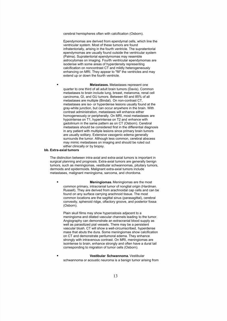
Ependmomas are derived !rom ependmal cells- ,hich line theventricular sstem. ost o! these tumors are !oundin!ratentoriall- arising in the !ourth ventricle. The supratentorialependmomas are usuall !ound outside the ventricular sstem
6<alma7. Supratentorial ependmomas ma resem%leastroctomas on imaging. &ourth ventricular ependmomas areisodense ,ith some areas o! hperdensit representingcalci!ication on noncontrast T and mildl heterogeneouslenhancing on )(. The appear to !ill the ventricles and maextend up or do,n the !ourth ventricle.
Metastases. etastases represent one
0uarter to one third o! all adult %rain tumors 6+avis7. ommonmetastases to %rain include lung- %reast- melanoma- renal cellcarcinoma- G(- and G/ tumors. et,een $C and =#J o! allmetastases are multiple 6indal7. ?n non'contrast T-metastases are iso' or hperdense lesions usuall !ound at thegra',hite 4unction- %ut can occur an,here in the %rain. @ith
contrast administration- metastases ,ill enhance eitherhomogeneousl or peripherall. ?n )(- most metastases arehpointense on T1- hperintense on T2 and enhance ,ithgadolinium in the same pattern as on T 6?s%orn7. ere%ralmetastasis should %e considered !irst in the di!!erential diagnosisin an patient ,ith multiple lesions since primar %rain tumorsare usuall solitar. Extensive vasogenic edema generallsurrounds the tumor. Although less common- cere%ral a%scessma mimic metastases on imaging and should %e ruled outeither clinicall or % %iops.
)). Extra$axial tumors
The distinction %et,een intra'axial and extra'axial tumors is important insurgical planning and prognosis. Extra'axial tumors are generall %enigntumors- such as meningiomas- vesti%ular sch,annomas- pituitar tumors-
dermoids and epidermoids. alignant extra'axial tumors includemetastases- malignant meningioma- sarcoma- and chordoma.
Meningiomas. eningiomas are the most
common primar- intracranial tumor o! nonglial origin 6ardman-)ussell7. The are derived !rom arachnoidal cap cells and can %e!ound on an sur!ace carring arachnoid tissue. The mostcommon locations are the sagittal sinus 6parasagittal7- cere%ralconvexit- sphenoid ridge- ol!actor groove- and posterior !ossa6?s%orn7.
<lain skull !ilms ma sho, hperostosis ad4acent to ameningioma and dilated vascular channels leading to the tumor. Angiograph can demonstrate an extracranial %lood suppl as
,ell as parasiti*ed pial vessels. There ma %e a persistentvascular %lush. T ,ill sho, a ,ell'circumscri%ed- hperdensemass that a%uts the dura. Some meningiomas sho, calci!icationon T and demonstrate peritumoral edema. The enhancestrongl ,ith intravenous contrast. ?n )(- meningiomas areisointense to %rain- enhance strongl and o!ten have a dural tailcorresponding to migration o! tumor cells 6?s%orn7.
-esti)ular Sch/annoma. Desti%ular
sch,annoma or acoustic neuroma is a %enign tumor arising !rom
13

8/10/2019 NCH Student
http://slidepdf.com/reader/full/nch-student 14/68
the nerve sheath o! the vesti%ular portion o! cranial nerve D(((. Assuch- the are seen onl in the cere%ellopontine angle- o!ten ,ithextension into the internal acoustic canal 6(A7. ?n T the areiso' or hpodense and enhance strongl. The %one ,indo,s masho, ,idening o! the (A. ?n )(- vesti%ular sch,annomas are,ell circumscri%ed- enhancing masses that extend into the (A6Gold%erg7.
&ituitary Adenomas. <ituitar adenomas
represent approximatel 1CJ o! all primar intracranial tumors6)ussell7. The are grouped into microadenomas 6KL 1C mm7and macroadenomas 6M 1C mm7. ost adenomas are slo,gro,ing and #CJ are endocrinologicall active- ,ith the mostcommon tpe %eing the prolactinoma 6)ussell7. icroadenomasappear hpodense on contrast enhanced T and )( %ecausethe normal pituitar enhances more strongl. acroadenomasappear as isodense intra' and suprasellar masses that enhancestrongl on T and )(. sts- hemorrhage- or necrosis maalso %e seen in the macroadenoma.
vi. Hydroce(halus
drocephalus is a condition in ,hich the ventricles %ecome enlarged and intracranial pressure %ecomes elevated.(t is commonl divided into t,o tpes: communicating- ,hich is due to the ina%ilit o! the arachnoid granulations toade0uatel a%sor% S&9 non'communicating- ,hich is caused % an o%struction o! S& !lo, ,ithin the ventricles.ommunicating hdrocephalus ma occur a!ter su%arachnoid hemorrhage i! the su%arachnoid %lood and its %'products impair the !unction o! the arachnoid villi. ?n noncontrast T and )(- these patients have smmetricenlargement o! all ventricles- ,hich is particularl noticea%le in the third ventricle and temporal horns o! the lateralventricles. Non'communicating hdrocephalus ma occur ,ith intraventricular tumors or csts. (n these cases thereis enlargement o! the ventricles proximal to the occlusion and normal si*ed ventricles distal.
vii. Cere)ral A)scess
ere%ral a%scesses are !oci o! parenchmal %acterial in!ections that result !rom contiguous spread- directinoculation- or hematogenous spread. ematogenous spread !rom an extracranial site such as the lung o!tenresults in multiple lesions- ,hereas local spread !rom mastoid air cells or paranasal sinuses usuall creates asolitar lesion 6ellar7.
The appearance on T or )( depends on the age o! the a%scess. (n the earl cere%ritis stage o! cere%ral a%scess
63 to # das7- T or )( ma sho, a small- ill'de!ined area o! mild enhancement at the gra',hite inter!ace. &rom# das to 2 ,eeks- the late cere%ritis stage develops- and T and )( sho, a thin ring o! enhancement around anecrotic center. +uring the earl and late capsule stages 6,eeks to months7- a thicker- more de!ined ring o!enhancement !orms ,ith a surrounding area o! edema 6immerman7.
f. %maging in S(inal *isease
i. S(inal Trauma
<rompt recognition o! spinal in4uries and neurologic de!icits are essential in the success!ul treatment o! the multiplin4ured patient 6ohlman7. As descri%ed previousl- plain x'ra is a !ast and eas !irst test to evaluate spinal in4uriesost !ractures and su%luxations can %e seen on plain x'ra- %ut patients ,ith point tenderness or neurologicalde!icit should undergo !urther evaluation. T o! the spine ,ith sagittal reconstruction is ver use!ul !or identi!ingthe anatomical detail o! the !ractures seen on x'ra or to !ind !ractures not seen on plain x'ra. )( is use!ul inidenti!ing spinal cord compression or contusion and discBligamentous in4ur.
The !our tpes o! spinal in4ur %ased on direction o! !orces applied to the verte%ral column are: 17 hper!lexion- 27hperextension- 37 axial loading- "7 rotational in4ur 6rant',ad*ski7. per!lexion in4uries usuall result in anterior,edging and compression !ractures o! the verte%ral %odies. (! su!!icient !lexion !orces are applied to the spine-these !ractures ma %e associated ,ith posterior ligamentous in4ur or !acet su%luxation 6locked !acets7.
perextension in4uries are common in the cervical spine- and o!ten result in !ractures o! the posterior elementssuch as the lamina- lateral masses- and !acets. &re0uentl there is rupture o! the anterior longitudinal ligament anddisruption o! the disc space. (n patients ,ith congenitall narro, spinal canals or degenerative disc disease-hperextension can cause central cord sndrome.
Axial loading in4uries are common in the cervical and thoracolum%ar segments o! the spine. A direct vertical !orce is
14
8/10/2019 NCH Student
http://slidepdf.com/reader/full/nch-student 15/68
applied to the spine as in diving or 4umping in4uries resulting in compression or %urst !ractures o! the verte%ral%odies.
)otational in4uries are usuall associated ,ith other !orces such as lateral %ending- !lexion- or extension. These!orces can result in unilateral !acet dislocations and in the cervical spine.
ii. *egenerative S(inal *isease
+egenerative spinal disease is the most commonl seen disease in most neurosurgical practices. Advances in Tand )( have %een ver important in the diagnosis o! spinal stenosis and disc herniation.aa. S(ondylosis and s(inal stenosis.
As the interverte%ral discs age- the %ecome desiccated- the annular!i%ers %ecome ,eak- and the discs %ulge or herniate. @hen patientsdevelop spondlosis- the !orm osteophtes along the discoverte%ral 4unction that can compromise the spinal canal 6)esnick7. There are t,o!orms o! spinal stenosis- congenital or ac0uired. The ac0uired !orm isusuall the result o! spinal degeneration caused % %ulging discs-osteophtes- and ligamentum hpertroph- ,hich are %est visuali*ed onT %one ,indo,s. T melograph 6T per!ormed a!ter intrathecalin4ection o! contrast7 and )( ,ill sho, compression o! the thecal sac.<atients ,ith severe prolonged stenosis ma sho, signal a%normalit,ith the spinal cord.
)). *isc )ulge and herniation.
ulging o! the interverte%ral disc is a common phenomenon in peopleover age 2C and ma %e asmptomatic 6oden7. As the annulus !i%rosisages- it %ecomes ,eaker- thus allo,ing the nucleus pulposus to %ulge%eond the verte%ral %od margins. @hile T or T melograph canaccuratel diagnose herniated discs- )( is the pre!erred test %ecause o!its a%ilit to de!ine the relationship o! disc material to S&- %one- and so!ttissue in %oth axial and sagittal planes. )( is also less invasive than Tmelograph.
iii. S(inal Tumors
Spinal tumors can %e divided into three groups- 17 extradural- 27 intradural extramedullar- and 37 intramedullarmasses.
aa. Extradural s(inal masses.
The most common extradural masses are metastases to the %one. ?therextradural masses include %enign and malignant tumors o! %one andmultiple meloma. etastases o!ten present ,ith pain- pathologic!ractures- and neurologic de!icit consistent ,ith location. ?n plain x'ra-metastases ma appear as multi!ocal- ltic lesions or verte%ral !ractures6?lcott7. T is good !or delineating the %on anatom o! osteoltic areas-and- ,ith the in4ection o! intrathecal contrast- can sho, neurologicalelement compression. ontrast )( allo,s %etter examination o! thespinal cord and so!t tissues surrounding the spine. etastatic lesions are
usuall multiple and strongl enhancing.
)). %ntradural extramedullary s(inal masses.
ost intradural- extramedullar spinal masses are either nerve sheathtumors or meningiomas. Sch,annomas and neuro!i%romas %oth originate!rom the Sch,ann cell %ut di!!er grossl and histologicall. Sch,annomasare ,ell'circumscri%ed round or dum%%ell'shape tumors that ma %e solidor cstic 6urger7. Neuro!i%romas are poorl circumscri%ed masses- ,hicho!ten have nerve !i%ers coursing through them and are more !re0uentlsolid. The imaging characteristics o! these t,o tumors are similar. ?n T-
15
8/10/2019 NCH Student
http://slidepdf.com/reader/full/nch-student 16/68
there is o!ten ,idening o! the neural !oramen. ?n T1 ,eighted )(- thelesions are iso' or hperintense and enhance strongl ,ith contrast. ?nT2 ,eighted images- the are usuall hperintense. The ,ill displace %utnot invade the spinal cord or nerve roots ,ithin the thecal sac.
The histolog o! spinal meningiomas is identical to intracranialmeningiomas. ?n )(- the are isointense on T1 and T2 ,eighted
images and enhance ,ell. A dural tail ma %e o%served.
cc. %ntramedullary masses.
(ntramedullar masses are !ound ,ithin the spinal cord parenchma. ostare gliomas- either ependmoma or astroctoma. Ependmomas arise!rom ependmal cells in the central canal. The are !ound in all segmentso! the spinal cord and ma have a %etter cleavage plane %et,een tumorand spinal cord than the astroctoma. A su%tpe that is !re0uentl !ound inthe conus medullaris- the mxopapillar ependmoma- is histologicall%enign and has a good prognosis. An ependmoma ma undergo csticdegeneration and hemorrhage 6cormick7. Their appearance isisointense to spinal cord on T1 ,eighted images- hperintense on T2- andenhance homogeneousl 6?s%orn7.
Astroctomas o! the spinal cord usuall involve multiple segments anddi!!usel ,iden the cord. The occur more !re0uentl in the cervical cord.T ma sho, thinning o! the pedicles or ,idening o! the interpediculardistance. ?n )(- the are iso' to hpointense on T1- hperintense on T2-and enhance strongl.
eferences
1. ellar AF- Sahar A- <raiss (: rain a%scess- revie, o! => cases over a period o! 3C ears. F Neurol Neurosurg <schiatr3$:5#5'5$=- 1>53.
2. indal )8- Sa,aa )- Heavens E- Hu FF: Surgical treatment o! multiple %rain metastases. F Neurosurg 5>:21C'21$- 1>>3.3. oden S+- +avis +?- +ina TS et al: A%normal magnetic resonance scans o! the lum%ar spine in asmptomatic patients. F
one Foint Surg 52:"C3'"C=- 1>>C.". ohlman - +ucker T- Hucas FT: Spine and spinal cord in4uries. (n )othman )- and Simeone &A- eds.- pp.$$1'5#5.
<hiladelphia- @.. Saunders- 1>=2.#. o**ola &G- Gorelick <- Fensen F: Epidemiolog o! intracranial hemorrhage- Neuroimaging lin N Amer 2:1'1C- 1>>2.$. rant'a,ad*ki - <ost F+: Trauma. (n Ne,ton T- <otts +G- eds.:odern neuroradiolog- vol 1- computed tomograph
o! the spine and spinal cord- pp1">'1=$- 1>=#.5. rocker - )a%in - Hevin A: linical and surgical management o! head in4ur- Neuroimaging lin N Amer 1:3=5'3>$- 1>>1.=. urger <- Scheithauer @- Dogel &S: <eripheral nerve. (n Surgical <atholog o! the Nervous Sstem and its overings-
pp. $$1'53C. Ne, Oork- hurchill Hivingstone (nc.- 1>>1.>. +avis <- udgins <A- <eterman S- o!!man F: +iagnosis o! cere%ral metastases: dou%le'dose delaed T vs. contrast'
enhanced ) imaging- AFN) 12:2>3'3CC- 1>>1.1C. +harker S)- hargava N: ilateral epidural hematoma- Acta Neurochir 6@ien7 11C:2>'32- 1>>1.11. Gold%erg (- Havi E- Atlas S@: Extral'axial %rain tumors. (n Atlas S@ 6ed.7: agnetic )esonance (maging o! the rain and
Spine- pp "23'"=5. <hiladelphia- Hippincott')aven <u%lishers- 1>>$.12. ardman F: Nonglial tumors o! the nervous sstem. (n Schochet SS Fr.- ed. The linical Neurosciences. Ne, Oork-
hurchill Hivingstone. 1>=3.13. ashimoto N- Sakaki%ara T- Oamamoto 8- et al:T,o !luid'%lood densit levels in chronic su%dural hematoma- F Neurosurg
55:31C'11- 1>>2.1". Heeds NE- Sa,aa )- DanTassel <- aman HA: (ntracranial hemorrhage in the oncologic patient- Neuroimaging lin N
Amer 2:11>'3$- 1>>2.1#. 8ell A- immerman )+- Sno, ) et al: ead trauma: comparison o! ) and T ' experience in 1CC patients- AFN)
>:$>>'5C=- 1>==.1$. acpherson - acpherson <- Fennett : T incidence o! intracranial contusion and hematoma in relation to the
presence- site- and tpe o! skull !racture- lin )adiolog "2:321'$- 1>>C.15. cormick <- Torres )- <ost 8+- Stein : (tramedullar ependmoma o! the spinal cord. F Neurosurg $2:#23'#32- 1>>C.1=. ?hman- F- eiskanen ?: Timing o! operation !or ruptured supratentorial aneursms: a prospective randomi*ed stud. F
Neurosurg =C:""C'"$- 1>>".
16
8/10/2019 NCH Student
http://slidepdf.com/reader/full/nch-student 17/68
1>. ?lcott E@- +illon@<: <lain !ilm clues to the diagnosis o! spinal epidural neoplasm and in!ection. Neuroradiol 3#:2=='2>2-1>>3.
2C. ?s%orn AG: +iagnostic Neuroradiolog. St. Houis- os%'Oear ook- (nc.- 1>>".21. ?s%orn AG: (ntracranial aneursms. (n and%ook o! Neuroradiolog- pp 5>'="- St. Houis- os%'Oear ook- (nc.- 1>>1.22. ?s%orn AG: (ntroduction to ere%ral Angiograph. <hiladelphia- arper and )o,- <u%lishers- (nc.- 1>=C.23. <alma H- elli <- antore G: Supratentorial ependmomas o! the !irst t,o decades o! li!e: long'term !ollo,'up o! 2C cases
6including t,o su%ependmomas7. Neurosurg 32:1$>'15#- 1>>3.
2". <erl F- Turski <A- asark TF: ) angiograph: Techni0ues and clinical applications. (n Atlas S@ 6ed.7: agnetic)esonance (maging o! the rain and Spine- pp 1#"5'1$1=. <hiladelphia- Hippincott')aven <u%lishers- 1>>$.2#. )esnick +: +egenerative diseases o! the verte%ral column. )adiol 1#$:3'1"- 1>=#.2$. )ussell +S- )u%enstein HF: <atholog o! Tumors o! the Nervous Sstem- #th ed- altimore- @illiams and @ilkins- 1>=>.25. DanGi4n F- Dan +onger 8F: The time course o! aneursmal hemorrhage on computed tomograph- Neuroradiol 23:1#3-
1>=2.2=. Dilla!ana T: <hsics and instrumentation. (n Hee S- )ao 8 6eds.7: ranial omputed Tomograph- pp 1'"$. cGra,'ill-
(nc.- 1>=3.2>. @ehrli &@- cGo,an F: The %asis o! ) contrast. (n Atlas S@ 6ed.7: agnetic )esonance (maging o! the rain and
Spine- pp 2>'"=. <hiladelphia- Hippincott')aven <u%lishers- 1>>$.3C. @il%erger FE- arris - +iamond +H: Auto su%dural hematoma: mor%idit- mortalit- and operative timings- F Neurosurg
5":212'1=- 1>>1.31. immerman )+- @eingarten 8: Neuroimaging o! cere%ral a%scesses. Neuroimaging lin N Amer 1:1'1$- 1>>1.
A.0. %ntracranial hy(ertension
a. Clinical Manifestation of Acute and Chronic %ntracranial Hy(ertension
i. Cere)ros(inal Fluid CSF2 &hysiology
S& is produced % choroid plexus in the lateral- third- and !ourth ventricle through an ultra!iltration process.Normall S& is produced at the rate o! C.3ccBminute 6or "CC'#CC cc a da7. S& circulates through the ventricularsstem- then do,n to the spinal su%arachnoid space- %e!ore it returns to the intracranial cavit. S& is generalla%sor%ed % the arachnoid granulation over the cere%ral convexit and returned to the vascular sstem. Someminor a%sorption occurs near the cranial nerve root sheaths. /nder extremel high pressure- S& ma %e resor%edover the ventricular ependma in proportion to the pressure gradient. The total volume o! S& in an adult is a%out
1#Ccc- meaning that the S& volume is turned over more than 3 times a da. ?nl a%out "Ccc o! the total %odS& volume is !ound in the ventricular sstem.
+e!inition: Normal intracranial pressure 6(<7 is generall considered to %e less than 2Cmm g. The onro'8elliedoctrine states that the total intracranial volume- consisting o! %rain- %lood- cere%rospinal !luid 6S&7- and otherpathological entit- remains !ixed. The cere%ral per!usion pressure 6<<7 is de!ined as ean Arterial <ressure6A<7 minus the (ntracranial pressure 6(<7. P<<LA<'(<Q (n management o! increased intracranial pressure-an e!!ort is made to maintain the (< %elo, 2C mmg and the << a%ove 5C mmg.
ii. Etiology3 Elevated intracranial (ressure is the final common (ath/ay of a variety of intracranial (athology.
The most common cause o! elevated (< is traumatic %rain in4ur. Trauma can lead to hematoma
6epidural- su%dural- and intraparenchmal7 !ormation ,ith resultant mass e!!ect on the involvedhemisphere or lo%e. ?ther parenchmal lesions such as cere%ral contusion can also exert signi!icantpressure on the surrounding %rain tissue. Edema and hperemia 6pro%a%l due to loss o! cere%ralautoregulation7 can cause !urther increase (<- leading to secondar in4ur a!ter the initial insult.
oth primar and metastatic neoplasm o! the %rain can cause %reakdo,n o! %lood %rain %arrier- edema-
mass e!!ect- and increased (<. Generall- %enign neoplasms do not cause (< elevation unless their si*e%ecomes su%stantial- or i! the %lock S& !lo,. alignant neoplasms- %ecause o! the %reakdo,n o! %lood%rain %arrier- readil cause edema- mass e!!ect- and (< elevation. ?%struction o! the ventricular sstem% a mass lesion can lead to o%structive hdrocephalus and intracranial hpertension.
drocephalus can occur in t,o !orms- communicating or o%structive. ommunicating hdrocephalus
generall occurs as a result o! prior in!ection- hemorrhage- or arachnoid irritation. Normal pressurehdrocephalus 6N<7 in the elderl population ma occur ,ithout the a%ove predisposing !actors. )arel-a cere%rospinal !luid 6S&7 producing neoplasm such a choroid plexus papilloma can cause S&overproduction and result in communicating hdrocephalus. ?%structive hdrocephalus is the result o!S& o%struction at strategic points o! the S& path,a. ommon causes include intraventricular or
17

8/10/2019 NCH Student
http://slidepdf.com/reader/full/nch-student 18/68
periventricular tumors- %rain herniation causing trapping o! the lateral ventricle- intraventricularhemorrhage- or a0ueductal stenosis. A com%ination o! these t,o tpes o! hdrocephalus can occur.
Denous sinus throm%osis can occur as the result o! sstemic hpercoagula%le states or trauma. )isk
!actors include the use o! oral contraceptive and certain connective tissue disorders- including lupuserthematosis. S& a%sorption depends on !lo, across the arachnoid granulations. ecause o! thereduced venous drainage- increased intracranial %lood volume leads to decreased S& resorption-resulting in intracranial hpertension.
(diopathic intracranial hpertension is a disease most commonl !ound in adult !emale patients. ?%esit isa predisposing !actor. eadache and progressive loss o! visual acuit are the primar smptoms. hronicelevation o! (< causes papilloedema- ,hich could %e detected on !undoscopic examination.
Sstemic hpertension can cause %reakdo,n o! the %lood %rain %arrier and causes hpertensive
encephalopath. This can increase (< %ecause o! higher A< and cere%ral %lood volume.
b. Clinical &resentation of %ntracranial Hy(ertension
Acute intracranial hpertension most !re0uentl presents ,ith a constellation o! smptoms including headache- alteredmental status 6level o! consciousness7- nauseaBvomiting- and occasionall sudden death. (n addition to the a%ovesmptoms- chronic intracranial hpertension can present ,ith cranial nerve palsies 6third or sixth cranial nerve7- ataxia-memor distur%ance- personalit changes- or urinar incontinence. oth acute and chronic intracranial hpertension cancause sei*ures.
c. Emergency Management of %ntracranial Hy(ertension
ead position: Simple head elevation can promote venous return !rom the head and reduce (<. (t should %e notedthat head elevated does lo,er the mean arterial pressure suppling the %rain- possi%l negating the %ene!iciale!!ects o! increased venous drainage.
perventilation: Though ver e!!ective in reducing (< through its cere%ral vasoconstrictive e!!ect- hperventilationcauses reduced cere%ral %lood !lo, 6&7. This can lead to secondar hpoxic in4ur. ?nl mild to moderatedegree o! hperventilation is recommended. 6p?2 M 3Cmmg7 The patient need to %e mechanicall ventilated tohave desired controlled ventilator e!!ects. The e!!ect o! hperventilation is generall transient. 6"='52 hours7
+iureticsBperosmolar agents: annitol can %e used to dra, ,ater out o! the %rain tissue % osmotic !orces. Thisreduces %rain tissue volume and (<. ?ther agents that can %e used include !urosemide and urea. These agentsshould generall %e ,eaned o!! to prevent re%ound cere%ral edema.
Sedation and paraltic agents. Agitation and muscle tremorsBspasms can arti!iciall elevate (<. en*odia*epines-narcotics- and i! necessar- chemical paraltic agents should %e emploed.
S& drainage ,ith an external ventricular drain- or much more rarel ,ith a S& shunt- can lo,er the (<. ar%iturate coma can %e induced ,ith pento%ar%ital to reduce cere%ral %lood !lo, 6&7 and cere%ral meta%olic
re0uirement o! ?2 6)?27. lose arterial pressure monitoring is mandator %ecause o! its cardio'depressivee!!ect. ost clinicians advocate continuous electroencephalographic 6EEG7 monitoring to assess the end point o!%urst suppression o! EEG.
Avoid hpotension in order to maintain <<. Evacuate the causative intracranial mass lesion- including hematoma or neoplasm. A depressed skull !racture can
also cause elevated (<. The underling in4ur ma not %e reversi%le.
.1. *iagnosis and Management of Head Trauma
a. 4nderstand and Assign the !lasgo/ Coma Scale.
The Glasgo, oma Scale ,as developed in order to standardi*e the neurologic assessment o! patients ,ith head in4ur. (t,as speci!icall designed to %e easil per!ormed %ased upon clinical data- and to have a lo, rate o! intero%server varia%ilit.(n addition- the Glasgo, oma Scale score is correlated ,ith outcome in that patients ,ith a higher Glasgo, oma Scalescore have a statisticall %etter outcome than patients ,ith a lo,er Glasgo, oma Scale score.
Glasgo, oma Scale:
&oints 5est Eye (ening 5est -er)al es(onse 5est Motor es(onse
$ ' ' ?%es
# ' ?riented Hocali*es pain
18

8/10/2019 NCH Student
http://slidepdf.com/reader/full/nch-student 19/68
" Spontaneous on!used @ithdra,s to pain
3 To speech (nappropriate &lexor 6decorticate7
2 To pain (ncomprehensi%le Extensor 6decere%rate7
1 None None None
%.
The Glasgo, oma Scale score is determined % adding the values !or ee opening- ver%al response- and motor response.<ossi%le values range !rom 3 to 1#. Note that this scale rates the %est response onl. (n patients ,ho are intu%ated- in ,homassessment o! %est ver%al response cannot %e per!ormed- notation o! this is made in the Glasgo, oma Scale score %adding a t to the end o! the score. (n patients ,ho are intu%ated- the %est possi%le score ,ould there!ore %e 11t. ertainnumerical values o! the Glasgo, oma Scale have particular clinical signi!icance. <atients ,ith a Glasgo, oma Scale o! 5or less are considered to %e comatose. <atients ,ith a Glasgo, oma Scale score o! = or less are considered to havesu!!ered a severe head in4ur.
c. ecogni6e the &resentation of 5rain Herniation Syndromes in the Setting of Trauma3
+istortion o! the midline %rain structures secondar to %rain trauma ma lead to speci!ic com%inations o! signs andsmptoms ,hich are collectivel re!erred to as herniation sndromes. (n general- these smptoms result !rom the distortiono! midline %rain structures secondar to %rain s,elling- hdrocephalus- or intracranial mass lesions. @hile numerous
herniation smptoms have %een descri%ed- the t,o most commonl seen sndromes in the setting o! trauma are uncalherniation and tonsillar herniation.i. 4ncal herniation3
ost o!ten results !rom a laterall placed mass displacing the %rain stem contralaterall and pushing the uncus o!the temporal lo%e mediall over the tentorial edge.
Earl Sign: (psilateral pupillar dilation
Hate Signs:
omplete ipsilateral third nerve pals
Hoss o! consciousness ontralateral hemiplegia 6secondar to mass7
(psilateral hemiplegia 6secondar to compression o! contralateral cere%ral peduncle against edge o!
tentorium P8ernohan’s notchQ7 &laccid paralsis
ii. Tonsillar herniation3
)esults !rom do,n,ard displacement o! the cere%ellar tonsils through the !oramen magnum- causing compressiono! the cervicomedullar 4unction. &re0uentl secondar to posterior !ossa mass. a %e precipitated % lum%arpuncture in the presence o! such a mass.
Signs: ead tiltBneck pain
)espirator arrest
Hoss o! consciousness
&laccid paralsis
d. %nitiate Management of Elevated %ntracranial &ressure in Head Trauma3
rain in4ur a!ter acute head trauma can %e divided into t,o categories. The !irst categor is primar in4ur- ,hich is su!!eredat the time o! impact. The second categor is secondar in4ur- ,hich ma occur at an time !rom that point !or,ard. ?ne o!the most important causes o! secondar %rain in4ur in head trauma is !elt to %e elevated intracranial pressure 6(<7.
(n treatment o! the patient ,ith head trauma- the possi%ilit o! elevated intracranial pressure should al,as %e considered.anagement o! the traumati*ed patient %egins ,ith the primar surve and resuscitation.
19
8/10/2019 NCH Student
http://slidepdf.com/reader/full/nch-student 20/68
Steps involved include:
Air,a patenc ,ith cervical spine control: (t is important to esta%lish the presence o! a patent air,a. (! such anair,a is not present- one should %e esta%lished. This ma include the use o! chin li!t or 4a, thrust- clearance o!!oreign %odies- endotracheal intu%ation- or creation o! a surgical air,a. (t is important to consider that the cervicalspine ma %e in4ured and that it should %e maintained in a neutral position during an o! the a%ove maneuvers.
reathing control. The chest should %e examined and the rate and depth respirations determined. (nade0uac maindicate the need !or mechanical ventilation. igh concentrations o! oxgen should %e administered. <neumothoraxshould %e treated.
irculator and hemorrhage control. The 0ualit- rate- and regularit o! the pulse should %e determined. Sights o!ma4or hemorrhage should %e identi!ied and treated.
+isa%ilit. A %rie! neurologic examination should %e per!ormed. The Glasgo, oma Scale should %e determined.<upils should %e assessed !or si*e- e0ualit- and reaction.
+uring the secondar surve- a more complete neurologic examination should %e per!ormed including evaluation o! thepatient’s strength- sensation- re!lexes- and remaining cranial nerves.
&ollo,ing the secondar surve- appropriate imaging studies should %e o%tained. (n a patient ,ith o%vious cranio!acialtrauma- mechanism o! in4ur su!!icient to produce %rain in4ur- or a distur%ed level o! consciousness- a T scan o! the head,ithout contrast should %e per!ormed. The presence o! !ractures- !oreign %odies- space occuping lesions- or hemorrhage
should %e noted- as ,ell as the ventricular si*e.
(ntracranial pressure monitoring should %e considered in the !ollo,ing situations:
<atients ,ith an a%normal admission T scan and Glasgo, oma Scale score o! 3 to = a!ter cardiopulmonar resuscitation.
?r
<atients ,ith a normal head T ,ith a Glasgo, oma Scale score o! 3 to = and the presence o! t,o or more o! the !ollo,ing!eatures: Age over "C ears- unilateral or %ilateral motor posturing- sstolic %lood pressure less than >C mmg.
The current pre!erred modalit !or monitoring o! intracranial pressure is the placement o! a ventricular catheter6ventriculostom7. /se o! !i%eroptic or strain gauge pressure monitors can %e considered.
Elevated intracranial pressure has %een sho,n to have de!inite prognostic implications in a patient ,ith severe %rain in4ur.(n addition- it is generall held that treatment o! elevated intracranial pressure ma improve outcome in the patient ,ithsevere %rain in4ur. The currentl recommended threshold !or treatment o! elevated intracranial pressure is 2C to 2# mmg.(nterpretation and treatment o! intracranial pressure %ased on this threshold value should %e corro%orated % !re0uentclinical examination and assessment o! the cere%ral per!usion pressure. urrent recommendations suggest that << should%e a minimum o! $C to 5C mm o! mercur. (t is important to consider that- ,hile A< is an important determining !actor in the<<- it has also %een sho,n that lo, A< is an independent predictor o! poor outcome.
A!ter elevated intracranial pressure has %een identi!ied- treatment should %e initiated. (n general- treatment should proceedin a step,ise !ashion- %eginning ,ith the least onerous treatment modalities. Escalation o! treatment should proceed onla!ter !ailure o! less onerous modalities. The suggested hierarch o! treatment includes the !ollo,ing therapeutic modalities:
od positioning. The head should %e elevated 3C degrees. The neck should %e maintained in a neutral position.ompression o! the 4ugular veins should %e avoided.
aintenance o! homeostasis. Euvolemia should %e esta%lished. Arterial %lood gases should %e measured ,ith thegoal o! maintaining the <?2 in the >C to 1CC'mm mercur range and the <?2 in the 3# to "C'mm mercur range.
ild sedation. This is most !re0uentl carried out using a com%ination o! %en*odia*epines andBor narcotics. External ventricular drainage. At this stage- placement o! a ventriculostom- i! not done previousl !or (<
measurement- should %e considered. The reservoir is generall placed # to 1C cm a%ove ear level or- alternativel-placed at ear level and opened at regular intervals.
/se o! osmotic diuretics. annitol is the most commonl used osmotic diuretic. (t is most !re0uentl given in a doseo! C.2# to 1.C gramsBkg (.D. over 1# minutes. (! the e!!ect o! treatment ,ith annitol is transient- the dose ma %e
20
8/10/2019 NCH Student
http://slidepdf.com/reader/full/nch-student 21/68
repeated- so long as the serum osmolalit remains less than or e0ual to 32C m?smBH and the patient remainseuvolemic.
oderate hperventilation. At this stage- moderate hperventilation to a <?2 o! 3C to 3# mm o! mercur can %econsidered.
Second tier therapies including %ar%iturate therap. At this stage- %ar%iturate therap can %e considered.<ento%ar%ital is the most commonl used %ar%iturate !or the treatment o! re!ractor intracranial hpertension.)ecommended loading dose is 1C mgBkg over 3C minutes !ollo,ed % a maintenance dose o! 1 mgBkg per hour as
a continuous in!usion. The dose is then titrated to achieve serum <ento%ar%ital levels in the range o! 3 to " mgBdl oran electroencephalographic pattern o! %urst suppression. <otential complications o! this modalit o! treatment arenumerous- ,ith hpotension %eing the primaril dose'limiting toxicit. ar%iturate treatment o! re!ractor intracranialhpertension has %een sho,n to decrease mortalit %ut has not %een sho,n to improve neurologic outcome.
The a%ove treatment algorithm assumes that all signi!icant cranial space'occuping lesions have %een appropriatelsurgicall treated. +uring implementation o! the a%ove algorithm- it is important to consider the possi%ilit that a ne,intracranial lesion has developed. ecause o! this- a repeat T scan o! the head should %e considered prior to escalation o!therap.
Note that the a%ove treatment algorithm does not include the use o! corticosteroids. The use o! glucocorticoids is notrecommended !or improving outcome or reducing intracranial pressure in patients ,ith severe head in4ur. The routine useo! prophlactic anticonvulsant medication is not recommended !or the prevention o! posttraumatic sei*ures in the patient,ithout a premor%id sei*ure disorder. The use o! anticonvulsants ma %e considered- in the !irst 5 das a!ter in4ur- toprevent earl posttraumatic sei*ures in patients ,ho are at high risk. <hentoin and car%ama*epine are the most commonl
used agents. &inall- nutritional support o! %rain in4ured patients should %e instituted ,ithin 5 das o! in4ur.
ecogni6e and %nitiate Management of Concussion 5rain Contusion and *iffuse Axonal %n,ury.
i. Concussion.
ere%ral concussion is a di!!use %rain in4ur thought to %e caused % acceleration'deceleration in4ur to the %rain.ere%ral concussion is a spectrum o! in4uries- ranging !rom mild to severe.
ild concussion is de!ined as no loss o! consciousness ,ith transient neurologic distur%ance.
oderate concussion is de!ined as loss o! consciousness ,ith complete recover occurring in less than #
minutes.
Severe cere%ral concussion is de!ined as unconsciousness lasting greater than # minutes.
Evaluation and treatment o! patients ,ith cere%ral concussion remains controversial. @orkup includes completehistor and phsical examination- ,ith neurological examination. ?ther tests include cervical spine x'ras and otherradiographs as indicated- %lood alcohol level and urine drug screen- and T scan o! the head in all patients exceptthose ,ho are completel asmptomatic and neurologicall normal.
Treatment o! patients ,ith cere%ral concussion ,ho have a Glasgo, oma Scale score o! 1" or 1# is usuallexpectant. ost patients should undergo hospital admission ,ith !re0uent neurological examinations. Theseinclude all patients ,ith an a%normal T scan- histor o! loss o! consciousness- decreased or decreasing level o!consciousness- severe headache- under the in!luence o! alcohol or drugs- have phsical examination evidence o!S& rhinorrhea or otorrhea- signi!icant associated in4uries- no relia%le companion at home- una%le to returnpromptl- or are amnestic !or the in4ur. ?nl those patients ,ho do not mani!est an o! the prior criteria should %econsidered !or discharge !rom the hospital. (! hospital discharge is considered- an o! the previousl listed signs orsmptoms should prompt a return to the hospital. A ,ritten head in4ur ,arning sheet should %e issued.Neurosurgical !ollo,'up should %e scheduled.
(n addition to !re0uent neurologic examinations- patients admitted a!ter cere%ral concussion ma %e treated ,ithTlenol or ver mild doses o! narcotic pain medication !or headache. Nausea and vomiting- ,hich are !re0uentlpresent a!ter mild or severe concussion- should %e treated ,ith non'phenothia*ine antiemetic medication.+ischarge ma %e considered ,hen the patient is neurologicall normal- nausea and vomiting has ceased- andheadache has ceased or is ade0uatel controlled.
All patients ,ho have su!!ered a cere%ral concussion should %e counseled regarding the possi%le occurrence o!post concussive sndrome. <rominent smptoms include headache- mild impairment o! memor- dse0uili%rium-and alteration o! mood. These smptoms usuall regress spontaneousl %ut ma persist !or ,eeks to months.
21
8/10/2019 NCH Student
http://slidepdf.com/reader/full/nch-student 22/68
ii. *iffuse axonal in,ury3
+i!!use axonal in4ur is the most severe !orm o! di!!use %rain in4ur. (t is !elt to %e the most common cause o!prolonged posttraumatic coma that is not due to mass lesion or ischemia. +i!!use axonal in4ur is characteri*ed %!ocal hemorrhagic lesions involving the corpus callosum- rostral mid %rain- superior cere%ellar peduncles com%ined,ith microscopic evidence o! ,idespread axonal damage. <atients ,ith di!!use axonal in4ur !re0uentl mani!estdecorticate or decere%rate posture and autonomic ds!unction in addition to their prolonged coma. Elevated
intracranial pressure is !re0uentl a%sent. are is primaril supportive. (n patients ,ith prolonged coma- theprognosis is generall poor- ,ith a #CJ mortalit and ,ith an approximatel 2#J incidence o! !avora%le outcome.
iii. Cere)ral Contusion.
A cere%ral contusion is a !ocal %rain in4ur caused primaril % impact o! the %rain sur!ace and the %on ridges o!the calvarium. ere%ral contusions are !re0uentl !ound in the region o! the !rontal poles- anterior skull %ase-ad4acent the sphenoid ridge- and at the temporal poles. ?ther locations include the cere%ellar hemispheres and theoccipital poles. A characteristic pattern o! cere%ral contusion called the coup and contrecoup in4ur is !re0uentlseen. The coup contusion occurs at the sight o! impact and the contrecoup contusion occurs in the %rain at thepoint diametricall opposite the point o! impact.
Treatment o! cere%ral contusion is guided % the neurologic examination. The patient should %e admitted to thehospital !or o%servation and !re0uent neurologic examinations. (ntracranial pressure monitoring and treatmentshould %e instituted !or the comatose patient. @hile surgical treatment- consisting o! de%ridement o! the contused
%rain tissue- is not routinel recommended- it can %e considered in patients ,ith re!ractor intracranialhpertension.
e. ecogni6e and %nitiate Management of Acute Su)dural and E(idural Hematoma7 %ncluding Surgical %ndications.
iv. Su)dural hematoma.
(n the acute traumatic su%dural hematoma- %lood collects %et,een the dura mater and sur!ace o! the %rain. ostcommonl- the %leeding results !rom tearing o! %ridging veins located over the convexit o! the %rain sur!ace.leeding originating !rom a small cortical arter represents the second most common source. Associatedintracranial lesions- particularl cere%ral contusions- are !ound in at least #CJ o! patients ,ith acute traumaticsu%dural hematoma.
<atients presenting ,ith acute traumatic su%dural hematoma ma range !rom normal to deepl comatose. /nlike
the epidural hematoma- to %e descri%ed later- the most common presentation o! the patient ,ith an acute traumaticsu%dural hematoma is that o! a patient rendered unconscious at the time o! in4ur ,ithout regaining consciousnessprior to presentation.
Neurologic !indings ma %e secondar to mass e!!ect- elevated intracranial pressure- andBor associated %rainin4uries. Elevated intracranial pressure is a common !inding and evaluation and treatment o! elevated intracranialpressure- as outlined a%ove- should %e instituted immediatel. The acute traumatic su%dural hematoma is mostcommonl treated surgicall. ?%servation should onl %e considered in those patients ,ith small 6less than 1C mmthick7 su%dural hematomas ,ho are neurologicall intact. ecause the acute traumatic su%dural hematomaconsists o! solid %lood clot- %urr hole drainage is inade0uate !or relie! o! mass e!!ect. A craniotom should %eper!ormed and all easil assessa%le %lood clots should %e removed. <ostoperativel- patients !re0uentl mani!estintracranial hpertension- and this should %e treated as outlined a%ove.
?utcome a!ter treatment o! acute traumatic su%dural hematoma is related to a num%er o! !actors- particularlpreoperative neurologic status. ortalit ranges !rom greater than 5#J in those patients ,ho present ,ith a GS
o! 3 to # to minimal in patients ,ho present ,ith GS o! 12 to 1#. Time !rom in4ur to surgical decompression-elevated intracranial pressure- and associated %rain lesions also have a detrimental e!!ect on outcome.
v. E(idural hematoma.
(n the acute epidural hematoma- %lood collects %et,een the inner sur!ace o! the calvarium and the dura mater.ost commonl- the acute epidural hematoma results !rom !racture o! the skull- stripping the dura mater !rom theinner ta%le o! the skull- and causing laceration o! meningeal vessels or dural sinuses. leeding !rom the middlemeningeal arter is responsi%le !or man supratentorial epidural hematomas. (n contrast to the su%duralhematoma- the patient har%oring an acute epidural hematoma ma have an initial loss o! consciousness- !ollo,ed% a %rie! lucid interval- !ollo,ed % progressive neurologic decline. This presentation is seen in approximatel 1B3
22

8/10/2019 NCH Student
http://slidepdf.com/reader/full/nch-student 23/68
o! patients having an acute epidural hematoma.
Treatment o! acute epidural hematoma is generall surgical. <atients ,ith small epidural hematomas not traversinga meningeal arter vein or ma4or sinus- ,ho present greater than $ hours a!ter in4ur ma %e considered !or anonoperative therap. o,ever- admission ,ith !re0uent neurologic examination and a lo, threshold !or repeat Tscanning is mandator. &or all other patients- operative treatment is recommended. As ,ith the acute su%duralhematoma- the acute epidural hematoma is comprised o! a solid %lood clot. There!ore- %urr hole drainage is
inade0uate !or removing the intracranial mass. A craniotom should %e per!ormed and the entire %lood clotevacuated. A!ter evacuation- the dura mater should %e opened to inspect the su%dural space. &re0uentl- asu%dural hematoma ma %e encountered ,hich ,as not o%vious on the preoperative T scan. <ostoperativel- thepatient is monitored !or signs o! elevated intracranial pressure. Elevated intracranial pressure- i! detected- is treatedas outlined a%ove.
As in the acute traumatic su%dural hematoma- outcome a!ter treatment o! acute epidural hematoma is related to avariet o! !actors- the most important %eing preoperative neurologic status. ortalit ranges !rom less than 1#J inthose patients ,ho present ,ith GS o! less than = to ver lo, in patients ,ho present ,ith GS = to 1#.
!. ecogni6e and %nitiate Management of &enetrating Trauma %ncluding !unshot 8ounds.
<enetrating in4uries include all in4uries ,here the scalp and skull are violated % !oreign o%4ects including knives- sticks-pencils- arro,s- and %ullets. Ho, velocit in4uries- such as those produced % a kni!e %lade- produce %rain in4ur along thetract o! the kni!e. igh velocit in4uries- such as those produced % a %ullet- produce %oth local in4ur along the tract o! the
%ullet- as ,ell as remote in4ur in the cavit produced in the ,ake o! the %ullet.
The si*e o! the cavit produced % a %ullet is related to its kinetic energ- as ,ell as its shape. The kinetic energ isproportional to the mass o! the %ullet as ,ell as the s0uare o! its velocit. The shape o! the %ullet not onl a!!ects the velocit%ut also its a%ilit to trans!er its kinetic energ to the %rain tissue. ullets that tum%le or de!orm on impact trans!er a greaterproportion o! their kinetic energ to %rain tissue and thus produce a more severe in4ur.
A!ter resuscitation and primar surve- an evaluation o! the patient ,ith a penetrating in4ur includes a thorough phsicalexamination and neurological examination. The location o! entr and exit ,ounds should %e ,ell documented. Extent o!tissue loss should like,ise %e documented. All patients ,ho have had penetrating %rain in4ur should undergo a T scan o!the head. oronal T scan o! the head should %e considered in patients ,ith involvement o! the anterior skull %ase.ere%ral angiograph should %e considered in patients ,here vascular in4ur is suspected. ere%ral angiograph should %egiven particular consideration ,here the tract o! the in4ur passes close to a ma4or vascular structure or in patients ,ith asigni!icant su%arachnoid hemorrhage or delaed hematoma.
As in patients ,ith severe non'penetrating %rain in4ur- elevated intracranial pressure should %e suspected. The algorithm !othis is descri%ed a%ove. (n addition- in patients ,ith penetrating %rain in4ur- intracranial pressure monitoring is indicated,hen it is not possi%le to assess the neurological examination accuratel or ,hen the need to evacuate a mass lesion isunclear.
As ,ith the open depressed skull !racture- most penetrating %rain in4uries re0uire surgical treatment.
Entrance and exit ,ounds. Treatment o! small %ullet entrance ,ounds ,here the scalp is not devitali*ed and thepatient has no signi!icant intracranial patholog ma %e treated ,ith local ,ound care and closure. ore extensive,ounds ,ith nonvia%le scalp- %one- or dura ma re0uire operative de%ridement %e!ore primar closure and duralgra!ting. @atertight dural closure is recommended. This includes patients ,ith signi!icant !ragmentation o! the skull.
(ntraparenchmal lesions. (ntraparenchmal lesions resulting in signi!icant mass e!!ect ma re0uire de%ridement o!necrotic %rain tissue and removal o! easil accessi%le %one !ragments. This includes patients ,ith signi!icantintracranial hematomas.
(n4uries resulting in communication %et,een an open'air sinus and the intracranial space should %e closed in a,atertight !ashion.
)outine removal o! %one !ragments or missile !ragments remote !rom the entr site is not recommended. Dascular in4uriesdetected on arteriograph ma re0uire surgical or endovascular treatment as indicated. ere%rospinal !luid leaks ,hich donot close spontaneousl or ,hich do not resolve a!ter the primar surger should %e treated ,ith temporar S& diversion.Those ,hich do not respond to temporar S& diversion ma re0uire surgical treatment. <atients ,ith penetrating %rainin4ur should %e administered %road'spectrum anti%iotics prophlacticall. The duration o! this treatment is some,hatcontroversial. &inall- the use o! anti'sei*ure prophlaxis in the !irst ,eek o! penetrating %rain in4ur is recommended to
23

8/10/2019 NCH Student
http://slidepdf.com/reader/full/nch-student 24/68
prevent earl posttraumatic sei*ures. As ,ith severe non'penetrating %rain in4ur- long'term prophlactic treatment ,ithanticonvulsants to prevent late posttraumatic sei*ures is not recommended.
ecogni6e and 4nderstand the &rinci(les of Management of (en7 Closed7 and 5asilar S+ull Fractures %ncluding
Cere)ros(inal Fluid 9ea+ in Chronic Su)dural Hematoma in Children and Adults.
i. (en s+ull fractures.
The treatment o! skull !ractures ,ith overling laceration is primaril %ased on ,hether the !racture is depressed ornon'depressed. ?pen non'depressed !ractures ma %e treated ,ith inspection- cleansing- and scalp suturing ,ithan accepta%l lo, rate o! in!ection. ?pen depressed skull !ractures present a signi!icant risk o! in!ection. The aregenerall treated ,ith operative irrigation- de%ridement- and removal o! the depressed !ragments. &re0uentl- this,ill necessitate !urther surgical procedures to correct the resulting cosmetic de!ormit.
ii. Closed fractures.
losed skull !ractures re0uire no speci!ic treatment. As noted a%ove- the patient har%oring a skull !racture is atincreased risk o! !ormation o! epidural hematoma. <atients har%oring this in4ur should %e treated as !or thosepatients ,ith severe concussion- unless smptoms o! elevated intracranial pressure develop. Traditionall- closeddepressed skull !ractures ,ere treated surgicall to elevate the depressed !ragments. (t has since %een sho,n thatthis practice does not result in a decreased incidence o! posttraumatic epileps and has there!ore %eena%andoned. Treatment o! patients ,ith depressed- closed !ractures- except ,here there is a signi!icant cosmetic
de!ormit- should %e treated as outlined a%ove !or closed- non'depressed !ractures.iii. 5asilar s+ull fractures.
A %asilar skull !racture involves the cranial %ase. The most common sites are the !loor o! the anterior cranial !ossaand the temporal %one. Evidence that a patient ma have su!!ered a %asilar skull !racture includes raccoon eesand %attle sign 6%ruising posterior to the ear- possi%l including the mastoid process7. ost %asilar skull !racturesdo not re0uire treatment in the a%sence o! associated %rain in4ur. The are a sign that a signi!icant %lo, has %eendelivered to the cranium. ost patients ,ith signi!icant %asilar skull !racture should %e o%served ,ith !re0uentneurological examinations !or 2" hours a!ter the in4ur. ?ccasionall- complications o! %asilar skull !racture maresult. These include posttraumatic cere%rospinal !luid leakage- optic nerve in4ur- and !acial nerve in4ur. Theincidence o! posttraumatic cere%rospinal !luid leak a!ter a closed head in4ur is approximatel 2J. The mostcommon sites o! cere%rospinal !luid egress are the nose 6S& rhinorrhea7 and ear 6S& otorrhea7. ost traumaticS& leaks resolve ,ith nonoperative treatment including head elevation. (t is important to insure that the S&leakage does resolve- as persistent S& rhinorrhea carries ,ith it an approximatel 2#J risk o! meningitis- as ,ellas a risk o! tension pneumocephalus.
iv. &osttraumatic CSF lea+.
Treatment o! the posttraumatic S& leak ,hich does not resolve ,ith head elevation includes repeated highvolume lum%ar punctures- as ,ell as continuous catheter S& drainage. There is a !inite risk o! inducing tensionpneumocephalus using those modalities. &or leaks ,hich persist despite these measures- surgical treatmentshould %e considered.
v. Chronic su)dural hematoma.
As descri%ed a%ove- the chronic su%dural hematoma is a collection o! %lood and %lood products %et,een the innersur!ace o! the dura mater and the outer sur!ace o! the %rain. /nlike the acute traumatic su%dural hematoma- onseto! smptoms and detection o! the su%dural hematoma occurs much later in the course. The hallmark o! the chronicsu%dural hematoma is %lood products- visuali*ed on the T scan- ,hich are isodense or hpointense ,ith respectto %rain tissue. (t is theori*ed that- like the acute traumatic su%dural hematoma- the source o! the %lood products islacerated %ridging veins resulting !rom acceleration'deceleration !orces applied to the skull. @hile this is usuall theresult o! trauma- trauma ma %e mild- remote- and not remem%ered % the patient or !amil. an patients ,hopresent ,ith a chromic su%dural hematoma have one or more risk !actors. These risk !actors include:
Advanced age and cere%ral atroph.
ale sex.
oagulopath.
(ntracranial hpotension secondar to S& shunting procedures
hronic alcoholism.
hronic su%dural hematomas tend to enlarge slo,l over time. The most common mechanism o! enlargement
24
8/10/2019 NCH Student
http://slidepdf.com/reader/full/nch-student 25/68
appears to %e multiple episodes o! re'%leeding- ho,ever- other mechanisms ma also %e important.
Smptoms at presentation include smptoms o! increased intracranial pressure 6headache- papilledema-decreased level o! consciousness7- as ,ell as those o! hemispheric mass e!!ect 6hemiparesis- dsphasia- tremors-and dstonia. Sei*ures occur occasionall.
Treatment o! chronic su%dural hematoma is generall surgical. @hile treatment ,ith prolonged %edrest- head
elevation- and osmotic diuretics has produced accepta%le results- the risks o! prolonged immo%ili*ation in thesegenerall de%ilitated patients !re0uentl out,eigh the %ene!its.
There are multiple surgical options !or the treatment o! chronic su%dural hematoma including:
T,ist drill hole and drainage.
ultiple %urr holes and drainage.
raniotom and drainage ,ith stripping o! mem%ranes.
ematomas that are not !ull li0ue!ied ma re0uire craniotom. (n addition- recurrence rate a!ter evacuation o!chronic su%dural hematoma is su%stantial.
<ostoperativel- elevated intracranial pressure is unusual. <atients are generall maintained supine ,ith minimal
elevation o! the head !or at least 2" hours to allo, some re'expansion o! the %rain. ecause o! the su%stantial rateo! recurrence- neurosurgical !ollo,'up is essential.
vi. Chronic su)dural hematoma in children.
Hike the adult chronic su%dural hematoma- %lood and %lood products- in the childhood chronic su%dural hematoma-accumulate %et,een the inner sur!ace o! the dura and the %rain sur!ace. (n children- it is particularl important- andsometimes di!!icult- to distinguish %et,een the chronic su%dural hematoma and the su%dural hgroma- in ,hichcere%rospinal !luid collects %et,een the dura and arachnoid mem%rane. &urthermore- lo,'densit !luid ma collect%et,een the arachnoid and pia mater secondar to communicating hdrocephalus.
@hile trauma is the most common cause o! chronic su%dural hematoma in children- the possi%ilit o! non'accidental trauma 6child a%use7 must al,as %e considered- particularl in children less than 2 ears o! age.oagulopath remains a ma4or risk !actor.
(n children- presenting smptoms are usuall those o! elevated intracranial pressure including vomiting- letharg-irrita%ilit- or increase in the head si*e. Sei*ures are also seen.
The treatment o! chronic su%dural hematoma in children is di!!erent than that o! adults. /nlike adult patients- veroung patients ma have an open !ontanel- !acilitating percutaneous aspiration o! the su%dural hematoma. &ailureo! this treatment modalit generall re0uires placement o! a su%dural to peritoneal shunt.
eferences"Suggested eading3
1. ullock )- hesnut ) et al. Guidelines !or the management o! severe head in4ur. Fournal o! Neurotrauma 13: $3>'53"-1>>$
2. ullock )- hesnut ) et al. Guidelines !or the management o! severe traumatic %rain in4ur. Fournal o! Neurotrauma 15:"#1'$25- 2CCC
3. ommittee on Trauma o! the American ollege o! Surgeons: Advanced Trauma Hi!e Support Student anual. hicago- (H- American ollege o! Surgeons- 1>>C
". Eisen%erg - Aldrich E& 6eds7. anagement o! ead (n4ur. Neurosurger linics o! North America 2: 2#1'#C1- 1>>1#. El'8adi - 8au!man 6eds7. hronic Su%dural ematoma. Neurosurger linics o! North America 11: 3>#'#$5- 2CCC$. &lorin )E. anagement and prognosis o! penetrating %rain in4ur. Fournal o! Neurotrauma 6in press7
5.#. *iagnosis and Management of 5rain Tumor and A)scess
a. Neuroe(ithelial tumors
25

8/10/2019 NCH Student
http://slidepdf.com/reader/full/nch-student 26/68
i. Astrocytoma
Astroctomas- ,hich arise !rom astroctes- are the most common primar %rain tumor. Astroctomas arehistologicall graded %ased on cellularit- anaplasia- mitotic !igures- endothelial proli!eration and necrosis. @ell'di!!erentiated astroctomas 6@+A7 have mild hpercellularit and minimal nuclear pleomorphism. The tpicalloccur in children and oung adults ,ho present ,ith a sei*ure or headaches. ?n T or )( scan the usuallappear as a non'enhancing mass lesion. Anaplastic Astroctomas 6AA7 have moderate cellularit and nuclear
pleomorphism ,ith mitotic activit. oderate endothelial proli!eration can %e present %ut no necrosis. These lesionsare tpicall !ound in mid'li!e as enhancing lesions ,ith mass e!!ect. The o!ten present ,ith sei*ures- headachesandBor !ocal neurological !indings. Glio%lastoma multi!orme 6G7 is characteri*ed % hpercellularit- dramaticnuclear pleomorphism- endothelial proli!eration- mitotic !igures and necrosis. These patients are usuall olderadults ,ho present ,ith headaches- sei*ures or !ocal neurologic !indings. ?n T or )( scan a G tpicallappears as an irregular ring'enhancing lesion ,ith signi!icant mass e!!ect and edema.
Survival correlates ,ith grade: @+A #'1C ears- AA 2'3 ears and G 1'1.# ear. Astroctomas can initiallpresent as a lo,'grade tumor and su%se0uentl convert to a higher grade. +e novo AA and G tumors occur as,ell.
Treatment is %ased on grade. ontrovers exists !or the optimal treatment o! @+A. ost agree that a @+A in non'elo0uent %rain ,ith mass e!!ect should %e resected. A stereotactic %rain %iops !or diagnosis is an option !orsmptomatic lesions in areas o! the %rain ,here open surger carries an increased risk o! causing a de!icit.)adiation is controversial in sta%le tumors %ut is o!ten used i! gro,th is demonstrated. AA and G are tpicall
treated % surgical resection !ollo,ed % radiation and N/ chemotherap. Surger is indicated !or diagnosis- torelieve mass e!!ect- and- possi%l- to decrease the tumor %urden. AA or G in the motor strip- language areas orother elo0uent %rain regions are o!ten %iopsied rather than resected %ecause o! the high risk o! surger in theseareas. (t has %een di!!icult to prove that the extent o! tumor resection has an e!!ect on patient survival. As a rule-surger is never curative. N/ impregnated ,a!ers implanted in the tumor %ed a!ter resection o! a recurrent AA oG have proven to %e e!!icacious. )ecurrence ,ithin a centimeter or t,o o! the resection site is tpical regardlesso! the treatment given a!ter surgical resection. Surger !or recurrent AA and G is controversial. ost surgeonsagree that reoperation is indicated !or the relie! o! headaches or neurological de!icits due to mass e!!ect.
<athologicall- several tpes o! astroctomas exist- such as !i%rillar- protoplasmic and gemistoctic astroctomas.Glio%lastomas also have giant cell and gliosarcoma variants.
A separate group o! astroctoma is the 4uvenile piloctic astroctoma 6F<A7. This group is either not graded orconsidered Grade C. F<A are distinct %ecause the %ehave in a more %enign !ashion and ,hen completel resectedcan %e cured % surger alone. The are tpicall discrete cstic lesions ,ith an enhancing mural nodule.istologicall- F<A are composed o! loose and dense regions o! stellate astroctes. These have )osenthal !i%ersthat indicate slo, gro,th. F<A are tpicall !ound in children and oung adults. The tend to occur in the cere%ellarhemisphere- optic nerve- hpothalamus and %rainstem. ere%ellar F<A o!ten present ,ith signs o! increasedintracranial pressure 6headache- nausea- vomiting7 due to hdrocephalus. F<A also can present ,ith cere%ellards!unction such as gait ataxia or ipsilateral extremit dsmetria. )arel- F<A can undergo malignant degeneration.Su%arachnoid seeding does occur rarel ,ith F<A and pro%a%l carries a poorer prognosis. This has %een seen,ith hpothalamic tumors. ?ptic nerve F<A and @+A tumors are associated ,ith neuro!i%romatosis tpe ((.
<leomorphic xanthoastroctomas are astroctic neoplasms !ound in oung adults ,ith a long histor o! sei*ures.The are usuall super!icial in the cere%ral cortex and ma consist o! a mural nodule associated ,ith a cst. Theare tpicall slo, gro,ing %ut malignant trans!ormation does occur. Su%ependmal giant cell astroctomas aretpicall !ound at the !oramen o! onro in patients ,ith tu%erous sclerosis.
Gliomatosis cere%ri is a condition ,here there is di!!use in!iltration o! the entire %rain ,ith an astroctic tumor.
ii. ligodendroglial tumors
The ma4orit o! oligodendrogliomas present in oung adulthood ,ith the onset o! sei*ures. )adiographicall-calci!ications are tpical. The classic histologic appearance is o! homogeneousl appearing cells ,ith a !ried eggappearance and chicken ,ire vessel pattern. Similar to @+A- patients can have long term survival. alignanttrans!ormation does occur. These tumors are called anaplastic oligodendrogliomas. (n general- surgical resection isrecommended ,hen possi%le !or diagnosis- to relieve mass e!!ect- and resect as much tumor as sa!el as possi%le.)adiation therap is controversial- %ut is pro%a%l %ene!icial. <D 6procar%a*ine- N/- R vincristine7 chemotheraphas %een sho,n to %e %ene!icial in the treatment o! oligodendrogliomas.
26

8/10/2019 NCH Student
http://slidepdf.com/reader/full/nch-student 27/68
iii. E(endymal tumors
Ependmomas tpicall arise !rom the lining o! the ventricular sstem and usuall occur in children and oungadults. The !loor o! the !ourth ventricle is a common location. Ependmomas tpicall present ,ith hdrocephalusand increased (<. <resenting smptoms include nausea- vomiting- headache- gait ataxia- diplopia and vertigo.Ependmomas have a signi!icant potential !or S& seeding and thus drop metastasis. omplete surgicalresection has %een sho,n to improve survival and should %e attempted i! there is minimal %rainstem invasion.
<ostoperative radiation and chemotherap is usuall administered. No clear consensus exists !or gradingependmomas %ut the term anaplastic ependmoma is sometimes used !or more malignant appearing tumors.<aradoxicall- intramedullar spinal cord ependmomas- ,hich are histologicall identical- ma %e cured % surgeralone.
xopapillar ependmomas are a variant o! ependmomas. This tumor occurs in the conus or !ilum terminale o!the spinal cord. omplete resection is pro%a%l curative. Su%ependmomas occur in anterior lateral ventricles orposterior !ourth ventricle. The are %enign slo, gro,ing tumors that are tpicall !ound incidentall at autops.o,ever su%ependmomas can cause hdrocephalus !rom o%struction o! cere%rospinal !luid path,as.Smptomatic or enlarging tumors should %e removed. (n elderl patients insertion o! a D< shunt is a via%le option i!o%struction o! S& path,as is present.
iv. Mixed gliomas
ixed gliomas occur and appear histologicall as a com%ination o! neoplastic oligodendroctes and astroctes.
These tumors are re!erred to as oligo'astroctomas or anaplastic oligo'astroctomas. ?!ten the name is shortenedto the dominant cell tpe onl.
v. Choroid (lexus tumors
Tumors o! the choroid plexus are called choroid plexus papillomas 6<<7. (n children less than 2 ears o! age- theusuall are located in the lateral ventricle and present ,ith hdrocephalus. (n adults << usuall occur in the !ourthventricle- !oramen o! Huschka or cere%ellopontine angle. The treatment is surgical resection- though elderlpatients ,ith an asmptomatic cere%ellopontine angle tumor ma %e !ollo,ed ,ith serial imaging studies.)ecurrence should %e treated aggressivel ,ith reoperation- ,hen possi%le- due to a !avora%le prognosis. ?ne ort,o percent o! choroid plexus tumors are carcinomatous. horoid plexus carcinoma carries a poor prognosis.
vi. Neuronal and mixed Neuronal$glial tumors
Ganglioctoma is a tumor composed o! large a%normal mature neurons. The are primaril supratentorial ,ith themost common location %eing the temporal lo%e. The ma4orit o! patients are in their !irst t,o decades o! li!e.Surgical resection- ,hen complete- is curative. Daria%le radiographic !eatures occur- ranging !rom an enhancingmural nodule to a ring enhancing mass ,ith calci!ications.
+splastic ganglioctoma o! the cere%ellum 6Hhermite +uclos +isease7 is a non'neoplastic mass o! hpertrophicgranular cell neurons ,hich expands the cere%ellar !olia. These tumors can cause mass e!!ect and hdrocephalusand tpicall occur in oung adults. )esection 6total or su%total7 andBor shunting are therapeutic options.
+sem%roplastic neuroepithelial tumors 6+NET7 are a hamartomatous- supratentorial- predominantl temporallo%e lesion composed primaril o! glial cells. The usuall present ,ith sei*ures. )adiographicall these tumorso!ten lack edema and have a multinodular appearance. (nner ta%le skull erosion or de!ormation ma %e present.
Gangliogliomas are tumors consisting o! large mature neurons and a neoplastic glial component. This tumor a!!ectspatients o! all ages ,ith the ma4orit diagnosed in oung adults ,ho o!ten have a long histor o! sei*ures. Surgicalresection even i! su%total can %e curative. Surger is recommended !or diagnosis and- on occasion- !or control o!sei*ures.
vii. &ineal tumors
<ineal tumors are tumors arising !rom pineoctes. The ,ell'di!!erentiated pineoctoma occurs in mid'li!e as adiscrete contrast enhancing mass in the posterior third ventricleBpineal region. The poorl di!!erentiatedpineo%lastoma has a similar location and enhances ,ith contrast %ut sho,s signs o! local invasion and is prone toS& dissemination. The complex o! pineo%lastoma and %ilateral retino%lastoma is called trilateral tumor. (n generalpineoctomas have a good prognosis ,hile pineo%lastomas ,ith su%arachnoid spread are aggressive and carr apoor prognosis. <ineal tumors o!ten present ,ith hdrocephalus due to o%struction o! the a0ueduct o! Slvius.ompression o! the dorsal mid%rain % a pineal tumor can result in <arinaud’s sndrome o! pupillar mdriasis-paralsis o! upga*e- and convergence retractorius.
27

8/10/2019 NCH Student
http://slidepdf.com/reader/full/nch-student 28/68
viii. Em)ryonal tumors
Neuro%lastoma is a small cell neoplasm ,ith neuro%lastic di!!erentiation arising in the deep cere%ral hemisphereso! oung children 6K# rs. o! age7. A variant is the ganglioneuro%lastoma that has a preponderance o! ganglionappearing cells.
The ol!actor neuro%lastoma 6esthesioneuro%lastoma7 is a neuro%lastic tumor arising !rom the nasal epithelium
,ith cri%i!orm plate involvement. There is a %imodal age distri%ution in adolescents and older adults. <atientspresent ,ith nasal o%struction and or epistaxis. omplete surgical resection ,ith com%ined cranio'!acial resectionis the treatment o! choice- ,ith a generall !avora%le prognosis. Ad4uvant radiation therap is generallrecommended.
Ependmo%lastoma is a rare small cell em%ronal neoplasm ,ith prominent ependmo%lastic rosettes. (t tpicalloccurs in the cere%rum o! children less than !ive ears o! age. (ts propensit !or craniospinal dissemination o!tenleads to death ,ithin a ear.
)etino%lastoma is a retinal neoplasm that occurs in children K 3 ears o! age. ?phthalmoscopic exam is diagnosticgiving the characteristic ,hite re!lex. These tumors have %oth hereditar 6earlier onset and %ilateral7 and sporadic!orms. Surgical resection ,ith or ,ithout radiation can %e curative.
edullo%lastoma or primitive neuroectodermal tumor 6<NET7 o! the cere%ellum is a small cell neoplasm %elieved toarise !rom the external granular laer o! the cere%ellum. This tumor arises in the vermis o! children and oung
adults although cases in older patients have %een reported. )adiographicall- these are homogeneousl enhancingmasses o! the cere%ellar vermis. S& tumor seeding can produce drop metastases- even at the time o! diagnosis.Surgical resection- com%ined ,ith radiation and chemotherap- ma lead to signi!icant long'term survivals. )arel-metastasis to %one- lmph nodes and lung have %een reported. Dariants include desmoplastic medullo%lastoma-medullomo%lastoma- and melanoctic medullo%lastoma. edullomo%lastoma occurs in children- ,ith apropensit !or %os. ere%ral 6supratentorial7 and spinal <NET’s occur- %ut ,ith much less !re0uenc.
). Tumors of cranial and s(inal nerves
i. Sch/annomas
Sch,annomas 6neurinoma- neurilemmoma7 are tumors composed o! Sch,ann cells that arise along cranial orspinal nerves. The vesti%ular sch,annoma 6acoustic neuroma7 is pro%a%l the most common sch,annoma andarises tpicall !rom the superior vesti%ular nerve. Desti%ular sch,annomas tpicall present ,ith tinnitus andsensori'neural hearing loss. &acial num%ness !ollo,s ,hen the tumor reaches approximatel 2.# cm. (psilateralcoordination di!!iculties and mild !acial nerve ,eakness tpicall do not occur until the tumor diameter is greater
than 3 cm. )adiographicall- these tumors enhance ,ith contrast and extend into the internal auditor canal.omplete surgical resection is curative. earing and !acial nerve preservation is dependent on tumor si*e andpreoperative level o! nerve !unction. (n older patients or poor surgical candidates- stereotactic radiosurger is ane!!ective treatment !or tumor si*e K 2.#cm. Some proponents o! radiosurger !eel that it should %e used as theprimar treatment !or all %ut the largest tumors. ilateral vesti%ular sch,annomas occur ,ith neuro!i%romatosistpe ((.
ii. Neurofi)romas
Neuro!i%romas are a nerve sheath tumor composed o! Sch,ann cells- !i%ro%lasts and perineural cells. This tumorcan occur in isolation or in associated ,ith neuro!i%romatosis. This tumor ma %e solitar- plexi!orm or occur as amixed neuro!i%romaBsch,annoma. These tumors arise !rom nerves in the su%cutaneous tissue or in theneuro!oramena. lassic spinal tumors have a dum%%ell appearance ,hen the extend across the neuro!oramena.Surgical resection is indicated in smptomatic lesions. alignant trans!ormation in neuro!i%romas is rare %ut should%e suspected ,ith increasing si*e or pain.
iii. Malignant (eri(heral nerve sheath tumors
alignant peripheral nerve sheath tumors: neurogenic sarcoma- anaplastic neuro!i%roma- and malignantsch,annoma are rare malignant tumors arising !rom the non'neural elements o! nerves. These have a poorprognosis ,ith death resulting ,ithin the ear. omplete resection is usuall not !easi%le.
c. Tumors of the meninges
eningioma 6su%tpes: meningothelial- transitional- !i%rous- psammomatous- angiomatous- microcstic- secretor- clear cell-choroid- lmphoplasmacte'rich- metaplastic variants- atpical- anaplastic7.
28

8/10/2019 NCH Student
http://slidepdf.com/reader/full/nch-student 29/68
eningiomas are tpicall solitar- %enign- slo, gro,ing- extra'axial tumors arising !rom arachnoid cap cells in the craniumand spine. The most common locations are parasagittal- convexit- tu%erculum sella and sphenoid ridge. A small percentagema %e intraventricular. eningiomas rarel occur in children %ut ,hen the do there is a predilection !or the posterior !ossaand ventricle. Hess than !ive percent o! meningiomas are malignant- characteri*ed % %rain invasion and increased mitoticactivit. eningiomas are most common in middle age and elderl ,omen. ommon smptoms include headache- sei*ures-,eakness- and mental status changes. &ocal neurologic de!icits on presentation depend on the site o! origin o! the tumor.)adiographicall these tumors are ,ell circumscri%ed- homogeneousl enhancing lesions. There ma %e hperostosis o! the
underling skull. There is o!ten a tail o! dural enhancement at the edge o! the tumor a!ter contrast administration on imagingstudies. The primar treatment o! smptomatic surgicall accessi%le tumors is surgical resection. Surger i! complete iso!ten curative. Harge lesions ma re0uire em%oli*ation o! intra'operativel inaccessi%le vascular suppl prior to surgicalresection to decrease intraoperative %leeding. Small or asmptomatic meningiomas in older individuals can %e !ollo,ed andtreatment recommended i! gro,th is demonstrated. )adiation therap andBor radiosurger are treatment alternatives !orrecurrent tumors or i! surger carries an increased risk o! complications. There is an association %et,een meningiomas andneuro!i%romatosis tpe 2 and an a%normalit in the long arm o! chromosome 22.
d. Cysts and tumor$li+e lesions
Numerous csts and tumor'like lesions can occur in the %rain including )athke’s cle!t cst- epidermoid cst- dermoid cst-colloid cst o! the third ventricle- enterogenous cst 6neuroenteric cst7- neuroglial cst- other csts- lipoma- granular celltumor 6choristoma- pituictoma7- hpothalamic neuronal hamartoma- nasal glial heterotopias.
e. Tumors of the anterior (ituitary
<ituitar adenomas are slo, gro,ing- %enign tumors that arise in the anterior pituitar gland. <ituitar tumors are dividedinto hormone secreting tumors and non'secretors. Tumors less than 1 cm in diameter are re!erred to as microadenomas,hile tumors larger than 1 cm are called macroadenomas. <rolactinomas are pituitar adenomas that secrete the hormoneprolactin. <rolactinomas o!ten present ,ith amenorrhea in ,omen and loss o! li%ido in men. The primar treatment !orprolactinomas is medication- usuall %romocrptine analogues. romocrptine o!ten results in a decrease in tumor si*e %utdoes not kill tumor cells. (t usuall must %e continued !or li!e or tumor recurrence is the rule. Adenomas that secrete gro,thhormone produce gigantism i! present %e!ore pu%ert and acromegal i! present a!ter pu%ert. AT secreting adenomasresult in ushing’s disease. percortisolism is characteri*ed clinicall % centripetal o%esit- moon !acies- %u!!alo hump-glucose intolerance- hpertension and impaired ,ound healing. Non'secreting macroadenomas ,ith suprasellar extensioncan compress the optic chiasm. ompression o! the optic chiasm !rom %elo, o!ten produces a %itemporal hemianopsia.Spontaneous hemorrhage or in!arction o! a pituitar adenoma is re!erred to as pituitar apoplex and can result in suddenvisual loss or hpocortisolism. Emergent surger is sometimes necessar !or pituitar apoplex.
The primar treatment !or patients ,ith secreting pituitar adenomas producing ushing’s disease- acromegal- orsmptomatic non'secreting tumors is surgical resection. Surger is tpicall per!ormed through the transsphenoidal route.
avernous sinus invasion % adenoma cells o!ten makes a surgical cure di!!icult. )ecurrent tumors or those ,ith cavernoussinus invasion can %e treated ,ith re'operation or radiation therap.
f. Metastasis
etastases are the most common %rain tumor. Tumors that !re0uentl spread to the %rain- in order o! decreasing incidenceare: lung- %reast- skin- colon- and kidne. ommonl- lung- throid- renal cell and melanoma metastases can %ecomehemorrhagic. The treatment o! solitar lesions is surgical resection !ollo,ed % radiation therap. &or patients ,ith multipleasmptomatic lesions- surger is reserved !or diagnosis onl. &or patients ,ith controlled sstemic disease %ut multiple %rainmetastases- a large smptomatic accessi%le lesion could %e considered !or resection. @hole %rain radiation is generallgiven !or multiple %rain metastases.
g. 5rain A)scess
rain a%scesses arise % several mechanisms including hematogenous spread- penetrating trauma- surger- or local spread!rom the paranasal sinuses- mastoid air cells or emissar veins. The peak incidence is in oung men due to the occurrence
o! middle ear and paranasal sinus in!ections in addition to congenital heart disease. ?ther predisposing !actors include?sler'@e%er')endu sndrome ,ith pulmonar arteriovenous !istulae- endocarditis- congenital heart disease- dental ,orkand immunosuppression. Smptoms consist o! headache- !ever- sei*ures andBor neurological de!icit. The ma4orit o! %raina%scesses are solitar.
Aero%ic and anaero%ic %acterial a%scesses occur. A%scess cultures in one third o! patients gro, multiple organisms.ommon organisms %ased on the site o! origin include Streptococcus species !rom the !rontalBethmoid sinus9 acteroides!ragilis !rom chronic mastoiditisBotitis9 Staph. aureus or entero%acteriacea !ollo,ing penetrating trauma or surger9 Strep.viridans and Strep. pneumonia in cases o! congenital heart disease9 Staph aureus and Strep. pneumonia in cases o!endocarditis. (n immunosuppressed patients- toxoplasma gondii- nocardia- mco%acteria- east and !ungal a%scesses occur?utside the /S- tu%erculomas- csticercosis- echinococcus- schistosomiasis and strongoloidiasis are more common.
29

8/10/2019 NCH Student
http://slidepdf.com/reader/full/nch-student 30/68
(n the !irst stage o! %rain in!ection- there is in!lammation o! the %rain- termed earl cere%ritis. This stage occurs in the !irst 3'#das a!ter inoculation. The T scan appearance o! cere%ritis is that o! an ill'de!ined hpodense contrast enhancing area.This coalesces to a late cere%ritis stage during das "'13 ,ith irregular rim enhancement. This is !ollo,ed- at approximatelda 1"- % a collagen reticulum encapsulation ,ith a necrotic center 6earl capsule stage7 ?n T scan or )( scan thisappears as a ring enhancing mass o!ten ,ith the a%scess ,all !acing the ventricle appearing the thinnest. The !inal stage isthe late capsule stage in ,hich there is a three'laer capsule: an outer gliotic laer- a middle collagenous laer and an inner
granulation laer. These can persist !or months on imaging studies %e!ore ultimate resolution.
Anti%iotics are the mainsta o! treatment in all cases. Empiric treatment o! a presumed %acterial a%scess re0uires coverage!or %oth aero%es and anaero%es. Surger is usuall indicated to con!irm the diagnosis o! an a%scess and !or culture andsensitivit o! speci!ic organisms. Stereotactic aspiration is the treatment o! choice. Aspiration ma need to %e repeated%e!ore resolution occurs. ?!ten t,o to three ,eeks o! anti%iotic treatment are needed %e!ore a si*e decrease is seen onimaging studies. (n general " $ ,eeks o! intravenous anti%iotics are o!ten used- !ollo,ed % a period o! oral anti%iotics.<atients ,ith nocardia a%scesses- or patients in ,hom treatment has !ailed a!ter the third aspiration- should considersurgical resection ,hen accessi%le. ?!ten- aspiration alone can treat signi!icant mass e!!ect and prevent rupture o! thea%scess into the ventricular sstem. Dentricular rupture o! a %acterial %rain a%scess is o!ten !atal.
5.0. *iagnosis and Management of Headaches
a. Ma,or Causes of %ntracranial Hemorrhage.
i. :no/ the ma,or causes of intracranial hemorrhage3 -asculo(athy in the aged hy(ertension andamyloidosis27 aneurysm7 vascular malformation7 tumor7 and coagulo(athy.
aa. Hy(ertension.
Spontaneous intracere%ral hemorrhage ma occur ,ith %oth acute andchronic hpertension. Acute hpertension su!!icient to producespontaneous intracere%ral hemorrhage is sometimes seen ,ith eclampsiaand drug intoxication ,ith smpathomimetic drugs. hronic hpertensionma lead to vascular changes o! the %asal per!orating arteries including!ormation o! harcot'ouchard aneursms. The most common locations!or these hemorrhages are 6in decreasing order o! !re0uenc7:
asal ganglia
Thalamus <ons
ere%ellum
ere%ral ,hite matter
rainstem
)). Amyloid angio(athy.
This is characteri*ed % a deposition o! %eta amloid protein ,ithin themeningeal and cortical vessels. The clinical course is characteri*ed %multiple lo%ar intracere%ral hemorrhages. )isk !actors include advancedage and +o,n’s sndrome. +e!initive diagnosis re0uires pathologicexamination.
cc. Aneurysm.
ere%ral aneursms result !rom ,eakening o! the ,all o! ma4orintracranial arteries. These aneursms are generall saccular- though!usi!orm aneursms also occur. Saccular aneursms usuall occur at%ranch points along the arterial tree. @hen a saccular aneursm ruptures-it most commonl produces su%arachnoid hemorrhage. (ntracere%ral-intraventricular- and su%dural hemorrhage are also seen. ommon siteso! aneursm rupture include 6in decreasing order o! !re0uenc7
30

8/10/2019 NCH Student
http://slidepdf.com/reader/full/nch-student 31/68
Anterior communicating arter
<osterior communicating arter
iddle cere%ral arter
asilar arter
Derte%ral arter
ultiple aneursms are seen in approximatel one'!ourth o! patients. )isk!actors include hpertension- smoking- !amil histor- and collagenvascular disease.
dd. -ascular malformation.
Dascular mal!ormations include the high !lo, arteriovenous mal!ormation-cavernous angioma- venous angioma- and capillar telangiectasias.
The high !lo, arteriovenous mal!ormation
results !rom a direct communication %et,een the arterial %loodsuppl and draining veins ,ithout normal interposed capillaries.These are usuall visi%le on arteriograph. Saccular aneursmare !re0uentl seen on !eeding vessels. (ntraparenchmal
hemorrhage is most commonl seen- though su%arachnoid andsu%dural hemorrhage ma occur.
avernous angiomas consist o! irregularl
!ormed vascular channels- ,ithout intervening %rainparenchma. &lo, is usuall lo, and these lesions- there!ore- donot routinel mani!est themselves on arteriograph. emorrhage-,hen it occurs- is tpicall intraparenchmal. &amil histor is arisk !actor.
Denous angiomas result !rom a cluster o!
normal medullar veins draining into a central enlarged venouschannel. The arterial sstem is uninvolved. emorrhage isin!re0uent and usuall intraparenchmal. an are associated,ith cavernous angiomas.
apillar telangiectasias are clusters o! dilated
capillaries ,ith lo, !lo,. The are not visi%le on arteriograph.The are sometimes associated ,ith ?sler'@e%er')endusndrome- Houis'arr sndrome- %urn'ason sndrome- andSturge'@e%er sndrome.
ee. Tumor.
oth primar and metastatic tumors ma hemorrhage. ?! the malignantprimar %rain tumors- glio%lastoma multi!orme most commonl presents,ith hemorrhage. @hile lung carcinoma is the most common hemorrhagiccere%ral metastasis- melanoma- choriocarcinoma- and renal cellcarcinoma are the most likel metastases to present ,ith cere%ralhemorrhage. ost hemorrhages are intraparenchmal.
ff. Coagulo(athy.
Even in the a%sence o! underling cere%ral patholog- coagulopath-either secondar to underling medical disease or iatrogenicall induced-increases the risk !or intracranial hemorrhage. <articularl in elderlpatients- su%dural hematoma is the most common mani!estation.(ntraparenchmal hemorrhage ma also occur- particularl in the settingo! anticoagulation a!ter cere%ral in!arction.
). ecogni6e the Sym(toms and Signs of Su)arachnoid7 Cere)ral7 and Cere)ellar Hemorrhage.
31

8/10/2019 NCH Student
http://slidepdf.com/reader/full/nch-student 32/68
i. Su)arachnoid hemorrhage.
The most common smptom o! su%arachnoid hemorrhage is explosive onset o! severe headache. The headache isdescri%ed % the patient as the ,orst headache o! m li!e. The patient ma complain o! neck pain and sti!!ness-as ,ell as photopho%ia. Ho, %ack pain ma also %e present. Nausea and vomiting ma %e present. There ma %ea histor o! transient loss o! consciousness.
Signs o! su%arachnoid hemorrhage include nuchal rigidit- positive 8ernig’s sign- andBor positive rud*inski’s sign. A depressed level o! consciousness ma also %e present. ?cular mani!estations include hemorrhages andpapilledema 6secondar to elevated intracranial pressure7. &ocal neurologic de!icit- particularl third nerve palsipsilateral to the site o! aneursm rupture- ma %e present and in some instances ma help to locali*e theaneursm.
ii. %ntracere)ral hemorrhage.
eadache ma %e present a!ter intracere%ral hemorrhage. <atients ,ith nondominant hemorrhages ma complaino! ,eakness or num%ness on their nondominant side.
Signs o! intracere%ral hemorrhage are tpicall those o! hemispheric neurologic de!icit. <atients ,ith dominant'sided lesions are !re0uentl !ound to have hemiparesis and hemianesthesia. +sphasia is usuall present- thoughthe patient ma present ,ith !rank aphasia. <atients ,ith nondominant lesions tpicall have contralateralhemiparesis and hemianesthesia. @hile speech is !re0uentl normal- neglect ma %e pro!ound.
(! the hemorrhage is large- the patient ma present ,ith signs o! elevated intracranial pressure including depressedlevel o! consciousness- decorticate or decere%rate posturing- or !laccid are!lexia.
iii. Cere)ellar Hemorrhage.
The a,ake patient ,ith cere%ellar hemorrhage ma complain o! severe headache- ,hich ma %e locali*ed to thesu%occipital or upper cervical region.
Smptoms include those o! cere%ellar and lo,er %rainstem ds!unction- as ,ell as hdrocephalus- ,hich is!re0uentl present. Signs o! cere%ellar ds!unction include ipsilateral dsmetria- nstagmus ,hen looking to theside o! the lesion- and di!!iculties ,ith speech. Ho,er %rainstem ds!unction tpicall mani!ests as di!!icult ,iths,allo,ing. (n the presence o! hdrocephalus- signs o! elevated intracranial pressure ma ensue. These include adecreased level o! consciousness- decorticate or decere%rate posturing- and !laccid are!lexia.
c. A((ly *iagnostic Tools in Evaluation of Acute Headache CT7 M%7 and 9um)ar &uncture2.
(n the patient ,ho presents ,ith the ,orst headache o! m li!e a su%arachnoid hemorrhage should al,as %e suspected. A!ter initial resuscitation and complete neurologic examination- head T ,ithout contrast is mandator. (n addition toveri!ing the presence or a%sence o! su%arachnoid %lood- the T scan should %e care!ull scrutini*ed !or other intracranialpatholog- including hdrocephalus.
(! the T scan is positive !or su%arachnoid hemorrhage- the patient should undergo cere%ral arteriograph. The arteriogramshould %e scrutini*ed !or the location o! the aneursm- as ,ell as !or an associated lesions including additional unrupturedaneursms- arteriovenous mal!ormations- and the presence or a%sence o! vasospasm.
(! the initial head T is negative !or su%arachnoid hemorrhage- and the patient has a histor that is highl suspicious !orsu%arachnoid hemorrhage- a lum%ar puncture should %e per!ormed. A small needle should %e utili*ed and onl a smallvolume o! !luid should %e ,ithdra,n. Non'clotting %lood- xanthochromia- or a red %lood cell count greater than 1CC-CCC,hich does not drop signi!icantl !rom the !irst to the last tu%e are all highl suggestive o! su%arachnoid hemorrhage. (! thelum%ar puncture meets an o! these criteria- the patient should undergo arteriograph as descri%ed a%ove.
(! the cere%ral arteriogram is negative- in the !ace o! a T scan indicative o! su%arachnoid hemorrhage or a positive lum%arpuncture- the patient should %e admitted to the hospital !or o%servation. At this point- an )( scan o! the %rain ,ith and,ithout contrast should %e considered. )( arteriograph- i! availa%le- should %e included. The purpose o! this stud is torule out other structural causes o! su%arachnoid hemorrhage including angiographicall occult vascular mal!ormations andtumor. (! no %leeding source is identi!ied ,ith the )( scan- most clinicians recommend repeating the arteriogram in 5 to 1"das. ?ccasionall- an intracranial aneursm that ,as o%scured % mass e!!ect- throm%osis- or vasospasm- ma %edetected.
d. 4nderstand the Natural History and 5road Treatment Strategies of %ntracranial Aneurysms and -ascularMalformations.
i. %ntracranial aneurysms $ natural history.
32

8/10/2019 NCH Student
http://slidepdf.com/reader/full/nch-student 33/68
aa. u(tured intracranial aneurysms.
(n the patient ,ho survives their initial aneursm rupture- the most seriouscomplication is re%leeding. The rate o! re%leeding is approximatel "J inthe !irst 2" hours and decreases to 1 to 2J per da !or the !irst t,o,eeks. A!ter the !irst t,o ,eeks- the rate o! re%leeding drops toapproximatel 3J per ear. A second potential serious complication o!
aneursmal su%arachnoid hemorrhage is a vasospasm. Dasospasm isde!ined as delaed narro,ing o! large and medium si*e arteries at the%ase o! the %rain. (! severe- this ma lead to cere%ral in!arction. The risko! smptomatic cere%ral vasospasm is maximum %et,een " and 11 daso! su%arachnoid hemorrhage. The incidence o! vasospasm- detected %angiograph- is approximatel 5CJ. The incidence o! smptomaticvasospasm 6resulting in neurologic de!icit7 is approximatel 2# to 3CJ.The ma4or risk !actor !or the development o! vasospasm is the amountand location o! su%arachnoid %lood visuali*ed on the T scan. This isgraded using the &isher sstem.
!rou( 5lood on CT Scan
1 No %lood detected
2 +i!!use or vertical laers K 1mm thick
3 Hocali*ed clot andBor vertical laer ML 1mm thick
"(ntracere%ral or intraventricular clot ,ith di!!use or no SA
)). 4nru(tured intracranial aneurysm $ natural history.
?ccasionall aneursms are discovered prior to rupture. Again- theprimar risk is rupture. The availa%le in!ormation suggests that the annualrate o! rupture !or an unruptured intracranial aneursm is %et,een 1Jand 3J per ear. The risk o! rupture is a!!ected % several !actors. Hargeraneursms- particularl those greater than or e0ual to 1C mm in diameter-have a higher rate o! rupture. Aneursms located in the posteriorcommunicating arter- verte%ro%asilarBposterior cere%ral arter- and in the
%asilar tip are more likel to rupture.
cc. Treatment strategies for intracranial aneurysms.
The goal o! intracranial aneursm treatment is to prevent %leeding orre%leeding. Additionall- in the patient ,ith a ruptured intracranialaneursm- treatment is also instituted to lessen the risk o! smptomaticvasospasm.
(n patients ,ith su%arachnoid hemorrhage due to aneursm rupture-outcome is related to the modi!ied unt'ess grade. igher'gradepatients have a statisticall poorer outcome.
!rade *escri(tion
C /nruptured aneursm
1 Asmptomatic- or mild headache and slightnuchal rigidit
1aNo acute meningealB%rain reaction- %ut ,ith!ixed neurologic de!icit
2ranial nerve pals 6e.g. (((- (D7- moderate tosevere headache- nuchal rigidit
33

8/10/2019 NCH Student
http://slidepdf.com/reader/full/nch-student 34/68
3 ild !ocal de!icit- letharg- or con!usion
"Stupor- moderate to severe hemiparesis- earldecere%rate rigidit
#+eep coma- decere%rate rigidit- mori%undappearance
Add one grade !or serious sstemic disease 6e.g. TN-+- severe atherosclerosis- ?<+7- or severevasospasm on arteriograph
The patient ,ith aneursmal su%arachnoid hemorrhage should %eadmitted to the neurological intensive care unit. Ade0uate oxgenationand respiration should %e assured. The patient should %e maintainednormotensive. pertension should not %e treated unless the sstolic%lood pressure rises a%ove 1$C mm o! mercur. drocephalus- ,hich ispresent in at least 1#J o! patients ,ith su%arachnoid hemorrhage- should%e treated ,ith cautious external ventricular drainage i! smptomatic.)outine use o! external ventricular drainage is controversial. ere%ralselective calcium channel %lockers lessen the risk o! smptomaticvasospasm and are started at this time. /se o! anti'!i%rinoltic agents and
corticosteroids ma %e considered.
Treatment o! the ruptured intracranial aneursm should at this point %eentertained. +irect surgical exclusion o! the aneursm !rom the circulation6craniotom and clipping7 remains the gold standard. Endovascularo%literation o! the aneursm through the use o! detacha%le coils ma %econsidered in certain situations 6di!!icult or unclippa%le aneursms-medicall unsta%le patient7. The long'term results o! this modalit o!treatment are unkno,n and currentl under stud.
Treatment o! unruptured aneursms takes into account %oth the risk o!rupture- as ,ell as age o! the patient- severit and progression o!smptoms- and treatment alternatives. ?lder patients have a statisticallshorter li!e span and there!ore lo,er cumulative risk o! rupture. )ecentrecommendations suggest that smptomatic intradural aneursms o! allsi*es should %e considered !or treatment. /nruptured aneursms o! allsi*es in patients ,ith su%arachnoid hemorrhage due to another treatedaneursm should %e considered !or treatment. Treatment !or unrupturedsmall 6less than 1C mm7 aneursms is not generall recommended exceptin special circumstances. These include aneursms near the 1C mm si*e-those ,ith daughter sac !ormation- and in patients ,ith !amil histor o!aneursm or aneursmal su%arachnoid hemorrhage. Asmptomaticaneursms greater than or e0ual to 1C mm in diameter ,arrant strongconsideration !or treatment.
ii. Arteriovenous Malformations $ Natural History
aa. is+s.
The primar risk o! arteriovenous mal!ormations is rupture. The rate o!rupture has %een estimated to %e approximatel "J per ear !orsmptomatic arteriovenous mal!ormations and 2J per ear !orasmptomatic arteriovenous mal!ormations. The rate o! arteriovenousmal!ormation rupture appears to %e higher in small mal!ormations than inlarge mal!ormations. The second risk o! arteriovenous mal!ormation issei*ures. Nearl $CJ o! patients ,ith an arteriovenous mal!ormation havea histor o! sei*ures. Harger mal!ormations are associated ,ith a greater
34

8/10/2019 NCH Student
http://slidepdf.com/reader/full/nch-student 35/68
risk o! preoperative sei*ures.
)). Treatment strategies for arteriovenous malformations.
Treatment !or arteriovenous mal!ormations is recommended ,hen the risko! su%se0uent hemorrhage is greater than the risk o! treatment. The risko! treatment ma %e estimated using the Spet*ler'artin classi!ication:
!raded Feature&oints
Assigned
Si6e of arteriovenous malformation
Small 6K3cm7 1
edium 63'$cm7 2
Harge 6M$cm7 3
Elo;uence of ad,acent )rain
Nonelo0uent C
Elo0uent 1
&attern of venous drainage
Super!icial onl C
+eep 1
Grade L Psi*eQ I Pelo0uenceQ I Pvenous drainageQ
There are several treatment options !or arteriovenous mal!ormations.These include craniotom and direct surgical excision- em%oli*ation usinginterventional radiologic techni0ues- and Gamma 8ni!e radiosurger.ecause the risk o! treatment rises ,ith increasing Spet*ler'artin grade-the !ollo,ing recommendations have %een made:
Grade ( or (( arteriovenous mal!ormations
should %e treated. Grade ((( lesions should %e treated i!
smptomatic. Grade (D or D arteriovenous mal!ormations
should %e treated onl ,hen signi!icant or repetitive intracere%ralhemorrhage has occurred or the patient is experiencingprogressive neurologic disa%ilit.
The treatment modalit o! choice remains
controversial. Surgical removal remains the gold standard. @ithall treatment modalities- the goal remains complete removal oro%literation o! the arteriovenous mal!ormation. This goal should%e pursued until it has %een achieved.
e. *ifferentiate the Sym(tomatology of Migraine7 Cluster7 and Tension Headache7 and Sinusitis Headache.i. Migraine headache. Features of classical migraine headache include3
?nset in childhood- adolescence or earl adulthood.
<ositive !amil histor.
ore common in !emales.
/suall unilateral- thro%%ing pain.
Nausea and vomiting common.
Scintillating scotomata.
+uration: Several hours.
35

8/10/2019 NCH Student
http://slidepdf.com/reader/full/nch-student 36/68
ii. Cluster headache.
uch more common in men.
<ain severe and retro'or%ital or temporal- usuall unilateral.
<ain at night. Accompanied % lacrimation- rhinorrhea- perior%ital edema.
+uration o! pain 2C to $C minutes and ma recur several times ,ithin a 2"'hour period.
<ain'!ree period o! months to ears is tpical.
iii. Tension headache.
?nset in adolescence or oung adulthood.
Non'!amilial.
<ain is tpicall %ilateral- %i!rontal- %itemporal- or su%occipital.
<ain descri%ed as thro%%ing or a sensation o! pressure.
Nausea and vomiting rare- no lacrimation or rhinorrhea.
ore !re0uent in a!ternoon or evening.
+uration hours to das.
<recipitated % stress- depression- or anxiet.
iv. Sinusitis.
istor o! allergic %ackground or !re0uent prior sinusitis.
/nilateral or %ilateral.
Hocali*ed pain and tenderness over a!!ected sinuses common.
Associated conditions include nasal o%struction- !evers- and chills. No histor o! scintillating scotomata-nausea- or vomiting.
eadache persists until sinusitis is cleared.
eferences 1. ederson F- A,ad (A- @ie%ers +?- Et all. )ecommendations !or the management o! patients ,ith unruptured intracranial
aneursms. Stroke 31:25"2- 2CCC.2. &isher - 8istler F<- +avis F. )elation o! cere%ral vasospasm to su%arachnoid hemorrhage visuali*ed % T scanning.
Neurosurger $:1'>- 1>=C.3. amilton G- Spet*ler )&. The prospective application o! a grading sstem !or arteriovenous mal!ormations. Neurosurger
3":2'5- 1>>".". unt @E- 8osnik EF. Timing and perioperative care in intracranial aneursm surger. lin Neurosurg 21:5>'=>- 1>5".
#. ?ndra SH- Troupp - George E+- Sch,a%% 8. The natural histor o! smptomatic arteriovenous mal!ormations o! the %rain: A 2"'ear !ollo,'up assessment. F Neurosurg 53:3=5'3>1- 1>>C.
$. <iepgras +G- Sundt T- )agoo,ansi AT- Stephens H. Sei*ure outcome in patients ,ith surgicall treated cere%ralarteriovenous mal!ormations. F Neurosurg 5=:#'11- 1>=3.
5.<. *iagnosis and Management of %schemic Cere)rovascular *isease
36
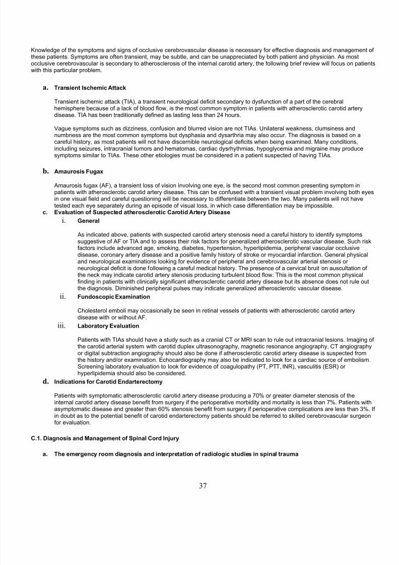
8/10/2019 NCH Student
http://slidepdf.com/reader/full/nch-student 37/68
8no,ledge o! the smptoms and signs o! occlusive cere%rovascular disease is necessar !or e!!ective diagnosis and management o!these patients. Smptoms are o!ten transient- ma %e su%tle- and can %e unappreciated % %oth patient and phsician. As mostocclusive cere%rovascular is secondar to atherosclerosis o! the internal carotid arter- the !ollo,ing %rie! revie, ,ill !ocus on patients,ith this particular pro%lem.
a. Transient %schemic Attac+
Transient ischemic attack 6T(A7- a transient neurological de!icit secondar to ds!unction o! a part o! the cere%ralhemisphere %ecause o! a lack o! %lood !lo,- is the most common smptom in patients ,ith atherosclerotic carotid arterdisease. T(A has %een traditionall de!ined as lasting less than 2" hours.
Dague smptoms such as di**iness- con!usion and %lurred vision are not T(As. /nilateral ,eakness- clumsiness andnum%ness are the most common smptoms %ut dsphasia and dsarthria ma also occur. The diagnosis is %ased on acare!ul histor- as most patients ,ill not have discerni%le neurological de!icits ,hen %eing examined. an conditions-including sei*ures- intracranial tumors and hematomas- cardiac dsrhthmias- hpoglcemia and migraine ma producesmptoms similar to T(As. These other etiologies must %e considered in a patient suspected o! having T(As.
b. Amaurosis Fugax
Amaurosis !ugax 6A&7- a transient loss o! vision involving one ee- is the second most common presenting smptom inpatients ,ith atherosclerotic carotid arter disease. This can %e con!used ,ith a transient visual pro%lem involving %oth ees
in one visual !ield and care!ul 0uestioning ,ill %e necessar to di!!erentiate %et,een the t,o. an patients ,ill not havetested each ee separatel during an episode o! visual loss- in ,hich case di!!erentiation ma %e impossi%le.
c. Evaluation of Sus(ected atherosclerotic Carotid Artery *isease
i. !eneral
As indicated a%ove- patients ,ith suspected carotid arter stenosis need a care!ul histor to identi! smptomssuggestive o! A& or T(A and to assess their risk !actors !or generali*ed atherosclerotic vascular disease. Such risk!actors include advanced age- smoking- dia%etes- hpertension- hperlipidemia- peripheral vascular occlusivedisease- coronar arter disease and a positive !amil histor o! stroke or mocardial in!arction. General phsicaland neurological examinations looking !or evidence o! peripheral and cere%rovascular arterial stenosis orneurological de!icit is done !ollo,ing a care!ul medical histor. The presence o! a cervical %ruit on auscultation o!the neck ma indicate carotid arter stenosis producing tur%ulent %lood !lo,. This is the most common phsical!inding in patients ,ith clinicall signi!icant atherosclerotic carotid arter disease %ut its a%sence does not rule outthe diagnosis. +iminished peripheral pulses ma indicate generali*ed atherosclerotic vascular disease.
ii. Fundosco(ic Examination
holesterol em%oli ma occasionall %e seen in retinal vessels o! patients ,ith atherosclerotic carotid arterdisease ,ith or ,ithout A&.
iii. 9a)oratory Evaluation
<atients ,ith T(As should have a stud such as a cranial T or )( scan to rule out intracranial lesions. (maging o! the carotid arterial sstem ,ith carotid duplex ultrasonograph- magnetic resonance angiograph- T angiographor digital su%traction angiograph should also %e done i! atherosclerotic carotid arter disease is suspected !romthe histor andBor examination. Echocardiograph ma also %e indicated to look !or a cardiac source o! em%olism.Screening la%orator evaluation to look !or evidence o! coagulopath 6<T- <TT- (N)7- vasculitis 6ES)7 orhperlipidemia should also %e considered.
d. %ndications for Carotid Endarterectomy
<atients ,ith smptomatic atherosclerotic carotid arter disease producing a 5CJ or greater diameter stenosis o! theinternal carotid arter disease %ene!it !rom surger i! the perioperative mor%idit and mortalit is less than 5J. <atients ,ithasmptomatic disease and greater than $CJ stenosis %ene!it !rom surger i! perioperative complications are less than 3J. (!in dou%t as to the potential %ene!it o! carotid endarterectom patients should %e re!erred to skilled cere%rovascular surgeon!or evaluation.
C.1. *iagnosis and Management of S(inal Cord %n,ury
a. The emergency room diagnosis and inter(retation of radiologic studies in s(inal trauma
37

8/10/2019 NCH Student
http://slidepdf.com/reader/full/nch-student 38/68
i. *iagnosis3
aa. &resentation3
There are approximatel 11-CCC ne, spinal cord in4uries per ear.
Mechanism of %n,ury3
otor vehicle crashes: "#J
&alls: 22J
Sports: 1"J
Diolence: 1"J
?ther: #J
ost patients 65#J7 are males. Sixt percent are %et,een the ages o! 1$and 3C. Slightl more than hal! o! all patients ,ith cervical spine trauma,ill present ,ith signs o! neurologic in4ur. /p to 1CJ o! patients ,ithcervical spine trauma ,ill develop signs o! neurologic in4ur a!terpresentation.
)). Association /ith other in,uries
&ive to ten percent o! unconscious patients ,ho have %een involved in a!all or motor vehicle accident ,ill have a cervical spine in4ur. onversel-$CJ o! patients ,ith a cervical spine !racture ,ill have at least one ma4orassociated in4ur. &ive to !i!teen percent o! patients ,ith a cervical spine!racture ,ill have a second verte%ral column !racture.
cc. History and (hysical examination clues to the (resence of s(inalin,ury
<ain'local pain- due to %oneBso!t tissue in4ur
'radicular pain- due to nerve root compressionDitals'%radcardia indicating loss o! smpathetic input to the heart !rom cervicalor high thoracic lesion
'hpotension due to loss o! smpathetic input tosstemic vasculature6spinal shock7External'tenderness- %ruising- or s,elling due to local so!t tissue damage
'palpa%le step'o!! due to malalignment
'paraspinous muscle spasm
'torticollis due to muscle spasm or unreducedverte%ral dislocation@eakness'partial vs. complete
'level: motor level is de!ined as the most caudal level ,ith grade (((6antigravit7 strength- assuming that more cranial levels have grade (DstrengthSensation'decreased vs. a%sent vs. hperesthesia
'level : sensor level is de!ined as the most caudal dermatome ,ith
38

8/10/2019 NCH Student
http://slidepdf.com/reader/full/nch-student 39/68
normal sensation
'saddle region needs to %e tested ' ma %e the onl region o! preservedsensation 6sacral sparing7)e!lexes'normal vs. a%sent vs. increased
'a%sent ma indicate the presence o! spinal shock
'increased ma indicate the presence o! an older complete or incompletein4urSphincter'decreased or a%sent tone
'loss o! voluntar contraction
'loss o! %ul%ocavernosis re!lexdd. %n the comatose or intoxicated (atient
The comatose or intoxicated ma %e una%le to transmit in!ormationregarding pain or cooperate ,ith strength or sensor testing. (ntracranialin4ur ma co'exist ,ith spinal in4ur. There!ore one must assume that all
comatose or intoxicated trauma patients have a spinal in4ur until provenother,ise.
lues to the presence o! a spinal in4ur in such a patient include:
'!laccid are!lexia
'diaphragmatic %reathing
'loss o! grimace to pain!ul stimuli %elo, speci!ic level
'loss o! ,ithdra,al response %elo, a speci!ic level
'loss o! spontaneous movement %elo, a speci!ic level
'loss o! sphincter tone
'priapismFran+el grading system
Fran+el !rade *esignation *efinition
ompleteneurological
in4ur A
No motor or sensor !unctiondetected %elo, level o! lesion
<reservedsensation onl
No motor !unction detected%elo, level o! lesion- somesensor !unction %elo, level o!
lesion preserved
<reservedmotor-
non!unctional
Some voluntar motor !unctionpreserved %elo, level o! lesion%ut too ,eak to serve an use!ulpurpose- sensation ma or manot %e preserved
<reservedmotor- !unctional
+&unctionall use!ul voluntarmotor !unction %elo, level o!in4ur is preserved
39
8/10/2019 NCH Student
http://slidepdf.com/reader/full/nch-student 40/68
Normal motor !unction
ENormal motor and sensor!unction %elo, level o! lesion-a%normal re!lexes ma persist
ii.iii. adiologic studies
aa. &lain x$rays
Static:'Need to see do,n to the 5'T1 disc space on the lateral plain x'rao! thecervical spine
'Anterior'<osterior and Hateral 6ervicalBThoracicBHum%ar7
Evaluate !or the presence o!:'<reverte%ral so!t tissue s,elling
'alalignment
'Derte%ral %od !ractures or loss o! verte%ral %od height
'<osterior element !ractures 6spinous process- transverse process-lamina- !acet7
'@idening o! interpedicular distance
'&racture o! dens
'?pen outh ?dontoid 6ervical7
'Evaluate !or the presence o!:'&racture o! dens
'@idening o! lateral masses o! 1+namic 6&lexionBExtension7:
'<er!ormed onl i! static x'ra are normal %ut there is a high suspicion o!ligamentous in4ur not %e visi%le on static x'ras
'an onl %e per!ormed in the conscious cooperative patient
'Evaluate !or the presence o!:'Derte%ral su%luxation
'@idening o! distance %et,een dens and arch o! 1
'A%normal kphosis or opening up o! posterior elements in !lexion)). Com(uted axial tomogra(hy
'as largel replaced conventional poltomograph
'ost use!ul !or assessing %one detail9 poor visuali*ation o! neuralelements
'/suall per!ormed ,henever the plain x'ras are a%normal in order to:'ore completel characteri*e a kno,n !racture
')ule out spinal canal compromise due to indriven %one !ragments ,ith akno,n !racture
')ule out occult !racture in the presence o! kno,n ligamentous in4ur
40
8/10/2019 NCH Student
http://slidepdf.com/reader/full/nch-student 41/68
'?ccasionall used to image the lo,er cervical spine ,hen this cannot %edone using plain x'ras
'a %e com%ined ,ith intrathecal contrast 6melograph7 in order tovisuali*e neural elements ,hen )( is contraindicated or not availa%le
cc. Magnetic resonance imaging M%2
'ost use!ul !or visuali*ing so!t tissue structures
')esolution o! %one in!erior to computed axial tomograph
'a %e per!ormed ,hen plain x'ras are a%normal in order to rule out:'Spinal cord in4urBhematomelia
'Traumatic interverte%ral disc herniation
'ematoma
'Higamentous in4ur
). %nitiate acute management of s(inal cord in,ury including immo)ili6ation7 steroids and systemic measures
i. %mmo)ili6ationaa. Cervical
Traumatic cervical spine in4ur should %e presumed to %e present until it isruled out % phsical examination and appropriate radiographic studies. (norder to prevent ne, or additional neurologic in4ur- immo%ili*ation o! thecervical spine should %e instituted as soon as possi%le. <atients ,ith akno,n or suspected cervical spine in4ur- or those ,ho are comatose orintoxicated at the scene o! in4ur- should ideall %e placed in a cervicalorthosis at the scene. Alternativel- i! an appropriate cervical orthosis isnot availa%le- the head and neck ma %e immo%ili*ed % placingsand%ags or to,el rolls on either side o! the head.
(! a cervical spine in4ur is !ound radiographicall- it is usuall imperative
that cervical spine immo%ili*ation %e continued until de!initivemanagement is complete. (n some patients this ma consist o! continuedtreatment in a cervical orthosis- com%ined ,ith %ed rest.
(n other patients- especiall those ,ith neurologic in4ur- cervical spinalinsta%ilit- or unreduced cervical spinal dislocation- in'line cervical tractionma %e used. ervical traction is !re0uentl applied using Gardner'@ellstongs secured to the skull. &ive pounds o! ,eight are initiall applied.ore ,eight is occasionall added in an attempt to reduce a spinaldislocation. @hile the patient is in cervical traction- neurologicexaminations should %e per!ormed !re0uentl. )adiographic examinationsshould %e per!ormed a!ter the initial application o! ,eight and a!ter ansu%se0uent change to assess !or changes in spinal alignment and thepresence o! overdistraction 6pulling apart o! the spine7. @eight should %eimmediatel reduced i! a decline in the neurologic examination or
overdistraction occurs. ervical traction is contraindicated in certain highlunsta%le cervical in4uries- as ,ell as in occipital'cervical dislocation.
&inall- some patients ma initiall %e placed in a halo vest. (n addition toe!!ective immo%ili*ation o! the upper and middle cervical spine- placementin a halo vest ma constitute de!initive therap !or a variet o! lesions.
)). Thoracolum)ar
As ,ith the cervical spine- thoracolum%ar spine !racture should %e
41

8/10/2019 NCH Student
http://slidepdf.com/reader/full/nch-student 42/68
presumed to %e present until it is ruled out % phsical examination andappropriate radiographic studies. <resent extrication and transporttechni0ues emphasi*e maintenance o! a neutral position during theprehospital phase. ost commonl- patients are transported to thehospital on a %ack%oard. The should not %e allo,ed to sit or stand priorto evaluation. +uring the phsical examination- patients should %ecare!ull logrolled % multiple personnel !or examination o! the %ack.
/nless the patient has a highl unsta%le lesion- the %ack%oard ma %eremoved ,hile the patient is !lat in %ed. @henever the patient is moved-!or example to the T or )( scanner- the patient should %e care!ullplaced %ack onto the %ack%oard.
ii. Steroidsaa. S(inal cord in,ury
)esults o! the NAS(S (( stud sho,ed that patients ,ith spinal cordin4ur ,ho ,ere treated ,ith methlprednisolone ,ithin eight hours o!in4ur had signi!icantl greater improvement in their neurologic !unction6motor- pinprick sensation- and touch sensation7 than those given aplace%o. The dose administered in the stud ,as 3CmgBkg given as a%olus over 1# minutes- !ollo,ed % an in!usion o! #." mgBkgBhr !or 23hours- %egun "# minutes a!ter completion o! the %olus.
)esults o! a !ollo,'up stud- NAS(S (((- concluded that patients ,ithacute spinal cord in4ur ,ho receive methlprednisolone ,ithin 3 hours o!in4ur should %e maintained on the treatment regimen !or 2" hours. @henmethlprednisolone is initiated 3 to = hours a!ter in4ur- patients should %emaintained on steroid therap !or "= hours.
)). Cauda e;uina in,ury
No data !rom randomi*ed- controlled studies.
cc. !unshot /ounds to the s(inal cord
No data !rom randomi*ed- controlled studies. o,ever- a recent
retrospective stud o! 2#" patients reported no neurological %ene!it !romintravenousl administered steroids a!ter a gunshot ,ound to the spine61'H17. oth in!ectious and nonin!ectious complications ,ere more!re0uent in the group receiving steroids. (t ,as recommended thatpatients ,ith spinal cord in4ur secondar to a gunshot ,ound to the spinenot %e treated ,ith steroids.
iii. Systemic measuresaa. es(iratory
aintenance o! an ade0uate air,a and %reathing remains the !irstpriorit in the trauma patient. Spinal cord in4ured patients ma su!!er !rominade0uate respirator !unction due to paralsis o! the intercostal musclesor diaphragm. oncomitant in4uries ma also compromise respirator!unction. aintenance o! spinal alignment is critical during intu%ation. (!
endotracheal intu%ation is needed- it is %est per!ormed in con4unction ,ithin'line cervical traction- nasotracheall 6i! there is no evidence o! %asilarskull !racture7 or !i%eropticall.
)). Cardiac
A!ter air,a and %reathing a%normalities are treated- ade0uate per!usionmust %e assured. potension in the patient ,ith a spinal in4ur ma %edue to intravascular volume loss due to %leeding or spinal cord in4ur.These t,o processes ma co'exist. (denti!ication and control o! %leeding
42

8/10/2019 NCH Student
http://slidepdf.com/reader/full/nch-student 43/68
is the !irst priorit. potension- due to loss o! smpathetic vascular tone-and %radcardia- due to loss o! smpathetic innervation o! the heart- arethe most important elements o! spinal shock due to spinal cord in4ur.(nitial treatment o! spinal shock consists o! intravascular volumeexpansion. (n patients re!ractor to this therap- smpathomimetic drugsma %e needed
cc. !enitourinary
<lacement o! an ind,elling urinar catheter is mandator in the severelin4ured patient !or monitoring o! urine output volume. (n the patient ,ith aspinal in4ur- urinar catheteri*ation during the period %et,eenidenti!ication o! the in4ur and de!initive treatment !acilitates nursing careand avoids unnecessar patient movement. (n the neurologicall in4uredpatient- the %ladder ma not !unction normall. ontinuous urinardrainage prevents the complication o! %ladder rupture due tooverdistension.
c. 4nderstand the definition and su)se;uent management (rinci(les of the unsta)le s(ine
i. 4nsta)le s(ine3 *efinition
Spinal insta%ilit has %een de!ined as the loss o! the a%ilit o! the spine under phsiologic loads to maintainrelationships %et,een verte%rae in such a ,a that there is neither damage nor su%se0uent irritation to the spinalcord or nerve roots and - in addition- there is no development o! incapacitating de!ormit or pain due to structuralchanges. Notice that this is a clinical de!inition.
Numerous sets o! radiographic criteria have %een developed in an attempt to predict ,hich patients are or ,ill%ecome unsta%le a!ter a spinal in4ur. The most commonl used is the three'column model o! +enis. (n this model-the spine is divided into a posterior- middle- and anterior column. The posterior column includes all o! the posterior%on and ligamentous elements ,hile the middle column includes the posterior longitudinal ligament and all o! theelements comprising the posterior one third o! the verte%ral %od and interverte%ral disc. The anterior column iscomprised o! the remaining portions o! the verte%ral %od and interverte%ral disc- as ,ell as the anteriorlongitudinal ligament. (n4uries ,ith incompetence o! t,o or three columns are in!erred to %e unsta%le. The three'column theor applies to the thoracolum%ar spine onl.
ii. Management (rinci(les
As descri%ed a%ove- initial management o! the unsta%le spine consists o! immo%ili*ation o! the in4ured verte%ralsegment ,hile the patient is %eing sta%ili*ed and other in4uries are %eing ruled out. A!ter this initial phase-management decisions are generall %ased on three !actors:aa. Need for decom(ression of neural elements.
General indications !or neural decompression ,ill %e discussed in thenext section. (t is important to consider that- in some cases-decompressive procedures ma !urther desta%ili*e the unsta%le spine orrender the sta%le spine potentiall unsta%le.
)). Need to mo)ili6e the (atient as soon as (ossi)le.
Some unsta%le spinal in4uries ma %e potentiall treata%le ,ith prolonged%ed rest. o,ever- % avoiding prolonged periods o! %ed rest- earl
surgical sta%ili*ation o! the unsta%le spine ma help to preventatelectasis- pneumonia- deep venous throm%osis- and decu%iti. (naddition- earl sta%ili*ation allo,s the patient to %egin reha%ilitationearlier- potentiall reducing the complications o! 4oint contracture anddeconditioning- as ,ell as potentiall reducing time o!! ,ork.
cc. Need to sta)ili6e the s(ine that is not li+ely to heal /ithout surgicalintervention.
(n general- in4uries that are not likel to heal include those ,ith ,ideldisplaced !ractures- unreduced or unreduci%le dislocations- severe
43
8/10/2019 NCH Student
http://slidepdf.com/reader/full/nch-student 44/68
de!ormit- or severe ligamentous in4ur. Also include in this group arein4uries ,hich have %een treated ,ith prolonged %ed rest or %racing ,hichhave not healed correctl.
Surgical sta%ili*ation has t,o components. The !irst component isarthrodesis- or !usion. The goal o! !usion is to induce ad4acent verte%raea%ove and %elo, the in4ur to heal together into a solid %lock o! %one-
eliminating an potential movement %et,een them. This usuall involvesplacement o! %one gra!t %et,een the verte%rae. one gra!t ma %e placedanteriorl- %et,een ad4acent verte%ral %odies- or posteriorl- %et,eenad4acent laminae- !acets- or transverse processes.
The second component o! surgical sta%ili*ation involves internal !ixation6instrumentation7. This provides immediate strength and maintainsanatomic alignment during the time it takes !or !usion to occur. (nternal!ixation usuall involves the implantation o! some com%ination o! ,ire-hooks- scre,s- andBor rods. (nternal !ixation is not a su%stitute !or !usion. A general principle is that all internal !ixators ,ill eventuall !ail i! !usiondoes not occur.
d. 4nderstand management (rinci(les in s(inal cord in,ury including indications for decom(ressive surgery andtreatment of the medical com(lication associated /ith cord in,ury s+in7 )ladder7 )o/el movement7 res(iratory2
i. %ndications for surgery
The most compelling indication !or decompressive surger in spinal cord in4ur is the presence o! an incompleteneurologic in4ur ,ith persistent neural compression at the site o! in4ur. ompression ma %e due to indriven %one!ragments- traumatic disc herniation- epidural hematoma- or persistent verte%ral malalignment. @hile there iscurrentl de%ate regarding the most appropriate timing o! decompression in the presence o! a sta%le neurologicexamination- most clinicians agree that it should %e done emergentl in the presence o! a rapidl decliningneurologic examination.
The goal o! decompressive surger is restoration o! a normal spinal canal ,ithout additional in4ur to the neuralelements. This- in theor- ,ill !acilitate neurologic recover. A revie, o! the most common causes o! neuralcompression reveals that most are ventral processes. There!ore- technicall demanding ventral decompressiveprocedures are !re0uentl necessar. As has %een previousl stated- these procedures ma !urther desta%ili*e theunsta%le spine or render the sta%le spine potentiall unsta%le.
ii. Treatment of medical com(lications associated /ith cord in,ury
aa. S+in3
<atients ,ith spinal cord in4ur are at particular risk !or skin %reakdo,ndue to ina%ilit to sense pain and prolonged periods o! %ed rest. This ma%e !urther aggravated % incontinence and the necessit o! ,earing anorthosis. All insensate regions should %e surveed regularl. @hile in %ed-spinal cord in4ured patients should %e turned ever t,o hours. onprominences 6sacrum- heels7 should %e protected. Shearing !orces- ,hich!re0uentl occur during trans!ers- should %e avoided. o,el and %ladderds!unction should %e treated accordingl as descri%ed %elo,. Allorthoses and ,heel chairs should %e !itted appropriatel and ad4usted toaccommodate ,eight loss or gain.
)). 5ladder3
<atients ,ith spinal cord in4ur !re0uentl su!!er !rom some !orm o! urinar%ladder ds!unction. This ma mani!est as a lo,er motor neuron %ladder,ith over!lo, incontinence and urinar retention. Alternativel- an uppermotor neuron %ladder ma %e present ,ith incontinence and reduced%ladder capacit. (n either instance- the goals o! treatment are to maintaina !unctional lo,er urinar tract !ree o! in!ection and to preserve renal!unction. As has %een descri%ed a%ove- the initial treatment o! urinar%ladder ds!unction is usuall placement o! an ind,elling urinar catheter.
44
8/10/2019 NCH Student
http://slidepdf.com/reader/full/nch-student 45/68
A!ter the acute period- sterile- intermittent catheteri*ation is pre!era%le tothe long'term use o! an ind,elling urinar catheter. (ntermittentcatheteri*ation should initiall %e per!ormed ever " hours and urinarvolumes should not %e permitted to exceed "#Ccc. (! possi%le- patientsma %e taught to per!orm sel!'catheteri*ation. )enal !unction should %eclosel monitored long'term.
cc. 5o/el movement3
<atients ,ith spinal cord in4ur also !re0uentl su!!er !rom some !orm o!%o,el ds!unction. The goals o! management are !or the patient to %e !reeo! incontinence and to develop a predicta%le %o,el routine ,ithout !ecalimpaction. <atients ,ith lo,er motor neuron %o,els !re0uentl re0uiremanual removal o! !eces. Those ,ith upper motor neuron %o,els mare0uire dail digital rectal stimulation or suppositories. Ade0uate dail!luids and !i%er intake !acilitate development o! a %o,el routine. +iarrheama %e due either to excessive laxative intake or !ecal impaction.
dd. es(iratory3
All trauma patients are suscepti%le to atelectasis and pneumonia duringthe acute phase. This is especiall true in the patient ,ith concomitant
traumatic %rain in4ur. (ncentive spirometr should %e instituted earl intreatment. (! the patient is not a%le to participate- consideration should %egiven to the use o! intermittent positive pressure %reathing 6(<<7.
Additional pro%lems arise in the spinal cord in4ured patient. The phrenicnerve is supplied % the 3- "- and # roots. <atients ,ith a " orhigher level o! in4ur most o!ten re0uire placement o! a tracheostom andthe assistance o! a ventilator. <atients ,ith a $ or higher level o! in4ur,ill re0uire some assistance ,ith cough and clearing o! secretions.<atients ,ith a lo, cervical or thoracic level o! in4ur remain at higher risk!or atelectasis- pneumonia- and respirator !ailure- despite normal phrenicnerve !unction- due to ds!unction o! the intercostal musculature. Allshould %e started on incentive spirometr- as ,ell as exercise programs toincrease strength in the remaining muscles o! inspiration. <rophlacticclearing o! secretions should %e encouraged.
eferences3
1. racken - Shepard F- ollins @& et al. A randomi*ed- controlled trial o! methlprednisolone or naloxone in thetreatment o! acute spinal'cord in4ur. NEF 322:1"C#'1"11- 1>>C
2. racken - Shepard F- ol!ord T) et al. Administration o! methlprednisolone !or 2" or "= hours or tirili*ad meslate !or"= hours in the treatment o! acute spinal cord in4ur. FAA 255:1#>5'1$C"- 1>>5
3. +enis &. The three column spine and its signi!icance in the classi!ication o! acute thoracolum%ar in4uries. Spine =:=15- 1>=3". +eDivo F- )utt )+- lack 8F- et al. Trends in spinal cord in4ur demographics and treatment outcomes %et,een 1>53 and
1>=$. Arch <hs ed )eha%il 53:"2"- 1>>2#. &rankel H- ancock +?- slop G et al. Dalue o! postural reduction in initial management o! closed in4uries o! the spine
,ith paraplegia and tetraplegia: <art 1. <araplegia 5:15>- 1>$>'1>5C
$. ear )&- Daccaro A)- esa FF et al. Steroids and gunshot ,ounds to the spine. Neurosurger "1:#5$'#="- 1>>55. @hite AA- <an4a%i . linical iomechanics o! the Spine. <hiladelphia- Hippincott- 1>5=
45

8/10/2019 NCH Student
http://slidepdf.com/reader/full/nch-student 46/68
C.#. *iagnosis and Management of Non$Traumatic Nec+ and 5ac+ &ro)lems
%ntroduction
+egenerative diseases o! the spine provide the tpical neurosurgeon ,ith the %ulk o! his or her practice. Ho, %ack pain is the mostcommon speci!ic complaint generating a phsician visit- and is the most o!ten cited reason !or missed ,ork. (t is estimated that 5#Jo! people ,ill visit a health care pro!essional at some time in their lives !or %ack pain. Estimates o! prevalence suggest that at angiven time 1 in # adults in the /S ,ill complain o! lo, %ack discom!ort i! asked.
The direct and indirect economic impact o! lo, %ack complaints is enormous. +irect costs o! treatment are estimated at U2C'3C %illiona ear. Some 2CC million person'das o! ,ork are lost per ear due to lo, %ack pro%lems. Ho, %ack pain has recentl ranked $th inhospital'%ed das per ear. learl- e!!icient and e!!ective management o! degenerative conditions o! the spine is crucial- %oth at thelevel o! the primar care provider- and at the level o! the su%specialt spine surgeon.
The purpose o! this chapter is to give the student an overvie, o! ho, patients present ,ith degenerative complaints o! the lum%arand cervical spine. The !ocus ,ill %e on identi!ing smptom complexes that point to speci!ic pathological entities that are surgicalltreated. Special attention ,ill %e paid to identi!ing signs and smptoms that are suggestive o! serious patholog that ma threatenli!e or neurologic !unction and re0uire urgent attention.
a. Categories of Sym(toms3
(n order to have an orderl approach to the patient ,ith spine related complaints- the phsician needs to %e !amiliar ,ith the%road categories o! smptoms ,ith ,hich patients ,ill present. The di!!erential diagnosis o! spine related pain is extensive. Although the vast ma4orit o! patients have %enign- non'surgical pro%lems- ver serious medical illness can present ,ith thechie! complaint o! %ack- neck- or extremit pain. @ith experience- common presenting sndromes emerge- and allo, morerational triage o! patients into groups ,ho need !urther evaluation- and perhaps surgical re!erral- and those ,ho are %estmanaged ,ithout extensive testing and re!errals.
i. Axial S(ine &ain
<ain in the spine ,ith or ,ithout radiation into the extremities is the most common presenting compliant related tospinal disease. (nitial evaluation should center on o%taining a detailed histor o! the patient’s complaints andprevious medical histor. A histor o! recent trauma should raise the suspicion o! a !racture. haracter- severit-!re0uenc- inciting and relieving events should %e thoroughl explored. (n0uir a%out associated smptoms- such
as !ever and visceral pain can suggest diagnoses and avenues o! investigation. The timing o! pain can also lead toappropriate investigation. orning sti!!ness and pain that relents as the da progresses suggests an in!lammatordisorder- ,here nocturnal pain associated ,ith recum%enc is a much more ominous smptom- %eing seen ,ithmalignant- destructive lesions.
Activit'related %ack pain ,ithout neurologic smptoms or !indings is 0uite common. (n the a%sence o! other,orrisome smptoms or !indings- most patients can %e success!ull managed ,ith a %rie! reduction in activit leveland analgesics. The optimum duration and content o! non'operative treatment is controversial. Some practitionersadvocate strict %ed rest- ,hile others avoid it. )eduction o! general activit level ,hile avoiding an speci!ic incitingevents- coupled ,ith non'steroidal agents- is the mainsta o! most clinicians’ !irst line therap.
Evaluation !or pure axial spine pain that does not resolve ,ith conservative measures can include plainradiographs- magnetic resonance imaging- computed tomograph- melograph- electrophsiologic studies- anddiscograph. A set o! plain !ilms can exclude !racture- and identi! serious %on patholog such as a destructivetumor. o,ever- !or so!t tissue imaging the )( is the stud o! choice. T can give detailed in!ormation a%out %on
anatom. elograph is sensitive to canal and !oraminal patholog- and ,hen paired ,ith a post'melogram Tscan- can %e ver use!ul in the patient in ,hom an )( is not an option. +iscograph is controversial- and ,ill not%e discussed at length.
ii. adiculo(athy
)e!erred pain into the upper or lo,er extremities o!ten accompanies %ack or neck pain. )e!erred pain can %e theinitial smptom o! a compressed nerve root % a ruptured disc or neural !oraminal stenosis !rom osteophtes.)adicular pain is usuall descri%ed as sharp or even shock'like- and ma %e associated ,ith certain activities orpositions. The distri%ution o! the pain ma not al,as %e classic- and o!ten doesn’t respect dermatomaldistri%utions.
46
8/10/2019 NCH Student
http://slidepdf.com/reader/full/nch-student 47/68
Sensor changes are also o!ten seen- ,ith complaints o! tingling and num%ness %eing ver common. ?nexamination decreased sensation to pinprick and light touch are !ound in a dermatomal distri%ution in manpatients. (t is interesting that areas o! re!erred pain and sensor loss o!ten are di!!erent. aking determinations o!level o! nerve root compression solel !rom pain or sensor distri%ution is o!ten di!!icult.
otor ,eakness is also seen in nerve root compression sndromes. uscle innervation is more constant and has
less overlap than sensor innervation and is %etter at predicting level o! patholog. otor de!icits that are o! a morelong'standing nature can have signi!icant ,asting. pore!lexia in the appropriate distri%ution is also seen.aa. Cervical
ervical radiculopath can present acutel- as ,ith a traumatic ruptureddisc- or can %e o! a more chronic and intermittent nature- as is seen in!oraminal narro,ing !rom osteophtes. Tpicall- the in!erior nerve root isa!!ected 6e.g. #'$ disc a%normalities a!!ect the $ nerve root7. #'$ and$'5 are the most commonl a!!ected segments.
A # radiculopath tpicall presents ,ith pain in the shoulder and theupper part o! the lateral arm. <aresthesias are o!ten seen in the moredistal part o! the a!!ected dermatome. +eltoid ,eakness is seencommonl ,ith a # radiculopath. iceps or %rachioradialis ,eaknesscan %e seen ,ith a $ radiculopath along ,ith the appropriate
hpore!lexia. <aresthesias and !rank sensor loss are more distal- andcan extend into the hand. )oot compression at 5 produces triceps,eakness and a decreased triceps re!lex. <ain extending into the distal!orearm or hand is common. Sensor loss is commonl seen in the hand.
)). 9um)ar
Sciatica is a classic sndrome o! lo,er lum%ar nerve root compression.Ho, %ack pain- that ma or ma not have %een associated ,ith some sorto! trauma- is commonl antecedent to the onset o! leg pain % das to a!e, ,eeks. <ain tends to %e more proximal- and in a slightl di!!erentdistri%ution than sensor changes. otor ,eakness is also seen- %ut can%e missed i! dnamic testing is not done. All patients should %e asked tostand on their toes and heels- as con!rontational testing ,ill miss su%tlemotor de!icits in the lo,er extremities. As in the cervical spine- thepathologic level usuall a!!ects the caudal nerve root 6e.g. H#'Sl discproduces an S1 radiculopath7. H#'S1 and H"'# are over,helmingl themost common levels a!!ected. The upper lum%ar spine is a!!ected less!re0uentl.
The classic S1 radiculopath results in pain do,n the %ack o! the leg andinto the heel or !oot. Sensor loss is usuall over the lateral aspect o! the!oot. <lantar'!lexion ,eakness is seen- %ut can %e su%tle. A loss o! the Achilles re!lex is also !airl speci!ic to S1. The H# radiculopath producessimilar pain- %ut the sensor smptoms tend to %e over the dorsum o! the!oot. @eakness in dorsi!lexion o! the !oot 6or more speci!icall extensorhallicus longus7 is the motor !inding associated ,ith H#. There is not arelia%l reproduci%le re!lex associated ,ith H#.
iii. Cauda E;uina Syndrome
The cauda e0uina sndrome is important to recogni*e- as prompt surgical attention ma %e necessar in cases o!acute patholog. <atients tpicall present ,ith lo, %ack pain and di!!use lo,er extremit complaints. inimal ora%sent leg pain is seen- in contrast to the predominance o! extremit pain seen in the radicular sndromes. o,el-and- more commonl- %ladder ds!unction- are also seen. (n man patients this goes unrecogni*ed until %ladderdistention leads to over!lo, incontinence. ecause o! the vague nature o! the complaints- and the common lack o!severe pain associated ,ith a large disc herniation producing a cauda sndrome- dela in diagnosis is notuncommon. <atients ,ith %ack pain and complaints o! lo,er extremit ,eakness should %e care!ull examined torule out a cauda e0uina sndrome. @eakness can %e di!!use- and var !rom su%tle to paraplegia. Sensor !indings
47
8/10/2019 NCH Student
http://slidepdf.com/reader/full/nch-student 48/68
are varia%le- %ut o!ten !ound in the perineal area. hecking a post void residual can give a 0uick initial assessmento! %ladder !unction- and should %e done prior to placing a !ole. )e!lex changes are varia%le- %ut in general revealdi!!use hpore!lexia. <atients in ,hom the diagnosis is suspected should undergo urgent imaging ,ith )( or amelogram.
)ecover !rom a severe cauda e0uina sndrome- even ,ith prompt surgical management- can %e ver slo, andincomplete. ladder !unction is o!ten the slo,est smptom to improve. This is another reason to keep a high index
o! suspicion !or this uncommon entit in patients presenting ,ith %ack pain and complaints o! lo,er extremit,eakness.
iv. Myelo(athy
elopath is the clinical presentation o! patholog a!!ecting spinal cord !unction. The di!!erential diagnosis !orcauses o! melopath is large and includes trauma- meta%olic- degenerative- in!lammator- toxic- in!ectious- andneoplastic etiologies. +egenerative conditions o! the spine ma produce the smptoms o! melopath. (n maninstances the onset o! the melopath is insidious- and smptoms and signs su%tle. Hongstanding melopath-un!ortunatel- is rarel reversi%le. Earl identi!ication o! patients ,ith progression o! melopath is essential toprevent permanent loss o! neurologic !unction. There!ore- in patients ,ho present ,ith neck or thoracic spine pain-the histor and phsical exam should %e tailored to exclude melopath.
The most general signs o! melopath are those o! upper motor neuron ds!unction. Su%tle smptoms includedi!!icult ,ith !ine motor control o! the hands and !ingers- gait pro%lems and insta%ilit- and num%ness.perre!lexia- increased tone- and ,eakness are the hallmarks o! the clinical exam. A%normal plantar response
and o!!man’s sign are !re0uent a%normal re!lexes seen in patients ,ith melopath. /rinar ds!unction- such hashesitanc- !re0uenc- and incontinence- is also seen- %ut tends not to %e severe.aa. Chronic
<rogression o! the melopath can %e ver slo, and gradual- or step,ise.(t is not uncommon !or the onset to %e so insidious that patients are 0uitedisa%led %e!ore the seek medical evaluation !or their smptoms. are!ulhistor and examination can direct the level o! suspicion. (n general-smptoms that a!!ect the hands and upper extremities should promptcervical evaluation- ,hile isolated lo,er extremit smptoms and a trunksensor level are more suspicious !or a thoracic lesion. ?!ten in chronicmelopath the distinction can %e di!!icult. (n patients ,ith chronicmelopath an )( is the stud o! choice to evaluate the spinal canal. (npatients ,ho are una%le to have an )(- melograph and Tmelograph is ade0uate and gives good in!ormation a%out the spinalcanal.
)). Acute
+egenerative disease ma lead to acute onset o! melopath. Acute discherniation can %e seen in the setting o! trauma- %ut also ,ithoutsigni!icant in4ur. <atients ,ho present ,ith the acute onset o!melopathic smptoms deserve urgent evaluation ,ith )( ormelograph. (! patholog such as an acutel ruptured disc causing spinalcord compression is !ound- surgical evaluation should %e sought./n!ortunatel- patients ,ith complete spinal cord in4uries onl in!re0uentlmake !ull recoveries.
Acute ,orsening o! cervical melopath in the setting o! cervical stenosis
can %e seen in the !ace o! !airl minor trauma. (n patients ,ith acutemelopath ,ithout o%vious !racture- %ut signi!icant degenerative disease-cervical stenosis should %e suspected.
). S(ecific Conditions
i. The Herniated *isc
The central portion o! the interverte%ral disc is the nucleus pulposus. /nder certain pathologic conditions it marupture through the annulus !i%rosis and into spaces occupied % neurologic structures. entral- large disc rupturecausing compression o! the spinal cord or cauda e0uina ma %e seen. (t is- ho,ever- much more common to !ind a
48

8/10/2019 NCH Student
http://slidepdf.com/reader/full/nch-student 49/68
posterior'lateral rupture producing nerve root compression and radiculopath.
echanisms producing radicular pain are poorl understood. +irect compressive e!!ects certainl pla a role. Thedorsal root ganglion appears especiall sensitive to compressive e!!ects. )ecent animal models have suggested arole !or %iochemical !actors leading to in!lammation. (ncreasingl- experimental evidence suggests that themechanisms leading to pain generation are more complex than once thought.aa. 9um)ar
Hum%ar radiculopath is commonl kno,n as sciatica. The classicpresentation is in ounger patients ,ho ,ill present ,ith a histor o! %ackpain !ollo,ed in a !e, das to ,eeks % intense leg discom!ort-paresthesias- and radicular ,eakness as descri%ed in the previoussection.
So!t disc herniations in the lum%ar spine leading to radicular complaintsare seen most o!ten in the 3rd through $th decade o! li!e. Estimates o!prevalence var ,idel in the literature- !rom as lo, as 2J to high as"CJ.
Non'surgical therapies !or smptoms due to lum%ar disc herniation areplenti!ul. The natural histor o! radiculopath is one o! improvement inman individuals. A variet o! therapies are success!ul in helping patients
get through ver pain!ul periods. ?ral or epidural steroids can %e 0uitesuccess!ul in managing lum%ar radiculopath- although the results areo!ten temporar. <hsical therap- chiropractic manipulation- and a host o! other devices and regimens are used and promoted. +e!initive evidenceon the superiorit o! an particular approach to non'surgical therap islacking. All patients ,ithout severe neurologic de!icit should undergo atrial o! non'surgical therap. The duration o! non'surgical therap is notset- and is o!ten driven % the patient’s a%ilit to continue to tolerate theirsmptoms. &re0uentl " ,eeks- and- pre!era%l 2'3 months o! non'surgical treatment are recommended.- (t is- ho,ever- common !or patients,ith severe pain or neurologic de!icit to %e operated upon more 0uickl.
The lum%ar laminectom !or discectom is one o! the most ,idelper!ormed spinal procedures. +espite this- indications continue to %ede%ated. Except !or patients ,ith a cauda e0uina sndrome- non'
operative therap is al,as an option. There is prospective data !rom@e%er that ,ould suggest that patients that undergo discectom improvemore 0uickl over the short term. This %ene!it appears to dissipate % "ears. @ith those disclaimers- most spine surgeons ,ould agree thatreasona%le operative indications ,ould include 17 large midline discherniation ,ith resulting cauda e0uina sndrome9 27 nerve rootcompression ,ith pain and signi!icant motor and sensor de!icit9 37 nerveroot compression ,ith or ,ithout neurologic de!icit and incapacitating painthat !ails to improve ,ith non'surgical measures9 "7 recurrence o!incapacitating episodes o! H< and sciatica that prevent the patient !romleading a normal li!e.
(n patients ,ith clinical radiculopath and concordant imaging !indings asuccess!ul surgical outcome can %e expected in =C'>#J o! patients.)ecurrence rates are reported at 2'12J. The incidence o! seriouscomplications is ver lo, 6K2J7.
)). Cervical
ervical radiculopath !rom nerve root compression can %e caused % aherniated disc or !rom !oraminal narro,ing !rom osteophtes. The rootcompression sndromes produced % these conditions have %eendescri%ed a%ove. Neck pain associated ,ith degenerative disc diseaseand osteophtes ,ill improve in the ma4orit o! people ,ithout invasivetreatment9 although there is certainl a group that ,ill go on to have
49

8/10/2019 NCH Student
http://slidepdf.com/reader/full/nch-student 50/68
chronic smptoms.
The natural histor o! cervical radiculopath is not as ,ell characteri*edas that o! cervical melopath !rom degenerative disease. )adiculopath,ill improve ,ith time in man patients. o,ever- it is impossi%le to de!inestrict rules on the length o! non'surgical therap to undertake %e!oresurger should %e considered. ervical radiculitis !rom a so!t disc
herniation ma %e less likel to improve spontaneousl as that due toosteophtes. Non'surgical therap can include oral or epidural steroids-cervical traction- phsical therap- %racing- and man others.
(n care!ull selected patients ,ith radicular smptoms and evidence o!nerve root compression on their imaging studies- more than >CJ canexpect a !avora%le outcome ,ith care!ul surgical management. Seriouscomplications are rare 6K1J7.
ii. S(inal Stenosis
Spinal stenosis is the narro,ing o! the cross'sectional diameter o! the spinal canal to such an extent thatneurologic smptoms or signs are produced. The sndromes produced % lum%ar and cervical stenosis are 0uitedistinct and ,ill %e discussed separatel.
aa. 9um)ar
Hum%ar stenosis classicall produces neurogenic claudication.Neurogenic claudication is leg pain produced % ,alking or standing thatis tpicall relieved % a change o! position such as s0uatting- leaningover or sitting do,n. Heg pain can %e in a variet o! distri%utions- and%ecomes 0uite de%ilitating. <atients o!ten report associated paresthesias.Neurologic examination ma %e normal at rest- though sensor de!icitsand hpore!lexia are sometimes seen. @hen motor ,eakness is !ound itcan %e associated ,ith ,asting- as stenosis is usuall a slo,ingprogressive disease. Approximatel 2B3 o! patients ,ith smptomaticspinal stenosis ,ill present ,ith some variet o! the classic picture o!neurogenic claudication.
Ac0uired spinal stenosis is caused % advanced degenerative disease o!the disc- !acets and ligaments. The hpertroph o! the !acets andassociated ligaments- such as the ligamentum !lavum- com%ine ,ith%ulging discs to produce %oth central and lateral narro,ing. ost patients,ith ac0uired lum%ar stenosis are in their $th to 5th decade or %eond.
Surgical treatment !or lum%ar stenosis involves decompressivelaminectom and ma re0uire medial !acetectomies !or lateral recessstenosis and !oraminal stenosis. ore recentl- surgeons have %eenexploring the role o! lum%ar !usion in the treatment o! spinal stenosis inthe older population. The role o! !usion and instrumented !usion in thissetting is et to %e !ull determined. The reader is re!erred to thesuggested readings !or more on this topic.
Earl improvement a!ter surger !or lum%ar spinal stenosis is the rule
6M>CJ7 in patients ,ith a postural component to their pain. o,ever- lateprogression o! smptoms is not uncommon. A large revie, % Turner etal. suggests that good results are maintained in approximatel $"J o!patients over time.
)). Cervical
ervical spondlotic melopath 6S7 is the clinical entit produced %cervical stenosis. S usuall progresses slo,l- in a step,ise !ashion.This melopath can %e 0uite su%tle in the earl stages- and some
50

8/10/2019 NCH Student
http://slidepdf.com/reader/full/nch-student 51/68
patients ,ill have signi!icant disa%ilit %e!ore seeking appropriate medicalcare. The most common presenting complaints include neck pain- gaitdi!!iculties- and hand num%ness and clumsiness. Hoss o! %o,el and%ladder control is uncommon earl in S. ?ccasionall patients ,illpresent ,ith acute and pro!ound spinal cord in4ur a!ter mild trauma6usuall a hperextension in4ur7. ore common is a step,ise decline inspinal cord !unction.
The tpical patient ,ith S is older than #C and male. en are seennearl t,ice as o!ten as ,omen. elopathic !indings dominate thephsical examination o! patients ,ith S. (ncreased re!lexes in %oth theupper and lo,er extremities ,ith lo,er extremit spasticit are common.<athologic re!lexes such as a%inski and o!!man are also o!ten positive.Hhermitte’s sign 6electric- shock'like pain radiating do,n the spine onneck !lexion7 is classicall descri%ed- %ut occurs in a small minorit o!patients. omplicating the clinical picture in S is the lo,er motorneuron !indings that can %e seen secondar to nerve root compression.@asting- !asciculations- and hpoactive re!lexes can %e seen in the upperextremities due to nerve root compression.
The di!!erential diagnosis o! S includes multiple sclerosis-sringomelia- spinal cord tumor- su%acute com%ined degeneration- and
normal pressure hdrocephalus. Special care should %e taken in patients,ith %oth upper and lo,er motor neuron signs- as amotrophic lateralsclerosis and S can %e di!!icult to distinguish.
Surgical decompression o! the cervical spinal cord ,ill %e recommended% most neurosurgeons in the setting o! an signs o! melopath andsigni!icant cervical canal stenosis. +e!icits ac0uired % patients ,ith Sare rarel completel corrected % surger- so most surgeons ,ill tend too!!er decompression as earl as possi%le. (n patients ,ith signi!icantcervical stenosis ,ithout signs or smptoms o! melopath operativeindications are less clear. The role o! !usion in the treatment o! S isde%ated- and is %eond the scope o! this chapter.
Surgical results in the large series availa%le suggest that in 5#'>CJ o!cases the melopath can %e sta%ili*ed or improved. The incidence o!
,orsening o! melopath ,ith surger is lo, 6K1J7. ?ther complicationsare approach' related and the reader is re!erred to the suggestedreadings.
eferences"Suggested eading3
1. en*el E. ervical spondlotic melopath: <osterior surgical approaches- in <rinciples o! Spinal Surger- ene*es A-Sonntag D8 6eds79 cGra,'ill- Ne, Oork- 1>>$.
2. itchon <@. anagement o! lum%ar herniated disks- in <rincipals o! Spinal Surger- ene*es A- Sonntag D8 6eds79cGra,'ill- Ne, Oork- 1>>$.
3. 8at* FN- Hipson SF- Harson G- et al. The outcome o! decompressive laminectom !or degenerative lum%ar stenosis. Fone Foint Surg 53A: =C>'1$- 1>>1.
". Saunders )H. orpectom !or cervical spondlotic melopath- in <rinciples o! Spinal Surger- ene*es A- Sonntag D8
6eds79 cGra,'ill- Ne, Oork- 1>>$.#. @e%er . Hum%ar disc herniation: a controlled- prospective stud ,ith ten ears o! o%servation. Spine =: 131- 1>=3.$. @olcott @<- alik F- Sha!!re (- Sha!!re E- Fane FA. +i!!erential diagnosis o! surgical disorders o! the spine- in Spine
Surger- en*el E 6ed79 hurchill Hivingstone- Ne, Oork- 1>>>.
*.1. *iagnosis and Management of &eri(heral Nerve %n,ury and Entra(ment
51

8/10/2019 NCH Student
http://slidepdf.com/reader/full/nch-student 52/68
This revie, is intended to present a set o! general principles ,hich can %e applied clinicall to %oth evaluate and treat a %roadspectrum o! peripheral nerve pro%lems ,hich include traumatic in4uries and associated entrapment neuropathies.
a. &eri(heral nerve in,uries
i. Clinical evaluation
<eripheral nerve in4ur or disease can cause smptoms o! pain- dsesthesias- and either partial or complete loss o!sensor and motor !unction. A thorough clinical histor- phsical examination- electrodiagnostic evaluation- andrelevant radiographic studies should %e per!ormed to distinguish a peripheral nerve pro%lem !rom one involving thespinal cord or %rain- %one- or so!t tissues. (n addition- earl neurosurgical consultation should %e o%tained.
The strength o! individual muscles or muscle groups is graded. A sensor exam is per!ormed ,hich includes testing!or light touch- pinprick- t,o'point discrimination- vi%ration- and proprioception accordingl. (t is help!ul to testsensation in the autonomous *ones o! a nerve ,here there is minimal overlap !rom ad4acent nerves. The presenceo! Tinels sign is use!ul to locali*e a nerve in4ur. The Tinel's sign re!ers to paresthesias elicited % tapping along thecourse o! a nerve. <rogressive distal advancement o! a Tinels sign over time can %e use!ul clinicall to !ollo, thecourse o! regenerating sensor axons. o,ever- the presence o! a Tinel’s sign does not guarantee motor recover.)eturn o! s,eating in an autonomous *one signi!ies smpathetic nerve !i%er regeneration. )e!lex changes are alsosensitive and earl indicators o! nerve damage.
oth electromograph 6EG7 and nerve conduction studies 6NS7 are use!ul to distinguish an upper !rom lo,ermotor neuron disorder as ,ell as diagnose a primar muscle disease. &or EG studies- a needle electrode isplaced through the skin into a speci!ic muscle and the activit at rest and electrical response to graded musclecontractions are determined. The nerve conduction stud involves stimulation and recording along the course o!peripheral nerves. This allo,s one to measure the velocit and amplitude o! the propagating nerve action potential.
An understanding o! the !unctional anatom o! the peripheral nervous sstem can o!ten permit the clinician tolocali*e nerve in4uries and lesions ,ith a high degree o! accurac:
The %rachial plexus tpicall originates !rom the !i!th through eighth cervical spinal nerve roots as ,ell as the !irstthoracic root and innervates all o! the muscles o! the upper extremit. These nerve roots 4oin to !orm trunks- ,hich!urther su%divide into divisions- then cords- then- more distall- individual peripheral nerve %ranches. Severe traumatransmitted to the most proximal portions o! the %rachial plexus ma produce a preganglionic in4ur ,ith avulsion o!spinal nerve roots !rom the spinal cord. (t is important to determine ,hether avulsion o! the ventral or dorsal rootshas occurred- since direct repair o! a peripheral nerve ,hose contri%uting spinal roots have %een disconnected
!rom the spinal cord ,ill not restore !unction. A orners sndrome- characteri*ed % ptosis- miosis- and anhidrosisindicates avulsion o! the ipsilateral proximal = andBor T1 spinal nerve roots. ?ther phsical signs o! proximalnerve root avulsion are elevation o! the ipsilateral hemidiaphragm 6phrenic nerve7- scapular ,inging 6long thoracicnerve7- and ,eakness o! the rhom%oid muscles 6dorsal scapular nerve7. All o! these nerves originate proximallalong spinal nerves.
The lum%o'sacral plexus arises !rom the !irst lum%ar through the !ourth sacral spinal nerves. The !emoral ando%turator nerves arise !rom the anterior divisions o! H2'". The sciatic nerve is the largest nerve in the %od andarises !rom the H"'S" spinal nerves. This nerve passes through the sciatic notch and travels do,n the %ack o! theleg ,here it %ranches into peroneal and ti%ial nerves usuall 4ust a%ove the popliteal !ossa.
ii. %maging of (eri(heral nerve lesions
(maging techni0ues such as 'ras- T- and- most recentl- )( can %e valua%le diagnostic tools in evaluatingperipheral nerve lesions. ervical spine !ractures are !re0uentl associated ,ith %rachial plexus in4uries- as ,ell as
in4uries o! the proximal spinal nerves and roots. hest radiographs ma sho, unilateral elevation o! the diaphragmas a signature o! phrenic nerve paralsis 63'#7 !rom in4ur to the proximal upper cervical spinal nerves and roots.id'humeral !ractures are associated ,ith radial nerve in4uries ,hile mid!orearm !ractures o! the ulna or radius areassociated ,ith median or ulnar nerve in4uries- respectivel. ip and proximal !emur !ractures are associated ,ithsciatic nerve in4uries ,hile more distal !emur !ractures are associated ,ith peroneal or ti%ial nerve in4uries.
elograph in con4unction ,ith a T scan are use!ul to visuali*e meningeal diverticula and a%normalities o! thespinal nerve roots- ! indings o! ,hich also indicate a spinal nerve root avulsion in4ur.
T is a%le to delineate so!t tissue mass lesions such as tumors. )( has proven to %e much more e!!ective inresolving the !ine anatomical detail o! so!t tissues. /sing conventional and enhanced )( techni0ues- it has %een
52

8/10/2019 NCH Student
http://slidepdf.com/reader/full/nch-student 53/68
possi%le to visuali*e %oth normal and a%normal peripheral nerve structures. Ne, techni0ues such as )(neurograph make it possi%le to image and reconstruct the complex peripheral nerve anatom as ,ell as pinpointregions o! patholog. )( can also %e used to image signal changes in denervated muscle.
iii. !rading of (eri(heral nerve in,ury
The severit or grade o! a peripheral nerve in4ur is determined % the magnitude and duration o! the applied !orceso! in4ur. Seddon de!ined 3 grades o! nerve in4ur 6neurapraxia- axonotmesis- and neurotmesis 7 %ased on the
extent o! in4ur to the three structural components o! the peripheral nerve descri%ed a%ove. Neura(raxia- the mildest grade o! nerve in4ur- is characteri*ed % a reduction or complete %lockage o!
conduction across a segment o! nerve. Axonal continuit is maintained and nerve conduction is preserved%oth proximal and distal to the lesion %ut not across the lesion. Neurapraxia can result !rom directmechanical compression- ischemia secondar to vascular compromise- meta%olic derangements- ordiseases or toxins causing demelination o! the nerve. onduction is restored once either the meta%olicderangement is corrected or remelination occurs. Neurapraxic in4uries are usuall reversi%le and a !ullrecover can occur ,ithin das to ,eeks.
Axonotmesis represents a more severe grade o! nerve in4ur and is characteri*ed % interruption o! the
axons ,ith preservation o! the surrounding connective tissue high,a ,hich can support axonalregeneration. +istal @allerian degeneration 6axon and melin degenerate distal to site o! in4ur7 o! theaxons occurs over a several da period a!ter ,hich direct electrical stimulation o! the disconnected distalnerve stump ,ill not give rise to a nerve conduction or muscle response. )ecover can occur throughaxonal regeneration due to the preservation o! the connective tissue high,a ,hich consists o! Sch,ann
cells and their %asal lamina. The Sch,ann cells proli!erate and !orm longitudinal conduits 6i.e. the %andso! ungner7 through ,hich axons regenerate. Axonotmetic in4uries usuall recover over a period o!months. The timing and degree o! recover depends on several !actors ,hich include the extent o!retrograde axonal loss- as ,ell as the time to regenerate and reinnervate target muscles andBor sensorend organs. As a general rule- peripheral nerve !i%ers regenerate at a rate o! approximatel 1 mm per daor 1 inch per month. There!ore- more proximal in4uries re0uire longer time intervals !or regenerating axonsto reinnervate their targets.
Neurotmesis is the severest grade o! peripheral nerve in4ur. Neurotmetic in4uries are characteri*ed %
disruption o! the axon- melin- and connective tissue high,a components o! the nerve. There!ore-recover through regeneration cannot occur. This grade o! in4ur encompasses nerve lesions ,hereexternal continuit o! the nerve is preserved %ut intraneural !i%rosis occurs and %locks axonalregeneration. Neurotmetic in4uries also include nerves ,hose continuit has %een completel interrupted.Since the necessar high,as !or axonal regeneration are a%sent- surger is re0uired to remove anintervening road%locks in the !orm o! scar tissue as ,ell as to re'esta%lish continuit o! the nerve.
?n the %asis o! clinical smptoms and phsical !indings alone- it is o!ten di!!icult to di!!erentiate neurapraxic-axonotmetic- and neurotmetic grades o! nerve in4ur- especiall in the acute setting. Nerve conduction studies- %othsensor and motor- are use!ul during and a!ter the !irst ,eek !ollo,ing an in4ur to distinguish neurapraxic !romaxonotmetic and neurotmetic grades.
iv. Treatment Strategies3
Trauma is the most !re0uent cause o! peripheral nerve lesions. Nerve in4uries are caused % traction- compression-sharp laceration- and missile in4ur 6gun shot ,ounds7. Traction in4ur is o!ten associated ,ith a !racture ordislocation.. An understanding o! the mechanism o! in4ur is extremel help!ul in determining the severit o! thelesion and to guide clinical management.
Traumatic peripheral nerve in4uries can %e classi!ied into open and closed in4uries. +ecision making !or openin4uries is relativel straight!or,ard. (mmediate repair o! acute sharp lacerating in4uries 6i.e. glass or knives7 should%e undertaken ,ith the goal o! per!orming a primar end to end suture repair. o,ever- not all transecting in4urieslead themselves to a primar repair. (t the ends are ragged or contused- a delaed repair is pre!era%le todemarcate normal !rom a%normal neural tissue.
The decision making process in treating closed traumatic peripheral nerve in4uries is more complex. The ma4orit o!closed traumatic in4uries are due to stretch andBor compressive !orces. An associated expanding hematomaproducing a compartment sndrome ma re0uire emergent surger to avoid irreversi%le nerve in4ur. ecausenerves are o!ten contained in a neurovascular %undle- there is potential !or com%ined vascular and neural trauma Adelaed onset o! a neural de!icit due to a traumatic pseudoaneursm ma also re0uire urgent attention. Anangiogram is necessar ,hen damage to vascular structures is suspected clinicall. (n the ma4orit o! closed
53
8/10/2019 NCH Student
http://slidepdf.com/reader/full/nch-student 54/68
traumatic in4uries- ho,ever- nerves are not actuall transected. (nstead- a lesion in continuit representing thedamaged segment o! nerve ma %e produced ,hich results in either a neurapraxic- axonotmetic- neurotmetic- orcom%ination o! these grades o! in4ur.
(n the case o! compression or stretching- it i! o!ten not possi%le to immediatel determine the grade o! the in4ur. Apartial nerve in4ur associated ,ith muscle denervation usuall indicates an axonotmetic grade o! in4ur. <atients,ith such in4uries should %e !ollo,ed ,ith serial clinical and electrodiagnostic examinations to document recover
and con!irm the diagnosis. These patients do not re0uire immediate surgical intervention. An enlarging hematomacan convert a partial nerve in4ur into a complete in4ur. omplete nerve in4uries produce severe muscledenervation and ma represent either an axonotmetic or neurotmetic grade o! in4ur. (t is critical to distinguish%et,een these t,o grades o! in4ur over time- since the latter re0uires a surgical repair !or recover to occur.<atients are there!ore !ollo,ed closel over a several month period looking !or clinical and electrodiagnosticevidence o! nerve regeneration and muscle reinnervation.
uscles should %e reinnervated ,ithin t,o ears !ollo,ing a traumatic nerve in4ur i! recover o! use!ul motor!unction is to occur. eond this point- denervated muscles undergo irreversi%le atroph and replacement ,ith !at.There!ore- it is necessar to time a surgical exploration so that a success!ul nerve repair results in musclereinnervation ,ithin t,o ears o! the in4ur. A use!ul rule o! thum% is to !ollo, a patient !or 3 to " months to allo,an element o! neurapraxia to resolve as ,ell as permit axonal regeneration to occur %eond the point o! in4ur. (!there is no clinical or electrodiagnostic evidence o! muscle reinnervation- then a surgical exploration usingintraoperative electrophsiological monitoring should %e per!ormed.
Another approach in the management o! these traumatic peripheral nerve in4uries is to operate earl 6i.e. as soonas medicall !easi%le7. The rationale !or this approach is the !ollo,ing: 17 less scarring and there% easierdissection o! peripheral nerve elements9 and 27 intraoperative evaluation o! anatomical and electrophsiologicalcontinuit. o,ever- it remains controversial ,hether an earlier surgical repair leads to a %etter recover o!peripheral nerve !unction.
b. Entra(ment neuro(athies
<eripheral nerve entrapment descri%es the mechanical irritation % ,hich a speci!ic peripheral nerve %ecomes locall in4uredin a vulnera%le anatomic site. <eripheral nerve entrapments produce !ocal distur%ances o! nerve !unction. Nerve entrapmentma occur at an site as a result o! non'speci!ic local lesions including !racture callus- hematomas- and %enign or malignanttumors. There are several anatomical sites ,here peripheral nerves run in relativel con!ined spaces and are there!ore atincreased risk o! compression. The di!!erential diagnosis o! entrapment neuropathies includes an disease process thatdamages nerves in a !ocal manner: i.e. degenerative- hereditar- vascular- in!lammator- and meta%olic. <redisposing
!actors include repetitive activities involving the a!!ected extremit- tenosnovitis- rheumatoid arthritis- acromegal-alcoholism- amloidosis- mucopolsaccharidosis- gout- sarcoid- vitamin $ de!icienc- dia%etes- trauma- and conditionsaltering !luid %alance including pregnanc- oral contraceptives- and hpothroid mxedema.
i. Car(al tunnel syndrome
Median nerve compression %eneath the !lexor retinaculum o! the ,rist is the most common entrapmentneuropath. @omenMmen 2:1
aa. Sym(toms
(ntermittent num%ness and paresthesias along
!lexor aspects o! thum%- index- and middle !ingers- as ,ell asradial side o! "th !inger ,ith or ,ithout pain
<ain ma radiate to the !orearm and upper arm
Smptoms are ,orse ,ith repetitive use o! the
hand.
<ain a,akens patients !rom sleep
)). Exam
<halen’s maneuver 6!lexion o! ,rist ,ith el%o,
extended !or $C seconds reproducing smptoms )everse <halen’s maneuver 6extension o!
,rist !or $C seconds7
Tinel’s sign 6locali*ed pain or paresthesia in
54
8/10/2019 NCH Student
http://slidepdf.com/reader/full/nch-student 55/68
the cutaneous distri%ution o! the nerve ,hen it is percussed7 Sensor loss in median nerve distri%ution
6altered light touch and later t,o'point discrimination7
Thenar muscle ,asting 6H?A& muscles:
lum%ricals (-((- opponens pollicis- a%ductor pollicis %revis- !lexorpollicis %revis7
cc. *iagnostic studies
Nerve conduction studies sho, locali*ed
slo,ing o! nerve conduction velocit or decreased sensoramplitude in the sensor !i%ers across the ,rist
Signs o! muscle denervation o! thenar
musculature on EG 6electromogram7 ,ith advanced diseasedd. *ifferential *iagnosis
$ radiculopath
<roximal median nerve compression
Anterior interosseus sndrome
Hateral cord o! %rachial plexus compression
)anaud’s disease B vascular
Generali*ed peripheral neuropath
Amotrophic lateral sclerosis
ee. Treatment
Nonsurgical
Avoid precipitating activit
Dolar ,rist splint in neutral position
Short course o! nonsteroidals or
prednisone Hocal steroid in4ection into carpal
tunnel
+iuretic i! premenstrual
)ecommended !or patients ,ith mild-
intermittent- or acute smptoms Surgical' arpal Tunnel release
(ndicated !or thenar muscle ,eakness
or atroph
+enervation % EG 6axonotmesis7
&ailure o! nonsurgical management
ii. 4lnar Nerve Entra(ment
/lnar nerve entrapment in the region o! the el%o, is the second most !re0uentl seen compression neuropath. Asthe ulnar nerve descends do,n the arm it %ecomes super!icial %ehind the medial epicondle at the el%o,. At thispoint it travels %et,een the heads o! !lexor carpi ulnaris 6cu%ital tunnel7 and !inall passes through the ulnar tunnel6Guon’s canal7 to enter the hand. The anatomic cu%ital tunnel is a !i%roosseous ring !ormed % the medialepicondle and the proximal part o! the ulna. The ulnar nerve is vulnera%le to compromise !rom compression- scar
!ixation- or traction- as it ,inds around the medial epicondle. <atients su%4ected to immo%ili*ation 6e.g. anesthesia-coma- restrained positions7 are at risk !or prolonged pressure on the ulnar nerve.
aa. Sym(toms
<ain at the el%o,
<aresthesias in ulnar side o! "th and #th digits
6palm and dorsum7
Exacer%ated ,ith repetitive !lexion
55
8/10/2019 NCH Student
http://slidepdf.com/reader/full/nch-student 56/68
)). *iagnosis
@eakness o! pinching- grip- "th and #th !lexors
<ositive &roment’s sign 6(na%ilit to adduct the
thum% against the index !inger ,ithout !lexing the interphalangeal 4oint7
@eakness o! third palmar interosseous ,ith
a%duction o! #th digit 6@arten%erg’s sign7 la,ing posture o! little and ring !ingers
6%enediction posture7 <oint tenderness 6Tinel’s sign7 a%ove el%o,
6ligament o! Struthers7- at el%o, 6trauma7- or %elo, el%o,6cu%ital tunnel7- ,ith radiation into the "th and #th !ingers.
Electrodiagnostics sho, motor nerve
conduction slo,ing across the el%o,- reduced sensor actionpotential- and denervation in ulnar innervated muscles 6intrinsichand muscles7
cc. *ifferential *iagnosis
/lnar neuropath at Guon’s canal in the hand
= radiculopath
Thoracic outlet sndrome 6medial cord o!
%rachial plexus ='T17
)anaud’s disease
dd. Treatment
Nonsurgical
Avoid repetitive !lexion and pressure
on the nerve Splint el%o, in extension
El%o, pad
Surgical
/lnar nerve decompression andBor
anterior transposition 6su%cutaneous- intramuscular- or
su%muscular7 i! progressive de!icits or o%4ective,eakness
iii. Thoracic outlet syndrome
The %rachial neurovascular %undle goes through the thoracic outlet to enter the arm. Thoracic outlet sndrome iscaused % %on- !ascial- and muscular structures that inter!ere ,ith the neurovascular %undle. A !i%rous %and ,ithinthe scalenius anterior muscle- a cervical ri%- or its remnant ma result in angulation or compression o! the lo,ertrunk o! the %rachial plexus or =BT1 roots and su%clavian vessels.
aa. *iagnosis
<aresthesias in !orearm and hand commonl
precede the development o! pain Atroph o! intrinsic hand muscles
<ain or paresthesias ,hen arms held
overhead Sensor loss in territories o! ulnar and medial
cutaneous nerves
Adson’s maneuver 6o%literation o! the pulse
,ith a !ull %reath and head in extension or turned to side7)). Treatment
Nonsurgical
orset to prevent elevation o! the
56

8/10/2019 NCH Student
http://slidepdf.com/reader/full/nch-student 57/68
arms or hands ild Smptoms ma respond to
stretching phsiotherap
Surgical
Exploration !or re!ractor smptoms
iv. Meralgia &aresthetica
Entrapment o! the lateral !emoral cutaneous nerve is re!erred to as meralgia paresthetica 6merosLthigh9algoLpain7. The lateral !emoral cutaneous nerve is a %ranch o! the H2 and H3 nerve roots and is (urely sensory. (texits the pelvis to enter the thigh at the upper lateral end o! the inguinal ligament. The most !re0uent location o!entrapment is medial to its origin on the anterior iliac spine.
aa. Sym(toms
Num%ness- %urning- or tingling o! lateral thigh
<ositive Tinel’s at the level o! the inguinal
ligament @orse standing or extending the leg
etter sitting
Associated ,ith o%esit andBor pregnanc
)). Treatment
Nonsurgical
@eight loss
)emove constricting %inders- corsets-
tight %elts- tight 4eans Surgical
SteroidBlocal anesthetic test in!iltration
around the nerve at the inguinal ligament
Hateral !emoral cutaneous nerve
surgical decompression 6high recurrence rate7 orproximal transection o! nerve
E.1. *iagnosis and Management of Hydroce(halus and S(inal *ysra(hism
a. Hydroce(halus
i. *efinition of acute and chronic hydroce(halus3
drocephalus is the im%alance %et,een spinal !luid production and a%sorption leading to a %uild'up o!cere%rospinal !luid 6S&7.
ii. Normal (hysiology and (atho(hysiology of hydroce(halus3
ere%rospinal !luid 6S&7 is a li0uid that normall surrounds the %rain and spinal cord- serving to cushion the
nervous tissue as ,ell as ,ash a,a meta%olic %products. (t pro%a%l serves other !unctions- ,hich are not as,ell understood. S& is made ,ithin the %rain % a speciali*ed tissue kno,n as choroid plexus. This speciali*edmodi!ied cu%oidal epithelium secretes S& at a constant rate o! .35 ccBminute- or roughl #CC ccs per da. TheS& production rate is energ dependent and constant. The composition o! this !luid is similar to an ultra!iltrate o!plasma. horoid plexus in !ound ,ithin the " ventricles o! the %rain- the t,o lateral ventricles- the midline thirdventricle and the !ourth ventricle o! the posterior !ossa. The S& normall circulates through these ventricles %,a o! communication path,as. The t,o lateral ventricles 4oin the third ventricle % ,a o! the &oramena o!onro. The third ventricle 4oins the !ourth ventricle % ,a o! the A0ueduct o! Slvius- and S& exits the !ourthventricle % ,a o! the midline !oramen o! agendie and the lateral &oramina o! Huschka. S& then circulates upover the convexit o! the cere%ral hemispheres ,here it is a%sor%ed % another speciali*ed tissue- the arachnoidgranulations. The arachnoid granulations are a single cell laer o! cu%oidal epithelium ,hich allo, S& to cross
57
8/10/2019 NCH Student
http://slidepdf.com/reader/full/nch-student 58/68

8/10/2019 NCH Student
http://slidepdf.com/reader/full/nch-student 59/68
Skull radiographs can demonstrate !indings o! raised intracranialpressure- are inexpensive- and are readil availa%le in most phsicians’o!!ices. Separation o! the cranial sutures- deminerali*ation o! the sellaturcica- or a F'shaped sella ma indicate chronicall raised intracranialpressure.
)). 4ltrasound3
(n the ne,%orn in!ant ,ith an open !ontanelle- sonograph at the %edsidecan demonstrate the ventricular si*e and large su%dural collections. Smallsu%durals can %e missed. (nsonation through the mastoid can image theposterior !ossa and rule out "th ventricular masses.
cc. Com(uteri6ed tomogra(hy3
This is the imaging modalit o! choice !or screening !or hdrocephalus. (tis relativel inexpensive and is o! su!!icient detail to rule out most tumors,hich might o%struct the ventricular sstem.
dd. Magnetic resonance imaging3
)( gives the highest resolution image o! the %rain. (t has the advantageo! multiplanar imaging- ,hich can %e use!ul in determining su%tleties suchas agenesis o! the &oramen o! onro or a0ueductal stenosis. @ithresolution do,n to C.#mm- a )( is unlikel to miss even the smallest o!tumors.
vi. *ifferential diagnosis of hydroce(halusaa. Acute3
The di!!erential diagnosis o! acute hdrocephalus is age dependent. (n thepremature in!ant- it ,ill most commonl %e secondar to a spontaneousintraventricular hemorrhage. (n the ne,%orn- it ma %e secondar to acongenital a%normalit o! the S& path,as- ma %e secondar toneonatal meningitis- or ma %e caused % a congenital %rain tumor.
)). Chronic3
hronic hdrocephalus is caused % a slo,l gro,ing %rain tumor untilproven other,ise. (t ma %e secondar to a congenital a%normalit suchas a0ueductal stenosis- or possi%l to one o! the chronic meningitides- %utT or )( must rule out a mass lesion !irst.
vii. Treatment of hydroce(halus3
The treatment o! hdrocephalus is dependent upon its cause. Acute hdrocephalus secondar to hemorrhage orin!ection is o!ten transient and can %e managed % temporar S& drainage- either % serial lum%ar punctures6H<s7 or % placement o! a temporar ventricular drain until the underling patholog has %een dealt ,ith. hronichdrocephalus has classicall %een treated % a shunt- ,hich is a plastic tu%e and valve sstem ,hich o!!ers amanmade plum%ing sstem to replace the natural one ,hich is no longer ,orking. This most commonl involvesplacement o! a tu%e into the ventricle ,hich exits the skull through a drilled hole 6%urr hole7 and is connected to aone ,a pressure regulating valve and then a distal tu%e ,hich drains excess !luid into another %od cavit ,here itcan %e a%sor%ed. ost o!ten- this is the peritoneum.
). S(ina 5ifidai. *efinition of S(ina 5ifida3
Spina %i!ida is a developmental a%normalit in ,hich there is incomplete !usion o! the dorsal elements composingthe roo! o! the spinal canal. (! this %irth de!ect is skin covered- it is termed occulta. (! the de!ect is open and theneural elements are exposed- it is termed cstica or aperta.
59
8/10/2019 NCH Student
http://slidepdf.com/reader/full/nch-student 60/68
ii. Normal develo(ment and a)normal develo(ment related to s(ina )ifida3
(n the course o! normal development- the human em%ro at da 1= o! gestation is composed o! 3 primordial laerso! tissue- the ectoderm- the mesoderm- and the endoderm. Shortl therea!ter- the ectoderm %egins to develop t,oraised areas- one on either side o! the primitive streak. These !olds o! tissue comprise the neural crest tissue-,hich curves together and !uses across the midline. This !usion expands in %oth the rostral and caudal directions to!orm the neural tu%e. The anterior neuropore closes at around da 2" o! li!e and the posterior neuropore around
da 2=. &ailure o! closure o! the anterior neuropore ,ill cause anencephal- ,hereas !ailure o! closure o! theposterior neuropore is associated ,ith spina %i!ida cstica.iii. Signs of s(ina )ifida
aa. (en3
Spina %i!ida cstica o! aperta is %eing increasingl diagnosed % prenatalultrasound. @hen not diagnosed prenatall- it generall %ecomes readilapparent at %irth- ,ith the !etus %eing %orn ,ith a large head and amelomeningocele. This condition is usuall associated ,ith additionala%normalities such as pes cavus de!ormit o! the !eet and neurogenic%o,el and %ladder.
)). Closed3
Spina %i!ida occulta can %e a variant o! normal- ,ith #J o! the population
demonstrating incomplete !usion o! the neural arches on spine x'ra. osto! the time- this is not associated ,ith neural a%normalities. At times- theincomplete arch is accompanied % other midline lum%ar ectodermala%normalities. These include an a%normal pit in the skin- representing arudimentar sinus tract- an a%normal lipomatous collection- a tu!t o! hair-or an area o! cutis aplasia 6a%normal skin similar to a %irth mark7. @hen!ound on screening phsical exam- this should alert the phsician to thepossi%ilit o! an underling dsraphism. The !ilum terminale is the terminalextension o! the pia o! the spinal cord. (t !orms a small linear structure-,hich normall connects the end o! the conus medullaris to the dura atthe end o! the thecal sac. (n the !etus- the spinal cord extends to the endo! the sacral spinal canal- %ut over time- there exists a di!!erential rate o!gro,th %et,een the verte%rae and the neural elements such that the endo! the spinal cord migrates rostrall ,ithin the spinal canal. At %irth- theend o! the conus in normall around H3- and % six months o! age- it isnormall %et,een T12 and H2. (n cases o! a%normal development- athickened !ilum terminale- a spinal lipoma- or the %on spicule associated,ith diastematomelia ma serve to tether the spinal cord and prevent thenormal rostral migration. This tethering ,ill lead to progressiveds!unction o! the distal spinal cord- that ,hich controls %o,el and%ladder !unction- sexual !unction- and distal lo,er extremit !unction.
iv. Sym(toms of s(ina )ifidaaa. (en3
Spina %i!ida aperta is generall too o%vious to %e smptomatic. The in!anthas de!ormit o! the lo,er extremities- an enlarged head circum!erence-and a neural tu%e de!ect- ,hich is o%vious. (n some instance- thesein!ants ma develop smptoms o! hind%rain compression secondar to a
hiari mal!ormation. This mal!ormation is commonl !ound in patients ,ithspina %i!ida aperta and results in their %rainstem structures %eing in theircervical spinal canal. hiari smptoms at this age ma consist o! drooling-!eeding di!!iculties- a hoarse or high pitched cr- vocal cord paralsis orother signs o! lo,er cranial nerve ds!unction.
)). Closed3
The smptomatic spina %i!ida occulta tpicall %ecomes smptomatic%eond in!anc- tpicall !ollo,ing a gro,th spurt. lassicall- the oungchild ,ho has %ecome toilet trained %egins to experience urinar
60

8/10/2019 NCH Student
http://slidepdf.com/reader/full/nch-student 61/68
incontinence and urgenc. This is o!ten accompanied % %ack painexacer%ated % exercise- similar to the sndrome o! lum%arpseudoclaudication seen in elderl adults. Num%ness o! the legs-dsesthesias in the lo,er extremities and motor smptoms can also %eseen. ost o! these children ,ill have a histor o! chronic constipation.
v. adiogra(hic diagnosis of s(ina )ifida
aa. &lain radiogra(hs3
<lain radiographs o! the spine are generall diagnostic. ?ccasionall-more su%tle a%normalities ,ill %e !ound such as the midline upper lum%arcalci!ication o! diastematomelia or ,idening o! the pedicles !rom achronic intraspinal mass such as a spinal arachnoid cst.
)). 4ltrasound3
(n the ne,%orn period- the dorsal arches o! the spine are cartilaginousand have et to ossi!. (n the !irst !e, ,eeks o! li!e- ultrasound over thedistal spine can ade0uatel image the spinal canal and cord to determinethe level o! the conus medullaris- detect intraspinal lipomas and !luidcollections such as sringomelia. At this age- a thickened !ilum terminalecan also %e detected.
cc. CT and CT myelogra(hy3
The T scan is an excellent mode o! imaging a%normal %one anatomand is important in de!ining a%normal segmentation de!ects such as%utter!l verte%rae or diastematomelia. (n the )( era- melograph isonl rarel per!ormed in the child.
dd. M%3
This is the imaging modalit o! choice !or de!ining a%normalities o! theneural elements associated ,ith spina %i!ida occulta or tethering o! thespinal cord. (n the ne,%orn period )( ma %e di!!icult due to respiratorand cardiac motion arti!act. (! the in!ant is clinicall sta%le- most pediatric
neurosurgeons pre!er to ,ait until the in!ant is 3'$ months old to per!ormthis stud.
vi. Treatment3
(n the in!ant %orn ,ith a melomeningocele- repair is usuall per!ormed ,ithin the !irst "= hours o! li!e. The earlrepair o! a leaking melomeningocele is %elieved to prevent the development o! meningitis. Ninet percent o! thesein!ants ,ill also have hdrocephalus and ,ill re0uire placement o! a ventriculo'peritoneal 6D<7 shunt. The repairedmelomeningocele al,as scars into the ,alls o! the spinal canal at the site o! repair9 there!ore- all o! these in!antshave- % de!inition- a tethered spinal cord. ?ver time- at least a !ourth o! children ,ith spina %i!ida ,ill %ecomesmptomatic !rom this tethering and ,ill re0uire !urther surgeries to untether the spinal cord.
(n the asmptomatic in!ant noted to have spina %i!ida occulta detected % a midline lum%ar cutaneous signaturemark- treatment continues to %e controversial. ost pediatric neurosurgeons ,ill untether the asmptomatic in!antdue to reports o! progressive neural ds!unction noted in man in!ants- ,hich are !ollo,ed over time ,ithout
treatment. Given that all neonates are incontinent- it is di!!icult to assess %o,el and %ladder !unction at this age-and once lost- surgical intervention cannot relia%l restore !unction- %ut can onl halt the deterioration9 there!ore-ade0uate evidence exists to support prophlactic untethering.
E.#. *iagnosis and Management of Surgically Treata)le &ain &ro)lems7 Movement *isorders7 and E(ile(sy
61

8/10/2019 NCH Student
http://slidepdf.com/reader/full/nch-student 62/68
a. ecogni6ing the Features of Trigeminal and !losso(haryngeal Neuralgia7 Causalgia and Cancer &ain7 %ndicationsfor Surgical eferral and S(ectrum of Surgical Thera(eutic (tions.
i. Trigeminal Neuralgia3
Trigeminal neuralgia- also kno,n as tic douloureux- is a sudden lancinating pain usuall lasting !or a !e, secondsthat is con!ined to the distri%ution o! one or more o! the %ranches o! the trigeminal nerve on one side o! the !ace.
This pain is o!ten triggered % sensor stimulus. There is no neurologic de!icit associated ,ith this pain. The patient,ill o!tentimes have a trigger point or position some,here ,ithin the trigeminal nerve distri%ution that stimulatesthis pain. &acial pain that does not !it into this classi!ication is usuall re!erred to as atpical !acial pain. A conditionkno,n as status trigeminus exists ,hen the trigeminal neuralgia'like pain occurs as tic'like spasms in rapidsuccession.
The disease is not uncommon- ,ith an incidence o! approximatel " per 1CC-CCC people. ?ccasionall- there is anassociation ,ith S- ,ith 1=J o! patients ,ith %ilateral trigeminal neuralgia having S and 2J o! patients ,ithmultiple sclerosis sho,ing evidence o! trigeminal neuralgia.
The exact pathophsiologic cause o! trigeminal neuralgia is not kno,n- %ut it is pro%a%l likel due to irritation orcompression o! the trigeminal nerve- particularl its large diameter melinated a tpe !i%ers. This causes ana%normal transmission o! sensor stimuli through the poorl melinated a delta and c tpe pain !i%ers. ommoncauses include vascular compression o! the trigeminal nerve at the root entr *one- posterior !ossa tumor ,ithcompression o! the nerve- or a multiple sclerosis pla0ue ,ithin the %rain stem. Nearl hal! o! autops specimens
,ill sho, evidence o! compression o! the trigeminal nerve root entr *one in patients ,ho have never hadsmptoms o! trigeminal neuralgia- ,hereas nearl all patients ,ith trigeminal neuralgia ,ill sho, this tpe o!compression. ost commonl the superior cere%ellar arter is the culprit causing compression %ut other posterior!ossa vessels ma also cause compression. <atients ,ith tumors can also sho, compression either o! the nerve orthe entr *one.
(n order to de!initivel recogni*e trigeminal neuralgia- one rule out other causes o! !acial pain. &ive other ma4orcauses must %e considered in the di!!erential diagnosis o! trigeminal neuralgia including dental disease- disease inthe or%it- temporal arteritis- tumor- and herpes *oster. (n particular- herpes *oster pain tends to %e continuous-rather than sudden and lancinating- and is accompanied % other signs. These include characteristic vesicles andcrusting that usuall !ollo, the pain and pain noted in the distri%ution o! the !irst division o! the trigeminal nerve6D17. To delineate these causes- one must per!orm a detailed histor and phsical examination. This includes anaccurate description o! the distri%ution o! the pain as ,ell as the 0ualit o! the pain. The division or divisions o! thetrigeminal nerve that are involved should %e characteri*ed. Trigeminal neuralgia pain is characteristicallparoxsmal 6sudden7 pain interrupted % pain !ree intervals. (! pain is al,as present- then it is unlikel !or the painto %e trigeminal neuralgia. (! the patient has %een tried on medications- a !ull description o! the dosages that have%een used- the duration o! their use- and an side e!!ects must %e noted.
?n phsical exam the patient should have a normal neurologic exam unless the patient has undergone surger inthe past ,hich ma have caused some neurologic de!icit. (! a neurologic de!icit is present then one should lookinstead
&or a %rain tumor or other %rain lesion as a cause o! the a%normalit. A simple series o! tests to look at trigeminalnerve !unction include testing the corneal re!lex- testing !acial sensation in all three divisions o! the trigeminalnerve- and testing the patient’s %ite 6,hich assesses the motor !unction through the motor %ranch o! the trigeminalnerve7. To rule out or%ital disease- one should assess extraocular movement !unction.
(t is important to di!!erentiate true trigeminal neuralgia pain and pain that is induced % a tumor- ,hich is usuallcharacteri*ed % atpical !acial pain. ?ne o! the atpical !eatures o! tumor'related pain is that the pain is usuall
constant and neurologic a%normalities are o!ten present- usuall o! sensor tpe. True trigeminal neuralgia patientsare tpicall older individuals ,ith ,hereas tumor patients can %e o! signi!icantl ounger age.
Trigeminal neuralgia is usuall treated % medications. (n !act- the most common medication- ar%ama*epine6Tegretol7- ,ill give accepta%le relie! in $>J o! patients. <atients on this medication should %e checked !or a relativeleukopenia. aclo!en and ga%apentin can %e used i! these medications are not e!!ective. ?ther medications can %etried including phentoin and clona*epam %ut i! these medications sho, poor %ene!it !or the patient surgicaltherap is considered.
Surgical re!erral is recommended !or patients that are re!ractor to medical management or ,hen the side e!!ectso! the medications exceed the risks and the dra,%acks that ,ould %e noted !or surger.
62

8/10/2019 NCH Student
http://slidepdf.com/reader/full/nch-student 63/68
Surgical methods !or the treatment o! trigeminal pain are numerous and deal ,ith di!!erent portions o! the trigeminanerve.
aa. Extracranial &rocedures
?ne can attempt to %lock the transmission o!
the pain signal using a percutaneous trigeminal rhi*otom. Theo%4ect o! this procedure is to attempt a selective destruction o!the a delta and c !i%ers ,hich mediate nociception and topreserve the a alpha and %eta !i%ers ,hich mediate touch andother sensor parameters. The techni0ues most ,idel utili*edinclude radio!re0uenc thermocoagulation- glcerol in4ection intoeckel’s cave- and percutaneous microcompression via in!lationo! a &ogart catheter %alloon.
)arel- peripheral nerve a%lation or
neurectomma %e per!ormed. These procedures are usuallreserved !or elderl patients ,hose remaining li!e span ma notexceed the time to recurrence o! the pain. This recurrence o!pain is secondar to nerve regeneration and occurs ,ithin 1='3$months- %ut it ma %e !ollo,ed % a repeat o! that procedure i!necessar.
)). %ntracranial &rocedures
?ne common procedure is the microvascular
decompression 6D+7- in ,hich a microsurgical exploration o!the root entr *one is per!ormed via a craniectom o! theposterior !ossa %one. The vessel that is impinging on the nerve isdisplaced using a nona%sor%a%le insulator such as Te!lon !elt or(valon sponge to a%sor% the pulsations o! the vessel impingingon the nerve.
A ne,er- less invasive means o! treatment is
stereotactic radiosurger. (n this treatment- a small collimatorsi*e 6" mm7 is used to place a 5C'>C G lesioning dose on theportion o! the trigeminal nerve as it enters the %rain stem in theroot entr *one. All e!!orts are made to keep the =CJ isodosecurve outside o! the %rain stem.
(n!re0uentl- an extradural su%temporal
approach to section the trigeminal nerve or an open intraduralexposure o! the nerve !ollo,ed % sectioning ma %e per!ormed.
ii. !losso(haryngeal Neuralgia
Glossopharngeal Neuralgia is a much less common !orm o! pain compared to trigeminal neuralgia %ut is also asevere lancinating tpe o! pain. The pain is in the distri%ution o! the glossopharngeal and vagus nerves andincludes pain radiating to the throat and the %ase o! the tongue- occasionall to the ear- as ,ell as to the neck.Sometimes this pain is associated ,ith coughing or salivation. (n rare instances the patient ma have evidence o!cardiac arrest- convulsions- sncope- or hpertension. The pain ma %e triggered % talking- che,ing- and
s,allo,ing- all tasks that utili*e the (th and th cranial nerve re!lex arcs.
?ne ma tr to treat initial pain using cocaine over the tonsillar pillars and !ossa %ut usuall patients ,ithglossopharngeal neuralgia ,ill re0uire surgical treatment. Surgical treatment includes either microvasculardecompression or sectioning o! the glossopharngeal nerve via either an extra or intracranial approach. Theintracranial approach ma provide %etter results. (n the intracranial approach- the patient ,ill undergo sectioning o!all o! the preganglionic glossopharngeal nerve !i%ers as ,ell as the upper one'third or t,o !i%ers 6,hichever islarger7 o! the vagus nerve. ?ccasionall patients ma have pro%lems either ,ith their cardiovascular sstem or ,iththeir s,allo,ing and there!ore re0uire monitoring- particularl over the !irst 2" to "= hours in order to treat anvagus nerve complication. /nlike trigeminal neuralgia- medications do not appear to %e e!!ective and this particularpain sndrome seems to re0uire surgical management !or long term %ene!it.
63

8/10/2019 NCH Student
http://slidepdf.com/reader/full/nch-student 64/68
iii. Causalgia
ausalgia is also kno,n as re!lex smpathetic dstroph and- more recentl- as complex regional pain sndrome6)<S7. <atients ,ith complex regional pain sndrome 6)<S7 usuall sho, some mani!estation o! a partialperipheral nerve in4ur including autonomic ds!unction- severe %urning or gna,ing tpe o! pain- and trophicchanges in the involved extremit. <reviousl the pain had %een divided into minor and ma4or causalgia !orms- theminor !orm usuall occurring a!ter nonpenetrating trauma and ma4or causalgia occurring a!ter high velocit missile
in4uries in victims o! ,ar.
Theories regarding the pathogenesis o! complex regional pain sndrome initiall invoked ideas o! electricaltransmission %et,een smpathetic nerves and a!!erent pain !i%ers. ore recentl it has %een postulated thatnorephinephrine released at smpathetic terminals- together ,ith hpersensitivit secondar to denervation orsprouting- induces the complex regional pain sndrome.
ost commonl- patients present ,ith %urning pain in a single lim%- most prominent in the distal extent o! thatextremit. ost patients ,ill sho, the onset ,ithin 2" hours or less o! the actual time o! in4ur though it ma takedas or ,eeks to develop. Allodnia is o!ten present and is de!ined as pain induced % a non'noxious stimulus. (npatients ,ith )<S- allodnia is a common !inding.
?n examination- patients ma sho, either vasodilator !indings- ,ith a ,arm and pink extremit- or vasoconstrictor!indings- ,ith a cold- mottled- %lue skin change- along ,ith their other neurologic !indings. The patient ma sho,trophic changes including sti!! 4oints- tapering !ingernails- long coarse hair or loss o! hair- dr scal skin- and
alterations in their s,eating ranging !rom anhidrosis to hperhidrosis.
There are no good tests at this time that ,ill con!irm or exclude the diagnosis o! )<S. (n order to assessimprovement one can onl assess the su%4ective impression o! improvement % the patient. edications have %eentried %ut are largel ine!!ective. These medications include the use o! tricclic antidepressants as ,ell as painmedications. ?ne'!ourth or less o! patients ,ill sho, satis!actor long lasting relie! a!ter a series o! smpathetic%locks. Surgical smpathectom- usuall involving an exposure 6i! the pain is in the upper extremit7 to the upperportion o! the thoracic chain has- in some studies- %een sho,n to signi!icantl relieve pain. Spinal cord stimulation-,hich is the most recent treatment %eing utili*ed- also seems to have some success in treating causalgia. (tse!!ectiveness is usuall tested preoperativel % a spinal cord stimulator trial. A needle is utili*ed !or percutaneousplacement o! a lead electrode over the spinal cord. (t is placed in the cervicothoracic area !or the upper extremitsmptoms and in the thoracolum%ar region 6most commonl the lo,er thoracic area7 lo,er extremit smptoms.
iv. Cancer &ain
ancer pain ma %e extremel di!!icult to treat. an malignant tumors ma cause pain- particularl i! the cancer
itsel! is locali*ed peripherall. ancer ,ithin the NS- particularl the %rain and spinal cord- tends not to sho, painsmptomatolog except !or headache- and can %e treated ,ith other means.
<atients ,ith cancer pain are usuall re!erred ,hen opioid 6narcotic7 medication management has %een !ound to %eine!!ective. These patients ma undergo a trial o! either an (D narcotic pain medication regimen or- more recentl- amorphine pump in!usion. The intrathecal morphine pump utili*es a su%cutaneousl placed pump- usuall in thea%dominal region- ,hich slo,l and continuousl instills the narcotic ,ithin the S&- usuall into the lum%ar cistern.This has %een !ound to %e 0uite e!!ective in man tpes o! cancer pain and has %ecome a ,idel used treatmentoption. ore aggressive and invasive surgical techni0ues ma also %e utili*ed %ut have !ound signi!icantl greaterdis!avor ,ith the advent o! the intrathecal morphine pump. These other procedures include cordotom 6either openor percutaneous7- dorsal root entr *one lesioning- and commissural melotom to treat %ilateral pain. ?ne maalso consider deep %rain stimulation in the peria0ueductal or periventricular gra matter !or treatment o! cancerpain. edial thalamotom has %een tried %ut is controversial. Stereotactic mesial encephalotom has even %eenutili*ed !or head- neck- !acial- and upper extremit pain. All o! these more invasive techni0ues have !ound muchless !avor since the introduction o! intrathecal morphine pump treatment.
). ecogni6e movement disorders amena)le to surgical intervention7 including &ar+inson=s disease7 dystonia7s(asticity and hemifacial s(asm7 indications for surgical referral and the s(ectrum of surgical thera(eutic o(tions
i. &ar+inson=s disease
<arkinson’s disease is characteri*ed % a triad o! %radkinesia or akinesia- tremor- and cog,heel rigidit. <rior tothe earl 1>#C’s the onl treatment that sho,ed some %ene!it ,as ligation o! the anterior choroidal arter. Theresults ,ere 0uite varia%le- ho,ever. (t ,as soon discovered that most patients ,ith !avora%le outcomes hadlesions that included the glo%us pallidus. ecause o! this- techni0ues !or lesioning this site developed and thislesioning provided %ene!it. (nitiall- anterior and dorsal pallidotom %ecame the accepted procedure. This mainlimproved rigidit %ut had minimal e!!ect on tremor or %radkinesia. The thalamus then %ecame a lesion target-
64
8/10/2019 NCH Student
http://slidepdf.com/reader/full/nch-student 65/68
particularl the ventral lateral thalamus. This site o! treatment provided marked improvement in tremor on thecontralateral side. (t could not %e per!ormed %ilaterall %ecause o! an unaccepta%l high incidence o! complications,ith the %ilateral procedure- including dsarthria and gait distur%ance- as ,ell as an unaccepta%le level o! memordistur%ance.
@hen H'dopa ,as introduced in the late 1>$C’s- surgical treatment !or <arkinson’s disease diminished signi!icantl.o,ever- in the earl 1>>C’s a resurgence o! surgical treatment occurred %ecause it ,as !ound that medications
onl provided %ene!it !or several ears. Tissue transplantation developed as one method o! treatment 6e.g. ,ithadrenal medullar tissue or !etal tissue7- %ut sho,ed onl modest %ene!its. Hesioning procedures- including theposterior ventral lateral pallidotom- ,ere later re'explored. The sho,ed signi!icant %ene!it in end'stage patientsand %ecame the accepted !orm o! surgical treatment in the earl and mid'1>>C’s. <atients sho,ed someimprovement- particularl in the dskinesias or a%normal movements induced % their medication- as ,ell as someimprovement in their rigidit and tremor. uch less improvement ,as seen in %radkinesia- akinesia- or gaitdistur%ance.
ore recentl- deep %rain stimulation has %een utili*ed as a treatment !or <arkinson’s disease. riteria !or re!erralinclude <arkinson’s disease smptoms that do not improve ,ith maximal medical treatment or i! unaccepta%le sidee!!ects result ,hen control is o%tained. /suall patients ,ill do ,ell ,ith medical treatment !or the !irst several ears%ut re0uire surgical treatment to provide improvement a!ter that period. (t is possi%le that patients should %ere!erred earlier !or surgical treatment %ecause o! preliminar evidence that deep %rain stimulation per!ormed%ilaterall ma actuall reduce the severit- progression- and course o! the disease.
+eep %rain stimulation !or <arkinson’s disease is almost al,as no, directed at the su%thalamic nucleus. Theprocedure ma %e per!ormed either % staged unilateral procedures or occasionall as a %ilateral procedure. Thestaged unilateral procedure appears to have lo,er risk !or the patient- particularl in postoperative recover- %ut dore0uire a second operative setting. The %ilateral procedure has the advantage o! com%ining the surgical treatmentsall in a single da %ut patients ma sho, some di!!icult postoperativel %ecause o! the micro'lesioning e!!ecto%tained %ilaterall at the time o! implantation.
ii. *ystonia
Treatment !or dstonia has mainl evolved !rom treatment alread developed !or <arkinson’s disease. +stonia-ho,ever- di!!ers in its clinical presentation !rom <arkinson’s disease. Der rigid muscle contraction or spasm inmultiple muscle groups creats a distorted posture. an patients ,ith dstonia ,ill note severe pain secondar tothe dstonic muscle contractions. <reliminar evidence suggests that pallidotom procedures ma %e o! signi!icant%ene!it. Additionall- current ,ork is under,a exploring deep %rain stimulation directed at %oth the glo%us pallidusand the su%thalamic nucleus. At present- ho,ever- there is no generall accepted surgical treatment !or dstonia.
iii. S(asticity
Spasticit is an uninhi%ited re!lex arc %et,een alpha motor neurons and (a a!!erents !or muscle spindles resulting ina hpertonic state. ?!tentimes patients ,ill sho, clonus and ma sho, involuntar movements as ,ell. The lack o!inhi%ition o! the re!lex arc results !rom a lesion in the upper motor neuron path,as causing a%sence o! inhi%itorin!luence on the motor neurons- including %oth the alpha motor neurons- as ,ell as the gamma motor neurons6intra!usal !i%ers7. an tpes o! NS in4ur ma cause spasticit- %ut the most common are cere%ral in4ur 6e.g.stroke7- spinal cord in4ur 6e.g. trauma to the spinal cord7- congenital a%normalities 6e.g. cere%ral pals or spinaldsraphism7- and multiple sclerosis.
Spasticit is noted on phsical examination % hperactive muscle stretch re!lexes- increased resistance to passivemovement- and- occasionall- simultaneous activation o! opposing antagonistic muscle groups. <atients o!tentimeshave pain accompaning this rigidit. (n addition to causing pain- spasticit ma disrupt activities o! dail living %making the patient una%le to sit in a ,heelchair ,ith or ,ithout special modi!ication- or even la con!orta%l in %ed.Some patients ,ill develop a spastic %ladder ,ith spontaneous empting and lo, capacit. /lcers ma alsodevelop %ecause o! characteristic postures that develop including scissoring o! the legs or hper!lexion o! thethighs. (n cases o! spasticit !ollo,ing spinal cord in4ur- the onset o! the spasticit ma %e delaed an,here !romdas to months a!ter the initial in4ur- particularl in cases ,here spinal shock or hpotonia and hpore!lexia exist inthe earl period. Some level o! spasticit ma actuall %e help!ul in these patients- usuall to provide support ,hensitting in the ,heelchair and to help in %racing the patient- %ut in other respects the spasticit is ver limiting !or thepatient and re0uires medical andBor surgical treatment.
Spasticit is graded % a score kno,n as the Ash,orth score. This is a use!ul score graded on 1'# ,ith 1 sho,ingno increase in tone and # sho,ing the e!!ected muscle as %eing rigid in !lexion or extension.
edical treatment !or spasticit includes stretching- to prevent muscle and 4oint contractures- removal o! inciting
65
8/10/2019 NCH Student
http://slidepdf.com/reader/full/nch-student 66/68
stimuli- and use o! various medications. The most common medications utili*ed include %aclo!en ,hich activatesga%a'% receptors- dia*epam- ,hich activates ga%a'a receptors- and dantrolene- ,hich reduces calcium !lux throughcalcium channels ,ithin the sarcoplasmic reticulum. ?!ten- ho,ever- these medications have signi!icantundesira%le side e!!ects culminating in the need !or surgical treatment.
<atients are re!erred !or surgical treatment ,hen the are noted to %e re!ractor to medical treatment or ,hen theside e!!ects o! the medication %ecome intolera%le. ?rthopedic procedures- including tendon releases and
motomies- are sometimes per!ormed- particularl !or !ixed de!ormities. Neurosurgical interventions includenona%lative procedures- such as the intrathecal %aclo!en pump- and a%lative procedures- including in4ections-rhi*otomies- neurolses- and neurectomies. !ar the most common procedures no, utili*ed are the selectivedorsal rhi*otom and the intrathecal %aclo!en pump.
Selective dorsal rhi*otom utili*es intraoperative EG and electrophsiologic stimulation to evaluate the re!lex arcinvolved in the spasticit. This procedure acts % interrupting the a!!erent component o! the pathologic re!lex arc.<articularl in children ,ith cere%ral pals- this operation has %een noted to produce improvement- %oth inam%ulator and nonam%ulator patients. (t usuall does not provide much improvement in upper extremit- trunk- orhead and neck'related spasticit.
The intrathecal %aclo!en pump- on the other hand- provides improvement in all o! these respects. The deviceconsists o! an implated pump that delivers an intrathecal in!usion o! %aclo!en through a catheter in the lum%arregion. The dosage can %e ad4usted to maximi*e improvement in spasticit ,ithout reducing tone to such a degreethat the patient is handicapped % hpotonia. <rior to placement o! a %aclo!en pump a %aclo!en trial is per!ormed in
,hich incremental test doses o! #C- 5#- or 1CC ugs o! intrathecal %aclo!en are in!used via lum%ar puncture or atemporar catheter. (! a positive improvement in Ash,orth score is noted- the patient then %ecomes a candidate !orplacement o! the pump itsel!. (ntrathecal %aclo!en pump implantation is a minor procedure per!ormed as anoutpatient- %ut does re0uire continued !ollo,'up !or re!ills o! the pump and ad4ustments o! the rate o! in!usion tomaximi*e %ene!its.
iv. Hemifacial s(asm
emi!acial Spasm is a condition in ,hich spasmodic- involuntar- usuall intermittent- usuall unilateral- andpainless contractions o! the !acial muscles occur. (n rare instances- the contractions ma %e limited to 4ust oneportion o! the !acial nerve distri%ution. <atients ma occasionall have excessive lacrimation. /suall the or%icularisoculi start the contractions- %ut the usuall progress to involve the entire hal! o! the !ace. The condition usuallincreases in !re0uenc until the a%ilit to see out o! the a!!ected ee is impaired. (t is critical to distinguish this !rom!acial mokmia- a continuous !acial spasm. ?ne must also distinguish hemi!acial spasm !rom %lepharospasm- a%ilateral spasmodic closure o! the or%icularis oculi muscles. (t is interesting to note that hemi!acial spasm andpalatal moclonus are the onl involuntar movement disorders that can still persist during sleep. (n other tpes o!
movement disorders- the smptoms dissipate ,ith the induction o! sleep. emi!acial spasm sho,s a predilection!or the le!t side- usuall appears sometime a!ter the teenage ears- and is more common in ,omen. Alteration o!cranial nerve D((( !unction is noted !re0uentl as ,ell.
The pathogenesis o! the disease appears to %e compression o! the !acial nerve at the root entr *one % an arteror other vascular structure. ost commonl this arter is the anterior in!erior cere%ellar arter %ut other veins orarteries ma cause compression as ,ell. Tpicall the vessel that contacts the root entr *one causes othersmptoms as ,ell 6e.g. compression o! the vesti%ular nerves causes vertigo- cochlear nerve compression causestinnitus or hearing loss7. ?ccasionall- %enign tumors- csts- multiple sclerosis- or %on a%normalities ma causehemi!acial spasm due to irritation o! the !acial nerve. (n hemi!acial spasm the !acial motor nucleus ma %e involvedsecondaril as the result o! the root entr *one compression via a phenomena similar to kindling.
<atients should undergo an )( scan to rule out a posterior !ossa mass such as a tumor or AD. ?ccasionall theo!!ending vessel ma %e seen on these studies. +iagnostic ,ork'up other,ise ,ill %e negative. edicalmanagement is usuall limited to %otulinum 6otox7 in4ections. otulinum toxin in4ections ma %e e!!ective intemporaril treating hemi!acial spasm and particularl %lepharospasm- %ut- in general- hemi!acial spasm is asurgicall treated disease. ecause o! this- patients ,ith this disease- ,ith limitation o! vision or signi!icant limitationo! their activities o! dail living- ,ill present !or surgical treatment.
There are a num%er o! options !or surgical treatment. A%lative procedures are e!!ective %ut the ,ill leave thepatient ,ith !acial paresis or paralsis. The current procedure o! choice is microvascular decompression 6D+7- in,hich the o!!ending vessel is phsicall moved o!! o! the nerve and cushioning material similar to that utili*ed in thetreatment o! trigeminal neuralgia is interposed. Though it is common !or a patient to sho, mild hemi!acial spasmimmediatel a!ter decompression- the smptoms usuall have signi!icantl improved % 2'3 das !ollo,ing theprocedure. &acial ,eakness- remarka%l- is a rare postoperative complication %ut hearing decrease ma occur in
66
8/10/2019 NCH Student
http://slidepdf.com/reader/full/nch-student 67/68
as man as 1C'33J o! patients. Some patients ,ill not resolve the hemi!acial spasm even a!ter microvasculardecompression. Approximatel 1CJ o! the patients ,ill sho, recurrence o! smptoms.
c. 4nderstanding the general classification of sei6ure disorders7 definition of intracta)le e(ile(sy7 and the )roadcategories of surgical intervention for e(ile(sy including invasive electrodes7 resective and disconnective surgery.
i. Classification of sei6ure disorders3
Sei*ures are de!ined as sudden cere%ral neuronal discharges that result in alterations o! motor !unction- %ehavior-
or consciousness. This discussion- o! course- ,ill %e limited and cannot cover all aspects o! sei*ure diagnosis andtreatment- %ut ,ill give %rie! descriptions o! each o! the ma4or tpes.
Sei*ures are usuall divided into either generali*ed or partial sei*ures. Generali*ed sei*ures involve %ilateralsmmetric snchronous activation o! %oth cere%ral hemispheres ,ith evidence o! a non'!ocal onset and loss o!consciousness at the ver start o! the sei*ure. Generali*ed sei*ures account !or approximatel "CJ o! sei*ures.
There are six ma4or tpes o! generali*ed sei*ures: lonic sei*ures- characteri*ed % smmetric %ilateral snchronous semirhthmic 4erking o! the extremities
Tonic sei*ures- ,hich include sudden sustained increase in tone o!ten accompanied % a characteristic
cr or grunt as air is !orced through the adducted vocal cords Generali*ed tonic clonic sei*ures- ,hich usuall involve a generali*ed sei*ure that evolves !rom tonic to
clonic motor activit A%sence sei*ures 6previousl called petit'mal sei*ures7- in ,hich there is impaired consciousness %ut onl
mild or no motor movement oclonic sei*ures- characteri*ed % shock'like %od 4erks
Atonic sei*ures 6sometimes called drop attacks7- ,hich involve sudden %rie! loss o! tone that ma cause
!alls.
<artial sei*ures- on the other hand- involve onl one hemisphere or one !ocal center in the %rain at the onset. @henpartial sei*ures exist- one should conduct a thorough evaluation !or a potential structural lesion. Surger is mostuse!ul in the treatment o! this tpe o! sei*ure. <artial sei*ures are divided into simple partial- complex partial- andpartial sei*ures ,ith secondar generali*ation.
Simple partial sei*ures have no impairment o! consciousness and ma involve either motor signs-
autonomic signs- or sensor smptoms omplex partial sei*ures involve an alteration o! consciousness- usuall ,ith a loss o! consciousness and
automatisms 6e.g. che,ing- picking ,ith the !ingers- or lipsmacking7 and- o!tentimes- an autonomic aura-e.g. gastric discom!ort.
<artial sei*ures ma generali*e- ,ith simple partial or complex partial sei*ures evolving to generali*ation-
or- in some instances- ,ith a simple partial sei*ure evolving to a complex partial sei*ure that then evolvesto a generali*ed sei*ure.
Some patients ma have sei*ures due to a kno,n etiolog- also sometimes kno,n as smptomatic or secondarsei*ures. Etiologies include stroke- tumor- and mesial temporal sclerosis. ?ther sei*ures- sometimes re!erred to asprimar sei*ures or idiopathic sei*ures- ma occur ,ithout an underling cause- e.g. 4uvenile moclonic epileps.
ii. %ntracta)le e(ile(sy
T,ent percent o! patients ,ith epileps ,ill continue to have sei*ures even ,ith antiepileptic medication. Surger%ecomes a consideration !or these patients under the classi!ication o! medicall re!ractor sei*ures. /suall thesei*ure disorder must %e severe- ,ith the sei*ures %eing medicall re!ractor ,ith several trials o! tolera%lemedication !or at least one ear- ,ith signi!icant disa%ilit to the patient. (n !unctional terms- the patient has usuall%een tried on t,o di!!erent monotherap antiepileptic drugs at high doses and has had one attempt at poltherap.
iii. Surgical intervention for e(ile(sy3
aa. %nvasive electrodes3
(nvasive techni0ues are sometimes re0uired to de!initel identi! a sei*ure
67

8/10/2019 NCH Student
http://slidepdf.com/reader/full/nch-student 68/68
!ocus. These techni0ues include su%dural strip electrode placement as,ell as depth electrode placement. Su%dural strip electrode placementinvolves creation o! small %urr holes through ,hich electrodes are thenpassed in a su%dural location over the %rain sur!ace to determine- in aninvasive !ashion- and ,ith more precision- the actual sei*ure !ocus. +epthelectrodes ma %e placed- usuall stereotacticall- to locali*e the sei*ure!ocus as ,ell. (n cases ,here the general region o! a sei*ure !ocus is
kno,n %ut the exact location is not- su%dural grid electrodes ma %eplaced via craniotom. These grids also allo, mapping to %e per!ormed-either in surger- or in an epileps unit- in order to evaluate motor!unction- language !unction- and other vital centers that ma %e near theepileptogenic !ocus.
)). esective surgery3
!ar the most common epileps operation is temporal lo%ectom ,ithresection o! the amgdala and hippocampus. /suall- i! surger isper!ormed on the dominant hemisphere- the temporal lo%e is removed!rom the temporal tip to a point approximatel "'# cm posteriorl. (!resection is proceeded %eond this distance- speech centers ma %einvolved. Nondominant temporal lo%e resections can go !urther %ack-usuall $'5 cm. (ntraoperative electrocorticograph is used in some
centers to guide the resection. <atients undergoing such a temporallo%ectom in an epileps center can usuall expect a $C'5CJ incidence o! sei*ure control 6de!ined as the a%sence o! sei*ure activit7 ,ith or ,ithoutmedications. ?ther patients ma experince a reduction in sei*ure!re0uenc- even i! not rendered totall sei*ure'!ree.
cc. *isconnective surgery3
+isconnection surgeries are usuall utili*ed ,hen elo0uent %rain isinvolved- or to separate the electrical activit o! the t,o cere%ralhemispheres. +isconnective surger includes corpus callosotom6particularl use!ul ,hen drop attacks are the most disa%ling sei*uretpe7- hemispherectom 6utili*ed !or unilateral sei*ures ,ith ,ide spreadhemispheric lesions and pro!ound contralateral neurologic de!icit7- andmultiple su%pial transsections 6ST7. (n ST- the cortex is transected at #mm intervals- interrupting the hori*ontal spread o! sei*ures- ,hile sparingthe verticall oriented !unctional !i%ers. ost recentl- the vagus nervestimulator has %een used to interrupt the spread o! sei*ure activit. Thisdevice consists o! a pulse generator coupled to an electrode sstemaround the le!t vagus nerve. The exact method o! inhi%ition o! sei*ures isunclear- %ut it has %een noted to have signi!icant %ene!it in %oth !ocalsei*ures as ,ell as generali*ed sei*ures.
eferences3
1. Ash,orth : <reliminar Trial o! arisoprodal in ultiple Sclerosis. <ractitioner 1>2:#"C'#"2- 1>$".2. Huns!ord H+- Ap!el%aum )(: hoice o! Surgical Therapeutic odalities !or Treatment o! Trigeminal Neuralgia. lin
Neurosurg 32:31>'333- 1>=#.3. Engel FF: Surger !or Sei*ures. N Engl F ed 33":$"5'$#2- 1>>$." Haitinen HD ergenhein AT ari* N(: Heksell’s <osterior Dentral <allidotom and the Treatment o! <arkinson’s +isease F