neoadjuvant antiangiogenic therapy reveals contrasts in...
TRANSCRIPT
Research Article
Neoadjuvant antiangiogenic therapy revealscontrasts in primary and metastatic tumor efficacyJohn ML Ebos1,*, Michalis Mastri1, Christina R Lee2, Amanda Tracz1, John M Hudson2, Kristopher
Attwood3, William R Cruz-Munoz2, Christopher Jedeszko2, Peter Burns2,4 & Robert S Kerbel2,4
Abstract
Thousands of cancer patients are currently in clinical trials evaluat-ing antiangiogenic therapy in the neoadjuvant setting, which is thetreatment of localized primary tumors prior to surgical intervention.The rationale is that shrinking a tumor will improve surgicaloutcomes and minimize growth of occult micrometastatic disease—thus delaying post-surgical recurrence and improving survival. Butapproved VEGF pathway inhibitors have not been tested in clinicallyrelevant neoadjuvant models that compare pre- and post-surgicaltreatment effects. Using mouse models of breast, kidney, and mela-noma metastasis, we demonstrate that primary tumor responses toneoadjuvant VEGFR TKI treatment do not consistently correlate withimproved post-surgical survival, with survival worsened in certainsettings. Similar negative effects did not extend to protein-basedVEGF pathway inhibitors and could be reversed with altered dose,surgical timing, and treatment duration, or when VEGFR TKIs arecombined with metronomic ‘anti-metastatic’ chemotherapy regi-mens. These studies represent the first attempt to recapitulate thecomplex clinical parameters of neoadjuvant therapy in mice andidentify a novel tool to compare systemic antiangiogenic treatmenteffects on localized and disseminated disease.
Keywords antibodies; neoadjuvant; surgery; tyrosine kinase inhibitors; VEGF
Subject Categories Cancer; Vascular Biology & Angiogenesis
DOI 10.15252/emmm.201403989 | Received 19 February 2014 | Revised 23
September 2014 | Accepted 25 September 2014 | Published online 31 October
2014
EMBO Mol Med (2014) 6: 1561–1576
See also: D Biziato & M De Palma (December 2014)
Introduction
Eight inhibitors that block the vascular endothelial growth factor
(VEGF) pathway have now been approved as first- or second-line
treatment in twelve different late-stage cancer types, thus validating
antiangiogenesis as a therapeutic modality in treating established
metastatic disease and late-stage glioblastoma (Jayson et al, 2012).
Stemming from these approvals, several hundred phase II and III
trials were initiated to evaluate VEGF pathway inhibitors in earlier
stage disease, that is, neoadjuvant (pre-surgical) and adjuvant (post-
surgical) treatment settings (Ebos & Kerbel, 2011). Such ‘periopera-
tive’ treatments are unique in that they typically have defined treat-
ment durations (unlike in late-stage or advanced disease, where
treatments are variable depending on response) and are guided by
the hypothesis that drug efficacy in advanced metastatic disease
would elicit equal or greater improvements in the earlier stages
(Tanvetyanon et al, 2005). These benefits—shown with radiation
and chemotherapy (Van Cutsem et al, 2009)—would theoretically
include control of localized primary cancers which, in turn, would
prevent occult micrometastatic disease and improve progression-free
survival (PFS) (Ebos & Kerbel, 2011). However, based on recent clini-
cal and preclinical observations, there is growing concern that VEGF
pathway inhibitors may not be effective in this setting (Ebos & Kerbel,
2011). First, there have been five failed phase III adjuvant trials with
VEGF pathway inhibitors, including four with the VEGF neutralizing
antibody bevacizumab (in combination with chemotherapy or an
anti-HER2 antibody) in colorectal carcinoma (CRC) (AVANT and
C-08) (de Gramont et al, 2012) and triple-negative and HER2+breast
carcinoma (BEATRICE and BETH, respectively) (Cameron et al,
2013), and one with the VEGF receptor tyrosine kinase inhibitor
(RTKI) sorafenib in hepatocellular carcinoma (HCC) (Bruix et al,
2014). Second, growing preclinical evidence suggests that unex-
pected collateral consequences of angiogenesis inhibition may limit
efficacy in preventing growth of micrometastatic lesions (Mountzios
et al, 2014). Indeed, we and others have demonstrated that VEGF
pathway inhibitors can elicit both tumor- and host-mediated reac-
tions to therapy that can offset (reduce) benefits, or even facilitate,
early-stage metastatic disease in certain instances (Ebos et al, 2009;
Paez-Ribes et al, 2009). Though these latter results have thus far not
been confirmed clinically in patients with advanced metastatic
disease when therapy is removed (Miles et al, 2010; Blagoev et al,
2013), they underscore a gap in our current understanding of how
antiangiogenic therapy may work in different disease stages. They
1 Genitourinary Section, Department of Medicine, Roswell Park Cancer Institute, Buffalo, NY, USA2 Biological Sciences Platform, Sunnybrook Research Institute, Toronto, ON, Canada3 Department of Biostatistics and Bioinformatics, Roswell Park Cancer Institute, Buffalo, NY, USA4 Department of Medical Biophysics, University of Toronto, Toronto, ON, Canada
*Corresponding author. Tel: +1 716 8454464; E-mail: [email protected]
ª 2014 The Authors. Published under the terms of the CC BY 4.0 license EMBO Molecular Medicine Vol 6 | No 12 | 2014 1561
Published online: October 31, 2014

also raise questions about the translational value of preclinical stud-
ies in predicting clinical outcomes. This is of immediate concern as
few preclinical studies have tested VEGF pathway inhibitors in
clinically appropriate models of late-stage metastatic disease (Guerin
et al, 2013), and even fewer still have modeled treatments in the peri-
operative setting with spontaneous metastatic disease similar to
patients. For this reason, there is an urgent need to develop predictive
preclinical models to evaluate the efficacy of different VEGF pathway
inhibitors in localized versus micrometastatic disease.
Neoadjuvant therapy may offer significant value in this regard
(de John, 2012). Two recent phase III trials examining bevacizumab
(with chemotherapy) in the neoadjuvant setting demonstrated
improved pathological complete response (pCR) (Bear et al, 2012;
von Minckwitz et al, 2012a), and there are numerous neoadjuvant
trials underway or completed in renal cell carcinoma (RCC) with
VEGFR TKIs such as sunitinib (NCT00849186), axitinib
(NCT01263769) and pazopanib (NCT01512186) (Bex & Haanen,
2014). The rationale behind such trials is based on several
presumed/theoretical advantages of antiangiogenic therapy in the
neoadjuvant setting. These include (i) primary tumor debulking to
improve surgical margins and spare tissue or organs (such as neph-
ron sparing in RCC), (ii) to assess treatment efficacy for potential
use in post-surgical recurrent disease, and (iii) to prevent occult
metastatic lesions not detectable at time of surgery (van der Veldt
et al, 2008; Silberstein et al, 2010; Ebos & Kerbel, 2011; Fumagalli
et al, 2012; Schott & Hayes, 2012; Bex & Haanen, 2014). Surpris-
ingly, few preclinical studies have examined pre-surgical therapy
(Padera et al, 2008; de Souza et al, 2012), and none have estab-
lished appropriate parameters in preclinical models of spontaneous
metastatic disease to compare the effects of neoadjuvant antiangio-
genic treatment. Such studies could serve as a predictive tool to
compare pre-surgical primary tumor responses to systemic therapy
to post-surgical benefits, such as delayed metastatic disease and
improved survival.
Using established models of spontaneous metastasis following
surgical removal of orthotopically grown tumors in mice (Francia
et al, 2011), we have developed a methodical approach to evaluate
neoadjuvant therapy and assess the value of primary tumor responses
as predictors of eventual (post-surgical) metastatic recurrence. Our
results show that primary tumor responses and post-surgical meta-
static recurrence rates after VEGFR TKI treatment do not consistently
correlate, and reveal the potential that primary tumor reduction can
be offset by worsened post-surgical survival. Importantly, such effects
could be minimized with altered dose, surgical timing, and treatment
durations, as well as the addition of metronomic chemotherapy
regimens. Interestingly, protein-based VEGF pathway inhibitors
(including VEGF and VEGFR-2 inhibitors) provide an example of how
drug efficacies can differ within drug classes. Taken together, our
models help to distinguish therapeutic efficacy as ‘anti-primary’ and
‘anti-metastatic’ (or both), could help explain some recent high-
profile trial failures, and may serve to predict outcomes for patients
currently receiving neoadjuvant antiangiogenic therapy.
Results
Optimal neoadjuvant treatment and surgical parameters differ inmultiple metastatic models
To evaluate neoadjuvant therapy in mice, we first defined an opti-
mal window for neoadjuvant therapy using four tumor models of
spontaneous metastasis that involved orthotopic implantation of
tumor cells followed by primary tumor resection. Human tumor
xenograft models included breast (LM2-4LUC+) (Ebos et al, 2008),
melanoma (WM113/6-4L) (Cruz-Munoz et al, 2008), and kidney
(SN12-PM6LUC+) cell lines in SCID mice, while a mouse syngenic
model utilized the kidney cell line (RENCALUC+) (Tracz et al, 2014)
in BALB/c. Optimization of the models to evaluate neoadjuvant
therapy examined three parameters: (i) determination of metastatic
potential (MP), used to identify the tumor size prior to surgery
necessary to ensure sufficient metastatic disease; (ii) optimal surgi-
cal time (OST), used to define a tumor growth period sufficient to
elicit spontaneous metastasis; and (iii) residual cancer burden
(RCB), used to allow for potential comparisons with clinical parame-
ters of pCR, monitoring of surgical variability as well as exclusion of
mice with obvious non-localized disease at surgery (detailed in
Supplementary Results and Supplementary Fig S1A–H).
Primary tumor responses following neoadjuvant sunitinibtreatment do not correlate with post-surgical survival inmetastatic kidney and melanoma models
Short-term neoadjuvant sunitinib treatments were compared in
three models of varying response to therapy in the pre-surgical
setting. In the first, SN12-PM6LUC+ cells were implanted into the
subcapsular space of kidneys in SCID mice and randomized
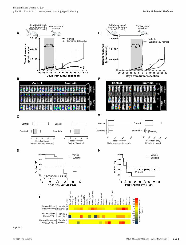
▸Figure 1. Primary tumor response to neoadjuvant sunitinib treatment is not predictive of post-surgical survival in multiple models of metastasis.
A BLI of SCID mice bearing orthotopic human SN12-PM6LUC+ renal tumors receiving neoadjuvant sunitinib for 14 days.B BLI of mice before and after nephrectomy (SN12-PM6LUC+ model).C Corresponding quantification of resected kidney BLI (left panel) and kidney weight (right panel) following neoadjuvant sunitinib treatment cessation (SN12-PM6LUC+ model).D Post-surgical survival (SN12-PM6LUC+ model).E BLI of BALB/c mice bearing orthotopic mouse RENCALUC+ renal tumors receiving neoadjuvant sunitinib for 14 days.F BLI of mice before and after nephrectomy (RENCALUC+ model).G Corresponding quantification of resected kidney BLI (left panel) and kidney weight (right panel) following neoadjuvant sunitinib treatment cessation (RENCALUC+ model).H Post-surgical survival (RENCALUC+ model).I Heatmap summary of metastatic distribution by visual scoring at individual mouse endpoint following neoadjuvant sunitinib treatment and tumor resection
(SN12-PM6LUC+, RENCALUC+, and human WM113/6-4L melanoma tumor model).
Data information: Symbols and bars for box and whiskers plot: median (line), upper/lower quartile (box), min/max (error bars). Survival analysis: hazard ratio (HR),confidence interval (CI), overall survival (OS) based on Kaplan–Meier or Cox regression analysis. N = 8–12 mice per group. BLI, bioluminescence imaging; Neoadj. Tx,neoadjuvant treatment; *P < 0.05, **P < 0.01 compared to control.
EMBO Molecular Medicine Vol 6 | No 12 | 2014 ª 2014 The Authors
EMBO Molecular Medicine Neoadjuvant antiangiogenic therapy John M L Ebos et al
1562
Published online: October 31, 2014
H
Befo
resu
rger
y
Control Sunitinib DPI
25
29
27
49
Afte
rsu
rger
y
Bio
lum
ines
cenc
e(p
hoto
ns/s
ec)
Days from tumor resection
Bio
lum
ines
cenc
e(p
hoto
ns/s
ec)
-30 -25 -20 -15 -10 -5 0 5 10 15 20 25 300
5.0 10 8
1.0 10 9
1.5 10 9 VehicleSunitinib (60 mg/kg)
VehicleSunitinib (60 mg/kg)
VehicleSunitinib
VehicleSunitinib
010
020
030
040
010
08060 120
140
A
B
C
D
I
E
F
H
Orthotopic (renal) tumor implanta�on (S12-PM6LUC+ cells)
Primary tumor resec�on
NeoAdj.Tx
Orthotopic (renal) tumor implanta�on
(RencaLUC+ cells)Primary tumor
resec�on
NeoAdj.Tx
0.40.30.20.1
0-0.1-0.2-0.3-0.4-0.5-0.6
Fold
-Cha
nge
Com
pare
d to
Co
ntro
l
# M
ice
Lung
s/D
iaph
ragm
Axill
ary
LNs
Rena
l LN
sLu
mba
r LN
sIn
guin
al L
Ns
Brac
hial
LN
sm
uscl
eLo
cal r
egro
wth
Kidn
ey (a
djac
ent)
Asci
tes
Thym
usAd
rena
l gla
nds
Ova
ries
Blad
der
Brai
nM
amm
ary
fat p
adH
eart
Live
rSt
omac
hSp
leen
Panc
reas
Inte
s�ne
s
Vehicle 12Sunitinib 12
Vehicle 3Sunitinib 7
Human Kidney(SN12-PM6LUC+)
Mouse Kidney(RencaLUC+)
Control Sunitinib DPI
29
35
30
51
020
040
060
0 010
020
030
040
0
**
G
Human Melanoma(WM113/6-4L)
VehicleSunitinib
87
Afte
rsu
rger
y
p=0.0079
Control
Suni�nib
Resected Kidney(Bioluminecence, % control)
Resected Kidney(Bioluminecence, % control)
Resected Kidney(Weight, % control)
Resected Kidney(Weight, % control)
Control
Suni�nib
Control
Suni�nib
Control
Suni�nib
Befo
resu
rger
y
Figure 1.
ª 2014 The Authors EMBO Molecular Medicine Vol 6 | No 12 | 2014
John M L Ebos et al Neoadjuvant antiangiogenic therapy EMBO Molecular Medicine
1563
Published online: October 31, 2014
into groups of corresponding size based on bioluminescence (see
Materials and Methods for details). Neoadjuvant sunitinib treatment
(60 mg/kg/day for 14 days) yielded no reductions in overall biolu-
minescence (BLI) (Fig 1A and B), or overall kidney weight or kidney
BLI following surgical removal, compared to vehicle controls
(Fig 1C, left and right panel, respectively). However, upon treat-
ment cessation and surgical removal of the kidney, sunitinib-treated
mice had significantly decreased overall survival (Fig 1D). In a
second model, BALB/c mice bearing orthotopic RENCALUC+ tumors
received neoadjuvant treatment (60 mg/kg sunitinib for 14 days)
yielding significantly reduced pre-surgical BLI (Fig 1E and F) and
reduced resected kidney BLI and weights (see Fig 1G, left and right
panel, respectively). However, these significant pre-surgical benefits
did not lead to improvements in post-surgical survival (Fig 1H). A
third model yielded similar differences in pre- and post-surgical
comparisons with vehicle-treated controls. SCID mice bearing ortho-
topic melanoma (WM113/6-4L) cells treated with neoadjuvant suni-
tinib (60 mg/kg for 14 days) led to a trend of reduced tumor size
and weight following surgical resection but yielded a trend of wors-
ened post-surgical survival (Supplementary Fig S2A–C, both did not
reach statistical significance). Metastatic sites at the endpoint were
visually assessed in all three models and compared to respective
vehicle controls to test whether neoadjuvant treatment influenced
post-surgical metastatic disease progression patterns. Stomachs and
spleens had consistent increases in metastasis compared to controls
in response to treatment in all models, but no clear trends suggested
therapy-induced progression pattern differences (Fig 1I). Together,
all three models showed that pre-surgical effects of neoadjuvant
treatment did not predict for similar effects in the post-surgical
setting with benefits (or non-benefits) leading to consistently wors-
ened outcomes (i.e., no benefit or decreased survival).
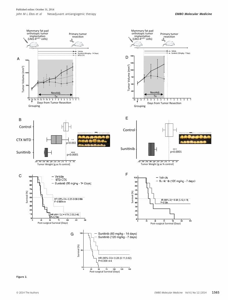
Dose, treatment duration, and surgical timing can improveneoadjuvant sunitinib treatment efficacy outcomes in ametastatic breast model
We next undertook experiments to determine whether treatment
dose, duration, and surgical resection timing had the potential to
impact post-surgical outcomes following pre-surgical neoadjuvant
therapy (Fumagalli et al, 2012). Following implantation of human
LM2-4LUC+ breast cells into the mammary fat pads of SCID mice,
sunitinib (60 mg/kg) was administered daily for 14 days prior to
surgical removal of the primary tumor. Separately, the chemothera-
peutic drug cyclophosphamide (CTX) was administered at the maxi-
mum tolerated dose (MTD) for the same period (Schott & Hayes,
2012). Both sunitinib and CTX MTD led to significant reductions in
primary tumor volume (Fig 2A) and reduced excised tumor weight
(Fig 2B) compared to vehicle-treated controls. CTX MTD neoadju-
vant treatment led to a delay in post-surgical metastatic recurrence
and improved survival; however, a similar benefit was not seen
following neoadjuvant sunitinib treatment (Fig 2C). In a separate
study using the identical cell line and implantation protocol, SCID
mice bearing orthotopic breast tumors were treated for a shortened
period (7 days) with neoadjuvant sunitinib at a higher dose
(120 mg/kg). This condensed neoadjuvant treatment protocol
yielded similar significant reductions in primary tumor volume and
weight (Fig 2D and E, respectively) as compared to controls, with
an improved post-surgical survival (Fig 2F). Shorter (7 days) higher-
dose (120 mg/kg/day) neoadjuvant sunitinib treatment showed
significantly improved survival compared to sunitinib administered
in lower doses over a longer period (60 mg/kg over 14 days, respec-
tively) (Fig 2G). Interestingly, similar observations were made in
the same model with a vascular disrupting agent, OXi4503. When
given neoadjuvantly as one high dose (50 mg/kg) on day 1 of the
7 day treatment period, it provided a significant survival advantage
over a lower dose (10 mg/kg) given twice in 14 days. These post-
surgical differences contrasted with the significant benefits observed
in the pre-surgical setting following neoadjuvant therapy (Supple-
mentary Fig S3A–G). These results confirm that primary tumor
response to neoadjuvant antiangiogenic therapy can be highly diver-
gent in predicting post-surgical improvements in survival, and treat-
ment dose, duration, and surgical timing are critical parameters
influencing the predictive potential of primary (pre-surgical) neoad-
juvant response benefit. Short-term, high-dose sunitinib or VDA
therapy may offer improved post-surgical outcomes compared to
longer-term, lower-dose treatment.
Benefits of neoadjuvant therapy before and after surgery dependon the mode of VEGF pathway inhibition
Current clinical trials involving neoadjuvant therapy and VEGF
pathway inhibition include small molecule VEGFR TKIs as well as
drugs that block VEGF binding its receptor (typically to VEGF recep-
tor 2). Recent preclinical comparisons of protein-based inhibitors of
extracellular binding of VEGF ligand to VEGFRs suggest differential
benefits in primary versus metastatic disease when compared to
VEGFR TKIs. This may depend on model, drug type, and dose used
(Paez-Ribes et al, 2009; Chung et al, 2012; Cooke et al, 2012;
Sennino et al, 2012; Guerin et al, 2013), but this has not been exam-
ined in the neoadjuvant setting where the effects of pre-surgical
▸Figure 2. Modulating neoadjuvant sunitinib dose and surgical timing can improve post-surgical survival.
A–C SCID mice implanted with LM2-4LUC+ human breast cancer cells in the mammary fat pad and treated with three neoadjuvant regimens: vehicle, sunitinib (60 mg/kg/day), or CTX MTD (100 mg, 3 times weekly) for 14 days. (A) Comparison of tumor volume by caliper measurement, and (B) comparison of tumor weightfollowing surgery (36 days post-implantation), with images of excised tumors shown (side panel). (C) Post-surgical survival following neoadjuvant CTX MTD orsunitinib treatment.
D–G SCID mice implanted with LM2-4LUC+ human breast cancer cells in the mammary fat pad and treated with vehicle or sunitinib (120 mg/kg/day) for 7 days.(D) Comparison of tumor volume by caliper measurement, and (E) comparison of tumor weight following surgery (30 days post-implantation), with images ofexcised tumors shown (side panel). (F) Post-surgical survival following short-term (high-dose) sunitinib treatment compared to control. (G) Post-surgical survivalcomparison of short-term sunitinib treatment at either high (120 mg/kg/day) or lower (60 mg/kg/day) doses.
Data information: Symbols and bars for box and whiskers plot: median (line), upper/lower quartile (box), min/max (error bars). Survival analysis: hazard ratio (HR),confidence interval (CI), overall survival (OS) based on Kaplan–Meier or Cox regression analysis. N = 8–15 mice per group. Neoadj. Tx, neoadjuvant treatment; **P < 0.01,***P < 0.001 compared to control.
EMBO Molecular Medicine Vol 6 | No 12 | 2014 ª 2014 The Authors
EMBO Molecular Medicine Neoadjuvant antiangiogenic therapy John M L Ebos et al
1564
Published online: October 31, 2014
A
B
-16 -15 -14 -13 -12 -11 -10 -9 -8 -7 -6 -5 -4 -3 -2 -1 0 10
500
1000
1500
VehicleSunitinib (60mg/kg - 14 Days)MTD CTX
C
-10 -9 -8 -7 -6 -5 -4 -3 -2 -1 0 1 2 3 4 5 6 70
250
500
750
1000
1250
1500
VehicleSunitinib (120mg/kg - 7 Days)
D
E
F
Mammary fat pad orthotopic tumor
implanta�on (LM2-4LUC+ cells)
Mammary fat pad orthotopic tumor
implanta�on (LM2-4LUC+ cells)
Primary tumor resec�on
Primary tumor resec�on
Tum
or V
olum
e (m
m3 )
Days from Tumor Resec�on
Tum
or V
olum
e (m
m3 )
Days from Tumor Resec�onGroupingGrouping
**
NeoAdj.Tx
NeoAdj.Tx
0 25 50 75 100 125 1500
50
100
Sunitinib (120 mg/kg - 7 days)Sunitinib (60 mg/kg - 14 days)
HR (95% CI)= 0.28 (0.11,0.62)P=0.009
G
p=0.002
p<0.0001p<0.0001
Control
CTX MTD
Suni�nib
Control
Suni�nib
Tumor Weight [g as % control]Tumor Weight [g as % control]
Post-surgical Survival (Days) Post-surgical Survival (Days)
Surv
ival
(%)
Surv
ival
(%)
**
Surv
ival
(%)
Post-surgical Survival (Days)
Figure 2.
ª 2014 The Authors EMBO Molecular Medicine Vol 6 | No 12 | 2014
John M L Ebos et al Neoadjuvant antiangiogenic therapy EMBO Molecular Medicine
1565
Published online: October 31, 2014

primary tumor response can be compared directly (i.e., in the same
mouse) to post-surgical metastasis and survival. We undertook
neoadjuvant treatment comparisons of protein-based and TKI-based
VEGF/VEGFR inhibition with multiple drugs. This included two
VEGF neutralizing antibodies (B20 and G6.31), a VEGFR-2 blocking
adnectin (CT322), and two VEGF RTKIs, sunitinib or axitinib.
Animals bearing orthotopic LM2-4LUC+ tumors were treated for
14 days prior to surgical resection of the primary tumor (see Supple-
mentary Table S1 for treatment schedule and dosing). Significant
differences in excised tumor weight were observed in all groups
(Fig 3A) compared to vehicle-treated controls. However, only extra-
cellular VEGF/VEGFR-2 inhibitors G6.31, B20, and CT322 showed
significant benefits in post-surgical survival compared to control
following surgery and treatment cessation, with no improvements
observed following sunitinib or axitinib therapy (Fig 3B). Similar
comparisons between VEGF RTKIs and antibodies were performed
in human melanoma, human kidney, and mouse kidney tumor
models. The cells and treatments included LM2-4LUC+ (sunitinib,
axitinib, and B20), WM113/6-4L (sunitinib and B20), RENCALUC
(sunitinib and axitinib), and SN12-PM6LUC+ (sunitinib only). Data
from a minimum of two models were analyzed together by stan-
dardizing individual mouse data to respective vehicle-treated
controls (see Materials and Methods for details). This allowed for
graphing to be depicted as having pro- or anti-primary (pre-surgical)
tumor benefits and pro- or anti-metastatic (post-surgical) benefits,
where overall survival is used as a surrogate for metastasis (see
Fig 3C, top left panel for illustration). Grouped analysis of mice
from multiple models confirmed that VEGF RTKI and VEGF anti-
body neoadjuvant treatments yielded pre-surgical anti-primary
benefits, with translation to significant post-surgical anti-metastatic
benefits only observed in the VEGF antibody groups (Fig 3C and D).
Interestingly, grouped analysis of pooled pre- and post-surgical data
allowed for evaluation of general treatment–response trends (see
Materials and Methods for details). Spearman rank correlation
analysis showed that primary tumor benefits (as compared to
control) were significantly correlated to overall outcomes in
axitinib-, sunitinib-, and B20-treated animals (Fig 3D). This suggests
that the magnitude response of the primary tumor at time of
resection (as measured by comparing to vehicle-treated group
averages) following neoadjuvant treatment may serve as a predictor
of overall post-surgical benefits, independent of whether benefit
was observed as a group in the pre- or post-surgical setting. Taken
together, our results demonstrate that the pre-surgical efficacy of
neoadjuvant therapy with an extracellular VEGF inhibitor on the
primary tumor is more predictive of post-surgical survival outcomes
than VEGFR TKI therapy and that the magnitude of tumor response
after neoadjuvant therapy may be an independent surrogate marker
of overall post-surgical benefits.
Tumor-independent host responses to therapy influenceexperimental metastasis and differ among severaltreatment types
Since neoadjuvant treatment involves systemic therapy for localized
disease, it is possible that non-tumor host responses may influence
the extravasive potential of circulating tumor cells prior to surgery.
Using a model of experimental metastasis, we administered several
anticancer regimens to SCID mice for 7 days prior to i.v. inoculation
of human breast (LM2-4LUC+) and melanoma (MeWo) cancer cells.
Treatments included chemotherapy such as CTX or UFT (a 5-fluoro-
uracil oral prodrug) administered as MTD or low-dose metronomic
(LDM) regimens, radiation (XRT), OXi4503, an ALK/c-Met inhibitor
crizotinib (PF1066), sunitinib, and several extracellular VEGF path-
way inhibitors, including, B20, G6.31, CT322, and DC101—an anti-
body blocking VEGFR-2 function (see Supplementary Table S1). In
the LM2-4LUC+ breast cell model, preconditioning mice with XRT,
MTD CTX, and sunitinib lead to significant increases in metastasis
and a decrease in survival compared to control (Fig 4A), as has
been previously shown (Ebos et al, 2009; Ebos & Kerbel, 2011). In
contrast, a range of outcomes were observed with CT322, G6.31,
and B20, OXi4503, LDM UFT, LDM CTX, and LDM CTX/UFT with
moderate detriments or improvements in overall survival seen
compared to control (Fig 4A). In a similar experiment using MeWo
cells, Cox regression analysis was used to stratify pre-treatment
effects on eventual survival outcomes compared to control. Thera-
pies listed ranged from favoring control to treatment in the following
order: sunitinib > LDM CTX/UFT > crizotinib > CT322 > OXI5403 >
DC101 > G6.31 > B20 (Fig 4B). Interestingly, in preconditioning stud-
ies in both human breast and melanoma tumor cell models, anti-VEGF
antibodies B20 and G6.31 yielded improved overall survival outcome
compared to sunitinib monotherapy, but these were not significant.
Taken together, various anticancer therapies demonstrate a range of
increased or decreased survival compared to control, with extracellu-
lar VEGF pathway inhibitors showing more benefit than intracellular
TKI-based therapy. Identifying systemic ‘host’ responses to therapy
which facilitate metastasis in an experimental metastasis model could
explain potential differential outcomes with therapy in a systemic
neoadjuvant treatment setting.
▸Figure 3. Pre-surgical effects of neoadjuvant protein-based VEGF pathway inhibition predict for improved post-surgical survival in multiplemetastasis models.
A Comparison of excised orthotopic LM2-4LUC+ breast tumor weights following 14-day neoadjuvant therapy with VEGF RTKIs (sunitinib or axitinib), protein-basedneutralizing antibodies to VEGF (G6.31 or B20), or a VEGFR-2 blocking adnectin (CT322).
B Forest plot summary of post-surgical Cox regression survival analysis following neoadjuvant treatment cessation for groups described in (A).C Combined analysis of pre- and post-surgical effects to assess effects on primary tumor and metastatic growth following 14 days of neoadjuvant treatment with
sunitinib, axitinib, or B20. Models include LM2-4LUC+ (red circle), WM113/6-4L (green square), SN12-PM6LUC+ (blue triangle), and RENCALUC+ (purple diamond).D Corresponding values for primary tumor burden, survival hazard ratio, and Spearman coefficient analysis (see Materials and Methods for details).
Data information: Symbols and bars for box and whiskers plot: median (line), upper/lower quartile (box), min/max (error bars). Survival analysis: hazard ratio (HR),confidence interval (CI), overall survival (OS) based on Kaplan–Meier or Cox regression analysis. N = 6–15 mice per group. Treatment (Tx), vehicle control (Veh). Opensymbols were used to indicate data points from animals that were still alive when the experiments were terminated. Crossed lines represent the standard deviation ofthe vehicle-treated (gray cross) and drug-treated (black cross) from primary tumor burden data (vertical line) and median survival data (horizontal line). *P < 0.05,**P < 0.01, ***P < 0.001 compared to control.
EMBO Molecular Medicine Vol 6 | No 12 | 2014 ª 2014 The Authors
EMBO Molecular Medicine Neoadjuvant antiangiogenic therapy John M L Ebos et al
1566
Published online: October 31, 2014

-50 0 50 100-100
-50
0
50
155 260
-5 0 0 5 0 1 0 0-1 0 0
-5 0
0
5 0
1 5 5 2 6 0
-1 0 0 -5 0 0 5 0 1 0 0-1 0 0
-5 0
0
5 0
1 0 0
Anti-Primary
Pro-Primary
-50 0 50 100-100
-50
0
50
155 260Effect on overall survival (post-surgical)
[% change compared to control]
Effec
t on
prim
ary
tum
or (p
re-s
urgi
cal)
[% c
hang
e co
mpa
red
to co
ntro
l]
Suni�nib
B20
Axi�nib
D Pre-Surgical Post-Surgical Pre- & Post- Surgical
Drug – Dose – Dura�onPrimary tumor burden
(% change) Hazard Ra�o Spearman Coefficient
p-value p-value p-valueSuni�nib – 60mg/kg – 14 days -32.434 <0.0001 (***) 1.244 0.366 -0.437 0.002 (**)Axi�nib – 100mg/kg – 14 days -34.091 0.002 (**) 1.179 0.698 -0.657 0.010 (*)B20 – 5mg/kg – 14 days -33.778 <0.0001 (***) 0.421 0.038 (*) -0.493 0.026 (*)
C
63/62146/11272/6857/57
Median SurvivalVehicle/Treated (days)
65/78146/162
57/5857/56
Median SurvivalVehicle/Treated (days)
Median SurvivalVehicle/Treated (days)
Mouse
Breast (LM2-4LUC+) Melanoma (WM113/6-4L)Kidney (SN12-PM6LUC+)Kidney (RENCALUC+)
Human
Model
0 .0 1 0 .1 1 1 0 1 0 0
C T3 2 2
G 6 .3 1
B 2 0
A x it in ib
S u n it in ib 0 .9 8 (0 .4 6 ,2 .0 7 ) , 0 .9 4 9 , 6 2 .0
0 .1 7 (0 .0 6 ,0 .5 1 ) , 0 .0 0 1 , 9 2 .0
0 .2 1 (0 .0 7 ,0 .6 1 ) , 0 .0 0 4 , 8 8 .0
0 .3 4 (0 .1 2 ,0 .9 6 ) , 0 .0 4 1 , 7 8 .0
H R (9 5 % C I) , P -v a lu e , M e d ia n O S
S u rv iv a l F a v o rs T x S u rv iv a l F a v o rs V e h
* *
* *
*
0 .9 4 (0 .2 9 ,3 .1 0 ) , 0 .9 2 0 , 5 8 .0
-8 0 -7 0 -6 0 -5 0 -4 0 -3 0 -2 0 -1 0 0 1 0 2 0
C T 3 2 2
G 6 .3 1
B 2 0
A xit in ib
S u n it in ib
V e h ic le
* * *
**
* **
* **
* **
T u m o r W e ig h t [g a s % c o n tro l]
* * *
A Effect on primary tumor (pre-surgical)Breast Model (LM2-4LUC+)
Effect on overall survival (post-surgical)Breast Model (LM2-4LUC+)B
Tumor
Pro-Metastasis
Anti-Metastasis
p<0.0001
p<0.0001
p<0.0001
p<0.0001
p<0.0001
Figure 3.
ª 2014 The Authors EMBO Molecular Medicine Vol 6 | No 12 | 2014
John M L Ebos et al Neoadjuvant antiangiogenic therapy EMBO Molecular Medicine
1567
Published online: October 31, 2014
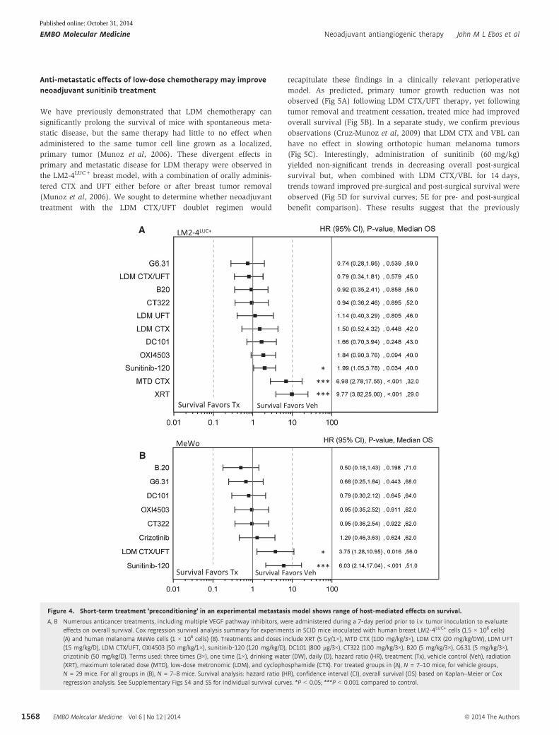
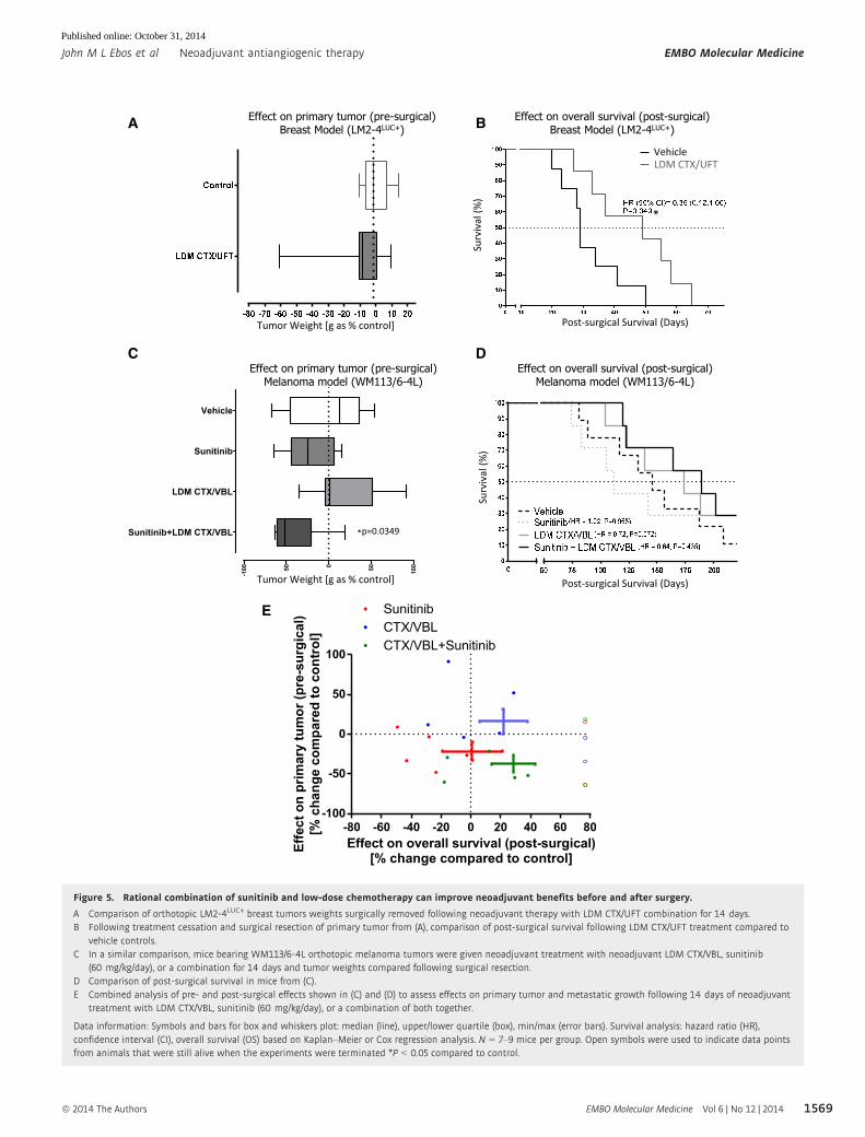
Anti-metastatic effects of low-dose chemotherapy may improveneoadjuvant sunitinib treatment
We have previously demonstrated that LDM chemotherapy can
significantly prolong the survival of mice with spontaneous meta-
static disease, but the same therapy had little to no effect when
administered to the same tumor cell line grown as a localized,
primary tumor (Munoz et al, 2006). These divergent effects in
primary and metastatic disease for LDM therapy were observed in
the LM2-4LUC+ breast model, with a combination of orally adminis-
tered CTX and UFT either before or after breast tumor removal
(Munoz et al, 2006). We sought to determine whether neoadjuvant
treatment with the LDM CTX/UFT doublet regimen would
recapitulate these findings in a clinically relevant perioperative
model. As predicted, primary tumor growth reduction was not
observed (Fig 5A) following LDM CTX/UFT therapy, yet following
tumor removal and treatment cessation, treated mice had improved
overall survival (Fig 5B). In a separate study, we confirm previous
observations (Cruz-Munoz et al, 2009) that LDM CTX and VBL can
have no effect in slowing orthotopic human melanoma tumors
(Fig 5C). Interestingly, administration of sunitinib (60 mg/kg)
yielded non-significant trends in decreasing overall post-surgical
survival but, when combined with LDM CTX/VBL for 14 days,
trends toward improved pre-surgical and post-surgical survival were
observed (Fig 5D for survival curves; 5E for pre- and post-surgical
benefit comparison). These results suggest that the previously
A
B
******
*
****
Survival Favors Tx Survival Favors Veh
Survival Favors Tx Survival Favors Veh
LM2-4LUC+
MeWo
Figure 4. Short-term treatment ‘preconditioning’ in an experimental metastasis model shows range of host-mediated effects on survival.
A, B Numerous anticancer treatments, including multiple VEGF pathway inhibitors, were administered during a 7-day period prior to i.v. tumor inoculation to evaluateeffects on overall survival. Cox regression survival analysis summary for experiments in SCID mice inoculated with human breast LM2-4LUC+ cells (1.5 × 10⁶ cells)(A) and human melanoma MeWo cells (1 × 10⁶ cells) (B). Treatments and doses include XRT (5 Gy/1×), MTD CTX (100 mg/kg/3×), LDM CTX (20 mg/kg/DW), LDM UFT(15 mg/kg/D), LDM CTX/UFT, OXI4503 (50 mg/kg/1×), sunitinib-120 (120 mg/kg/D), DC101 (800 lg/3×), CT322 (100 mg/kg/3×), B20 (5 mg/kg/3×), G6.31 (5 mg/kg/3×),crizotinib (50 mg/kg/D). Terms used: three times (3×), one time (1×), drinking water (DW), daily (D), hazard ratio (HR), treatment (Tx), vehicle control (Veh), radiation(XRT), maximum tolerated dose (MTD), low-dose metronomic (LDM), and cyclophosphamide (CTX). For treated groups in (A), N = 7–10 mice, for vehicle groups,N = 29 mice. For all groups in (B), N = 7–8 mice. Survival analysis: hazard ratio (HR), confidence interval (CI), overall survival (OS) based on Kaplan–Meier or Coxregression analysis. See Supplementary Figs S4 and S5 for individual survival curves. *P < 0.05; ***P < 0.001 compared to control.
EMBO Molecular Medicine Vol 6 | No 12 | 2014 ª 2014 The Authors
EMBO Molecular Medicine Neoadjuvant antiangiogenic therapy John M L Ebos et al
1568
Published online: October 31, 2014
-100 -50 0 50 100
Sunitinib+LDM CTX/VBL
LDM CTX/VBL
Sunitinib
Vehicle
Tumor weight (g, % of control)
*
C
BA
D
Effect on primary tumor (pre-surgical)Breast Model (LM2-4LUC+)
Effect on overall survival (post-surgical)Breast Model (LM2-4LUC+)
Effect on primary tumor (pre-surgical)Melanoma model (WM113/6-4L)
Effect on overall survival (post-surgical)Melanoma model (WM113/6-4L)
E
Effect on overall survival (post-surgical)[% change compared to control]
Effe
ct o
n pr
imar
y tu
mor
(pre
-sur
gica
l)[%
cha
nge
com
pare
d to
con
trol
]
-80 -60 -40 -20 0 20 40 60 80-100
-50
0
50
100
CTX/VBLCTX/VBL+Sunitinib
Sunitinib
p=0.0349
Surv
ival
(%)
Surv
ival
(%)
Post-surgical Survival (Days)
Post-surgical Survival
VehicleLDM CTX/UFT
(Days)
Tumor Weight [g as % control]
Tumor Weight [g as % control]
*
Figure 5. Rational combination of sunitinib and low-dose chemotherapy can improve neoadjuvant benefits before and after surgery.
A Comparison of orthotopic LM2-4LUC+ breast tumors weights surgically removed following neoadjuvant therapy with LDM CTX/UFT combination for 14 days.B Following treatment cessation and surgical resection of primary tumor from (A), comparison of post-surgical survival following LDM CTX/UFT treatment compared to
vehicle controls.C In a similar comparison, mice bearing WM113/6-4L orthotopic melanoma tumors were given neoadjuvant treatment with neoadjuvant LDM CTX/VBL, sunitinib
(60 mg/kg/day), or a combination for 14 days and tumor weights compared following surgical resection.D Comparison of post-surgical survival in mice from (C).E Combined analysis of pre- and post-surgical effects shown in (C) and (D) to assess effects on primary tumor and metastatic growth following 14 days of neoadjuvant
treatment with LDM CTX/VBL, sunitinib (60 mg/kg/day), or a combination of both together.
Data information: Symbols and bars for box and whiskers plot: median (line), upper/lower quartile (box), min/max (error bars). Survival analysis: hazard ratio (HR),confidence interval (CI), overall survival (OS) based on Kaplan–Meier or Cox regression analysis. N = 7–9 mice per group. Open symbols were used to indicate data pointsfrom animals that were still alive when the experiments were terminated *P < 0.05 compared to control.
ª 2014 The Authors EMBO Molecular Medicine Vol 6 | No 12 | 2014
John M L Ebos et al Neoadjuvant antiangiogenic therapy EMBO Molecular Medicine
1569
Published online: October 31, 2014

reported ‘anti-metastatic’ properties of low-dose metronomic
chemotherapy regimens may also include the prevention of micro-
metastatic disease following short-term pre-surgical therapy and
may reverse the muted or worsened post-surgical effects observed in
previously described neoadjuvant treatments with sunitinib.
A preclinical neoadjuvant efficacy score (NES) to compareperioperative treatment benefits
To better understand the pre- and post-surgical effects of neoadju-
vant therapy, we sought to develop a descriptive model to compare
(i) pre-surgical effects on the primary tumor, and (ii) post-surgical
outcomes. Using excised tumor weights and median survival ratios
from experiments listed in Figs 1–3 and 5 (all standardized to
vehicle-treated control groups), we established a neoadjuvant
efficacy score (NES) to generate a value of overall benefit of therapy
as ‘anti-primary’ (pre-surgical) and ‘anti-metastatic’ (post-surgical)
(see Materials and Methods). Importantly, NES values allow assess-
ment of overall therapeutic impact and identification of neoadjuvant
benefit or detriments (Fig 6A). In LM2-4LUC+, WM113/6, and
RENCALUC+ models, VEGF RTKIs sunitinib or axitinib showed low
NES values because anti-primary effects did not translate into anti-
metastatic effects, something that was in contrast to protein-based
VEGF therapy, which showed improved overall NES values
(LM2-4LUC+ and WM113/6 models only) (Fig 6B). Furthermore,
comparisons of varied neoadjuvant treatment durations and doses
can improve NES values. Short-term VDA (OXi4503) (50 mg/kg,
one dose) and sunitinib (120 mg/kg/day) treatment over 7 days
(NES 1.33 and 0.73) were superior to the same treatments at lower
doses over 14 days (NES 0.50 and 0.57, respectively, see Fig 6B
upper panel). In the WM113/6 model, NES values for LDM CTX/
VBL and sunitinib combined therapy (NES 0.67) show improvement
over sunitinib and LDM CTX/VBL treatment alone (NES �0.01 and
0.03, respectively). Intriguingly, and based on the experiments with
sunitinib treatment in the SN12-PM6LUC+ model described in Fig 1,
low NES values demonstrate the lack of overall benefit in this
neoadjuvant treatment strategy in both primary and metastatic
disease (NES �0.12). Importantly, no consistent trends were
observed that suggest neoadjuvant treatment influenced a preferred
location of eventual metastasis compared to vehicle-treated controls
(see Supplementary Results and Supplementary Fig S6). Taken
together, the use of descriptive NES values offers the potential to
serve as a predictor of anti-primary and anti-metastatic efficacy, as
well as to serve as a tool to compare treatments and predict drug
combination strategies to improve overall outcome.
Discussion
In the present study, we have identified the optimal surgical param-
eters necessary to examine short-term pre-surgical neoadjuvant
treatment effects on primary tumor growth and its influence on
(post-surgical) metastatic recurrence after therapy cessation.
Multiple therapeutic strategies were used, including protein-based
inhibitors of the VEGF ligand or receptor binding, small molecule
drugs targeting intracellular VEGFRs (e.g., VEGFR TKIs), vascular
disrupting agents, as well as chemotherapy. Herein, we demonstrate
that the pre-surgical benefits of neoadjuvant therapy do not
consistently predict for post-surgical disease recurrence and
survival, with correlations dependent on treatment dose, surgical
timing, treatment duration, and mode of VEGF pathway inhibition.
Preclinical neoadjuvant models can be used to uncover (and differ-
entiate) between ‘anti-primary’ and ‘anti-metastatic’ treatment
effects, and potentially uncover rational treatment combination
strategies to improve perioperative outcomes.
We previously demonstrated that short-term treatment with suni-
tinib prior to intravenous (i.v.) inoculation of breast and melanoma
cells could accelerate metastasis and shorten survival, despite cessa-
tion of treatment (Ebos et al, 2009). This, along with another similar
study (Paez-Ribes et al, 2009), raised the possibility that VEGF path-
way inhibition may change the natural history of tumor progression
after antiangiogenic therapy and include potential metastasis-
promoting effects. However, it remains an open question of how
clinically relevant these preclinical findings are (Miles et al, 2010;
Blagoev et al, 2013), particularly since there is growing evidence
that differential efficacies of anti-VEGF pathway inhibitors extend to
not only disease stage (i.e., primary tumor versus micro- and
macro-metastatic disease) but also among treatment types (i.e., TKIs
versus antibodies). For the latter question, mode of VEGF pathway
inhibition may play a key role in explaining different outcomes clini-
cally and preclinically (Ebos et al, 2009; Singh & Ferrara, 2012;
Guerin et al, 2013). For instance, it has been recently shown that
antibodies neutralizing VEGF do not increase experimental lung
metastasis in mice preconditioned to therapy, suggesting these
effects may be specific to VEGFR TKIs only and may be dose depen-
dent (Chung et al, 2012; Cooke et al, 2012; Welti et al, 2012). Our
results in an experimental model of metastasis where mice were
preconditioned by therapy confirm this differential effect and extend
these results to compare inhibitors of VEGFR-2 and a VDA, along
with direct comparisons to radiation and chemotherapy (including
MTD and LDM). We found that VEGFR TKIs (along with chemother-
apy and radiation) can lead to decreases in overall survival, but
differ from other VEGF inhibition strategies, where no detrimental
effects on survival were observed. Critically, despite this difference,
protein-based VEGF/R inhibitors (G6.31, B20, CT322, and DC101)
still did not result in a significant survival benefit in experimental
metastasis models, something not tested in previous publications
(Chung et al, 2012). This could explain why sunitinib-induced bene-
fits in primary tumor reduction following neoadjuvant treatment did
not translate into post-surgical survival in all instances, and why
similar antibody treatment led to improvements in overall survival.
One important consideration is that the half-life of biologics such as
antibodies is significantly different than for small molecule drugs
(2 weeks vs. 12–24 h, respectively). This means target inhibition
likely persists long after the neoadjuvant treatment window (some-
thing observed with neoadjuvant bevacizumab clinically (Starlinger
et al, 2012)) and makes preclinical comparisons with TKIs challeng-
ing to assess in the perioperative setting.
For our studies, we chose to evaluate the neoadjuvant setting to
determine whether these putative ‘pro-metastatic’ treatment effects
could be observed in a clinically relevant model. In this regard,
neoadjuvant therapy could potentially allow for testing of off-target
‘host’ effects (since it involves systemic treatment) and allow differ-
entiation between primary tumor responses and post-surgical
disease recurrence following treatment cessation. Critically, our
neoadjuvant therapy model also has the potential to predict drug
EMBO Molecular Medicine Vol 6 | No 12 | 2014 ª 2014 The Authors
EMBO Molecular Medicine Neoadjuvant antiangiogenic therapy John M L Ebos et al
1570
Published online: October 31, 2014

combinations by uncovering therapies that may yield benefits in
primary tumor reduction or metastatic prevention, but not necessar-
ily both. Indeed, therapies with only ‘anti-metastatic’ properties
raise the importance of testing drugs in mouse models where these
effects would not be overlooked (such as only testing in localized
disease) (Palmieri et al, 2007; Steeg et al, 2009). In this regard, our
results confirm the potential ‘anti-metastatic’ effects of LDM doublet
combination of UFT/CTX and raise the potential for future use in
the pre-surgical setting. It is possible that LDM co-administration
may abrogate any negative effects of TKI therapy, if observed. Inter-
estingly, current clinical trials with sunitinib in combination with
LDM chemotherapy are now under evaluation in several late-stage
C h a n g e fro m C o n tro l (% )
-10 0
-50 0 5 0
1 0 0
Ax it in ib (1 0 0 m g /k g )
S u n it in ib (6 0 m g /k g )
S u n it in ib (6 0 m g /k g )
S u n it in ib (6 0 m g /k g )
L D M C T X /VB L
C T 3 2 2
B 2 0
L D M C T X /VB L + S u n it in ib (6 0 m g /k g )
Ax it in ib (1 0 0 m g /k g )
L D M C T X /U F T
S u n it in ib (6 0 m g /k g )
L D M C T X /U F T
O X I4 5 0 3 (1 0 m g /k g )
C T 3 2 2
S u n it in ib (1 2 0 m g /k g )
M T D C T X
G 6 .3 1
B 2 0
O X I4 5 0 3 (5 0 m g /k g )
e ffe c t o n o v e ra ll s u rv iv a l
e ffe c t o n p r im a ry tu m o r
(7 )
G re a te s tB e n e fit N E S
-4 6%87% M e t 1 .3 3
(1 4 )
(7 )
(1 4 )
(1 4 )
(1 4 )
(1 4 )
(1 4 )
(1 4 )
(7 )
(1 4 )
(1 4 )
(1 4 )
(1 4 )
(1 4 )
(1 4 )
(1 4 )
(1 4 )
P r im 0 .9 2
P r im 0 .2 4
P r im 0 .4 5
P r im 0 .8 9
N o n e -0 .1 5
M e t 0 .8 4
P r im 0 .7 3
P r im 0 .6 9
P r im -0 .0 2
P r im 0 .5 8
M e t 0 .0 3
M e t 0 .5 7
P r im 0 .1 8
P r im 0 .5 7
P r im 0 .4 1
M e t 0 .4 4
P r im 0 .6 7
P r im 0 .3 1
-5 2%
-2 5%
-4 6%
-5 1%
6%
-3 4%
-4 6%
-4 2%
-2 2%
-5 6%
16%
-2 2%
-2 0%
-5 9%
-2 1%
-6%
-3 8%
-3 5%
-1%
-2%
40%
39%
-9%
50%
26%
27%
-2 3%
2%
19%
35%
-1%
-2%
20%
38%
29%
-5%
D a y s T x
(1 4 )
Brea
stM
elan
oma
Kidn
eyEffect of treatment on
Primary TumorNeoadjuvant efficacy
score (NES)Indication where treatment has
greatest benefit
BenefitBenefit 2primary
metastasis
<1<1
Effect of treatment on Overall Survival
Both / None
Pre-surgical Post-surgical
A
B
Kidn
eyM
ouse
Hum
an
Figure 6. Comparison of primary tumor and overall survival benefits to define neoadjuvant efficacy score.
A Neoadjuvant efficacy score (NES) was defined as the difference between measured benefit of primary tumor response to therapy, and overall survival (OS) benefitafter therapy is stopped and tumor resected. All values are compared to control with negative (�) and positive (+) indicating improvement or detriment for primarytumor benefit, respectively, and vice versa for overall survival benefit.
B NES values for all therapies tested in breast, melanoma, and kidney models shown in Figs 1–3 and 5. See Materials and Methods for NES value determination details.
ª 2014 The Authors EMBO Molecular Medicine Vol 6 | No 12 | 2014
John M L Ebos et al Neoadjuvant antiangiogenic therapy EMBO Molecular Medicine
1571
Published online: October 31, 2014
disease types, including in breast (NCT00513695) and in pediatric
tumors (Navid et al, 2013), as well as in the neoadjuvant setting in
RCC (with cyclophosphamide) (Khattak et al, 2012).
One key rationale for the use of neoadjuvant chemotherapy in
breast cancer patients is that pCR observed in the primary tumor
may have value in predicting future (post-surgical) benefit (by
delaying recurrence) (Fumagalli et al, 2012). It may also have the
advantage of guiding clinicians’ choice of post-surgical therapy in
the case of recurrence if a potent initial response is observed (von
Minckwitz et al, 2012b). However, there is no preclinical evidence
to support this relationship for antiangiogenic therapy nor have the
treatment variables such as dose, treatment duration, and OST been
tested for their potential to influence post-surgical survival
outcomes in models of spontaneous (post-surgical) metastatic
disease. In terms of dose, our findings showing that different
concentrations of sunitinib and OXi4503 can improve initial differ-
ential effects between pre- and post-surgical treatment effects could
be of immediate clinical importance. This follows previous preclini-
cal studies demonstrating that higher VEGFR TKI dosing in mice can
improve efficacy when compared with the same drug given in lower
concentrations, more frequently (Wang et al, 2011). Clinically, the
use of sunitinib given to patients in doses of 50 mg daily for an
intermittent 4 weeks on/2 weeks off schedule showed no improve-
ment when given in a continuous, lower dose (37.5 mg daily, with
no breaks) (Motzer et al, 2012). Indeed, higher sunitinib dosing is
currently under study in mRCC patients with progressive disease if
toxicity is tolerated (Bjarnason et al, 2013; Pili et al, 2013). Our
results showing increased dose and shortened surgical window
overcoming putative negative (or negligible) post-surgical impact on
overall survival could warrant consideration in clinical neoadjuvant
trials with VEGFR TKIs, where parameters of tumor dosing and
tumor size are still being investigated in terms of assessing overall
benefit (Kroon et al, 2013). Already evidence from retrospective
studies investigating pre-surgical cytoreductive sunitinib treatment
in RCC suggest that parameters of treatment stage (Bex et al, 2011)
and primary tumor reduction (Abel et al, 2011) may play a signifi-
cant role in patient outcomes. In this regard, our results demonstrat-
ing that the magnitude of primary tumor response following
neoadjuvant therapy correlates with overall survival could support
these findings. Furthermore, it is also possible that alterations in
standard pre-surgical dosing could alleviate concerns about poten-
tial break periods, or gaps in treatment, that typically occur in
patients receiving neoadjuvant therapy (e.g., because of toxicity).
Related to this, recent retrospective studies in RCC patients receiving
pre-surgical VEGFR TKIs showed an increase in proliferative tumor
endothelial cells (ECs) in those patients who had a longer treatment
break before surgery (Ebos & Pili, 2012; Griffioen et al, 2012). But
the same study showed that bevacizumab did not yield similar
elevations in proliferating ECs. In our studies, we found that eleva-
tions in proliferating tumor cell populations in the resected primary
tumor following neoadjuvant therapy (as measured by Ki67 levels)
may correlate with post-surgical survival benefits. Interestingly, we
found that increases in tumoral Ki67 following neoadjuvant B20 and
CT322 treatment predicted for decreased survival, whereas the
opposite was observed following sunitinib treatment with elevated
Ki67 levels predicting for prolonged survival (see Supplementary
Results and Supplementary Fig S7). The basis for this difference
is currently unknown but could merit further investigations that
examine whether treatment gaps can influence post-surgical survival
and/or metastatic disease distribution in the neoadjuvant setting.
Taken together, while traditional cytotoxic treatments (such as
chemotherapy) in the neoadjuvant setting have typically resulted in
improved survival following surgical intervention, similar benefits
with antiangiogenic therapy remain largely untested. Herein, we
have identified a novel methodology for evaluating neoadjuvant effi-
cacy using a spontaneous surgical metastasis model and show how
it can be used to explain differential efficacies of VEGF pathway
inhibitors seen preclinically and clinically. These findings may be
immediately relevant to numerous perioperative trials underway in
patients.
Materials and Methods
Drugs and doses used
Drugs used in this study include SU11248/sunitinib malate,
AG013736/ axitinib, and an ALK/c-Met inhibitor, crizotinib/PF1066
(Pfizer Inc, New York); UFT, a 5-fluorouracil pro-drug (Taiho,
Japan); anti-VEGF antibodies, G6.31 and B20 (Genentech, Roche);
targeted adnectin inhibitor of VEGFR-2, CT322 (Adnexus, Waltham,
MA); anti-VEGFR-2 antibody, DC101 (ImClone Systems/Eli Lilly,
New York); vascular disrupting agent (VDA), OXi4503 (Oxigene,
San Francisco, CA); cyclophosphamide (CTX) (Baxter Oncology
GmbH, Mississauga, Ontario, Canada); vinblastine sulfate (VBL).
All doses, treatment durations, and formulations are summarized in
Supplementary Information.
Mouse tumor models
Animal tumor model studies were performed in strict accordance
with the recommendations in the Guide for Care and Use of Labora-
tory Animals of the National Institutes of Health and according to
guidelines of the Canadian Council on Animal Care. Protocols used
were approved by the Sunnybrook Health Sciences Centre Animal
Care Committee (for studies using LM2-4LUC+, SN12-PM6LUC+,
MeWo, and WM113/6-4L cell lines) and by the institutional Animal
Care and Use Committee (IACUC) at Roswell Park Cancer Institute
(for studies using LM2-4LUC+ and RENCALUC+; Protocol: 1227M).
Experimental metastasis assays
LM2-4LUC+ (1.5 × 10⁶ cells) or human MeWo melanoma (1 × 10⁶
cells) were injected directly into the tail vein of 6- to 8-week-old
female CB-17 SCID mice (Charles River, Canada) as previously
described (Ebos et al, 2009).
Human xenograft and mouse syngenic orthotopic tumor implantation
and primary tumor resection
LM2-4LUC+ cells (2 × 10⁶ cells in 50 µl), WM113/6-4L (1 × 10⁶ cells
in 150 µl), SN12-PM6LUC+ (2 × 10⁶ cells in 5 µl), and RENCALUC+
(4 × 10⁴ cells in 5 µl) were orthotopically implanted, respectively,
into the right inguinal mammary fat pads (right flank), dermis (right
flank), or kidney (subcapsular space) of 6- to 8-week-old female
CB-17 SCID or BALB/c mice as previously described (Munoz et al,
2006; Cruz-Munoz et al, 2008; Tracz et al, 2014). Primary mela-
noma and breast tumor size was assessed regularly with vernier
EMBO Molecular Medicine Vol 6 | No 12 | 2014 ª 2014 The Authors
EMBO Molecular Medicine Neoadjuvant antiangiogenic therapy John M L Ebos et al
1572
Published online: October 31, 2014
calipers using the formula width2 × length × 0.5. Breast and kidney
tumor models utilized bi-weekly bioluminescent monitoring, which
has previously been demonstrated to parallel overall tumor burden
(Ebos et al, 2009). Prior to neoadjuvant therapy, mice were grouped
according to tumor burden (melanoma and breast according to
tumor size, and kidney according to bioluminescence), ensuring that
the mean average was not statistically different. Representative
examples of neoadjuvant pre-treatment sorting to standardize group-
ing are shown for SN12-PM6LUC+ (Supplementary Fig S8A),
RENCALUC+ (Supplementary Fig S8B), WM113/6-4L (Supplementary
Fig S8C), and LM2-4LUC+ cells (Supplementary Fig S8D). Optimal
surgical times (OST) were determined by assessing metastatic poten-
tial (MP) following surgery at various time points to determine prob-
ability of spontaneous metastasis not derived from local invasion
(see Supplementary Fig S1E–G). OST determination was made using
vehicle-treated controls from various experiments. No difference in
metastatic disease progression patterns or survival has been noted
between vehicles or between vehicle and untreated animals.
Approximate tumor weights to determine OST were based on previ-
ous studies with LM2-4LUC+ and WM113/6-4L tumor sizes of
~400 mm3 and ~200 mm3, respectively as previously described
(Cruz-Munoz et al, 2008; Ebos et al, 2009). For kidney tumor
model, 1–2 days after cessation of neoadjuvant therapy, kidney
nephrectomy was performed. Excised kidneys were examined for
encapsulated tumor. If any tumor invasion into the peritoneal space
was noted, the mouse was removed from the study. For kidney
models, in any rare instance where no tumor was ever present at
any time before and after surgery and treatments (determined by
BLI or visible macroscopically), mice were excluded from the study
so as not to give potential false positive or negative bias to results.
Defining parameters for establishing a neoadjuvanttreatment period
Residual cancer burden (RCB)
During surgical resection of primary tumor, any residual or localized
tumor invasion was noted and RCB was broadly compared to clini-
cal pathological complete response (pCR). Acceptable RCB was
defined as not having visual residual tumor or obvious local inva-
sion. The presence of bioluminescent-positive tumor following
surgery (shown in Supplementary Fig S1F) indicated micrometastatic
disease, but was considered acceptable since this detection sensitiv-
ity exceeds clinical comparisons. Unacceptable RCB was defined as
any localized invasion or unresectable primary tumor mass. In
melanoma and breast models, skin or peritoneal wall was removed
if invasion was noted, with clear margins taken of normal tissue. A
mouse was excluded from the study if clear margins were not attain-
able. In the kidney model, any subcapsular tumors growing outside
of the kidney were noted as ‘invasive’ and ensured to not have
invaded surrounding tissue prior to nephrectomy.
Justification of timing between treatment cessation and surgery
All treatments in the experimental metastasis model or neoadjuvant
perioperative model included treatments that were halted 24 h
before surgery. We previously demonstrated therapy cessation in
this timeframe (Ebos et al, 2009), and pre-surgical sunitinib treat-
ment in RCC patients has included 24 h in certain trials, though this
varies among ongoing clinical trials (Schrader et al, 2012).
Neoadjuvant Efficacy Score (NES)
Independent parameters of neoadjuvant efficacy in our studies
included primary tumor response (PTR), which compared resected
primary tumor weight (RPTW) following treatment to control, and
overall survival response (OSR), which compared the median
survival after treatment to control. These quantitative measure-
ments were combined as a descriptive analysis of overall treatment
benefit as a neoadjuvant efficacy score (NES), with the aim to
compare PTR and OSR among treatments. The following equation
was used to establish the NES, with indication (i.e., PTR or OSR or
none) with the greatest benefit noted.
PTR ð%Þ ¼ TreatedRPTW ðAVGÞ � Vehicle RPTW ðAVGÞVehicle RPTW ðAVGÞ � 100
OSR ð%Þ ¼ TreatedOS ðmedianÞ � VehicleOS ðmedianÞVehicleOS ðmedianÞ �100
NES ¼ OSR� PTR
100
Statistical analysis
Results were subjected to statistical analysis using the GraphPad
Prism software package v.4.0 (GraphPad Software Inc., San Diego,
CA), IBM SPSS Statistics v22.0 (IBM Corp., Armonk, NY), and SAS
v9.3 (Cary, NC). All growth curves shown are represented as
mean � standard deviation (SD). Overall survival was summarized
using the Kaplan–Meier method with the association between treat-
ment group and survival evaluated using the two-sided log-rank
test. Cox regression models were used to obtain hazard ratio esti-
mates, with corresponding 95% confidence intervals, for comparing
treatment groups to control. Correlation plots for combined pre- and
post-surgical summary analysis (Figs 3C and 5E) were conducted as
follows. Pre-surgical primary tumor effects: Resected tumor or
tumor-bearing kidney weights normalized to control animals follow-
ing one-sample one-tailed t-test comparison. Post-surgical survival
effects: HR based on Cox regression analysis; median survival based
on Kaplan–Meier analysis. Pre-/post-surgical correlation: Spearman
rank correlation used one-tail test for significance (linear regression
avoided due to censored data). Student’s t-tests were one-tailed and
unpaired. A minimum significance level of 0.05 was used for all
analysis.
Supplementary information for this article is available online:
http://embomolmed.embopress.org
AcknowledgementsWe would like to thank Dr. Alejandro Godoy and Dr. Gary Smith for valuable
technical assistance with staining protocols; Dr. Ping Xu for OXi4503 dosing
advice; Dr. Roberto Pili for the RENCALUC+ cells; Petia Stefanova for construc-
tion of the TMAs; and several companies for drug. These include Pfizer for
sunitinib and axitinib (Dr. J. Christensen); ImClone Systems for DC101 (Dr.
Bronek Pytowski); Genentech for B.20 and G6.31 (Dina Washington); Adnexus
for CT322 (Eric Furfine); Taiho Pharmaceuticals for UFT (Teiji Takechi). This
study has been carried out with grant support from the following: Canadian
Breast Cancer Foundation (to R.S.K); the Canadian Institutes of Health and
ª 2014 The Authors EMBO Molecular Medicine Vol 6 | No 12 | 2014
John M L Ebos et al Neoadjuvant antiangiogenic therapy EMBO Molecular Medicine
1573
Published online: October 31, 2014

Science (to R.S.K); the Ontario Institute for Cancer Research (to R.S.K); and the
Roswell Park Alliance Foundation (to J.M.L.E).
Author contributionsStudy conception and design (JMLE, CRL, MM, RSK). Acquisition of data (JMLE,
CRL, MM, AT, WRC, CJ). Analysis and interpretation of data (JMLE, CRL, MM, AT,
JMH, KA, PB, WRC, CJ). Manuscript writing (JMLE, CRL, MM, RSK).
Conflict of interestThe authors declare that they have no conflict of interest.
References
Abel EJ, Culp SH, Tannir NM, Tamboli P, Matin SF, Wood CG (2011) Early
primary tumor size reduction is an independent predictor of improved
overall survival in metastatic renal cell carcinoma patients treated with
sunitinib. Eur Urol 60: 1273 – 1279
Bear HD, Tang G, Rastogi P, Geyer CE Jr, Robidoux A, Atkins JN, Baez-Diaz L,
Brufsky AM, Mehta RS, Fehrenbacher L et al (2012) Bevacizumab added
to neoadjuvant chemotherapy for breast cancer. N Engl J Med 366:
310 – 320
Bex A, Blank C, Meinhardt W, van Tinteren H, Horenblas S, Haanen J (2011)
A phase II study of presurgical sunitinib in patients with metastatic
clear-cell renal carcinoma and the primary tumor in situ. Urology 78:
832 – 837
Bex A, Haanen J (2014) Do targeted agents offer clinical benefit as presurgical
therapy? World J Urol 32: 3 – 8
Bjarnason GA, Naveen BS, Knox JJ, Kollmannsberger M, Reaume N, zalewski P,
ZMacfarlane RJ, MacKean MJ, Hotte SJ, Yick D et al (2013) A phase II
multicenter study of the efficacy and safety of sunitinib given on an
individualized schedule as first-line therapy for metastatic renal cell
cancer. ASCO Annual Meeting #TPS4594
Blagoev KB, Wilkerson J, Stein WD, Motzer RJ, Bates SE, Fojo AT (2013)
Sunitinib does not accelerate tumor growth in patients with metastatic
renal cell carcinoma. Cell Rep 3: 277 – 281
Bruix JTT, Mazzaferro V, Chau G, Yang J, Kudo M, Cai J, Poon RT, Han K, Tak
W, Lee HC et al (2014) STORM: A phase III randomized, double-blind,
placebo-controlled trial of adjuvant sorafenib after resection or ablation
to prevent recurrence of hepatocellular carcinoma (HCC). ASCO Annual
Meeting #4006
Cameron D, Brown J, Dent R, Jackisch C, Mackey J, Pivot X, Steger GG, Suter
TM, Toi M, Parmar M et al (2013) Adjuvant bevacizumab-containing
therapy in triple-negative breast cancer (BEATRICE): primary results of a
randomised, phase 3 trial. Lancet Oncol 14: 933 – 942
Chung AS, Kowanetz M, Wu X, Zhuang G, Ngu H, Finkle D, Komuves L, Peale
F, Ferrara N (2012) Differential drug class-specific metastatic effects
following treatment with a panel of angiogenesis inhibitors. J Pathol 227:
404 – 416
Cooke VG, LeBleu VS, Keskin D, Khan Z, O’Connell JT, Teng Y, Duncan MB,
Xie L, Maeda G, Vong S et al (2012) Pericyte depletion results in
hypoxia-associated epithelial-to-mesenchymal transition and
metastasis mediated by met signaling pathway. Cancer Cell 21:
66 – 81
Cruz-Munoz W, Man S, Xu P, Kerbel RS (2008) Development of a preclinical
model of spontaneous human melanoma central nervous system
metastasis. Cancer Res 68: 4500 – 4505
Cruz-Munoz W, Man S, Kerbel RS (2009) Effective treatment of advanced
human melanoma metastasis in immunodeficient mice using
combination metronomic chemotherapy regimens. Clin Cancer Res 15:
4867 – 4874
Ebos JM, Kerbel RS (2011) Antiangiogenic therapy: impact on invasion,
disease progression, and metastasis. Nat Rev Clin Oncol 8:
210 – 221
Ebos JM, Lee CR, Bogdanovic E, Alami J, Van Slyke P, Francia G, Xu P,
Mutsaers AJ, Dumont DJ, Kerbel RS (2008) Vascular endothelial growth
factor-mediated decrease in plasma soluble vascular endothelial growth
factor receptor-2 levels as a surrogate biomarker for tumor growth. Cancer
Res 68: 521 – 529
Ebos JM, Lee CR, Cruz-Munoz W, Bjarnason GA, Christensen JG, Kerbel RS
(2009) Accelerated metastasis after short-term treatment with a potent
inhibitor of tumor angiogenesis. Cancer Cell 15: 232 – 239
Ebos JM, Pili R (2012) Mind the gap: potential for rebounds during
antiangiogenic treatment breaks. Clin Cancer Res 18: 3719 – 3721
The paper explained
ProblemA hypothesis guiding the clinical development of new cancer therapy isthat benefits observed in patients with late-stage metastatic diseasemay also have benefits in the treatment of earlier cancer stages. Withthe approval of antiangiogenic drugs that target the vascular endothelialgrowth factor (VEGF) pathway in the metastatic setting, several trialshave been initiated in the pre-surgical setting, which aim to treat alocalized primary tumor before it is removed (termed ‘neoadjuvant’therapy). Yet there are few, if any, preclinical studies that aim to recapit-ulate neoadjuvant treatment, and none have involved rigorous compar-ison of multiple VEGF pathway inhibition strategies. The reason for thisis, at least in part, because few preclinical tumor models effectivelymimic spontaneous metastatic growth following surgical resection ofan orthotopic tumor. Additional challenges include the need to definethe optimal time to start/stop neoadjuvant therapy and perform surgery,as well as allow comparisons of different doses and drug types—all ofwhich may influence pre- and post-surgical disease progression.
ResultsIn this study, we used four mouse models of spontaneous metastaticdisease—each established by first implanting cells orthotopically andthen surgically removing the primary tumor. We then identified theoptimal surgical parameters necessary to compare effects of short-term pre-surgical neoadjuvant treatment on post-surgical metastaticrecurrence after therapy cessation. Our results show that primarytumor reductions following neoadjuvant VEGF RTKIs do not consis-tently correlate with post-surgical metastatic recurrence rates, whichcan be offset by reduced survival in some instances. Importantly, wefound that negative post-surgical effects could be reversed withincreased dose, shorter treatment duration, and earlier surgical times.Interestingly, protein-based VEGF pathway inhibitors (such as antibod-ies) provide an example of how drug efficacies can differ within drugclasses, with survival benefits typically improved.
ImpactWe describe for the first time a novel preclinical methodology toexamine the effects of neoadjuvant therapy in a clinically relevantsetting. These models can be used as a tool to differentiate ‘anti-primary’ and ‘anti-metastatic’ effects. Furthermore, we present a novelscoring system that could serve to predict drug combinations thatmaximize neoadjuvant VEGF RTKI treatment benefits. This includesaltering dose and treatment duration to improve post-surgicaloutcomes as well as combination with low-dose metronomic chemo-therapy regimens shown to have anti-metastatic activity.
EMBO Molecular Medicine Vol 6 | No 12 | 2014 ª 2014 The Authors
EMBO Molecular Medicine Neoadjuvant antiangiogenic therapy John M L Ebos et al
1574
Published online: October 31, 2014
Francia G, Cruz-Munoz W, Man S, Xu P, Kerbel RS (2011) Mouse models of
advanced spontaneous metastasis for experimental therapeutics. Nat Rev
Cancer 11: 135 – 141
Fumagalli D, Bedard PL, Nahleh Z, Michiels S, Sotiriou C, Loi S, Sparano JA,
Ellis M, Hylton N, Zujewski JA et al (2012) A common language in
neoadjuvant breast cancer clinical trials: proposals for standard definitions
and endpoints. Lancet Oncol 13: e240 – e248
de Gramont A, Van CE, Schmoll HJ, Tabernero J, Clarke S, Moore MJ,
Cunningham D, Cartwright TH, Hecht JR, Rivera F et al (2012)
Bevacizumab plus oxaliplatin-based chemotherapy as adjuvant treatment
for colon cancer (AVANT): a phase 3 randomised controlled trial. Lancet
Oncol 13: 1225 – 1233
Griffioen AW, Mans LA, de Graaf AM, Nowak-Sliwinska P, de Hoog CL, de Jong
TA, Vyth-Dreese FA, van Beijnum JR, Bex A, Jonasch E (2012) Rapid
angiogenesis onset after discontinuation of sunitinib treatment of renal
cell carcinoma patients. Clin Cancer Res 18: 3961 – 3971
Guerin E, Man S, Xu P, Kerbel RS (2013) A model of postsurgical advanced
metastatic breast cancer more accurately replicates the clinical efficacy of
antiangiogenic drugs. Cancer Res 73: 2743 – 2748
Jayson GC, Hicklin DJ, Ellis LM (2012) Antiangiogenic therapy-evolving view
based on clinical trial results. Nat Rev Clin Oncol 9: 297 – 303
de John K (2012) Speeding discoveries with neoadjuvant studies. Cancer
Discov 2: 294
Khattak MA, Edmonds K, Khabra K, Sohaib A, Pennert K, Pickering L, Gore
M, Larkin J (2012) Interim results of a phase II study of sunitinib and
low dose metronomic cyclophosphamide in advanced renal cell cancer.
ESMO Annual Meeting #827P
Kroon BK, de Bruijn R, Prevoo W, Horenblas S, Powles T, Bex A (2013)
Probability of downsizing primary tumors of renal cell carcinoma by
targeted therapies is related to size at presentation. Urology 81:
111 – 115
Miles D, Harbeck N, Escudier B, Hurwitz H, Saltz L, Van Cutsem E, Cassidy J,
Mueller B, Sirzen F (2010) Disease course patterns after discontinuation of
bevacizumab: pooled analysis of randomized phase III trials. J Clin Oncol
29: 83 – 88
von Minckwitz G, Eidtmann H, Rezai M, Fasching PA, Tesch H, Eggemann H,
Schrader I, Kittel K, Hanusch C, Kreienberg R et al (2012a) Neoadjuvant
chemotherapy and bevacizumab for HER2-negative breast cancer. N Engl J
Med 366: 299 – 309
von Minckwitz G, Untch M, Blohmer JU, Costa SD, Eidtmann H, Fasching PA,
Gerber B, Eiermann W, Hilfrich J, Huober J et al (2012b) Definition and
impact of pathologic complete response on prognosis after neoadjuvant
chemotherapy in various intrinsic breast cancer subtypes. J Clin Oncol 30:
1796 – 1804
Motzer RJ, Hutson TE, Olsen MR, Hudes GR, Burke JM, Edenfield WJ, Wilding
G, Agarwal N, Thompson JA, Cella D et al (2012) Randomized phase II trial
of sunitinib on an intermittent versus continuous dosing schedule as
first-line therapy for advanced renal cell carcinoma. J Clin Oncol 30:
1371 – 1377
Mountzios G, Pentheroudakis G, Carmeliet P (2014) Bevacizumab and
micrometastases: revisiting the preclinical and clinical rollercoaster.
Pharmacol Ther 141: 117 – 124
Munoz R, Man S, Shaked Y, Lee C, Wong J, Francia G, Kerbel RS (2006) Highly
efficacious non-toxic treatment for advanced metastatic breast cancer
using combination UFT-cyclophosphamide metronomic chemotherapy.
Cancer Res 66: 3386 – 3391
Navid F, Baker SD, McCarville MB, Stewart CF, Billups CA, Wu J, Davidoff AM,
Spunt SL, Furman WL, McGregor LM et al (2013) Phase I and clinical
pharmacology study of bevacizumab, sorafenib, and low-dose
cyclophosphamide in children and young adults with refractory/recurrent
solid tumors. Clin Cancer Res 19: 236 – 246
Padera TP, Kuo AH, Hoshida T, Liao S, Lobo J, Kozak KR, Fukumura D, Jain RK
(2008) Differential response of primary tumor versus lymphatic metastasis
to VEGFR-2 and VEGFR-3 kinase inhibitors cediranib and vandetanib. Mol
Cancer Ther 7: 2272 – 2279
Paez-Ribes M, Allen E, Hudock J, Takeda T, Okuyama H, Vinals F, Inoue M,
Bergers G, Hanahan D, Casanovas O (2009) Antiangiogenic therapy elicits
malignant progression of tumors to increased local invasion and distant
metastasis. Cancer Cell 15: 220 – 231
Palmieri D, Chambers AF, Felding-Habermann B, Huang S, Steeg PS (2007)
The biology of metastasis to a sanctuary site. Clin Cancer Res 13:
1656 – 1662
Pili R, adelaiye R, Miles KM, Ciamporcero E, Sotomayor P, Bjarnason GA
(2013) Overcoming sunitinib-induced resistance by dose escalation in
renal cell carcinoma: evidence in animal models and patients. ASCO
Annual Meeting #4582
Schott AF, Hayes DF (2012) Defining the benefits of neoadjuvant
chemotherapy for breast cancer. J Clin Oncol 30: 1747 – 1749
Schrader AJ, Steffens S, Schnoeller TJ, Schrader M, Kuczyk MA (2012)
Neoadjuvant therapy of renal cell carcinoma: a novel treatment option in
the era of targeted therapy? Int J Urol 19: 903 – 907
Sennino B, Ishiguro-Oonuma T, Wei Y, Naylor RM, Williamson CW,
Bhagwandin V, Tabruyn SP, You WK, Chapman HA, Christensen JG et al
(2012) Suppression of tumor invasion and metastasis by concurrent
inhibition of c-Met and VEGF signaling in pancreatic neuroendocrine
tumors. Cancer Discov 2: 270 – 287
Silberstein JL, Millard F, Mehrazin R, Kopp R, Bazzi W, Diblasio CJ, Patterson
AL, Downs TM, Yunus F, Kane CJ et al (2010) Feasibility and efficacy of
neoadjuvant sunitinib before nephron-sparing surgery. BJU Int 109:
1270 – 1276
Singh M, Ferrara N (2012) Modeling and predicting clinical efficacy for drugs
targeting the tumor milieu. Nat Biotechnol 30: 648 – 657
de Souza BM, Chaves KB, Chammas R, Schor N, Bellini MH (2012)
Endostatin neoadjuvant gene therapy extends survival in an orthotopic
metastatic mouse model of renal cell carcinoma. Biomed Pharmacother
66: 237 – 241
Starlinger P, Alidzanovic L, Schauer D, Maier T, Nemeth C, Perisanidis B,
Tamandl D, Gruenberger B, Gruenberger T, Brostjan C (2012) Neoadjuvant
bevacizumab persistently inactivates VEGF at the time of surgery despite
preoperative cessation. Br J Cancer 107: 961 – 966
Steeg PS, Anderson RL, Menash B, Chambers AF, Eccles SA, Hunter K, Itoh K,
Kang Y, Matrisian LM, Sleeman JP et al (2009) Preclinical drug
development must consider the impact on metastasis. Clin Cancer Res 15:
4529 – 4530
Tanvetyanon T, Clark JI, Campbell SC, Lo SS (2005) Neoadjuvant therapy: an
emerging concept in oncology. South Med J 98: 338 – 344
Tracz A, Mastri M, Lee CR, Pili R, Ebos JM (2014) Modeling spontaneous
metastatic renal cell carcinoma (mRCC) in mice following nephrectomy.
J Vis Exp e51485
Van Cutsem E, Labianca R, Bodoky G, Barone C, Aranda E, Nordlinger B,
Topham C, Tabernero J, Andre T, Sobrero AF et al (2009) Randomized
phase III trial comparing biweekly infusional fluorouracil/leucovorin alone
or with irinotecan in the adjuvant treatment of stage III colon cancer:
PETACC-3. J Clin Oncol 27: 3117 – 3125
van der Veldt AA, Meijerink MR, van den Eertwegh AJ, Bex A, de Gast G,
Haanen JB, Boven E (2008) Sunitinib for treatment of advanced
ª 2014 The Authors EMBO Molecular Medicine Vol 6 | No 12 | 2014
John M L Ebos et al Neoadjuvant antiangiogenic therapy EMBO Molecular Medicine
1575
Published online: October 31, 2014

renal cell cancer: primary tumor response. Clin Cancer Res 14:
2431 – 2436
Wang X, Zhang L, Goldberg SN, Bhasin M, Brown V, Alsop DC, Signoretti S,
Mier JW, Atkins MB, Bhatt RS (2011) High dose intermittent sorafenib
shows improved efficacy over conventional continuous dose in renal cell
carcinoma. J Transl Med 9: 220
Welti JC, Powles T, Foo S, Gourlaouen M, Preece N, Foster J, Frentzas S,
Bird D, Sharpe K, van Weverwijk A et al (2012) Contrasting effects of
sunitinib within in vivo models of metastasis. Angiogenesis 15:
623 – 641
License: This is an open access article under the
terms of the Creative Commons Attribution 4.0
License, which permits use, distribution and reproduc-
tion in any medium, provided the original work is
properly cited.
EMBO Molecular Medicine Vol 6 | No 12 | 2014 ª 2014 The Authors
EMBO Molecular Medicine Neoadjuvant antiangiogenic therapy John M L Ebos et al
1576
Published online: October 31, 2014