neonatal cholestasis: recent insights
TRANSCRIPT

REVIEW Open Access
Neonatal cholestasis: recent insightsRamy Mohamed Ghazy1* and Mohamed Ahmed Khedr2
Abstract
Background: Neonatal physiological jaundice is a common benign condition that rarely extends behind the secondweek of life; however, it may interfere with the diagnosis of a pathological condition termed neonatal cholestasis (NC).The latter is a critical, uncommon problem characterized by conjugated hyperbilirubinaemia. This review aims tohighlight the differences between physiological and pathological jaundice, identify different causes of NC, and providea recent approach to diagnosis and management of this serious condition.
Main text: NC affects 1/2500 live births, resulting in life-threatening complications due to associated hepatobiliary ormetabolic abnormalities. NC is rarely benign and indicates the presence of severe underlying disease. If jaundiceextends more than 14 days in full-term infants or 21 days in preterm infants, the serum bilirubin level fractionated intoconjugated (direct) and unconjugated (indirect) bilirubin should be measured. A stepwise diagnostic approach startswith obtaining a complete history, and a physical examination which are valuable for the rapid diagnosis of theunderlying disease. The most frequently diagnosed causes of NC are biliary atresia (BA) and idiopathic neonatalhepatitis (INH). The early diagnosis of NC ensures more accurate management and better prognosis. Despite theunavailability of any specific treatments for some causes of NC, the patient can benefit from nutritional managementand early medical intervention. Future research should attempt to shed light on methods of screening for NC,especially for causes that can be effectively treated either through proper nutritional support, appropriatechemotherapeutic management, or timely surgical intervention.
Conclusion: Further attention should be paid for diagnosis and treatment of NC as it may be misdiagnosed asphysiological jaundice; this may delay the proper management of the underlying diseases and aggravates itscomplications.
Keywords: Biliary atresia, Neonatal cholestasis, Management of jaundice, Physiological jaundice, Surgical causes ofneonatal jaundice
Highlights
� Definition of neonatal cholestasis.� Points of differentiation between physiological and
pathological jaundice.� Causes of neonatal cholestasis.� Recent approach to diagnosis and management of
neonatal cholestasis
BackgroundNeonatal physiological jaundice is a common benigncondition that rarely extends behind the second week oflife; unfortunately, it may delay the diagnosis of a
pathological condition termed neonatal cholestasis (NC).The latter is a life-threatening, infrequent problem char-acterized by conjugated hyperbilirubinaemia. In manydiseases associated with NC, multiple organs and bodysystems may be affected, and early intervention may sig-nificantly affect the outcome [1]. If jaundice extendsmore than 2 weeks in full-term infants or 3 weeks inpreterm infants, the serum bilirubin level should be frac-tionated into conjugated and unconjugated bilirubin toexclude the diagnosis of NC [2].In this review, I will highlight the main differences be-
tween physiological and pathological jaundice, identifydifferent reasons of NC and provide a recent approachto diagnosis and management of many causes of NC.NC is considered when the conjugated bilirubin level
exceeds 1 mg/dL with a total bilirubin level ≤ 5 mg/dL orwhen the conjugated bilirubin is ≥ 20% of the total
© The Author(s). 2019 Open Access This article is distributed under the terms of the Creative Commons Attribution 4.0International License (http://creativecommons.org/licenses/by/4.0/), which permits unrestricted use, distribution, andreproduction in any medium, provided you give appropriate credit to the original author(s) and the source, provide a link tothe Creative Commons license, and indicate if changes were made.
* Correspondence: [email protected] Institute of Public Health, Alexandria University, 65 Garidet St., ElHoreya Rd., El Shatby, Alexandria, EgyptFull list of author information is available at the end of the article
Egyptian PediatricAssociation Gazette
Ghazy and Khedr Egyptian Pediatric Association Gazette (2019) 67:9 https://doi.org/10.1186/s43054-019-0009-3

bilirubin that is > 5 mg/dL. It is crucial to detect andrefer patients with NC to specialized medical centres toimprove their management and prognosis [3].
EpidemiologyGlobally, NC affects 1/2500 live births, and this inci-dence rate has remained steady from 1985 to 2017. Bil-iary atresia (BA) and idiopathic neonatal hepatitis (INH)are the most frequently diagnosed aetiologies [4].
Clinical presentationNeonates may have immature bile acid excretion, result-ing in a state of cholestasis [5]. This state may extendfor the first 6 months of infancy, with increased vulner-ability to other cholestatic agents. This fact makes NC isatypical feature of neonatal liver disease rather than alate manifestation [6]. NC is suspected when there is aprolongation of jaundice, necessitating further work-upfor cholestasis [7]. Patients’ complaints are usually re-lated to fat-soluble vitamin insufficiency, i.e. prolongedprothrombin time. Clinical findings include related tothe associated diseases, i.e. cardiac lesions in the case ofAlagille syndrome.
CausesAny factor that impedes the bile flow from the hepato-cytes to the sphincter of Oddi results in a state of chole-stasis [8]. NC can be due to either intra/extra-hepaticbile duct (IHBD/EHBD) obstruction or hepatocellulardisease (defects in membrane transport, embryogenesis,or metabolic dysfunctions) [7]. Aetiologies of NC can befurther divided into those that are surgically correctableand those that are not (Fig. 1).
Surgically correctable aetiologiesBiliary atresiaBA is the most common cause of liver transplantation(LT) in paediatric patients and needs urgent manage-ment to prevent liver cirrhosis. BA is a unique disease tothe neonatal period representing the end result of adamaging inflammatory process with unclear aetiologyaffecting both intra- and extrahepatic bile ducts [9]. Inci-dence of BA varies worldwide ranging from about 1 in5–10,000 live births in Japan, China and Taiwan toabout 1 in 15–20,000 in Europe [10, 11]. Four differentvariants of BA can be distinguished based on clinical orlaboratory features: isolated disease, cystic BA, virally as-sociated BA especially with cytomegalovirus infection,and BA with splenic malformation syndrome. While theaetiology of BA is not fully understood with many inter-esting possibilities for different clinical patterns, NC isthe key feature of BA especially when associated withpale stool and dark urine in a healthy infant.
The biochemical features of the disease include directhyperbilirubinaemia, raised liver transaminases, raisedalkaline phosphatase and γ-glutamyl transpeptidase;however, these findings may overlap with many othercauses of NC. Non-dilatation of biliary tract, absent ornon-contractile gallbladder, positive triangular cord andsub-capsular hepatic flow, and right hepatic hypertrophyare the main findings by abdominal ultrasound that helpin the diagnosis of BA [12]. The presence of bile ductproliferation, bile plug, a small cell infiltrate, and portalfibrosis and the absence of sinusoidal fibrosis and giantcells are the major histopathological findings in BA [13].Duodenal aspiration and analysis for bile have been
used for diagnosis of BA. Because of the poor differen-tiating rule of technetium-labelled iminodiacetic acidderivatives, this technique now is less commonly used.Endoscopic retrograde cholangiopancreatography is an-other recent tool for diagnosis of BA, but it highly inva-sive. On-table cholangiography remains the goldstandard for diagnosis [14].The usual management of BA is a surgical attempt
to restore bile flow using the Kasai portoenterostomy(KPE) technique. The aim of KPE is to restore, albeitimperfectly, the residual intrahepatic biliary systemwith the gastrointestinal tract and delay the progressto liver fibrosis. In case of failure of KPE, LT shouldbe considered [15].
Choledochal malformation (choledochal cyst [CC])CC is a congenital dilation of the biliary tree not second-ary to an obstruction. It is a benign condition but can becomplicated by cholestasis, cholelithiasis, cholangitis, bil-iary cirrhosis, pancreatitis, and malignant transformation[16]. Females are four times as likely as males to experi-ence CC [17]. The flux of pancreatic enzymes into thebiliary tree is observed in one third to two thirds of pa-tients due to anomalies in the pancreaticobiliary duct.Biliary epithelium exposure to these caustic enzymesmay contribute to CC formation [18]. Abdominal pain,NC, and right upper quadrant mass are the classic pres-entation [19]. CC is classified as follows: type I, fusiformdilatations of the common bile duct (CBD); type II, truediverticula of the CBD; type III CC (choledochoceles),intraduodenal dilations of the common channel; typeIVA CC, multiple IHBD/EHBD dilatations; type IVB CC,EHBD dilatation; and type V CC (Caroli’s disease), cysticdilation of the IHBDs [20]. Abdominal ultrasound dem-onstrates abnormal dilatation of the CBD. Laboratoryresults reveal conjugated hyperbilirubinaemia, increasedγ-glutamyl transpeptidase (GGT) level, and mild eleva-tion of liver transaminase levels. Albumin and globulinlevels are normal. It is essential to differentiate CC fromcystic BA, and approximately one tenth of BA patientsmay have cystic components of EHBDs [21, 22]. Before
Ghazy and Khedr Egyptian Pediatric Association Gazette (2019) 67:9 Page 2 of 14

surgical intervention, the precise diagnosis should bereached, as this can affect the management strategy andprognosis [23]. Intraoperative cholangiogram and patho-logic examination (absence of an epithelial lining andpresence of a grossly visible inner cyst wall) can effect-ively differentiate between both entities [24, 25]. Surgicalcyst excision is the treatment of choice [20]. Laparo-scopic hepaticojejunostomy is a new feasible treatmentoption achieving a better outcome than traditional treat-ment modalities with fewer complications [26].
Inspissated bile syndrome (IBS)IBS is a rare cause of NC resulting from the obstructionof the EHBDs by either bile plugs or sludge withoutchemical defects of the bile, anatomical abnormalities,or liver cell damage [27]. IBS accounts for 8% of thesurgically treatable causes of NC, with an estimatedincidence of 1/175,000 live births [28, 29]. Systemic in-fection, haemolysis, total parental nutrition (TPN),rapid weight loss, progressive familial intrahepatic cho-lestasis (PFIC), citrin deficiency, and drugs (ceftriaxone)can cause IBS [30]. IBS can resolve spontaneously withor without the administration of oral ursodeoxycholicacid (UDCA) (10–20 mg/kg). Failed medical manage-ment indicates the need for endoscopic retrogradecholangiopancreatography, percutaneous transhepatic
cholangiography, or irrigation of the biliary tree withsaline or a mucolytic agent through a cholecystostomy[30, 31]. Co-administration of N-acetylcysteine andglucagon can effectively treat IBS [32]. Omega-threepolyunsaturated fatty acids (500 mg four times perday) can be used as an alternative to surgical inter-vention [27].
Spontaneous biliary duct perforation (SBDP)SBDP is a rare disease involving the perforation of thebile ducts and gallbladder in the absence of CC, and it iscommonly diagnosed in early infancy [33]. SBDP occursat the confluence of the cystic and common hepaticducts. The cause is mainly idiopathic, although somecases are associated with pancreaticobiliary malunion ordistal CBD obstruction by either stones or atresia. Basedon the site of perforation, most perforations are situatedanteriorly (at the junction of the cystic duct and theCBD) and can be controlled by adjacent structures. Ifthat control fails, bile leaks into the peritoneal cavity,resulting in bilious ascites [34, 35]. Clinically, patientscomplain of abdominal distension, vomiting, discolor-ation of hydroceles or hernia sacs, NC, and clay-coloured stool. Patients are usually healthy despite thepresence of bilious ascites unless there is an associatedinfection. Persistent vomiting may be the only symptom
Fig. 1 Causes of neonatal jaundice
Ghazy and Khedr Egyptian Pediatric Association Gazette (2019) 67:9 Page 3 of 14

of posterior perforation. Less commonly, there is anacute deterioration associated with sudden abdominalpain, abdominal distension, fever, and vomiting [30].SBDP can be treated conservatively with broad-spectrum
antibiotics [36], endoscopic retrograde pancreatography[34], and percutaneous transhepatic cholangiography [37].Failed conservative therapy indicates the need for surgicalintervention (biliary intestinal reconstruction) [38].Although SBDP has a good prognosis, it may be compli-cated by biliary fistulas or portal vein thrombosis [38, 39].
Non-surgical causesIdiopathic neonatal hepatitis (INH)INH is a term historically applied to infants presentingwith idiopathic NC or neonatal hepatitis. There is anincreasing number of identified aetiologies producingneonatal hepatitis or cholestasis including infectious andmetabolic causes such as tyrosinemia, alpha-1 antitryp-sin deficiency (A1ATD), and galactosemia [6]. INHrepresents 15% of the causes of NC, characterized histo-logically by the presence of giant cells and prolongedidiopathic intrahepatic cholestasis. Immature hepatocyteinjury caused by infection, biliary obstruction, or meta-bolic disease results in multinucleated giant cell gener-ation [40]. In addition to the elevated levels of GGT andalkaline phosphatase among BA patients, anti-smoothmuscle antibodies are new biomarkers that are useful indifferentiating between BA and INH. The antibody levelsare significantly higher in patients with BA than in thosewith INH [41].
Infection
Sepsis Inflammatory mediators (i.e. bacterial endotoxinand lipopolysaccharides) cause NC by triggering therelease of cytokines [42]. NC is considered to be a severecondition that may complicate neonatal septicaemia.Prolonged NC will aggravate liver dysfunction, leadingto liver failure and the failure of other organs. Manage-ment of neonatal sepsis-associated NC at the early stageis mandatory to prevent its sequelae and maintain nor-mal growth and development [43].
Infection Infection (toxoplasma, rubella, cytomegalo-virus, and herpes simplex virus types 1 and 2) is anaetiological factor for NC, including BA [9, 44]. TORCHIgM antibodies were detected in 8.5% of patients withBA and in 23% of the non-BA group; cytomegalovirus isthe most commonly diagnosed agent [45].
Metabolic disease
Alpha one antitrypsin deficiency (A1ATD) Alpha oneantitrypsin (ATA) is a glycoprotein synthesized mainly
by the liver. ATA is the main protease inhibitor (Pi)[46], protecting against lung damage by inhibiting theneutrophil elastase enzyme [47]. A1ATD is among themost common Mendelian hereditary liver disease inCaucasians, affecting 1/1800 live births [48]. AlthoughA1ATD is a common disorder, it is underdiagnosed,especially in patients with liver disease [49].In paediatric patients, BA is generally the most com-
mon indication for LT, and A1ATD is the most fre-quently identified genetic cause [47]. Among the knowngenetic variants (> 100), approximately 30 alleles haveclinical implications [50]. The PiM gene is the normalgene; PiS and Piz are the most common deleteriousgenes [50, 51]. Abnormal proteins accumulate in hepato-cytes, inducing hepatitis, hepatic fibrosis, and cirrhosis[52]. A1ATD should be considered when examining in-fants with NC. Genotyping is recommended irrespectiveof the serum A1AT concentration [53].
Galactosemia The process of galactose metabolism isillustrated in Fig. 2 [54]. Classic galactosemia is an auto-somal recessive disorder [55] affecting 1/16,000–60,000live births [56]. It presents during the neonatal periodand is a potentially lethal disorder that can lead tochronically debilitating complications [57].Clinically, patients are asymptomatic at birth; after a
few days of galactose ingestion, they develop poor feed-ing, weight loss, vomiting, diarrhea, lethargy, malnutri-tion, failure to thrive (FTT) (the most common), andhypotonia. Untreated patients may develop hepatocellu-lar damage, NC, severe hemolysis, and cataracts.Advanced cases develop life-threatening conditions, i.e.acute neonatal toxicity syndrome characterized byhypoglycemia, food intolerance, NC, hepatosplenome-galy, hepatocellular insufficiency, renal tubular dysfunc-tion, hypotonia, and sepsis especially with gram-negativeorganism (E. coli is the commonest), pseudotumour cer-ebri with bulging fontanel, progressive liver disease,mental retardation, and ovarian failure [58, 59].Reducing substances in urine that do not react with
glucose oxidase as a method of newborn screening(NBS) can be an initial test; however, it is not totallyvalid [60]. High levels of red blood cell galactose 1 phos-phate and/or galactitol (detected even after galactoserestriction) in the blood and/or urine are more specific.Diagnosis should not be based mainly on elevated gal-actose 1 phosphate levels [> 10mg/dl], as in benign vari-ants, patients have a normal level [61]. The levels ofgalactose 1 phosphate should be measured 3 and 9months after dietary restriction, and then yearly [62].High urinary and serum galactitol levels are detectedeven after galactose restriction [63]. Low levels of redblood cell galactose-1-phosphate uridyltransferase(GALT) activity (≤ 10% of control activity) are diagnostic
Ghazy and Khedr Egyptian Pediatric Association Gazette (2019) 67:9 Page 4 of 14

for galactosemia. A negative result does not exclude thedisease, and full gene sequencing may be required [64].GALT mutational analysis has been used in some NBSprogrammes to improve screening outcome [65]. A life-long dietary lactose-free diet is the mainstay of treat-ment; however, this diet may be insufficient to preventlong-term sequelae [66]. Dietary restriction should bestarted even before confirming the diagnosis, and cal-cium and vitamin D should be supplemented. Agalactose-free diet results in reversal of the acute symp-toms and normal growth, with complete recovery ofliver function. However, long-term sequelae such asspeech impairments may persist into adulthood [62, 67].The administration of an aldose reductase inhibitor isanother treatment option because galactitol is an im-portant pathogenic metabolite [68], but its role may beless efficient in galactokinase deficiency rather than inclassical galactosemia [69].
Tyrosinemia I (TYR-1) (hepatorenal tyrosinemia) Theprocess of phenylalanine metabolism is illustrated in
Fig. 3 [70]. Tyrosinemia (type 1-III) is an autosomal re-cessive disease associated with a high level of blood tyro-sine [71]. The three types of the disease differgenetically, enzymatically, and clinically, and TYR-1-associated liver dysfunction is a useful differentiatingpoint [72]. TYR-1 results in symptoms before the secondyear of life; however, acute liver and renal dysfunctionmay manifest earlier [73]. Some patients present aftertheir second birthday with isolated coagulopathy orother signs of liver dysfunction, renal tubular dysfunc-tion, hypophosphatemic rickets, and FTT. Moreover,succinyl acetone inhibits aminolevulinate dehydratase,which causes bouts of abdominal pain, polyneuropathy,an increase in δ-aminolaevulinic acid in the urine, andother manifestations resembling acute intermittent por-phyria [74]. Neurologic crises, manifesting as painfulepisodes affecting extremities and/or abdomen, accom-panied by hypertension and hyponatremia, may presentat any time and may result in respiratory failure anddeath [75]. Survivors may develop hepatomas that oftentransition to hepatocellular carcinoma (HCC), with a
Fig. 2 Galactose metabolism and the effect of enzyme defect on its metabolism
Ghazy and Khedr Egyptian Pediatric Association Gazette (2019) 67:9 Page 5 of 14

lifetime risk as high as 37% [76]. HCC may be the firstrecognized clinical event [77]. Newborns referred tometabolic centres for elevated tyrosine and/or succinylacetone levels suspected of having TYR-1 should receiveclinical and laboratory evaluations as soon as possible.NBS using blood/urinary succinyl acetone levels is pre-dicted to identify all affected infants [78]. Plasma aminoacids and liver function tests, including the prothrombintime, should be investigated. Patients should restricttheir tyrosine and phenylalanine intake to maintain theirplasma levels within the non-harmful range [73]. Thisprevents corneal tyrosine crystal formation. If the bloodconcentration of phenylalanine becomes too low (<20 μmol/L), additional natural protein should be addedto the diet [79]. Early treatment with nitisinone is associ-ated with lower rates of LT, without any improvementin the neurological complications [80]. LT, as a treat-ment for TYR-1, is limited to patients who have devel-oped malignancies or decompensated liver disease andin those who do not have access to or are refractory tonitisinone [81]. Successful LT is expected to restore liverfunction and reduce the risk of HCC [82].
Citrin deficiency Mutation of the human SLC25A13gene encoding a mitochondrial aspartate/glutamate car-rier isoform 2 can lead to its deficiency. The deficiencyof this carrier results in neonatal intrahepatic cholestasiscaused by citrin deficiency (NICCD), FTT, dyslipidaemia
(intermediate phenotype), and adult-onset fatal disease,namely, citrullinemia type II [83]. NICCD manifestsearly in infancy with NC, diffuse fatty liver, parenchymalcellular infiltration associated with hepatic fibrosis, hy-poalbuminemia, coagulopathy, liver dysfunction with orwithout hypoglycaemia, and galactosemia. Citrullinelevels are elevated in the early neonatal period, followedby increases in the levels of arginine, threonine, methio-nine, phenylalanine, tyrosine, and galactose [84–86]. Theprevalence of NICCD is underestimated because of themajority of the affected infants die early or are misdiag-nosed as having INH [85]. DNA analysis or Western blotanalysis of defective proteins in lymphocytes are themost reliable diagnostic tools [87]. Dietary supplemen-tation with a lactose-free formula, a medium- chaintriglyceride-enriched formula, fat-soluble vitamins,UDCA, and phenobarbital is strongly recommended. LTis another treatment option in cases of medical treat-ment failure [84].
Cystic fibrosis (CF) CF is an autosomal recessive dis-ease that results from a mutation in the CF transmem-brane regulator (CFTR) [88]. Recently, a novel mutationof CF, c. 3871 G > T, was identified [89]. Classic CFmanifests with pulmonary symptoms, meconium ileus,recurrent pancreatitis or pancreatic insufficiency, andfocal biliary cirrhosis due to the obstruction of theIHBDs [90]. NC occurs more frequently in patients with
Fig. 3 Pathophysiology of phenylalanine metabolism
Ghazy and Khedr Egyptian Pediatric Association Gazette (2019) 67:9 Page 6 of 14

meconium ileus and TPN. The associated clay stool andconjugated hyperbilirubinaemia may result in a misdiag-nosis of BA [91]. The abnormal expression of CFTR inthe apical surface of the biliary epithelium affects bilefluidity and viscosity, resulting in abnormal bile accumu-lation [92, 93]. NC resolves, on average, by 9.2 monthsof age; if not, hepatobiliary complications will arise [94].NBS can be accomplished by measuring the immuno-
reactive trypsinogen level in dried blood spot. If the re-sult measured ≥ 62 ng/mL, testing for CFTR mutationsthrough DNA sequencing is mandatory. Patients with ≥2 mutations are affected with the disease, and those withone mutation are diagnosed as carriers. This diagnosticapproach had a sensitivity and positive predictive valueof 92% and 34% respectively [90].The mainstay of treatment is to delay the progression
to cirrhosis and development of portal hypertension.Management of CF should include multi-specialityteam, including pulmonologist, hepatologist, otolaryn-gology specialist, dietitian, radiologist, psychologist, andsurgeon.Poor nutritional intake is a common problem and
caused by poor appetite, malabsorption, and increasedcaloric expenditure. If there is pancreatic insufficiency,evaluation of plasma levels of fat-soluble vitamins afterthe initiation of enzyme and vitamin supplementation3–6 months after initiation or change in vitamin therapyand yearly subsequently should be done [95]. For moredetails on nutritional requirements for cholestatic pa-tient please, read the article at https://www.sciencedir-ect.com/science/article/pii/S111066381730054X.Reduction of hyperbilirubinaemia can be achieved
either medically (UDCA 20–30 mg/kg body weight/day) or by cholangiogram. In addition to its immuno-regulatory function, UDCA has a cytoprotective effectagainst toxic bile acids and it displaces them. Further-more, UDCA stimulates calcium-activated chloridechannels [96].However, progression to stenosis and fibrosis of the
hepatic ducts and CBD may occur, and surgical correc-tion becomes necessary. LT is offered for patients withend-stage liver disease provided that there is any contra-indication [93]. LT to treat CF-related liver diseaseaccounted for 3.5% of all paediatric LTs over a 16-yearperiod [94].
Bile acid synthesis disorders (BASDs) BASDs are dis-orders of primary bile acid (cholic and chenodeoxycholicacid) synthesis that result in liver injury due to the accu-mulated toxic intermediate metabolites. Abnormalitiesin bile excretion result in the retention of other toxicmetabolites within the liver. BASDs should be includedin the differential diagnosis of NC [97]. The effects ofBASDs on the liver range from persistent cholestasis to
acute hepatitis or liver failure. Clinically, patients presentwith NC, FTT, hepatosplenomegaly, rickets, evidence offat malabsorption, and bleeding. Neurologic effects in-clude seizures, developmental delay, deafness, blindness,and neuromuscular weakness [98]. UDCA disruptsserum bile acid levels, and liver transaminases are ele-vated with normal GGT. Urinary bile acids should bemeasured to identify the synthetic defect. Liver biopsy isnot diagnostic [97]. Treatment with cholic acid, notursodiol, suppresses the production of toxic metabolitesand maintains normal growth and development [98].
Genetic causes
Alagille syndrome (ALGS) (arteriohepatic dysplasia,Alagille–Watson syndrome) is a multisystem autosomaldominant disorder. The majority of cases are caused bythe JAG1 gene mutation [99]. NOTCH 1 mutations arediagnosed in < 1% of the cases, indicating a higher riskof renal disease [100]. ALGS affects the liver, heart, skel-eton, eyes, kidneys, and central nervous system, and itleads to characteristic facial features [101]. ALGS is arare disorder affecting 1/70,000 live births, based on thepresence of neonatal liver disease; however, it is under-estimated because that metric does not take into accountthe reduced penetrance of the condition [102]. In new-borns, bile duct paucity may be present in conjunctionwith ductal proliferation, resulting in its misdiagnosis asBA [103]. However, before molecular genetic testing,variable expression can be determined through segrega-tion analysis, and it has been suggested that the presenceof only one feature is sufficient to make the diagnosis inextended family members [104] (Fig. 4). ALGS is nowdefined by both its genotype and its phenotype. Bile ductpaucity is not a characteristic of ALGS, but it is presentin trisomy 21, CF, congenital infections, A1ATD, andZellweger and Ivemark syndromes [101]. All patientsshould undergo liver and renal function tests, lipid pro-filing, serum bile acids tests, and clotting studies. Oph-thalmic examinations, spinal X-rays, abdominalultrasounds, echocardiographs, scintiscans, and liver bi-opsies may be needed in ALGS patients to diagnose sys-temic complications. All patients should undergo regulargrowth monitoring, receive nutritional support, and havemeticulous renal and pancreatic function follow-up[101]. Fortunately, intense pruritus due to associatedliver disease can be treated with choleretic agents suchas UDCA, cholestyramine, rifampicin, or naltrexone. Bil-iary diversion is a helpful procedure and can be benefi-cial before LT. In certain cases, partial external biliarydiversion has also been demonstrated to be successful[105]. The success of the LT is affected by the associatedcomorbidities [106].
Ghazy and Khedr Egyptian Pediatric Association Gazette (2019) 67:9 Page 7 of 14

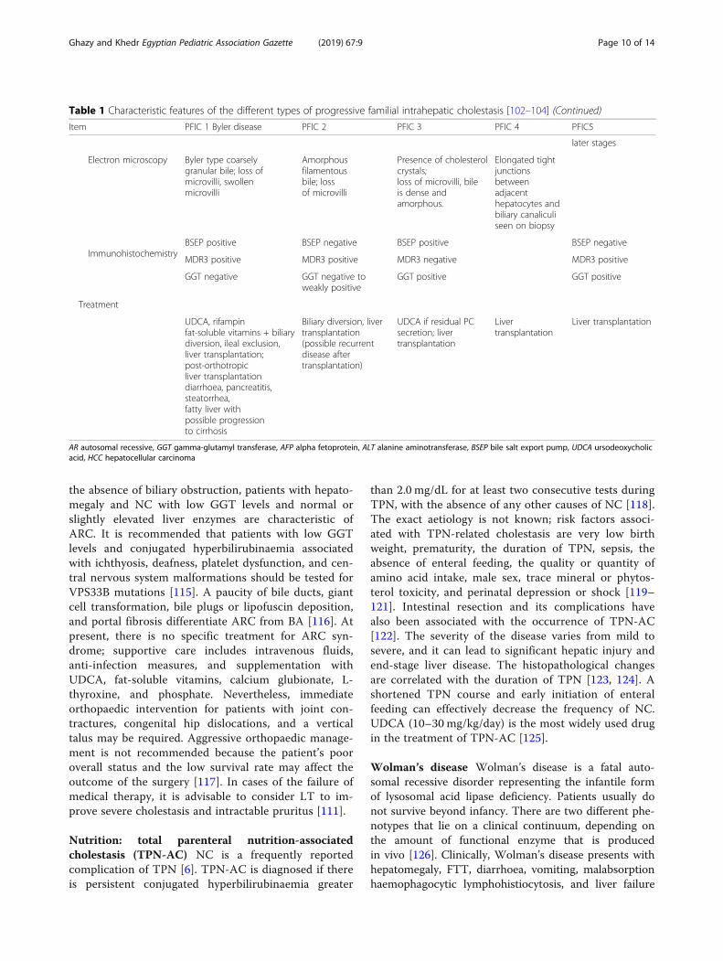
Progressive familial intrahepatic cholestasis (PFIC)PFIC is a heterogeneous group of rare, autosomal re-cessive disorders resulting from defects in the mecha-nisms involved in bile formation with typical clinical,biochemical, and histological features. PFIC presentswith intrahepatic cholestasis in infancy or childhood[107]. The worldwide incidence is 1/50,000 to 1/100,000, with equal distribution between the sexes [108].The course of the disease involves portal hyperten-sion, liver failure, cirrhosis, and HCC along with sev-eral extra-hepatic manifestations. The five types ofPFIC are identified in Table 1 [109, 110].
Arthrogryposis renal dysfunction cholestasis (ARC)syndrome ARC syndrome is a rare fatal autosomal re-cessive multisystem disorder involving the liver, kidney,skin, central nervous system, and musculoskeletal sys-tem; it is caused by mutations in the VPS33B or VIPARgene [111]. ARC syndrome includes arthrogryposis(muscle atrophy, radial deviation of the wrist, dislocationof both hip joints, flexion contracture of the knee joints,
and calcaneo-valgus), renal tubular acidosis, and NC[112]. Half of the cases have ichthyosis, and one fourthof those with ARC syndrome may have platelet anomal-ies. Agenesis of the corpus callosum is reported in morethan one fifth of cases. One tenth of patients with ARCsyndrome have congenital cardiovascular anomalies,deafness, recurrent infections, and internal bleeding dueto coagulation dysfunctions. Mild or atypical symptomsmay delay diagnosis. The prognosis of ARC syndrome ispoor, and the majority of patients die during infancy[113]. The pathogenesis characteristic of ARC syndromeprimarily involved degeneration of the anterior motorneurons, whereas the severity of arthrogryposis may betraced to placental insufficiency during pregnancy, witholigohydramnios in the mother and foetal growth re-striction. Osteopenia and pathological fractures are at-tributable to impaired renal tubular reabsorption andsecondary hyperparathyroidism [114]. Renal tubular dys-function manifests in the form of Fanconi syndrome,renal tubular acidosis, nephrogenic diabetes insipidus,glucosuria, aminoaciduria, and phosphaturia [114]. In
Fig. 4 Algorithm for diagnosis of Alagille syndrome (ALGS)
Ghazy and Khedr Egyptian Pediatric Association Gazette (2019) 67:9 Page 8 of 14

Table 1 Characteristic features of the different types of progressive familial intrahepatic cholestasis [102–104]
Item PFIC 1 Byler disease PFIC 2 PFIC 3 PFIC 4 PFIC5
Both genders are equally affected, incidence 1/50000–100,000
Genetics
Inheritance AR AR AR AR AR
Gene ATP8B1/F1C1 ABCB11/BSEP ABCB4/MDR3 TJP2 NR1H4
Protein Familial intrahepaticcholestasis 1 (FICI)
Bile salt export protein(BSEP)
Multidrug resistanceprotein 3 (MDR3)
TJP2 protein FXR, the keyregulator of BSmetabolism
Chromosome 18q21-q22 2q24 7q21 9q12 12q23.1
Location Wide tissue distributionincluding almost allepithelial cells; onapical membranes
Hepatocyte canalicularmembrane
Hepatocyte canalicularmembrane
Tight junctions Bile canaliculi, FXR ishighly expressed inliver and pancreaticβ cells
Pathophysiology
Function ofhereditarydefect
Aminophospholipidflippase
Bile acid secretion Phosphatidylcholinesecretion
Integral tightjunction protein(claudin-1)
Farnesoid Xreceptor loss
Clinical findings
Age of onset Neonates Neonates 1 month–20 years Infancy,childhood
Neonate
Course Progressive Progressive Progressive Progressive Rapidly progressive
Cholestasis Chronic Chronic Chronic Chronic Chronic
Pruritus Severe Severe Moderate Severe Severe
Others Other features includeshort stature, diarrhoea,hepatosplenomegaly,malabsorption,pancreatitis, respiratorydisease, andoccasionallysensorineural hearingloss
Growth failuregallbladder stones
Later onset cholestasis,portal hypertension,minimal pruritus, gallbladder stone, cupperaccumulation in livertissue, and increase inurinary cupper.
Intrahepaticcholestasis, earlychildhood liverfailure, portalhypertension,neurological andrespiratorysymptoms
Coagulopathya directconsequence of theloss of FXR function.Failure to thrive andascites, gallstonespleural effusions, andintraventricularhaemorrhage at birth
Risk of malignancy – +HCC/cholangiocarcinoma(in 30% of patients)
+ +HCC
Laboratory findings
Serum GGT Normal/low Normal/low High Normal or mildlyincreased
Low to normal
Serum ALT Mildly elevated > 5 × normal > 5 × normal Elevated Elevated
Serum AFP Normal Elevated Normal Elevated Elevated
Serum primary bileacid concentration
Very high + normalcholesterol
Very high High
Biliary bile acidsecretion
Low Low Low
Liver biopsy
Histology Minimal giant celltransformation,intracanalicularcholestasis, no ductalproliferation, minimalinflammation. Latefibrosis
Giant celltransformation,intracanalicularcholestasis, noductular proliferation,moderateinflammation, fibrosis,extramedullaryhemopoiesis
Giant celltransformation,intracanalicularcholestasis, ductularproliferation, moderateinflammation, markedfibrosis, lipid crystalswithin bile ducts, andfibroobliterative bileduct lesions
Ductular reaction,diffuse giant celltransformation,and ballooning ofhepatocytes andintralobularcholestasis
Intralobularcholestasis,diffuse giant celltransformation,ballooninghepatocytes, andductular reaction.Micronodularcirrhosis and fibrosiswere evident at
Ghazy and Khedr Egyptian Pediatric Association Gazette (2019) 67:9 Page 9 of 14
the absence of biliary obstruction, patients with hepato-megaly and NC with low GGT levels and normal orslightly elevated liver enzymes are characteristic ofARC. It is recommended that patients with low GGTlevels and conjugated hyperbilirubinaemia associatedwith ichthyosis, deafness, platelet dysfunction, and cen-tral nervous system malformations should be tested forVPS33B mutations [115]. A paucity of bile ducts, giantcell transformation, bile plugs or lipofuscin deposition,and portal fibrosis differentiate ARC from BA [116]. Atpresent, there is no specific treatment for ARC syn-drome; supportive care includes intravenous fluids,anti-infection measures, and supplementation withUDCA, fat-soluble vitamins, calcium glubionate, L-thyroxine, and phosphate. Nevertheless, immediateorthopaedic intervention for patients with joint con-tractures, congenital hip dislocations, and a verticaltalus may be required. Aggressive orthopaedic manage-ment is not recommended because the patient’s pooroverall status and the low survival rate may affect theoutcome of the surgery [117]. In cases of the failure ofmedical therapy, it is advisable to consider LT to im-prove severe cholestasis and intractable pruritus [111].
Nutrition: total parenteral nutrition-associatedcholestasis (TPN-AC) NC is a frequently reportedcomplication of TPN [6]. TPN-AC is diagnosed if thereis persistent conjugated hyperbilirubinaemia greater
than 2.0 mg/dL for at least two consecutive tests duringTPN, with the absence of any other causes of NC [118].The exact aetiology is not known; risk factors associ-ated with TPN-related cholestasis are very low birthweight, prematurity, the duration of TPN, sepsis, theabsence of enteral feeding, the quality or quantity ofamino acid intake, male sex, trace mineral or phytos-terol toxicity, and perinatal depression or shock [119–121]. Intestinal resection and its complications havealso been associated with the occurrence of TPN-AC[122]. The severity of the disease varies from mild tosevere, and it can lead to significant hepatic injury andend-stage liver disease. The histopathological changesare correlated with the duration of TPN [123, 124]. Ashortened TPN course and early initiation of enteralfeeding can effectively decrease the frequency of NC.UDCA (10–30 mg/kg/day) is the most widely used drugin the treatment of TPN-AC [125].
Wolman’s disease Wolman’s disease is a fatal auto-somal recessive disorder representing the infantile formof lysosomal acid lipase deficiency. Patients usually donot survive beyond infancy. There are two different phe-notypes that lie on a clinical continuum, depending onthe amount of functional enzyme that is producedin vivo [126]. Clinically, Wolman’s disease presents withhepatomegaly, FTT, diarrhoea, vomiting, malabsorptionhaemophagocytic lymphohistiocytosis, and liver failure
Table 1 Characteristic features of the different types of progressive familial intrahepatic cholestasis [102–104] (Continued)
Item PFIC 1 Byler disease PFIC 2 PFIC 3 PFIC 4 PFIC5
later stages
Electron microscopy Byler type coarselygranular bile; loss ofmicrovilli, swollenmicrovilli
Amorphousfilamentousbile; lossof microvilli
Presence of cholesterolcrystals;loss of microvilli, bileis dense andamorphous.
Elongated tightjunctionsbetweenadjacenthepatocytes andbiliary canaliculiseen on biopsy
ImmunohistochemistryBSEP positive BSEP negative BSEP positive BSEP negative
MDR3 positive MDR3 positive MDR3 negative MDR3 positive
GGT negative GGT negative toweakly positive
GGT positive GGT positive
Treatment
UDCA, rifampinfat-soluble vitamins + biliarydiversion, ileal exclusion,liver transplantation;post-orthotropicliver transplantationdiarrhoea, pancreatitis,steatorrhea,fatty liver withpossible progressionto cirrhosis
Biliary diversion, livertransplantation(possible recurrentdisease aftertransplantation)
UDCA if residual PCsecretion; livertransplantation
Livertransplantation
Liver transplantation
AR autosomal recessive, GGT gamma-glutamyl transferase, AFP alpha fetoprotein, ALT alanine aminotransferase, BSEP bile salt export pump, UDCA ursodeoxycholicacid, HCC hepatocellular carcinoma
Ghazy and Khedr Egyptian Pediatric Association Gazette (2019) 67:9 Page 10 of 14

[127]. The exact disease incidence is unknown but is es-timated to be approximately 1/500,000 live births [128].There is no or very minimal lysosomal acid lipase activ-ity (< 1% of normal), resulting in heavy accumulation ofcholesteryl esters and triglycerides in visceral organs, i.e.liver and bone marrow, which means that patients usu-ally present within the first 2–4 months of life [128,129]. Adrenal infiltration is leading to necrosis and calci-fication of the adrenal glands in approximately 50% ofpatients. Intestinal involvement results in chronicdiarrhoea or steatorrhea secondary to the diseaseprocess itself and the resultant severe malabsorption[129]. Oxysterol levels are a new biomarker for thediagnosis of Wolman’s disease and are correlated withthe clinical management of the disease [130]. Intes-tinal malabsorption, hepatic impairment, and adrenalinsufficiency explain the very poor prognosis of theseyoung patients [131].
ManagementGeneral medical managementMost children having NC are malnourished and requirean adequate provision of caloric requirements to preventand treat malnutrition associated with steatorrhea andmalabsorption. Affected patients should receive 125% ofthe recommended dietary allowance based on ideal bodyweight [132]. Medium-chain triglyceride oil should beadministered in a dose of 1–2 mL/kg/d in 2–4 divideddoses in expressed breast milk. In non-breast feed, a mix-ture of puffed rice powder and MCT to milk can makefeeds energy -dense. Essential fatty acids should consti-tute 2–3% of the energy provided. Vegetable protein at2–3 g/kg/d is recommended. 1,25-Dihydroxyvitamin D3
(0.05–0.2 μg/kg/d) is recommended in the presence ofsignificant bone changes or patients having severe chole-stasis. Vitamin K is administered at a dose of 5 mg intra-muscular, subcutaneously or intravenously, at diagnosisto correct the coagulopathy. Water soluble vitamins aregiven orally 1–2 times the recommended daily allow-ance. Vitamin supplementation should be continued till3 months after resolution of jaundice [133].
Specific treatmentSpecial infant formula and diets are recommended forchildren with specific diagnosis (galactosemia, fructose-mia, and tyrosinemia). Treatment with nitisinone (1 mg/kg/d) in addition to dietary restriction leads to rapid re-duction of toxic metabolites in tyrosinemia. Specifictherapy is recommended for patients with CMV, herpes,and toxoplasmosis-related NC. Antibiotics need to beadministered in patients with bacterial sepsis based onthe site of infection and the performed culture. There isno role for steroids in INH.
In infants with pruritus due to severe cholestasis,UDCA (20mg/kg/d), rifampicin (5–10mg/kg/d), andphenobarbitone (5–10mg/kg/d) are drugs of choice.Kasai’s operation entails removal of the atretic extrahe-patic tissue and a Roux-en-Y jejunal loop anastomosis tothe hepatic hilum. Patients’ bilirubin normalizes afterKPE if it was performed before the end of the thirdmonth [134]. About 20% of all patients undergoing KPEduring infancy survive into adulthood with their nativeliver [135]. In children with PFIC without decompen-sated cirrhosis, external and internal biliary diversionhas been shown to be of benefit [136].
Liver transplantationLiver transplantation is the standard therapy for decom-pensated cirrhosis due to any cause, and it is now wellestablished. Any baby who has had failed KPE (bilirubinremains > 2mg/dL, 6 months after surgery) should bereferred to a transplant centre.
ConclusionNC may be misdiagnosed as physiological jaundice; thismay delay the proper management of the underlying dis-ease and aggravates its complications. General practi-tioner and health care workers should be able todifferentiate between physiological jaundice and NC es-pecially BA; consequently, they would refer the affectedpatients to pediatric hepatologist as early as possible. Inaddition, parents must know when they should seekmedical advice and when to suspect NC.
AbbreviationsA1ATD: Alpha-1 antitrypsin deficiency; ALGs: Alagille syndrome;ARC: Arthrogryposis renal dysfunction cholestasis; BA: Biliary atresia;BASDs: Bile acid synthesis disorders; CBD: Common bile duct;CC: Choledochal cyst; CF: Cystic fibrosis; CFTR: Cystic fibrosis transmembraneregulator; EHBDs: Extra-hepatic bile ducts; FFT: Failure to thrive;GALT: Galactose-1-phosphate uridyltransferase; GGT: Gamma-glutamyltranspeptidase; HCC: Hepatocellular carcinoma; IHBDs: Intrahepatic bile ducts;INH: Idiopathic neonatal hepatitis; KPE: Kasai portoenterostomy; LT: Livertransplantation; NBS: Newborn screening; NC: Neonatal cholestasis;NICCD: Neonatal intrahepatic cholestasis caused by citrin deficiency;PFIC: Progressive familial intrahepatic cholestasis; TPN: Total parentalnutrition; TPN-AC: Total parenteral nutrition-associated cholestasis;UDCA: Ursodeoxycholic acid
AcknowledgmentsI would like to thank Dr. Salma Nagy and Dr. Bassam Ayoub for revising thismanuscript.
Authors’ contributionsRMG is the author of the manuscript, searched the literature, wrote themanuscript, designed the figure and tables, and finally sent the manuscriptfor publication. MAK revised the manuscript and wrote the paragraph about“Biliary atresia” section. Both authors read and approved the final manuscript.
FundingI did not receive any fund for this publication.
Availability of data and materialsAll data generated or analysed during this study are included in thispublished article.
Ghazy and Khedr Egyptian Pediatric Association Gazette (2019) 67:9 Page 11 of 14

Ethics approval and consent to participateNot applicable
Consent for publicationNot applicable
Competing interestsThe authors declare that they have no competing interests.
Author details1High Institute of Public Health, Alexandria University, 65 Garidet St., ElHoreya Rd., El Shatby, Alexandria, Egypt. 2National Liver Institute, MenoufiaUniversity, Shibin Al Kawm, Egypt.
Received: 9 July 2019 Accepted: 9 October 2019
References1. Hoerning A, Raub S, Dechêne A, Brosch MN, Kathemann S, Hoyer PF et al
(2014) Diversity of disorders causing neonatal cholestasis–the experience ofa tertiary pediatric center in Germany. Front Pediatr 2:65
2. Hartley J. The jaundiced baby. Atlas of Pediatric Hepatology. Cham:Springer; 2018. p. 1–16
3. Götze T, Blessing H, Grillhösl C, Gerner P, Hoerning A (2015) Neonatalcholestasis–differential diagnoses, current diagnostic procedures, andtreatment. Front Pediatr 3:43
4. Moyer V, Freese DK, Whitington PF, Olson AD, Brewer F, Colletti RB et al(2004) Guideline for the evaluation of cholestatic jaundice in infants:recommendations of the North American Society for PediatricGastroenterology, Hepatology and Nutrition. J Pediatr Gastroenterol Nutr39(2):115–128
5. Suchy FJ, Balistreri WF, Heubi JE, Searcy JE, Levin RS (1981) Physiologiccholestasis: elevation of the primary serum bile acid concentrations innormal infants. Gastroenterology. 80(5 pt 1):1037–1041
6. Suchy FJ (2004) Neonatal cholestasis. Pediatr Rev 25(11):388–3967. Fawaz R, Baumann U, Ekong U, Fischler B, Hadzic N, Mack CL et al (2017)
Guideline for the evaluation of cholestatic jaundice in infants: jointrecommendations of the North American Society for PediatricGastroenterology, Hepatology, and Nutrition and the European Society forPediatric Gastroenterology, Hepatology, and Nutrition. J PediatrGastroenterol Nutr 64(1):154–168
8. Fischler B, Lamireau T (2014) Cholestasis in the newborn and infant. Clin ResHepatol Gastroenterol 38(3):263–267
9. Betalli P, Davenport M. Biliary atresia and other congenital disorders of theextrahepatic biliary tree. Pediatric Hepatology and Liver Transplantation.Cham: Springer; 2019. p. 129–144
10. Wada H, Muraji T, Yokoi A, Okamoto T, Sato S, Takamizawa S et al (2007)Insignificant seasonal and geographical variation in incidence of biliaryatresia in Japan: a regional survey of over 20 years. J Pediatr Surg 42(12):2090–2092
11. Chardot C, Carton M, Spire-Bendelac N, Le Pommelet C, Golmard J-L, AuvertB (1999) Epidemiology of biliary atresia in France: a national study 1986–96.J Hepatol 31(6):1006–1013
12. El-Guindi MA-S, Sira MM, Sira AM, Salem TA-H, El-Abd OL, Konsowa HA-Set al (2014) Design and validation of a diagnostic score for biliary atresia. JHepatol 61(1):116–123
13. Russo P, Magee JC, Boitnott J, Bove KE, Raghunathan T, Finegold M et al(2011) Design and validation of the biliary atresia research consortiumhistologic assessment system for cholestasis in infancy. Clin GastroenterolHepatol 9(4):357–362 e2
14. Ghazy RM, Adawy NM, Khedr MA, Tahoun MM (2018) Biliary atresia recentinsight. Egypt Pediatric Assoc Gazette 66(1):1–8
15. Kasai M, Kimura S, Asakura Y, Suzuki H, Taira Y, Ohashi E (1968) Surgicaltreatment of biliary atresia. J Pediatr Surg 3(6):665–675
16. Huang CS, Huang CC (2010) Choledochal cysts: differences betweenpediatric and adult patients. J Gastrointest Surg 14(7):1105–1110
17. Rozel C, Garel L, Rypens F, Viremouneix L, Lapierre C, Décarie JC et al (2011)Imaging of biliary disorders in children. Pediatr Radiol 41(2):208–220
18. Lee SE, Jang J-Y, Lee Y-J, Choi DW, Lee WJ, Cho B-H et al (2011)Choledochal cyst and associated malignant tumors in adults: a multicentersurvey in South Korea. Arch Surg 146(10):1178–1184
19. Davenport M, Basu R (2005) Under pressure: choledochal malformationmanometry. J Pediatr Surg 40(2):331–335
20. Soares KC, Arnaoutakis DJ, Kamel I, Rastegar N, Anders R, Maithel S et al(2014) Choledochal cysts: presentation, clinical differentiation, andmanagement. J Am Coll Surg 219(6):1167–1180
21. Arora A, Patidar Y, Khanna R, Alam S, Rastogi A, Negi SS (2012) Cystic biliaryatresia: confounding and intriguing. J Pediatr 161(3):562
22. Jiexiong F, Minju L, Hongfeng T, Weizhong G, Shaoyong Y (2003) Clinicaland pathological characteristics of cystic lesions of extrahepatic bile duct inneonates. Acta Paediatr 92(10):1183–1189
23. Verkade HJ, Bezerra JA, Davenport M, Schreiber RA, Mieli-Vergani G,Hulscher JB et al (2016) Biliary atresia and other cholestatic childhooddiseases: advances and future challenges. J Hepatol 65(3):631–642
24. Caponcelli E, Knisely AS, Davenport M (2008) Cystic biliary atresia: anetiologic and prognostic subgroup. J Pediatr Surg 43(9):1619–1624
25. Lobeck IN, Sheridan R, Lovell M, Dupree P, Tiao GM, Bove KE (2017) Cysticbiliary atresia and choledochal cysts are distinct histopathologic entities. AmJ Surg Pathol 41(3):354–364
26. Qiao G, Li L, Li S, Tang S, Wang B, Xi H et al (2015) Laparoscopic cystexcision and Roux-Y hepaticojejunostomy for children with choledochalcysts in China: a multicenter study. Surg Endosc 29(1):140–144
27. Jun WY, Cho MJ, Han HS, Bae SH (2016) Use of omega-3 polyunsaturatedfatty acids to treat inspissated bile syndrome: a case report. PediatrGastroenterol Hepatol Nutr 19(4):286–290
28. Davenport M, Betalli P, D’Antiga L, Cheeseman P, Mieli-Vergani G, Howard E(2003) The spectrum of surgical jaundice in infancy. J Pediatr Surg 38(10):1471–1479
29. Fitzpatrick E, Jardine R, Farrant P, Karani J, Davenport M, Mieli-Vergani Get al (2010) Predictive value of bile duct dimensions measured byultrasound in neonates presenting with cholestasis. J PediatrGastroenterol Nutr 51(1):55–60
30. Makin E, Davenport M. Biliary atresia and other causes of surgical jaundicein infancy. Diseases of the Liver and Biliary System in Children 2017:413–429
31. Heaton N, Davenport M, Howard E (1991) Intraluminal biliary obstruction.Arch Dis Child 66(12):1395–1398
32. Berrani H, Vasies I, Cron J, Bachy B, Le Dosseur P, Mouterde O (2015)Association of N-acetylcysteine and glucagon during percutaneouscholangiography in the treatment of inspissated bile syndrome. ArchPediatr 22(3):300–302
33. Murphy JT, Koral K, Soeken T, Megison S (2013) Complex spontaneous bileduct perforation: an alternative approach to standard porta hepatisdrainage therapy. J Pediatr Surg 48(4):893–898
34. Barnes BH, Narkewicz MR, Sokol RJ (2006) Spontaneous perforation of thebile duct in a toddler: the role of endoscopic retrogradecholangiopancreatography in diagnosis and therapy. J Pediatr GastroenterolNutr 43(5):695–697
35. Davenport M, Heaton N, Howard E (1991) Spontaneous perforation of thebile duct in infants. Br J Surg 78(9):1068–1070
36. Gobbi D, Leon FF, Gasparella P, Gamba P, Betalli P (2011) Conservativetreatment of spontaneous biliary perforation. Pediatr Int 53(4):594–595
37. Kasat L, Borwankar S, Jain M, Naregal A (2001) Spontaneous perforation ofthe extrahepatic bile duct in an infant. Pediatr Surg Int 17(5–6):463–464
38. Chardot C, Iskandarani F, De Dreuzy O, Duquesne B, Pariente D, Bernard Oet al (1996) Spontaneous perforation of the biliary tract in infancy: a seriesof 11 cases. Eur J Pediatr Surg 6(06):341–346
39. Livesey E, Davenport M (2008) Spontaneous perforation of the biliary tractand portal vein thrombosis in infancy. Pediatr Surg Int 24(3):357–359
40. Ledesma-Ramírez S (2017) Idiopathic neonatal hepatitis. Revista Mexicanade Pediatría 83(6):208–214
41. Joob B, Wiwanitkit V (2017) Anti-smooth muscle antibodies and liverenzymes in differentiation of extrahepatic biliary atresia and idiopathicneonatal hepatitis: concern in laboratory medicine view. Afr J PaediatrSurg 14(3):60
42. Elferink RO (2003) Cholestasis. Gut 52(suppl 2):ii42–iii843. Rosenthal P (2001) Neonatal hepatitis and congenital infections. Liver
Disease Children. 2:239–25244. Carvalho E, Ivantes CAP, Bezerra JA (2007) Extrahepatic biliary atresia: current
concepts and future directions. J Pediatr 83(2):105–12045. Sira M, Sira A, Elhenawy I, Khalil F (2016) Prevalence of serological markers
of TORCH infections in biliary atresia and other neonatal cholestaticdisorders. Peertechz J Pediatr Ther 2(1):013–017
Ghazy and Khedr Egyptian Pediatric Association Gazette (2019) 67:9 Page 12 of 14

46. Kaczor MP, Sanak M, Szczeklik A (2007) Rapid and inexpensive detection ofα1-antitrypsin deficiency-related alleles S and Z by a real-time polymerasechain reaction suitable for a large-scale population-based screening. J MolDiagn 9(1):99–104
47. Campbell KM, Arya G, Ryckman FC, Alonso M, Tiao G, Balistreri WF et al(2007) High prevalence of α-1-antitrypsin heterozygosity in children withchronic liver disease. J Pediatr Gastroenterol Nutr 44(1):99–103
48. Perlmutter DH, Brodsky JL, Balistreri WF, Trapnell BC (2007) Molecularpathogenesis of alpha-1-antitrypsin deficiency-associated liver disease: ameeting review. Hepatology. 45(5):1313–1323
49. Luisetti M, Seersholm N (2004) α1-Antitrypsin deficiency· 1: epidemiology ofα1-antitrypsin deficiency. Thorax. 59(2):164–169
50. Kalsheker N (2009) α1-Antitrypsin deficiency: best clinical practice. J ClinPathol 62(10):865–869
51. de Serres FJ (2003) Alpha-1 antitrypsin deficiency is not a rare disease but adisease that is rarely diagnosed. Environ Health Perspect 111(16):1851
52. Yachha SK, Srivastava A (2010) Alpha-1 antitrypsin deficiency related liverdisease: is it worth a search in India? Indian Pediatr 47(12):1011–1012
53. Regev A, Guaqueta C, Molina EG, Conrad A, Mishra V, Brantly ML et al (2006)Does the heterozygous state of alpha-1 antitrypsin deficiency have a role inchronic liver diseases? Interim results of a large case-control study. J PediatrGastroenterol Nutr 43(1):S30–SS5
54. Calderon FR, Phansalkar AR, Crockett DK, Miller M, Mao R (2007) Mutationdatabase for the galactose-1-phosphate uridyltransferase (GALT) gene. HumMutat 28(10):939–943
55. Berry G, Nissim I, Gibson J, Mazur A, Lin Z, Elsas L et al (1997) Quantitativeassessment of whole body galactose metabolism in galactosemic patients.Eur J Pediatr 156(1):S43–SS9
56. Coss K, Byrne J, Coman D, Adamczyk B, Abrahams J, Saldova R et al (2012)IgG N-glycans as potential biomarkers for determining galactose tolerancein Classical Galactosaemia. Mol Genet Metab 105(2):212–220
57. Waisbren SE, Potter NL, Gordon CM, Green RC, Greenstein P, GubbelsCS et al (2012) The adult galactosemic phenotype. J Inherit Metab Dis35(2):279–286
58. Saudubray J-M, Berghe G, Walter JH (2012) Inborn metabolic diseases: Springer59. Ammoury RF, Ghishan FK (2014) Inborn errors of carbohydrate metabolism.
Liver Disease Children 435, 61–14760. Bosch AM (2006) Classical galactosaemia revisited. J Inherit Metab Dis 29(4):
516–52561. Berry GT (2008) Galactosemia and amenorrhea in the adolescent. Ann N Y
Acad Sci 1135(1):112–11762. Welling L, Bernstein LE, Berry GT, Burlina AB, Eyskens F, Gautschi M
et al (2017) International clinical guideline for the management ofclassical galactosemia: diagnosis, treatment, and follow-up. J InheritMetab Dis 40(2):171–176
63. Chen J, Yager CT, Reynolds RA, Segal S (2002) Identification of galactitol andgalactonate in red blood cells by gas chromatography/mass spectrometry.Clin Chim Acta 322(1–2):37–41
64. Lindhout M, Rubio-Gozalbo ME, Bakker JA, Bierau J (2010) Direct non-radioactive assay of galactose-1-phosphate: uridyltransferase activity using highperformance liquid chromatography. Clin Chim Acta 411(13–14):980–983
65. Coelho AI, Lourenço S, Trabuco M, Silva MJ, Oliveira A, Gaspar A et al (2015)Functional correction by antisense therapy of a splicing mutation in theGALT gene. Eur J Hum Genet 23(4):500
66. Bosch A, Maurice-Stam H, Wijburg F, Grootenhuis M (2009) Remarkabledifferences: the course of life of young adults with galactosaemia and PKU.J Inherit Metab Dis 32(6):706
67. Hoffmann B, Wendel U, Schweitzer-Krantz S (2011) Cross-sectional analysisof speech and cognitive performance in 32 patients with classicgalactosemia. J Inherit Metab Dis 34(2):421–427
68. Coelho AI, Rubio-Gozalbo ME, Vicente JB, Rivera I (2017) Sweet and sour: anupdate on classic galactosemia. J Inherit Metab Dis 40(3):325–342
69. Berry GT (1995) The role of polyols in the pathophysiology ofhypergalactosemia. Eur J Pediatr 154(2):S53–S64
70. Mitchell G, Grompe M, Lambert M, Tanguay R (2001) The metabolic andmolecular bases of inherited disease
71. Nakamura K, Tanaka Y, Mitsubuchi H, Endo F (2007) Animal models oftyrosinemia. J Nutr 137(6):1556S–1560S
72. Tomoeda K, Awata H, Matsuura T, Matsuda I, Ploechl E, Milovac T et al (2000)Mutations in the 4-hydroxyphenylpyruvic acid dioxygenase gene are responsiblefor tyrosinemia type III and hawkinsinuria. Mol Genet Metab 71(3):506–510
73. Chinsky JM, Singh R, Ficicioglu C, van Karnebeek CD, Grompe M, Mitchell G,et al. Diagnosis and treatment of tyrosinemia type I: a US and Canadianconsensus group review and recommendations. Genet Med. 2017;19(12):1–16.
74. Nakamura K, Matsumoto S, Mitsubuchi H, Endo F (2015) Diagnosis andtreatment of hereditary tyrosinemia in Japan. Pediatr Int 57(1):37–40
75. Mitchell G, Larochelle J, Lambert M, Michaud J, Grenier A, Ogier H et al(1990) Neurologic crises in hereditary tyrosinemia. N Engl J Med 322(7):432–437
76. Schady DA, Roy A, Finegold MJ (2015) Liver tumors in children withmetabolic disorders. Transl Pediatr 4(4):290
77. Castilloux J, Laberge A-M, Martin SR, Lallier M, Marchand V (2007) “Silent”tyrosinemia presenting as hepatocellular carcinoma in a 10-year-old girl. JPediatr Gastroenterol Nutr 44(3):375–377
78. De Jesús VR, Adam BW, Mandel D, Cuthbert CD, Matern D (2014)Succinylacetone as primary marker to detect tyrosinemia type I in newbornsand its measurement by newborn screening programs. Mol Genet Metab113(1):67–75
79. King LS, Trahms C, Scott CR (2017) Tyrosinemia type I80. Geppert J, Stinton C, Freeman K, Fraser H, Clarke A, Johnson S et al (2017)
Evaluation of pre-symptomatic nitisinone treatment on long-term outcomesin Tyrosinemia type 1 patients: a systematic review. Orphanet J Rare Dis.12(1):154
81. Mohan N, McKiernan P, Kelly D, Preece M, Green A, Buckels J et al (1999)Indications and outcome of liver transplantation in tyrosinaemia type 1. EurJ Pediatr 158(2):S049–SS54
82. Arnon R, Annunziato R, Miloh T, Wasserstein M, Sogawa H, Wilson M et al(2011) Liver transplantation for hereditary tyrosinemia type I: analysis of theUNOS database. Pediatr Transplant 15(4):400–405
83. Treepongkaruna S, Jitraruch S, Kodcharin P, Charoenpipop D, Suwannarat P,Pienvichit P et al (2012) Neonatal intrahepatic cholestasis caused by citrindeficiency: prevalence and SLC25A13 mutations among Thai infants. BMCGastroenterol 12(1):141
84. Ohura T, Kobayashi K, Tazawa Y, Abukawa D, Sakamoto O, Tsuchiya S et al(2007) Clinical pictures of 75 patients with neonatal intrahepatic cholestasiscaused by citrin deficiency (NICCD). J Inherit Metab Dis 30(2):139–144
85. Kimura A, Kage M, Nagata I, Mushiake S, Ohura T, Tazawa Y et al (2010)Histological findings in the livers of patients with neonatal intrahepaticcholestasis caused by citrin deficiency. Hepatol Res 40(3):295–303
86. Song Y-Z, Li B-X, Chen F-P, Liu S-R, Sheng J-S, Ushikai M et al (2009)Neonatal intrahepatic cholestasis caused by citrin deficiency: clinical andlaboratory investigation of 13 subjects in mainland of China. Dig Liver Dis41(9):683–689
87. Tokuhara D, Iijima M, Tamamori A, Ohura T, Takaya J, Maisawa S et al (2007)Novel diagnostic approach to citrin deficiency: analysis of citrin protein inlymphocytes. Mol Genet Metab 90(1):30–36
88. O'Sullivan BP, Freedman SD (2009) Cystic fibrosis. Lancet. 373(9678):1891–1904
89. Eminoglu TF, Polat E, Gökçe S, Ezgü FS, Senel S, Apaydin S (2013) Cysticfibrosis presenting with neonatal cholestasis simulating biliary atresia in apatient with a novel mutation. Indian J Pediatr 80(6):502–504
90. Kharrazi M, Yang J, Bishop T, Lessing S, Young S, Graham S et al (2015)Newborn screening for cystic fibrosis in California. Pediatrics. 136(6):1062–1072
91. Leeuwen L, Magoffin AK, Fitzgerald DA, Cipolli M, Gaskin KJ. Cholestasis andmeconium ileus in infants with cystic fibrosis and their clinical outcomes.Arch Dis Child. 2014;99(5):443-7.
92. Parisi GF, Di Dio G, Franzonello C, Gennaro A, Rotolo N, Lionetti E, et al.Liver disease in cystic fibrosis: an update. Hepat Mon. 2013;13(8)
93. Greenholz SK, Krishnadasan B, Marr C, Cannon R (1997) Biliary obstruction ininfants with cystic fibrosis requiring Kasai portoenterostomy. J Pediatr Surg32(2):175–180
94. Molmenti EP, Squires RH, Nagata D, Roden JS, Molmenti H, Fasola CG et al(2003) Liver transplantation for cholestasis associated with cystic fibrosis inthe pediatric population. Pediatr Transplant 7(2):93–97
95. Hollander FM, de Roos NM, Heijerman HG (2017) The optimal approach tonutrition and cystic fibrosis: latest evidence and recommendations. CurrOpin Pulm Med 23(6):556–561
96. Kobelska-Dubiel N, Klincewicz B, Cichy W (2014) Liver disease in cysticfibrosis. Prz Gastroenterol 9(3):136–141
97. Setchell KD, Heubi JE (2006) Defects in bile acid biosynthesis-diagnosis andtreatment. J Pediatr Gastroenterol Nutr 43(1):S17–S22
Ghazy and Khedr Egyptian Pediatric Association Gazette (2019) 67:9 Page 13 of 14

98. Lane E, Murray KF (2017) Neonatal cholestasis. Pediatr Clin 64(3):621–63999. Oda T, Elkahloun AG, Pike BL, Okajima K, Krantz ID, Genin A et al (1997)
Mutations in the human Jagged1 gene are responsible for Alagillesyndrome. Nat Genet 16(3):235
100. McDaniell R, Warthen DM, Sanchez-Lara PA, Pai A, Krantz ID, Piccoli DA et al(2006) NOTCH2 mutations cause Alagille syndrome, a heterogeneousdisorder of the notch signaling pathway. Am J Hum Genet 79(1):169–173
101. Turnpenny PD, Ellard S (2012) Alagille syndrome: pathogenesis, diagnosisand management. Eur J Hum Genet 20(3):251
102. Danks D, Campbell P, Jack I, Rogers J, Smith A (1977) Studies of the aetiologyof neonatal hepatitis and biliary atresia. Arch Dis Child 52(5):360–367
103. Saleh M, Kamath BM, Chitayat D (2016) Alagille syndrome: clinicalperspectives. Appl Clin Genet 9:75–82
104. Dhorne-Pollet S, Deleuze J, Hadchouel M, Bonaiti-Pellie C (1994)Segregation analysis of Alagille syndrome. J Med Genet 31(6):453–457
105. Mattei P, von Allmen D, Piccoli D, Rand E (2006) Relief of intractable pruritis inAlagille syndrome by partial external biliary diversion. J Pediatr Surg 41(1):104–107
106. Kamath BM, Schwarz KB, Hadžic N (2010) Alagille syndrome and livertransplantation. J Pediatr Gastroenterol Nutr 50(1):11–15
107. Amer S, Hajira A (2014) A comprehensive review of progressive familialintrahepatic cholestasis (PFIC): genetic disorders of hepatocanaliculartransporters. Gastroenterology Res 7(2):39
108. Schwab P, Racsa P, Rascati K, Mourer M, Meah Y, Worley K (2019) Aretrospective database study comparing diabetes-related medicationadherence and health outcomes for mail-order versus communitypharmacy. J Manag Care Spec Pharm 25(3):332–340
109. Davit-Spraul A, Gonzales E, Baussan C, Jacquemin E (2009) Progressivefamilial intrahepatic cholestasis. Orphanet J Rare Dis 4(1):1
110. Kullak-Ublick GA, Beuers U, Paumgartner G (2000) Hepatobiliary transport. JHepatol 32:3–18
111. Zhou Y, Zhang J (2014) Arthrogryposis–renal dysfunction–cholestasis(ARC) syndrome: from molecular genetics to clinical features. Ital JPediatr 40(1):77
112. Taha D, Khider A, Cullinane AR, Gissen P (2007) A novel VPS33B mutation inan ARC syndrome patient presenting with osteopenia and fractures at birth.Am J Med Genet 143(23):2835–2837
113. Abdullah MA, Al-Hasnan Z, Okamoto E, Abomelha AM (2000)Arthrogryposis, renal dysfunction and cholestasis syndrome. Saudi Med J21(3):297–299
114. Malaki M, Mandana R, Ghaffari S (2012) ARC syndrome with complex renalproblems: nephrocalcinosis, proximal and hyperkalemic distal RTA andnephrogenic diabetes insipidus. Saudi J Kidney Dis Transpl 23(4):804
115. Bull LN, Mahmoodi V, Baker AJ, Jones R, Strautnieks SS, Thompson RJ et al(2006) VPS33B mutation with ichthyosis, cholestasis, and renal dysfunctionbut without arthrogryposis: incomplete ARC syndrome phenotype. J Pediatr148(2):269–271
116. Jang JY, Kim KM, Kim G-H, Yu E, Lee J-J, Park YS et al (2009) Clinicalcharacteristics and VPS33B mutations in patients with ARC syndrome. JPediatr Gastroenterol Nutr 48(3):348–354
117. Jang WY, Cho T-J, Bae JY, Jung HW, Ko JS, Park MS et al (2011) Orthopaedicmanifestations of arthrogryposis-renal dysfunction-cholestasis syndrome. JPediatr Orthop 31(1):107–112
118. Kirk JM (2008) Neonatal jaundice: a critical review of the role and practice ofbilirubin analysis. Ann Clin Biochem 45(5):452–462
119. Wright K, Ernst KD, Gaylord MS, Dawson JP, Burnette TM (2003) Increasedincidence of parenteral nutrition-associated cholestasis with aminosyn PFcompared to trophamine. J Perinatol 23(6):444
120. Albers MJ, de Gast-Bakker D-AH, van Dam NA, Madern GC, Tibboel D (2002)Male sex predisposes the newborn surgical patient to parenteral nutrition–associated cholestasis and to sepsis. Arch Surg 137(7):789–793
121. Whitfield P, Iyer K (1998) The role of phytosterols in the pathogenesis ofliver complications of pediatric parenteral nutrition. Nutrition. 14(1):158–164
122. Sondheimer JM, Asturias E, Cadnapaphornchai M (1998) Infection andcholestasis in neonates with intestinal resection and long-term parenteralnutrition. J Pediatr Gastroenterol Nutr 27(2):131–137
123. Buchman AL, Iyer K, Fryer J (2006) Parenteral nutrition–associated liverdisease and the role for isolated intestine and intestine/liver transplantation.Hepatology. 43(1):9–19
124. Zambrano E, El-Hennawy M, Ehrenkranz RA, Zelterman D, Reyes-Múgica M(2004) Total parenteral nutrition induced liver pathology: an autopsy seriesof 24 newborn cases. Pediatr Dev Pathol 7(5):425–432
125. Chen H-L, Chen H-L, Liu Y-J, Feng C-H, Wu C-Y, Shyu M-K et al (2005)Developmental expression of canalicular transporter genes in human liver. JHepatol 43(3):472–477
126. Reiner Ž, Guardamagna O, Nair D, Soran H, Hovingh K, Bertolini S et al(2014) Lysosomal acid lipase deficiency–an under-recognized cause ofdyslipidaemia and liver dysfunction. Atherosclerosis. 235(1):21–30
127. Tinsa F, Romdhane MB, Boudabous H, Hadj IB, Brini I, Tebib N, et al. A NovelMutation c. 153 C> A in a Tunisian Girl With Wolman Disease and UnusualPresentation: Hemophagocytic Lymphohistiocytosis. J Pediatr HematolOncol. 2019;41(3):e193–e196.
128. Porto AF (2014) Lysosomal acid lipase deficiency: diagnosis and treatmentof Wolman and cholesteryl ester storage diseases. Pediatr Endocrinol Rev:PER 12:125–132
129. Bernstein DL, Hülkova H, Bialer MG, Desnick RJ (2013) Cholesteryl esterstorage disease: review of the findings in 135 reported patients with anunderdiagnosed disease. J Hepatol 58(6):1230–1243
130. Ghosh A, Cooper J, Church H, Jones SA, Wu H (2017) Plasma oxysterols as aputative biomarker for infantile onset lysosomal acid lipase deficiency(Wolman disease). Mol Genet Metab 120(1):S52–SS3
131. Muntoni S, Wiebusch H, Funke H, Ros E, Seedorf U, Assmann G (1995)Homozygosity for a splice junction mutation in exon 8 of the geneencoding lysosomal acid lipase in a Spanish kindred with cholesterol esterstorage disease (CESD). Hum Genet 95(5):491–494
132. Feranchak AP, Sokol R (2007) Medical and nutritional management ofcholestasis in infants and children. Liver Dis Children 3:190–231
133. Venigalla S, Gourley GR, editors. Neonatal cholestasis. Semin Perinatol; 2004;28(5):348–55. Elsevier
134. Sokol RJ, Shepherd RW, Superina R, Bezerra JA, Robuck P, Hoofnagle JH(2007) Screening and outcomes in biliary atresia: summary of a NationalInstitutes of Health workshop. Hepatology. 46(2):566–581
135. Bhatia V, Bavdekar A (2013) Management of acute liver failure in infants andchildren: consensus statement of the pediatric gastroenterology chapter,Indian academy of pediatrics. Indian Pediatr 50(5):477–482
136. Sharma D, Shah UH, Sibal A, Chowdhary SK (2010)Cholecystoappendicostomy for progressive familial intrahepatic cholestasis.Indian Pediatr 47(7):626–628
Publisher’s NoteSpringer Nature remains neutral with regard to jurisdictional claims inpublished maps and institutional affiliations.
Ghazy and Khedr Egyptian Pediatric Association Gazette (2019) 67:9 Page 14 of 14