neonatal diethylstilbestrol exposure alters the metabolic
TRANSCRIPT

Washington University School of Medicine Washington University School of Medicine
Digital Commons@Becker Digital Commons@Becker
Open Access Publications
11-2012
Neonatal diethylstilbestrol exposure alters the metabolic profile of Neonatal diethylstilbestrol exposure alters the metabolic profile of
uterine epithelial cells uterine epithelial cells
Yan Yin Washington University School of Medicine in St. Louis
Congxing Lin Washington University School of Medicine in St. Louis
G. Michael Veith Washington University School of Medicine in St. Louis
Hong Chen Washington University School of Medicine in St. Louis
Maulik Dhandha Washington University School of Medicine in St. Louis
See next page for additional authors
Follow this and additional works at: https://digitalcommons.wustl.edu/open_access_pubs
Recommended Citation Recommended Citation Yin, Yan; Lin, Congxing; Veith, G. Michael; Chen, Hong; Dhandha, Maulik; and Ma, Liang, ,"Neonatal diethylstilbestrol exposure alters the metabolic profile of uterine epithelial cells." Disease Models & Mechanisms. 5,6. 870-880. (2012). https://digitalcommons.wustl.edu/open_access_pubs/1231
This Open Access Publication is brought to you for free and open access by Digital Commons@Becker. It has been accepted for inclusion in Open Access Publications by an authorized administrator of Digital Commons@Becker. For more information, please contact [email protected].

Authors Authors Yan Yin, Congxing Lin, G. Michael Veith, Hong Chen, Maulik Dhandha, and Liang Ma
This open access publication is available at Digital Commons@Becker: https://digitalcommons.wustl.edu/open_access_pubs/1231
dmm.biologists.org870
INTRODUCTIONEndocrine-disrupting chemicals (EDCs) are defined by the USEnvironmental Protection Agency as a group of substances that“interfere with biosynthesis, secretion, transport, elimination andfunction of naturally existing hormones in the human body”.Exposure to endocrine disruptors during crucial developmentaltime windows increases an individual’s risk of developing a varietyof diseases, including inborn errors, infertility, obesity and cancer(Crisp et al., 1998). One of the most well-known EDCs isdiethylstilbestrol (DES), a synthetic estrogen that was widelyprescribed in the 1940s-1970s to prevent miscarriage. During thattime period, millions of women and their offspring were exposedto this compound, which was later shown to have teratogenic andoncogenic effects on many organ systems especially thereproductive tract (Ma, 2009). Since then, the mechanismsunderpinning DES-related pathogenesis have been intensivelystudied and several animal models have been established(McLachlan, 1977; McLachlan et al., 1980; Newbold andMcLachlan, 1982). The most widely used model for DES researchis the neonatal-DES model, which was first described by McLachlanand co-workers in 1982 (Newbold and McLachlan, 1982). Femalemice receiving 1 mg/kg body weight/day of DES in the first 5 daysof their lives exhibited uterine malformations, including metaplasia
and reduced adenogenesis. These mice were overweight, subfertileand developed uterine adenocarcinoma later in life (McLachlan etal., 1982; Newbold and McLachlan, 1982; Newbold et al., 2007).Although the mouse phenotypes do not fully recapitulate theclinical findings (Hatch et al., 2001; Hoover et al., 2011), this modelis nonetheless valuable for understanding how EDCs impactreproductive organ function in general. Notably, most of these DESeffects are mediated through the estrogen receptor (ESR1), asshown by the finding that Esr1-null mice are largely resistant toDES-induced phenotypes (Couse et al., 2001).
The effect of DES on the uterus is of particular interest becauseuterine metaplasia is one of the most common health problems forwomen exposed to DES before birth (DES-daughters), which couldlead to infertility. The neonatal uterus is composed of a simplecolumnar luminal epithelium, a fibroblast mesenchymal (stromal)layer and smooth muscles. It is well known that stromal cells playan inductive role in the differentiation of the uterine epithelium(UE) during reproductive tract patterning (Cunha, 1972). We andothers have shown that the neonatal uterus is prone to DES-mediated teratogenic effects, exhibiting marked morphological andgene expression changes shortly after DES exposure (Huang et al.,2005; Ma et al., 1998; Miller et al., 1998; Yin et al., 2008). A recentstudy demonstrated that the uterine epithelial ESR1 is dispensiblefor the proliferative response but is required for suppressing UEapoptosis as well as lactoferrin induction, indicating both cell-autonomous and non-cell-autonomous mechanisms in estrogensignaling (Winuthayanon et al., 2010).
To better understand the dynamic regulation of uterine biologyby DES treatment, we performed tissue-specific cDNA microarrayanalyses and discovered that DES-elicited gene expression changesoccurred mostly in the UE. A core group of genes involved inadipogenesis and glucose metabolism, especially the CCAAT-enhancer-binding proteins (C/EBPs) and peroxisome proliferator-activated receptor (PPAR), were induced by DES in the UE. We
Disease Models & Mechanisms 5, 870-880 (2012) doi:10.1242/dmm.009076
1Division of Dermatology, Department of Medicine and 2Department ofDevelopmental Biology, Washington University School of Medicine, St Louis, MO 63110, USA*Author for correspondence ([email protected])
Received 9 November 2011; Accepted 23 May 2012
© 2012. Published by The Company of Biologists LtdThis is an Open Access article distributed under the terms of the Creative Commons AttributionNon-Commercial Share Alike License (http://creativecommons.org/licenses/by-nc-sa/3.0), whichpermits unrestricted non-commercial use, distribution and reproduction in any medium providedthat the original work is properly cited and all further distributions of the work or adaptation aresubject to the same Creative Commons License terms.
SUMMARY
Developmental exposure to diethylstilbestrol (DES) causes reproductive tract malformations, affects fertility and increases the risk of clear cell carcinomaof the vagina and cervix in humans. Previous studies on a well-established mouse DES model demonstrated that it recapitulates many features ofthe human syndrome, yet the underlying molecular mechanism is far from clear. Using the neonatal DES mouse model, the present study usesglobal transcript profiling to systematically explore early gene expression changes in individual epithelial and mesenchymal compartments of theneonatal uterus. Over 900 genes show differential expression upon DES treatment in either one or both tissue layers. Interestingly, multiple componentsof peroxisome proliferator-activated receptor- (PPAR)-mediated adipogenesis and lipid metabolism, including PPAR itself, are targets of DES inthe neonatal uterus. Transmission electron microscopy and Oil-Red O staining further demonstrate a dramatic increase in lipid deposition in uterineepithelial cells upon DES exposure. Neonatal DES exposure also perturbs glucose homeostasis in the uterine epithelium. Some of these neonatalDES-induced metabolic changes appear to last into adulthood, suggesting a permanent effect of DES on energy metabolism in uterine epithelialcells. This study extends the list of biological processes that can be regulated by estrogen or DES, and provides a novel perspective for endocrinedisruptor-induced reproductive abnormalities.
Neonatal diethylstilbestrol exposure alters themetabolic profile of uterine epithelial cellsYan Yin1, Congxing Lin1, G. Michael Veith1, Hong Chen1, Maulik Dhandha1 and Liang Ma1,2,*
RESEARCH ARTICLED
iseas
e M
odel
s & M
echa
nism
s
DM
M

Disease Models & Mechanisms 871
DES induces ectopic adipogenesis RESEARCH ARTICLE
further demonstrated that activation of PPAR was essential forinitiation of abnormal adipogenesis in UE cells. This workimplicates the adipogenic program as a downstream target of DESin the UE, which leads to the hypothesis that DES and possiblyother EDCs might activate similar genetic pathways in otherestrogen-responsive tissues, especially the adipocytes, to causeadulthood obesity.
RESULTSNeonatal DES exposure alters uterine cell metabolismpredominantly in the epitheliumThe prevailing hypothesis is that the reproductive pathology inDES-daughters is defined by transient molecular changes duringthe crucial developmental period when DES is administrated. Toreveal potential mechanisms involved in this complex process, wesought to capture early DES-induced gene expression changes inthe UE and uterine mesenchyme (UM) using cDNA microarrayanalyses. We separated UE from the UM from vehicle (oil)- or DES-treated postnatal day 5 (P5) mice, and prepared biological triplicatesof RNA from pooled specimens (n≥3). Those samples wereanalyzed on two MouseWG-6 BeadChips, which detects 45,200transcripts, including more than 26,000 annotated genes in theNCBI RefSeq database. A difference of at least twofold in signalintensity of each given probe set with a P value less than 0.05 wasconsidered statistically significant. As a result of this analysis, 981transcripts were found differentially expressed in the UE, UM orboth between oil- and DES-treated groups (supplementary materialTable S1 and the Gene Expression Omnibus, GEO# GSE37969).Hierarchical clustering analysis showed that among thesedifferentially regulated genes (DRGs), the majority (80%) were eitherchanged exclusively in the UE or showed a more prominentdysregulation in the UE (Fig. 1A, red boxes). Around 10% of theDRGs were similarly regulated in both tissue layers, and another10% showed altered gene expression in the UM alone (Fig. 1A, greenand black boxes, respectively). When plotted on a log2-scaled starglyph, it is obvious that the fold-changes of most DRGs were morepronounced in the UE, either up- or downregulated (Fig. 1B). Thenumber of DRGs is summarized in Fig. 1C, which furtherdemonstrates that the UE contains more unique DRGs comparedwith the UM. The UE-specific DRGs were further analyzed andcategorized by their biological functions following the criteria ofthe Gene Ontology Consortium. Genes involved in metabolismaccounted for the largest proportion of the DRGs (16.7%), followedby those regulating cell growth and maintenance (12.5%), cellcommunication (10.1%) and signal transduction (8.1%; Fig. 1D;supplementary material Table S2). These results indicated thatneonatal DES treatment affected biology of the UE more than itdid the UM, even though the UE expresses only marginal levels ofestrogen receptor-, the key mediator of DES action (Korach etal., 1988).
Neonatal DES exposure alters adipogenesis and lipid metabolismin the UEFurther analysis of the 213 DRGs involved in metabolic regulationaccording to the KEGG (Kyoto Encyclopedia of Genes andGenomes) pathway database showed that metabolism of complexlipids was the most affected cellular metabolic pathways(supplementary material Table S3) (Kanehisa et al., 2006). Careful
examination of genes involved in fatty acid transport andmetabolism showed that many were expressed abnormally in theUE (Fig. 2A, red boxes). Ppar, a gene encoding the key regulatorof adipocyte differenctiation, insulin sensitivity, inflammation andthe endocrine signaling pathway (Barak et al., 1999; Fischer-Posovszky et al., 2007; Tontonoz et al., 1994), showed markedupregulation in DES-treated UE in our microarray. We furtherverified this result using quantitative real-time reversetranscription-polymerase chain reaction (RT-PCR), andconfirmed a tenfold upregulation in DES-treated UE (Fig. 2B).Furthermore, both western blotting and immunohistochemistryclearly demonstrated elevated PPAR protein levels in DES-treated UE (Fig. 2C-E). PPAR is a nuclear hormone receptor thatbelongs to the peroxisome proliferator-activated receptor genefamily. Both natural ligands, such as unsaturated fatty acids, andsynthetic ligands, such as thiazolidinediones (TZDs), can activatePPAR (Lehmann et al., 1995). Once activated, cytoplasmicPPAR translocates into the nucleus where it heterodimerizes withretinoid X receptor (RXR), and directly regulates genetranscription by binding to PPAR-response elements (PPREs)through its DNA binding domain. Consistent with the observedupregulation of PPAR expression, many known PPARtranscriptional targets, including Gpr109a, Acsl1, Rarb and Arnt1(Inoue et al., 2005; James et al., 2003; Jeninga et al., 2009; Karniket al., 2009), also showed marked changes in expression upon DEStreatment in the UE (Fig. 2B). Notably, the expressions of thesegenes were either low or unchanged in the UM, which is consistentwith a cell-autonomous role for PPAR in mediatingtranscriptional regulation. Collectively, these data suggest that thePPAR signaling cascade is abnormally activated in the UE byneonatal DES treatment in the mouse.
In addition to the PPAR pathway, expressions of many othergenes implicated in adipocyte differentiation were also perturbedby DES exposure. Besides PPARs, C/EBPs are the other crucialfactors regulating preadipocyte differentiation. It is well establishedthat preadipocyte differentiation starts with activation of C/EBPand C/EBP, followed by activation of C/EBP and PPAR (Wu etal., 1996; Yeh et al., 1995). This process is positively regulated bytranscription factors Krüppel-like factor 4 (KLF4), KLF6, earlygrowth response gene 2 (EGR2), and negatively regulated bytransmembrane proto-oncogene Delta-like 1 (DLK1) (Birsoy et al.,2008; Boyle et al., 2009; Li et al., 2005). Interestingly, we detecteda significant increase in expression of aforementioned positiveregulators, including Cebpa, Cebpb, Klf4, Klf6 and Egr2 in the UEby quantitative RT-PCR analyses (Fig. 2A,B). Accumulation ofnuclear KLF4 protein was also evident by immunofluorescence (Fig.2F,G). By contrast, expression of the negative regulator Dlk1 wasdecreased by DES in the UE (Fig. 2B). These data suggest that thecore transcriptional cascade favoring adipogenesis is ectopicallyswitched on in the luminal epithelial cells of the neonatal uterusby DES treatment.
Possibly as a result of this ectopic activation of the adipogenicsignaling pathway, expressions of many genes involved in lipidmetabolism were also altered. As shown in Fig. 2A,H, the expressionof long-chain fatty acid transport protein 1, encoded by Slc27a1,was dramatically increased in the luminal epithelium by DEStreatment. Similarly, the expression of Slc27a2, which encodes fattyacid transport protein 2, was also induced in the UE by DES
Dise
ase
Mod
els &
Mec
hani
sms
D
MM

dmm.biologists.org872
DES induces ectopic adipogenesisRESEARCH ARTICLE
treatment. Moreover, the expression of Agpat1, a gene encodingan acyltransferase that converts phospholipid lysophosphatidic acid(LPA) into phosphatidic acid (PA), and that of Dgat2, a geneencoding diglyceride acyltransferase that catalyzes the formationof triglycerides from diacylglycerol and Acyl-CoA, were bothupregulated in the UE (Fig. 2H). On the other hand, the geneencoding phospholipid transfer protein (Pltp) was significantlydownregulated in the UE. Taken together, these data suggest that,in addition to turning on the lipogenic signaling cascades, neonatalDES treatment also alters expression of genes responsible for lipidtransport and metabolism in UE cells.
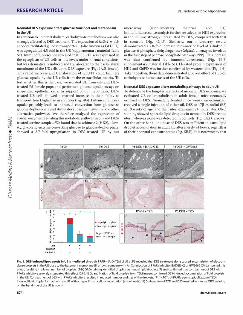
DES induces PPAR-dependent adipogenesis in the UEOur gene profiling and subsequent analyses indicated that ectopicadipogenesis is induced by DES in UE cells, which prompted us toevaluate the ultrastructure of the UE for evidence of adipogenesis.Transmission electron microscopy (TEM) on ultra-thin sectionsof P5 uterus revealed many conspicuous, electron-dense dropletslocated close to the basal side of DES-treated UE cells, which wererarely detected in control UE (Fig. 3A,B, arrows). As themorphology of these droplets resembles lipid droplets observed inmany adipocytes, we stained frozen uterine sections with Oil-RedO (ORO), a lysochrome diazo dye that binds neutral lipids. Normal
Fig. 1. DES alters uterine gene expression primarily in the epithelium. (A)Hierarchical clustering heatmap of differentially regulated genes by DES in the UEand UM. Green to red, color range gradient of mean abundance (−0.7 to 0.7). Each column represents a pool of more than three animals. Red box, genes whoseexpression was altered by DES primarily in the UE; green box, genes regulated by DES similarly in the UE and UM; black box, genes altered by DES primarily in theUM. (B)Star glyph distribution of the fold-change of differentially regulated gene in the uterus; log2 values of the mean fold-change. Red, UM; blue, UE. (C)Venndiagram of UE and UM DRGs. (D)Pie chart showing classification of DRGs based on Gene Ontology Consortium biological processes. Note that some genesmight have multiple functions and could be represented in more than one category.
Dise
ase
Mod
els &
Mec
hani
sms
D
MM

Disease Models & Mechanisms 873
DES induces ectopic adipogenesis RESEARCH ARTICLE
uterus at P5 did not stain for ORO; however, prominent ORO-staining was evident in DES-treated luminal epithelial cells closeto the basement membrane, which coincides with the abundantdroplets seen by TEM (Fig. 3E,F, arrows). These data demonstratethat in merely 5 days, DES transiently activated the adipogenicprogram in the UE, rendering them lipogenic.
Because PPAR is considered the key modulator of adipogenesis,we tested the hypothesis that DES-induced lipogenic effects aremediated through PPAR. We pooled neonatal female mice intofour groups which received oil, DES, or DES in combination withPPAR inhibitors GW9662 or BADGE, and examined their uteriat P5. Indeed, co-treatment with either inhibitor attenuated DES-induced lipid accumulation in the UE, resulting in a significantlyreduced number of lipid droplets (Fig.3C,D,G,H,I) (oil, 0.25±0.16;DES, 6.91±1.15; GW+DES, 2.32±0.80; BADGE+DES, 1.90±0.57droplets/m; *P<1×10⁻⁴). In addition, combined treatments also
reduced the size of the residual lipid droplets (Fig. 3C,D,I). Whenneonatal mice received five daily injections of Pioglitazone, a TZD-class PPAR agonist that selectively stimulates PPAR activity, lipiddroplet accumulation was evident in the UE (Fig. 3J). Interestingly,these droplets were present throughout the UE with no subcellularconfinement (Fig. 3J, inset, arrowheads). When DES was co-injected with TZD, however, intense ORO staining was detectedonly on the basal side of the UE (Fig. 3K, arrows, compare with3F,J). Taken together, these data clearly demonstrated that PPAR,induced by neonatal DES exposure, is both necessary and sufficientto cause aggregation of lipid droplets in the UE. Moreover, DESnot only upregulates PPAR expression but must also increaseendogenous PPAR ligand expression and accumulation in the UEto ectopically activate this program. On the other hand, genesinvolved in lipid trafficking must also be affected by DES to elicitthe distinct basally-localized lipid droplet phenotype in the UE.
Fig. 2. DES affects lipid metabolism and transport. (A)Hierarchical clustering heatmap of genes involved in lipid trafficking and metabolism. Green to red,color range gradient of mean abundance (−1.0 to 1.0). Red box, genes whose expression was altered by DES primarily in the UE. (B)Real-time RT-PCR survey ofgenes involved in adipocyte differentiation in the uterus. Expression of each gene was normalized to that of Rpl7; normalized expression by oil UE wasconsidered to be 1.0. (C)Western blot on whole uterine lysates showed markedly increased PPAR protein in DES-treated uteri. GAPDH served as a loadingcontrol. (D,E)Immunohistochemistry of PPAR showed increased staining in the DES-treated UE. Inset shows that PPAR was predominantly detected in the UEnuclei (arrow). (F,G)Increased KLF4 protein was detected in the UE nuclei by immunofluorescence. Red, KLF4; blue, nuclei. Insets show magnifiedimmunofluorescence signal of the boxed region. (H)Real-time RT-PCR validated genes involved in fatty acid transport and metabolism in oil- or DES-treated UEand UM. Data are presented as mean + s.d. of three samples analyzed in each treatment group from corresponding tissues. *P<0.01, **P<0.05.
Dise
ase
Mod
els &
Mec
hani
sms
D
MM
dmm.biologists.org874
DES induces ectopic adipogenesisRESEARCH ARTICLE
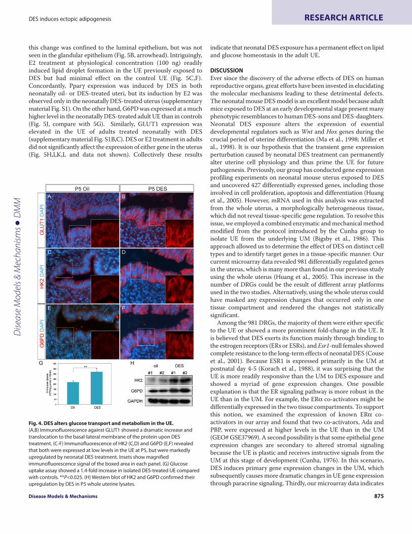
Neonatal DES exposure alters glucose transport and metabolismin the UEIn addition to lipid metabolism, carbohydrate metabolism was alsostrongly affected by DES treatment. The expression of Slc2a1, whichencodes facilitated glucose transporter 1 (also known as GLUT1),was upregulated 4.5-fold in the UE (supplementary material TableS1). Immunofluorescence revealed that GLUT1 was expressed inthe cytoplasm of UE cells at low levels under normal conditions,but was dramatically induced and translocated to the basal-lateralmembrane of the UE cells upon DES exposure (Fig. 4A,B, insets).This rapid increase and translocation of GLUT1 could facilitateglucose uptake by the UE cells from the extracellular matrix. Totest whether this is the case, we isolated UE from oil- and DES-treated P5 female pups and performed glucose uptake assays onsuspended epithelial cells. In support of our hypothesis, DES-treated UE cells showed a marked increase in their ability totransport free D-glucose in solution (Fig. 4G). Enhanced glucoseuptake probably leads to increased conversion from glucose toglucose-6-phosphate and stimulates subsequent glycolysis or otheralternative pathways. We therefore analyzed the expression ofcrucial enzymes regulating this metabolic pathway in oil- and DES-treated uterine samples. We found that hexokinase 2 (HK2), a low-Km glycolytic enzyme converting glucose to glucose-6-phosphate,showed a 3.7-fold upregulation in DES-treated UE by our
microarray (supplementary material Table S1).Immunofluorescence analysis further revealed that HK2 expressionin the UE was strongly upregulated by DES, compared with thatin controls (Fig. 4C,D). Similarly, our microarray resultsdemonstrated a 2.8-fold increase in transcript level of X-linked Gglucose-6-phosphate dehydrogenase (G6pdx), an enzyme involvedin the first step of pentose phosphate pathway (PPP). This increasewas also confirmed by immunofluorescence (Fig. 4E,F;supplementary material Table S1). Elevated protein expression ofHK2 and G6PD was further confirmed by western blot (Fig. 4H).Taken together, these data demonstrated an overt effect of DES oncarbohydrate homeostasis of the UE cells.
Neonatal DES exposure alters metabolic pathways in adult UETo determine the long-term effects of neonatal DES exposure, weevaluated UE cell metabolism in adult female mice neonatallyexposed to DES. Neonatally treated mice were ovariectomized,received a single injection of either oil, DES or 17-estrodial (E2)at 10 weeks of age, and their uteri examined 24 hours later. OROstaining showed sporadic lipid droplets in neonatally DES-treateduteri, whereas none was detected in controls (Fig. 5A,D, arrows).On the other hand, one dose of DES was sufficient to cause lipiddroplet accumulation in adult UE after merely 24 hours, regardlessof their neonatal exposure status (Fig. 5B,E). It is noteworthy that
Fig. 3. DES-induced lipogenesis in UE is mediated through PPAR. (A-D)TEM of UE at P5 revealed that DES treatment alone caused accumulation of electron-dense droplets in the UE close to the basement membrane (B, arrows, compare with A). Co-injection of PPAR inhibitor BADGE (C) or GW9662 (D) dampened thiseffect, resulting in a lower number of droplets. (E-H)ORO staining identified droplets as neutral lipid droplets (F) and confirmed that co-treatment of DES withPPAR inhibitors severely attenuated this effect (G,H). (I)Quantification of lipid droplets from TEM images confirmed DES-induced accumulation of lipid dropletsin the UE. Co-treatment of DES with PPAR inhibitors resulted in reduced number and size of the droplets. *P<1×10⁻⁴. (J)PPAR agonist pioglitazone (TZD)induced lipid droplet formation in the UE without specific subcellular localization (arrowheads). (K)Co-injection of TZD and DES resulted in intense ORO stainingon the basal side of the UE (arrows).
Dise
ase
Mod
els &
Mec
hani
sms
D
MM
Disease Models & Mechanisms 875
DES induces ectopic adipogenesis RESEARCH ARTICLE
this change was confined to the luminal epithelium, but was notseen in the glandular epithelium (Fig. 5B, arrowhead). Intriguingly,E2 treatment at physiological concentration (100 ng) readilyinduced lipid droplet formation in the UE previously exposed toDES but had minimal effect on the control UE (Fig. 5C,F).Concordantly, Ppar expression was induced by DES in bothneonatally oil- or DES-treated uteri, but its induction by E2 wasobserved only in the neonatally DES-treated uterus (supplementarymaterial Fig. S1). On the other hand, G6PD was expressed at a muchhigher level in the neonatally DES-treated adult UE than in controls(Fig. 5J, compare with 5G). Similarly, GLUT1 expression waselevated in the UE of adults treated neonatally with DES(supplementary material Fig. S1B,C). DES or E2 treatment in adultsdid not significantly affect the expression of either gene in the uterus(Fig. 5H,I,K,L and data not shown). Collectively these results
indicate that neonatal DES exposure has a permanent effect on lipidand glucose homeostasis in the adult UE.
DISCUSSIONEver since the discovery of the adverse effects of DES on humanreproductive organs, great efforts have been invested in elucidatingthe molecular mechanisms leading to these detrimental defects.The neonatal mouse DES model is an excellent model because adultmice exposed to DES at an early developmental stage present manyphenotypic resemblances to human DES-sons and DES-daughters.Neonatal DES exposure alters the expression of essentialdevelopmental regulators such as Wnt and Hox genes during thecrucial period of uterine differentiation (Ma et al., 1998; Miller etal., 1998). It is our hypothesis that the transient gene expressionperturbation caused by neonatal DES treatment can permanentlyalter uterine cell physiology and thus prime the UE for futurepathogenesis. Previously, our group has conducted gene expressionprofiling experiments on neonatal mouse uterus exposed to DESand uncovered 427 differentially expressed genes, including thoseinvolved in cell proliferation, apoptosis and differentiation (Huanget al., 2005). However, mRNA used in this analysis was extractedfrom the whole uterus, a morphologically heterogeneous tissue,which did not reveal tissue-specific gene regulation. To resolve thisissue, we employed a combined enzymatic and mechanical methodmodified from the protocol introduced by the Cunha group toisolate UE from the underlying UM (Bigsby et al., 1986). Thisapproach allowed us to determine the effect of DES on distinct celltypes and to identify target genes in a tissue-specific manner. Ourcurrent microarray data revealed 981 differentially regulated genesin the uterus, which is many more than found in our previous studyusing the whole uterus (Huang et al., 2005). This increase in thenumber of DRGs could be the result of different array platformsused in the two studies. Alternatively, using the whole uterus couldhave masked any expression changes that occurred only in onetissue compartment and rendered the changes not statisticallysignificant.
Among the 981 DRGs, the majority of them were either specificto the UE or showed a more prominent fold-change in the UE. Itis believed that DES exerts its function mainly through binding tothe estrogen receptors (ERs or ESRs), and Esr1-null females showedcomplete resistance to the long-term effects of neonatal DES (Couseet al., 2001). Because ESR1 is expressed primarily in the UM atpostnatal day 4-5 (Korach et al., 1988), it was surprising that theUE is more readily responsive than the UM to DES exposure andshowed a myriad of gene expression changes. One possibleexplanation is that the ER signaling pathway is more robust in theUE than in the UM. For example, the ER co-activators might bedifferentially expressed in the two tissue compartments. To supportthis notion, we examined the expression of known ER co-activators in our array and found that two co-activators, Ada andPBP, were expressed at higher levels in the UE than in the UM(GEO# GSE37969). A second possibility is that some epithelial geneexpression changes are secondary to altered stromal signalingbecause the UE is plastic and receives instructive signals from theUM at this stage of development (Cunha, 1976). In this scenario,DES induces primary gene expression changes in the UM, whichsubsequently causes more dramatic changes in UE gene expressionthrough paracrine signaling. Thirdly, our microarray data indicates
Fig. 4. DES alters glucose transport and metabolism in the UE.(A,B)Immunofluorescence against GLUT1 showed a dramatic increase andtranslocation to the basal-lateral membrane of the protein upon DEStreatment. (C-F)Immunofluorescence of HK2 (C,D) and G6PD (E,F) revealedthat both were expressed at low levels in the UE at P5, but were markedlyupregulated by neonatal DES treatment. Insets show magnifiedimmunofluorescence signal of the boxed area in each panel. (G)Glucoseuptake assay showed a 1.4-fold increase in isolated DES-treated UE comparedwith controls. **P<0.025. (H)Western blot of HK2 and G6PD confirmed theirupregulation by DES in P5 whole uterine lysates.
Dise
ase
Mod
els &
Mec
hani
sms
D
MM

dmm.biologists.org876
DES induces ectopic adipogenesisRESEARCH ARTICLE
that DES exclusively induces Esr1 expression in the UE but not inthe UM (GEO# GSE37969), which might contribute to increasedESR signaling in the UE. Finally, it is also possible that, in additionto ESRs, other steroid hormone receptor(s) transduce DES signalingin UE cells. One such candidate is the orphan nuclear receptorestrogen-receptor-related receptors (ERR, ERR and ERR),previously reported to interact with DES, which suppresses co-activator binding and affects transcription of downstream targetsin trophoblast cells (Tremblay et al., 2001). We found in ourmicroarray that Esrra, which encodes ERR, was highly expressedin the UE, and was upregulated 2.8-fold (P0.00047) by DEStreatment. On the other hand, its expression was low in the UMand unaffected by DES (supplementary material Table S1 and GEO#GSE37969).
The most interesting finding in our study was the ectopicaccumulation of lipid droplets in the UE, as evidenced by bothultrastructural and histological analyses. This abnormal activationof adipogenesis results from aberrant lipid metabolism and
trafficking in the UE upon DES exposure. As a consequence of thealtered lipid metabolism, UE cells might use alternative metabolicpathways for energy storage and/or consumption, which could leadto undesired epigenetic changes (Wellen et al., 2009) and causedisease later in life. Thus, this abnormal adipogenesis could alsobe a potential mechanism that contributes to the etiology of DES-induced uterine metaplasia.
Previous studies have reported that estrogen treatment in adultmice resulted in increased lipid biosynthesis (Bourke et al., 1991;Stacey et al., 1991). In this study we provide additional mechanisticinsights into this process by both gene profiling experiments andfunctional analyses. The program of adipocyte differentiation istightly regulated through a genetic cascade mainly involvingsequential activation of C/EBP and C/EBP, followed by PPARand C/EBP (Tontonoz et al., 1994). We found that DES ectopicallyactivated the adipogenic program by affecting the expression ofboth positive and negative regulators of this pathway. Activationof PPAR is a crucial event in this process, as clearly demonstrated
Fig. 5. Neonatal DES treatment alters adult UE metabolic homeostasis. (A-F)ORO staining of uteri of ovariectomized (OVX) adult mice. No staining wasdetected in the OVX UE of control animals (A), whereas a number of lipid droplets were observed in the OVX UE neonatally exposed to DES (D, arrows). Dashedlines outline the UE. DES induced ORO staining in the luminal UE of both groups (B,E, arrows), without affecting the glandular epithelium (B, arrowhead). E2treatment induced ORO staining in the UE of animals previously exposed to DES (F, arrow), but had little effect on neonatally oil-treated UE (C). (G-L)Expressionof G6PD assessed by immunofluorescence. G6PD was higher in the UE with neonatal DES exposure (J) and remained high when treated with DES or E2 (K,L).Neonatally oil-treated UE had lower G6PD levels regardless of hormone treatment in adulthood (G-I).
Dise
ase
Mod
els &
Mec
hani
sms
D
MM

Disease Models & Mechanisms 877
DES induces ectopic adipogenesis RESEARCH ARTICLE
by antagonist and agonist experiments showing that it is bothnecessary and sufficient to induce ectopic lipid droplet formationin the UE. The exact mechanism through which DES activatesPPAR is still not clear, although a previous study suggested thatPPAR could be regulated by ovarian hormones duringimplantation (Li et al., 2004). We found that elevation of Ppartranscript was not observed until 18 hours after DES treatment(data not shown), suggesting that its regulation by DES might besecondary and mediated by other upstream factors. One suchcandidate is C/EBP, which is a known activator of PPAR in 3T3-L1 adipocytes and has previously been shown to be a direct targetof 17-estradiol in the adult mouse uterus (Gellersen et al., 2010).The subcellular localization of DES-induced lipid droplets, however,cannot be achieved by activating PPAR alone (Fig. 3J), indicatingthat other lipid-interacting molecules must also be induced tocontribute to this phenotype.
PPAR has two isoforms in the mouse resulting from alternativeusage of 5� exons (Tontonoz et al., 1994; Zhu et al., 1995). PPAR1is expressed ubiquitously at low levels whereas PPAR2 is highlyexpressed in the adipose tissue and large intestine (Mansen et al.,1996; Tontonoz et al., 1994). PPAR1 is induced by DES in neonataluterus, but is suppressed in abdominal fat pad (supplementarymaterial Fig. S2B). By contrast, PPAR2 is absent in the uterus butinduced by DES in the fat pad (supplementary material Fig. S2B).These results indicate tissue-specific regulation of Ppar isoformsby DES. Recent epidemiological studies indicate that exposure toEDCs during early development is associated with overweight,obesity and type 2 diabetes later on (Gladen et al., 2000; Goncharovet al., 2008; Smink et al., 2008; Vasiliu et al., 2006). Other than itsadverse effects on the reproductive tract, neonatal DES exposurealso leads to the development of obesity in adult animals as youngas 2 months of age and throughout adulthood (Newbold et al.,2007). Newbold et al. clearly showed that all white fat depotsweighed more in DES-treated mice than in controls, especially theinguinal and retroperitoneal fat pads, whereas brown fat depotweights were not affected. This DES-induced weight gain is ESR1-dependent (Couse et al., 2000). The fact that ESR1 is only expressedin white but not brown adipocytes (Rodriguez-Cuenca et al., 2005)is sufficient to explain the differential response of white and brownadipose tissues to neonatal DES treatment. We speculate thatneonatal DES exposure might also activate the adipogenic programthrough ESR1 in mesenchymal cells, leading to an increasednumber of adipocytes at an early age. Indeed, we showed that asearly as P5, increased Ppar2 transcript and PPAR protein wereevident in the abdominal fat pad (white adipose tissue) of miceexposed neonatally to DES (supplementary material Fig. S2B,C).Future investigations addressing molecular changes in fat tissuesin DES-treated mice will further our understandings on how DES,and possibly other EDCs, cause obesity and other related diseases.
Lipogenesis and glycolysis are two closely regulated processessharing common intermediate metabolites such asdihydroxyacetone phosphate (DHAP), which is generated byglycolysis and serves as a main source of glycerol backbone forlipogenesis. Thus, perturbation of one process often affects theother. We observed altered glucose homeostasis in DES-treated UE,including elevated GLUT1 expression and enhanced glucosetransport. These findings are in line with previous reports thatestradiol alters glucose transporter expression and glucose uptake
in adult mouse and pre-pubertal rat uteri (Kim and Moley, 2009;Welch and Gorski, 1999). In addition, we found that several keyenzymes involved in glycolysis, including HK2, ALDO1 and PGK1,are all upregulated in UE cells by DES (Fig. 4 and data not shown).Abnormal glucose metabolism in the UE is also evidenced by theabnormal expression of enzymes involved in the PPP: G6PD, theenzyme responsible for the first and rate-limiting step of PPP, isstrongly activated in the UE (Fig. 4). Notably, utilization of PPPleads to production of NADPH, which provides the reducing powerneeded in cellular biosynthesis, particularly lipogenesis (Chascioneet al., 1987). Whether the PPP is the preferred choice of glucosemetabolism in DES-treated UE cells requires further investigation.
Our findings also indicate that neonatal DES exposure has a long-lasting effect on UE metabolism. We observed a higher number oflipid droplets in ovariectomized adult UE neonatally exposed toDES, as well as increased G6PD and GLUT1 expression. These datasuggest that DES elicited permanent changes in gene expression,which was independent of ovarian hormones. Moreover, neonatalDES exposure altered adult UE response to estrogens, as evidencedby lipid droplet accumulation as well as molecular markers.Previously Newbold and co-workers reported that a high dose ofneonatal DES treatment desensitized uterine responsiveness to E2in juvenile mice, as measured by uterine weight gains (Newbold etal., 2004). By contrast, we found that early DES exposure rendersthe adult uterus more sensitive to E2-induced lipid dropletformation as well as to some gene expressions (PPAR). On theother hand, neonatal DES dampened the E2 regulation of Arntlexpression (supplementary material Fig. S1A). Therefore, early DESexposure leads to a more complex and pleiotropic estrogenicresponsiveness in the adult uterus. It is likely that epigenetic changescaused by DES are the underlying mechanism for the long-termeffects of DES.
In summary, we have demonstrated that neonatal DES treatmentdramatically alters the metabolic pathways in UE cells. It is not clearat this point whether the mouse DES phenotype is relevant toclinical findings; nevertheless, our observations provide a novelmechanism through which DES, and possibly other EDCs, canaffect homeostasis of the female reproductive tract.
METHODSMiceAll mice were housed in the animal facility at WashingtonUniversity with controlled light and dark cycles and handled inaccordance with National Institutes of Health guidelines. Allprocedures were approved by the Washington UniversityInstitutional Animal and Use Committee. Time-pregnant CD-1mice were purchased from Charles River Breeding Laboratory(Wilmington, MA). DES was prepared and injected subcutaneouslyinto female pups from P1 to P5 as described previously (Huang etal., 2005). PPAR inhibitors bisphenol A diglycidyl ether (BADGE)and GW9662 were purchased from Sigma-Aldrich (St Louis, MO).Stock solution (50×) was prepared in DMSO at a concentration of150 mg/ml for BADGE and 10 mg/ml for GW9662. Workingsolutions were made fresh in sterile PBS on the day of injection.PPAR inhibitor was administered daily intraperitoneally at aconcentration of 30 mg/kg for BADGE (Naveiras et al., 2009) or 2mg/kg for GW9662 (De Backer et al., 2009). PPAR agonistpioglitazone (Sigma) was dissolved in either pure corn oil or DES
Dise
ase
Mod
els &
Mec
hani
sms
D
MM

dmm.biologists.org878
DES induces ectopic adipogenesisRESEARCH ARTICLE
solution and injected subcutaneously (20 mg/kg) into neonatalfemale mice from P1 to P5 (Ji et al., 2009). Routine ovariectomywas performed on 8-week-old mice exposed to oil or DES from P1to P5. The mice were allowed to recover for 2 weeks, followed byone subcutaneous injection of either 100 l corn oil, 100 l DES(20 g) or 100 l E2 (100 ng) (Sigma). Uteri were harvested forfixation or RNA extraction 24 hours later.
Separation of UE cellsTo isolate UE cells, we followed a protocol described previouslywith slight modifications (Bigsby et al., 1986). Briefly, whole uteriwere dissected from P5 mice of the same treatment group andpooled accordingly. Each uterine horn was cut into 3-4 mm piecesand rinsed in Ca2+-free, Mg2+-free Hank’s balanced salt solution(HBSS). Tissues were digested in 1% trypsin in HBSS at 4°C for 1hour on a rotating platform. An equal volume of 5 mg/ml bovineserum albumin (BSA) was added to stop the digestion. Gentlepressure was applied along each piece with tweezers to squeezeout the epithelia, which were transferred to 1.5-ml tubes bypipetting. The epithelial cells were collected by low-speedcentrifugation. The remaining uterine tissue containing mostlymesenchymal cells was transferred to 1.5-ml tubes and spun down.
RNA extraction and quantitative RT-PCRTotal RNA was isolated from the cell and tissue pellets with RNAStat-60 (Tel-Test, Friendswood, TX) following the manufacturer’sinstructions. Primer design, reverse transcription and real-time RT-PCR were performed as previously described (Yin et al., 2008).Student’s t-test was performed on biological triplicates and P<0.05was considered statistically significant.
cDNA microarray and data analysisOil- or DES-treated P5 mice were harvested and the epitheliaseparated from the mesenchyme. UE or UM tissues from three orfour animals of the same treatment group were pooled togetherand RNA isolated to make one sample. Three RNA samples of eachtissue per treatment group were used for microarray analysis forstatistical purposes. Total RNA was cleaned using RNeasy Kit(Qiagen, Valencia, CA) and 1 g purified RNA of each sample wassubmitted to the Genome Technology Access Center at WashingtonUniversity School of Medicine. A total of 12 samples were randomlyapplied to two Illumina MouseWG-6 BeadChips, hybridized andimaged as per the manufacturer’s instructions. An averagenormalization algorithm was used to normalize the signals and isavailable upon request. Normalized signals were first filtered byintensity to eliminate genes that were not expressed in any of thefour groups. Average signal intensity and fold-change for each probewere then calculated (oil UE versus DES UE and oil UM versusDES UM). The remaining data were further filtered to eliminatethose significantly changed by no more than twofold (P<0.05 wasconsidered significant) in any of the two comparisons. Hierarchicalclustering was performed on DRGs using the program Cluster 3.0and the output dendrograms and heatmaps were visualized usingthe program Treeview (Eisen et al., 1998). Star Glyph was plottedin Microsoft Excel 2007. The data discussed in this paper have beendeposited in NCBI’s Gene Expression Omnibus (Edgar et al., 2002)and are accessible through GEO series accession number GSE37969(http://www.ncbi.nlm.nih.gov/geo/query/acc.cgi?accGSE37969).
Western blot, immunohistochemistry, immunofluorescence andOil-Red O stainingWestern blot, immunohistochemistry and immunofluorescencewere performed as described previously (Yin et al., 2008; Yin et al.,2011). Antibodies and dilutions used were: 1:100(immunohistochemistry) and 1:1000 (western blot) for PPAR (CellSignaling); 1:5000 (western blot) for GAPDH (Cell Signaling); 1:200(immunofluorescence) for KLF4 (Santa Cruz Biotechnology); 1:100(immunofluorescence) for G6PD and GLUT1 (generous gifts fromKelle Moley, Washington University, St Louis, MO); 1:100 for HK2(Cell Signaling); 1:1000 for Alexa-Fluor-594-conjugated goat anti-rabbit (Invitrogen) and 1:5000 for HRP-conjugated goat anti-rabbitsecondary antibodies. For ORO staining, tissues were fixed in 4%paraformaldehyde briefly, washed through a series of Tissue-Tekoptimal cutting temperature compound (OCT)-sucrose solutions,and embedded in OCT Slides of 10-m frozen sections werewashed in water once, twice in 100% propylene glycol (Sigma) andthen stained in 0.7% ORO in propylene glycol for 7 minutes withagitation at 60°C. Slides were then washed in 85% propylene glycolbriefly, rinsed in water and mounted with glycerin jelly.
Transmission electron microscopyUterus was fixed in 4% PFA and 2.5% glutaraldehyde, contrastedwith osmium tetroxide and embedded in resin. Ultrathin sectionswere cut and examined under a Hitachi H7600 TEM system. Tocount lipid droplets in each image, a line was drawn along thebasement membrane of the UE cells, its length measured. All
TRANSLATIONAL IMPACT
Clinical issueIn utero exposure to endocrine-disrupting chemicals (EDCs) is associated withreproductive tract malformations, cancer, obesity and type 2 diabetes later inlife. Diethylstilbestrol (DES), a synthetic estrogen that was prescribed in the1940s-1970s to prevent miscarriage, is a prototype EDC that causesreproductive tract malformations and cancers in exposed populations. Anestablished mouse model has shown that neonatal DES exposure results inuterine malformations, uterine adenocarcinoma and weight gain in adult mice.However, the molecular mechanisms underlying DES toxicity have beenelusive.
ResultsThe authors use a genetic profiling approach to uncover genes regulated byDES in the mouse uterus and identify several new targets, including genesinvolved in lipid and glucose metabolism. They show that DES activates thetranscriptional program for adipogenesis in the uterine epithelium, andidentify peroxisome proliferator-activated receptor- (Ppar) as a crucial playerin this process. Furthermore, they found that DES exposure altered glucosemetabolism in the uterine epithelium. Finally, the authors show that neonatalDES treatment elicits permanent metabolic changes in the uterus that are stilldetectable in adult mice.
Implications and future directions These findings indicate that DES exposure causes marked metabolic changesin uterine epithelial cells, some of which persist into adulthood, providing newclues regarding how EDCs might cause reproductive problems. In addition toaffecting uterine epithelium, DES also induces PPAR expression in neonataladipose tissue; the authors propose that in utero exposure to other EDCs mightcontribute to adult obesity via activation of the adipogenic program throughsimilar molecular mechanisms. Future studies that investigate molecularchanges in adipose tissues in DES-exposed mice will further understanding onhow DES, and possibly other EDCs, contribute to obesity and related diseases.
Dise
ase
Mod
els &
Mec
hani
sms
D
MM

Disease Models & Mechanisms 879
DES induces ectopic adipogenesis RESEARCH ARTICLE
droplets in the included UE cells were counted. The averagenumber of lipid droplets per length was calculated from images(five images per sample, n3 per treatment). Student’s t-tests wereperformed for statistical analysis.
Glucose uptake assayGlucose uptake assays were performed on freshly isolated UE cellsfrom oil- or DES-treated mice on P5 following previously describedmethods with modifications (Frolova et al., 2009; Keawpradub andPurintrapiban, 2009). Briefly, enriched UE cells were washed andincubated in 1× Krebs-Ringer buffer (125 mM NaCl, 4.7 mM KCl,2 mM CaCl2, 2.4 mM MgSO4, 25 mM HEPES, 0.5% BSA and 1.2mM K2HPO4) containing 1 mM glucose for 10 minutes. Cells werespun down then resuspended in 1 ml 1× Krebs-Ringer buffercontaining 1 mM glucose and 0.1 Ci/ml 2-deoxy-D-glucose, [1-14C] (MP Biomedicals) for exactly 1 minute. Cytochalasin B (5 lof 50 mM stock solution) was added to the reaction to stop glucoseuptake. Cell pellets were washed and lysed in 0.05 N NaOH.Radioactivity taken up by the cells was determined using ascintillation counter. Aliquots from each sample were used todetermine the protein concentration using the Bradford assay.Reactions were normalized to total protein, and glucose uptakeexpressed as DPM/mg protein/10 minutes. Three pools of UE cellsreceiving control or hormone treatment were used for statisticalanalysis.ACKNOWLEDGEMENTSWe thank Drs Antonina Frolova for technical advice and Kelle Moley for GLUT1 andG6PD antibodies. We thank the Genome Technology Access Center in theDepartment of Genetics at Washington University School of Medicine for helpwith genomic analysis. We thank Jaclynn Lett and Washington University in StLouis Department of Otolaryngology, Research Center for Auditory and VisualStudies funded by the National Institutes of Health [grant number P30 DC004665]for providing technical assistance on electron microscopy.
COMPETING INTERESTS STATEMENTThe authors have nothing to declare.
AUTHOR CONTRIBUTIONSY.Y. and L.M. conceived and designed experiments. Y.Y., C.L., G.M.V. and H.C.performed the experiments. Y.Y., C.L., M.D. and L.M. analyzed data. Y.Y., C.L. andL.M. wrote the paper.
FUNDING This work is supported by the National Institutes of Health (NIH) [grant numbersES014482 and ES016597 to L.M.]. The Genome Technology Access Center ispartially supported by National Cancer Institute (NCI) Cancer Center Support Grant[grant number P30 CA91842] to the Siteman Cancer Center and by a grant fromthe Institute of Clinical and Translational Science/Clinical and Translational ScienceAwards (ICTS/CTSA) [grant number UL1RR024992] from the National Center forResearch Resources (NCRR), a component of the NIH, and NIH Roadmap forMedical Research. This publication is solely the responsibility of the authors anddoes not necessarily represent the official view of the NCRR or NIH.
SUPPLEMENTARY MATERIALSupplementary material for this article is available athttp://dmm.biologists.org/lookup/suppl/doi:10.1242/dmm.009076/-/DC1
REFERENCESBarak, Y., Nelson, M. C., Ong, E. S., Jones, Y. Z., Ruiz-Lozano, P., Chien, K. R., Koder,
A. and Evans, R. M. (1999). PPAR gamma is required for placental, cardiac, andadipose tissue development. Mol. Cell 4, 585-595.
Bigsby, R. M., Cooke, P. S. and Cunha, G. R. (1986). A simple efficient method forseparating murine uterine epithelial and mesenchymal cells. Am. J. Physiol. 251,E630-E636.
Birsoy, K., Chen, Z. and Friedman, J. (2008). Transcriptional regulation ofadipogenesis by KLF4. Cell Metab. 7, 339-347.
Bourke, J. E., Dank, S., Wilce, P. A. and Martin, L. (1991). Effect of estradiol andprogesterone on phosphatidylinositol metabolism in the uterine epithelium of themouse. J. Steroid Biochem. Mol. Biol. 39, 337-342.
Boyle, K. B., Hadaschik, D., Virtue, S., Cawthorn, W. P., Ridley, S. H., O’Rahilly, S.and Siddle, K. (2009). The transcription factors Egr1 and Egr2 have opposinginfluences on adipocyte differentiation. Cell Death Differ. 16, 782-789.
Chascione, C., Elwyn, D. H., Davila, M., Gil, K. M., Askanazi, J. and Kinney, J. M.(1987). Effect of carbohydrate intake on de novo lipogenesis in human adiposetissue. Am. J. Physiol. 253, E664-E669.
Couse, J. F., Curtis Hewitt, S. and Korach, K. S. (2000). Receptor null mice revealcontrasting roles for estrogen receptor alpha and beta in reproductive tissues. J.
Steroid. Biochem. Mol. Biol. 74, 287-296.Couse, J. F., Dixon, D., Yates, M., Moore, A. B., Ma, L., Maas, R. and Korach, K. S.
(2001). Estrogen receptor-alpha knockout mice exhibit resistance to thedevelopmental effects of neonatal diethylstilbestrol exposure on the femalereproductive tract. Dev. Biol. 238, 224-238.
Crisp, T. M., Clegg, E. D., Cooper, R. L., Wood, W. P., Anderson, D. G., Baetcke, K. P.,Hoffmann, J. L., Morrow, M. S., Rodier, D. J., Schaeffer, J. E. et al. (1998).Environmental endocrine disruption: an effects assessment and analysis. Environ.
Health Perspect. 106 Suppl. 1, 11-56.Cunha, G. R. (1972). Tissue interactions between epithelium and mesenchyme of
urogenital and integumental origin. Anat. Rec. 172, 529-541.Cunha, G. R. (1976). Stromal induction and specification of morphogenesis and
cytodifferentiation of the epithelia of the Mullerian ducts and urogenital sinusduring development of the uterus and vagina in mice. J. Exp. Zool. 196, 361-370.
De Backer, O., Elinck, E., Priem, E., Leybaert, L. and Lefebvre, R. A. (2009).Peroxisome proliferator-activated receptor gamma activation alleviatespostoperative ileus in mice by inhibition of Egr-1 expression and its downstreamtarget genes. J. Pharmacol. Exp. Ther. 331, 496-503.
Edgar, R., Domrachev, M. and Lash, A. E. (2002). Gene Expression Omnibus: NCBIgene expression and hybridization array data repository. Nucleic Acids Res. 30, 207-210.
Eisen, M. B., Spellman, P. T., Brown, P. O. and Botstein, D. (1998). Cluster analysisand display of genome-wide expression patterns. Proc. Natl. Acad. Sci. USA 95, 14863-14868.
Fischer-Posovszky, P., Wabitsch, M. and Hochberg, Z. (2007). Endocrinology ofadipose tissue-an update. Horm. Metab. Res. 39, 314-321.
Frolova, A., Flessner, L., Chi, M., Kim, S. T., Foyouzi-Yousefi, N. and Moley, K. H.(2009). Facilitative glucose transporter type 1 is differentially regulated byprogesterone and estrogen in murine and human endometrial stromal cells.Endocrinology 150, 1512-1520.
Gellersen, B., Reimann, K., Samalecos, A., Aupers, S. and Bamberger, A. M. (2010).Invasiveness of human endometrial stromal cells is promoted by decidualization andby trophoblast-derived signals. Hum. Reprod. 25, 862-873.
Gladen, B. C., Ragan, N. B. and Rogan, W. J. (2000). Pubertal growth anddevelopment and prenatal and lactational exposure to polychlorinated biphenylsand dichlorodiphenyl dichloroethene. J. Pediatr. 136, 490-496.
Goncharov, A., Haase, R. F., Santiago-Rivera, A., Morse, G., McCaffrey, R. J., Rej, R.and Carpenter, D. O. (2008). High serum PCBs are associated with elevation ofserum lipids and cardiovascular disease in a Native American population. Environ.
Res. 106, 226-239.Hatch, E. E., Herbst, A. L., Hoover, R. N., Noller, K. L., Adam, E., Kaufman, R. H.,
Palmer, J. R., Titus-Ernstoff, L., Hyer, M., Hartge, P. et al. (2001). Incidence ofsquamous neoplasia of the cervix and vagina in women exposed prenatally todiethylstilbestrol (United States). Cancer Causes Control 12, 837-845.
Hoover, R. N., Hyer, M., Pfeiffer, R. M., Adam, E., Bond, B., Cheville, A. L., Colton, T.,Hartge, P., Hatch, E. E., Herbst, A. L. et al. (2011). Adverse health outcomes inwomen exposed in utero to diethylstilbestrol. N. Engl. J. Med. 365, 1304-1314.
Huang, W. W., Yin, Y., Bi, Q., Chiang, T. C., Garner, N., Vuoristo, J., McLachlan, J. A.and Ma, L. (2005). Developmental diethylstilbestrol exposure alters geneticpathways of uterine cytodifferentiation. Mol. Endocrinol. 19, 669-682.
Inoue, I., Shinoda, Y., Ikeda, M., Hayashi, K., Kanazawa, K., Nomura, M.,Matsunaga, T., Xu, H., Kawai, S., Awata, T. et al. (2005). CLOCK/BMAL1 is involvedin lipid metabolism via transactivation of the peroxisome proliferator-activatedreceptor (PPAR) response element. J. Atheroscler. Thromb. 12, 169-174.
James, S. Y., Lin, F., Kolluri, S. K., Dawson, M. I. and Zhang, X. K. (2003). Regulationof retinoic acid receptor beta expression by peroxisome proliferator-activatedreceptor gamma ligands in cancer cells. Cancer Res. 63, 3531-3538.
Jeninga, E. H., Bugge, A., Nielsen, R., Kersten, S., Hamers, N., Dani, C., Wabitsch,M., Berger, R., Stunnenberg, H. G., Mandrup, S. et al. (2009). Peroxisomeproliferator-activated receptor gamma regulates expression of the anti-lipolytic G-protein-coupled receptor 81 (GPR81/Gpr81). J. Biol. Chem. 284, 26385-26393.
Dise
ase
Mod
els &
Mec
hani
sms
D
MM

dmm.biologists.org880
DES induces ectopic adipogenesisRESEARCH ARTICLE
Ji, S., Kronenberg, G., Balkaya, M., Farber, K., Gertz, K., Kettenmann, H. andEndres, M. (2009). Acute neuroprotection by pioglitazone after mild brain ischemiawithout effect on long-term outcome. Exp. Neurol. 216, 321-328.
Kanehisa, M., Goto, S., Hattori, M., Aoki-Kinoshita, K. F., Itoh, M., Kawashima, S.,Katayama, T., Araki, M. and Hirakawa, M. (2006). From genomics to chemicalgenomics: new developments in KEGG. Nucleic Acids Res. 34, D354-D357.
Karnik, P., Tekeste, Z., McCormick, T. S., Gilliam, A. C., Price, V. H., Cooper, K. D. andMirmirani, P. (2009). Hair follicle stem cell-specific PPARgamma deletion causesscarring alopecia. J. Invest. Dermatol. 129, 1243-1257.
Keawpradub, N. and Purintrapiban, J. (2009). Upregulation of glucose uptake in L8myotubes by the extract from Lagerstroemia speciosa: a possible mechanism ofaction. Maejo Int. J. Sci. Technol. 3, 472-485.
Kim, S. T. and Moley, K. H. (2009). Regulation of facilitative glucose transporters andAKT/MAPK/PRKAA signaling via estradiol and progesterone in the mouse uterineepithelium. Biol. Reprod. 81, 188-198.
Korach, K. S., Horigome, T., Tomooka, Y., Yamashita, S., Newbold, R. R. andMcLachlan, J. A. (1988). Immunodetection of estrogen receptor in epithelial andstromal tissues of neonatal mouse uterus. Proc. Natl. Acad. Sci. USA 85, 3334-3337.
Lehmann, J. M., Moore, L. B., Smith-Oliver, T. A., Wilkison, W. O., Willson, T. M. andKliewer, S. A. (1995). An antidiabetic thiazolidinedione is a high affinity ligand forperoxisome proliferator-activated receptor gamma (PPAR gamma). J. Biol. Chem. 270,12953-12956.
Li, D., Yea, S., Li, S., Chen, Z., Narla, G., Banck, M., Laborda, J., Tan, S., Friedman, J.M., Friedman, S. L. et al. (2005). Kruppel-like factor-6 promotes preadipocytedifferentiation through histone deacetylase 3-dependent repression of DLK1. J. Biol.Chem. 280, 26941-26952.
Li, Q., Cheon, Y. P., Kannan, A., Shanker, S., Bagchi, I. C. and Bagchi, M. K. (2004). Anovel pathway involving progesterone receptor, 12/15-lipoxygenase-derivedeicosanoids, and peroxisome proliferator-activated receptor gamma regulatesimplantation in mice. J. Biol. Chem. 279, 11570-11581.
Ma, L. (2009). Endocrine disruptors in female reproductive tract development andcarcinogenesis. Trends Endocrinol. Metab. 20, 357-363.
Ma, L., Benson, G. V., Lim, H., Dey, S. K. and Maas, R. L. (1998). Abdominal B (AbdB)Hoxa genes: regulation in adult uterus by estrogen and progesterone and repressionin mullerian duct by the synthetic estrogen diethylstilbestrol (DES). Dev. Biol. 197,141-154.
Mansen, A., Guardiola-Diaz, H., Rafter, J., Branting, C. and Gustafsson, J. A. (1996).Expression of the peroxisome proliferator-activated receptor (PPAR) in the mousecolonic mucosa. Biochem. Biophys. Res. Commun. 222, 844-851.
McLachlan, J. A. (1977). Prenatal exposure to diethylstilbestrol in mice: toxicologicalstudies. J. Toxicol. Environ. Health 2, 527-537.
McLachlan, J. A., Newbold, R. R. and Bullock, B. C. (1980). Long-term effects on thefemale mouse genital tract associated with prenatal exposure to diethylstilbestrol.Cancer Res. 40, 3988-3999.
McLachlan, J. A., Newbold, R. R., Shah, H. C., Hogan, M. D. and Dixon, R. L. (1982).Reduced fertility in female mice exposed transplacentally to diethylstilbestrol (DES).Fertil. Steril. 38, 364-371.
Miller, C., Degenhardt, K. and Sassoon, D. A. (1998). Fetal exposure to DES results inde-regulation of Wnt7a during uterine morphogenesis. Nat. Genet. 20, 228-230.
Naveiras, O., Nardi, V., Wenzel, P. L., Hauschka, P. V., Fahey, F. and Daley, G. Q.(2009). Bone-marrow adipocytes as negative regulators of the haematopoieticmicroenvironment. Nature 460, 259-263.
Newbold, R. R. and McLachlan, J. A. (1982). Vaginal adenosis and adenocarcinoma inmice exposed prenatally or neonatally to diethylstilbestrol. Cancer Res. 42, 2003-2011.
Newbold, R. R., Jefferson, W. N., Padilla-Banks, E. and Haseman, J. (2004).Developmental exposure to diethylstilbestrol (DES) alters uterine response toestrogens in prepubescent mice: low versus high dose effects. Reprod. Toxicol. 18,399-406.
Newbold, R. R., Padilla-Banks, E., Snyder, R. J., Phillips, T. M. and Jefferson, W. N.(2007). Developmental exposure to endocrine disruptors and the obesity epidemic.Reprod. Toxicol. 23, 290-296.
Rodriguez-Cuenca, S., Monjo, M., Proenza, A. M. and Roca, P. (2005). Depotdifferences in steroid receptor expression in adipose tissue: possible role of the localsteroid milieu. Am. J. Physiol. Endocrinol. Metab. 288, E200-E207.
Smink, A., Ribas-Fito, N., Garcia, R., Torrent, M., Mendez, M. A., Grimalt, J. O. andSunyer, J. (2008). Exposure to hexachlorobenzene during pregnancy increases therisk of overweight in children aged 6 years. Acta Paediatr. 97, 1465-1469.
Stacey, K., Beasley, B., Wilce, P. A. and Martin, L. (1991). Effects of female sexhormones on lipid metabolism in the uterine epithelium of the mouse. Int. J.
Biochem. 23, 371-376.Tontonoz, P., Hu, E. and Spiegelman, B. M. (1994). Stimulation of adipogenesis in
fibroblasts by PPAR gamma 2, a lipid-activated transcription factor. Cell 79, 1147-1156.
Tremblay, G. B., Kunath, T., Bergeron, D., Lapointe, L., Champigny, C., Bader, J. A.,Rossant, J. and Giguere, V. (2001). Diethylstilbestrol regulates trophoblast stem celldifferentiation as a ligand of orphan nuclear receptor ERR beta. Genes Dev. 15, 833-838.
Vasiliu, O., Cameron, L., Gardiner, J., Deguire, P. and Karmaus, W. (2006).Polybrominated biphenyls, polychlorinated biphenyls, body weight, and incidenceof adult-onset diabetes mellitus. Epidemiology 17, 352-359.
Welch, R. D. and Gorski, J. (1999). Regulation of glucose transporters by estradiol inthe immature rat uterus. Endocrinology 140, 3602-3608.
Wellen, K. E., Hatzivassiliou, G., Sachdeva, U. M., Bui, T. V., Cross, J. R. andThompson, C. B. (2009). ATP-citrate lyase links cellular metabolism to histoneacetylation. Science 324, 1076-1080.
Winuthayanon, W., Hewitt, S. C., Orvis, G. D., Behringer, R. R. and Korach, K. S.(2010). Uterine epithelial estrogen receptor alpha is dispensable for proliferation butessential for complete biological and biochemical responses. Proc. Natl. Acad. Sci. USA
107, 19272-19277.Wu, Z., Bucher, N. L. and Farmer, S. R. (1996). Induction of peroxisome proliferator-
activated receptor gamma during the conversion of 3T3 fibroblasts into adipocytesis mediated by C/EBPbeta, C/EBPdelta, and glucocorticoids. Mol. Cell. Biol. 16, 4128-4136.
Yeh, W. C., Cao, Z., Classon, M. and McKnight, S. L. (1995). Cascade regulation ofterminal adipocyte differentiation by three members of the C/EBP family of leucinezipper proteins. Genes Dev. 9, 168-181.
Yin, Y., Huang, W. W., Lin, C., Chen, H., MacKenzie, A. and Ma, L. (2008). Estrogensuppresses uterine epithelial apoptosis by inducing birc1 expression. Mol. Endocrinol.
22, 113-125.Yin, Y., Lin, C., Kim, S. T., Roig, I., Chen, H., Liu, L., Veith, G. M., Jin, R. U., Keeney, S.,
Jasin, M. et al. (2011). The E3 ubiquitin ligase Cullin 4A regulates meioticprogression in mouse spermatogenesis. Dev. Biol. 356, 51-62.
Zhu, Y., Qi, C., Korenberg, J. R., Chen, X. N., Noya, D., Rao, M. S. and Reddy, J. K.(1995). Structural organization of mouse peroxisome proliferator-activated receptorgamma (mPPAR gamma) gene: alternative promoter use and different splicing yieldtwo mPPAR gamma isoforms. Proc. Natl. Acad. Sci. USA 92, 7921-7925.
Dise
ase
Mod
els &
Mec
hani
sms
D
MM