neonatal exposure to a glyphosate based herbicide alters
TRANSCRIPT
Accepted Manuscript
Title: Neonatal exposure to a glyphosate based herbicide altersthe development of the rat uterus
Author: MarliseGuerrero Schimpf Marı́a M. Milesi Paola I.Ingaramo Enrique H. Luque Jorgelina Varayoud
PII: S0300-483X(16)30093-2DOI: http://dx.doi.org/doi:10.1016/j.tox.2016.06.004Reference: TOX 51682
To appear in: Toxicology
Received date: 22-12-2015Revised date: 26-5-2016Accepted date: 6-6-2016
Please cite this article as: Schimpf, MarliseGuerrero, Milesi, Marı́a M., Ingaramo,Paola I., Luque, Enrique H., Varayoud, Jorgelina, Neonatal exposure to aglyphosate based herbicide alters the development of the rat uterus.Toxicologyhttp://dx.doi.org/10.1016/j.tox.2016.06.004
This is a PDF file of an unedited manuscript that has been accepted for publication.As a service to our customers we are providing this early version of the manuscript.The manuscript will undergo copyediting, typesetting, and review of the resulting proofbefore it is published in its final form. Please note that during the production processerrors may be discovered which could affect the content, and all legal disclaimers thatapply to the journal pertain.
1
Neonatal exposure to a glyphosate based herbicide alters the development of
the rat uterus
Authors: MarliseGuerrero Schimpf, María M. Milesi, Paola I. Ingaramo, Enrique H. Luque,
JorgelinaVarayoud*
Instituto de Salud y Ambiente del Litoral, Facultad de Bioquímica y Ciencias Biológicas,
Universidad Nacional del Litoral - Consejo Nacional de Investigaciones Científicas y Técnicas,
Santa Fe, Argentina.
*Correspondingauthor:Instituto de Salud y Ambiente del Litoral (ISAL), Facultad de
Bioquímica y Ciencias Biológicas, Universidad Nacional del Litoral, Casilla de Correo 242,
Santa Fe 3000, Argentina. Tel.: +54 342 4575207; fax: +54 342 4575207.
E-mail address: [email protected] (J. Varayoud).
2
HIGHLIGHTS
Neonatal exposure to GBH lead to endometrial hyperplasia and increase proliferation
GBH disrupts proteins involved in uterine organogenetic differentiation
GBH exposure induced persistent increase of PR and Hoxa10 proteins
ABSTRACT
Glyphosate-based herbicides (GBHs) are extensively used to control weeds on both cropland and non-
cropland areas. No reports are available regarding the effects of GBHs exposure on uterine development.
We evaluated if neonatal exposure to a GBH affects uterine morphology, proliferation and expression of
proteins that regulate uterine organogenetic differentiation in rats. Female Wistar pups received saline
solution (control, C) or a commercial formulation of glyphosate (GBH, 2 mg/kg) by sc injection every 48
h from postnatal day (PND) 1 to PND7. Rats were sacrificed on PND8 (neonatal period) and PND21
(prepubertal period) to evaluate acute and short-term effects, respectively. The uterine morphology was
evaluated in hematoxylin and eosin stained sections. The epithelial and stromal immunophenotypes were
established by assessing the expression of luminal epithelial protein (cytokeratin 8; CK8), basal epithelial
proteins (p63 and pan cytokeratin CK1, 5, 10 and 14); and vimentin by immunohistochemistry (IHC). To
investigate changes on proteins that regulate uterine organogenetic differentiation we evaluated the
expression of estrogen receptor alpha (ERα), progesterone receptor (PR), Hoxa10 and Wnt7a by IHC. The
GBH-exposed uteri showed morphological changes, characterized by an increase in the incidence of
luminal epithelial hyperplasia (LEH) and an increase in the stromal and myometrial thickness. The
epithelial cells showed a positive immunostaining for CK8, while the stromal cells for vimentin. GBH
treatment increased cell proliferation in the luminal and stromal compartment on PND8, without changes
on PND21. GBH treatment also altered the expression of proteins involved in uterine organogenetic
differentiation. PR and Hoxa10 were deregulated both immediately and two weeks after the exposure.
ERα was induced in the stromal compartment on PND8, and was downregulated in the luminal epithelial
cells of gyphosate-exposed animals on PND21. GBH treatment also increased the expression of Wnt7a in
the stromal and glandular epithelial cells on PND21. Neonatal exposure to GBH disrupts the postnatal
uterine development at the neonatal and prepubertal period. All these changes may alter the functional
differentiation of the uterus, affecting the female fertility and/or promoting the development of
neoplasias.

3
Abbreviations
CK, cytokeratin; DES, diethylstilbestrol; EDCs, endocrine-disrupting chemicals; ER, estrogen
receptor alpha; GBHs, glyphosate-based herbicides; IARC, International Agency for Research on
Cancer; IHC, immuhistochemistry; IOD, integral optical density;LEH, luminal epithelial
hyperplasia; PND, postnatal day; PR, progesterone receptor;RfD, reference dose;U.S.EPA,
United StatesEnvironmental Protection Agency;Vv, Volume fraction.
Keywords: Glyphosate based herbicide, Uterus, Luminal epithelial hyperplasia, Progesterone
receptor, Hoxa10, Estrogen receptor alpha.

4
1. INTRODUCTION
Glyphosate (N-phosphonomethyl glycine) is the active ingredient of a number of broad-spectrum
herbicide formulations, widely used all over the world to control weeds on both cropland and
non-cropland areas (Baylis, 2000; Woodburn, 2000; Cerdeira et al., 2007; Duke and Powles,
2008). Commercial formulations of glyphosate include other chemical compounds that act as
solvents, adjuvants, preservatives or surfactants. Although these substances are classified as inert
compounds, it has been demonstrated that the formulations of glyphosate are more toxic than the
compound in its technical grade (Richard et al., 2005; BenachourandSeralini, 2009; Mesnage et
al., 2014). In Argentina, the areas of lands in transgenic glyphosate-resistant soybean production
have extensively increased, and that has been accompanied by an increase in the herbicide use
(Cerdeira et al., 2011). To date, more than 200 million litters of GBHs are applied every year in
our country (Aparicio et al., 2013).
Although glyphosate has been considered to have low persistency, the magnitude of
environmental impact depends on the rate and frequency of glyphosate application (Mamy et al.,
2010). In Argentina, a monitoring study carried out within the main area of soybean production,
revealed levels of glyphosate range from 0.1 to 0.7 mg/l in surface waters and 0.5 to 5 mg/kg in
sediments and soil (Peruzzo et al., 2008; Aparicio et al., 2013). Other studies reported the
presence of glyphosate residues in pre-harvest soybean (Arregui et al., 2004; Test Biotech, 2013)
and in crops at harvest (Agricultural Marketing Service - U.S. Department of Agriculture, 2013).
In addition, Curwin et al. (2007a,b) reported glyphosate detection in the urine of families living
in farms and nonfarm households, although the estimated exposure levels to glyphosate were
several orders of magnitude below thereference dose (RfD) proposed by the U.S. Environmental
Protection Agency (U.S. EPA, 1993).
In a recent report, a consensus statement analyzeddifferent results related to GBHs (Myers et al.,
2016).Some studies indicate that GBHs disrupt endocrine-signalling systems in vitro(Richard et
al., 2005; Gasnier et al., 2009; Thongprakaisang et al., 2013;Defarge et al., 2016). Few in vivo
studies have dealt with the effects of GBHs, and no reports are available regarding the
consequence of GBHs exposure during critical periods of developmentonthe female reproductive
tract.
5
The female reproductive tract and particularly the uterus are highly sensitive to developmentally
disruptive effects of hormonal steroids and natural or synthetic endocrine-disrupting chemicals
(EDCs) (Spencer et al., 2012; Varayoud et al., 2014). Transient disruption of the normal
developmental program has long-term adverse consequence for uterine function and reproductive
health (Varayoud et al., 2008; Varayoud et al., 2011; Milesi et al., 2012; Milesi et al., 2015). In
the present work we hypothesized that early postnatal exposure to a GBH might interfere with
normal uterine development and differentiation. We evaluated the effects of neonatal exposure to
a low dose of a GBH on the uterine morphology, the cell proliferation and the expression of
proteins involved in uterine organogenetic differentiation, such as, ERα, PR, Hoxa10 (a member
of the Hox gene family) and Wnt7a (a member of the Wnt gene family). The effects were
determined at two time points: i) shortly after the end of the exposure period (PND8, neonatal
period) to evaluate the acute response to GBH exposure, and ii) two weeks after the end of the
exposure period (PND21, prepubertal period), to investigate whether the effects persisted and/or
were manifested in a stage distant from the GBH exposure. The selection of proteins to be
evaluated was based on their role in uterine organogenetic differentiation. Hoxa10 and Wnt7a,
regulate several developmental pathways that guide uterine growth and differentiation during
embryogenesis and postnatal development (Benson et al., 1996; Miller and Sassoon, 1998;
Spencer et al., 2012). These molecules are also dynamically expressed in adult endometrium,
where they play a pivotal role on embryo implantation (Bagot et al., 2000; Dunlap et al., 2011).
Because of many EDCs exert their actions through the interaction with sex steroid hormone
receptors (Roy et al., 2009), we postulate that uterine ERα and PR proteins could be affected by
a GBH developmental exposure.
2. MATERIALS AND METHODS
2.1. Animals
All procedures used in this study were approved by the Institutional Ethic Committee of the
School of Biochemistry and Biological Sciences (Universidad Nacional del Litoral, Santa Fe,
Argentina), and were performed in accordance with the principles and procedures outlined in the
Guide for the Care and Use of Laboratory Animals issued by the U.S. National Academy of
Sciences. Inbred Wistar strain rats were bred at the Department of Human Physiology (Santa Fe,
6
Argentina) and housed under a controlled environment (22°C ± 2°C; lights on from 06:00 to
20:00 h) with free access to pellet laboratory chow (16–014007 Rat-Mouse Diet, Nutrición
Animal, Santa Fe, Argentina) and tap water. For more information regarding the food
composition, see Kass et al. (2012) and Andreoli et al. (2015). To minimize additional exposure
to EDCs, rats were housed in stainless steel cages with sterile pine wood shavings as bedding,
and tap water was supplied in glass bottles with rubber stoppers surrounded by a steel ring.
2.2. Experimental design
Pups were obtained from 8-10 timed-pregnant Wistar rats per group housed singly. After
parturition (PND0), pups were sexed according to anogenital distance and litters of eight pups
(preferably four males and four females) were left per mother. Female pups from each mother
were randomly assigned to the following neonatal treatment groups: 1) control group receiving
saline solution, and 2) GBH group receiving a commercial formulation of glyphosate dissolved
in saline solution (2 mg/kg b.w). The glyphosate formulation used was Roundup FULL II®, a
liquid water-soluble formulation containing 66.2% of glyphosate potassium salt, as its active
ingredient, coadjunvants and inert ingredients. Substances (40 µl) were administered by s.c.
injection in the nape of the neck every 48 h from PND1 to PND7. Each treatment day, the dose
was calculated based on the average body weight of the pups. The dose of GBH was selected
based on the reference dose (RfD)for glyphosate proposed by the U.S. Environmental Protection
Agency (U.S. EPA, 1993) Although the RfD for glyphosate is based on oral exposure, the
subcutaneous via is the unique administration route that warrants the whole incorporation of a
chemical compound when an early postnatal exposure model is used.Eight rats from each
neonatal treatment group were weighted and sacrificed by decapitation on PND8 and PND21 to
evaluate acute and short-term effects, respectively. Uterine horns were removed, fixed by
immersion in 4% paraformaldehyde buffer for 6 h at 4°C and processed for histology and IHC.
2.3. Histological analysis
Uterine longitudinal sections (5 μm thick) were stained with hematoxylin and eosin and
examined by light microscope (Olympus BH2 microscope; Olympus, Tokyo, Japan) to analyze
the uterine morphology. Three sections per animal separated 25 µm from each other were
evaluated. First, we quantified the number of luminal epithelial layers using a Dplan

7
40× objective (numerical aperture = 0.65; Olympus) on PND8 and 20× objective (numerical
aperture = 0.40; Olympus) on PND21. Luminal epithelial hyperplasia (LEH) was established as a
luminal epithelium with more than four cellular layers. A total of 10 fields were
evaluated/section and the results were expressed as % of incidence of LEH. The number of
uterine glands was determined on 10 randomly selected fields using a Dplan 20× objective.
Finally, the thickness of the subephitelial stroma and myometrium layers was analyzed by Image
Pro-Plus 5.0.2.9 system (Media Cybernetics, Silver Spring, MD), as previously described
(Ramos et al., 2002). Briefly, the images were recorded with a Spot Insight V3.5 color video
camera, attached to a microscope (Olympus). To spatially calibrate the Image Pro-Plus analyzer,
square grids from Neubauer’s chamber images were captured. At least 10 fields were recorded in
each section using a Dplan 40× objective (numerical aperture = 0.65; Olympus).
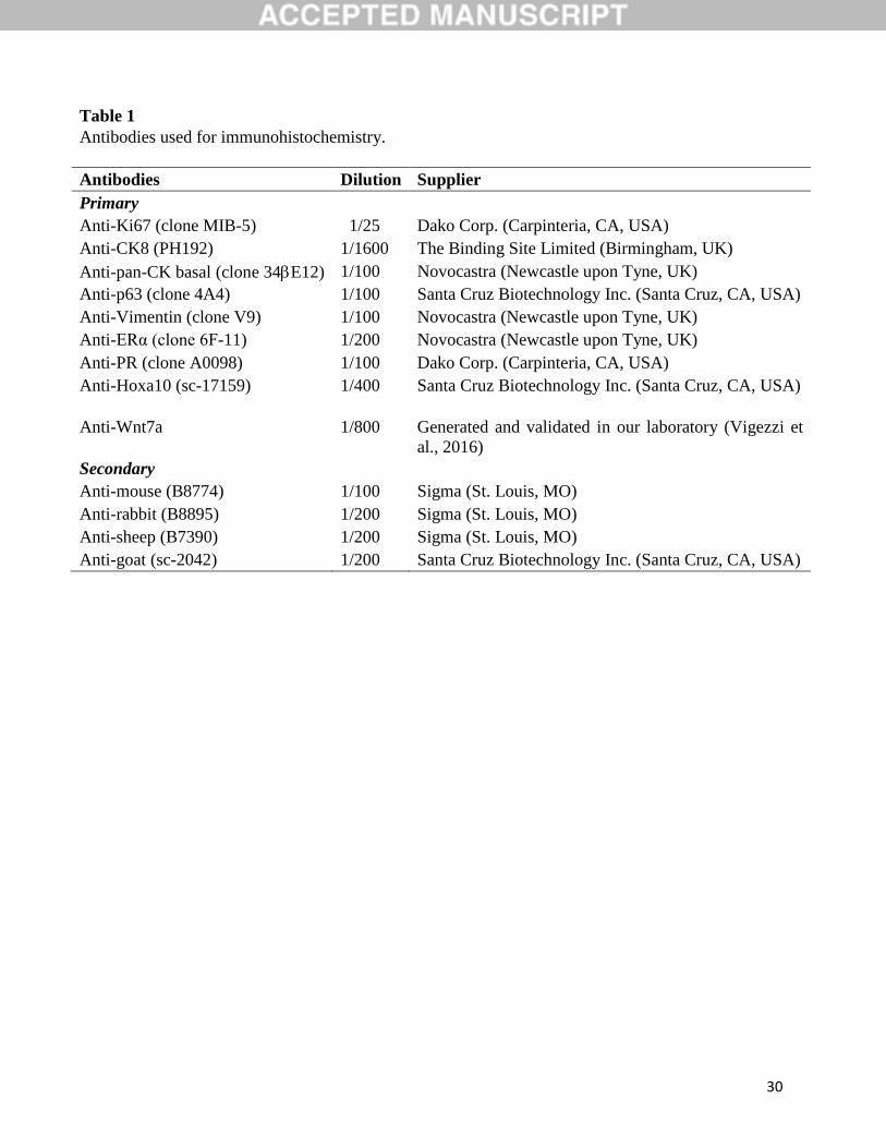
2.4. Immunohistochemistry
In order to determine the immunophenotype of uterine cells we evaluated the expression of
cytokeratins (molecular markers for different types of epithelial differentiation). We evaluated
the expression of CK8 (indicative of simple epithelium) and p63 and panCKs-CK1, 5, 10 and 14-
(indicative of stratified epithelium). To characterize the immunophenotype of the stromal cells
we used an antibody against vimentin, a cytoskeletal protein expressed in mesenchymal-derived
cells. Ki67 was used as a proliferation marker of cells in G1, G2, S and M cell cycle stages.
Primary antibodies against steroid receptors (ERα and PR), Hoxa10 and Wnt7a were used to
evaluate uterine organogenetic differentiation. Antibodies used for IHC were described in Table
1. For Wnt7a immunodetection we used a rabbit polyclonal antibody generated and tested in our
laboratory, according to previously described protocols (Rey et al., 2006;Varayoud et al., 2008).
The Wnt7a antigen included a region corresponding to amino acids 194-283 of the rat sequence
(accession no.EDL91365.1), and the antiserum was purified using antigen-linked affinity
chromatography (Hi-Trap NHS activated HP column; GE Healthcare, Buenos Aires, Argentina).
The specificity of the antibodies was determined using validation tests. First, 1 μg of Wnt-7a
antibody was adsorbed for 24 h at 4 °C with 10–20 μg of the antigenic peptide used to generate
the antibody. No staining of positive control tissues was observed by immunohistochemical
assays using the antibody-antigen complexes. In addition, the specificity of the antiserum was
tested via Western blotting.
8
A standard immunohistochemical technique, following protocols previously described by our
laboratory (Muñoz-de-Toro et al., 1998), was performed. Briefly, uterine longitudinal sections (5
µm thick) were deparaffinized and rehydrated in graded ethanol. After microwave pretreatment
for antigen retrieval, the endogenous peroxidase activity and non-specific binding sites were
blocked. Samples were incubated in a humid chamber with the specific primary antibody
(overnight at 4°C) and then with the corresponding biotin-conjugated secondary antibody (30
min at room temperature) (described in Table 1). Reactions were developed using the avidin-
biotin-peroxidase method and diaminobenzidine (DAB) (Sigma-Aldrich) as a chromogen
substrate. Samples were dehydrated and mounted with permanent mounting medium (Eukitt,
Sigma-Aldrich). For Ki67 immunodetection, the samples were counterstained with Mayer
hematoxylin (Biopur, Rosario, Argentina). Each immunohistochemical run included negative
controls in which the primary antibody was replaced by non-immune horse serum (Sigma-
Aldrich).
2.5. Quantification of cell proliferation
Cell proliferation was evaluated in all uterine compartments using the Olympus BH2 microscope
with a Dplan 100× objective (numerical aperture = 1.25; Olympus). In the luminal epithelium,the
proliferation rate was assessed as a percentage of Ki67-positive cells on a total of 2000
cells/section. In the subepithelial stroma and myometrium,the proliferation index was obtained
considering the volume fraction (Vv) of the Ki67-positive cells, calculated by applying the
followingformula by Weibel (1969): Vv = Pi/P, where Vv is the estimated volume fraction of the
object, Pi is the number of incident points over the positive cells, and P is the number of incident
points over all the cells in the studied population.To obtain the data for the point-counting
procedure, a glass disk with a squared grid of 0.8 mm × 0.8 mm was inserted into a focusing
eyepiece (Gundersen et al., 1988; Ramos et al., 2002).The results were expressed as Vv × 100.
The cell proliferation in the subepithelial stroma and myometrium was quantified on at least 10
randomly selected fields per section, and two sections per animal (separated 25 μm from each
other) were evaluated.

9
2.6. Quantification of protein expression by image analysis
The expression of ERα, PR, Hoxa10 and Wnt7a proteins in all tissue compartments of the uterus
was evaluated by image analysis, using the Image Pro-Plus 5.0.2.9 system (Media Cybernetics)
as previously described (Ramos et al., 2002). Immunostained images were captured with a Dplan
40× objective (numerical aperture =0.65; Olympus) attached to a Spot Insight V3.5 color video
camera. After convert each image into a gray scale, the integrated optical density (IOD) was
measured as a linear combination of the average gray intensity and the relative area occupied by
the positive cells (Ramos et al., 2001; Ramos et al., 2002). Because the IOD is a dimensionless
parameter, the results were expressed as arbitrary unit. In the subephitelial stroma and
myometrium, quantification was performed on at least 10 randomly selected fields per section,
and two sections per animal (separated 25 μm from each other) were evaluated. In the luminal
epithelium, quantification was performed in areas where luminal folds were not present, while in
the glandular epithelium, protein expression was measured on at least 10 endometrial glands of
each uterine sample. Because uterine gland formation in the rat occurs on PND9 (Branham et al.,
1985), quantification in the glandular epithelium was only performed on PND21.
2.7. Statistics
All data are expressed as the mean ± SEM. The incidence of LEH was analyzed by the Fisher’s
exact test. In order to analyzethe other variables weselecteda Mann-Whitney testdue to the small
sample size (n=8) and the impossibility to know the distribution of our variables under study
(Fay et al., 2010). p<0.01 (**) and p<0.05 (*) were accepted as significant.
3. RESULTS
No alterations in maternal care and nursing were detected between the experimental groups. No
signs of acute or chronic toxicity were observed in the litters, and no significant differences in
weight gain between treated and control pups were recorded during the experiment. At the end of
both the neonatal (PND8) and the prepubertal period (PND21) pup´sbody weights were similar
between the GBH-exposed animals (PND8: 14.28 ± 0.20 g and PND21: 35.56 ± 0.59 g) and
control animals (PND8: 14.22 ± 0.30 g and PND21: 36.55 ± 0.58 g).
10
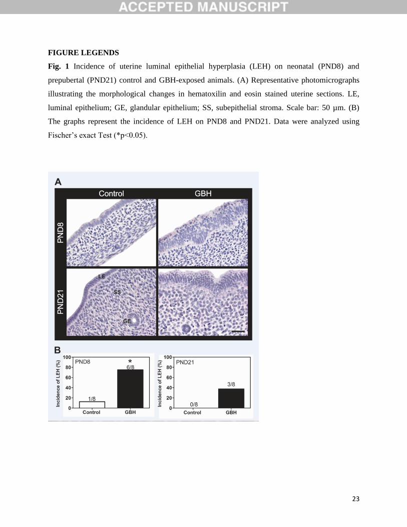
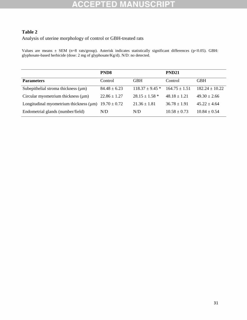
3.1. GBH exposure modified the uterine morphology
The uterus of control animals revealed typical morphological features with a simple columnar
luminal epithelium supported by stromal cells, and two thin layers of smooth muscle on PND8
(Fig. 1A). Two weeks later (PND21), the uterus showed well developed morphological features:
columnar luminal epithelium, simple tubular glands lined with simple cuboidal epithelium,
surrounded with a more stratified endometrial stroma and a thicker myometrium than those on
PND8. The neonatal GBH-exposed uteri exhibitedmorphological changes. We observed that the
75% (6/8) of female pups showed LEH (p<0.05, Fig. 1B) in association with an increase in the
thickness of subepithelial stroma and circular myometrium on PND8 (p<0.05) (Table 2). The
37.5% (3/8) of the GBH-exposed animals exhibited LEH on PND21; however, differences were
not significant (Fig. 1B). The thickness of the subepithelial stroma of PND21 GBH-treated
animals showed a trend to persist increased (p = 0.053, Table 2). Neither significant differences
relative to the thickness of the myometrium nor the number of glands were recorded on PND21
(Table 2).
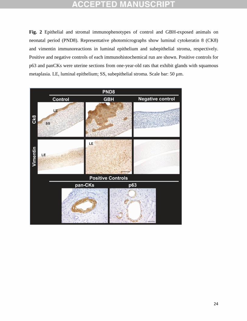
3.2. Uterine epithelial and stromal immunophenotypes in GBH-exposed animals
Then, we determined if the epithelial and stromal cells showed a normal uterine
immunophenotype in GBH-treated animals. Normal uterine epithelium expresses CK8 (simple
epithelium) and normalsubepithelial stroma expresses vimentin (Fig.2). If GBH treatment affects
uterine development, the immunphenotype of uterine compartments could be modified.GBH
exposure did not affect the immnuphenotype of the uterine compartments neither shortly after
the end of the exposure(PND8) nor two weeks after the end of the exposure (PND21). The
epithelial cells were immunoreactive for CK8 (simple epithelium) (Fig. 2) and were negative for
basal CKs (CK1, 5, 10 and 14) and p63 (data not shown). These expression profiles indicate the
absence of squamouscell metaplasia, i.e., change in columnar epithelial cells tostratified
squamous epithelium..
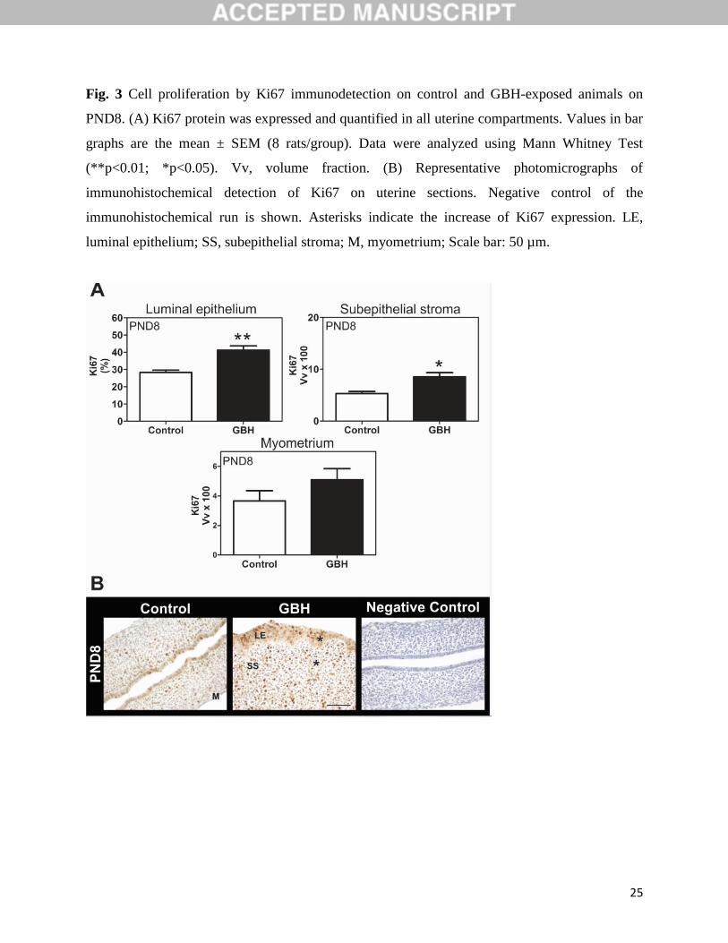
3.3. GBH exposure increased epithelial and stromal proliferation on PND8
The evaluation of Ki67 expression indicated that uterine epithelial and stromal proliferation was
robust in control animals on PND8. Neonatal GBH exposure elicited an increase in cell
proliferation in the epithelial (C: 28.34 ± 1.30%; GBH: 41.28 ± 2.45%, p<0.01) and the stromal
compartments (C: 5.31 ± 0.40; GBH: 8.55 ± 0.80, p<0.05) (Fig. 3, A and B). Endometrial cell
11
proliferation decreased markedly by PND21, reaching very low values (i.e. the percentage of
Ki67-positive cells for the epithelium was less than 0.5%), without changes between control and
GBH-treated animals (data not shown).
3.4. Expression of proteins involved in uterine organogenetic differentiation
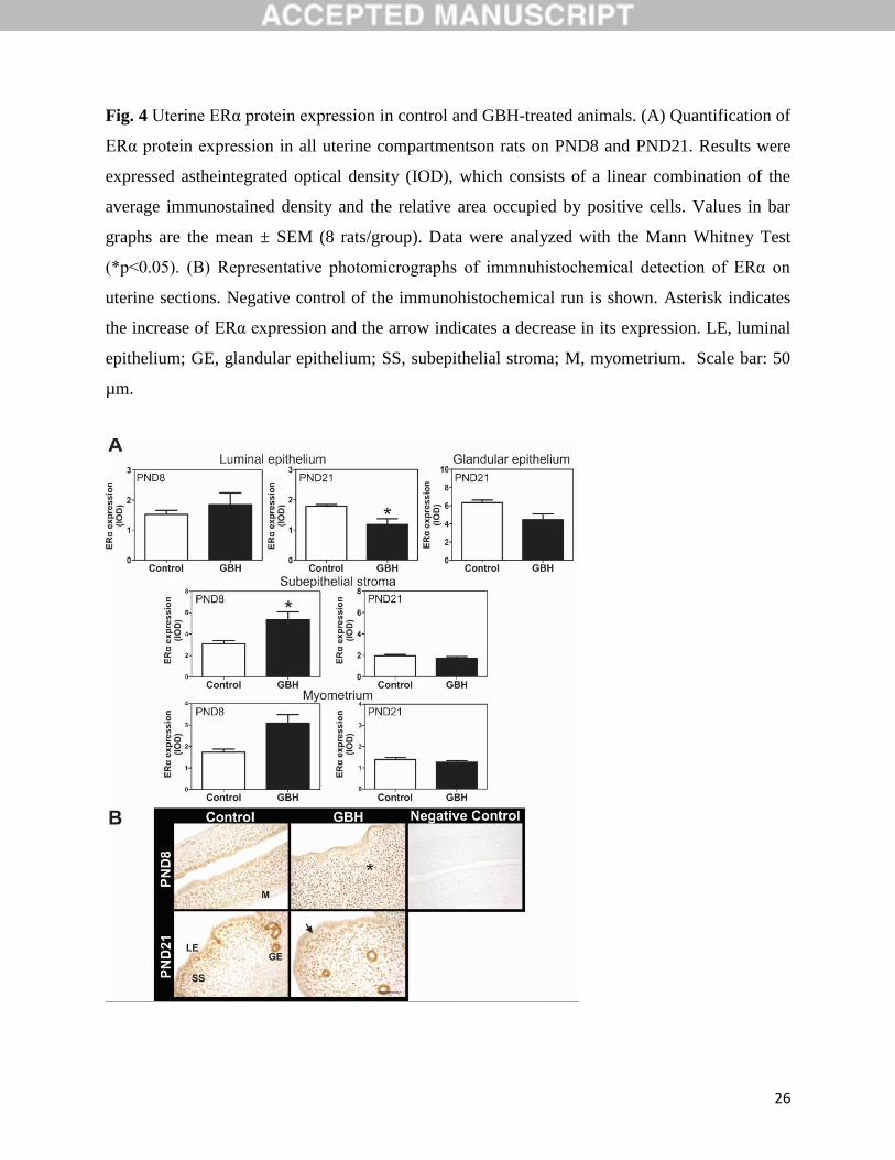
3.4.1. ERα
The quantification of uterine ERα on PND8 (neonatal period) and PND21 (prepubertal period) of
control and GBH-treated animals is presented in Fig. 4A. Female pups neonatally exposed to the
herbicide displayed an induction of ERα in the subepithelial stroma on PND8 (p<0.05, Fig. 4A).
Although GBH-induced changes reverted two weeks after the end of treatment (PND21), a
downregulation of ERα expression was detected in the luminal epithelium (p<0.05, Fig. 4A). The
ERα expression in the myometrium did not show statistically differences between control and
GBH-treated animals at both periods (Fig. 4A). Representative photomicrographs of these results
are shown in Fig. 4B.
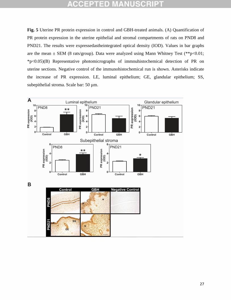
3.4.2. PR
Fig. 5A shows the results of PR quantification in control and GBH-exposed animals.
Surprisingly, on PND8, GBH treatment notably increased PR expression in both the luminal
epithelium and the stromal compartments (p<0.01, Fig. 5A). The deregulation of PR in the
subepithelial stroma persisted up to the prepubertal period (PND21, p<0.05). The quantification
of PR on myometrium was not performed because the detection of PR was weak on PND8 and
PND21, in both control and GBH-treated animals. Representative photomicrographs of these
results are shown in Fig. 5B.
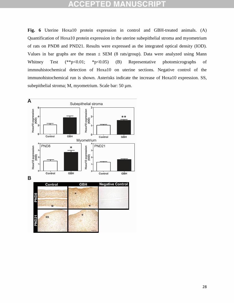
3.4.3. Hoxa10
No immunostaining for Hoxa10 was detectable in the luminal nor glandular epithelium at any
stage examined.In contrast, strong nuclear immunostaining for Hoxa10 was observed in the
subepithelial stroma and myometrium from PND8 animals.Fig. 6A shows the Hoxa10
quantification in the uteri of control and GBH-treated animals. On PND8, an up regulation of
Hoxa10 was observed in both the subepithelial stroma and the myometrium of GBH-exposed
animals (p<0.05, Fig. 6A). The changes observed in the stromal compartment persisted up to
12
PND21 (p<0.05), while the myometrial expression was similar to control rats (Fig. 6A).
Representative photomicrographs of these results are shown in Fig. 6B.
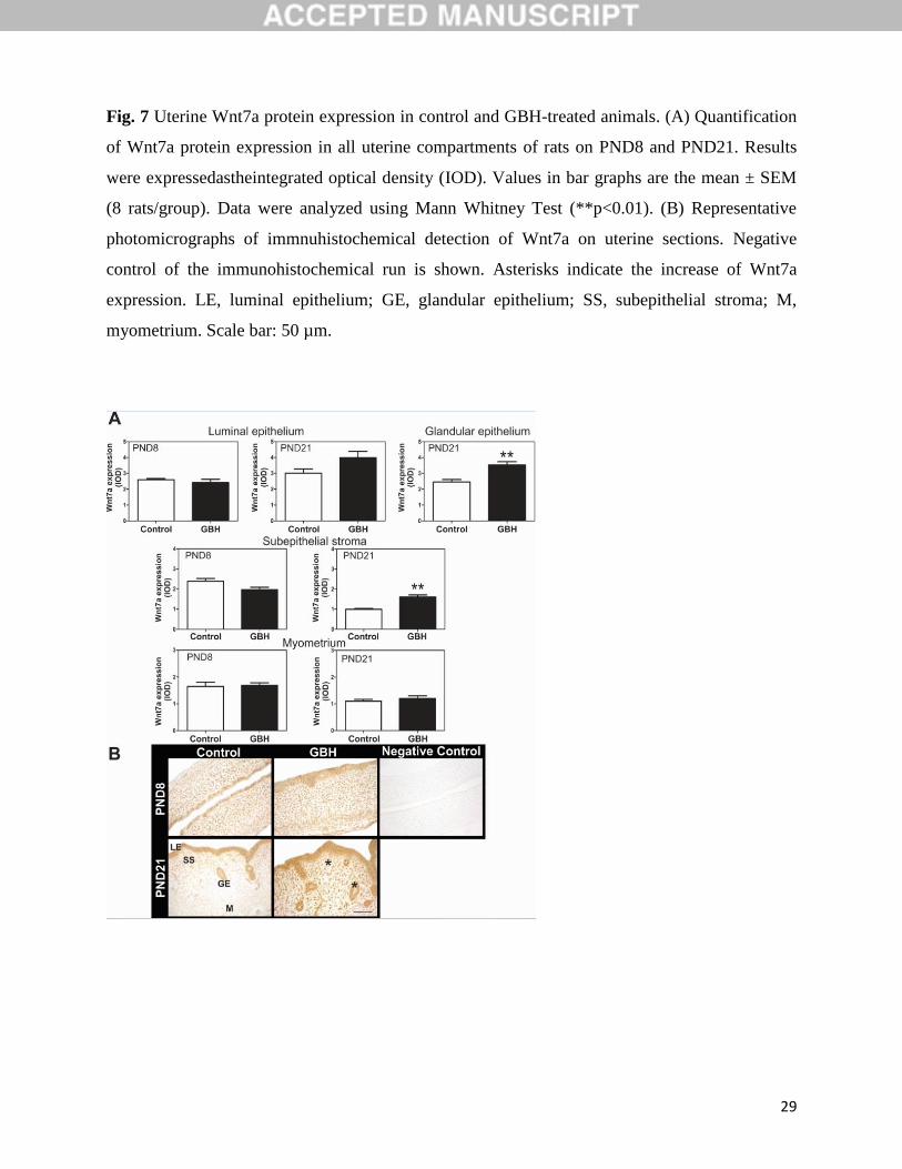
3.4.4. Wnt7a
No changes in Wnt7a expression were observed between the GBH-exposed female pups and
control animals in any of the uterine compartments on PND8. However, on PND21,an induction
of Wnt7a protein was evident in the stromal and glandular cells (p<0.01, Fig. 7A) of GBH-
treated animals. Representative photomicrographs of these results are shown in Fig. 7B.
4. DISCUSSION
To our knowledge, this is the first study showing that postnatal exposure to a GBH affects the
uterine morphology and the expression of proteins that regulate uterine organogenetic
differentiation in neonatal and prepubertal rats. The most relevant effects were incidence of LEH
(75% of animals), increase in stromal and circular myometrium thickness, increase in epithelial
and stromal proliferation, and induction of Hoxa10, PR and ERα on PND8. Two weeks after the
end of the GBH-exposure, some changes were remained, such as the deregulation of Hoxa10, PR
and ERα expression. During this period, a deregulation of Wnt7a uterine expression was also
observed.
The organogenetic development and differentiation of most reproductive tract organs is
completed during the fetal period; however, the uterus is not fully developed or differentiated at
birth. Establishment of tissue-specific histoarchitectureis completed postnatally in laboratory
rodents, domestic animals and presumably humans (Cunha, 1976; Bartol et al., 1999; Kurita and
Nakamura, 2008; Spencer et al., 2012). The functional capacity of the adult uterus is established
by developmental events associated with ‘programming’ of uterine tissues during prenatal and
postnatal life (Sassoon, 1999; Kobayashi and Behringer, 2003; Crain et al., 2008; Walker, 2011).
The postnatal development of the uterus is highly sensitive to a brief exposure to different
substances: hormonal steroids (estradiol, testosterone, progesterone) and others, in general
classified as EDCs. A brief exposure to substances with hormonal activity disrupts the uterine
development in the prepubertal period with consequences at adulthood (Varayoud et al., 2008;
13
Varayoud et al., 2011; Milesi et al., 2015). In the present study, we selected a model of exposure
during the first week of age, to evaluate the effects of GBH on postnatal uterine development.
Regarding morphological evaluation, GBH-exposed uteri showed LEH with a higher
proliferation rate on PND8. The immunophenotype of the epithelial cells indicates a normal
phenotype (CK8 immunoreactive cells, indicative of normal uterine epithelium). In addition, an
increase thickness and a higher proliferation rate were detected in the subepithelial stroma of
GBH-treated animals. Again, the stromal cells showed the typical immunophenotype of uterine
stroma (vimentin-immunoreactive cells, indicative of fibroblastic cells). Clearly, the increased
epithelial and stromal proliferation induced by neonatal GBH treatment was temporary and
reversible. The epithelial and stromal proliferation rate was very low on PND21, both in control
and GBH-exposed animals. However, the 37.5% of animals showed a hyperplastic epithelium on
PND21. All these results could indicate an increase susceptibility to uterine dysfunctions, such as
development of uterine carcinoma. Some evidences could indicate an association between GBH
and carcinogenesis. Glyphosate exposure to hormone-dependent breast cancer cells in vitro
resulted in increased cell proliferation (Thongprakaisang et al. 2013), while an in vivo study
suggested that a glyphosate formulation has tumor promoting potential in skin carcinogenesis in
mice (George et al., 2010). A recent report suggest an augmented risk of cutaneous melanoma
among subjects with exposure to pesticides (glyphosate, mancozeband maneb), in particular
among those exposed to occupational sun exposure (Fortes et al, 2016). A recent report showed
that there are controversial results related to the classification of the herbicide glyphosate as a
“probably carcinogenic to humans” (Portier et al, 2016). The International Agency for Research
on Cancer (IARC, World Health Organization)concluded that glyphosate is a ‘probable human
carcinogen’, putting it into IARC category 2A due to sufficient evidence of carcinogenicity in
animals, limited evidence of carcinogenicity in humans and strong evidence for two carcinogenic
mechanisms(IARC, 2015). However, the European Food Safety Authority (EFSA) concluded
that ‘glyphosate is unlikely to pose a carcinogenic hazard to humans and the evidence does not
support classification with regard to its carcinogenic potential’ (European Food Safety Authority,
2015). The authors concluded that owing to the potential public health impact of glyphosate, it is
essential that all scientific evidence relating to its possible carcinogenicity is publicly accessible
and reviewed transparently in accordance with established scientific criteria (Portier et al, 2016).

14
Subsequently, we investigated whether early postnatal exposure to a GBH induced changes in
the expression of proteins that regulate uterine organogenetic differentiation in neonatal (PND8)
and prepubertal (PND21) periods. Specifically, we found that the GBH exposurealtered the
expression of PR and Hoxa10 both, immediately and two weeks after the exposure.Both PR and
Hoxa10 are two key genes during embryo implantation and decidualization. Previously we
detected that when an EDC disrupts the Hoxa10 and PR expression during development the
animals showed a lower number of implantation sites during pregnancy (Varayoud et al, 2008,
2011; Milesi et al, 2012, 2015). Taking into account our and other results we could suggest that
GBH-postnatally exposed rats could show long-term effects such as subfertility. In addition, the
timing, nature, and severity of endocrine system impacts will vary depending on the levels and
timing of GBH exposures, the age and health status of exposed organisms. Exposures can trigger
a cascade of biological effects that may culminate later in chronic diseases (Myers et al, 2016).
ERα was induced in the stromal compartment on PND8, and was down-regulated in the luminal
epithelial cells of GBH-exposed animals on PND21. GBH treatment also increased the
expression of Wnt7a in the stromal and glandular epithelial cells on PND21. Several agents with
hormonal-like activity have been shown to disrupt the expression of developmental-related
genes. Similar to the effects observed in this study, female pups neonatally exposed to
endosulfan, an organochlorine pesticide recently banned in our country, showed an increased
Hoxa10 uterine expression on PND8 and PND21 (Milesi et al., 2012). Other authors reported a
dose-responsive increase in uterine Hoxa10 expression in 2-week-old mice following in utero
BPA exposure (Smith and Taylor, 2007). As for Wnt7a, a down-regulation in its expression has
been reported in 6-day-old female mice exposed to DES and Aroclor 1254 from PND1 to PND5
(Ma and Sassoon, 2006). Similar to the changes observed in the postnatal exposure model, when
mice were exposed in utero to a high dose of DES, low Wnt7a expression was detected at birth
that normalized at 5 days after delivery (Sassoon, 1999). In our study, the treatment with a GBH
increased the expression of Wnt7a in the stromal and glandular epithelial cells on PND21. It has
been reported that Wnt7a differentially regulates the uterine expression of Hoxa10 during
embryogenesis and adulthood (Miller and Sasson, 1998; Kitajewski and Sassoon, 2000). The
deregulation of both Wnt7a and Hoxa10 detected in our study, suggests a possible mechanistic
interaction in the GBH’s uterine disruption. In our study, PR and ERα were affected by GBH
exposure. A previous report of Thongprakaisang et al. (2013) showed that GBH affects ER
15
expression in mammary cells, producing an induction of ERα in the human T47D hormone-
dependent breast cancer cell line. The authors hypothesized that glyphosate may act as a weak
xenoestrogen, activating ERα (Thongprakaisang et al., 2013). In accordance with these authors
we detected an induction of ERα after GBH exposure. Taken together, these findings confirm
previous evidence that steroid receptors, Hoxa10 and Wnt7a are common targets of endocrine
disruption.
Even though the U.S. EPA has recently concluded that the weight of the evidence is not
sufficient to classified glyphosate as EDC (EPA’s conclusions for Glyphosate Tier 1 battery
screening, June 2015), different studies have shown a disruption of endocrine-signalling systems
in vitro(Thongprakaisang et al., 2013; Gasnier et al, 2009) . The present study shows for the first
time, endocrine disrupting effects of a GBH on the uterus in neonatal and prepubertal rats,
supporting the fact that GBHs might act as an EDC. At this moment, we began to study in new
experiments if a long-term exposure could affect the uterine functional differentiation, with the
intention to mimic the human exposure. More comprehensive toxicity experiments are needed
including those using “two hit” study designs, which examine early life exposures to GBHs
followed by later-life exposures to chemical or other environmental stressors (Myers et al, 2016).
As above-mentioned, chemical mixtures in formulations can have effects that are not predicted
from tests of single compounds(Rajapakse et al., 2002; Silva et al., 2002). GBHs themselves are
chemical mixtures; in addition to the inclusion of glyphosate (the active ingredient), these
herbicides include adjuvants such as surfactants, which can make GBH-product formulations
more toxic than glyphosate alone (Mesnage et al., 2014). In vitrostudies have demonstrated toxic
effects at low doses of GBHs in embryonic, fetal, placental and testicular cells (Richard et al.,
2005;Benachourand Seralini, 2009; Clair et al., 2012; Mesnage et al., 2013).
Uterine morphogenesis is governed by several hormonal, cellular and molecular mechanisms. As
previously stated, disruption of the normal developmental program by neonatal exposure to
EDCs might induce permanent changes in the structure and function of tissues and organs
(Newbold et al., 1990; Kitajewski and Sassoon, 2000; Bosquiazzo et al., 2013). Hoxa10 and
Wnt7a regulate anterior-posterior and radial patterning of the müllerian duct. Specifically, Wnt7a
plays a pivotal role in postnatal uterine myometrial and epithelial morphogenesis. It has been
16
reported that a deregulation in Wnt7a expression lead to abnormal multilayered epithelium
(Carta and Sassoon et al., 2004) and hyperplastic and disorganized myometrium (Miller and
Sassoon, 1998; Miller et al., 1998a). Altered expression of Hoxa10 and Wnt7a during
development results in uterine anomalies, as seen in mice exposed prenatally to DES (Miller et
al., 1998a,b; Block et al., 2000). Mice exposed to DES showed many uterine malformations,
characterized by luminal and glandular squamous metaplasia, endometrial hyperplasia and
increased risk of leiomyomas (Kitajewski and Sassoon, 2000). Similar to DES, in our work, a
brief exposure to a GBH during development lead to endometrial hyperplasia. These
organizationally induced alterations in uterine histoarchitecture might lead to reproductive
anomalies, such as infertility and early pregnancy loss, and could promote the development of
uterine neoplasias (Newbold et al., 1990; Newbold et al., 1997).
5. CONCLUSION
In summary, our results show that early postnatal exposure to a GBH, at dose similar to the RfD,
alters uterine morphology and the expression of proteins involved in uterine development and
differentiation. We considered that more studied should be conducted to fully understand the
effects of GBH on the female reproductive health, as well as, its molecular mechanism of action.
Currently, further experiments are underway to investigate whether the deregulation of steroid
receptors, Hoxa10 and Wnt7a following postnatal exposure to a GBH could affect the proper
uterine function along pregnancy and/or promote uterine neoplasias.
Conflict of interest statement
The authors declare that there are no conflicts of interest.
Acknowledgements
Financial support from the Argentine National Agency for the Promotion of Science and
Technology (ANPCyT, PICT 2011-1491, PICT 2014-1522;PICT 2014-1522), the Argentine
National Council for Science and Technology (CONICET, PIP 2011, 11220110100494) and the
Universidad Nacional del Litoral (CAI + D 2011, 501 20110100423 LI).
17
REFERENCES
Agricultural Marketing Service, 2013. Pesticide data program annual summary, program year
2011. In: Appendix C Distribution of Residues in Soybean by Pesticide. Washington, D.C: U.
S. Department of Agriculture.
Andreoli, M.F., Stoker, C., Rossetti, M.F., Alzamendi, A., Castrogiovanni, D., Luque,
E.H.,Ramos, J.G., 2015. Withdrawal of dietary phytoestrogens in adult male rats affects
hypothalamic regulation of food intake, induces obesity and alters glucose metabolism.
Mol.Cell.Endocrinol. 401, 111-119.
Aparicio, V.C., De Geronimo, E., Marino, D., Primost, J., Carriquiriborde, P.,Costa, J.L., 2013.
Environmental fate of glyphosate and aminomethylphosphonic acid in surface waters and soil
of agricultural basins. Chemosphere.93, 1866-1873.
Monitoring glyphosate residues in transgenic glyphosate-resistant soybean. Pest.Manag. Sci. 60,
163-166.
Bagot, C.N., Troy, P.J.,Taylor, H.S., 2000. Alteration of maternal Hoxa10 expression by in vivo
gene transfection affects implantation. Gene.Ther.7, 1378-1384.
Bartol, F.F., Wiley, A.A., Floyd, J.G., Ott, T.L., Bazer, F.W., Gray, C.A., Spencer, T.E.,
1999.Uterine differentiation as a foundation for subsequent fertility.J.Reprod.Fertil. Suppl.54,
287-302.
Baylis, A.D., 2000. Why glyphosate is a global herbicide: strengths, weaknesses and prospects.
Pest.Manag. Sci.56, 299-308.
Benachour, N.,Seralini, G.E., 2009. Glyphosate formulations induce apoptosis and necrosis in
human umbilical, embryonic, and placental cells. Chem. Res. Toxicol. 22, 97-105.
Benson, G.V., Lim, H., Paria, B.C., Satokata, I., Dey, S.K. and Maas, R.L., 1996. Mechanisms of
reduced fertility in Hoxa-10 mutant mice: uterine homeosis and loss of maternal Hoxa-10
expression. Development.122, 2687-2696.
Block, K., Kardana, A., Igarashi, P., Taylor, H.S., 2000. In utero diethylstilbestrol (DES)
exposure alters Hox gene expression in the developing mullerian system. Faseb.J. 14, 1101-
1108.
Bosquiazzo, V.L., Vigezzi, L., Muñoz-de-Toro, M., Luque, E.H., 2013. Perinatal exposure to
diethylstilbestrol alters the functional differentiation of the adult rat uterus.
J.Steroid.Biochem.Mol.Biol.138, 1-9.
Branham, W.S., Sheehan, D.M., Zehr, D.R., Ridlon, E., Nelson, C.J., 1985. The postnatal
ontogeny of rat uterine glands and age-related effects of 17 beta-estradiol. Endocrinology.117,
2229-2237.
Carta, L., Sassoon, D., 2004. Wnt7a is a suppressor of cell death in the female reproductive tract
and is required for postnatal and estrogen-mediated growth. Biol.Reprod.71, 444-454.
Cerdeira, A.L., Gazziero, D.L., Duke, S.O.,Matallo, M.B., 2011. Agricultural impacts of
glyphosate-resistant soybean cultivation in South America. J. Agric. Food.Chem.59, 5799-
5807.

18
Cerdeira, A.L., Gazziero, D.L., Duke, S.O., Matallo, M.B.,Spadotto, C.A., 2007. Review of
potential environmental impacts of transgenic glyphosate-resistant soybean in Brazil. J.
Environ. Sci. Health. B. 42, 539-549.
Clair, E., Mesnage, R., Travert, C., Seralini, G.E., 2012. A glyphosate-based herbicide induces
necrosis and apoptosis in mature rat testicular cells in vitro, and testosterone decrease at lower
levels. Toxicol. In. Vitro. 26, 269-279.
Crain, D.A., Janssen, S.J., Edwards, T.M., Heindel, J., Ho, S.M., Hunt, P., Iguchi, T., Juul, A.,
McLachlan, J.A., Schwartz, J., Skakkebaek, N., Soto, A.M., Swan, S., Walker, C., Woodruff,
T.K., Woodruff, T.J., Giudice, L.C.,Guillette, L.J. Jr., 2008. Female reproductive disorders:
the roles of endocrine-disrupting compounds and developmental timing. Fertil.Steril.90, 911-
940.
Cunha, G.R., 1976. Stromal induction and specification of morphogenesis and
cytodifferentiation of the epithelia of the Mullerian ducts and urogenital sinus during
development of the uterus and vagina in mice.J.Exp.Zool.196, 361-370.
Curwin, B.D., Hein, M.J., Sanderson, W.T., Striley, C., Heederik, D., Kromhout, H., Reynolds,
S.J.,Alavanja, M.C., 2007a. Pesticide dose estimates for children of Iowa farmers and non-
farmers. Environ. Res.105, 307-315.
Curwin, B.D., Hein, M.J., Sanderson, W.T., Striley, C., Heederik, D., Kromhout, H., Reynolds,
S.J., Alavanja, M.C., 2007b. Urinary pesticide concentrations among children, mothers and
fathers living in farm and non-farm households in Iowa. Ann. Occup. Hyg. 51, 53-65.
Defarge, N., Takacs, E., Lozano, V.L., Mesnage, R., Spiroux de Vendomois, J., Seralini, G.E.,
Szekacs, A., 2016. Co-formulants in glyphosate-based herbicides disrupt aromatase activity in
human cells below toxic levels. Int. J. Environ. Res. Public Health. 13, 1-17.
Dixon, D., Alison, R., Bach, U., Colman, K., Foley, G.L., Harleman, J.H., Haworth, R., Herbert,
R., Heuser, A., Long, G., Mirsky, M., Regan, K., Van Esch, E., Westwood, F.R., Vidal, J.,
Yoshida, M., 2014. Nonproliferative and proliferative lesions of the rat and mouse female
reproductive system. J. Toxicol. Pathol. 27, 1s-107s.
Duke, S.O., Powles, S.B., 2008. Glyphosate: a once-in-a-century herbicide. Pest.Manag. Sci. 64,
319-325.
Dunlap, K.A., Filant, J., Hayashi, K., Rucker, E.B. 3rd, Song, G., Deng, J.M., Behringer, R.R.,
DeMayo, F.J., Lydon, J., Jeong, J.W.,Spencer, T.E., 2011. Postnatal deletion of Wnt7a
inhibits uterine gland morphogenesis and compromises adult fertility in mice. Biol.Reprod.85,
386-396.
European Food Safety Authority. Final Addendum to the Renewal Assessment
Report.2015. http://registerofquestions.efsa.europa.eu/roqFrontend/outputLoader?output=ON-
4302
Fay, M.P., Proschan, M.A., 2010. Wilcoxon-Mann-Whitney or t-test? On assumptions for
hypothesis tests and multiple interpretations of decision rules. Stat. Surv. 4, 1-39.

19
Fortes, C., Mastroeni, S., Segatto, M.M., Hohmann, C., Miligi, L., Bakos, L., Bonamigo, R.,
2016. Occupational Exposure to Pesticides With Occupational Sun Exposure Increases the
Risk for Cutaneous Melanoma. J. Occup. Environ. Med. 58, 370-375.
Gasnier, C., Dumont, C., Benachour, N., Clair, E., Chagnon, M.C., Seralini, G.E., 2009.
Glyphosate-based herbicides are toxic and endocrine disruptors in human cell lines.
Toxicology. 262, 184-191.
George, J., Prasad, S., Mahmood, Z., Shukla, Y., 2010. Studies on glyphosate-induced
carcinogenicity in mouse skin: a proteomic approach. J.Proteomics.73, 951-964.
Gundersen, H.J., Bendtsen, T.F., Korbo, L., Marcussen, N., Moller, A., Nielsen, K., Nyengaard,
J.R., Pakkenberg, B., Sorensen, F.B., Vesterby, A., et al., 1988. Some new, simple and
efficient stereological methods and their use in pathological research and diagnosis. Apmis.
96, 379-394.
IARC WorkingGroup. Glyphosate. In: Some organophosphate insecticides and herbicides:
diazinon, glyphosate, malathion, parathion, and tetrachlorvinphos.2015..IARC Monogr.
Prog, 112, 1–92.
Kass, L., Altamirano, G.A., Bosquiazzo, V.L., Luque, E.H., Muñoz-de-Toro, M., 2012. Perinatal
exposure to xenoestrogens impairs mammary gland differentiation and modifies milk
composition in Wistar rats. Reprod. Toxicol.33, 390-400.
Kitajewski, J., Sassoon, D., 2000. The emergence of molecular gynecology: homeobox and Wnt
genes in the female reproductive tract. Bioessays.22, 902-910.
Kobayashi, A.,Behringer, R.R., 2003. Developmental genetics of the female reproductive tract in
mammals. Nat.Rev. Genet.4, 969-980.
Kruger, M., Schrodl, W., Pedersen, I., Shehata, A.A., 2014. Detection of glyphosate in
malformed piglets. J. Environ. Anal. Toxicol. 4, 1-2.
Kurita, T., Nakamura, H., 2008.Embryology of the uterus. In: Aplin, J.D. et al.(Eds),
Endometrium.Informa UK Ltd., London, pp. 1–18.
Ma, R., Sassoon, D.A., 2006. PCBs exert an estrogenic effect through repression of the Wnt7a
signaling pathway in the female reproductive tract. Environ.Health.Perspect.114, 898-904.
Mamy, L., Gabrielle, B.,Barriuso, E., 2010.Comparative environmental impacts of glyphosate
and conventional herbicides when used with glyphosate-tolerant and non-tolerant
crops.Environ. Pollut.158, 3172-3178.
Mesnage, R., Bernay, B., Seralini, G.E., 2013. Ethoxylated adjuvants of glyphosate-based
herbicides are active principles of human cell toxicity. Toxicology. 313, 122-128.
Mesnage, R., Defarge, N., Spiroux de Vendomois, J., Seralini, G.E., 2014. Major pesticides are
more toxic to human cells than their declared active principles. Biomed. Res. Int. 2014, 1-8.
Milesi, M.M., Alarcon, R., Ramos, J.G., Muñoz-de-Toro, M., Luque, E.H.,Varayoud, J., 2015.
Neonatal exposure to low doses of endosulfan induces implantation failure and disrupts
uterine functional differentiation at the pre-implantation period in rats. Mol.Cell.Endocrinol.
401, 248-259.
20
Milesi, M.M., Varayoud, J., Bosquiazzo, V.L., Muñoz-de-Toro, M., Luque, E.H., 2012. Neonatal
exposure to low doses of endosulfan disrupts the expression of proteins regulating uterine
development and differentiation. Reprod. Toxicol.33, 85-93.
Miller, C., Degenhardt, K., Sassoon, D.A., 1998a. Fetal exposure to DES results in de-regulation
of Wnt7a during uterine morphogenesis. Nat.Genet.20, 228-230.
Miller, C., Pavlova, A., Sassoon, D.A., 1998b. Differential expression patterns of Wnt genes in
the murine female reproductive tract during development and the estrous cycle. Mech.Dev.76,
91-99.
Miller, C., Sassoon, D.A., 1998. Wnt-7a maintains appropriate uterine patterning during the
development of the mouse female reproductive tract. Development. 16, 3201-3211.
Muñoz-de-Toro, M.M., Maffini, M.V., Kass, L.,Luque, E.H., 1998.Proliferative activity and
steroid hormone receptor status in male breast carcinoma.J. Steroid.Biochem.Mol.Biol.67,
333-339.
Myers, J.P., Antoniou, M.N., Blumberg, B., Carroll, L., Colborn, T., Everett, L.G., Hansen, M.,
Landrigan, P.J., Lanphear, B.P., Mesnage, R., Vandenberg, L.N., VomSaal, F.S., Welshons,
W.V., Benbrook, C.M., 2016. Concerns over use of glyphosate-based herbicides and risks
associated with exposures: a consensus statement. Environ. Health. 15, 1-13.
Newbold, R.R., Bullock, B.C., McLachlan, J.A., 1990. Uterine adenocarcinoma in mice
following developmental treatment with estrogens: a model for hormonal carcinogenesis.
Cancer.Res.50, 7677-7681.
Newbold, R.R., Jefferson, W.N., Padilla-Burgos, E., Bullock, B.C., 1997. Uterine carcinoma in
mice treated neonatally with tamoxifen. Carcinogenesis. 18, 2293-2298.
Peruzzo, P.J., Porta, A.A., Ronco, A.E., 2008. Levels of glyphosate in surface waters, sediments
and soils associated with direct sowing soybean cultivation in north pampasic region of
Argentina. Environ. Pollut.156, 61-66.
Portier, C.J., Armstrong, B.K., Baguley, B.C., et al., 2016. Differences in the carcinogenic
evaluation of glyphosate between the International Agency for Research on Cancer (IARC)
and the European Food Safety Authority (EFSA). J. Epidemiol. Community. Health. 1-5.
Rajapakse, N., Silva, E., Kortenkamp, A., 2002. Combining xenoestrogens at levels below
individual no-observed-effect concentrations dramatically enhances steroid hormone action.
Environ. Health. Perspect. 110, 917-921.
Ramos, J.G., Varayoud, J., Bosquiazzo, V.L., Luque, E.H., Muñoz-de-Toro, M., 2002. Cellular
turnover in the rat uterine cervix and its relationship to estrogen and progesterone receptor
dynamics.Biol.Reprod. 67, 735-742.
Ramos, J.G., Varayoud, J., Sonnenschein, C., Soto, A.M., Muñoz De Toro, M., Luque, E.H.,
2001.Prenatal exposure to low doses of bisphenol A alters the periductal stroma and glandular
cell function in the rat ventral prostate. Biol.Reprod. 65, 1271-1277.
Rey, F., Ramos, J.G., Stoker, C., Bussmann, L.E., Luque, E.H., Muñoz-de-Toro, M.,
2006.Vitellogenin detection in Caiman latirostris (Crocodylia: Alligatoridae): a tool to assess
environmental estrogen exposure in wildlife. J.Comp.Physiol. B. 176, 243-251.

21
Richard, S., Moslemi, S., Sipahutar, H., Benachour, N., Seralini, G.E., 2005. Differential effects
of glyphosate and roundup on human placental cells and aromatase. Environ. Health.
Perspect. 113, 716-720.
Roy, J.R., Chakraborty, S., Chakraborty, T.R., 2009.Estrogen-like endocrine disrupting
chemicals affecting puberty in humans--a review. Med.Sci.Monit.15, RA137-145.
Sassoon, D., 1999. Wnt genes and endocrine disruption of the female reproductive tract: a
genetic approach. Mole.Cell.Endocrinol.158, 1-5.
Shehata, A.A., Schrodl, W., Aldin, A.A., Hafez, H.M., Kruger, M., 2013. The effect of
glyphosate on potential pathogens and beneficial members of poultry microbiota in vitro.
Curr. Microbiol. 66, 350-358.
Silva, E., Rajapakse, N., Kortenkamp, A., 2002. Something from "nothing"--eight weak
estrogenic chemicals combined at concentrations below NOECs produce significant mixture
effects. Environ. Sci. Technol. 36, 1751-1756.
Smith, C.C., Taylor, H.S., 2007.Xenoestrogen exposure imprints expression of genes (Hoxa10)
required for normal uterine development. Faseb. J. 21, 239-246.
Spencer, T.E., Dunlap, K.A.,Filant, J., 2012. Comparative developmental biology of the uterus:
insights into mechanisms and developmental disruption. Mol.Cell.Endocrinol.354, 34-53.
Test Biotech, 2013. High levels of residues from spraying with glyphosate found in soybeans in
Argentina. http://www.testbiotech.org/en/node/926. Accessed 18 May 2016.
Thongprakaisang, S., Thiantanawat, A., Rangkadilok, N., Suriyo, T.,Satayavivad, J., 2013.
Glyphosate induces human breast cancer cells growth via estrogen receptors.
Food.Chem.Toxicol.59, 129-136.
U.S. EPA, 1993.EPA 738-F-93-011.Registration Eligibility Decision (RED) for
Glyphosate.Pag.3
http://nepis.epa.gov/Exe/ZyPDF.cgi/20000A5T.PDF?Dockey=20000A5T.PDF
U.S.EPA, 2015.EDSP (Endocrine Disruptor Screening Program): Weight of Evidence Analysis
of Potential Interaction with the Estrogen, Androgen or Thyroid Pathways. Chemical:
Glyphosate.Pag. 26. http://www.epa.gov/sites/production/files/2015-
06/documents/glyphosate-417300_2015-06-29_txr0057175.pdf
Varayoud, J., Ramos, J.G., Bosquiazzo, V.L., Lower, M., Muñoz-de-Toro, M., Luque, E.H.,
2011. Neonatal exposure to bisphenol A alters rat uterine implantation-associated gene
expression and reduces the number of implantation sites. Endocrinology. 152, 1101-1111.
Varayoud, J., Ramos, J.G., Bosquiazzo, V.L., Muñoz-de-Toro, M., Luque, E.H., 2008.
Developmental exposure to bisphenol A impairs the uterine response to ovarian steroids in the
adult. Endocrinology. 149, 5848-5860.
Varayoud, J., Ramos, J.G., Muñoz-de-Toro, M., Luque, E.H., 2014. Long-lasting effects of
neonatal bisphenol A exposure on the implantation process. Vitam.Horm.94, 253-275.
Vigezzi, L., Ramos, J.G., Kass, L., Tschopp, M.V., Muñoz-de-Toro, M., Luque, E.H,,
Bosquiazzo, V.L. 2016. A deregulated expression of estrogen-target genes is associated with
22
an altered response to estradiol in aged rats perinatally exposed to bisphenol A. Mol Cell
Endocrinol. 426, 33-42.
Walker, C.L., 2011. Epigenomic reprogramming of the developing reproductive tract and disease
susceptibility in adulthood.Birth.Defects.Res. A.Clin.Mol.Teratol.91, 666-671.
Weibel, E.R., 1969.Stereological principles for morphometry in electron microscopic
cytology.Int. Rev.Cytol.26, 235-302.
Woodburn, J., 2000. Glyphosate: production, pricing and use worldwide. Pest.Manag. Sci.56,
309-312.
23
FIGURE LEGENDS
Fig. 1 Incidence of uterine luminal epithelial hyperplasia (LEH) on neonatal (PND8) and
prepubertal (PND21) control and GBH-exposed animals. (A) Representative photomicrographs
illustrating the morphological changes in hematoxilin and eosin stained uterine sections. LE,
luminal epithelium; GE, glandular epithelium; SS, subepithelial stroma. Scale bar: 50 µm. (B)
The graphs represent the incidence of LEH on PND8 and PND21. Data were analyzed using
Fischer’s exact Test (*p<0.05).
24
Fig. 2 Epithelial and stromal immunophenotypes of control and GBH-exposed animals on
neonatal period (PND8). Representative photomicrographs show luminal cytokeratin 8 (CK8)
and vimentin immunoreactions in luminal epithelium and subepithelial stroma, respectively.
Positive and negative controls of each immunohistochemical run are shown. Positive controls for
p63 and panCKs were uterine sections from one-year-old rats that exhibit glands with squamous
metaplasia. LE, luminal epithelium; SS, subepithelial stroma. Scale bar: 50 µm.
25
Fig. 3 Cell proliferation by Ki67 immunodetection on control and GBH-exposed animals on
PND8. (A) Ki67 protein was expressed and quantified in all uterine compartments. Values in bar
graphs are the mean ± SEM (8 rats/group). Data were analyzed using Mann Whitney Test
(**p<0.01; *p<0.05). Vv, volume fraction. (B) Representative photomicrographs of
immunohistochemical detection of Ki67 on uterine sections. Negative control of the
immunohistochemical run is shown. Asterisks indicate the increase of Ki67 expression. LE,
luminal epithelium; SS, subepithelial stroma; M, myometrium; Scale bar: 50 µm.
26
Fig. 4 Uterine ERα protein expression in control and GBH-treated animals. (A) Quantification of
ERα protein expression in all uterine compartmentson rats on PND8 and PND21. Results were
expressed astheintegrated optical density (IOD), which consists of a linear combination of the
average immunostained density and the relative area occupied by positive cells. Values in bar
graphs are the mean ± SEM (8 rats/group). Data were analyzed with the Mann Whitney Test
(*p<0.05). (B) Representative photomicrographs of immnuhistochemical detection of ERα on
uterine sections. Negative control of the immunohistochemical run is shown. Asterisk indicates
the increase of ERα expression and the arrow indicates a decrease in its expression. LE, luminal
epithelium; GE, glandular epithelium; SS, subepithelial stroma; M, myometrium. Scale bar: 50
µm.
27
Fig. 5 Uterine PR protein expression in control and GBH-treated animals. (A) Quantification of
PR protein expression in the uterine epithelial and stromal compartments of rats on PND8 and
PND21. The results were expressedastheintegrated optical density (IOD). Values in bar graphs
are the mean ± SEM (8 rats/group). Data were analyzed using Mann Whitney Test (**p<0.01;
*p<0.05)(B) Representative photomicrographs of immnuhistochemical detection of PR on
uterine sections. Negative control of the immunohistochemical run is shown. Asterisks indicate
the increase of PR expression. LE, luminal epithelium; GE, glandular epithelium; SS,
subepithelial stroma. Scale bar: 50 µm.
28
Fig. 6 Uterine Hoxa10 protein expression in control and GBH-treated animals. (A)
Quantification of Hoxa10 protein expression in the uterine subepithelial stroma and myometrium
of rats on PND8 and PND21. Results were expressed as the integrated optical density (IOD).
Values in bar graphs are the mean ± SEM (8 rats/group). Data were analyzed using Mann
Whitney Test (**p<0.01; *p<0.05) (B) Representative photomicrographs of
immnuhistochemical detection of Hoxa10 on uterine sections. Negative control of the
immunohistochemical run is shown. Asterisks indicate the increase of Hoxa10 expression. SS,
subepithelial stroma; M, myometrium. Scale bar: 50 µm.
29
Fig. 7 Uterine Wnt7a protein expression in control and GBH-treated animals. (A) Quantification
of Wnt7a protein expression in all uterine compartments of rats on PND8 and PND21. Results
were expressedastheintegrated optical density (IOD). Values in bar graphs are the mean ± SEM
(8 rats/group). Data were analyzed using Mann Whitney Test (**p<0.01). (B) Representative
photomicrographs of immnuhistochemical detection of Wnt7a on uterine sections. Negative
control of the immunohistochemical run is shown. Asterisks indicate the increase of Wnt7a
expression. LE, luminal epithelium; GE, glandular epithelium; SS, subepithelial stroma; M,
myometrium. Scale bar: 50 µm.
30
Table 1
Antibodies used for immunohistochemistry.
Antibodies Dilution Supplier
Primary
Anti-Ki67 (clone MIB-5) 1/25 Dako Corp. (Carpinteria, CA, USA)
Anti-CK8 (PH192) 1/1600 The Binding Site Limited (Birmingham, UK)
Anti-pan-CK basal (clone 34E12) 1/100 Novocastra (Newcastle upon Tyne, UK)
Anti-p63 (clone 4A4) 1/100 Santa Cruz Biotechnology Inc. (Santa Cruz, CA, USA)
Anti-Vimentin (clone V9) 1/100 Novocastra (Newcastle upon Tyne, UK)
Anti-ERα (clone 6F-11) 1/200 Novocastra (Newcastle upon Tyne, UK)
Anti-PR (clone A0098) 1/100 Dako Corp. (Carpinteria, CA, USA)
Anti-Hoxa10 (sc-17159) 1/400 Santa Cruz Biotechnology Inc. (Santa Cruz, CA, USA)
Anti-Wnt7a 1/800
Generated and validated in our laboratory (Vigezzi et
al., 2016)
Secondary
Anti-mouse (B8774) 1/100 Sigma (St. Louis, MO)
Anti-rabbit (B8895) 1/200 Sigma (St. Louis, MO)
Anti-sheep (B7390) 1/200 Sigma (St. Louis, MO)
Anti-goat (sc-2042) 1/200 Santa Cruz Biotechnology Inc. (Santa Cruz, CA, USA)
31
Table 2
Analysis of uterine morphology of control or GBH-treated rats
Values are means ± SEM (n=8 rats/group). Asterisk indicates statistically significant differences (p<0.05). GBH:
glyphosate-based herbicide (dose: 2 mg of glyphosate/Kg/d). N/D: no detected.
PND8 PND21
Parameters Control GBH Control GBH
Subepithelial stroma thickness (µm) 84.48 ± 6.23 118.37 ± 9.45 * 164.75 ± 1.51 182.24 ± 10.22
Circular myometrium thickness (µm) 22.86 ± 1.27 28.15 ± 1.58 * 48.18 ± 1.21 49.30 ± 2.66
Longitudinal myometrium thickness (µm) 19.70 ± 0.72 21.36 ± 1.81 36.78 ± 1.91 45.22 ± 4.64
Endometrial glands (number/field) N/D N/D 10.58 ± 0.73 10.84 ± 0.54