neonatal thrombocytopenia
TRANSCRIPT

Seminars in Fetal & Neonatal Medicine (2008) 13, 256e264
ava i lab le a t www.sc iencedi rec t .com
journa l homepage : www.e l sev i er . com/ loca te /s iny
Neonatal thrombocytopenia
Irene Roberts a,*, Neil A. Murray b
a Department of Haematology, Imperial College London and Imperial Healthcare NHS Trust,Du Cane Road, London W12 0NN, UKb National Blood Service, John Radcliffe Hospital, Oxford, UK
KEYWORDSFetalthrombocytopenia;Neonatal alloimmunethrombocytopenia(NAIT);Neonatalthrombocytopenia;Platelet transfusion
* Corresponding author. Tel.: þ44 209335.
E-mail address: irene.roberts@imp
1744-165X/$ - see front matter ª 200doi:10.1016/j.siny.2008.02.004
Summary Thrombocytopenia (platelets <150 � 109/L) is one of the most common haemato-logical problems in neonates, particularly those who are preterm and sick. In those pretermneonates with early thrombocytopenia who present within 72 h of birth, the most commoncause is reduced platelet production secondary to intrauterine growth restriction and/or ma-ternal hypertension. By contrast, the most common causes of thrombocytopenia arising afterthe first 72 h of life, both in preterm and term infants, are sepsis and necrotizing enterocolitis.The most important cause of severe thrombocytopenia (platelets <50 � 109/L) is neonatalalloimmune thrombocytopenia (NAIT), as diagnosis can be delayed and death or long-term dis-ability due to intracranial haemorrhage may occur. Platelet transfusion is the mainstay oftreatment for severe thrombocytopenia. However, the correlation between thrombocytopeniaand bleeding is unclear and no studies have yet shown clinical benefit for platelet transfusionin neonates. Studies to identify optimal platelet transfusion practice for neonatal thrombocy-topenia are urgently required.ª 2008 Elsevier Ltd. All rights reserved.
Introduction
Sick neonates often develop thrombocytopenia. Of neo-nates admitted to neonatal intensive care units (NICUs), theplatelet count drops below 150 � 109/L in one in four babiesand to below 50 � 109/L in one in twenty. Many clinical con-ditions are associated with neonatal thrombocytopenia (NT)but, until recently, almost half of all cases were classified asidiopathic. However, recent studies detailing the naturalhistory of NT have identified reduced platelet productionas the main underlying mechanism of many idiopathic cases.This has led to newer classifications of NT based on thetiming of onset of thrombocytopenia (i.e. early versus late
8 383 2163; fax: þ44 208 742
erial.ac.uk (I. Roberts).
8 Elsevier Ltd. All rights reserved
NT; Table 1). These are more useful for neonatal cliniciansthan previous classifications, with their focus on associa-tions and rare disorders, and will help to facilitate system-atic studies to improve the management of NT.
The need for systematic studies is emphasized by thevariation in platelet transfusion practice between centres,with reported rates ranging from 2% to 9% for similar NICUpopulations.1e3 In addition, it is important to note that nostudy has yet shown clinical benefit of platelet transfusionin NT. Indeed, recent reports suggest possible adverseeffects, including increased mortality in transfused versusnon-transfused neonates with a similar degree of NT,4 andan increased risk of short bowel syndrome and/or cholesta-sis in transfused neonates surviving necrotizing enterocoli-tis (NEC).5 Furthermore, no study has yet defined themagnitude of increased bleeding risk in sick neonatesdeveloping severe NT.
.
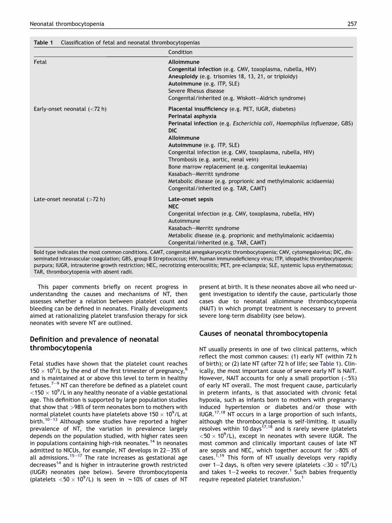
Table 1 Classification of fetal and neonatal thrombocytopenias
Condition
Fetal Alloimmune
Congenital infection (e.g. CMV, toxoplasma, rubella, HIV)Aneuploidy (e.g. trisomies 18, 13, 21, or triploidy)Autoimmune (e.g. ITP, SLE)Severe Rhesus diseaseCongenital/inherited (e.g. WiskotteAldrich syndrome)
Early-onset neonatal (<72 h) Placental insufficiency (e.g. PET, IUGR, diabetes)Perinatal asphyxia
Perinatal infection (e.g. Escherichia coli, Haemophilus influenzae, GBS)DIC
Alloimmune
Autoimmune (e.g. ITP, SLE)Congenital infection (e.g. CMV, toxoplasma, rubella, HIV)Thrombosis (e.g. aortic, renal vein)Bone marrow replacement (e.g. congenital leukaemia)KasabacheMerritt syndromeMetabolic disease (e.g. proprionic and methylmalonic acidaemia)Congenital/inherited (e.g. TAR, CAMT)
Late-onset neonatal (>72 h) Late-onset sepsis
NEC
Congenital infection (e.g. CMV, toxoplasma, rubella, HIV)AutoimmuneKasabacheMerritt syndromeMetabolic disease (e.g. proprionic and methylmalonic acidaemia)Congenital/inherited (e.g. TAR, CAMT)
Bold type indicates the most common conditions. CAMT, congenital amegakaryocytic thrombocytopenia; CMV, cytomegalovirus; DIC, dis-seminated intravascular coagulation; GBS, group B Streptococcus; HIV, human immunodeficiency virus; ITP, idiopathic thrombocytopenicpurpura; IUGR, intrauterine growth restriction; NEC, necrotizing enterocolitis; PET, pre-eclampsia; SLE, systemic lupus erythematosus;TAR, thrombocytopenia with absent radii.
Neonatal thrombocytopenia 257
This paper comments briefly on recent progress inunderstanding the causes and mechanisms of NT, thenassesses whether a relation between platelet count andbleeding can be defined in neonates. Finally developmentsaimed at rationalizing platelet transfusion therapy for sickneonates with severe NT are outlined.
Definition and prevalence of neonatalthrombocytopenia
Fetal studies have shown that the platelet count reaches150 � 109/L by the end of the first trimester of pregnancy,6
and is maintained at or above this level to term in healthyfetuses.7e9 NT can therefore be defined as a platelet count<150 � 109/L in any healthy neonate of a viable gestationalage. This definition is supported by large population studiesthat show that >98% of term neonates born to mothers withnormal platelet counts have platelets above 150 � 109/L atbirth.10e13 Although some studies have reported a higherprevalence of NT, the variation in prevalence largelydepends on the population studied, with higher rates seenin populations containing high-risk neonates.14 In neonatesadmitted to NICUs, for example, NT develops in 22e35% ofall admissions.15e17 The rate increases as gestational agedecreases14 and is higher in intrauterine growth restricted(IUGR) neonates (see below). Severe thrombocytopenia(platelets <50 � 109/L) is seen in w10% of cases of NT
present at birth. It is these neonates above all who need ur-gent investigation to identify the cause, particularly thosecases due to neonatal alloimmune thrombocytopenia(NAIT) in which prompt treatment is necessary to preventsevere long-term disability (see below).
Causes of neonatal thrombocytopenia
NT usually presents in one of two clinical patterns, whichreflect the most common causes: (1) early NT (within 72 hof birth); or (2) late NT (after 72 h of life; see Table 1). Clin-ically, the most important cause of severe early NT is NAIT.However, NAIT accounts for only a small proportion (<5%)of early NT overall. The most frequent cause, particularlyin preterm infants, is that associated with chronic fetalhypoxia, such as infants born to mothers with pregnancy-induced hypertension or diabetes and/or those withIUGR.17,18 NT occurs in a large proportion of such infants,although the thrombocytopenia is self-limiting. It usuallyresolves within 10 days17,18 and is rarely severe (platelets<50 � 109/L), except in neonates with severe IUGR. Themost common and clinically important causes of late NTare sepsis and NEC, which together account for >80% ofcases.1,19 This form of NT usually develops very rapidlyover 1e2 days, is often very severe (platelets <30� 109/L)and takes 1e2 weeks to recover.1 Such babies frequentlyrequire repeated platelet transfusion.1
258 I. Roberts, N.A. Murray
Neonatal thrombocytopenia secondary tochronic fetal hypoxia
Neonates with IUGR secondary to chronic fetal hypoxia, andthose born to mothers with pregnancy-induced hyperten-sion or diabetes, have a number of distinct haematologicalabnormalities that are present at birth. These includevarying degrees of NT, transient neutropenia and increasederythropoiesis (high numbers of circulating nucleated redcells with or without associated polycythaemia); many alsohave evidence on the blood film of hyposplenism (spher-ocytes, target cells and HowelleJolly bodies).18 Erythropoi-etin levels are increased and the severity of thehaematological abnormalities correlates both with serumerythropoietin levels and with the severity of the placentaldysfunction consistent with fetal hypoxia as a direct causeof the abnormalities.18,20 We and others have shown thatmegakaryocytopoiesis is severely impaired at birth in suchneonates, as shown by a marked reduction in circulatingmegakaryocytes and their precursor cells.1,18,21
Neonatal alloimmune thrombocytopenia
In NAIT, the platelet equivalent of haemolytic disease ofthe newborn, thrombocytopenia results from transplacen-tal passage of maternal platelet-specific antibodies topaternal human platelet antigens (HPA) expressed on fetalplatelets that the mother lacks. Sixteen HPAs have beenidentified but fetomaternal incompatibility between onlythree (HPA-1a, HPA-5b and HPA-15b), singly or in combina-tion, causes 95% of cases in Caucasian populations.22 Otherantibodies, such as anti-HPA-3a, are occasionally in-volved.23,24 Fetomaternal incompatibility for HPA-1a isresponsible for 75% of cases22e24 and occurs in 1:350 preg-nancies, although thrombocytopenia develops in only1:1000e1500 pregnancies.22 Recent data indicate that theability of an HPA-1a-negative woman to form anti-HPA-1ais controlled by the HLA DRB3*0101 allele such that HLADRB3*0101-positive women are 140 times more likely thanHLA DRB3*0101-negative women to make anti-HPA-1a,25
thereby explaining the frequency of the clinical problem.NAIT occurs in the first pregnancy in almost 50% of cases.
Thrombocytopenia is frequently extremely severe (plateletcount<20 � 109/L) and commonly results in major bleeding,particularly intracranial haemorrhage (ICH). The incidenceof ICH is difficult to ascertainprecisely, but large series reportits occurrence in 10e20% of untreated pregnancies.22,26,27 Asthrombocytopenia is frequently present in the fetus, often asearly as 20e24 weeks gestation,28,29 ICH might occur duringfetal development,30,31 sometimes resulting in fetal death.Fetal ICH is most common in untreated pregnancies, in whichit might account for up to 75% of all the fetuses and neonatesdeveloping such bleeds.24,32 The neonatal course in other-wise well neonates is variable, with thrombocytopeniaresolving in most cases within 1 week (with or without plate-let transfusion) with no long-term sequelae. However,thrombocytopenia can last for several weeks before resolv-ing, and in such cases it requires repeated platelet transfu-sion. In a minority of affected neonates ICH occurs for thefirst time following birth, adding to the overall mortalityand morbidity of the condition.
Neonatal ICH is most common in neonates whose mothersreceived no antenatal therapy.32 The neurodevelopmentaloutcome of neonates with untreated NAIT is poorer thantheir siblings where maternal treatment was instituted.33
However, the poorest outcome is seen in those neonateswith ICH; two-thirds of such cases show neurodevelopmen-tal problems, approximately half of which are severe, e.g.severe cerebral palsy and/or sensory impairment.22
The severe fetal and neonatal consequences of NAITmean that this disorder requires expert management withclose collaboration between experienced fetal medicinespecialists, haematologists and neonatologists. The guidingprinciple of therapy has been the knowledge that for motherswith known HPA antibodies, the fetal and neonatal course insubsequent pregnancies (with an antigen-positive fetus)closely reflects that in previously affected pregnancies.Thus, mothers with previous neonates suffering an ICH areat high risk of future children also having a severe course.
Antenatal management of neonatal alloimmunethrombocytopenia
Antenatal management of pregnancies at risk of NAITremainscontroversial. Three general approaches are used in differentcentres (these are reviewed in refs. [24,34] and [35]). First,an invasive approach, focused on ‘high-risk’ mothers withprevious severely affected children. This is based on repeatedfetal blood samples (FBS) and intrauterine transfusions (IUT)of HPA-compatible platelets in thrombocytopenic fetuses,combined with preterm delivery at 32e34 weeks. Second,a non-invasive approach based on monitoring by fetal ultra-sound scan (USS) and maternal intravenous weekly high-dose intravenous immunoglobulin therapy (IVIG) þ steroids;this is used most often in ‘low-risk’ women. Third, combina-tion therapy based on high-dose maternal IVIG togetherwith infrequent FBS to monitor the response to therapy duringpregnancy and to decide on the mode of delivery.
It has recently become clear that the rate of fetal lossand emergency preterm delivery associated with repeatedFBS and IUT approximates to the rate of fetal ICH inuntreated pregnancies and exceeds that in ‘low-risk’pregnancies treated with IVIG.22,36,37 As a result, therapyfor NAIT, even in high-risk cases, is increasingly movingtowards the non-invasive approach.
Recently, van den Akker et al. reported on theirexperience of treating 52 pregnant women (five witha previous sibling with ICH) with known HPA incompatibilitywith IVIG alone at a dose of 1 g/kg per week.38 IVIG wasbegun at 16 weeks if the previous sibling had an ICH and32 weeks if not. All the pregnancies resulted in live births;there were no ICHs and no neonatal deaths. As a result,these researchers have adopted this non-invasive regimenas their standard protocol for antenatal therapy for allmothers with known HPA incompatibilities.34
Management of affected neonates with neonatalalloimmune thrombocytopenia
Clinical symptoms range from asymptomatic neonates withincidental thrombocytopenia, to limited or widespread skinpetechiae or purpura, to symptoms of ICH (e.g. neonatal
Neonatal thrombocytopenia 259
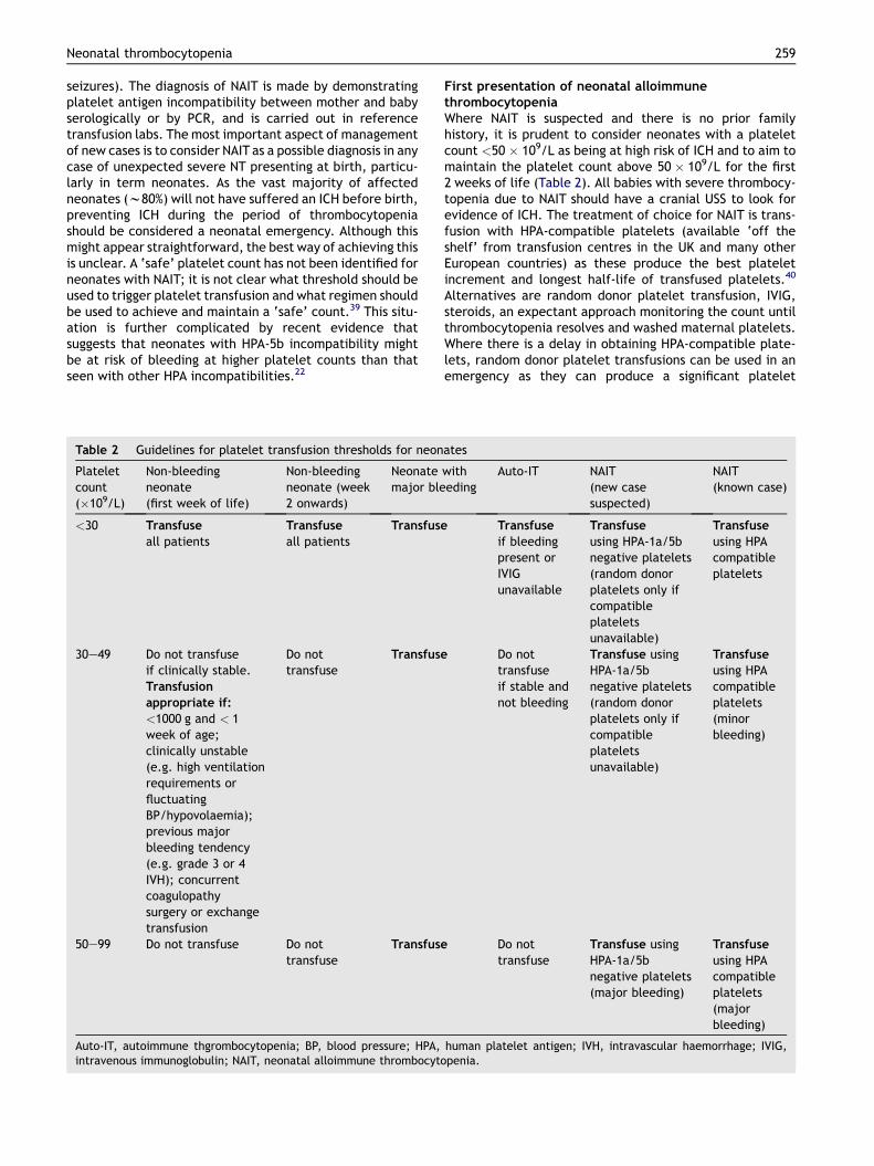
seizures). The diagnosis of NAIT is made by demonstratingplatelet antigen incompatibility between mother and babyserologically or by PCR, and is carried out in referencetransfusion labs. The most important aspect of managementof new cases is to consider NAIT as a possible diagnosis in anycase of unexpected severe NT presenting at birth, particu-larly in term neonates. As the vast majority of affectedneonates (w80%) will not have suffered an ICH before birth,preventing ICH during the period of thrombocytopeniashould be considered a neonatal emergency. Although thismight appear straightforward, the best way of achieving thisis unclear. A ‘safe’ platelet count has not been identified forneonates with NAIT; it is not clear what threshold should beused to trigger platelet transfusion and what regimen shouldbe used to achieve and maintain a ‘safe’ count.39 This situ-ation is further complicated by recent evidence thatsuggests that neonates with HPA-5b incompatibility mightbe at risk of bleeding at higher platelet counts than thatseen with other HPA incompatibilities.22
Table 2 Guidelines for platelet transfusion thresholds for neon
Plateletcount(�109/L)
Non-bleedingneonate(first week of life)
Non-bleedingneonate (week2 onwards)
Neonatemajor ble
<30 Transfuse
all patientsTransfuse
all patientsTransfuse
30e49 Do not transfuseif clinically stable.Transfusion
appropriate if:
<1000 g and < 1week of age;clinically unstable(e.g. high ventilationrequirements orfluctuatingBP/hypovolaemia);previous majorbleeding tendency(e.g. grade 3 or 4IVH); concurrentcoagulopathysurgery or exchangetransfusion
Do nottransfuse
Transfuse
50e99 Do not transfuse Do nottransfuse
Transfuse
Auto-IT, autoimmune thgrombocytopenia; BP, blood pressure; HPA,intravenous immunoglobulin; NAIT, neonatal alloimmune thrombocyto
First presentation of neonatal alloimmunethrombocytopeniaWhere NAIT is suspected and there is no prior familyhistory, it is prudent to consider neonates with a plateletcount <50 � 109/L as being at high risk of ICH and to aim tomaintain the platelet count above 50 � 109/L for the first2 weeks of life (Table 2). All babies with severe thrombocy-topenia due to NAIT should have a cranial USS to look forevidence of ICH. The treatment of choice for NAIT is trans-fusion with HPA-compatible platelets (available ‘off theshelf’ from transfusion centres in the UK and many otherEuropean countries) as these produce the best plateletincrement and longest half-life of transfused platelets.40
Alternatives are random donor platelet transfusion, IVIG,steroids, an expectant approach monitoring the count untilthrombocytopenia resolves and washed maternal platelets.Where there is a delay in obtaining HPA-compatible plate-lets, random donor platelet transfusions can be used in anemergency as they can produce a significant platelet
ates
witheding
Auto-IT NAIT(new casesuspected)
NAIT(known case)
Transfuse
if bleedingpresent orIVIGunavailable
Transfuse
using HPA-1a/5bnegative platelets(random donorplatelets only ifcompatibleplateletsunavailable)
Transfuse
using HPAcompatibleplatelets
Do nottransfuseif stable andnot bleeding
Transfuse usingHPA-1a/5bnegative platelets(random donorplatelets only ifcompatibleplateletsunavailable)
Transfuse
using HPAcompatibleplatelets(minorbleeding)
Do nottransfuse
Transfuse usingHPA-1a/5bnegative platelets(major bleeding)
Transfuse
using HPAcompatibleplatelets(majorbleeding)
human platelet antigen; IVH, intravascular haemorrhage; IVIG,penia.

260 I. Roberts, N.A. Murray
increment in NAIT.41 The role of IVIG is less clear. The avail-able literature suggests that any platelet increment follow-ing IVIG is likely to be delayed for 12e36 h,26,29 making itunacceptable as a monotherapy. In addition, a recent studyhas suggested that increases in platelet count followingIVIG therapy in some neonates with NAIT might be confusedwith natural platelet count recovery questioning the trueeffectiveness of neonatal IVIG therapy.32 In new cases with-out major bleeding and with an initial count >50 � 109/L,an expectant approach is appropriate. However, thrombo-cytopenia often worsens over the first few days, makingclose monitoring of the platelet count essential. In affectedneonates with active bleeding (e.g. new or worsening ICH,gastrointestinal, frank haematuria) it seems reasonable tomaintain the platelet count above 100 � 109/L (Table 2).
Known cases of neonatal alloimmune thrombocytopeniaNeonates born after treatment with antenatal maternalIVIG appear to have a reduced risk of major haemor-rhage,22,32 although some will still be born with plateletcounts <30 � 109/L. These neonates are also likely to beborn in specialist centres, the HPA-incompatibility will beknown and delivery can be planned with HPA-compatibleplatelets immediately available. It is possible that theseneonates can be maintained with lower platelet countsand a platelet threshold of 30 � 109/L rather than50 � 109/L, as they appear to have a reduced risk of ICH(see Table 2). However, this is not yet clear and awaitsevaluation by carefully designed clinical trials.
Neonatal autoimmune thrombocytopenia
NT secondary to transplacental passage of maternal plate-let autoantibodies occurs in babies born to mothers withidiopathic thrombocytopenic purpura (ITP) or systemiclupus erythematosus (SLE). Around 10% of infants ofaffected mothers develop thrombocytopenia. It is a lesscommon cause of NT than NAITP, affecting 1e5 in 10,000pregnancies.42,43 Thrombocytopenia is usually mild and ICHis rare (<1% of at-risk babies). In affected babies withsevere thrombocytopenia treatment with IVIG 1 g/kg for2 days is usually effective (see Table 2).44
Inherited thrombocytopenia
A number of rare inherited disorders present with throm-bocytopenia in the fetus or neonate (see Table 1). In mostcases, the thrombocytopenia is due to reduced plateletproduction secondary to abnormal haemopoietic stem celldevelopment and there are often associated congenitalanomalies, which are useful in guiding investigations andestablishing the diagnosis. Many of the recent advances inidentifying the molecular basis of a number of these disor-ders have informed both the diagnosis and the managementof neonates with unexplained, persistent NT (reviewed inref. [45]).
Neonatal thrombocytopenia and bleeding risk
From clinical experience it is clear that different neonateshave very different risks of bleeding for the same degree of
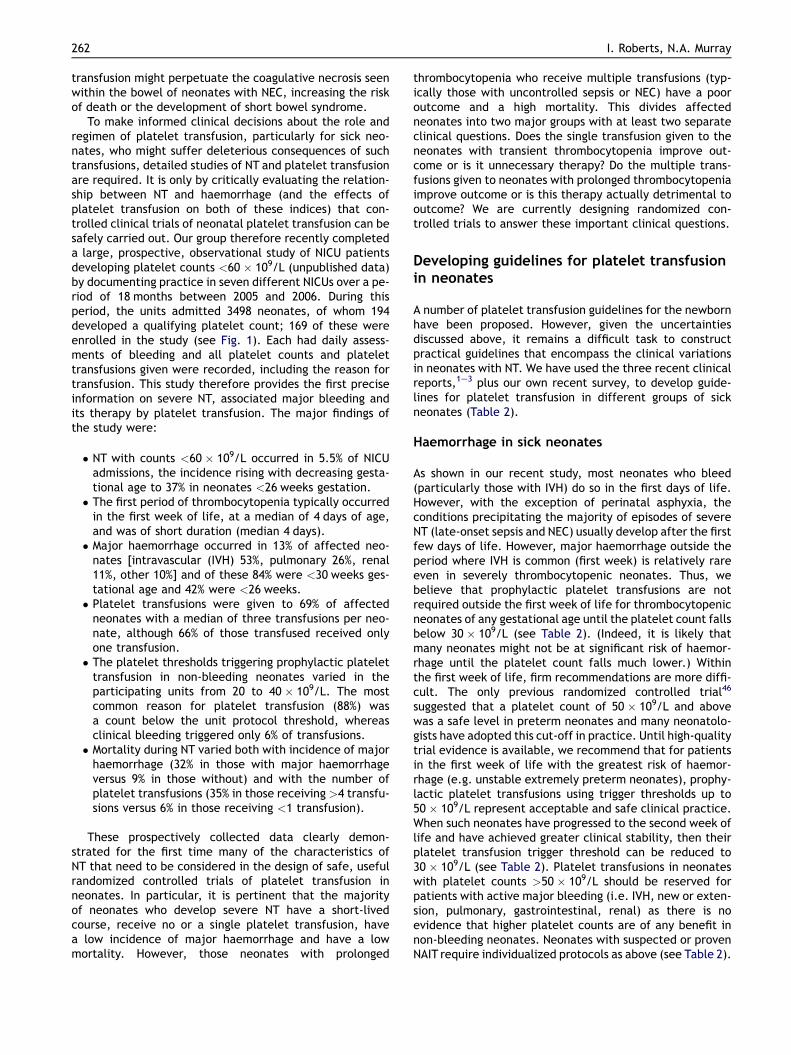
thrombocytopenia. Neonates with NAIT, for example, havea high risk of bleeding, possibly because some anti-HPAantibodies interfere with platelet function, although this isunproven as yet.24 Similarly, clinical experience suggeststhat thrombocytopenic neonates with IUGR have a lowrisk of major haemorrhage and those with sepsis or NECan intermediate risk, perhaps reflecting differences inplatelet function or co-existing coagulopathy. Althoughthese observations influence our clinical practice, there isvery little evidence that allows us to measure the risk ofbleeding accurately, at a particular platelet count, in anindividual neonate or group of neonates (e.g. those withIUGR). Indeed, no neonatal studies have addressed thisquestion and none has compared different groups of throm-bocytopenic neonates by underlying mechanism of throm-bocytopenia. We therefore recently carried out a detailedobservational study in 169 neonates with severe NT inwhom the prevalence and nature of major and minor hae-morrhage was recorded according to the cause of thethrombocytopenia (Fig. 1). In this study we found that ne-onates with NT secondary to IUGR or maternal pregnancy-induced hypertension formed the most common diagnosisin the group of babies with no major haemorrhage (38%) de-spite severe NT, whereas they formed a much smaller pro-portion of the group of babies who did develop majorhaemorrhage. Severe sepsis and NEC were the most com-mon diagnoses in those neonates who did bleed, suggestingthat these disorders do affect both platelet number andfunction.
Neonatal platelet transfusion
Platelet transfusion is the only specific therapy available totreat NT. However, no neonatal trials have yet evaluatedwhether platelet transfusion reduces haemorrhage orimproves outcome in NT. In the only randomized controlledtrial addressing this issue, Andrew et al.46 found no benefitin terms of haemorrhage of maintaining a normal plateletcount (platelets >150 � 109/L) by platelet transfusion inpreterm neonates compared to controls with moderatethrombocytopenia (platelets 50e150 � 109/L). This trialwas reported almost 15 years ago and neonates with plate-let counts <50 � 109/L were transfused and excluded fromanalysis. The relevance of this study to modern neonatalpractice is therefore limited, both because of considerablechanges in practice since the early 1990s and because thevast majority of platelet transfusions are now given toneonates with platelet counts <50 � 109/L.
The lack of clinical trial data in neonates has led touncertainties regarding optimum transfusion therapy, withthe result that NICUs often have quite different platelettransfusion protocols. Three publications1e3 retrospectivelydocument platelet transfusion practice in three separateNICUs in the USA, UK and Mexico. Despite the geographicaldifferences, several consistent observations are apparent inthese reports:
� Different platelet count triggers for transfusion are usedby each unit. As a result, platelet transfusion rates varyconsiderably between units: from 2% to 9.4% of admis-sions (some of this variation is explained because moretransfusions are given in NICUs with a high percentage
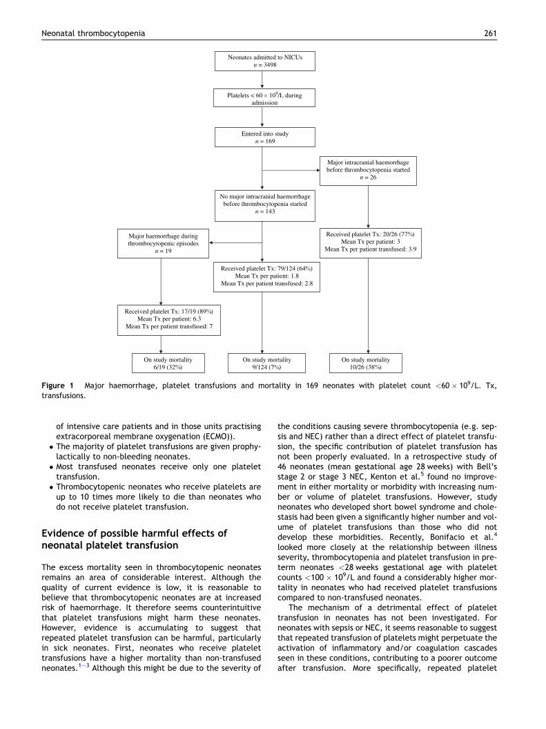
Platelets < 60 109/L during admission
Entered into study n = 169
Major intracranial haemorrhage before thrombocytopenia started
n = 26
No major intracranial haemorrhage before thrombocytopenia started
n = 143
Neonates admitted to NICUs n = 3498
Major haemorrhage during thrombocytopenic episodes
n = 19
Received platelet Tx: 79/124 (64 ) Mean Tx per patient: 1.8
Mean Tx per patient transfused: 2.8
Received platelet Tx: 17/19 (89 ) Mean Tx per patient: 6.3
Mean Tx per patient transfused: 7
Received platelet Tx: 20/26 (77 ) Mean Tx per patient: 3
Mean Tx per patient transfused: 3.9
On study mortality 6/19 (32 )
On study mortality 9/124 (7 )
On study mortality 10/26 (38 )
Figure 1 Major haemorrhage, platelet transfusions and mortality in 169 neonates with platelet count <60 � 109/L. Tx,transfusions.
Neonatal thrombocytopenia 261
of intensive care patients and in those units practisingextracorporeal membrane oxygenation (ECMO)).� The majority of platelet transfusions are given prophy-
lactically to non-bleeding neonates.� Most transfused neonates receive only one platelet
transfusion.� Thrombocytopenic neonates who receive platelets are
up to 10 times more likely to die than neonates whodo not receive platelet transfusion.
Evidence of possible harmful effects ofneonatal platelet transfusion
The excess mortality seen in thrombocytopenic neonatesremains an area of considerable interest. Although thequality of current evidence is low, it is reasonable tobelieve that thrombocytopenic neonates are at increasedrisk of haemorrhage. It therefore seems counterintuitivethat platelet transfusions might harm these neonates.However, evidence is accumulating to suggest thatrepeated platelet transfusion can be harmful, particularlyin sick neonates. First, neonates who receive platelettransfusions have a higher mortality than non-transfusedneonates.1e3 Although this might be due to the severity of
the conditions causing severe thrombocytopenia (e.g. sep-sis and NEC) rather than a direct effect of platelet transfu-sion, the specific contribution of platelet transfusion hasnot been properly evaluated. In a retrospective study of46 neonates (mean gestational age 28 weeks) with Bell’sstage 2 or stage 3 NEC, Kenton et al.5 found no improve-ment in either mortality or morbidity with increasing num-ber or volume of platelet transfusions. However, studyneonates who developed short bowel syndrome and chole-stasis had been given a significantly higher number and vol-ume of platelet transfusions than those who did notdevelop these morbidities. Recently, Bonifacio et al.4
looked more closely at the relationship between illnessseverity, thrombocytopenia and platelet transfusion in pre-term neonates <28 weeks gestational age with plateletcounts <100 � 109/L and found a considerably higher mor-tality in neonates who had received platelet transfusionscompared to non-transfused neonates.
The mechanism of a detrimental effect of platelettransfusion in neonates has not been investigated. Forneonates with sepsis or NEC, it seems reasonable to suggestthat repeated transfusion of platelets might perpetuate theactivation of inflammatory and/or coagulation cascadesseen in these conditions, contributing to a poorer outcomeafter transfusion. More specifically, repeated platelet
262 I. Roberts, N.A. Murray
transfusion might perpetuate the coagulative necrosis seenwithin the bowel of neonates with NEC, increasing the riskof death or the development of short bowel syndrome.
To make informed clinical decisions about the role andregimen of platelet transfusion, particularly for sick neo-nates, who might suffer deleterious consequences of suchtransfusions, detailed studies of NT and platelet transfusionare required. It is only by critically evaluating the relation-ship between NT and haemorrhage (and the effects ofplatelet transfusion on both of these indices) that con-trolled clinical trials of neonatal platelet transfusion can besafely carried out. Our group therefore recently completeda large, prospective, observational study of NICU patientsdeveloping platelet counts <60 � 109/L (unpublished data)by documenting practice in seven different NICUs over a pe-riod of 18 months between 2005 and 2006. During thisperiod, the units admitted 3498 neonates, of whom 194developed a qualifying platelet count; 169 of these wereenrolled in the study (see Fig. 1). Each had daily assess-ments of bleeding and all platelet counts and platelettransfusions given were recorded, including the reason fortransfusion. This study therefore provides the first preciseinformation on severe NT, associated major bleeding andits therapy by platelet transfusion. The major findings ofthe study were:
� NT with counts <60 � 109/L occurred in 5.5% of NICUadmissions, the incidence rising with decreasing gesta-tional age to 37% in neonates <26 weeks gestation.� The first period of thrombocytopenia typically occurred
in the first week of life, at a median of 4 days of age,and was of short duration (median 4 days).� Major haemorrhage occurred in 13% of affected neo-
nates [intravascular (IVH) 53%, pulmonary 26%, renal11%, other 10%] and of these 84% were <30 weeks ges-tational age and 42% were <26 weeks.� Platelet transfusions were given to 69% of affected
neonates with a median of three transfusions per neo-nate, although 66% of those transfused received onlyone transfusion.� The platelet thresholds triggering prophylactic platelet
transfusion in non-bleeding neonates varied in theparticipating units from 20 to 40 � 109/L. The mostcommon reason for platelet transfusion (88%) wasa count below the unit protocol threshold, whereasclinical bleeding triggered only 6% of transfusions.� Mortality during NT varied both with incidence of major
haemorrhage (32% in those with major haemorrhageversus 9% in those without) and with the number ofplatelet transfusions (35% in those receiving >4 transfu-sions versus 6% in those receiving <1 transfusion).
These prospectively collected data clearly demon-strated for the first time many of the characteristics ofNT that need to be considered in the design of safe, usefulrandomized controlled trials of platelet transfusion inneonates. In particular, it is pertinent that the majorityof neonates who develop severe NT have a short-livedcourse, receive no or a single platelet transfusion, havea low incidence of major haemorrhage and have a lowmortality. However, those neonates with prolonged
thrombocytopenia who receive multiple transfusions (typ-ically those with uncontrolled sepsis or NEC) have a pooroutcome and a high mortality. This divides affectedneonates into two major groups with at least two separateclinical questions. Does the single transfusion given to theneonates with transient thrombocytopenia improve out-come or is it unnecessary therapy? Do the multiple trans-fusions given to neonates with prolonged thrombocytopeniaimprove outcome or is this therapy actually detrimental tooutcome? We are currently designing randomized con-trolled trials to answer these important clinical questions.
Developing guidelines for platelet transfusionin neonates
A number of platelet transfusion guidelines for the newbornhave been proposed. However, given the uncertaintiesdiscussed above, it remains a difficult task to constructpractical guidelines that encompass the clinical variationsin neonates with NT. We have used the three recent clinicalreports,1e3 plus our own recent survey, to develop guide-lines for platelet transfusion in different groups of sickneonates (Table 2).
Haemorrhage in sick neonates
As shown in our recent study, most neonates who bleed(particularly those with IVH) do so in the first days of life.However, with the exception of perinatal asphyxia, theconditions precipitating the majority of episodes of severeNT (late-onset sepsis and NEC) usually develop after the firstfew days of life. However, major haemorrhage outside theperiod where IVH is common (first week) is relatively rareeven in severely thrombocytopenic neonates. Thus, webelieve that prophylactic platelet transfusions are notrequired outside the first week of life for thrombocytopenicneonates of any gestational age until the platelet count fallsbelow 30 � 109/L (see Table 2). (Indeed, it is likely thatmany neonates might not be at significant risk of haemor-rhage until the platelet count falls much lower.) Withinthe first week of life, firm recommendations are more diffi-cult. The only previous randomized controlled trial46
suggested that a platelet count of 50 � 109/L and abovewas a safe level in preterm neonates and many neonatolo-gists have adopted this cut-off in practice. Until high-qualitytrial evidence is available, we recommend that for patientsin the first week of life with the greatest risk of haemor-rhage (e.g. unstable extremely preterm neonates), prophy-lactic platelet transfusions using trigger thresholds up to50 � 109/L represent acceptable and safe clinical practice.When such neonates have progressed to the second week oflife and have achieved greater clinical stability, then theirplatelet transfusion trigger threshold can be reduced to30 � 109/L (see Table 2). Platelet transfusions in neonateswith platelet counts >50 � 109/L should be reserved forpatients with active major bleeding (i.e. IVH, new or exten-sion, pulmonary, gastrointestinal, renal) as there is noevidence that higher platelet counts are of any benefit innon-bleeding neonates. Neonates with suspected or provenNAIT require individualized protocols as above (see Table 2).
uncommon even in severely thrombocytopenicneonates.� Although the mainstay of treatment of neonatal
thrombocytopenia is platelet transfusion, the cor-relation between thrombocytopenia and bleedingis unclear and no studies have yet shown clinicalbenefit of platelet transfusion in neonates; studiesto identify optimal neonatal platelet transfusionpractice are an urgent priority.
Research agenda
� To define more clearly the safe lower limit forplatelet counts within the different groups of neo-nates in tandem with defining which neonates willbenefit from platelet transfusion for theirthrombocytopenia.� To ensure accurate diagnosis and to determine the
most effective fetal and neonatal therapy forNAIT, the most common cause of unexpected neo-natal mortality and morbidity associated with NT.
Neonatal thrombocytopenia 263
Platelet dose and product
No trial evidence is currently available regarding theoptimal volume (dose) of platelets to administer orwhen to administer further transfusions. Larger volumes(20 mL/kg) appear to result in larger and more sustainedrises in platelet count than smaller volumes (10 mL/kg),and are generally well tolerated (personal observations).As most neonates receive only one platelet transfusion itseems prudent to ensure that this results in a good plateletincrement by using a large volume strategy to minimize do-nor exposure. As highlighted in our study, a small number ofneonates whose thrombocytopenia is the result of markedplatelet consumption (e.g. NEC) might show no measurableresponse to repeated platelet transfusions. It is possiblethat such transfusions are having a positive effect at a mi-crovascular level not reflected by the circulating plateletcount. However, the accumulating evidence that repeatedplatelet transfusion might be detrimental to outcome insuch neonates only adds to the difficulty for neonatologistsin prescribing logical and effective therapy given thecurrent paucity of trial evidence.
For guidance on appropriate platelet products for trans-fusion in neonates, the reader is referred to the BritishCommittee for Standard in Haematology website.47
Conclusions
Neonatal thrombocytopenia remains a common clinicalproblem. Fortunately, most episodes of thrombocytopeniaare mild or moderate and resolve spontaneously withoutapparent clinical sequelae. For more severe episodes, therecent demonstration of impaired megakaryocytopoiesisand platelet production as a major contributor to NT is animportant advance both for our understanding of theunderlying disease processes and the potential for improvedtherapies. Clinical studies are now critically exploring howchanges in platelet count relate to the different causes ofNTand to the role of platelet transfusion in the managementof the different thrombocytopenic syndromes.
Practice points
� Neonatal thrombocytopenia is defined as a plateletcount <150 � 109/L in neonates of any viable ges-tational age.� The commonest cause of thrombocytopenia devel-
oping within 72 h of birth is intrauterine growth re-striction and/or maternal hypertension; this formof thrombocytopenia is self-limiting and rarelysevere.� The commonest cause of severe thrombocytopenia
developing within 72 h of life is NAIT; the majorcomplication is intracranial haemorrhage which isassociated with long-term neurodevelopmentalproblems in two-thirds of cases.� Our recent study indicates that most neonates who
bleed (particularly those with IVH) do so in the firstdays of life; major haemorrhage after this is
References
1. Murray NA, Howarth LJ, McCloy MP, et al. Platelet transfusionin the management of severe thrombocytopenia in neonatalintensive care unit patients. Transfus Med 2002;12:35e41.
2. Garcia MG, Duenas E, Sola MC, et al. Epidemiologic andoutcome studies of patients who received platelet transfusionsin the neonatal intensive care unit. J Perinatol 2001;21:415e20.
3. Del Vecchio A, Sola MC, Theriaque DW, et al. Platelet transfu-sions in the neonatal intensive care unit: factors predictingwhich patients will require multiple transfusions. Transfusion2001;41:803e8.
4. Bonifacio L, Petrova A, Nanjundaswamy S, Mehta R. Thrombo-cytopenia related neonatal outcome in preterms. IndianJ Pediatr 2007;74:269e74.
5. Kenton AB, Hegemier S, Smith EO, et al. Platelet transfusionsin infants with necrotizing enterocolitis do not lower mortalitybut may increase morbidity. J Perinatol 2005;25:173e7.
6. Pahal G, Jauniaux E, Kinnon C, Thrasher AJ, Rodeck CH. Normaldevelopment of human fetal hematopoiesis between eight andseventeen weeks’ gestation. Am J Obstet Gynecol 2000;183:1029e34.
7. Holmberg L, Gustavii B, Jonsson A. A prenatal study of fetalplatelet count and size with application to the fetus at riskof Wiskott Aldrich syndrome. J Pediatr 1983;102:773e81.
8. Forestier F, Daffos F, Galacteros F. Haematological values of163 normal fetuses between 18 and 30 weeks of gestation.Pediatr Res 1986;20:342e6.
9. Forestier F, Daffos F, Catherine N, Renard M, Andreux JP.Developmental hematopoiesis in normal human fetal blood.Blood 1991;77:2360e3.
10. Burrows RF, Kelton JG. Incidentally detected thrombocytope-nia in healthy mothers and their infants. N Engl J Med 1988;319:142e5.
264 I. Roberts, N.A. Murray
11. Burrows RF, Kelton JG. Thrombocytopenia at delivery: a pro-spective survey of 6715 deliveries. Am J Obstet Gynecol1990;162:731e4.
12. Burrows RF, Kelton JG. Fetal thrombocytopenia and its relationto maternal thrombocytopenia. N Engl J Med 1993;329:1463e6.
13. Sainio S, Jarvenpaa A-S, Renlund M, et al. Thrombocytopenia interm infants: a population-based study. Obstet Gynecol 2000;95:441e6.
14. Christensen RD, Henry E, Wiedmeier SE, et al. Thrombocytope-nia among extremely low birth weight neonates: data froma multihospital healthcare system. J Perinatol 2006;26:348e53.
15. Castle V, Andrew M, Kelton J, et al. Frequency and mechanismof neonatal thrombocytopenia. J Pediatr 1986;108:749e55.
16. Mehta P, Rohitkumar V, Neumann L, Karpatkin M. Thrombocy-topenia in the high risk infant. J Pediatr 1980;97:791e4.
17. Murray NA, Roberts IAG. Circulating megakaryocytes and theirprogenitors in early thrombocytopenia in preterm neonates.Pediatr Res 1996;40:112e9.
18. Watts TL, Roberts IAG. Haematological abnormalities in thegrowth-restricted infant. Semin Neonatol 1999;4:41e54.
19. Roberts IAG, Murray NA. Neonatal thrombocytopenia: newinsights into pathogenesis and implications for clinical manage-ment. Curr Opin Pediatr 2001;13:16e21.
20. Salvesen DR, Brudenell JM, Snijders RJ, Ireland RM,Nicolaides KH. Fetal plasma erythropoietin in pregnanciescomplicated by maternal diabetes mellitus. Am J Obstet Gyne-col 1993;168:88e94.
21. Watts TL, Murray NA, Roberts IAG. Thrombopoietin has a pri-mary role in the regulation of platelet production in pretermbabies. Pediatr Res 1999;46:28e32.
22. Ghevaert C, Campbell K, Walton J, et al. Management andoutcome of 200 cases of fetomaternal alloimmune thrombocy-topenia. Transfusion 2007;47:901e10.
23. Ouwehand WH, Smith G, Ranasinghe E. Management of severealloimmune thrombocytopenia in the newborn. Arch Dis ChildFetal Neonatal 2000;82:F173e5.
24. Bussel JB, Primiani A. Fetal and neonatal alloimmune thrombo-cytopenia: progress and ongoing debates. Blood Rev 2008;22:33e52.
25. Williamson LM, Hackett G, Rennie J, et al. The natural historyof fetomaternal alloimmunization to the platelet-specific anti-gen HPA-1a (PlA1, Zwa) as determined by antenatal screening.Blood 1998;92:2280e7.
26. Mueller-Eckhardt C, Kiefel V, Grubert A, et al. 348 cases of sus-pected neonatal alloimmune thrombocytopenia. Lancet 1989;1:363e6.
27. Bussel JB, Zacharoulis S, Kramer K, et al. Clinical and diagnos-tic comparison of neonatal alloimmune thrombocytopenia tonon-immune cases of thrombocytopenia. Pediatr Blood Cancer2005;45:176e83.
28. Giovangrandi Y, Daffos F, Kaplan C, et al. Very early intracra-nial haemorrhage in alloimmune fetal thrombocytopenia. Lan-cet 1990;336:310.
29. Bussel JB, Zabusky MR, Berkowitz RL, McFarland JG. Fetal al-loimmune thrombocytopenia. N Engl J Med 1997;337:22e6.
30. Singh SA, Pollard J, Singhal N. Fetomaternal alloimmunethrombocytopenia presenting as intracerebral bleeding inutero. Indian J Pediatr 2005;72:269.
31. Sharif U, Kuban K. Prenatal intracranial hemorrhage and neuro-logic complications in alloimmune thrombocytopenia. J ChildNeurol 2001;16:838e42.
32. te Pas AB, Lopriore E, van den Akker ES, et al. Postnatal man-agement of fetal and neonatal alloimmune thrombocytopenia:the role of matched platelet transfusion and IVIG. Eur J Pediatr2007;166:1057e63.
33. Ward MJ, Pauliny J, Lipper EG, Bussel JB. Long-term effects offetal and neonatal alloimmune thrombocytopenia and its ante-natal treatment on the medical and developmental outcomesof affected children. Am J Perinatol 2006;23:487e92.
34. van den Akker ES, Oepkes D. Fetal and neonatal alloimmunethrombocytopenia. Best Pract Res Clin Obstet Gynaecol 2008;22:3e14.
35. Murphy MF, Bussel JB. Advances in the management of alloim-mune thrombocytopenia. Br J Haematol 2007;136:366e78.
36. Overton TG, Duncan KR, Jolly M, Letsky E, Fisk NM. Serial ag-gressive platelet transfusion for fetal alloimmune thrombocy-topenia: platelet dynamics and perinatal outcome. Am JObstet Gynecol 2002;186:826e31.
37. Berkowitz RL, Kolb EA, McFarland JG, et al. Parallel random-ized trials of risk-based therapy for fetal alloimmune thrombo-cytopenia. Obstet Gynecol 2006;107:91e6.
38. van den Akker ES, Oepkes D, Lopriore E, Brand A, Kanhai HH.Noninvasive antenatal management of fetal and neonatal al-loimmune thrombocytopenia: safe and effective. Br J ObstetGynaecol 2007;114:469e73.
39. Bassler D, Greinacher A, Okascharoen C, et al. A systematicreview and survey of the management of unexpected neonatalalloimmune thrombocytopenia. Transfusion 2008;48:92e8.
40. Allen D, Verjee S, Rees S, Murphy MF, Roberts DJ. Platelettransfusion in neonatal alloimmune thrombocytopenia. Blood2007;109:388e9.
41. Kiefel V, Bassler D, Kroll H, et al. Antigen-positive platelettransfusion in neonatal alloimmune thrombocytopenia (NAIT).Blood 2006;107:3761e3.
42. Kelton JG. Idiopathic thrombocytopenic purpura complicatingpregnancy. Blood Rev 2002;16:43e6.
43. Bussel JB. Immune thrombocytopenia in pregnancy: autoim-mune and alloimmune. J Reprod Immunol 1997;37:35e61.
44. Gill KK, Kelton JG. Management of idiopathic thrombocytope-nic purpura in pregnancy. Semin Hematol 2000;37:275e89.
45. Alter BP. Diagnosis, genetics, and management of inheritedbone marrow failure syndromes. Hematol Am Soc HematolEduc Prog; 2007:29e39.
46. Andrew M, Vegh P, Caco C. A randomized, controlled trial ofplatelet transfusions in thrombocytopenic premature infants.J Pediatr 1993;123:285e91.
47. British Committee for Standard in Haematology, Availableonline at: http://www.bcshguidelines.com.