nephrology rounds riki buchwald, id fellow december 17 th 2008
TRANSCRIPT

Nephrology Rounds
Riki Buchwald, ID fellowDecember 17th 2008

Case
46 y old AA man with h/o GSW to right trochanter in 8/07, s/p ORIF at OSH
Admitted to Bellevue 9/07; found to have wound infection/OM with polyresistant Pseudomonas
Extensive debridement performed but hardware left in place
Underwent long-term treatment with polymyxin from 10/07 on. Course complicated by renal failure in 11/07 that resolved with polymyxin dose adjustment.

Case
Hardware removed on 3/12/08
Wound cx with MRSA
Received 4 week course of vancomycin and 6 week course of polymyxin after hardware removal; course completed at the end of April

Case Readmitted in 6/08 with increasing hip pain and persistent
drainage
Imaging c/w erosion of the right femoral head with joint space loss, septic arthritis and chronic osteomyelitis with sinus tract to the skin surface
Debridement and washout performed on 6/18/08: OR cx grew MRSA
Treated with vancomycin
Developed worsening non oliguric renal failure with creatinine increase from 1.1 on admission to 6.8 mg/dl over 4 weeks

Clinical History PMH: - Diabetes, A1c 7.9% in 10/2007
- HTN - Anemia
- Remote h/o syphilis, treated
SH: no tobacco or drug abuse
Meds: insulin, lisinopril, iron, MVI, folic acid, omeprazole, escitalopram, cyclobenzaprine, SQ heparin
ROS: several weeks of darkened urine, leg swelling; denied: dysuria, macro-hematuria, SOB, fevers, joint pain,
skin rash

Physical Exam
BP 150/89 HR 93 T 97.2 97% RA
Middle aged pt, appearing depressed, NAD
Sitting in wheelchair
Neck supple
Lungs: CTA
Heart: reg, nl S1 S2
Abdomen: soft, nontender
Right thigh with surgical scar, sutures in place, mild swelling
and chronic skin changes, no frank drainage
Ext: b/l 3+ LE edema

Laboratory Data
Wbc 11.4, 73% PMN,18% Lymph, Eos WNL Hgb 7.8 Plt 332 Hepatic: 42/64/201/0.2/8.2/3.4
Protein electrophoresis:
TP 7.7, albumin 2.4 Globulins:
alpha 1, alpha2, beta WNL,
gamma 2.6 (0.5-1.3); diffuse bands

Laboratory Data
Creatinine BUN
6/17 1.1 11
6/21 1.5 18
6/30 2.7 36
7/06 3.9 47
7/18 6.8 59
7/30 8.9 64
7/30: K: 5.2, Ca: 8.8, Phos: 6.0 Mg: 2.4
6/24: UA: protein >300 mg/dl, WBC 2-5,RBC packed, fine granular casts, RBC casts
7/02: Urine protein: 2g/day

Laboratory Data HIV: negative
Hep B: SAb positive, SAg negative
Hep C: negative
Syphilis: IgG/TPPA positive, RPR negative

Any ideas?

A Diagnostic Test was Performed

Normal glomerulus
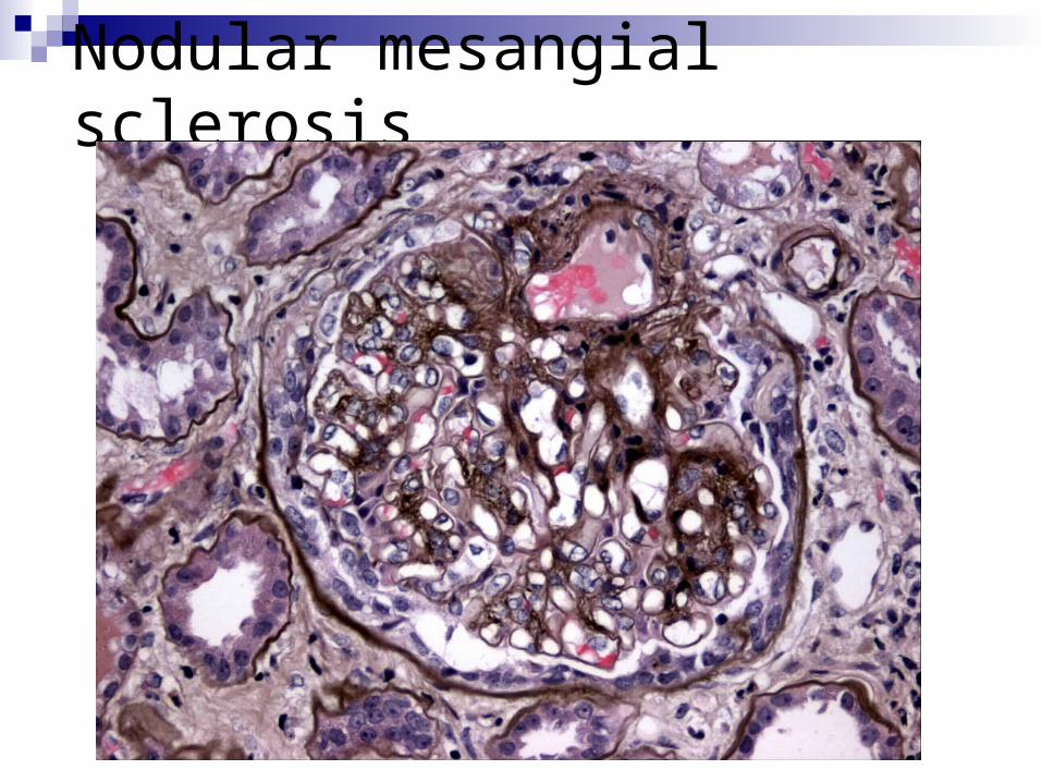
Nodular mesangial sclerosis
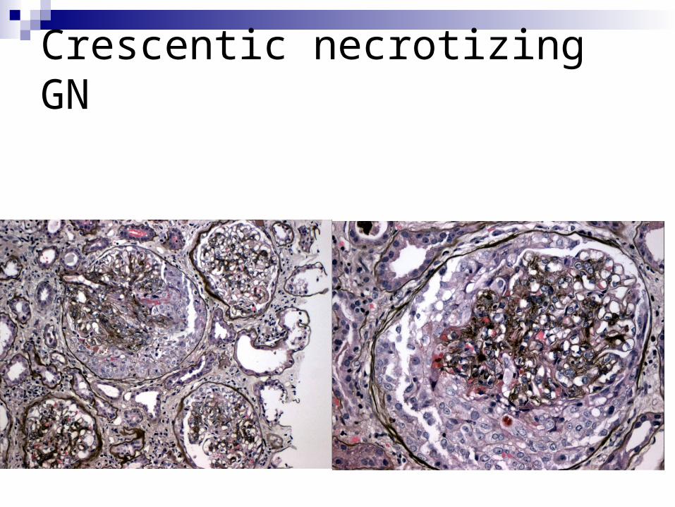
Crescentic necrotizing GN

RBC casts
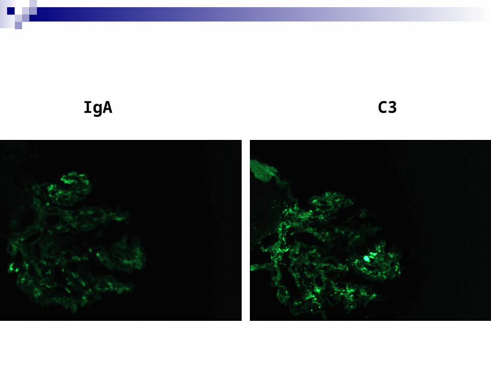
IgA C3

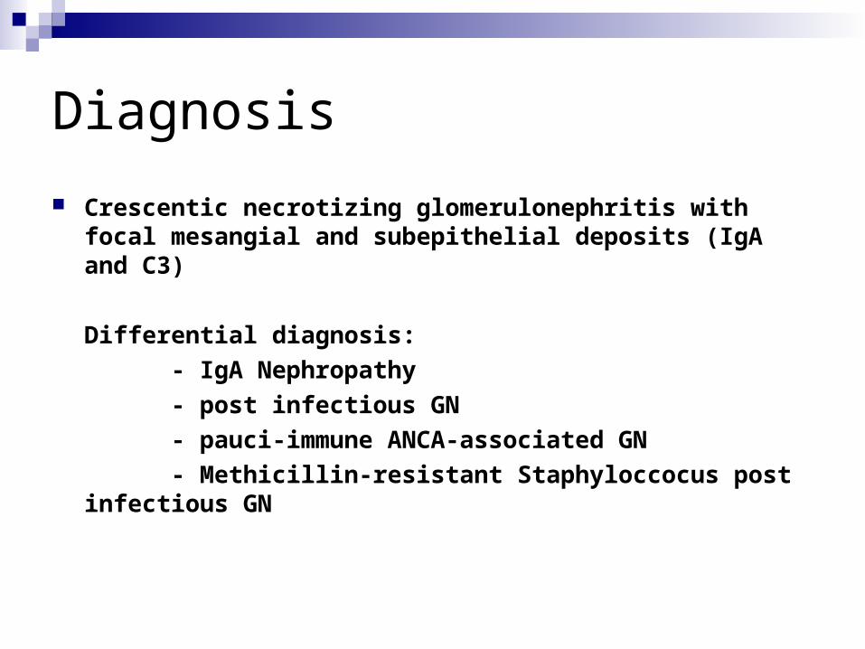
Diagnosis
Crescentic necrotizing glomerulonephritis with focal mesangial and subepithelial deposits (IgA and C3)
Differential diagnosis:
- IgA Nephropathy
- post infectious GN
- pauci-immune ANCA-associated GN
- Methicillin-resistant Staphyloccocus post infectious GN

IgA nephropathy Postinfectious GN
Laboratory Data
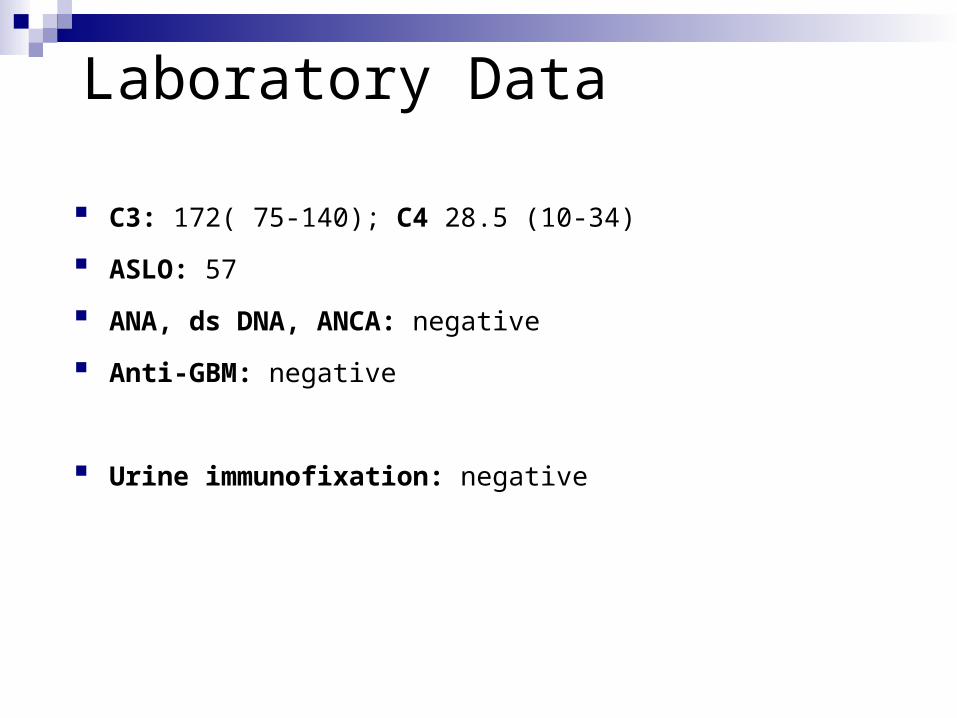
C3: 172( 75-140); C4 28.5 (10-34)
ASLO: 57
ANA, ds DNA, ANCA: negative
Anti-GBM: negative
Urine immunofixation: negative

Final Diagnosis
MRSA- post infectious GN

Objectives
Postinfectious Glomerulonephritis (PIGN)
Current trends in PIGN in adults
Staphylococcus and IgA dominant PIGN

Postinfectious Glomerulonephritis Acute postinfectious GN (APIGN) = disease of childhood Commonly following a streptococcal infection (= APSGN)
Clinical presentation: 3 phase sequence: infection - interval - nephritic syndrome
Course of disease: 1 week: onset of diuresis4 weeks: normalization of creatinine3-6 months: resolution of hematuria; resolution of mesangial hypercellularityYears: resolution of proteinuria
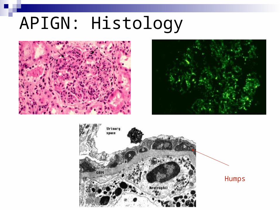
APIGN: Histology
Humps

APIGN: Outcome Long term follow up studies: excellent prognosis for most children with the epidemic form
A Japanese study followed 138 children with non-epidemic form: None developed renal insufficiency, all had normal serum complement within 12 weeks, resolution of proteinuria within 3 yrs and hematuria within 4 yrs (Kasahara T et al, Pediatr Int 2001; 43: 364)
A 12-17 yrs f/u study of 534 children and adults in Trinidad showed complete recovery in 96.5% (Potter EV et al, NEJM 1982; 307: 725)
A 2005 study from Brazil studied 56 patients for 5.4 yrs who had APIGN related to an outbreak of Streptococcus zooepidemicus: 30% with HTN, 49 % with reduced GFR, 22% with microalbuminuria (Sesso R et al, Nephrol Dial Transplant 2005; 20:1808)
Literature reports recovery rate in adults 53-76%

APIGN: What is New in Adults?Retrospective studies:
Keller CK et al, Q J Med 1994; 87: 97- Germany 1984-1993; 30 patients
Montseny JJ et al, Medicine 1995; 74: 63 - France 1976 - 1993; 76 patients
Moroni G et al, Nephrol Dial Transplant 2002; 17: 1204 - Italy 1979-1999; 50 patients
Nasr SH et al, Medicine 2008; 87: 21 - Columbia University 1995-2005; 92 patients

APIGN in Adults % of all renal biopsies: 0.6% - 4.6%
Median age 49 - 58 yrs
Underlying disease: 40-50%- Alcoholism +/- cirrhosis 2 - 57%- Diabetes 8 - 29%- COPD 7 - 33%- IVDU 3 - 27%- Malignancy 5 - 10%
Moroni G et al 2002
Nocomorbidities
+ comorbidities

APIGN: Presentation
Nephritic syndrome: 60%
Nephrotic Syndrome: 30-50%
Mean serum creatinine:
1.5-6.4 mg/dl
(with comorbidities/crescentic GN)
Mean 24 hr-protein:
3.6 g (with comorbidities)
Endocapillary proliferation: 70-100% Crescents (> 20-30%) : 14 - 36% Interstitial infiltration: 30 - 80% ATN: 20 - 40%
IF: C3 deposits: 93 - 100% C1: 18 - 35% IgG deposits: 55 - 65% IgM/IgA: 30 - 45%
EM: Mesangial deposits: 33 - 90% Subendothelial: 44 - 75% Humps: 94 - 100%

Sites of Infection and Microbiology
Streptococcus: 14-47% Staphylococcus: 12-24% Gram negatives: 1-22% 24-59% w/o microbiologic diagnosis
Nasr et al: Mean latent period: 3 weeks
2 weeks (endocarditis), 3 weeks (SSTI), 4 weeks (URI)
8% of patients simultaneous diagnosis (20% of pt with endocarditis and 27% with PNA)
URI: 24-44% SSTI: 5-25% Lung: 16-18% Endocarditis: 1-13% Dental: 0-13% UTI: 1-12%
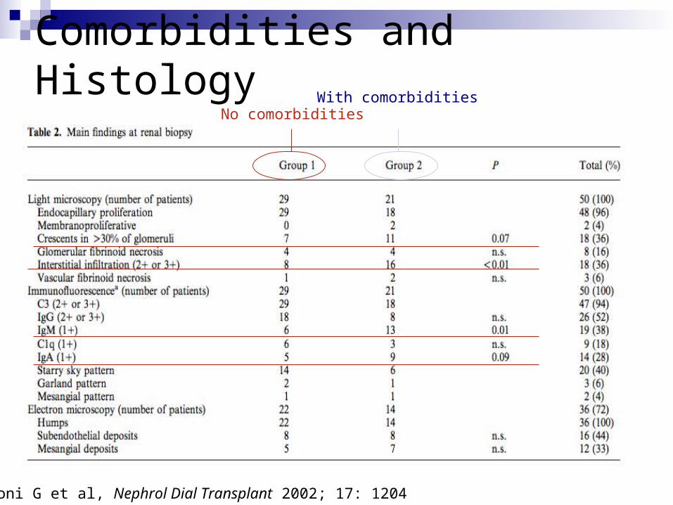
Comorbidities and HistologyNo comorbidities
With comorbidities
Moroni G et al, Nephrol Dial Transplant 2002; 17: 1204

Outcome CR 28-64% PRD 27-53% ESRD 4-17% Death 4-11%
Correlates of outcome:
- CR: younger age, no underlying disease h/o URI endocapillary disease, no crescents or subendothelial deposits no interstitial inflammation
- PRD: alcoholism nephrotic syndrome crescentic GN, interstitial fibrosis
- ESRD: higher baseline creatinine underlying diabetic GS
Nasr SH et al, Medicine 2008; 87: 21

% PIGN of all biopsies
% complete remission% with severe interstitial infiltration
% with “atypical” infection sites
Moroni G et al, Nephrol Dial Transplant 2002; 17: 1204

Do Steroids Matter ? Montseny et al:
17 pt (12 with crescentic GN) treated with steroids, 8 additionally with cyclophosphamide:2 died, 2 on HD, 3 with progressive CD, 5 with stable proteinuria, 5 with CR
Moroni et al:CR or partial remission in 54% treated with steroids vs 72% of untreated (but pt with steroids with higher creatinine and interstitial inflammation)
Nasr et al:33% of 52 pt treated with steroidsIndications: renal insufficiency with/without crescentsCR in 12/17 patients with steroid therapy and 10/23 without (p=0.116)
Nasr SH et al, Medicine 2008; 87: 21Moroni G et al, Nephrol Dial Transplant 2002; 17: 1204Montseny JJ et al, Medicine 1995; 74: 63

Staph and the Kidney
2 staphylococcal associated GN:
- acute proliferative exudative GN associated with
S. aureus endocarditis (resembling poststreptococcal GN)
- membranoproliferative GN associated with
S. epidermidis and ventricular shunt infections (“shunt nephritis”)
Nasr SH et al, Hum Pathol 2003, 34: 1235
MRSA and PIGN In 1980, Spector et al first reported 3 pt with S. aureus visceral
abscesses who developed acute mesangial proliferative GN with mesangial IgA deposits
In 1995, Koyama et al reported 10 pt who developed a rapidly progressive GN with nephrotic syndrome associated with MRSA infections (abdominal 8, PNA 2, arthritis 1, phlegmon 1)
Renal biopsy in 6 pt showed proliferative GN with various degrees of crescent formation and glomerular deposition of IgA , IgG and C3
Elevated serum IgA/IgG and immune complexes levels
High number of T cells with V+ usage in the TCR: ? Superantigen driven event
Named “MRSA Nephritis” or “Superantigen- related Nephritis”
Spector DA et al, Clin Nephrol 1980; 14: 256Koyama A et al, Kidney Internat 1995; 47: 207

MRSA and PIGN Recent reports: similar features after MSSA and MRSE infections
Clinical presentation: - acute RF with hematuria, severe proteinuria - onset 2-16 weeks after infection- +/- purpura, +/- hypocomplementemia
Mostly mesangial proliferative GN, often with crescents and (pre-) dominant mesangial IgA deposits
Several cases do not have subepithelial humps, the “hallmark” of PIGN
Treatment of infection lead to resolution of GN; however 40-60% of pt developed ESRD
Steroid treatment was related to the death in 2 people but recent report suggest positive outcome if used after cure of infection
Nagaba Y et al, Nephron 2002; 92: 297Yoh K et al, Nephrol Dial Transplant 2000; 15: 1170Shimizu Y et al, J Nephrol 2005; 18: 249Okuyama S, Clin Nephrol 2008; 70: 344

Pathogenesis
Link between staphylococcal enterotoxins and T cell/cytokine activation?
Superantigen triggered cytokine activation leads to class switching to IgA?
Link to a staphylococcal cell wall antigen that co-localizes in glomeruli of patients with MRSA nephritis?
Other IgA dominant immune responses against staphylococcal antigens? (eg an envelope antigen called ‘probable adhesin’ that is also found in IgA nephropathy)
Nagaba Y et al, Nephron 2002; 92: 297Yoh K et al, Nephrol Dial Transplant 2000; 15: 1170Shimizu Y et al, J Nephrol 2005; 18: 249
Diabetes, Staph and the Kidney
Nasr et al, Hum Pathol 2003; 34: 1235
In 2003, Nasr et al in New York reported 5 pt with DM who developed an IgA dominant GN after staphylococcal infection
Histology showed diabetic nephropathy with superimposed
endocapillary proliferation with neutrophils and some degree of
interstitial inflammation
IgA sole immunoglobulin in 3 cases; IF with mesangial or mesangial/capillary granular IgA and C3 staining
EM: all cases with predominantly mesangial deposits and sparse subepithelial deposits
Findings were similar to IgA nephropathy but all pt had low complement, endocapillary hypercellularity and humps


Endocapillary proliferation
Subendothelial and subepithelial deposits
Granular IgA
Nodular sclerosis

IGA-PIGN vs IgA nephropathy
IgA nephropathy:, IgA1 and J chain predominance?
Nasr SH et al, Kidney International 2007; 71: 1317

Diabetes and IgA nephropathy
Increased serum levels of IgA and IgA immune complexes
- secondary to (silent) mucosal infection
- abnormal IgA clearance (abnormal glycosylation or sialylation)
Thickened BM and mesangial sclerosis hinders subepithelial deposit formation >> predominantly mesangial deposition
Nasr SH et al, Kidney International 2007; 71: 1317

IgA predominant postinfectious GN Recently, Haas et al added 13 cases from John Hopkins University Selection criteria included IgA deposits + 3 or more subepithelial
humps, no clinical history Not only associated with staphylococcal infection
Haas M et al, Hum Pathol 2008; 39: 1309
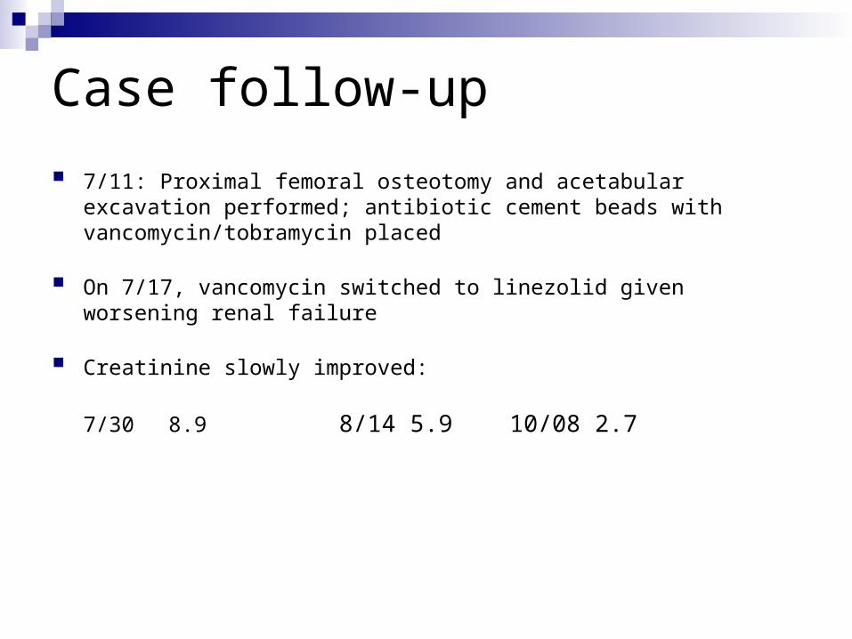
Case follow-up
7/11: Proximal femoral osteotomy and acetabular excavation performed; antibiotic cement beads with vancomycin/tobramycin placed
On 7/17, vancomycin switched to linezolid given worsening renal failure
Creatinine slowly improved:
7/30 8.9 8/14 5.9 10/08 2.7

Summary Epidemiology of APIGN is shifting
Diabetes, alcoholism and age emerge as major risk factor; prognosis is worse in pt with comorbidities and renal inflammation
Microbiology is changing and staphylococci are increasingly important in APIGN
Histologic pattern are changing, especially in immunocompromised persons
Summary
IgA predominant APIGN is recognized as 3rd entity of staphylococcal associated GN
IgA dominant PIGN can be associated with diabetic nephropathy
Exact pathologic diagnosis and pathogenesis is still under debate
This entity has to be differentiated from IgA nephropathy (and pauci-immune ANCA related GN)
Treatment of infection can lead to recovery; however, pt with underlying diabetic GS have poor prognosis
