nervous system - ankara Üniversitesi
TRANSCRIPT

NERVOUS SYSTEM

NERVOUS SYSTEM
•The nervous system of all animals is made up of groups of neurons that receive information from sensory systems, communicate with one another, and send information to motor systems.
•In invertebrates such as slugs, insects, etc., there is no real brain, just collections of neurons called ganglia. (singular = ganglion).
•The ganglia are distributed throughout various parts of the body.
•In vertebrates, the "head ganglion" or brain is extremely highly developed.

NERVOUS SYSTEM
The nervous system of vertebrates can be divided into:
1- The Central Nervous System (CNS).
2- The Peripheral Nervous System (PNS).

NERVOUS SYSTEM
In a mammal, the most obvious parts of the CNSfrom an external point of view are the cerebrum, cerebellum, brainstem, and
spinal cord.

NERVOUS SYSTEM
The peripheral nervous system includes all of
the nerves located outside the CNS. These are classified as:
The cranial nerves, which originate from cell groups within the brain.
The spinal nerves, which originate from the spinal cord.

NERVOUS SYSTEM
If we were to cut a slice through the brain or spinal cord, we would see that it is made up of:
- grey matter (areas containing neurons and unmyelinatedfibers)
-white matter (areas containing axons).
Both tissues include a number of glial cells.

•The skull and the vertebral column protect the central nervous system.
•It is also encased in membranes of connective tissue called the meninges.
•Starting with the outermost layer, the meninges are the
•dura mater,
•arachnoid, and
•pia mater.
•The arachnoid and the pia mater are linked together and are often considered a single membrane called the pia-arachnoid.
Meninges

Dura Mater
•The dura mater is the external layer and is composed of dense connective tissue continuous with the periosteum of the skull.

Dura Mater
•The dura mater that envelops the spinal cord is separated from the periosteum of the vertebrae by the epidural space, which contains thin-walled veins, loose connective tissue, and adipose tissue.
•This region is very convenient for epidural anesthesia.

Dura Mater
•The dura mater is always separated from the arachnoid by the thin subdural space.
•The internal surface of all dura mater, as well as its external surface in the spinal cord, is covered by simple squamousepithelium of mesenchymalorigin.

•The arachnoid (cobweblike) has two components: a layer in contact with the dura mater and a system of trabeculaeconnecting the layer with the pia mater.
•The cavities between the trabeculae form the subarachnoid space, which is filled with cerebrospinal fluid and is completely separated from the subdural space.
•This space forms a hydraulic cushion that protects the central nervous system from trauma.
•The subarachnoid space communicates with the ventricles of the brain.
Arachnoid

Arachnoid
•The arachnoid is composed of connective tissue devoid of blood vessels.
•The same type of simple squamous epithelium that covers the dura mater covers its surfaces.
•Because the arachnoid has fewer trabeculae in the spinal cord, it can be more clearly distinguished from the pia mater in that area.

Arachnoid
•In some areas, the arachnoid perforates the dura mater, forming protrusions that terminate in venous sinuses in the dura mater.
•These protrusions, which are covered by endothelial cells of the veins, are called arachnoid villi.
•Their function is to reabsorb cerebrospinal fluid into the blood of the venous sinuses.

•The pia mater is a loose connective tissue containing many blood vessels. Although it is located quite close to the nerve tissue, it is not in contact with nerve cells or fibers.
•Between the pia mater and the neural elements is a thin layer of neuroglial processes, adhering firmly to the pia mater and forming a physical barrier at the periphery of the central nervous system.
Pia Mater

•This barrier separates the central nervous system from the cerebrospinal fluid.
•The pia mater follows all the irregularities of the surface of the central nervous system and penetrates it to some extent along with the blood vessels. Squamous cells of mesenchymal origin cover pia mater.
Pia Mater

•Blood vessels penetrate the central nervous system through tunnels covered by pia mater—the perivascular spaces.
•The pia mater disappears before the blood vessels are transformed into capillaries.
•In the central nervous system, the blood capillaries are completely covered by expansions of the neuroglial cell processes.
Pia Mater

The Blood–brain Barrier(BBB)
•The blood–brain barrier is a functional barrier that prevents the passage of some substances, such as antibiotics and chemical and bacterial toxic matter, from the blood to nerve tissue.
•The blood–brain barrier results from the reduced permeability that is characteristic of blood capillaries of nerve tissue.

THE BLOOD-BRAIN BARRIER (BBB)
The blood–brain barrier allows the passage of water, some gases, and lipid-soluble molecules by passive diffusion, as well as the selective transport of molecules such as glucose and amino acids that are crucial to neural function.

The Blood–brain Barrier (BBB)
•Occluding junctions, which provide continuity between the endothelial cells of these capillaries, represent the main structural component of the barrier.
•The cytoplasm of these endothelial cells does not have the fenestrations found in many other locations, and very few pinocytotic vesicles are observed.
•The expansions of neuroglial cell processes that envelop the capillaries are partly responsible for their low permeability.

Blood–brain Barrier (BBB)
Subarachnoid space continues up into the brain tissue with piamater and blood vessels and it creates a sheath around blood vessels. It called perivascular liquor sheath.
Protoplasmic astrocytes are settled in this small areas.
So it creates the membrane limitans glia perivascularis.
Likewise, protoplasmic astrocytes are settled on the superficial ofbrain tissue. So it creates "membrana limitans gliae superficialis«
Protoplasmic astrocyte extensions have relationship with nerve cells and blood vessel.

Blood–brain Barrier (BBB)
“membrane limitans glia perivascularis”
and
“membrana limitans gliae superficialis” constitutes blood brainbarrier.
•Water, O2 and CO2, small water-soluble substances and some drugs easily cross the blood-brain barrier.
•Glucose, amino acids, some vitamins can also pass over the barrier.
•Ions (Na, K, Cl) can pass over the barrier by active transport.
•Antibiotics and some drugs can not pass.
•Heavy metals (Ag, Au, As) can not pass through the barrier.

NERVOUS SYSTEMLiquor cerebrospinalis=Cerebrospinal fluid CSF:
•Cerebrospinal fluid (CSF) is a clear, colorless body fluidfound in the brain and spine.
•Liquor is synthesized by cells of the choroid plexus and ventricular ependyma.
•It acts as a cushion or buffer for the brain's cortex, providing basic mechanical and immunological protection to the brain inside the skull.

NERVOUS SYSTEMLiquor cerebrospinalis=Cerebrospinal fluid (CSF):
•The CSF also serves a vital function in cerebral autoregulation of cerebral blood flow.
•The CSF occupies the subarachnoid space (between the arachnoidmater and the pia mater) and the four ventricles of the brainaround and inside the brain and spinal cord.
•It constitutes the content of the ventricles, cisterns, and sulci of the brain, as well as the central canal of the spinal cord.

NERVOUS SYSTEM
Blood-Cerebrospinal fluid barrier :
Blood-CSF barrier separates the cerebrospinal fluid and blood. It consists of three structurally distinct parts:
1) Ependymal cells
2) Basement membrane
3) Capillary endothelium

The Ventricles of the Brain
•The ventricles of the brain are a communicating network of cavities filled with cerebrospinal fluid (CSF) and located within the brain parenchyma.
•The ventricular system is composed of
-lateral ventricles (two piece),
-the third ventricle (one),
-the cerebral aqueduct, and
- the fourth ventricle.

The Ventricles of the Brain
•The choroid plexuses located in the ventricles produce CSF, which fills the ventricles and subarachnoid space, following a cycle of constant production and reabsorption.
•The ventricles are lined by a single layer of ciliated squamous or columnar ependymal cells.

The Ventricles of the Brain
Ependymal cells are connected to each other with zonula occludens, and located kinocilia and microvilli on their surface facing the cavity.
Cerebrospinal fluid flow occurs by the action of kinosilyum.
Microvilli absorb liquid from the liquor. Thus liquid remains clean.

Choroid Plexus
The choroid plexus is a highly specialized tissue that projects as elaborate folds with many villi into the four large ventricles of the brain. It is found in the roofs of the third and fourth ventricles and in parts of the walls of the two lateral ventricles, all regions in which the ependymal lining directly contacts the pia mater.

Choroid Plexus
•Each villus of the choroid plexus contains a thin layer of well-vascularized pia mater covered by cuboidalependymal cells.
•The main function of the choroid plexus is to remove water from blood and release it as cerebrospinal fluid (CSF).

Cerebrospinal fluid (CSF):
• This fluid completely fills the ventricles, the central canal of the spinal cord, the subarachnoid space, and the perivascular spaces.
•It is important for metabolism within the CNS and acts to absorb mechanical shocks.

Cerebrospinal fluid (CSF):
•CSF is clear, has a low density, contains Na+, K+, and Cl– ions but very little protein, and its only cells are normally very sparse lymphocytes.
•It is produced continuously across the walls of the choroid plexus villi and circulates through the ventricles and central canal, from which it passes into the subarachnoid space.
•There, arachnoid villi provide the main pathway for absorption of CSF into the venous circulation since there are no lymphatic vessels in CNS tissue.

NERVOUS SYSTEMThe central nervous system (CNS) consists of two parts.
1. Substantia grisea (gray matter)
2. Substantia alba (white matter)
•The principal structures of the CNS are the cerebrum, cerebellum, and spinal cord. It has virtually no connective tissue and is therefore a relatively soft, gel-like organ.

•When sectioned, the cerebrum, cerebellum, and spinal cord show regions of white (white matter) and gray (gray matter), differences caused by the differential distribution of myelin.
•The main components of white matter are myelinated axons and the myelin-producing oligodendrocytes.
•White matter does notcontain neuronal cellbodies, but microglia arepresent.

•Gray matter contains abundant neuronal cell bodies, dendrites, the initial unmyelinatedportions of axons, astrocytes, and microglial cells. This is the region wheresynapses occur.
• Gray matter is prevalent at the surface or cortex of the cerebrum and cerebellum, whereas white matter is present in more central regions.

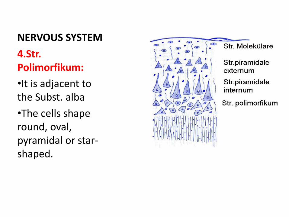
•Neuroscientists recognize four layers in the cerebral cortex with most neurons arranged vertically.
•The most abundant neurons are the efferent pyramidal neurons which come in many sizes.

1.Str. Moleculare:
•It is found under the pia mater.
•Painted pale than other layers.
•The cells are small and sparse.

2.Str. piramidale externum:
•It consists of small pyramid shaped cellslayer.
•The top of the pyramid is toward pia mater.
•Dendrites are directed to the upper layer.
•Axons extends to the substantia alba.

3.Str.piramidale internum:
•The cells of these layer is large and extend deeper.

NERVOUS SYSTEM
3.Str.piramidale internum:
•Dendrites extend into the molecular layer.
•Axons extend into the white matter.
NERVOUS SYSTEM
4.Str. Polimorfikum:
•It is adjacent to the Subst. alba
•The cells shaperound, oval, pyramidal or star-shaped.

Hippocampus
(Ammon’s horn):
•The hippocampus is located under the cerebral cortex.
•Negri bodies are found in the cytoplasm of certain nerve cellscontaining the virus of rabies, especially in the hippocampus.

Cerebellum
•Substantia grisea(gray matter), is on the outside of the cerebellum.
•Substantia alba(white matter) is on the inside of thecerebellum.

Cerebellum
•The cerebellar cortex has three layers :
-an outer molecular layer,
-a central layer of large Purkinje cells, and
-an inner granule layer.

Cerebellum
-an outer molecular layer,
-a central layer of large Purkinje cells, and
-an inner granule layer.

Cerebellum
•The Purkinje cells have a conspicuous cell body and their dendrites are highly developed, assuming the aspect of a fan.
•These dendrites occupy most of the molecular layer and are the reason for the sparseness of nuclei.

Cerebellum
The granuler layer is formed by very small neurons (the smallest in the body), which are compactly disposed, in contrast to the less cell-dense molecular layer.

Cerebellum
The cerebellar cortex regulates the balance functions,
and
•It coordinates muscular activity throughout the body.

Cerebellum:
Substantia alba (white matter) consists of myelinated nerve fibers and glial cells.

BRAINSTEM
•Located between the cerebrum and the spinal cord.
•Consist of the midbrain, pons, and medulla oblongata.
•Microscopically, it consists of deep gray matter surrounded by thewhite matter fiber tracts.
•Produce automatic behaviors necessary for survival.
•The brain stem controls the flow of messages between the brain and the rest of the body, and it also controls basic body functions such as breathing, swallowing, heart rate, blood pressure, consciousness, and whether one is awake or sleepy.

BRAINSTEM
Midbrain
•The midbrain, also called the mesencephalon, is a small region of the brain that serves as a relay center for visual, auditory, and motor system information.
•It regulates autonomic functions, those that the body carries out without conscious thought, such as digestion, heart rate, and breathing rate.

BRAINSTEM
Pons
•A major structure in the upper part of the brain stem is called the pons.
•The pons has two over-arching roles. The first is the regulation of breathing.
•In the pons, there is a structure called the pneumotaxic center.
•It controls the amount of air breathed and breaths per minute, which is known as the breathing rate.

BRAINSTEM
•Pons
•In addition, the pons is involved in the transmission of signals to and from other structures in the brain, such as the cerebrum or the cerebellum.
•The pons is also involved in sensations such as hearing, taste, and balance. Finally, the pons is also involved in the regulation of deep sleep.

BRAINSTEM
Medulla Oblongata:
•The medulla oblongata is located in the lower portion of thebrainstem.
•It is very important in things like heart rate and blood pressure.
•It's responsible for many reflexes in the body, or involuntarily controls, such as vomiting, sneezing, and coughing.

SPINAL CORD
•Unlike the cerebrumand cerebellum, in cross sections of the spinal cord, white matter is peripheral and gray matter is internal and has the general shape of an H.

SPINAL CORD
•In the center is anopening, the central canal, which develops from the lumen of the embryonic neural tube and is lined by ependymal cells.

SPINAL CORD
Substantia grisea(Gray matter):
•Horizontal arm of “H”is called commisuragrizea.
•Canalis centralis is located in the center of the spinal cord.
•Ependym cells are located in the inner side of the channel.

SPINAL CORD
•Ependymal cells are surrounded by a glialtissue (substantiagelatinosa centralis)
•Gray matter contains dorsal horns and ventral horns.

SPINAL CORD
•End portion of the dorsal horn is wrapped by fine glial tissue (substantia jelatinosa dorsalis).
•Ventral horn is larger than the dorsal horn.

SPINAL CORD
•Junctions of the dorsal and ventral horn is called pars intermedia.
•Pars intermediaprotrude to both sides at the thoracolumbarregion; theseprodrusions are named lateral horn.

SPINAL CORD
•White matter is divided from the dorsal and ventral (dorsal septum, ventral fissure).
•The nerve fibers in the white matter go from spinal cord to brain or go from the brain to spinal cord.

SPINAL CORD
The most important nerve cells present in the gray matter are:
1.Somatomotoric cells
2. Autonomic nerve cells
a) Sympathetic nerve cells
b)Parasympathetic nerve cells
3. Columnar cells
4. Reflex cells (Golgi type cells)

SPINAL CORD
1.Somatomotoric cells:
•They are located in the ventral horn. These cells haveefferent and multipolar feature.

SPINAL CORD
1.Somatomotoric cells:
•They are the largest cells in the spinal cord.
•Somatomotoric cells have two type:
•1. Small type (Gamma motor neurons)
•2. Large type (Alpha motor neurons).
Gamma motor neurons are responsible for contraction of muscle spindle.

SPINAL CORD
1.Somatomotoric cells:
•Alpha motor neurons innervateskeletal muscles.

SPINAL CORD
2.Autonomic nerve cells:
•The autonomic nervous system is a control system that acts largely unconsciously and regulates bodily functions such as the digestion, respiratory rate ect.
•Autonomic nerve cells have efferent feature.
•But they are responsible for vegetative organs innervation.

SPINAL CORD
3. Columnar cells•They are the second largest cell in the spinal cord. •Mainly, they are found in the dorsal horn.

SPINAL CORD
4. Reflex cells (Golgitype cells):
• Mostly, they are located in the dorsal horn.

SPINAL CORD
Substantia alba(White matter):
This structure consist of myelinated and un myelinated nerve fibers, scattered glia cells and blood vessels.

SPINAL CORD
Substantia alba(White matter):
•In cross section, the appearance of the nerve fibers are round.
•Nerve fibers, go out from the spinal cord, comes to brain. Or vice versa.

Peripheral Nervous SystemThe main components of the peripheral nervous system are
-ganglia,
-the peripheral nerves, and
-peripheral nerve endings.
Nerves are bundles of nerve fibers (axons) surrounded by glial cells and connective tissue.

PERIPHERAL NERVOUS SYSTEM
1. Ganglia:
•These are the anatomical structures.
•Ganglia occurs many of the nerve cells and nerve fibers (afferent, efferent).

PERIPHERAL NERVOUS SYSTEM
1. Ganglia:
•Nerves are bundles of nerve fibers (axons) surrounded by glial cells and connective tissue.

PERIPHERAL NERVOUS SYSTEM
1. Ganglia:
Ganglia are divided into two groups.
a)Cerebro-spinalganglia
b)Autonomicganglia

PERIPHERAL NERVOUS SYSTEM
a)Cerebro-spinalganglia :
•They are located on the cerebral and spinal nerve.
•Spinal ganglia is the best example. They are located in the intervertebral foramen.

PERIPHERAL NERVOUS SYSTEM
a)Cerebro-spinalganglia :
•Nerve cells are pseudounipolar type.
•These cells aresurrounded by mantocells (satellite cells).
•The nerve fibers are poor in myelin.
•Connective tissue are located between nerve cells and nerve fibers.

PERIPHERAL NERVOUS SYSTEM
b)Autonomic ganglia
•They work as a involuntary. Cells of autonomic ganglia are found in the brain, nucleus of the brainstem and lateral cornu of the spinal cord.
•Autonomic nerve cells in the ganglia are mostly multipolar.
•The primary function of the autonomic nervous system is to provide homeostasis.

PERIPHERAL NERVOUS SYSTEM
Autonomic ganglia:
Divided into two groups.
1. Sympathetic ganglia
2. Parasympathetic ganglia

PERIPHERAL NERVOUS SYSTEM
1. Sympathetic ganglia :
There are two types of ganglia of this system:
a) Paravertebral
ganglia
b) Prevertebral ganglia

PERIPHERAL NERVOUS SYSTEM
1. Sympathetic ganglia :
a)Paravertebral ganglia
•Located next to the spinal cord.
•The paravertebral ganglia are a series of ganglia which lie in a line lateral and parallel to the vertebral bodies of the spinal column. The ganglia are interconnected to each other and extend from the base of the skull to the sacrum.

PERIPHERAL NERVOUS SYSTEM
1. Sympathetic ganglia :
b) Prevertebral ganglia
•They are isolated ganglia in the chest or abdominal cavity.
•Nerve fibers are separated from paravertebral ganglia and come to prevertebral ganglia.
•Prevertebral ganglia are found in body cavity.
•The nerve fibers are distrubuted to the organs from prevertebralganglia.

PERIPHERAL NERVOUS SYSTEM
2. Parasympathetic ganglia:
Parasympathetic nerve axons goes to the organ without interruption.
•They make synapse in the organs wall.
•Neurons make network (plexus) in these synapses.
•This community is called intramural ganglia.

PERIPHERAL NERVOUS SYSTEM
2. Peripheral Nerves:
•They are macroscopic formations.
•Nerves are bundles of nerve fibers (axons) surrounded by
glial cells and connective tissue.
•Axons and Schwann cells of nerves are enclosed within connective tissue layers.
Externally is a dense, irregular fibrous coat called epineurium, which continues more deeply to also fill the space between bundles of nerve fibers.

PERIPHERAL NERVOUS SYSTEM
2. Peripheral Nerves:
Each such bundle is surrounded by the perineurium, a
sleeve of specialized connective tissue formed by layers of flattened epithelial-like cells.
The cells of each layer of the perineurium are joined at their edges by tight junctions, an arrangement that makes the perineurium a barrier to the passage of most macromolecules and has the important function of protecting the nerve fibers and helping maintain the internal microenvironment.

PERIPHERAL NERVOUS SYSTEM
2. Peripheral Nerves:
Within the perineurial sheath run the Schwann cell–covered axons and their enveloping connective tissue, the endoneurium.
The endoneurium consists of a sparse layer of loose connective tissue that merges with an external lamina of type IV collagen, laminin, and other proteins produced by the Schwann cells.

PERIPHERAL NERVOUS SYSTEM
3. Peripheral nerve endings:
•Some nerve endings simply terminates among other cells in the tissues they innervate.
•Some nerve endings terminate the mixed structure.
•Peripheral nerves are terminated by creating special structures in the body's end.
•These are efferent and afferent nerve endings.
•Motor nerve endings
•Sensory (sensible) nerve endings

PERIPHERAL NERVOUS SYSTEM
3. Peripheral nerve endings:
•Motor nerve endings:
•They finished in skeletal muscle.
•The nerve fibers loses myelinated, divided into several branch.
•Neurolemma and sarcolemma come face to face.
•A disc-shaped structure occurs.
•Neurolemma and sarcolemma are formed a synapses.
•Neurotransmitters are poured in the synaptic cleft.
•Ultimately, muscles contract.

PERIPHERAL NERVOUS SYSTEM
3. Peripheral nerve endings:
Motor nerve endings: Nerve ending of the smooth muscle and cardiac muscle is unmyelinated and made quite thin. They made a little swelling.
•Sensory (sensible) nerve endings:
•They are terminated in skeletal muscle, epithelial tissues and connective tissue.
PERIPHERAL NERVOUS SYSTEM
3. Peripheral nerve endings:
•Sensory (sensible) nerve endings:
•They are terminated in skeletal muscle, epithelial tissues and connective tissue.

REFERENCES:
Tanyolaç, A. (1999): Özel Histoloji. Yorum Basın Yayın Sanayi Ltd. Şti. Ankara.
Özer, A., Girgin, A., Alabay B., Liman, N., Özfiliz, N., Gülmez, N., Özcan, Z.,Yörük, M., Erdost, H., Aslan, Ş., Ergün, L., Zık, B. (2008): Veteriner ÖzelHistoloji. Nobel Yayın Dağıtım Tic. Ltd. Şti. Ankara
Dellmann, H. D., & Eurell, J. A. (1998). Textbook of Veterinary Histology,5th. Edn., Philadelphia, Lea and Febiger. P, 450.
Gartner, L.P. & Hiatt, J.L. (1997). Color textbook of Histology: W.B. SaundersCompany. Philadelphia, Pensilvanya, USA.
Junqueira, L. C., & Mescher, A. L. (2009). Junqueira's basic histology: text &atlas (12th ed.)/Anthony L. Mescher. New York [etc.]: McGraw-Hill Medical.