neural stages of spoken, written, and signed word...
TRANSCRIPT
ORIGINAL RESEARCH ARTICLEpublished: 02 July 2013
doi: 10.3389/fnhum.2013.00322
Neural stages of spoken, written, and signed wordprocessing in beginning second language learnersMatthew K. Leonard1,2†‡*, Naja Ferjan Ramirez 2,3‡, Christina Torres1,2, Marla Hatrak 3,Rachel I. Mayberry3$ and Eric Halgren1,2,4,5$
1 Department of Radiology, University of California, San Diego, La Jolla, CA, USA2 Multimodal Imaging Laboratory, Department of Radiology, University of California, San Diego, La Jolla, CA, USA3 Department of Linguistics, University of California, San Diego, La Jolla, CA, USA4 Department of Neurosciences, University of California, San Diego, La Jolla, CA, USA5 Kavli Institute for Brain and Mind, University of California, San Diego, La Jolla, CA, USA
Edited by:
John J. Foxe, Albert Einstein Collegeof Medicine, USA
Reviewed by:
John S. Butler, Albert EinsteinCollege of Medicine, USAEvie Malaia, University of Texas atArlington, USA
*Correspondence:
Matthew K. Leonard, MultimodalImaging Laboratory, Department ofRadiology, University of California,San Diego, 8950 Villa La Jolla Drive,Suite C101, La Jolla, CA 92037, USAe-mail: [email protected]†Present address:
Matthew K. Leonard, Department ofNeurological Surgery, University ofCalifornia, San Francisco,San Francisco, CA, USA‡These co-first authors havecontributed equally to this work.$These co-senior authors havecontributed equally to this work.
We combined magnetoencephalography (MEG) and magnetic resonance imaging (MRI)to examine how sensory modality, language type, and language proficiency interactduring two fundamental stages of word processing: (1) an early word encoding stage,and (2) a later supramodal lexico-semantic stage. Adult native English speakers whowere learning American Sign Language (ASL) performed a semantic task for spokenand written English words, and ASL signs. During the early time window, written wordsevoked responses in left ventral occipitotemporal cortex, and spoken words in leftsuperior temporal cortex. Signed words evoked activity in right intraparietal sulcus thatwas marginally greater than for written words. During the later time window, all threetypes of words showed significant activity in the classical left fronto-temporal languagenetwork, the first demonstration of such activity in individuals with so little secondlanguage (L2) instruction in sign. In addition, a dissociation between semantic congruityeffects and overall MEG response magnitude for ASL responses suggested shallowerand more effortful processing, presumably reflecting novice L2 learning. Consistent withprevious research on non-dominant language processing in spoken languages, the L2 ASLlearners also showed recruitment of right hemisphere and lateral occipital cortex. Theseresults demonstrate that late lexico-semantic processing utilizes a common substrate,independent of modality, and that proficiency effects in sign language are comparable tothose in spoken language.
Keywords: second language acquisition, sign language, speech, reading, proficiency, modality, magneto-
encephalography, MRI
INTRODUCTIONHumans acquire language in an astonishingly diverse set ofcircumstances. Nearly everyone learns a spoken language frombirth and a majority of individuals then follow this process bylearning to read, an extension of their spoken language experi-ence. In contrast to these two tightly-coupled modalities (writtenwords are a visual representation of phonological forms, spe-cific to a given language), there exists another language formthat bears no inherent relationship to a spoken form: Sign lan-guage. When deaf children are raised by deaf parents and acquiresign as their native language from birth, they develop profi-ciency within the same time frame and in a similar manner tothat of spoken language in hearing individuals (Anderson andReilly, 2002; Mayberry and Squires, 2006). This is not surprisinggiven that sign languages have sublexical and syntactic complex-ity similar to spoken languages (Emmorey, 2002; Sandler andLillo-Martin, 2006). Neural investigations of sign languages havealso shown a close correspondence between the processing ofsigned words in deaf (Petitto et al., 2000; MacSweeney et al.,2008; Mayberry et al., 2011; Leonard et al., 2012) and hearing
native signers (MacSweeney et al., 2002, 2006) and spoken wordsin hearing individuals (many native signers are also fluent in awritten language, although the neural basis of reading in deafindividuals is largely unknown). The predominant finding is thatleft anteroventral temporal, inferior prefrontal, and superior tem-poral cortex are the main loci of lexico-semantic processing inspoken/written (Marinkovic et al., 2003) and signed languages, aslong as the language is learned early or to a high level of profi-ciency (Mayberry et al., 2011). However, it is unknown whetherthe same brain areas are used for sign language processing inhearing second language (L2) learners who are beginning tolearn sign language. This is a key question for understanding thegeneralizability of L2 proficiency effects, and more broadly forunderstanding language mechanisms in the brain.
In contrast to the processing of word meaning, which occursbetween ∼200–400 ms after the word is seen or heard (Kutasand Federmeier, 2011), processing of the word form and sub-lexical structure appears to be modality-specific. Written wordsare encoded for their visual form primarily in left ventral occipi-totemporal areas (McCandliss et al., 2003; Vinckier et al., 2007;
Frontiers in Human Neuroscience www.frontiersin.org July 2013 | Volume 7 | Article 322 | 1
HUMAN NEUROSCIENCE
Leonard et al. Written, spoken, and signed words
Dehaene and Cohen, 2011; Price and Devlin, 2011). Spokenwords are likewise encoded for their acoustic-phonetic andphonemic forms in left-lateralized superior temporal cortex,including the superior temporal gyrus/sulcus and planum tem-porale (Hickok and Poeppel, 2007; Price, 2010; DeWitt andRauschecker, 2012; Travis et al., in press). Both of these pro-cesses occur within the first ∼170 ms after the word is pre-sented. While an analogous form encoding stage presumablyexists with similar timing for sign language, no such process hasbeen identified. The findings from monolingual users of spo-ken/written and signed languages to date suggest at least twoprimary stages of word processing: An early, modality-specificword form encoding stage (observed for spoken/written wordsand hypothesized for sign), followed by a longer latency responsethat converges on the classical left fronto-temporal language net-work where meaning is extracted and integrated independentof the original spoken, written, or signed form (Leonard et al.,2012).
Much of the world’s population is at least passingly famil-iar with more than one language, which provides a separate setof circumstances for learning and using words. Often, an L2 isacquired later with ultimately lower proficiency compared to thenative language. Fluent, balanced speakers of two or more lan-guages have little difficulty producing words in the contextuallycorrect language, and they understand words as rapidly and effi-ciently as words in their native language (Duñabeitia et al., 2010).However, prior to fluent understanding, the brain appears to gothrough a learning process that uses the native language as ascaffold, but diverges in subtle, yet important ways from nativelanguage processing. The extent of these differences (both behav-iorally and neurally) fluctuates in relation to the age at which L2learning begins, the proficiency level at any given moment dur-ing L2 learning, the amount of time spent using each languagethroughout the course of the day, and possibly the modality of thenewly-learned language (DeKeyser and Larson-Hall, 2005; vanHeuven and Dijkstra, 2010). Thus, L2 learning provides a uniqueopportunity to examine the role of experience in how the brainprocesses words.
In agreement with many L2 speakers’ intuitive experi-ences, several behavioral studies using cross-language translationpriming have found that proficiency and language dominanceimpact the extent and direction of priming (Basnight-Brownand Altarriba, 2007; Duñabeitia et al., 2010; Dimitropoulouet al., 2011). The most common finding is that priming isstrongest in the dominant to non-dominant direction, althoughthe opposite pattern has been observed (Duyck and Warlop,2009). These results are consistent with models of bilin-gual lexical representations, including the Revised HierarchicalModel (Kroll and Stewart, 1994) and the Bilingual InteractiveActivation + (BIA+) model (Dijkstra and van Heuven, 2002),both of which posit interactive and asymmetric connectionsbetween word (and sublexical) representations in both lan-guages. The BIA+ model is particularly relevant here, in thatit explains the proficiency-related differences as levels of acti-vation of the integrated (i.e., shared) lexicon driven by thebottom-up input of phonological/orthographic and word-formrepresentations.
An important question is how these behavioral proficiencyeffects manifest in neural activity patterns: Does the brain pro-cess less proficient words differently from more familiar words?Extensive neuroimaging and neurophysiological evidence sup-ports these models, and shows a particularly strong role for profi-ciency in cortical organization (van Heuven and Dijkstra, 2010).Two recent studies that measured neural activity with magnetoen-cephalography (MEG) constrained by individual subject anatomyobtained with magnetic resonance imaging (MRI) found that,while both languages for Spanish-English bilinguals evoked activ-ity in the classical left hemisphere fronto-temporal network,the non-dominant language additionally recruited posterior andright hemisphere regions (Leonard et al., 2010, 2011). These areasshowed significant non-dominant > dominant activity during anearly stage of word encoding (between ∼100–200 ms), continuingthrough the time period typically associated with lexico-semanticprocessing (∼200–400 ms). Crucially, these and other studies(e.g., van Heuven and Dijkstra, 2010) showed that language pro-ficiency was the main factor in determining the recruitment ofnon-classical language areas. The order in which the languageswere acquired did not greatly affect the activity.
These findings are consistent with the hemodynamic imag-ing and electrophysiological literatures. Using functional MRI(fMRI), proficiency-modulated differences in activity have beenobserved (Abutalebi et al., 2001; Chee et al., 2001; Perani andAbutalebi, 2005), and there is evidence for greater right hemi-sphere activity when processing the less proficient L2 (Dehaeneet al., 1997; Meschyan and Hernandez, 2006). While fMRI pro-vides spatial resolution on the order of millimeters, the hemo-dynamic response unfolds over the course of several seconds,far slower than the time course of linguistic processing in thebrain. Electroencephalographic methods including event-relatedpotentials (ERPs) are useful for elucidating the timing of activity,and numerous studies have found proficiency-related differ-ences between bilinguals’ two languages. One measure of lexico-semantic processing, the N400 [or N400 m in MEG; (Kutas andFedermeier, 2011)] is delayed by ∼40–50 ms in the L2 (Ardalet al., 1990; Weber-Fox and Neville, 1996; Hahne, 2001), andthis effect is constrained by language dominance (Moreno andKutas, 2005), in agreement with the behavioral and MEG studiesdiscussed above. In general, greater occipito-temporal activity inthe non-dominant language (particularly on the right), viewedin light of delayed processing, suggests that lower proficiencyinvolves less efficient processing that requires recruitment ofgreater neural resources. While the exact neural coding mecha-nism is not known, this is a well-established phenomenon thatapplies to both non-linguistic (Carpenter et al., 1999) and high-level language tasks (St George et al., 1999) at the neuronalpopulation level.
The research to date thus demonstrates two main findings:(1) In nearly all subject populations that have been examined,lexico-semantic processing is largely unaffected by languagemodality with respect to spoken, written, and signed lan-guage, and (2) lower proficiency involves the recruitment of anetwork of non-classical language regions that likewise appearto be modality-independent. In the present study, we sought todetermine whether the effects of language proficiency extend to
Frontiers in Human Neuroscience www.frontiersin.org July 2013 | Volume 7 | Article 322 | 2
Leonard et al. Written, spoken, and signed words
hearing individuals who are learning sign language as an L2.Although these individuals have extensive experience with a visuallanguage form (written words), their highly limited exposureto dynamic sign language forms allows us to investigate profi-ciency (English vs. ASL) and modality (spoken vs. written, vs.signed) effects in a single subject population. We tested a groupof individuals with a unique set of circumstances as they relate tothese two factors. The subjects were undergraduate students whowere native English speakers who began learning American SignLanguage (ASL) as an L2 in college. They had at least 40 weeksof experience, and were the top academic performers in theirASL courses and hence able to understand simple ASL signs andphrases. They were, however, unbalanced bilinguals with respectto English/ASL proficiency. Although there have been a few previ-ous investigations of highly proficient, hearing L2 signers (Nevilleet al., 1997; Newman et al., 2001), no studies have investigatedsign language processing in L2 learners with so little instruction.Likewise, no studies have investigated this question using meth-ods that afford high spatiotemporal resolution to determine boththe cortical sources and timing of activity during specific process-ing stages. Similar to our previous studies on hearing bilingualswith two spoken languages, here we combined MEG and struc-tural MRI to examine neural activity in these subjects while theyperformed a semantic task in two languages/modalities: spokenEnglish, visual (written) English, and visual ASL.
While it is not possible to fully disentangle modality andproficiency effects within a single subject population, these fac-tors have been systematically varied separately in numerousstudies with cross-language and between-group comparisons(Marinkovic et al., 2003; Leonard et al., 2010, 2011, 2012), and arewell-characterized in isolation. It is in this context that we exam-ined both factors in this group of L2 learners. We hypothesizedthat a comparison between the magnitudes of MEG responsesto spoken, written, and signed words would reveal a modality-specific word encoding stage between ∼100–200 ms (left superiorplanar regions for spoken words, left ventral occipitotempo-ral regions for written words, and an unknown set of regionsfor signed words), followed by stronger responses for ASL (thelower proficiency language) in a more extended network of brainregions used to process lexico-semantic content between ∼200–400 ms post-stimulus onset. These areas have previously beenidentified in spoken language L2 learners and include bilateralposterior visual and superior temporal areas (Leonard et al., 2010,2011). Finding similar patterns for beginning ASL L2 learnerswould provide novel evidence that linguistic proficiency effectsare generalizable, a particularly striking result given the vastlydifferent sensory characteristics of spoken English and ASL. Wefurther characterized the nature of lexico-semantic processingin this group by comparing the N400 effect across modalities,which would reveal differences in the loci of contextual inte-gration for relatively inexperienced learners of a visual secondlanguage.
MATERIALS AND METHODSPARTICIPANTSEleven hearing native English speakers participated in this study(10 F; age range = 19.74–33.16 years, mean = 22.42). All were
healthy adults with no history of neurological or psychologicalimpairment, and had normal hearing and vision (or wore correc-tive lenses that were applied in the MEG). All participants hadat least four academic quarters (40 weeks) of instruction in ASL,having reached the highest level of instruction at either UCSDor Mesa College. Participants were either currently enrolled in acourse taught in ASL or had been enrolled in such a course in theprevious month. One participant had not taken an ASL course inthe previous 4 months. Participants completed a self-assessmentquestionnaire that asked them to rate their ASL proficiency on ascale from 1 to 10. For ASL comprehension, the average score was7.1 ± 1.2; ASL production was 6.5 ± 1.9; Fingerspelling compre-hension was 6.4 ± 1.6; and fingerspelling production was 6.8 ±1.7. Six participants reported using ASL on a daily basis at thetime of enrollment in the study, while the remaining participantsindicated weekly use (one participant indicated monthly use).
Participants gave written informed consent to participate inthe study, and were paid $20/h for their time. This study wasapproved by the Institutional Review Board at the University ofCalifornia, San Diego.
STIMULI AND PROCEDUREIn the MEG, participants performed a semantic decision task thatinvolved detecting a match in meaning between a picture and aword. For each trial, subjects saw a photograph of an object for700 ms, followed by a word that either matched (“congruent”) ormismatched (“incongruent”) the picture in meaning. Participantswere instructed to press a button when there was a match;response hand was counterbalanced across blocks within subjects.Words were presented in blocks by language/modality for spo-ken English, written English, and ASL. Each word appeared oncein the congruent and once in the incongruent condition, anddid not repeat across modalities. All words were highly image-able concrete nouns that were familiar to the participants in bothlanguages. Since no frequency norms exist for ASL, the stimuliwere selected from ASL developmental inventories (Schick, 1997;Anderson and Reilly, 2002) and picture naming data (Bates et al.,2003; Ferjan Ramirez et al., 2013b). The ASL stimuli were pilotedwith four other subjects who had the same type of ASL instruc-tion to confirm that they were familiar with the words. Stimuluslength was the following: Spoken English mean = 473.98 ±53.17 ms; Written English mean = 4.21 ± 0.86 letters; ASL videoclips mean = 467.92 ± 62.88 ms. Written words appeared on thescreen for 1500 ms. Auditory stimuli were delivered through ear-phones at an average amplitude of 65 dB SPL. Written and signedword videos subtended <5 degrees of visual angle on a screenin front of the subjects. For all stimulus types, the total trialduration varied randomly between 2600 and 2800 ms (700 mspicture + 1500 ms word container + 400–600 ms inter-trialinterval).
Each participant completed three blocks of stimuli in each lan-guage/modality. Each block had 100 trials (50 stimuli in eachof the congruent and incongruent conditions) for a total of150 congruent and incongruent trials in each language/modality.The order of the languages/modalities was counterbalancedacross participants. Prior to starting the first block in each lan-guage/modality, participants performed a practice run to ensure
Frontiers in Human Neuroscience www.frontiersin.org July 2013 | Volume 7 | Article 322 | 3
Leonard et al. Written, spoken, and signed words
they understood the stimuli and task. The practice runs wererepeated as necessary until subjects were confident in their per-formance (no subjects required more than one repetition of thepractice blocks).
MEG RECORDINGParticipants sat in a magnetically shielded room (IMEDCO-AG, Switzerland) with the head in a Neuromag Vectorviewhelmet-shaped dewar containing 102 magnetometers and 204gradiometers (Elekta AB, Helsinki, Finland). Data were collectedat a continuous sampling rate of 1000 Hz with minimal filtering(0.1 to 200 Hz). The positions of four non-magnetic coils affixedto the subjects’ heads were digitized along with the main fidu-ciary points such as the nose, nasion, and preauricular pointsfor subsequent coregistration with high-resolution MR images.The average 3-dimensional Euclidian distance for head move-ment from the beginning of the session to the end of the sessionwas 7.38 mm (SD = 5.67 mm).
ANATOMICALLY-CONSTRAINED MEG (aMEG) ANALYSISThe data were analyzed using a multimodal imaging approachthat constrains the MEG activity to the cortical surface as deter-mined by high-resolution structural MRI (Dale et al., 2000). Thisnoise-normalized linear inverse technique, known as dynamicstatistical parametric mapping (dSPM) has been used extensivelyacross a variety of paradigms, particularly language tasks thatbenefit from a distributed source analysis (Marinkovic et al., 2003;Leonard et al., 2010, 2011, 2012; Travis et al., in press), and hasbeen validated by direct intracranial recordings (Halgren et al.,1994; McDonald et al., 2010).
The cortical surface was obtained in each participant witha T1-weighted structural MRI, and was reconstructed usingFreeSurfer. The images were collected at the UCSD RadiologyImaging Laboratory with a 1.5T GE Signa HDx scanner usingan eight-channel head coil (TR = 9.8 ms, TE = 4.1 ms, TI =270 ms, flip angle = 8◦, bandwidth = ±15.63 kHz, FOV = 24 cm,
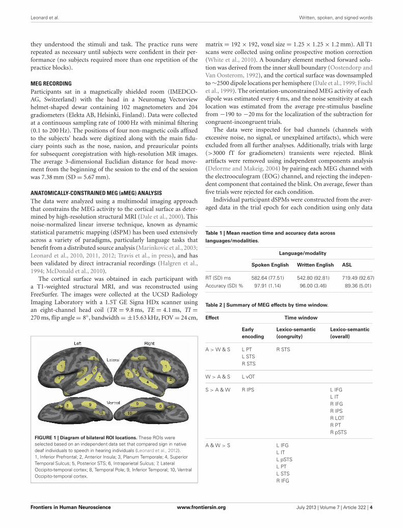
FIGURE 1 | Diagram of bilateral ROI locations. These ROIs wereselected based on an independent data set that compared sign in nativedeaf individuals to speech in hearing individuals (Leonard et al., 2012).1, Inferior Prefrontal; 2, Anterior Insula; 3, Planum Temporale; 4, SuperiorTemporal Sulcus; 5, Posterior STS; 6, Intraparietal Sulcus; 7, LateralOccipito-temporal cortex; 8, Temporal Pole; 9, Inferior Temporal; 10, VentralOccipito-temporal cortex.
matrix = 192 × 192, voxel size = 1.25 × 1.25 × 1.2 mm). All T1scans were collected using online prospective motion correction(White et al., 2010). A boundary element method forward solu-tion was derived from the inner skull boundary (Oostendorp andVan Oosterom, 1992), and the cortical surface was downsampledto ∼2500 dipole locations per hemisphere (Dale et al., 1999; Fischlet al., 1999). The orientation-unconstrained MEG activity of eachdipole was estimated every 4 ms, and the noise sensitivity at eachlocation was estimated from the average pre-stimulus baselinefrom −190 to −20 ms for the localization of the subtraction forcongruent-incongruent trials.
The data were inspected for bad channels (channels withexcessive noise, no signal, or unexplained artifacts), which wereexcluded from all further analyses. Additionally, trials with large(>3000 fT for gradiometers) transients were rejected. Blinkartifacts were removed using independent components analysis(Delorme and Makeig, 2004) by pairing each MEG channel withthe electrooculogram (EOG) channel, and rejecting the indepen-dent component that contained the blink. On average, fewer thanfive trials were rejected for each condition.
Individual participant dSPMs were constructed from the aver-aged data in the trial epoch for each condition using only data
Table 1 | Mean reaction time and accuracy data across
languages/modalities.
Language/modality
Spoken English Written English ASL
RT (SD) ms 582.64 (77.51) 542.80 (92.81) 719.49 (92.67)
Accuracy (SD) % 97.91 (1.14) 96.00 (3.46) 89.36 (5.01)
Table 2 | Summary of MEG effects by time window.
Effect Time window
Early
encoding
Lexico-semantic
(congruity)
Lexico-semantic
(overall)
A > W & S L PTL STSR STS
R STS
W > A & S L vOT
S > A & W R IPS L IFGL ITR IFGR IPSR LOTR PTR pSTS
A & W > S L IFGL ITL pSTSL PTL STSR IFG
Frontiers in Human Neuroscience www.frontiersin.org July 2013 | Volume 7 | Article 322 | 4
Leonard et al. Written, spoken, and signed words
from the gradiometers, and then these data were combined acrosssubjects by taking the mean activity at each vertex on the cor-tical surface and plotting it on an average brain. Vertices werematched across subjects by morphing the reconstructed corticalsurfaces into a common sphere, optimally matching gyral-sulcalpatterns and minimizing shear (Sereno et al., 1996; Fischl et al.,1999).
All statistical comparisons were made on region of interest(ROI) timecourses from these group data. ROIs were based ona separate data set not included in this study that comparedsigned and spoken word processing in congenitally deaf andhearing subjects using the same task presented here (Figure 1;Leonard et al., 2012). These ROIs were originally drawn on
the grand average activity across both deaf and hearing partic-ipants, and thus are not biased toward either signed or spokenwords. In the 80–120 ms time window, we specifically testedbilateral planum temporale (PT) and superior temporal sul-cus (STS) because these areas showed significant responses tospoken words, and are known to be involved in early wordencoding in the auditory modality (Uusvuori et al., 2008;Travis et al., in press). For the 150–200 ms time window, wewere specifically interested in ventral occipitotemporal (vOT)cortex because it is involved in written word form encod-ing (Vinckier et al., 2007). While there are no previous stud-ies of this stage for signed words, we selected bilateral intra-parietal sulcus (IPS) because it has been implicated in some
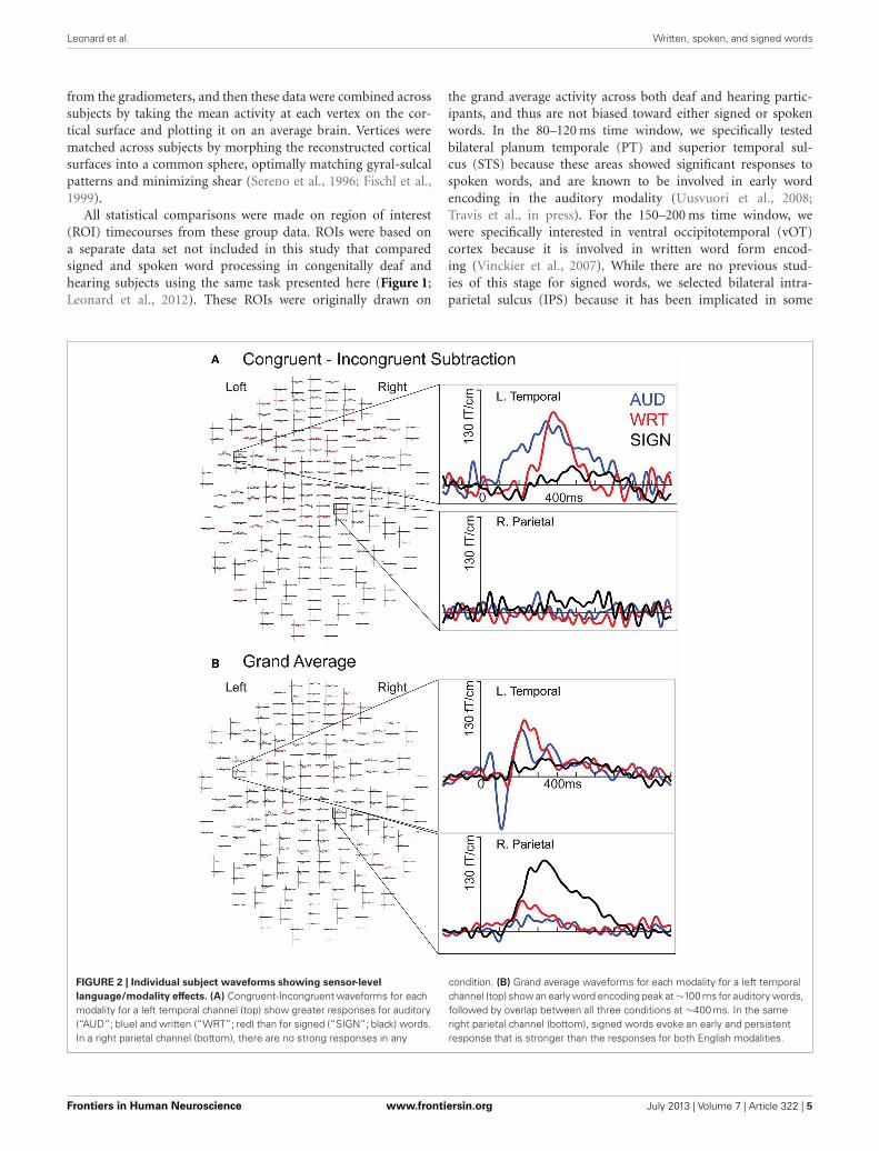
FIGURE 2 | Individual subject waveforms showing sensor-level
language/modality effects. (A) Congruent-Incongruent waveforms for eachmodality for a left temporal channel (top) show greater responses for auditory(“AUD”; blue) and written (“WRT”; red) than for signed (“SIGN”; black) words.In a right parietal channel (bottom), there are no strong responses in any
condition. (B) Grand average waveforms for each modality for a left temporalchannel (top) show an early word encoding peak at ∼100 ms for auditory words,followed by overlap between all three conditions at ∼400 ms. In the sameright parietal channel (bottom), signed words evoke an early and persistentresponse that is stronger than the responses for both English modalities.
Frontiers in Human Neuroscience www.frontiersin.org July 2013 | Volume 7 | Article 322 | 5
Leonard et al. Written, spoken, and signed words
studies of non-temporally specific sign processing (MacSweeneyet al., 2002; Emmorey et al., 2005), and because it showedthe strongest activity during this time window. For the lexico-semantic time window from 300 to 400 ms, we tested all tenbilateral ROIs. With the exceptions of IPS and lateral occip-itotemporal (LOT) cortex, these areas are typically involvedin lexico-semantic processing, including anteroventral tempo-ral areas that are hypothesized to be largely supramodal. Wealso included LOT because it has been implicated as a lexico-semantic area that is modulated by proficiency (Leonard et al.,2010, 2011).
RESULTSREACTION TIME AND ACCURACYThe following analyses use these abbreviations: A = auditorywords, W = written words, and S = signed words. Participantsperformed within ranges similar to those of native speakers andsigners on the semantic decision task in both reaction time andaccuracy compared to results in other studies (Leonard et al.,2012; Travis et al., in press). Table 1 shows the average and stan-dard deviations for each language/modality. A one-way ANOVAcomparing reaction times across modalities revealed a signifi-cant effect of modality [F(2, 30) = 12.21, p < 0.0002]. Consistentwith the fact that English was the subjects’ native and dom-inant language and ASL was a recently learned L2, reactiontimes were significantly faster for A than for S [t(10) = 6.85,p < 0.0001], and for W than for S [t(10) = 8.22, p < 0.0001].A and W were not significantly different. Similarly, there was asignificant effect of modality for accuracy on the semantic task[F(2, 30) = 17.31, p < 0.00001]. Participants were more accuratefor A than for S [t(10) = 5.13, p < 0.0001], and for W than for S[t(10) = 4.13, p = 0.002], although accuracy for S was still quitegood (nearly 90%). Accuracy for A and W were not significantlydifferent.
aMEG RESULTS SUMMARYThere were distinct patterns of neural activity related to the lan-guage/modality subjects saw or heard and language proficiency.These effects began during the earliest stages of word encod-ing (∼100 ms for auditory, and ∼150 ms for written and signedwords), and continued through lexico-semantic encoding (∼300–400 ms). Table 2 summarizes the main findings by time window,and the following sections statistically describe the effects shownin the table and figures. Figure 2 shows sensor-level data from asingle representative subject.
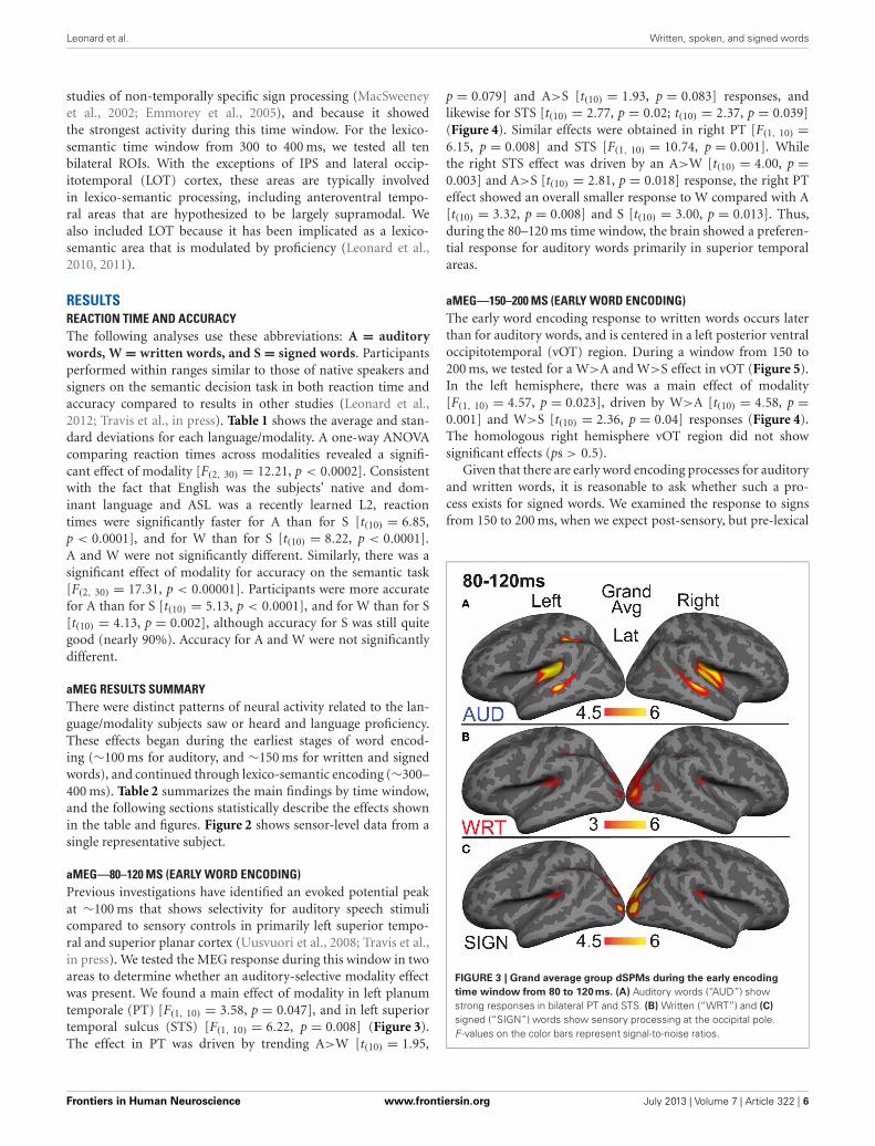
aMEG—80–120 MS (EARLY WORD ENCODING)Previous investigations have identified an evoked potential peakat ∼100 ms that shows selectivity for auditory speech stimulicompared to sensory controls in primarily left superior tempo-ral and superior planar cortex (Uusvuori et al., 2008; Travis et al.,in press). We tested the MEG response during this window in twoareas to determine whether an auditory-selective modality effectwas present. We found a main effect of modality in left planumtemporale (PT) [F(1, 10) = 3.58, p = 0.047], and in left superiortemporal sulcus (STS) [F(1, 10) = 6.22, p = 0.008] (Figure 3).The effect in PT was driven by trending A>W [t(10) = 1.95,
p = 0.079] and A>S [t(10) = 1.93, p = 0.083] responses, andlikewise for STS [t(10) = 2.77, p = 0.02; t(10) = 2.37, p = 0.039](Figure 4). Similar effects were obtained in right PT [F(1, 10) =6.15, p = 0.008] and STS [F(1, 10) = 10.74, p = 0.001]. Whilethe right STS effect was driven by an A>W [t(10) = 4.00, p =0.003] and A>S [t(10) = 2.81, p = 0.018] response, the right PTeffect showed an overall smaller response to W compared with A[t(10) = 3.32, p = 0.008] and S [t(10) = 3.00, p = 0.013]. Thus,during the 80–120 ms time window, the brain showed a preferen-tial response for auditory words primarily in superior temporalareas.
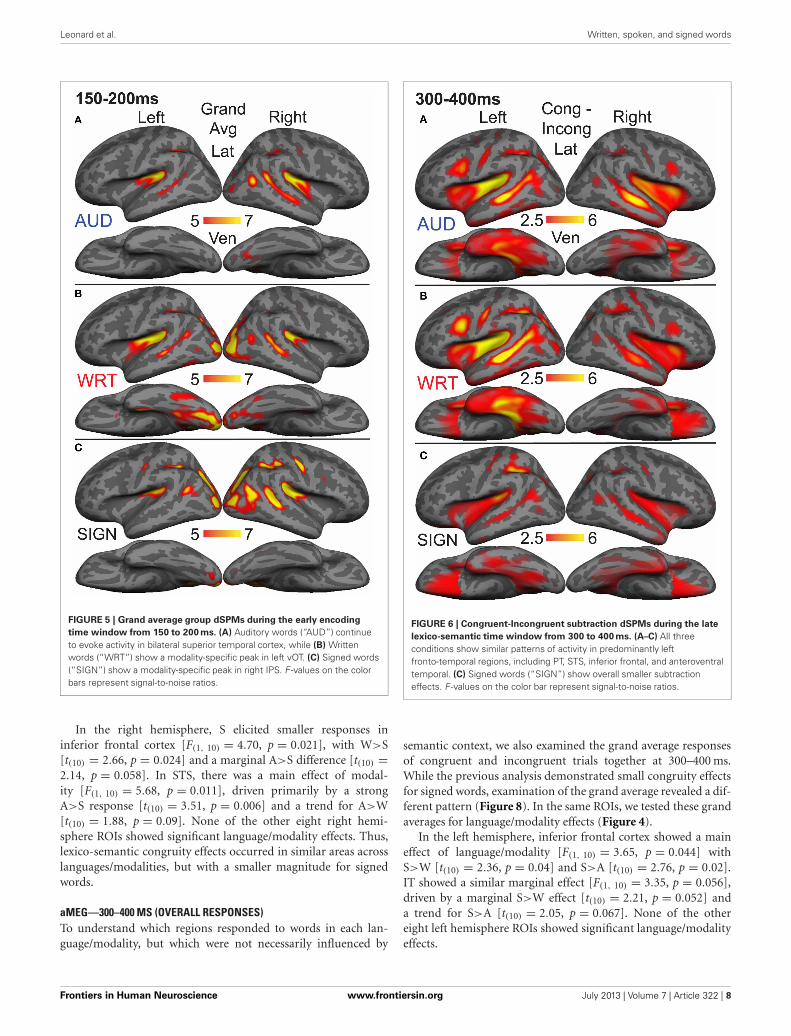
aMEG—150–200 MS (EARLY WORD ENCODING)The early word encoding response to written words occurs laterthan for auditory words, and is centered in a left posterior ventraloccipitotemporal (vOT) region. During a window from 150 to200 ms, we tested for a W>A and W>S effect in vOT (Figure 5).In the left hemisphere, there was a main effect of modality[F(1, 10) = 4.57, p = 0.023], driven by W>A [t(10) = 4.58, p =0.001] and W>S [t(10) = 2.36, p = 0.04] responses (Figure 4).The homologous right hemisphere vOT region did not showsignificant effects (ps > 0.5).
Given that there are early word encoding processes for auditoryand written words, it is reasonable to ask whether such a pro-cess exists for signed words. We examined the response to signsfrom 150 to 200 ms, when we expect post-sensory, but pre-lexical
FIGURE 3 | Grand average group dSPMs during the early encoding
time window from 80 to 120 ms. (A) Auditory words (“AUD”) showstrong responses in bilateral PT and STS. (B) Written (“WRT”) and (C)
signed (“SIGN”) words show sensory processing at the occipital pole.F -values on the color bars represent signal-to-noise ratios.
Frontiers in Human Neuroscience www.frontiersin.org July 2013 | Volume 7 | Article 322 | 6
Leonard et al. Written, spoken, and signed words
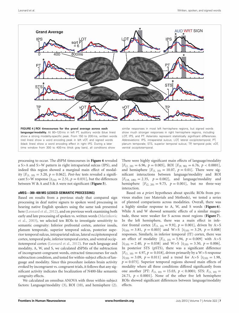
FIGURE 4 | ROI timecourses for the grand average across each
language/modality. At 80–120 ms in left PT, auditory words (blue lines)show a strong modality-specific peak. From 150 to 200 ms, written words(red lines) show a word encoding peak in left vOT, and signed words(black lines) show a word encoding effect in right IPS. During a latertime window from 300 to 400 ms (thick gray bars), all conditions show
similar responses in most left hemisphere regions, but signed wordsshow much stronger responses in right hemisphere regions, includingLOT, IPS, and PT. Asterisks represent statistically significant differences.Abbreviations: IPS, intraparietal sulcus; LOT, lateral occipitotemporal; PT,planum temporale; STS, superior temporal sulcus; TP, temporal pole; vOT,ventral occipitotemporal.
processing to occur. The dSPM timecourses in Figure 4 revealeda S>A and S>W pattern in right intraparietal sulcus (IPS), andindeed this region showed a marginal main effect of modal-ity [F(1, 10) = 3.20, p = 0.062]. Post-hoc tests revealed a signifi-cant S>W response [t(10) = 2.51, p = 0.031], but the differencesbetween W & A and S & A were not significant (Figure 5).
aMEG—300–400 MS (LEXICO-SEMANTIC PROCESSING)Based on results from a previous study that compared signprocessing in deaf native signers to spoken word processing inhearing native English speakers using the same task presentedhere (Leonard et al., 2012), and on previous work examining bothearly and late processing of spoken vs. written words (Marinkovicet al., 2003), we selected ten ROIs to investigate sensitivity tosemantic congruity: Inferior prefrontal cortex, anterior insula,planum temporale, superior temporal sulcus, posterior supe-rior temporal sulcus, intraparietal sulcus, lateral occipitotemporalcortex, temporal pole, inferior temporal cortex, and ventral occip-itotemporal cortex (Leonard et al., 2012). For each language andmodality, A, W, and S, we calculated dSPMs of the subtractionof incongruent-congruent words, extracted timecourses for eachsubtraction condition, and tested for within-subject effects of lan-guage and modality. Since this procedure isolates brain activityevoked by incongruent vs. congruent trials, it follows that any sig-nificant activity indicates the localization of N400-like semanticcongruity effects.
We calculated an omnibus ANOVA with three within-subjectfactors: Language/modality (3), ROI (10), and hemisphere (2).
There were highly significant main effects of language/modality[F(2, 20) = 6.96, p = 0.005], ROI [F(9, 90) = 6.76, p < 0.0001],and hemisphere [F(1, 10) = 10.07, p = 0.01]. There were sig-nificant interactions between language/modality and ROI[F(18, 180) = 2.35, p = 0.002], and language/modality andhemisphere [F(2, 20) = 9.75, p = 0.001], but no three-wayinteraction.
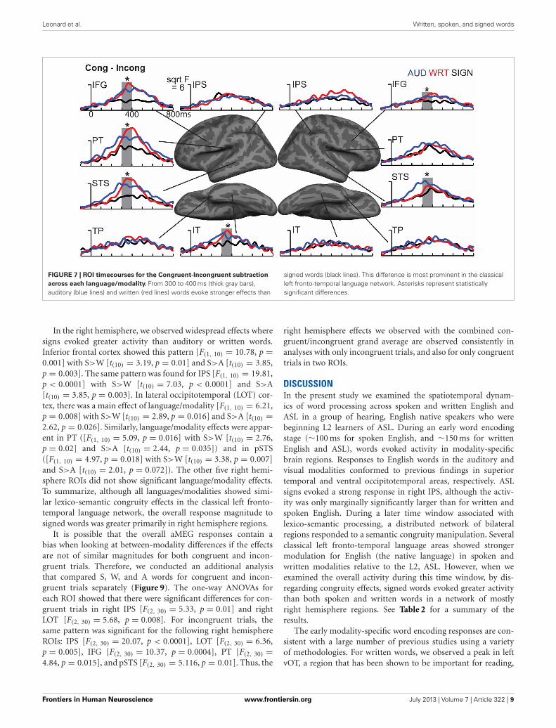
Based on a priori hypotheses about specific ROIs from pre-vious studies (see Materials and Methods), we tested a seriesof planned comparisons across modalities. Overall, there wasa highly similar response to A, W, and S words (Figure 6).While A and W showed semantic effects of a similar magni-tude, these were weaker for S across most regions (Figure 7).In the left hemisphere, there was a main effect in infe-rior frontal cortex [F(1, 10) = 9.92, p = 0.001], driven by A>S[t(10) = 3.81, p = 0.003] and W>S [t(10) = 3.29, p = 0.008]responses. Similarly, in inferior temporal (IT) cortex, there wasan effect of modality [F(1, 10) = 5.94, p = 0.009] with A>S[t(10) = 2.40, p = 0.038] and W>S [t(10) = 3.50, p = 0.006].In posterior STS (pSTS), there was a significant difference[F(1, 10) = 4.97, p = 0.018], driven primarily by a W>S response[t(10) = 3.09, p = 0.011] and a trend for A>S [t(10) = 1.98,p = 0.075]. Superior temporal regions showed main effects ofmodality where all three conditions differed significantly fromone another [PT: F(1, 10) = 15.03, p < 0.0001; STS: F(1, 10) =24.71, p < 0.0001]. None of the other five left hemisphereROIs showed significant differences between language/modalityeffects.
Frontiers in Human Neuroscience www.frontiersin.org July 2013 | Volume 7 | Article 322 | 7
Leonard et al. Written, spoken, and signed words
FIGURE 5 | Grand average group dSPMs during the early encoding
time window from 150 to 200 ms. (A) Auditory words (“AUD”) continueto evoke activity in bilateral superior temporal cortex, while (B) Writtenwords (“WRT”) show a modality-specific peak in left vOT. (C) Signed words(“SIGN”) show a modality-specific peak in right IPS. F -values on the colorbars represent signal-to-noise ratios.
In the right hemisphere, S elicited smaller responses ininferior frontal cortex [F(1, 10) = 4.70, p = 0.021], with W>S[t(10) = 2.66, p = 0.024] and a marginal A>S difference [t(10) =2.14, p = 0.058]. In STS, there was a main effect of modal-ity [F(1, 10) = 5.68, p = 0.011], driven primarily by a strongA>S response [t(10) = 3.51, p = 0.006] and a trend for A>W[t(10) = 1.88, p = 0.09]. None of the other eight right hemi-sphere ROIs showed significant language/modality effects. Thus,lexico-semantic congruity effects occurred in similar areas acrosslanguages/modalities, but with a smaller magnitude for signedwords.
aMEG—300–400 MS (OVERALL RESPONSES)To understand which regions responded to words in each lan-guage/modality, but which were not necessarily influenced by
FIGURE 6 | Congruent-Incongruent subtraction dSPMs during the late
lexico-semantic time window from 300 to 400 ms. (A–C) All threeconditions show similar patterns of activity in predominantly leftfronto-temporal regions, including PT, STS, inferior frontal, and anteroventraltemporal. (C) Signed words (“SIGN”) show overall smaller subtractioneffects. F -values on the color bar represent signal-to-noise ratios.
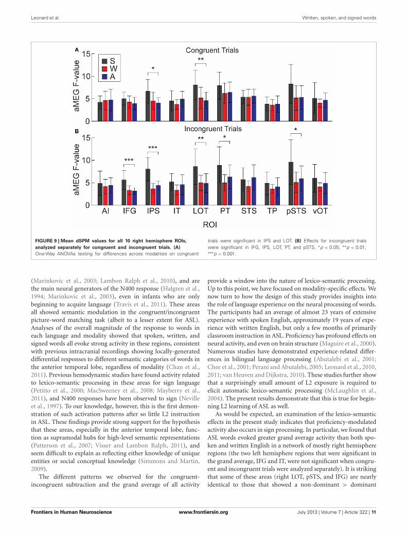
semantic context, we also examined the grand average responsesof congruent and incongruent trials together at 300–400 ms.While the previous analysis demonstrated small congruity effectsfor signed words, examination of the grand average revealed a dif-ferent pattern (Figure 8). In the same ROIs, we tested these grandaverages for language/modality effects (Figure 4).
In the left hemisphere, inferior frontal cortex showed a maineffect of language/modality [F(1, 10) = 3.65, p = 0.044] withS>W [t(10) = 2.36, p = 0.04] and S>A [t(10) = 2.76, p = 0.02].IT showed a similar marginal effect [F(1, 10) = 3.35, p = 0.056],driven by a marginal S>W effect [t(10) = 2.21, p = 0.052] anda trend for S>A [t(10) = 2.05, p = 0.067]. None of the othereight left hemisphere ROIs showed significant language/modalityeffects.
Frontiers in Human Neuroscience www.frontiersin.org July 2013 | Volume 7 | Article 322 | 8
Leonard et al. Written, spoken, and signed words
FIGURE 7 | ROI timecourses for the Congruent-Incongruent subtraction
across each language/modality. From 300 to 400 ms (thick gray bars),auditory (blue lines) and written (red lines) words evoke stronger effects than
signed words (black lines). This difference is most prominent in the classicalleft fronto-temporal language network. Asterisks represent statisticallysignificant differences.
In the right hemisphere, we observed widespread effects wheresigns evoked greater activity than auditory or written words.Inferior frontal cortex showed this pattern [F(1, 10) = 10.78, p =0.001] with S>W [t(10) = 3.19, p = 0.01] and S>A [t(10) = 3.85,p = 0.003]. The same pattern was found for IPS [F(1, 10) = 19.81,p < 0.0001] with S>W [t(10) = 7.03, p < 0.0001] and S>A[t(10) = 3.85, p = 0.003]. In lateral occipitotemporal (LOT) cor-tex, there was a main effect of language/modality [F(1, 10) = 6.21,p = 0.008] with S>W [t(10) = 2.89, p = 0.016] and S>A [t(10) =2.62, p = 0.026]. Similarly, language/modality effects were appar-ent in PT ([F(1, 10) = 5.09, p = 0.016] with S>W [t(10) = 2.76,p = 0.02] and S>A [t(10) = 2.44, p = 0.035]) and in pSTS([F(1, 10) = 4.97, p = 0.018] with S>W [t(10) = 3.38, p = 0.007]and S>A [t(10) = 2.01, p = 0.072]). The other five right hemi-sphere ROIs did not show significant language/modality effects.To summarize, although all languages/modalities showed simi-lar lexico-semantic congruity effects in the classical left fronto-temporal language network, the overall response magnitude tosigned words was greater primarily in right hemisphere regions.
It is possible that the overall aMEG responses contain abias when looking at between-modality differences if the effectsare not of similar magnitudes for both congruent and incon-gruent trials. Therefore, we conducted an additional analysisthat compared S, W, and A words for congruent and incon-gruent trials separately (Figure 9). The one-way ANOVAs foreach ROI showed that there were significant differences for con-gruent trials in right IPS [F(2, 30) = 5.33, p = 0.01] and rightLOT [F(2, 30) = 5.68, p = 0.008]. For incongruent trials, thesame pattern was significant for the following right hemisphereROIs: IPS [F(2, 30) = 20.07, p < 0.0001], LOT [F(2, 30) = 6.36,p = 0.005], IFG [F(2, 30) = 10.37, p = 0.0004], PT [F(2, 30) =4.84, p = 0.015], and pSTS [F(2, 30) = 5.116, p = 0.01]. Thus, the
right hemisphere effects we observed with the combined con-gruent/incongruent grand average are observed consistently inanalyses with only incongruent trials, and also for only congruenttrials in two ROIs.
DISCUSSIONIn the present study we examined the spatiotemporal dynam-ics of word processing across spoken and written English andASL in a group of hearing, English native speakers who werebeginning L2 learners of ASL. During an early word encodingstage (∼100 ms for spoken English, and ∼150 ms for writtenEnglish and ASL), words evoked activity in modality-specificbrain regions. Responses to English words in the auditory andvisual modalities conformed to previous findings in superiortemporal and ventral occipitotemporal areas, respectively. ASLsigns evoked a strong response in right IPS, although the activ-ity was only marginally significantly larger than for written andspoken English. During a later time window associated withlexico-semantic processing, a distributed network of bilateralregions responded to a semantic congruity manipulation. Severalclassical left fronto-temporal language areas showed strongermodulation for English (the native language) in spoken andwritten modalities relative to the L2, ASL. However, when weexamined the overall activity during this time window, by dis-regarding congruity effects, signed words evoked greater activitythan both spoken and written words in a network of mostlyright hemisphere regions. See Table 2 for a summary of theresults.
The early modality-specific word encoding responses are con-sistent with a large number of previous studies using a varietyof methodologies. For written words, we observed a peak in leftvOT, a region that has been shown to be important for reading,
Frontiers in Human Neuroscience www.frontiersin.org July 2013 | Volume 7 | Article 322 | 9

Leonard et al. Written, spoken, and signed words
FIGURE 8 | Grand average group dSPMs during the late
lexico-semantic time window from 300 to 400 ms. (A–C) All threeconditions show a similar pattern of activity in bilateral regions. (C) Signedwords (“SIGN”) show much stronger activity, particularly in the righthemisphere. F -values on the color bars represent signal-to-noise ratios.
and specifically for constructing written word-forms (McCandlisset al., 2003; Vinckier et al., 2007; Dehaene and Cohen, 2011; Priceand Devlin, 2011). Although there is evidence that it is a multi-modal region (Price and Devlin, 2003), it does seem to play animportant role in encoding written words. In addition to the loca-tion, the peak timing of the activity in this region at ∼170 msis consistent with previous electrophysiological and neuroimag-ing studies (McCandliss et al., 2003; McDonald et al., 2010).Additionally, although written and signed words are perceivedthrough the visual modality, signs did not evoke activity in thisregion in this group of beginning L2 learners of ASL. It is there-fore possible that early encoding activity in left vOT is specific tostatic written word forms.
Also consistent with previous studies, we observed thatareas typically associated with encoding spoken words include
a bilateral network of superior temporal and superior planarregions (Hickok and Poeppel, 2007; Price, 2010). Many of theseareas are sensitive to subtle sublexical and phonetic manipu-lations (Uusvuori et al., 2008) including the presence of thefundamental frequency (Parviainen et al., 2005) and alterationsin voice-onset time (Frye et al., 2007). Specific neural popula-tions within superior temporal cortex have been found to encodecategorical and phoneme-selective information within the first∼150 ms (Chang et al., 2010; Travis et al., in press). While themechanisms and specific representations in superior temporalareas are unknown, research suggests that between ∼60–150 ms,the brain encodes spoken word information at a sublexical level.The timing and location of the peak for spoken words in thepresent study is consistent with the majority of this previouswork.
To date, there have not been any investigations into an anal-ogous stage for sign encoding. In part, this may be due to thefact that most previous studies have used hemodynamic methodsthat do not afford sufficient temporal resolution to distinguishbetween early and late processing stages. During a time windowanalogous to the well-established encoding processes for writtenand spoken words, ASL signs showed an activity peak in right IPS,which was only marginally stronger than for English words. It isunclear whether such activity reflects linguistic encoding (anal-ogous to sublexical amplitude envelope information in spokenlanguage, for example) or quasi-gestural sensory characteristicsrelated to space and motion (Decety and Grèzes, 1999; Grossmanand Blake, 2002; Malaia et al., 2012). The early right IPS activ-ity has multiple possible interpretations, and may not be relatedto the fact that the stimuli were signed words, but rather to theproficiency of the participants in ASL. While prior studies havenot found right IPS to be modulated by language proficiency, theparticipants in those studies have typically possessed higher pro-ficiency in L2 (Leonard et al., 2010, 2011). In case studies of deafsigners with scant proficiency in any language, we have observedright IPS activation later at 300–350 ms (Ferjan Ramirez et al.,2013a). It is possible that modality and proficiency interact inparietal regions, perhaps reflecting a neural processing strategythat is uniquely useful for the dynamic visual linguistic content ofsign languages. To fully disentangle these effects, and to unam-biguously identify the analogous word encoding stage for signlanguages, it will be necessary to conduct studies with native deafand hearing signers and low proficiency deaf and hearing sign-ers using carefully controlled stimuli that separate linguistic andsensory levels of processing [similar to recent work with spokenwords (Travis et al., in press)]. These experiments are yet to becarried out, and the present results provide both anatomical andfunctional brain regions that can be used to test the interactionbetween proficiency and modality.
The results for word meaning and higher-level languageencoding processes were more definitive and demonstrated thatproficiency effects translate across spoken, written, and signedwords. Beginning at ∼200 ms, all three word types were pro-cessed in a highly similar left-lateralized network includinginferior frontal, superior temporal, and anteroventral tempo-ral areas. These regions have been hypothesized to provide coresupport for lexico-semantic encoding at a supramodal level
Frontiers in Human Neuroscience www.frontiersin.org July 2013 | Volume 7 | Article 322 | 10
Leonard et al. Written, spoken, and signed words
FIGURE 9 | Mean dSPM values for all 10 right hemisphere ROIs,
analyzed separately for congruent and incongruent trials. (A)
One-Way ANOVAs testing for differences across modalities on congruent
trials were significant in IPS and LOT. (B) Effects for incongruent trialswere significant in IFG, IPS, LOT, PT, and pSTS. ∗p < 0.05; ∗∗p < 0.01;∗∗∗p < 0.001.
(Marinkovic et al., 2003; Lambon Ralph et al., 2010), and arethe main neural generators of the N400 response (Halgren et al.,1994; Marinkovic et al., 2003), even in infants who are onlybeginning to acquire language (Travis et al., 2011). These areasall showed semantic modulation in the congruent/incongruentpicture-word matching task (albeit to a lesser extent for ASL).Analyses of the overall magnitude of the response to words ineach language and modality showed that spoken, written, andsigned words all evoke strong activity in these regions, consistentwith previous intracranial recordings showing locally-generateddifferential responses to different semantic categories of words inthe anterior temporal lobe, regardless of modality (Chan et al.,2011). Previous hemodynamic studies have found activity relatedto lexico-semantic processing in these areas for sign language(Petitto et al., 2000; MacSweeney et al., 2008; Mayberry et al.,2011), and N400 responses have been observed to sign (Nevilleet al., 1997). To our knowledge, however, this is the first demon-stration of such activation patterns after so little L2 instructionin ASL. These findings provide strong support for the hypothesisthat these areas, especially in the anterior temporal lobe, func-tion as supramodal hubs for high-level semantic representations(Patterson et al., 2007; Visser and Lambon Ralph, 2011), andseem difficult to explain as reflecting either knowledge of uniqueentities or social conceptual knowledge (Simmons and Martin,2009).
The different patterns we observed for the congruent-incongruent subtraction and the grand average of all activity
provide a window into the nature of lexico-semantic processing.Up to this point, we have focused on modality-specific effects. Wenow turn to how the design of this study provides insights intothe role of language experience on the neural processing of words.The participants had an average of almost 23 years of extensiveexperience with spoken English, approximately 19 years of expe-rience with written English, but only a few months of primarilyclassroom instruction in ASL. Proficiency has profound effects onneural activity, and even on brain structure (Maguire et al., 2000).Numerous studies have demonstrated experience-related differ-ences in bilingual language processing (Abutalebi et al., 2001;Chee et al., 2001; Perani and Abutalebi, 2005; Leonard et al., 2010,2011; van Heuven and Dijkstra, 2010). These studies further showthat a surprisingly small amount of L2 exposure is required toelicit automatic lexico-semantic processing (McLaughlin et al.,2004). The present results demonstrate that this is true for begin-ning L2 learning of ASL as well.
As would be expected, an examination of the lexico-semanticeffects in the present study indicates that proficiency-modulatedactivity also occurs in sign processing. In particular, we found thatASL words evoked greater grand average activity than both spo-ken and written English in a network of mostly right hemisphereregions (the two left hemisphere regions that were significant inthe grand average, IFG and IT, were not significant when congru-ent and incongruent trials were analyzed separately). It is strikingthat some of these areas (right LOT, pSTS, and IFG) are nearlyidentical to those that showed a non-dominant > dominant
Frontiers in Human Neuroscience www.frontiersin.org July 2013 | Volume 7 | Article 322 | 11
Leonard et al. Written, spoken, and signed words
pattern in hearing Spanish-English bilinguals (Leonard et al.,2010, 2011). The results for L2 ASL learners provide additionalevidence that these areas play an important role in processingwords in a less proficient language. The present results, togetherwith our previous findings, demonstrate that word processingin a less proficient L2 shows increased activity in these regions(particularly for semantically incongruent words) relative to wordprocessing in the native language. The recruitment of these areasfor both spoken and sign language L2 processing indicates thatthey function as an additional supramodal resource for processingmeaning in a non-dominant language.
The dissociation between semantic congruity and overallactivity across languages provides a finer-grained characteriza-tion of how proficiency affects neural processing. The English >
ASL congruity effects in left fronto-temporal areas could sug-gest shallower or less complete processing of semantic contentin the non-dominant language. The slower reaction times andlower accuracy for ASL support this hypothesis. However, giventhat subjects performed the task relatively well indicates thatsome neural processing strategy was used successfully. The ASL >
English responses in the grand average MEG activity acrossboth hemispheres suggest that additional neural resources wererecruited to perform the task, although perhaps not at the samesemantic depth. The overall stronger ASL > English differencesfor incongruent words compared to congruent words support thishypothesis. As these L2 learners improve their ASL proficiency, wepredict that the grand average activity will decrease to English-like levels, and the congruent/incongruent difference will likewiseincrease. This represents a testable hypothesis for tracking neuralprocessing strategies during development (Schlaggar et al., 2002;Brown et al., 2005) and later language acquisition in a bilingualcontext.
ACKNOWLEDGMENTSProject funded by NSF grant BCS-0924539, NIH grant T-32DC00041, an innovative research award from the Kavli Institutefor Brain and Mind, NIH grant R01DC012797, and a UCSDChancellor’s Collaboratories grant. We thank D. Hagler, A.Lieberman, K. Travis, T. Brown, P. Lott, M. Hall, and A. Dale forassistance.
REFERENCESAbutalebi, J., Cappa, S. F., and
Perani, D. (2001). The bilingualbrain as revealed by func-tional neuroimaging. Bilingual.Lang. Cogn. 4, 179–190. doi:10.1017/S136672890100027X
Anderson, D., and Reilly, J. (2002). TheMacArthur communicative devel-opment inventory: normative datafor american sign language. J. DeafStud. Deaf Educ. 7, 83–106. doi:10.1093/deafed/7.2.83
Ardal, S., Donald, M. W., Meuter, R.,Muldrew, S., and Luce, M. (1990).Brain responses to semantic incon-gruity in bilinguals. Brain Lang.39, 187–205. doi: 10.1016/0093-934X(90)90011-5
Basnight-Brown, D. M., and Altarriba,J. (2007). Differences in seman-tic and translation priming acrosslanguages: the role of languagedirection and language dominance.Mem. Cognit. 35, 953–965. doi:10.3758/BF03193468
Bates, E., D’amico, S., Jacobsen,T., Szekely, A., Andonova, E.,Devescovi, A., et al. (2003). Timedpicture naming in seven languages.Psychon. Bull. Rev. 10, 344–380. doi:10.3758/BF03196494
Brown, T. T., Lugar, H. M., Coalson,R. S., Miezin, F. M., Petersen,S. E., and Schlaggar, B. L.(2005). Developmental changesin human cerebral functionalorganization for word generation.Cereb. Cortex 15, 275–290. doi:10.1093/cercor/bhh129
Carpenter, P. A., Just, M. A., Keller,T. A., and Eddy, W. (1999).Graded functional activation
in the visuospatial system withthe amount of task demand.J. Cogn. Neurosci. 11, 9–24. doi:10.1162/089892999563210
Chan, A. M., Baker, J. M., Eskandar, E.,Schomer, D., Ulbert, I., Marinkovic,K., et al. (2011). First-pass selec-tivity for semantic categories inhuman anteroventral temporal lobe.J. Neurosci. 32, 9700–9705.
Chang, E. F., Rieger, J. W., Johnson,K., Berger, M. S., Barbaro, N.M., and Knight, R. T. (2010).Categorical speech representation inhuman superior temporal gyrus.Nat. Neurosci. 13, 1428–1432. doi:10.1038/nn.2641
Chee, M., Hon, N., Lee, H. L., andSoon, C. S. (2001). Relative lan-guage proficiency modulates BOLDsignal change when bilingualsperform semantic judgments.Neuroimage 13, 1155–1163. doi:10.1006/nimg.2001.0781
Dale, A. M., Fischl, B. R., and Sereno,M. I. (1999). Cortical surface-based analysis. I. Segmentationand surface reconstruction.Neuroimage 9, 179–194. doi:10.1006/nimg.1998.0395
Dale, A. M., Liu, A. K., Fischl, B. R.,Buckner, R. L., Belliveau, J. W.,Lewine, J. D., et al. (2000). Dynamicstatistical parametric mapping:combining fMRI and MEG forhigh-resolution imaging of corticalactivity. Neuron 26, 55–67. doi:10.1016/S0896-6273(00)81138-1
Decety, J., and Grèzes, J. (1999). Neuralmechanisms subserving the percep-tion of human actions. Trends Cogn.Sci. 3, 172–178. doi: 10.1016/S1364-6613(99)01312-1
Dehaene, S., and Cohen, L. (2011).The unique role of the visualword form area in reading. TrendsCogn. Sci. 15, 254–262. doi:10.1016/j.tics.2011.04.003
Dehaene, S., Dupoux, E., Mehler, J.,Cohen, L., Paulesu, E., Perani, D.,et al. (1997). Anatomical variabilityin the cortical representation of firstand second language. Neuroreport 8,3809–3815. doi: 10.1097/00001756-199712010-00030
DeKeyser, R., and Larson-Hall, J.(2005). “What does the criticalperiod really mean?,” in Handbookof Bilingualism: PsycholinguisticsApproaches, eds J. F. Kroll andA. M. B. D. Groot (New York,NY: Oxford University Press),88–108.
Delorme, A., and Makeig, S.(2004). EEGLAB: an opensource toolbox for analysisof single-trial EEG dynamics.J. Neurosci. Methods 134, 9–21. doi:10.1016/j.jneumeth.2003.10.009
DeWitt, I., and Rauschecker, J. P.(2012). Phoneme and wordrecognition in the auditory ventralstream. Proc. Natl. Acad. Sci. U.S.A.109, E505–E514. doi: 10.1073/pnas.1113427109
Dijkstra, T., and van Heuven, W. J.B. (2002). The architecture ofthe bilingual word recognitionsystem: from identification todecision. Bilingual. Lang. Cogn. 5,175–197. doi: 10.1017/S1366728902003012
Dimitropoulou, M., Duñabeitia, J. A.,and Carreiras, M. (2011). Maskedtranslation priming effects with lowproficient bilinguals. Mem. Cognit.
39, 260–275. doi: 10.3758/s13421-010-0004-9
Duñabeitia, J. A., Perea, M., andCarreiras, M. (2010). Maskedtranslation priming effects withhighly proficient simultaneousbilinguals. Exp. Psychol. 57, 98–107.doi: 10.1027/1618-3169/a000013
Duyck, W., and Warlop, N. (2009).Translation priming betweenthe native language and a sec-ond language: New evidencefrom Dutch-French bilinguals.Exp. Psychol. 56, 173–197. doi:10.1027/1618-3169.56.3.173
Emmorey, K., Grabowski, T. J.,McCullough, S., Ponto, L. L.,Hichwa, R. D., and Damasio, H.(2005). The neural correlates ofspatial language in English andAmerican Sign Language: a PETstudy with hearing bilinguals.Neuroimage 24, 832–840. doi:10.1016/j.neuroimage.2004.10.008
Emmorey, K. (2002). Language,Cognition and the Brain: Insightsfrom Sign Language Research.Mahwah: Lawrence ErlbaumAssociates.
Ferjan Ramirez, N., Leonard, M. K.,Torres, C., Hatrak, M., Halgren,E., and Mayberry, R. I. (2013a).Neural language processing inadolescent first-language learners.Cereb. Cortex doi: 10.1093/cercor/bht137. [Epub ahead of print].
Ferjan Ramirez, N., Lieberman, A. M.,and Mayberry, R. I. (2013b). Theinitial stages of first-language acqui-sition begun in adolescence: whenlate looks early. J. Child Lang. 40,391–414. doi: 10.1017/S0305000911000535
Frontiers in Human Neuroscience www.frontiersin.org July 2013 | Volume 7 | Article 322 | 12

Leonard et al. Written, spoken, and signed words
Fischl, B. R., Sereno, M. I., Tootell, R. B.H., and Dale, A. M. (1999). High-resolution intersubject averagingand a coordinate system for thecortical surface. Hum. Brain Mapp.8, 272–284.
Frye, R. E., Fisher, J. M., Coty, A.,Zarella, M., Liederman, J., andHalgren, E. (2007). Linear cod-ing of voice onset time. J. Cogn.Neurosci. 19, 1476–1487. doi:10.1162/jocn.2007.19.9.1476
Grossman, E. D., and Blake, R.(2002). Brain areas active duringvisual perception of biologicalmotion. Neuron 35, 1167–1175. doi:10.1016/S0896-6273(02)00897-8
Hahne, A. (2001). What’s differentin second-language processing.Evidence from event-related brainpotentials. J. Psycholinguist. Res. 30,251–266. doi: 10.1023/A:1010490917575
Halgren, E., Baudena, P., Heit, G.,Clarke, J. M., Marinkovic, K., andClarke, M. (1994). Spatio-temporalstages in face and word process-ing. 1. Depth-recorded potentialsin the human occipital, tempo-ral and parietal lobes. J. Physiol.(Paris) 88, 1–50. doi: 10.1016/0928-4257(94)90092-2
Hickok, G., and Poeppel, D. (2007).The cortical organization of speechprocessing. Nat. Rev. Neurosci. 8,393–402. doi: 10.1038/nrn2113
Kroll, J. F., and Stewart, E. (1994).Category interference in translationand picture naming: evidence forasymmetric connections betweenbilingual memory representations.J. Mem. Lang. 33, 149–174. doi:10.1006/jmla.1994.1008
Kutas, M., and Federmeier, K. D.(2011). Thirty years and counting:finding meaning in the N400 com-ponent of the event-related brainpotential (ERP). Annu. Rev. Psychol.62, 621–647. doi: 10.1146/annurev.psych.093008.131123
Lambon Ralph, M. A., Sage, K.,Jones, R. W., and Mayberry, E.J. (2010). Coherent concepts arecomputed in the anterior tem-poral lobes. Proc. Natl. Acad.Sci. U.S.A. 107, 2717–2722. doi:10.1073/pnas.0907307107
Leonard, M. K., Brown, T. T.,Travis, K. E., Gharapetian, L.,Hagler, D. J. Jr., Dale, A. M.,et al. (2010). Spatiotemporaldynamics of bilingual word pro-cessing. Neuroimage 49, 3286–3294.doi: 10.1016/j.neuroimage.2009.12.009
Leonard, M. K., Torres, C., Travis, K.E., Brown, T. T., Hagler, D. J. Jr.,Dale, A. M., et al. (2011). Language
proficiency modulates the recruit-ment of non-classical language areasin bilinguals. PLoS ONE 6:e18240.doi: 10.1371/journal.pone.0018240
Leonard, M. K., Ferjan Ramirez,N., Torres, C., Travis, K. E.,Hatrak, M., Mayberry, R. I.,et al. (2012). Signed words inthe congenitally deaf evoke typi-cal late lexicosemantic responseswith no early visual responsesin left superior temporal cortex.J. Neurosci. 32, 9700–9705. doi:10.1523/JNEUROSCI.1002-12.2012
Macsweeney, M., Woll, B., Campbell,R., McGuire, P. K., David, A. S.,Williams, S. C. R., et al. (2002).Neural systems underlying BritishSign Language and audio-visualEnglish processing in nativeusers. Brain 125, 1583–1593. doi:10.1093/brain/awf153
Macsweeney, M., Campbell, R., Woll,B., Brammer, M. J., Giampietro, V.,David, A. S., et al. (2006). Lexicaland sentential processing in Britishsign language. Hum. Brain Mapp.27, 63–76. doi: 10.1002/hbm.20167
MacSweeney, M., Capek, C. M.,Campbell, R., and Woll, B. (2008).The signing brain: the neurobi-ology of sign language. TrendsCogn. Sci. 12, 432–440. doi:10.1016/j.tics.2008.07.010
Maguire, E. A., Gadian, D. G.,Johnsrude, I. S., Good, C. D.,Ashburner, J., Frackowiak, R. S.,et al. (2000). Navigation-relatedstructural change in the hip-pocampi of taxi drivers. Proc. Natl.Acad. Sci. U.S.A. 97, 4398–4403.doi: 10.1073/pnas.070039597
Malaia, E., Ranaweera, R., Wilbur, R.B., and Talavage, T. M. (2012).Event segmentation in a visual lan-guage: neural bases of processingAmerican Sign Language predicates.Neuroimage 59, 4094–4101. doi:10.1016/j.neuroimage.2011.10.034
Marinkovic, K., Dhond, R. P., Dale,A. M., Glessner, M., Carr, V., andHalgren, E. (2003). Spatiotemporaldynamics of modality-specificand supramodal word process-ing. Neuron 38, 487–497. doi:10.1016/S0896-6273(03)00197-1
Mayberry, R. I., and Squires, B. (2006).“Sign language: Acquisition,” inEncyclopedia of Language andLinguistics II, 2nd Edn., edsK. Brown (Oxford: Elsevier),739–743.
Mayberry, R. I., Chen, J. K., Witcher, P.,and Klein, D. (2011). Age of acquisi-tion effects on the functional orga-nization of language in the adultbrain. Brain Lang. 119, 16–29. doi:10.1016/j.bandl.2011.05.007
McCandliss, B. D., Cohen, L., andDehaene, S. (2003). The visual wordform area: Expertise for reading inthe fusiform gyrus. Trends Cogn.Sci. 7, 293–299. doi: 10.1016/S1364-6613(03)00134-7
McDonald, C. R., Thesen, T., Carlson,C., Blumberg, M., Girard, H.M., Trongnetrpunya, A., et al.(2010). Multimodal imagingof repetition priming: usingfMRI, MEG, and intracranialEEG to reveal spatiotempo-ral profiles of word processing.Neuroimage 53, 707–717. doi:10.1016/j.neuroimage.2010.06.069
McLaughlin, J., Osterhout, L., andKim, A. (2004). Neural correlatesof second-language word learning:minimal instruction produces rapidchange. Nat. Neurosci. 7, 703–704.doi: 10.1038/nn1264
Meschyan, G., and Hernandez, A.E. (2006). Impact of languageproficiency and orthographictransparency on bilingual wordreading: an fMRI investigation.Neuroimage 29, 1135–1140. doi:10.1016/j.neuroimage.2005.08.055
Moreno, E. M., and Kutas, M. (2005).Processing semantic anomalies intwo languages: an electrophysiolog-ical exploration in both languagesof Spanish-English bilinguals.Cogn. Brain Res. 22, 205–220.doi: 10.1016/j.cogbrainres.2004.08.010
Neville, H., Coffey, S. A., Lawson,D. S., Fischer, A., Emmorey, K.,and Bellugi, U. (1997). Neuralsystems mediating american signlanguage: effects of sensory expe-rience and age of acquisition.Brain Lang. 57, 285–308. doi:10.1006/brln.1997.1739
Newman, A., Bavelier, D., Corina, D.,Jezzard, P., and Neville, H. (2001).A critical period for right hemi-sphere recruitment in AmericanSign Language. Nat. Neurosci. 5,76–80. doi: 10.1038/nn775
Oostendorp, T. F., and Van Oosterom,A. (1992). Source ParameterEstimation using Realistic Geometryin Bioelectricity and Biomagnetism.Helsinki: Helsinki University ofTechnology.
Parviainen, T., Helenius, P., andSalmelin, R. (2005). Corticaldifferentiation of speech andnonspeech sounds at 100 ms:implications for dyslexia. Cereb.Cortex 15, 1054–1063. doi:10.1093/cercor/bhh206
Patterson, K., Nestor, P. J., and Rogers,T. T. (2007). Where do you knowwhat you know. The representa-tion of semantic knowledge in the
human brain. Nat. Rev. Neurosci. 8,976–987. doi: 10.1038/nrn2277
Perani, D., and Abutalebi, J. (2005).The neural basis of first and sec-ond language processing. Curr.Opin. Neurobiol. 15, 202–206. doi:10.1016/j.conb.2005.03.007
Petitto, L. A., Zatorre, R. J., Gauna,K., Nikelski, E. J., Dostie, D., andEvans, A. C. (2000). Speech-likecerebral activity in profoundly deafpeople processing signed languages:implications for the neural basis ofhuman language. Proc. Natl. Acad.Sci. U.S.A. 97, 13961–13966. doi:10.1073/pnas.97.25.13961
Price, C. J., and Devlin, J. T. (2003).The myth of the visual word formarea. Neuroimage 19, 473–481. doi:10.1016/S1053-8119(03)00084-3
Price, C. J., and Devlin, J. T. (2011). Theinteractive account of ventral occip-itotemporal contributions to read-ing. Trends Cogn. Sci. 15, 246–253.doi: 10.1016/j.tics.2011.04.001
Price, C. J. (2010). The anatomy oflanguage: a review of 100 fMRIstudies published in 2009. Ann.N.Y. Acad. Sci. 1191, 62–88. doi:10.1111/j.1749-6632.2010.05444.x
Sandler, W., and Lillo-Martin,D. (2006). Sign Language andLinguistic Universals. Cambridge:Cambridge University Press. doi:10.1017/CBO9781139163910
Schick, B. (1997). The American SignLanguage Vocabulary Test. Boulder,CO: University of Colorado atBoulder.
Schlaggar, B. L., Brown, T. T., Lugar,H. M., Visscher, K. M., Miezin,F. M., and Petersen, S. E. (2002).Functional neuroanatomical differ-ences between adults and school-agechildren in the processing of singlewords. Science 296, 1476–1479. doi:10.1126/science.1069464
Sereno, M. I., Dale, A. M., Liu, A.,and Tootell, R. B. H. (1996). Asurface-based coordinate system fora canonical cortex. Neuroimage 3,S252. doi: 10.1016/S1053-8119(96)80254-0
Simmons, W. K., and Martin, A.(2009). The anterior temporallobes and the functional architec-ture of semantic memory. J. Int.Neuropsychol. Soc. 15, 645–649. doi:10.1017/S1355617709990348
St George, M., Kutas, M., Martinez,A., and Sereno, M. I. (1999).Semantic integration in read-ing: engagement of the righthemisphere during discourse pro-cessing. Brain 122, 1317–1325. doi:10.1093/brain/122.7.1317
Travis, K. E., Leonard, M. K., Brown,T. T., Hagler, D. J. Jr., Curran,
Frontiers in Human Neuroscience www.frontiersin.org July 2013 | Volume 7 | Article 322 | 13
Leonard et al. Written, spoken, and signed words
M., Dale, A. M., et al. (2011).Spatiotemporal neural dynamics ofword understanding in 12- to 18-month-old-infants. Cereb. Cortex21, 1832–1839. doi: 10.1093/cercor/bhq259
Travis, K. E., Leonard, M. K., Chan, A.M., Torres, C., Sizemore, M. L., Qu,Z., et al. (in press). Independenceof early speech processing fromword meaning. Cereb. Cortex doi:10.1093/cercor/bhs228. [Epubahead of print].
Uusvuori, J., Parviainen, T., Inkinen,M., and Salmelin, R. (2008).Spatiotemporal interaction betweensound form and meaning dur-ing spoken word perception.Cereb. Cortex 18, 456–466. doi:10.1093/cercor/bhm076
van Heuven, W. J. B., and Dijkstra,T. (2010). Language comprehension
in the bilingual brain: fMRI andERP support for psycholinguis-tic models. Brain Res. Rev. 64,104–122. doi: 10.1016/j.brainresrev.2010.03.002
Vinckier, F., Dehaene, S., Jobert, A.,Dubus, J. P., Sigman, M., andCohen, L. (2007). Hierarchicalcoding of letter strings in theventral stream: dissecting theinner organization of the visualword-form system. Neuron 55,143–156. doi: 10.1016/j.neuron.2007.05.031
Visser, M., and Lambon Ralph, M.A. (2011). Differential contribu-tions of bilateral ventral anteriortemporal lobe and left anteriorsuperior temporal gyrus to seman-tic processes. J. Cogn. Neurosci. 23,3121–3131. doi: 10.1162/jocn_a_00007
Weber-Fox, C., and Neville, H. J.(1996). Maturational constraints onfunctional specializations for lan-guage processing: ERP and behav-ioral evidence in bilingual speakers.J. Cogn. Neurosci. 8, 231–256. doi:10.1162/jocn.1996.8.3.231
White, N., Roddey, C.,Shankaranarayanan, A., Han,E., Rettmann, D., Santos, J.,et al. (2010). PROMO: Real-timeprospective motion correction inMRI using image-based tracking.Magn. Reson. Med. 63, 91–105. doi:10.1002/mrm.22176
Conflict of Interest Statement: Theauthors declare that the researchwas conducted in the absence of anycommercial or financial relationshipsthat could be construed as a potentialconflict of interest.
Received: 07 January 2013; accepted: 11June 2013; published online: 02 July2013.Citation: Leonard MK, Ferjan RamirezN, Torres C, Hatrak M, MayberryRI and Halgren E (2013) Neuralstages of spoken, written, andsigned word processing in beginningsecond language learners. Front. Hum.Neurosci. 7:322. doi: 10.3389/fnhum.2013.00322Copyright © 2013 Leonard, FerjanRamirez, Torres, Hatrak, Mayberryand Halgren. This is an open-accessarticle distributed under the terms of theCreative Commons Attribution License,which permits use, distribution andreproduction in other forums, providedthe original authors and source arecredited and subject to any copyrightnotices concerning any third-partygraphics etc.
Frontiers in Human Neuroscience www.frontiersin.org July 2013 | Volume 7 | Article 322 | 14