neuro-ophthalmology · papilloedema • disc margin disc swelling secondary to raised icp •...
TRANSCRIPT
NEURO-OPHTHALMOLOGY

Clinical Examination
• Visual Acuity • Colour Vision • Visual Fields • Pupils
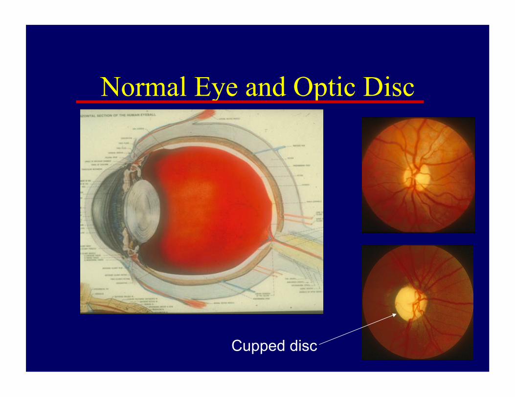
Normal Eye and Optic Disc
Cupped disc

The swollen optic disc
• Papilloedema
• Papillitis
• Malignant hypertension
• Ischaemic optic neuropathy
• Diabetic optic neuropathy
• CRVO
• Intraocular inflammation
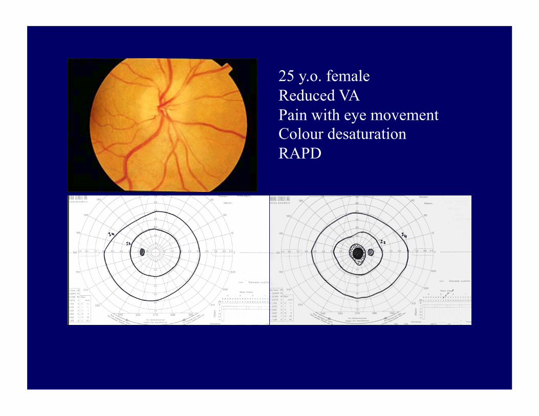
25 y.o. female Reduced VA Pain with eye movement Colour desaturation RAPD
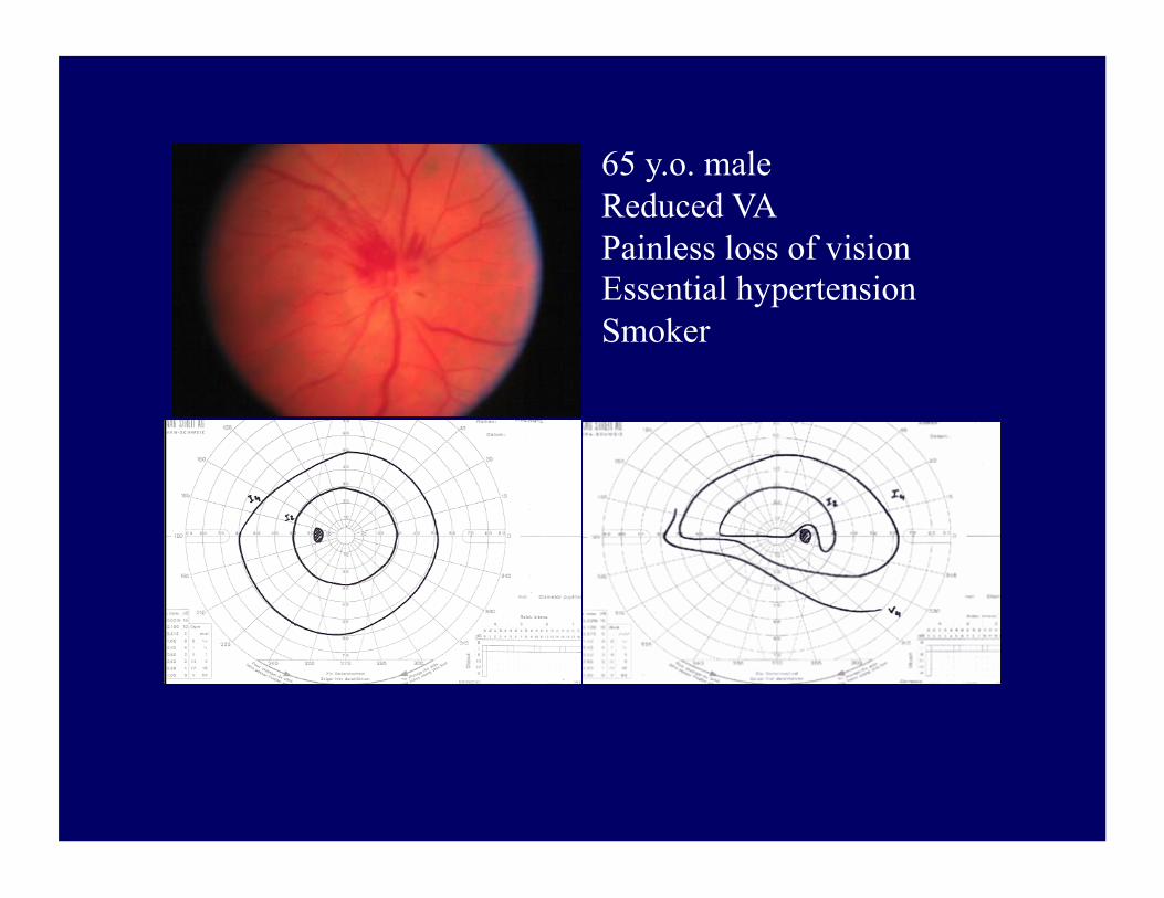
65 y.o. male Reduced VA Painless loss of vision Essential hypertension Smoker

The pale optic disc • Congenital
• Secondary to
• raised ICP
• vascular retinal disease
• optic neuritis
• optic nerve compression
• trauma
• Glaucoma
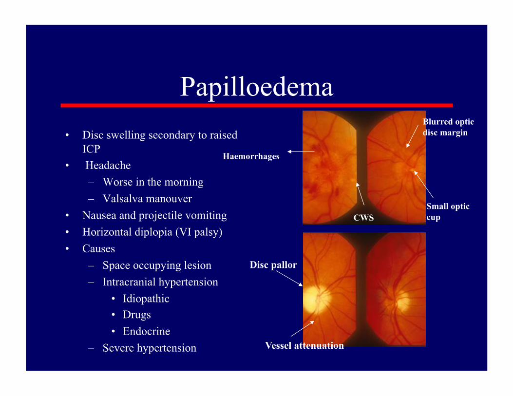
Papilloedema • Disc swelling secondary to raised
ICP • Headache
– Worse in the morning – Valsalva manouver
• Nausea and projectile vomiting • Horizontal diplopia (VI palsy) • Causes
– Space occupying lesion – Intracranial hypertension
• Idiopathic • Drugs • Endocrine
– Severe hypertension
Haemorrhages
CWS
Blurred optic disc margin
Small optic cup
Disc pallor
Vessel attenuation

Pupils
• First Order – Retina to Pretectal Nucleus in B/S (at level of Superior colliculus) • Second Order – Pretectal nucleus to E/W nucleus (bilateral innervation!) • Third Order – E/W nucleus to Ciliary Ganglion • Fourth Order – Ciliary Ganglion to Sphincter pupillae (via short ciliary nerves)

Pupil
• Constricted (mioisis) – Sympathetic
(pupillodilator) denervation
– Drugs • Pilocarpine • Morphine
• Dilated (mydriasis) – Parasympathetic
(pupilloconstrictor) denervation
– Lesion of the third CN – Drugs
• Atropine • Cocaine
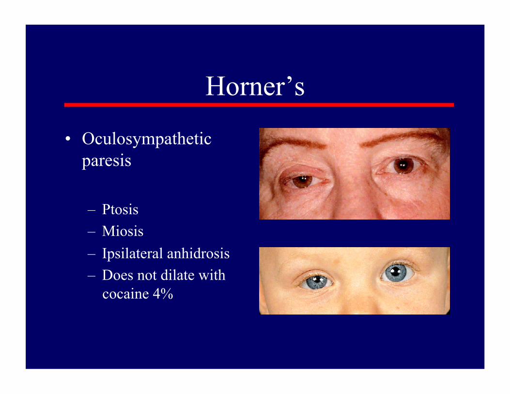
Horner’s
• Oculosympathetic paresis
– Ptosis – Miosis – Ipsilateral anhidrosis – Does not dilate with
cocaine 4%

Sympathetic Pathway • First Order – Posterior Hypothalamus to Ciliospinal centre of Budge (C8-T2) (Uncrossed in Brainstem) • Second Order – Ciliospinal centre of Budge to Superior Cervical Ganaglion • Third Order – Superior Cervical Ganglion to dilator pupillae muscle. (Close to ICA and joins V1 intracranially)
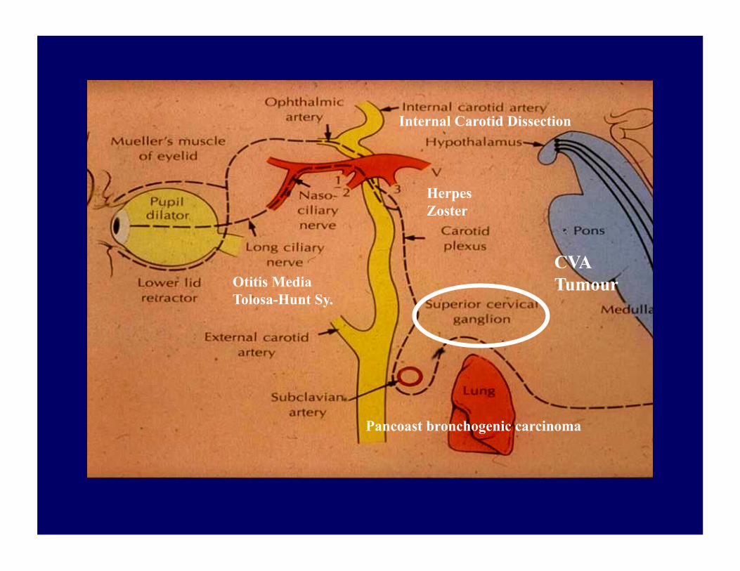
Pancoast bronchogenic carcinoma
Otitis Media Tolosa-Hunt Sy.
CVA Tumour
Internal Carotid Dissection
Herpes Zoster

Causes of Horner’s pupil • Central – B/S lesions (tumours, vascular and MS) Syringomyelia, Lat. Med. Syn., S.C. ca. • Preganglionic – Pancoast tumour, Carotid & Aortic aneurysms, Neck lesions/trauma. • Postganglionic – Cluster headaches, Nasopharyngeal tumours, Otitis media, Cavernous sinus mass and ICA disease. • Miscellaneous – Congenital (brachial plexus injury) Idiopathic.
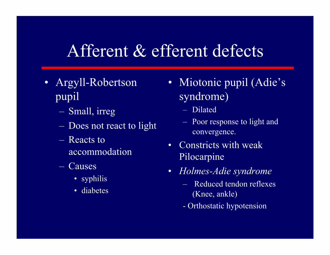
• Argyll-Robertson pupil – Small, irreg – Does not react to light – Reacts to
accommodation – Causes
• syphilis • diabetes
• Miotonic pupil (Adie’s syndrome) – Dilated – Poor response to light and
convergence.
• Constricts with weak Pilocarpine
• Holmes-Adie syndrome – Reduced tendon reflexes
(Knee, ankle) - Orthostatic hypotension
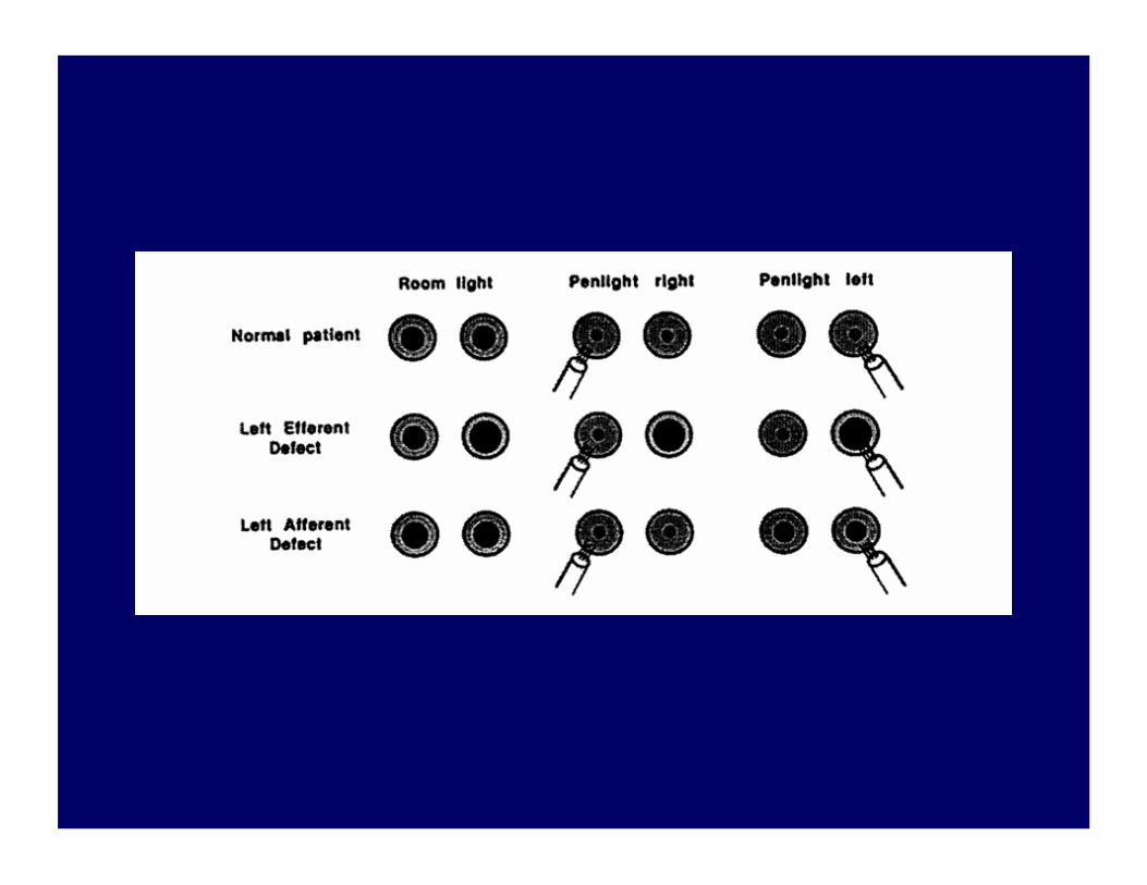
Afferent & efferent defects

Ocular motility abnormalities
• Third nerve palsy – Double vision – Eye turned down & out – Ptosis – Dilated pupil &
headache • Compressive lesion
• Sixth nerve palsy – Double vision – Eye turned in

Cranial Nerve Palsies Looking straight ahead

Posterior communicating artery aneurysm
III CN
Posterior cerebral artery
Chiasma
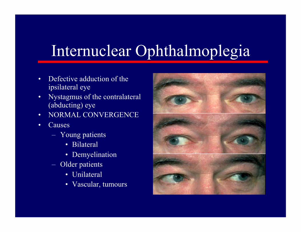
Internuclear Ophthalmoplegia • Defective adduction of the
ipsilateral eye • Nystagmus of the contralateral
(abducting) eye • NORMAL CONVERGENCE • Causes
– Young patients • Bilateral • Demyelination
– Older patients • Unilateral • Vascular, tumours

Myasthenia Gravis
• Fatigability • Double vision • Lid twitch • Ptosis • Normal reflexes &
sensation
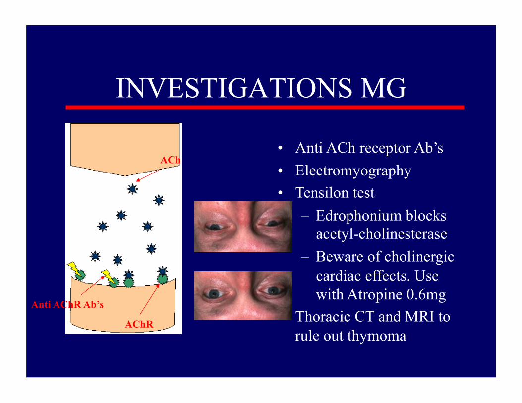
INVESTIGATIONS MG
• Anti ACh receptor Ab’s • Electromyography • Tensilon test
– Edrophonium blocks acetyl-cholinesterase
– Beware of cholinergic cardiac effects. Use with Atropine 0.6mg
• Thoracic CT and MRI to rule out thymoma
Anti AChR Ab’s AChR
ACh
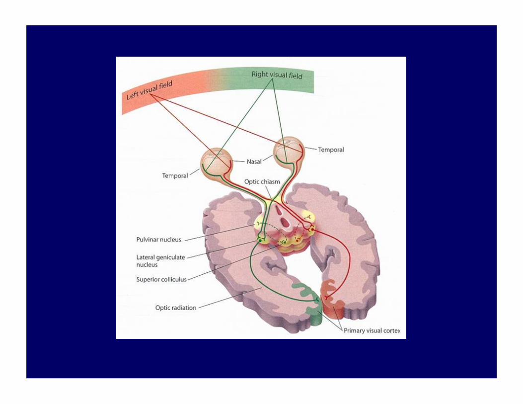
Localising the lesion
• Monocular visual field defects indicate lesions anterior to the optic chiasm
• Bitemporal defects are the hallmark of chiasmal lesions
• Binocular homonymous hemianopia result from lesions in the contralateral postchiasmal region
• Binocular quadrantanopias reflect optic tract lesions
