neurobiology and treatment of traumatic...
TRANSCRIPT

Editors
Ulrich F. Lanius
Sandra L. Paulsen
Frank M. Corrigan
Neurobiology and TreaTmeNT
of TraumaTicDissociaTioN
Toward an EmbodiEd SElf
Toward an EmbodiEd SElf
Lanius
Paulsen
Co
rrigan
Ne
ur
ob
iolo
gy a
nd Tr
ea
Tme
NT
of Tr
au
ma
Tic D
issoc
iaTio
N
This is the first book to synthesize emerging research in the neurobiology of attachment, trauma, and affect with new developments in effective clinical interventions. encompassing the contribu-
tions of leading researchers and practitioners, it offers neuroscientifically based understanding and practical guidance for clinicians working to heal people affected by traumatic dissociation.
The text discusses current neuroscientific research on dissociation and other types of traumat-ic stress including attachment, affective neuroscience, polyvagal theory, structural dissociation, and information processing theory. This includes the translation of important work with laborato-ry animals into clinical applications and the dissemination of research about all levels of the brain from the brainstem to the neocortex. The text integrates concepts from the affective and cogni-tive neurosciences and the study of consciousness. it describes a comprehensive model that guides treatment of traumatic sequelae, and integrates this model with stage-oriented treatment and such therapeutic interventions as emDr, somatic and body psychotherapy approaches, ego state thera-py, and adjunctive pharmacological interventions. readers are given hands-on practical guidance regarding clinical decision making, enabling them to make sound choices about interventions that will facilitate optimal treatment outcomes.
Key Features:◆ offers a truly comprehensive treatment approach to traumatic stress syndromes and dissociation
◆ Provides accessible, leading-edge research in neuroscience relevant to our understanding of attachment, traumatic stress, and dissociation
◆ contains hands-on suggestions about how to integrate emDr and somatic and body psychother-apy approaches with ego state therapy, and adjunctive pharmacological interventions
◆ The only text to encompass all levels of the brain beginning at the brainstem
Ulrich F. Lanius, PhD, Sandra L. Paulsen, PhD, Frank M. Corrigan, MD, Editors
Neurobiology and TreaTmeNT of TraumaTic DissociaTioN
“Through a skilled blending of emerging neuroscientific knowledge about emotion-al processes of mammalian brains and emerging clinical insights, each of the 22 chapters of this exceptional book will repeatedly provoke ‘aha’ moments of discovery. . . . The future of the field lies in credible neuropsychological syntheses, as superbly represented in this book.”
– From the Foreword by Jaak Panskeep, PhD
11 W. 42nd StreetNew York, NY 10036-8002 www.springerpub.com 9 780826 106315
ISBN 978-0-8261-0631-5

Neurobiology and Treatment of Traumatic Dissociation
This is a sample from NEUROBIOLOGY AND TREATMENT OF TRAUMATIC DISSOCIATION: TOWARD AN EMBODIED SELF VISIT THIS BOOK’S WEB PAGE BUY NOW REQUEST AN EXAM OR REVIEW COPY
© Springer Publishing Company

Ulrich F. Lanius, PhD, is a registered psychologist in West Vancouver, British Columbia, Canada, with a practice in clinical and neuropsychology. He has a particular interest in brain–behavior relationships with regard to attachment, trauma, and dissociation. His training includes eye move-ment desensitization and reprocessing (EMDR; EMDR Institute facilitator and EMDRIA approved consultant), sensorimotor psychotherapy, as well as neurofeedback. He specializes in the treatment of trauma and attachment-related problems, working from a client-centered perspective, integrat-ing EMDR, body therapy, ego state interventions, and neurotherapy. He has presented in North America as well as internationally, and has authored and coauthored a variety of book chapters and articles on both the treatment and the neurobiology of dissociation.
Sandra L. Paulsen, PhD, is a clinical and consulting psychologist who has accompanied many peo-ple on their extraordinary healing journeys for over two decades. She is a senior practitioner and instructor of advanced EMDR procedures and ego state therapy. She received 2 years of training in somatic therapy. She has published and spoken widely on the use of ego state therapy and EMDR to resolve complex and dissociative conditions. She is an EMDR Institute facilitator and EMDR International Association certified practitioner, consultant, and advanced specialty workshop instructor. She is a fellow of the International Society for the Study of Trauma and Dissociation. She has trained hundreds of mental health professionals in these special procedures since 1993.
Frank M. Corrigan, MD, is a consultant psychiatrist in Argyll, Scotland. Formerly full time, he now works part time in trauma therapy in Argyll and privately in Glasgow and at Manor Hall, Doune. His encounters in general psychiatry with the effects on suicidality of early trauma led to train-ing in dialectical behavior therapy, clinical hypnosis, EMDR, sensorimotor psychotherapy, and, most recently, brainspotting. He has an interest in understanding the impact of trauma on clinical presentations through awareness of laboratory work on stress and trauma and how this can be integrated with neuroscientific enquiry in humans.
This is a sample from NEUROBIOLOGY AND TREATMENT OF TRAUMATIC DISSOCIATION: TOWARD AN EMBODIED SELF VISIT THIS BOOK’S WEB PAGE BUY NOW REQUEST AN EXAM OR REVIEW COPY
© Springer Publishing Company

Neurobiology and Treatment of Traumatic Dissociation
Toward an Embodied Self
Ulrich F. Lanius, PhD, Sandra L. Paulsen, PhD, and Frank M. Corrigan, MD
Editors
This is a sample from NEUROBIOLOGY AND TREATMENT OF TRAUMATIC DISSOCIATION: TOWARD AN EMBODIED SELF VISIT THIS BOOK’S WEB PAGE BUY NOW REQUEST AN EXAM OR REVIEW COPY
© Springer Publishing Company

Copyright © 2014 Springer Publishing Company, LLC
All rights reserved.No part of this publication may be reproduced, stored in a retrieval system, or transmitted in any form or by any means, electronic, mechanical, photocopying, recording, or otherwise, without the prior permission of Springer Publishing Company, LLC, or authorization through payment of the appropriate fees to the Copyright Clearance Center, Inc., 222 Rosewood Drive, Danvers, MA 01923, 978–750-8400, fax 978–646-8600, [email protected] or on the Web at www.copyright.com.
Springer Publishing Company, LLC11 West 42nd StreetNew York, NY10036www.springerpub.com
Acquisitions Editor: Sheri W. SussmanProduction Editor: Shelby PeakComposition: Newgen Imaging
ISBN: 978-0-8261-0631-5e-book ISBN: 978-0-8261-0632-2
14 15 16 17 / 5 4 3 2 1
The author and the publisher of this work have made every effort to use sources believed to be reliable to provide information that is accurate and compatible with the standards generally accepted at the time of publication. The author and publisher shall not be liable for any special, consequential, or exemplary damages resulting, in whole or in part, from the readers’ use of, or reliance on, the information contained in this book. The publisher has no responsibility for the persistence or accuracy of URLs for external or third-party Internet websites referred to in this publication and does not guarantee that any content on such websites is, or will remain, accurate or appropriate.
Library of Congress Cataloging-in-Publication Data
Neurobiology and treatment of traumatic dissociation : towards an embodied self / [edited by] Ulrich F. Lanius, Sandra L. Paulsen and Frank M. Corrigan. p. ; cm.Includes bibliographical references.ISBN 978-0-8261-0631-5—ISBN 978-0-8261-0632-2 (e-ISBN)I. Lanius, Ulrich F., editor. II. Paulsen, Sandra, editor. III. Corrigan, Frank M., editor.[DNLM: 1. Dissociative Disorders—therapy. 2. Brain—physiopathology. 3. Stress Disorders, Traumatic— therapy. WM 173.6]RC553.D5616.85’23–dc23
2014006322
Special discounts on bulk quantities of our books are available to corporations, professional associations, pharmaceutical companies, health care organizations, and other qualifying groups. If you are interested in a custom book, including chapters from more than one of our titles, we can provide that service as well.
For details, please contact:Special Sales Department, Springer Publishing Company, LLC11 West 42nd Street, 15th Floor, New York, NY 10036–8002Phone: 877–687-7476 or 212–431-4370; Fax: 212–941-7842E-mail: [email protected]
Printed in the United States of America by McNaughton & Gunn.
This is a sample from NEUROBIOLOGY AND TREATMENT OF TRAUMATIC DISSOCIATION: TOWARD AN EMBODIED SELF VISIT THIS BOOK’S WEB PAGE BUY NOW REQUEST AN EXAM OR REVIEW COPY
© Springer Publishing Company

Contributors ixForeword by Jaak Panskepp, PhD xiPreface xvIntroduction: The Ubiquity of Dissociation by Sandra L. Paulsen and Ulrich F. Lanius xix
PART I. NEUROBIOLOGY
Introduction: Dissociation and Neurobiology 1Ulrich F. Lanius
1. Dissociation: Cortical Deafferentation and the Loss of Self 5Ulrich F. Lanius, Sandra L. Paulsen, and Frank M. Corrigan
2. Threat and Safety: The Neurobiology of Active and Passive Defense Responses 29Frank M. Corrigan
3. Peritraumatic Dissociation and Tonic Immobility: Clinical Findings 51Michelle J. Bovin, Elise Ratchford, and Brian P. Marx
4. A Social–Cognitive–Neuroscience Approach to PTSD: Clinical and Research Perspectives 69Ruth Lanius, Paul Frewen, Anthony Nazarov, and Margaret C. McKinnon
5. Dissociation and Endogenous Opioids: A Foundational Role 81Ulrich F. Lanius
6. Attachment, Neuropeptides, and Autonomic Regulation: A Vagal Shift Hypothesis 105Ulrich F. Lanius
Contents
This is a sample from NEUROBIOLOGY AND TREATMENT OF TRAUMATIC DISSOCIATION: TOWARD AN EMBODIED SELF VISIT THIS BOOK’S WEB PAGE BUY NOW REQUEST AN EXAM OR REVIEW COPY
© Springer Publishing Company
Share Neurobiology and Treatment of Traumatic Dissociation: Toward an Embodied Self

7. Defense Responses: Frozen, Suppressed, Truncated, Obstructed, and Malfunctioning 131
Frank M. Corrigan
8. The Clinical Sequelae of Dysfunctional Defense Responses: Dissociative Amnesia, Pain and Somatization, Emotional Motor Memory, and Interoceptive Loops 153
Frank M. Corrigan
9. Shame and the Vestigial Midbrain Urge to Withdraw 173 Frank M. Corrigan
10. Attachment and Attachment Repair 193 Frank M. Corrigan, Alistair Wilson, and Deirdre Fay
11. Dissociation, EMDR, and Adaptive Information Processing: The Role of Sensory Stimulation and Sensory Awareness 213
Ulrich F. Lanius and Uri Bergmann
PART II. TREATMENT
Introduction: Dissociation and Psychotherapy 243Sandra L. Paulsen and Ulrich F. Lanius
12. Seeing That Which Is Hidden: Identifying and Working With Dissociative Symptoms 247
Sandra L. Paulsen and Ulrich F. Lanius
13. The Compassionate Self 269 Frank M. Corrigan, Alistair Wilson, and Deirdre Fay
14. Stabilization Basics 289 Sandra L. Paulsen and Joan Golston
15. Stabilizing the Relationship Among Self-States 321 Sandra L. Paulsen and Joan Golston
16. Alexithymia, Affective Dysregulation, and the Imaginal: Resetting the Subcortical Affective Circuits 341
Sandra L. Paulsen, Katie O’Shea, and Ulrich F. Lanius
17. Fractionating Trauma Processing: TOTEMSPOTS and Other Attenuating Tactics 367
Sandra L. Paulsen and Ulrich F. Lanius
This is a sample from NEUROBIOLOGY AND TREATMENT OF TRAUMATIC DISSOCIATION: TOWARD AN EMBODIED SELF VISIT THIS BOOK’S WEB PAGE BUY NOW REQUEST AN EXAM OR REVIEW COPY
© Springer Publishing Company

18. Accelerating and Decelerating Access to Self-States 383 Sandra L. Paulsen
19. Integrating Body and Mind: Sensorimotor Psychotherapy and Treatment of Dissociation, Defense, and Dysregulation 399
Pat Ogden and Janina Fisher
20. Temporal Integration of Early Trauma and Neglect 423 Sandra L. Paulsen
21. Toward an Embodied Self: EMDR and Somatic Interventions 447 Ulrich F. Lanius and Sandra L. Paulsen
22. Opioid Antagonists and Dissociation: Adjunctive Pharmacological Interventions 471
Ulrich F. Lanius and Frank M. Corrigan
Index 499
This is a sample from NEUROBIOLOGY AND TREATMENT OF TRAUMATIC DISSOCIATION: TOWARD AN EMBODIED SELF VISIT THIS BOOK’S WEB PAGE BUY NOW REQUEST AN EXAM OR REVIEW COPY
© Springer Publishing Company

This is a sample from NEUROBIOLOGY AND TREATMENT OF TRAUMATIC DISSOCIATION: TOWARD AN EMBODIED SELF VISIT THIS BOOK’S WEB PAGE BUY NOW REQUEST AN EXAM OR REVIEW COPY
© Springer Publishing Company
Uri Bergmann, PhD—Private Practice, Commack and Bellmore, New York
Michelle J. Bovin, PhD—National Center for PTSD, VA Boston Healthcare System; Boston University School of Medicine, Boston, Massachusetts
Frank M. Corrigan, MD—Consultant Psychiatrist, Argyll, Scotland
Deirdre Fay, MSW, LICSW—Center for Integrative Healing, Boston, Massachusetts
Janina Fisher, PhD—Trauma Center, Boston, Massachusetts; Sensorimotor Psychotherapy Institute, Boulder, Colorado
Paul Frewen, PhD—Departments of Psychiatry, and Psychology, Western University of Canada, London, Ontario, Canada
Joan Golston, DCSW, LCSW—Private Practice, Seattle, Washington; ISSTD Board of Directors, McLean, Virginia
Ruth Lanius MD, PhD—Department of Psychiatry, Neuroscience, Western University of Canada, London, Ontario, Canada
Ulrich F. Lanius, PhD—Private Practice, West Vancouver, British Columbia, Canada
Brian P. Marx, PhD—National Center for PTSD, VA Boston Healthcare System; Boston University School of Medicine, Boston, Massachusetts
Margaret C. McKinnon, PhD—Department of Psychiatry and Behavioral Neurosciences, McMaster University, Hamilton, Ontario, Canada
Anthony Nazarov, PhD Cand.—Department of Psychiatry and Behavioral Neurosciences, McMaster University, Hamilton, Ontario, Canada
Pat Ogden, PhD—Founder and Director, Sensorimotor Psychotherapy Institute, Boulder, Colorado
Katie O’Shea, MA—Private Practice, Nampa, Idaho; EMDR-HAP, Hamden, Connecticut
Sandra L. Paulsen, PhD—Private Practice, Bainbridge Island, Washington
Elise Ratchford, BA—National Center for PTSD, VA Boston Healthcare System, Boston, Massachusetts
Alistair Wilson, MD—Consultant Psychiatrist, Glasgow, Scotland
Contributors
This is a sample from NEUROBIOLOGY AND TREATMENT OF TRAUMATIC DISSOCIATION: TOWARD AN EMBODIED SELF VISIT THIS BOOK’S WEB PAGE BUY NOW REQUEST AN EXAM OR REVIEW COPY
© Springer Publishing Company

This is a sample from NEUROBIOLOGY AND TREATMENT OF TRAUMATIC DISSOCIATION: TOWARD AN EMBODIED SELF VISIT THIS BOOK’S WEB PAGE BUY NOW REQUEST AN EXAM OR REVIEW COPY
© Springer Publishing Company

The diverse contributions to this book share insightful, indeed profound, apprecia-tions of neuropsychological dynamics of stressed minds and how they can be helped to emerge from the overwhelming effects of excessive negative affects. This excep-tional volume integrates diverse neurobiological perspectives on understanding how minds succumb to trauma, in order to provide skilled guidance on how to coordinate our modern understandings of the diverse resulting brain–mind changes with novel therapeutic strategies to optimally manage the affective sequelae of excessive stress. An integrated understanding of the affective networks of human and animal brains and their imbalances is writ large in this interprofessional compendium. This book offers 22 chapters of productive integration between neuroscientific analyses (the focus of the first 11 chapters) and clinical insights (the next 11).
In successive chapters we move from clear discussions of the emotional pro-cesses of the brain—knowledge arising from modern brain imaging as well as cross-species analyses of the neuroanatomies and neurochemistries of affective brain circuits and their responses to stress—toward exceptionally insightful ways to deal with the varieties of posttraumatic stress disorders. Within this synthesis of modern neuroscientific and psychotherapeutic knowledge, we find insights about the pat-terns of disorganization within overstressed brains and new evidence-based thera-peutics that may help troubled minds to heal—to emerge toward neuropsychological wholeness—more effectively than ever before.
It has become clearer during the past few decades that for optimal understand-ing of human psychiatric issues we must concurrently understand the neurobiolo-gies of mammalian brains and the accompanying affective dynamics of both human and animal minds, and how both need to be envisioned from modern evolutionary perspectives. Rarely have these lines of thought been blended as skillfully as here. To
Foreword:
Toward a Neuroaffective Understanding of Troubled Minds
This is a sample from NEUROBIOLOGY AND TREATMENT OF TRAUMATIC DISSOCIATION: TOWARD AN EMBODIED SELF VISIT THIS BOOK’S WEB PAGE BUY NOW REQUEST AN EXAM OR REVIEW COPY
© Springer Publishing Company
do this well, there is no reasonable alternative but to devote equal consideration to the ancestral passions of our animal past and our uniquely human capacity to think and peer into the future. Our raw emotional feelings, which arise from ancient brain regions we share with other animals, energize and direct higher cognitive processes, which begin to flow in narrow, ruminative ways in overstressed minds.
Throughout this book, one finds well-measured interprofessional arguments that concurrently avoid excessively ruthless neural reductionism as well as neurobi-ologically unbridled mentalizing. Rather, neural and psychological perspectives are blended to yield a rich tapestry of understanding, pregnant with therapeutic implica-tions. By effectively using evidence from our brethren (animals), the discussions in this book avoid strict behavioristic perspectives, which have commonly ruled pre-clinical theorizing in the past, and move toward more nuanced visions of how our understanding of human and animal minds, brains, and behaviors can be harmoni-ously integrated.
Contributions to this book offer rich and nuanced understandings of basic social emotions and attachment processes, allowing us to understand how higher order emotions such as guilt and shame arise from more fundamental substrates and how various developmental landscapes predispose minds and brains to become pro-foundly scarred. However, it is becoming increasingly clear that such wounds can be healed effectively if we apply our increasing understanding of the emotional pro-cesses of the brain with revolutionary therapeutic maneuvers, such as eye movement desensitization and reprocessing (EMDR) procedures, which have now been repeat-edly found to be exceptionally effective in countering the devastating psychological effects of stress. Through a skilled blending of emerging neuroscientific knowledge about emotional processes of mammalian brains and emerging clinical insights, each of the 22 chapters of this exceptional book will repeatedly provoke “aha” moments of discovery.
As this book blends brain and mind issues seamlessly, it illuminates the affec-tive dynamics of human minds with clarity and wisdom. In passing, it is worth not-ing that throughout the history of modern biological psychiatry, there has yet to be a new psychiatric medicine that has been developed through an understanding of brain emotional processes rather than by mere serendipity. What is the cause of this failure? Surely, it is partly due to the lack of adequate tools and scientific per-spectives. Among the latter, one of the most detrimental may have been the vast investment in behavioristic strategies that neglected the emotional mind of animals. As this book exemplifies, that is finally changing as clinical investigators are recog-nizing the enormous stretches of brain–mind evolution that we share with the other animals, especially in affective realms. Such perspectives have been placed front and center in this book, and they are effectively blended with our understanding of human emotional processes permitted by modern brain imaging. It is through such syntheses that a new era of understanding of psychiatric disorders, as well as the nature of human and animal minds, will emerge, which should fertilize more rapid progress than any other strategy so far considered. For instance, we can already envi-sion how future psychotherapy facilitators may be constructed, such as d-cycloserine at the present time, which may further facilitate the emotional learning that must transpire during psychotherapies. We can envision neuroscientific insights that will
This is a sample from NEUROBIOLOGY AND TREATMENT OF TRAUMATIC DISSOCIATION: TOWARD AN EMBODIED SELF VISIT THIS BOOK’S WEB PAGE BUY NOW REQUEST AN EXAM OR REVIEW COPY
© Springer Publishing Company

reveal how EMDR and other reconsolidation therapies help restructure imbalanced emotional circuits, potentially providing not only an understanding of underlying mechanisms of action but also additional clinical insights as we reveal how each therapeutic modality works.
In this context, I would share a personal experience with EMDR, facilitated by one of the editors of this book (SP). In 2009, my wife Anesa and I were concurrently diagnosed with lymphomas, with her prognosis, fortunately, less life threatening than mine. After many chemos, I was advised that the only treatment left that might save my life was tandem stem cell transplants—autologous followed by allogeneic. We proceeded to get treatment from the “Best in the West” at the “Fred Hutch” (more formally known as the Seattle Cancer Care Alliance). The first of my transplants was estimated to require 2 months of medical leave, but as successive life-threatening iat-rogenic side effects emerged, the process stretched through 4 months. Suffice to say, abundant negative affect weighed on my mind.
During this period, Sandra Paulsen kindly offered a sampler of EMDR therapy to see whether it might counteract and diminish/soften my negative feelings. She skillfully guided me through a sampler of EMDR procedures. As she systematically retrieved emotional feelings related to various medical problems/mistakes I had encountered, she applied her standard EMDR procedure. I easily retrieved emo-tional feelings under her clinical guidance, and I was amazed how rapidly those feel-ings dissolved once I started conjugate eye movement, following a smoothly flowing row of lights moving to the right and left on her EMDR apparatus. My negative feelings, whether anger, anxiety, or sadness, dissolved promptly once I initiated eye movements. The effect was repeatable in distinctly robust ways: Each time I evoked negative feelings from abundant autobiographical memories, the negative feelings faded promptly as soon as I initiated eye movements. We did this repeatedly with different primal affective states—rage, fear, and grief. The results were consistently clear. Repeatedly, with the onset of bilateral eye movements, the intensity of affect faded promptly. Each time! Surely, this should be an optimal way to reconsolidate affectively negative memories with less aversive hues.
This is eminently testable and needs to be experimentally evaluated across a substantial set of clients. I assume it will replicate, and may be one of, perhaps “the” fundamental source of emotional relief obtained with EMDR, which may allow for the reconsolidation of troublesome memories with diminished negative-affective intensity.
If this can be demonstrated, how might this occur neurodynamically? I would first note that exploratory eye movements represent a basic primate SEEKING response. Such scanning movements are organized in deep midbrain layers of the superior colliculi, just above the periaqueductal gray (PAG), which may be the most important brain region for engendering raw emotional affects since circuits for the most negative primal emotions—FEAR, RAGE, and PANIC/GRIEF—are concen-trated in the dorsal PAG, very close to the exploratory eye-orientation maps at the neuronal seam between the superior colliculi and the PAG.
There are neural connections between those eye-orienting response networks and the negative emotional primes, perhaps inhibitory gamma-aminobutyric acid (GABA) connections. This could provide a reasonable explanation for therapeutic
This is a sample from NEUROBIOLOGY AND TREATMENT OF TRAUMATIC DISSOCIATION: TOWARD AN EMBODIED SELF VISIT THIS BOOK’S WEB PAGE BUY NOW REQUEST AN EXAM OR REVIEW COPY
© Springer Publishing Company
effects: Namely, as one dwells on negative affects, arousing the dorsal PAG, the rapid shift of attention toward external events may engender direct inhibition of primal dorsal PAG emotional circuits for various negative affects. The retrieval of negative emotional memories into active neuronal reprocessing can allow “memory reconsolidation” processes to promote lasting dampening of the emotional intensity of the retrieved memories. By having systematically retrieved troublesome memo-ries, and rapidly establishing a euthymic affective state, the associated cognitive memories should be restored (i.e., reconsolidated) with a greatly diminished affec-tive penumbra. Of course, this is currently just a working hypothesis, but an emi-nently testable one. This neuromental hypothesis may highlight how a little bit of idiographic “me-search” may help promote illuminating nomothetic research on the efficacy of EMDR, a treatment modality that is so far begging for a cogent neu-roscientific explanation.
In this context, it is worth noting a general principle of reciprocal controls between inwardly directly basic affective-emotional (subcortical) and externally directed higher cognitive (neocortical) processes. When strongly aroused, these two global brain functions often work in reciprocal inhibitory ways. When people are emotionally aroused, there is disruption and narrowing of cognitive activities (rumination on one’s woes); conversely, when one is intently focusing on externally directed cognitive tasks, there is typically a global inhibition of affective cortical and subcortical processes (for an overview, see the Liotti and Panksepp chapter in the Textbook of Biological Psychiatry I edited for Wiley in 2004 [pp. 33–74]): In other words, just as emotional arousals can disrupt cognitive processing, intense engagements with cognitive issues can inhibit affective arousals. But this is just a working hypoth-esis, not a definitive conclusion.
This book is written in this same spirit. The diverse contributions to this vol-ume provide up-to-date reviews of how emotional stressors operate within the brain and how stressed minds may be helped to emerge from the psychoneural ravages of excessively sustained negative affective arousals. This book helps to counter the all-too-common trend during the past century of separating neuroscientific and psy-chological issues in clinical discourse. The future of the field lies in credible neuro-psychological syntheses, as superbly represented in this book.
Jaak PankseppProfessor and Baily Endowed Chair
of Animal Well-Being ScienceDepartment of Integrative Physiology
and NeuroscienceWashington State University
This is a sample from NEUROBIOLOGY AND TREATMENT OF TRAUMATIC DISSOCIATION: TOWARD AN EMBODIED SELF VISIT THIS BOOK’S WEB PAGE BUY NOW REQUEST AN EXAM OR REVIEW COPY
© Springer Publishing Company

Preface
Most psychologists treat the mind as disembodied, a phenomenon with little or no connection to the physical body. Conversely, physicians treat the body with no regard to the mind or the emotions. But the body and the mind are not separate, and we cannot treat one without the other.
—Candace Pert, in Molecules of Emotion (1999)
This book was conceived out of our shared vision that pivotal emerging develop-ments from the neurobiology of attachment, trauma, and affect can inform the clini-cal practice and, in particular, the use of certain contemporary clinical methods. That vision was to synthesize key neurobiological developments with effective develop-ments in clinical practice to offer both understanding and practical guidance for the many practitioners working to heal people burdened with traumatic sequelae.
We feel we have achieved that vision to a large degree; though the fact that there must be two parts to the book, neurobiology and treatment, reflects the status of the fields of inquiry—divided by science and practice. We have attempted to inte-grate them herein by weaving together the view from the warp and weft of those two outlooks, punctuating each part of the book with relevant understandings from the other.
We are pleased to have the chance to translate some of the important animal work from the laboratory studies to clinical applications. For example, the research on defense responses informs our understanding of human reactions under stress. The research on emotions informs all of human experience, including that encountered at times of traumatic stress. Also, much of the literature stops at the corticolimbic level and ignores the brainstem where the emotions, orienting, and defense responses are generated primarily—even if they are elaborated through thalamocortical projec-tions. This volume is unique in bringing in all levels of the brain from the brainstem, through the thalamus and basal ganglia, to the limbic structures, including the older
This is a sample from NEUROBIOLOGY AND TREATMENT OF TRAUMATIC DISSOCIATION: TOWARD AN EMBODIED SELF VISIT THIS BOOK’S WEB PAGE BUY NOW REQUEST AN EXAM OR REVIEW COPY
© Springer Publishing Company

forms of cortex, to the neocortex. We want to look at the neurochemistry of peri-traumatic dissociation and explore the effects on neuroplasticity and the eventual structural dissociation. Further, we want to help clinicians lay a sound foundation to support contemporary treatment methods with neuroscience.
We make an earnest plea about research conceptualization. Because of the dif-ficulty doing randomized controlled trials for polysymptomatic, posttraumatic pre-sentations such as major disorders of structural dissociation, it is essential that the concept of evidence be expanded to include that which is neuroscientifically feasible. If evidence for new treatments is based only on trials of manualized therapies in rig-idly defined groups, application to the dissociative disorders will remain forever out of reach, or the research has to exclude those very individuals we most want to help, with their significant clinical problems such as chronic suicidality, fragility, and ten-dency to decompensate and disorganize. But of all people, those most fragile patients need psychotherapy that is informed by not only clinical tradition but also enriched by the most promising leading-edge treatments, explicable at a neuroscientific level as research advances. Moreover, of all people, their treatment must be provided, not by students with manuals and clipboards, but by practitioners rich with a range of knowledge and tools, and driven by commitment, compassion, flexibility, and cre-ativity. Otherwise, they will be offered the same standardized treatment packages from which most will drop out or be excluded, their humanity again unseen, their complex nonverbal story again not heard as it reveals itself in a variety of symptoms. Those patients who were most severely traumatized at the most vulnerable devel-opmental stages are being excluded by research and treatment systems that cannot cope with the complexity of the presentations and cannot adapt to the need for long-term, highly specialized input. It is our hope that the findings and revelations of this book will help to explain why narrowly defined manualized treatment protocols will inevitably fail. Multimodal, creative, compassionate psychotherapy rooted in neuro-science and in healing traditions is required.
A word is in order about traditional versus contemporary treatments. While we surely appreciate and honor the traditions of our field, many of the recent pub-lications in complex trauma and dissociation do not incorporate some of the revela-tions of the neurobiology of trauma and affect, or they rely too heavily on traditional treatments. While all three of us are steeped in traditional psychological interven-tions (the first two authors are psychologists and the third is a psychiatrist), we have been privileged to also acquire a range of leading-edge methods in the treatment of trauma. It was important to us to include these newer ways of working with the clini-cal sequelae of trauma with an understanding of the multiple levels of the brain, the short-term neurochemical effects, and the long-term neuroplastic circuitry effects.
All of the treatment methods we discuss herein assume a theoretical basis of information processing that is blocked as a defensive and survival strategy. The treat-ments must both deal with the defensive blocking and enable information process-ing, which includes affect, soma, cognitive channels, and more, to resume processing for healing to occur. So, we incorporate herein not only psychoanalytic, cognitive behavioral, and hypnotic methods, but also specific ego state, somatic/sensorimo-tor methods, eye movement desensitization and reprocessing (EMDR), and varia-tions of EMDR suitable for working with trauma in the attachment period, the latter methods being explicitly information-processing methods that address affective and
This is a sample from NEUROBIOLOGY AND TREATMENT OF TRAUMATIC DISSOCIATION: TOWARD AN EMBODIED SELF VISIT THIS BOOK’S WEB PAGE BUY NOW REQUEST AN EXAM OR REVIEW COPY
© Springer Publishing Company

somatic modes of processing. Ego state therapy explicitly addresses defensive and dissociative structural blockages to information processing, adopted early in life. Our approach emphasizes decreasing internal conflict between the patient’s dissoci-ated viewpoint and the viewpoint of the aggressor adopted via introjection, which assumes structural proportions in dissociative disorders and complex trauma. Ego state therapy is both old and newly important, with its inclusion in conjunction with EMDR and attachment trauma processing.
It is impossible to be comprehensive in all the detail needed for full explication and treatment of dissociative disorders, but the structure of the approach—chemical, anatomical, and clinical—is novel and important. Even if a specific interpretation is disputed, there is in this volume a coherent framework in which research is under-stood. That framework can continue to provide a way of viewing the trauma disor-ders and can inform further discussion and research at the biological level as well as at the clinical.
All three of us have leadership roles in training for the treatment of trauma using these leading-edge methods, and are excited to share our understandings and experience with the many people who have eagerly attended our trainings and con-sultation groups. We are grateful to the patients whose courage to free themselves from their heavy burden of suffering has required them to walk a long and arduous road, and we are privileged to have walked that road with them. We know how hard it is for them to trust anyone, and their trust in us requires an enormous act of faith on their part. We also are grateful to our students and consultees, whose questions have forced us to formulate our hypotheses and articulate explanations that might have otherwise remained gray. We are heartened by the gratitude and appreciation of so many. We are humbled and grateful for what we have learned from countless colleagues and contributors, and no list would be long enough to pay homage to those who have gone before. The ancient tradition of honoring our scholarly and clinical ancestors informs our gratitude, each in our own way. We par-ticularly want to thank some of the people at the forefront of their respective fields who have inspired us: Candace Pert, Jaak Panksepp, Onno van der Hart, Bessel van der Kolk, Francine Shapiro, Alan Schore, John G. and Helen Watkins, Richard Kluft, Catherine Fine, and the countless others whose names are rarely far from our lips. We also thank the enduring support and patience of our friends and family who supported us during the creation of this work. Frank Corrigan is also specifically grateful to Pete Redgrave, Peter Carr, Rajiv Raju, David Finn, and Andrew Harkin for discussion of early drafts.
REFERENCE
Pert, C. (1999). Molecules of emotion: The science between mind-body medicine. New York, NY: Scribner.
This is a sample from NEUROBIOLOGY AND TREATMENT OF TRAUMATIC DISSOCIATION: TOWARD AN EMBODIED SELF VISIT THIS BOOK’S WEB PAGE BUY NOW REQUEST AN EXAM OR REVIEW COPY
© Springer Publishing Company

This is a sample from NEUROBIOLOGY AND TREATMENT OF TRAUMATIC DISSOCIATION: TOWARD AN EMBODIED SELF VISIT THIS BOOK’S WEB PAGE BUY NOW REQUEST AN EXAM OR REVIEW COPY
© Springer Publishing Company

Introduction:
The Ubiquity of Dissociation
Sandra L. PauLSen and uLrich F. LaniuS
This book makes an attempt to embody both neuroscience and treatment in the field of dissociation, the dialectic of those two themes being reflected in Part I, Neurobiology, and Part II, Treatment. We try to provide our readers with a neurobiological frame-work that directly informs treatment and clinical practice. Let us briefly explore the neurobiology and treatment parts in turn.
PART I. TOWARD A NEUROBIOLOGICAL MODEL OF DISSOCIATION
What happens in the brain when an experience is overwhelming? Traumatic disso-ciation is a complex phenomenon that can be viewed from diverse clinical and sci-entific perspectives. This book attempts to integrate recent findings in cognitive and affective neuroscience, integrating neurobiological research to provide a comprehen-sive model of dissociation. Our understanding of these neurobiological processes has been strongly influenced by Jaak Panksepp’s (1998) work: Affective Neuroscience.
Clinicians and researchers report that people show different experiential, psycho physiological, and neurobiological responses to trauma. They hypothesize two subtypes of trauma response, one characterized predominantly by hyperarousal and the other primarily dissociative, each one representing unique pathways to chronic stress-related psychopathology (e.g., Lanius, Bluhm, Lanius, & Pain, 2005).
This is a sample from NEUROBIOLOGY AND TREATMENT OF TRAUMATIC DISSOCIATION: TOWARD AN EMBODIED SELF VISIT THIS BOOK’S WEB PAGE BUY NOW REQUEST AN EXAM OR REVIEW COPY
© Springer Publishing Company

Dissociation—Multiple Phenomena
Dissociation includes a wide variety of phenomena like depersonalization, derealiza-tion, amnesia, and identity disturbances. Further, dissociative symptoms can occur in a wide variety of disorders, such as dissociative disorders; posttraumatic stress disorder (PTSD), including the more complex posttraumatic presentations (e.g., complex PTSD; disorder of extreme stress not otherwise specified); somatoform dis-orders; personality disorders (e.g., borderline personality disorder); attachment dis-orders; and many others. Moreover, dissociative experiences are not only reported by a considerable proportion of psychiatric patients but also by “healthy” people.
The Autonomic Nervous System (ANS)—Sympathetic, Dorsal Vagal, and Ventral Vagal
The hyperarousal response in PTSD—also referred to as fight-or-flight response—has been studied in great detail. It is understood to be mediated by sympathetic nervous system arousal, which involves a release of catecholamines, especially adrenaline (epinephrine) and noradrenaline (norepinephrine). The release of these neurotrans-mitters is commonly associated with a narrowing of consciousness, that is, a specific focus on the threatening event associated with the mobilization of active defensive responses (also see Chapter 2, Threat and Safety: The Neurobiology of Defense Responses).
However, the nature of the ANS is such that an activation of the sympathetic nervous system is complemented by an activation of the parasympathetic nervous system, commonly dorsovagal activation. It is this parasympathetic activation that is the underpinning of the dissociative response.
This parasympathetic activation is a normal neurobiological mechanism that is evident not only in lower animals like reptiles but also in mammals and humans. It is commonly associated with a passive defensive response (also see Chapter 7, Defense Responses: Frozen, Suppressed, Truncated, Obstructed, and Malfunctioning; as well as Chapter 8, The Clinical Sequelae of Dysfunctional Defense Responses: Dissociative Amnesia, Pain and Somatization, Emotional Motor Memory, and Interoceptive Loops). These passive defensive responses are at least in part mediated by anesthetic and dissociative neurochemicals, namely, endogenous opioids and endocannabinoids that reduce the perception of physical pain as well as emotional pain. Moreover, the release of these anesthetic neurochemicals results in a lowering of consciousness and interferes with the integration of information, for example, information processing. The organism is concerned with survival and minimizing the use of energy, and reflects the core of the dissociative experience.
Ubiquity Is Hardwired—Everybody Can Dissociate
It has been documented that even what can be considered innocuous and minor stressors like giving students an unsolvable math task will result in significant anal-gesia (Bandura, Cioffi, Taylor, & Brouillard, 1988). That is, a dissociative response
This is a sample from NEUROBIOLOGY AND TREATMENT OF TRAUMATIC DISSOCIATION: TOWARD AN EMBODIED SELF VISIT THIS BOOK’S WEB PAGE BUY NOW REQUEST AN EXAM OR REVIEW COPY
© Springer Publishing Company

like anesthesia occurs in nonclinical populations that are exposed to what are normal everyday stressors. Now, this dissociative response, under normal circumstances, will not result in significant posttraumatic sequelae. For this to happen, there likely needs to be concurrent cholinergic activation associated with hyperarousal and a narrowing of attention as well reduced opioid receptor density that is attributable to prior attachment experience (also see Chapter 5). We suggest that dissociation reflects a normal underlying neurobiological mechanism that is present not only in humans. Rather, it is a basic hardwired neurobiological mechanism that occurs in mammals and humans.
Learned Helplessness (LH), Tonic Immobility (TI), and Anesthetic Neurochemicals
In humans, the alteration and lowering of consciousness due to anesthetic neuro-chemicals is commonly described as peritraumatic dissociation, a phenomenon that has been associated with the severity of PTSD. Relevant animal models in this regard are LH and TI. The relevance of these animal models to dissociation is dis-cussed in Part I of the book, specifically in Chapter 3, Peritraumatic Dissociation and Tonic Immobility: Clinical Findings and in Chapter 5, Dissociation and Endogenous Opioids: A Foundational Role.
In Chapter 1, Dissociation: Cortical Deafferentation and the Loss of Self, we describe how the narrowed focus with regard to sensory input in conjunction with a lowering of consciousness results in the decreased capacity to integrate this infor-mation across multiple levels in the brain, giving rise to what has been referred to as “structural dissociation” or fragmented self-states, as well as other dissociative symptoms that are commonly considered pathological; both positive, for example, hypermnesias, flashbacks, somatization; as well as negative, for example, amnesia, alexithymia, numbing, paresthesias, and so forth.
Severity of Peritraumatic Dissociation and Attachment
We suggest that increased severity of peritraumatic dissociation will give rise to increasingly pathological dissociative symptoms. The severity of the peritraumatic response in turn is likely attributable to alterations in brain functioning, both at the receptor and synaptic level that are influenced by a history of prior trauma as well as affected by an individual’s prior attachment history (see Chapter 10, Attachment and Attachment Repair). This conceptualization makes evolutionary sense, in that off-spring that is not initially protected by its mother or father is more likely to become killed, eaten, or victimized by a predator. Thus, an increased availability of a pas-sive defense response like immobility, freezing, diminished perception of pain, and peritraumatic dissociation potentially not only confers a survival advantage but also reduces suffering.
Further, in Chapter 6, Attachment, Neuropeptides, and Autonomic Regulation: A Vagal Shift Hypothesis, we suggest how some possible neuro-biological underpinnings with regard to ventral engagement are intrinsic to the
This is a sample from NEUROBIOLOGY AND TREATMENT OF TRAUMATIC DISSOCIATION: TOWARD AN EMBODIED SELF VISIT THIS BOOK’S WEB PAGE BUY NOW REQUEST AN EXAM OR REVIEW COPY
© Springer Publishing Company

attachment relationship on one hand, but also how we can potentially shift our cli-ents or patients from states of sympathetic hyperarousal or dorsovagal shutdown toward social engagement and a ventral vagal state. By drawing on the ventral vagal system, we can maximize affective regulation and information processing in trauma-focused interventions. Finally, in Chapter 9, Shame and the Vestigal Midbrain Urge to Withdraw, we discuss the specific role of the emotion of shame and its contribution to dissociation.
Integrative Capacity
Moreover, both peritraumatic dissociation as well as structural dissociation inter-fere with the integrative capacity of the brain as well as information processing. Chapter 11, Dissociation, EMDR, and Adaptive Information Processing: The Role of Sensory Stimulation and Sensory Awareness, discusses the role of sensory awareness and stimulation in the experience of an embodied self as it relates to our understand-ing of treatment interventions like EMDR, specifically, but also other somatically ori-ented interventions like sensorimotor psychotherapy.
PART II. TREATMENT: BEING EMBODIED AND SAFELY TELLING THE TRUTH
When dissociation is a significant part of the clinical presentation, it often presents a barrier to effective treatment. It tends to interfere with clients’ sense of their own body, their ability to experience emotion, and emotional regulation. Moreover, dis-sociation tends to result in unpredictable ego state shifts and the continuity of self.
In Part II, we describe a neurobiologically informed treatment approach that incorporates numerous elements from the armamentarium of contemporary psy-chotherapeutic procedures that address the key elements of dissociative disor-ders. This approach is intended to guide the client in a safe and paced manner to experience an embodied sense of self, one that is truthful to oneself, allowing an integrated self-system that operates with mutual cooperation and compas-sion. Chapter 12, Seeing That Which Is Hidden: Identifying and Working With Dissociative Symptoms, discusses some of the basic skills required for identifying and treating dissociative spectrum disorders. Chapter 13, The Compassionate Self, discusses the foundational role of safe embodiment, mindfulness, and reparative interventions in the treatment of dissociative symptoms as well as how that relates to attachment experience.
Ebb and Flow, Affect Modulation, and the Window of Tolerance
Most dissociative clients have lived their lives by disconnecting from intolerable real-ities and emotions. Since the material was too painful to process and integrate, and the child had little or no assistance with handling emotions, any waves of emotion then tend to become truncated at the peak of the wave. The child never experiences
This is a sample from NEUROBIOLOGY AND TREATMENT OF TRAUMATIC DISSOCIATION: TOWARD AN EMBODIED SELF VISIT THIS BOOK’S WEB PAGE BUY NOW REQUEST AN EXAM OR REVIEW COPY
© Springer Publishing Company

the natural ebb and flow of emotion as facilitated in Mom’s or Dad’s loving arms. As a result, the client has both limited affect tolerance and a great deal of emotional intensity that is commonly held in the body, an intensity from which our clients naturally disconnect. The challenge and art of therapy is in finding a means to assist the integration of such intense emotional experience into the client’s sense of self, without overwhelming the client. Chapter 20, Temporal Integration of Early Trauma and Neglect, discusses the use of the time domain in titrating traumatic overwhelm, particularly using an approach that targets developmental time sequence.
Indeed, if the intensity of painful experience is too great, the client will be flooded and ultimately shut down. No information processing or integration of experience takes place. We suggest that the integration of experience can be facili-tated by one or more of the following strategies: (a) increasing client resources: It is necessary to increase the client’s resources so that the tolerance for affect can be greater than the magnitude of the pain. Chapter 14, Stabilization Basics, focuses on providing therapists with a conceptual understanding of different aspects of resourcing and stabilization and how that relates to therapeutic interventions; (b) decreasing client pain: Titrating the intensity of experience that the pain is manage-able; (c) staying within the window of tolerance. These strategies can be summarized in the concept of the “window of tolerance” (Ogden, Minton, & Pain, 2006; Siegel, 1999). Trauma processing and therapy itself can only take place in the midrange of activation, neither extremely high nor extremely low. This midrange describes an area, the window of tolerance, in which the client can tolerate affect, bodily sensa-tion, and painful awareness, while at the same time remaining aware of the present context: “I am safe now . . . it’s no longer happening.” All of the methods described in this book are designed to increase resources, on one hand, or titrate emotional intensity on the other.
Association and Dissociation—Accelerator and Brakes
The organizing principle for this book and all the therapies in it is dissociation and, conversely, association. Dissociation results when one doesn’t keep up with one’s associative housekeeping, the synthesis of experience that we all engage in, sometimes in REM sleep. The fragmented sensory channels of experience remain unintegrated and separate from ordinary consciousness. Through information pro-cessing, whether facilitated by EMDR or other therapeutic methods like sensorimo-tor psychotherapy, those channels become associated, synthesized, and integrated, thus “processed to an adaptive resolution” (Shapiro, 1995, 2001). The association of that which had been dissociated pathologically is the path to mental health for the trauma-related conditions, and for those conditions that result when developmental milestones couldn’t be successfully navigated because a child didn’t have sufficient help from caretakers.
Adaptive information processing, the theory behind EMDR, postulates that as long as there is an adaptive neural network available to associatively link to, process-ing can occur, leading to an adaptive resolution. For dissociative clients, blocked pro-cessing can occur for several reasons, including insufficient adaptive neural networks,
This is a sample from NEUROBIOLOGY AND TREATMENT OF TRAUMATIC DISSOCIATION: TOWARD AN EMBODIED SELF VISIT THIS BOOK’S WEB PAGE BUY NOW REQUEST AN EXAM OR REVIEW COPY
© Springer Publishing Company

insufficient linkages to adaptive neural networks, object relations conflicts that block associative linkages to adaptive neural networks, and insufficient tolerance of affect or insufficient neocortical integration to sustain information processing.
All of the therapeutic methods and interventions discussed in Part II share a view that processing affect and somatic sensory information is an integral part of an embodied sense of self. They have in common that they all make use of association and dissociation toward the goal of integration and symptom reduction artfully, just as one uses the accelerator and brake to pace forward movement when driving a car, steering around obstacles in one’s path.
Integrating Different Information Processing Therapies
Part II focuses on integrating different information-processing approaches, with a view toward an embodied self. At the same time, it should be noted that these pro-cedures are primarily derived from clinical experience and case studies rather than based on controlled research.
Body-Oriented and Somatic Therapies
The emphasis is on the felt sense of the body and integrating sensory and motor pro-cessing. We view this approach as foundational with regard to trauma processing and an embodied sense of self. Chapter 19, Integrating Body and Mind: Sensorimotor Psychotherapy and the Treatment of Dissociation, Defense, and Dysregulation, dis-cusses the use of body-oriented psychotherapy, specifically sensorimotor psycho-therapy with the dissociative disorders.
Ego State Therapy
Ego state therapy adds ego energy—the subjective experience of “I”—to disowned or dissociated experience. This quality of I-ness by definition is highly affective and somatically oriented. By extension, one could use an object–state therapy that can also be used to distance from I-ness by utilizing a cognitive or imaginal perspective for distance and insight. Watkins and Watkins (1997) described object cathexis and ego cathexis as variously owning or disowning the felt sense of an experience to titrate the intensity of experience. Ego state therapy then involves the artful use of adding or subtracting ego and object energy, thus facilitating in mindfulness both smooth and efficient state switching, thereby making linkages across hypothesized neural networks. Chapter 15, Stabilizing the Relationship Among Self-States, focuses on increasing awareness of different parts of the self and ultimately creating a more stable sense of self. Chapter 17, Fractionating Trauma Processing: TOTEMSPOTS and Other Attenuating Tactics, highlights the important role of fractionation and titration of traumatic experience in order to regulate the intensity of trauma-focused interven-tions as well as assisting in maximizing their effectiveness. Chapter 18, Accelerating
This is a sample from NEUROBIOLOGY AND TREATMENT OF TRAUMATIC DISSOCIATION: TOWARD AN EMBODIED SELF VISIT THIS BOOK’S WEB PAGE BUY NOW REQUEST AN EXAM OR REVIEW COPY
© Springer Publishing Company

and Decelerating Access to Self-States, discusses ego state interventions and how to use them to both slow down as well as accelerate information processing.
EMDR
The standard EMDR protocol integrates somatic awareness, affect, and cognition, relying on external sensory stimulation to assist with the integration of information. It has been our clinical experience that, with dissociative clients, the more somati-cally focused EMDR is practiced, the deeper and more lasting are its therapeutic effects. Thus, with dissociative clients the integration of somatic interventions can be of great benefit. However, in severe dissociative disorders, this is rarely sufficient to ensure efficient information processing, and the use of ego state interventions in conjunction with EMDR is commonly necessary. Chapter 21, Toward an Embodied Self: EMDR and Somatic Interventions, describes an approach that integrates somatic interventions with EMDR to facilitate effective information processing. Chapter 16, Alexithymia, Affective Dysregulation, and the Imaginal: Resetting the Subcortical Affective Circuits, focuses on an innovative EMDR-informed approach using affec-tive mentalization and imagery to decrease emotional numbing and alexithymia, one of the core symptoms of traumatic dissociation.
Of the interventions described, EMDR is the only one that has a considerable amount of controlled research to support its use. At the same time, it is understood that EMDR cannot be safely conducted for a client who is at odds with his or her own body, who cannot tolerate affect, and who has insufficient self-structure to tolerate the work, without considerable preparations and modifications.
Adjunctive Pharmacological Interventions—Opioid Antagonists
This is an innovative approach to assist with both stabilization and information pro-cessing. While some limited research supports the use of opioid antagonists with dissociative symptoms, limited or adverse effects are also reported. We try to make an effort to provide the reader with an understanding of the effects of opioid antago-nists and their effective use to assist with psychotherapeutic interventions in disso-ciative spectrum disorders.
We suggest that therapists, using their neurobiological understanding of the phenomenology of dissociation, integrate these therapeutic interventions, using them in the treatment of traumatic dissociation in conjunction with each other “toward an embodied self.”
REFERENCES
Bandura, A., Cioffi, D., Taylor, C. B., & Brouillard, M. E. (1988). Perceived self-efficacy in coping with cognitive stressors and opioid activation. Journal of Personality and Social Psychology, 55(3), 479–488.
This is a sample from NEUROBIOLOGY AND TREATMENT OF TRAUMATIC DISSOCIATION: TOWARD AN EMBODIED SELF VISIT THIS BOOK’S WEB PAGE BUY NOW REQUEST AN EXAM OR REVIEW COPY
© Springer Publishing Company

Lanius, R. A., Bluhm, R., Lanius, U., & Pain, C. (2005). A review of neuroimaging studies in PTSD: Heterogeneity of response to symptom provocation. Journal of Psychiatric Research, 155, 45–56.
Ogden, P., Minton, K., & Pain, C. (2006). Trauma and the body: A sensorimotor approach to psychother-apy. New York, NY: W. W. Norton.
Panksepp, J. (1998). Affective neuroscience: The foundations of human and animal emotions. New York, NY: Oxford University Press.
Shapiro, F. (1995). Eye movement desensitization and reprocessing: Basic principles, protocols and proce-dures (1st ed.). New York, NY: Guilford Press.
Shapiro, F. (2001). Eye movement desensitization and reprocessing: Basic principles, protocols, and proce-dures (2nd ed.). New York, NY: Guilford Press.
Siegel, D. J. (1999). The developing mind: Toward a neurobiology of interpersonal experience. New York, NY: Guilford Press.
Watkins, J. G., & Watkins, H. H. (1997). Ego-state theory and therapy. New York, NY: W. W. Norton.
This is a sample from NEUROBIOLOGY AND TREATMENT OF TRAUMATIC DISSOCIATION: TOWARD AN EMBODIED SELF VISIT THIS BOOK’S WEB PAGE BUY NOW REQUEST AN EXAM OR REVIEW COPY
© Springer Publishing Company

ShareNeurobiology and Treatment of Traumatic Dissociation: Toward an Embodied Self
This is a sample from NEUROBIOLOGY AND TREATMENT OF TRAUMATIC DISSOCIATION: TOWARD AN EMBODIED SELF VISIT THIS BOOK’S WEB PAGE BUY NOW REQUEST AN EXAM OR REVIEW COPY
© Springer Publishing Company

PART I: NEUROBIOLOGY
INTRODUCTION
Dissociation and NeurobiologyuLrich F. LaniuS
TRAUMATIC DISSOCIATION
Traumatic dissociation is a complex phenomenon that can be viewed from diverse clinical and scientific perspectives. We suggest that the capacity to dissociate is an innate neurobiological process that occurs not only to humans but also to mammals. That is, the dissociative response shares basic hardwired affective responses evident in animals. The chapters in this book attempt to integrate basic science research with findings in the affective and cognitive neurosciences relevant to our understanding of traumatic stress syndromes and dissociative disorders. A comprehensive model of dissociation is proposed, with a view toward providing a functional mechanism with regard to the phenomenology of dissociative symptoms, including what has been referred to as structural dissociation.
Peritraumatic Dissociation, Anesthetic Neurochemicals, and Structural Dissociation
We suggest that the phenomenon of peritraumatic dissociation has a central role in the genesis of other dissociative symptoms and it is associated with the release of anesthetic neurochemicals that alter the communication between lower and higher brain structures, leading to a lack of integration of traumatic experience, somatoform symptoms, as well as separate self-states. In our view, such a neurobiologically based understanding of dissociation is not only relevant to our understanding of traumatic stress syndromes and dissociative disorders but, in many cases, also with regard to other types of psychopathology.
This is a sample from NEUROBIOLOGY AND TREATMENT OF TRAUMATIC DISSOCIATION: TOWARD AN EMBODIED SELF VISIT THIS BOOK’S WEB PAGE BUY NOW REQUEST AN EXAM OR REVIEW COPY
© Springer Publishing Company

Toward a Neurobiological Understanding
The chapters in the first part of this volume contain a number of overlapping but occasionally slightly divergent views with regard to the role of different neurobio-logical processes that inform our understanding of traumatic dissociation.
They all share in common a strong neurobiological and neuroscience focus that is informed by the seminal work Affective Neuroscience by Jaak Panksepp (1998). Other influences include the work of Pierre Janet, who pioneered the field of disso-ciation with his concept of “disaggregation” that, in many ways, remains current to this day. Furthermore, the work of Allan Schore has influenced our understanding of the impact of attachment on later dissociative symptoms. Last, but not least, we want to express our indebtedness to the work of Bessel van der Kolk, McFarlane, and Weisaeth (1996) and that of Onno van der Hart, Nijenhuis, and Steele (2006).
In Chapter 1, Cortical Deafferentation: Dissociation and the Loss of Self, Ulrich F. Lanius, Sandra L. Paulsen, and Frank M. Corrigan propose an innovative theory that suggests a neurobiologically based functional mechanism of dissociation. The chapter integrates both clinical and basic neuroscience research with models of dis-sociation proposed in the writings of Pierre Janet as well of van der Hart et al. (2006) with regard to structural dissociation.
In Chapter 2, Threat and Safety: The Neurobiology of Defense Responses, Frank M. Corrigan discusses the role of the midbrain in defense responses to threat and how this relates to structural dissociation.
In Chapter 3, Peritraumatic Dissociation and Tonic Immobility: Clinical Findings, Michelle J. Bovin, Elise Ratchford, and Brian Marx discuss the relationship of peritraumatic dissociation to tonic immobility and other defensive responses.
In Chapter 4, A Social–Cognitive–Neuroscience Approach to PTSD: Clinical and Research Perspectives, Ruth Lanius, Paul Frewen, Anthony Nazarov, and Margaret McKinnon discuss recent neuroscience research with regard to alterations of rest-ing-state connectivity in the brain, for example, the default mode network, and how these relate to the clinical and neurobiological understanding of complex PTSD, par-ticularly with regard to social and emotional functions, such as motivation and emo-tion, self-referential processing, empathy, moral reasoning, and mentalizing (theory of mind).
In Chapter 5, Dissociation and Endogenous Opioids: A Foundational Role, Ulrich F. Lanius discusses the central role of endogenous opioids with regard to dor-sal vagal activation and the phenomenology of dissociative symptoms.
In Chapter 6, Attachment, Neuropeptides, and Autonomic Regulation: A Vagal Shift Hypothesis, Ulrich F. Lanius highlights the role of oxytocin in attachment and vasopressin in active defensive responses as well as the interrelationship of these neuropeptides with the opioid system, with a view of shifting the nervous system away from dorsal vagal activation toward ventral vagal engagement.
In Chapter 7, Dysfunctional Defense Responses: Frozen, Suppressed, Truncated, and Obstructed, Frank M. Corrigan discusses the relationship of different types of dysfunctional defense responses to dissociative symptoms.
In Chapter 8, The Clinical Sequelae of Dysfunctional Defense Responses: Dissociative Amnesia, Pain and Somatization, Emotional Motor Memory, and
This is a sample from NEUROBIOLOGY AND TREATMENT OF TRAUMATIC DISSOCIATION: TOWARD AN EMBODIED SELF VISIT THIS BOOK’S WEB PAGE BUY NOW REQUEST AN EXAM OR REVIEW COPY
© Springer Publishing Company

Interoceptive Loops, Frank M. Corrigan discusses the role of endogenous cannabi-noids and endogenous opioids and their modulatory role in high- and low-arousal per-itraumatic dissociation, dissociative amnesia, pain phenomena, and somatization.
In Chapter 9, Shame and the Vestigial Midbrain Urge to Withdraw, Frank M. Corrigan discusses the central role of shame with regard to dissociation as well as its relationship to both social learning and hardwired affective circuits in the midbrain.
In Chapter 10, Attachment and Attachment Repair, Frank M. Corrigan, Alistair Wilson, and Deirdre Fay discuss the relevance of attachment to our understanding of the phenomenology of dissociation as well as the role of attunement with regard to attachment repair.
Finally, in Chapter 11, Dissociation, EMDR, and Adaptive Information Processing: The Role of Sensory Stimulation and Sensory Awareness, Ulrich F. Lanius and Uri Bergmann describe a neurobiological model with regard to the func-tional mechanism of EMDR treatment that informs our understanding of traumatic dissociation and its treatment.
REFERENCES
Janet, P. (1889). L’automatisme psychologique. Paris, France: Felix Alcan.Panksepp, J. (1998). Affective neuroscience: The foundations of human and animal emotions. New
York, NY: Oxford University Press.Schore, A. N. (2001). The effects of early relational trauma on right brain development, affect
regulation, and infant mental health. Infant Mental Health Journal, 22, 201–269.van der Hart, O., Nijenhuis, E., & Steele, K. (2006). The haunted self: Structural dissociation and
the treatment of chronic traumatization. New York, NY: W. W. Norton.van der Kolk, B. A., McFarlane, A., & Weisaeth, L. (Eds.). (1996). Traumatic stress: The effects of
overwhelming experience on mind, body, and society. New York, NY: Guilford Press.
This is a sample from NEUROBIOLOGY AND TREATMENT OF TRAUMATIC DISSOCIATION: TOWARD AN EMBODIED SELF VISIT THIS BOOK’S WEB PAGE BUY NOW REQUEST AN EXAM OR REVIEW COPY
© Springer Publishing Company

This is a sample from NEUROBIOLOGY AND TREATMENT OF TRAUMATIC DISSOCIATION: TOWARD AN EMBODIED SELF VISIT THIS BOOK’S WEB PAGE BUY NOW REQUEST AN EXAM OR REVIEW COPY
© Springer Publishing Company

CHAPTER 1
Dissociation: Cortical Deafferentation and the Loss of Self
uLrich F. LaniuS, Sandra L. PauLSen, and Frank M. corrigan
Subcortical structures do not serve simply as pathways linking the two hemispheres but play an essential coordinating role in the integration of hemispheric activity.
—J. Sergent (1990)
. . . the diverse inputs to the hippocampal formation from the brainstem and certain limbic and neocortical areas provide special conditions for the integration of information from interoceptive and exteroceptive sys-tems. It is premised that such an integration is essential for a sense of self, without which self-referenced memory could not occur.
—Paul MacLean (1993)
THE BRAIN—AN ASSOCIATIVE ORGAN
The extraordinarily complex organization of the highly evolved human brain per-mits processing of massive amounts of sensory, motor, autonomic, affective, and cognitive information. It is imperative to be able to select for salience and to forget much of what passes through even those brain areas that are more accessible to conscious awareness. The fully integrated, optimally functioning human being can then effortlessly attend to selected stimuli and lay down memories important for
This is a sample from NEUROBIOLOGY AND TREATMENT OF TRAUMATIC DISSOCIATION: TOWARD AN EMBODIED SELF VISIT THIS BOOK’S WEB PAGE BUY NOW REQUEST AN EXAM OR REVIEW COPY
© Springer Publishing Company

the continuing well-being of the self. The development of this integrated self is dis-rupted by traumatic experiences, those that are not assimilated at an affective and autonomic level.
Loss of Integrative Capacity—Toward a Functional Mechanism of Dissociation
Patients with dissociative disorders and traumatic stress syndromes present with self-states that are relatively discrete, discontinuous, and resistant to integration. The present chapter suggests neurobiological mechanisms to account for dissocia-tive symptoms in general and structural dissociation in particular. Specifically, we suggest the following elements of a functional mechanism of dissociation based on peritraumatic neurochemical disturbances, which give rise to nonintegrated and dysfunctional neural pathways:
Peritraumatic dissociation (PD) is associated with the release of endogenous 1. opioids and other anesthetic neurochemicals that alter communication between lower and higher brain structures.Specifically, endogenous opioids inhibit the thalamus, resulting in a decou-2. pling of higher brain structures, such as limbic cortex and neocortex, from the brainstem.As a result of a failure to convey input into the cortex, horizontal integration 3. between hemispheres across the corpus callosum is impaired.The lack of integration between brain levels and across the corpus callosum 4. promotes a failure of integration of a traumatic experience.We hypothesize that at the core of structural dissociation and the develop-5. ment of separate self-states or emotional parts (EPs) is an opioid-induced deafferentation of basic affective circuits that are mobilized under threat, resulting in their remaining separated from ordinary conscious awareness.
Brain Architecture Reflects Horizontal Layers
MacLean’s triune brain model (1990) provides a structure for the understanding of emotional functioning and dissociation. McLean suggests that evolutionary develop-ment has resulted in three layers: the lower or reptilian brain, the limbic system, and the neocortex.
The oldest of the three, the reptilian brain, includes the brain stem—medulla, pons, cerebellum, mesencephalon, as well as the oldest basal nuclei; the globus pal-lidus; and the olfactory bulbs. The reptilian brain is responsible for control of the body’s vital functions, for example, heart rate, breathing, body temperature, balance, as well as sensorimotor processes. In addition, it is here, in the upper area of the rep-tilian brain, in the mesodiencephalon, the hypothalamus, and midbrain, where basic affects or emotions are generated. In particular, the periaqueductal gray (PAG) in the midbrain is fundamental to all the basic affective systems, although the limbic sys-tem adds behavioral complexity, especially to the social emotions (Panksepp, 1998).
This is a sample from NEUROBIOLOGY AND TREATMENT OF TRAUMATIC DISSOCIATION: TOWARD AN EMBODIED SELF VISIT THIS BOOK’S WEB PAGE BUY NOW REQUEST AN EXAM OR REVIEW COPY
© Springer Publishing Company

The limbic system or paleomammalian brain is believed to have emerged at a later stage in early mammals. The limbic system has vast interconnections with the neocortex, so that brain functions are not either purely limbic or purely cortical, but a mixture of both. The main structures of the limbic brain are the hippocampus, the amygdala, the hypothalamus, and the phylogenetically transi-tional cingulate cortex. However, it is important to recognize that while the limbic system provides the substrate for memories of emotional experiences and feeling and allowing learning from them, the basic affects are generated at the level of the brainstem within the PAG.
Lastly, the neocortex assumed importance in primates to ultimately culminate in the human brain. It is divided into left and right hemispheres that allow the emer-gence of higher cognitive functions such as language, abstract thought, and imagi-nation. The growth of the neocortex gives rise to the possibility of disconnections in humans between advanced cognitive functions and basic affective responding.
These three parts of the brain do not operate independently of one another. Rather, through numerous interconnections these different layers influence one another and, under normal circumstances, function as an interconnected whole. Moreover, in humans in particular, the neocortex frequently dominates the lower levels. However, under threat this may no longer be the case.
Brain Architecture Also Reflects Vertical Columns
While on one hand the organization of the brain reflects different horizontal lay-ers, at the same time the brain is also organized vertically in a columnar fashion, with adjacent columns that share similar function. This was initially documented at the cortical level by Mountcastle (1997). Connections between the different cellular layers of the cortex allow the subtle excitations and inhibitions necessary for learn-ing through neuroplasticity, so there is even potential for disrupted communication within a vertical column.
Columnar organization is also evident at other levels of the brain, such as in the midbrain PAG (Bittencourt, Carobrez, Zamprogno, Tufik, & Schenberg, 2004). Cortical columns typically relate to specific sensory functions, reflecting the local connectivity of the cerebral cortex while the longitudinal columns of the PAG along the aqueduct relate to specific defensive behaviors. A third way in which there is vertical organization is through looped circuits, for example, cortico-basal ganglia-thalamo-cortical loops, which are partially segregated to allow for rapid access to one of a range of responses (also see Chapter 8, The Clinical Sequelae of Dysfunctional Defense Responses: Dissociative Amnesia, Pain and Somatization, Emotional Motor Memory, and Interoceptive Loops). Reduced functioning of one of the stations on the line, such as the thalamus, impairs the selection and evaluation of goal-directed actions.
Connections “up” and “down” are much denser than connections that occur horizontally. That is, there are many more connections vertically than there are side to side. This columnar nature allows precision with regard to conveying specific sen-sory information, which is adaptive because sensory fields have little overlap, and
This is a sample from NEUROBIOLOGY AND TREATMENT OF TRAUMATIC DISSOCIATION: TOWARD AN EMBODIED SELF VISIT THIS BOOK’S WEB PAGE BUY NOW REQUEST AN EXAM OR REVIEW COPY
© Springer Publishing Company

likely results in the effective conduction of information from the lower brain struc-tures to the higher brain structures.
Sensory Integration Plays a Critical Role in Horizontal and Vertical Integration
The brain relies on both functional specialization and functional integration, where the integration within and among specialized areas is mediated by effective con-nectivity. The structural organization of the brain allows connectivity not only hori-zontally at the level of the mesencephalon but also across the hemispheres, and it promotes connectivity with regard to “bottom-up” and “top-down” processes. We suggest that in the absence of vertical integration, horizontal integration, for example, of sensory input, emotions, and thoughts, cannot occur at higher levels. We propose that the lack of vertical integration is at the core of the development of separate self-states, for example, structural dissociation.
Moreover, we suggest that at the core of both vertical and horizontal integra-tion is integration of sensory input that allows the inner world of the brain to relate to the outer world, the external environment (also see Chapter 11, Dissociation, EMDR and Adaptive Information Processing: The Role of Sensory Stimulation and Sensory Awareness). The processing of sensory information is central to the brain’s capacity to predict and is thus ultimately at the core of survival. Sensory integration is the neurological process that organizes sensation from one’s own body—both proprioceptive and interoceptive—with exteroceptive sensory input from the envi-ronment. This allows goal-directed behavior and effective movement of one’s body within a given environment. That is, sensory integration deals with how the brain processes multiple sensory modality inputs into usable functional outputs. Vision, sound, touch, and smell need to be integrated horizontally for adaptive function-ing and goal-directed movements. For example, hand–eye coordination requires the integration of what we visually perceive about an object with what we tactilely per-ceive about that same object. Information from the two senses is combined within the brain to increase the ability to manipulate an object. Thus, sensory integration is essential to functioning: for most activities, we need to integrate multiple sensory inputs not only to comprehend our surroundings but also to respond adaptively to our environment.
How Does the Brain Conduct Sensory Integration?
In our view, the integration of brain functioning both horizontally and vertically at different levels of the brain is at the core of information processing. We further sug-gest that dissociative phenomena are the result of the breakdown in both horizontal and vertical integration of brain functioning. Let us have a brief look at how sensory integration is mediated at the different levels, for example, both higher and lower brain structures in the brain, as this will assist our understanding of what actually may happen in the brain when we dissociate. We therefore would like to highlight
This is a sample from NEUROBIOLOGY AND TREATMENT OF TRAUMATIC DISSOCIATION: TOWARD AN EMBODIED SELF VISIT THIS BOOK’S WEB PAGE BUY NOW REQUEST AN EXAM OR REVIEW COPY
© Springer Publishing Company

the role of a number of brain structures that are important for the integration of information.
At higher levels, that is, just below the cerebral cortex, sensory integration is mediated at the level of the thalamus. It also contributes to the highlighting of par-ticular sensory inputs or emotional experiences by increasing the receptivity of some cortical areas while decreasing the activation of others. Further, information that is projected by the thalamus into cortical areas can be subsequently transferred across the hemispheres across the corpus calossum. In the lower brain structures, at the level of the mesencephalon, sensory integration likely occurs at the level of the col-liculi, both superior and inferior. Below, we discuss how these brain structures are relevant for integration of sensory input as well as what constitutes the essential ele-ments of a sense of self. Let us discuss each of those structures in turn.
A Switchboard—The Role of the Thalamus in Vertical and Horizontal Integration
The thalamus has been likened to a switchboard for information exchange between a variety of subcortical areas and the cerebral cortex, for example, both the limbic system and the neocortex. The thalamus consists of a midline-paired symmetrical structure situated between the cerebral cortex and the midbrain. It sits atop the brainstem at the center of the brain, surrounded by the basal ganglia and limbic structures.
It is at the level of the thalamus where the different types of sensory input con-verge and are ultimately relayed to the respective cortical areas—all sensory infor-mation with the exception of smell travels through the thalamus before reaching the cortex. It is within the higher cortical areas, at the level of the corpus callosum, where horizontal integration of information carried by different cortical columns can occur. That is, for the most part only information that is projected by the thalamus into cor-tical areas can be efficiently transferred across the hemispheres.
The thalamus also plays a role in cortical oscillations, a phenomenon that has been related to cognitive-temporal binding and information processing (also see Chapter 11), thus affecting cortical connectivity. Further, as a regulator of sensory information, the thalamus plays an important role in attentional processes, including states of alertness and arousal. This function is mediated by the nonspecific nuclei. Not surprisingly, damage to the thalamus can lead to comatose states (e.g., Uzan et al., 2003).
Experimental research shows a down-regulation of thalamic activity with increasing levels of arousal in nontraumatized populations, probably mediated by the reticular nucleus that is involved in the interaction of attention and arousal (Portas et al., 1998), where the reticular nucleus is a target for noradrenergic fibers from the locus coeruleus (LC; Bentivoglio, Kultas-Ilinsky, & Ilinsky, 1993). Another study of the interaction of attention and arousal highlighted the medial pulvinar, another thalamic nucleus (Coull, Jones, Egan, Frith, & Maze, 2004). The latter may be of particular relevance in traumatic stress syndromes, in that it has projections to anterior and posterior cingulate cortex (Shibata & Yukie, 2009), as well as receiving afferents from sensory integration areas in the deep layers of the superior colliculi.
This is a sample from NEUROBIOLOGY AND TREATMENT OF TRAUMATIC DISSOCIATION: TOWARD AN EMBODIED SELF VISIT THIS BOOK’S WEB PAGE BUY NOW REQUEST AN EXAM OR REVIEW COPY
© Springer Publishing Company

Specifically, it responds to cholinergic innervation from the mesopontine nuclei and has a high density of opioid receptors.
Krystal, Bennett, Bremner, Southwick, and Charney (1995) make a persuasive argument that the thalamus has a key role in dissociation by altering sensory input to cortical and limbic structures. That is, high levels of arousal during traumatic experiences may result in alterations in sensory processing in the thalamus, which in turn disrupts the transmission of sensory information to the frontal cortex, cingulate gyrus, amygdala, and hippocampus.
Indeed, the thalamus not only transmits sensory information bottom-up but also relays cognitive information top-down, thus allowing the regulation of lower brain structures. Each cortical area has associated subnuclei of the thala-mus with sharper delineations in the specific sensory areas. Cortico-thalamic (top-down) projections roughly outnumber thalamo-cortical (bottom-up) con-nections by a factor of nine. The thalamus processes sensory information and relays it—each of the primary sensory relay areas receives strong “back projec-tions” from the cerebral cortex. That is, the thalamic nuclei have strong recipro-cal connections with the cerebral cortex. The resulting thalamo-cortical-thalamic circuits are believed to be involved not only with self-awareness but also with consciousness.
All areas of cingulate cortex have reciprocal connections with different groups of dorsal thalamic nuclei (Shibata & Yukie, 2009). Thalamic afferents are integral to information transfer from subcortical areas to the cortex and for passing informa-tion among cortical areas for memory, pain awareness, motor responses, and visu-ospatial processing. The anterior cingulate cortex (ACC) has connections with the mediodorsal nucleus of the thalamus for affective responses and autonomic regu-lation. The ventral posterior cingulate cortex selects sensory events on the basis of self-relevance, and information is relayed anteriorly through association, orienta-tion, premotor and visceromotor, or autonomic areas of cingulate cortex (Vogt & Laureys, 2009). These areas all receive afferents from the mediodorsal thalamus but have specific inputs, mostly reciprocal, from other thalamic nuclei. Deafferentation at the mediodorsal nucleus leaves the cortex short of potential for a full emotional response to an environmental event. Furthermore, areas of cingulate cortex are likely deafferented by variable disconnections in the thalamus, interfering with the integration of affect and cognition.
Further, thalamic deafferentation may in part account for hippocampal shrinkage in traumatic stress syndromes. That is, the dorsal tegmental nuclei, the ventral tegmental nuclei, and the anterior thalamic nucleus are connected to the hippocampus via the mammillo-thalamic tract (Haines, 2003). Thus, decreased information relayed to hippocampal structures may induce alterations in the hippocampus.
Ultimately, we hypothesize that on one hand deafferentation at the level of spe-cific nuclei of the thalamus likely accounts for the development of some traumatized self-states (e.g., EPs with a strong subcortical dominance). On other occasions, specific nuclei of the thalamus may also be involved in state switching, among different mul-tilevel loops, as well as with the occurrence of adaptive self-states (e.g., apparently normal personalities [ANPs]), in response to changes in internal or external stimuli.
This is a sample from NEUROBIOLOGY AND TREATMENT OF TRAUMATIC DISSOCIATION: TOWARD AN EMBODIED SELF VISIT THIS BOOK’S WEB PAGE BUY NOW REQUEST AN EXAM OR REVIEW COPY
© Springer Publishing Company
Superior (SC) and Inferior Colliculi (IC) and Sensory Integration
We know that decorticate animals—those whose cerebral cortex has been removed—as well as hydranencephalic children exhibit consciousness and respond to their envi-ronment (Merker, 2007). They clearly can respond to environmental input perceived through their senses, likely integrated at the level of the SC and IC.
The SC and IC—two of each—are also often referred to as the corpora quad-rigemina or tectum. They are a composite substructure of the mesencephalon, that is, the midbrain that is part of the brainstem. Their general function is to coordinate behavioral responses toward specific points in “ego-centric” or body-centered space. The superior colliculi allow the integration of sensory input with other information from the body, in conjunction with basic affective responses mediated by the PAG, to promote survival of the organism. This ultimately suggests that the basis of the self is in the somatic motor map between the superior colliculi and the PAG (Panksepp, 2003). In fact, Panksepp has suggested that the last structure to evidence activity in the face of imminent death is the PAG (J. Panksepp, personal communication, April 19, 2009). That is, the capacity for integration of information in the lower brain structures depends on the colliculi.
The superior colliculi are the primary integrating centers for eye movements. That is, if there is sufficient input from the retina, the activation in the colliculi will produce a saccadic eye movement. The superficial layer of the superior colliculi, which receives projections from the retina, is interconnected with the intermediate and deep layers for integration of visual information with somatosensory, auditory, and even olfactory signals. The IC lie caudal in close vicinity to the superior col-liculi, and their neurons are implicated in integrating auditory and somatosensory input. Animal defense responses that occur at the level of the PAG are likely elicited through stimulation of the superior colliculus (e.g., Dringenberg, Dennis, Tomaszek, & Martin, 2003) and the ICs (e.g., Brandão, Melo, & Cardoso, 1993). Essentially, the superior and IC mediate orienting and goal-directed behavior through the integra-tion of sensory input, and their proximity to the PAG allows generation of affective and defensive responses without delay.
Inputs from the retina are projected to the superior colliculus and to the lat-eral geniculate nucleus of the thalamus, which in turn projects to the visual cortex. Similarly, the medial geniculate nucleus is the principal relay between the ICs and the auditory cortex. Multimodal sensory and visual information is provided to both the ventral posterior and the anterior cingulate cortices through the medial pulvinar nucleus of the thalamus (Shibata & Yukie, 2009). Finally, the ventral posterior nucleus is the key somatosensory relay that sends tactile and proprioceptive information to the somatosensory cortex.
Moreover, the superior colliculi are the only places outside of the cerebral cor-tex in which fast oscillations in the gamma range occur (e.g., Brecht, Goebel, Singer, & Engel, 2001). As suggested earlier, such oscillatory activity in the thalamus and the neocortex appears to have a role in “binding,” that is, integrating disparate ele-ments of unitary conscious percepts (also see Chapter 11). As such, apart from sen-sory integration, gamma oscillations may play a role in cortico-collicular integration (Merker, 2007). This may not only allow information traveling bottom-up from the
This is a sample from NEUROBIOLOGY AND TREATMENT OF TRAUMATIC DISSOCIATION: TOWARD AN EMBODIED SELF VISIT THIS BOOK’S WEB PAGE BUY NOW REQUEST AN EXAM OR REVIEW COPY
© Springer Publishing Company

brain stem and mesodiencephalic system to gain access to the cortex in all its ramifi-cations, but also allow for top-down cortical activity to influence the mesodienceph-alic system. This mechanism may be the principal step with regard to sensory and conscious awareness.
The Role of the Corpus Callosum in Horizontal Integration
The corpus callosum is the largest connective pathway in the human brain, consti-tuted of nerve fibers that connect the left and right hemispheres, thus facilitating interhemispheric communication. The corpus callosum transfers motor, sensory, affective, and cognitive information between the brain hemispheres. De Bellis et al. (1999) found that maltreated children with posttraumatic stress disorder (PTSD) had smaller callosal volumes. While these authors attributed this to catecholamines and steroid hormones, for example, cortisol, affecting brain development, we suggest that this effect may at least in part be attributable to decreased neural transmission to the cortex. That is, we suggest that ultimately callosal function depends on thalamic functioning. In the absence of information being relayed into the upper cortical areas, there is no activation of the upper cortical areas, nor is there information available that can be horizontally integrated between the hemispheres.
Trauma Impairs Sensory Integration
We hypothesize that under threat sensory integration is reduced to essential levels or, in extreme cases, is done away with completely. Underthreat sensory integration no longer occurs at the level of the higher brain structures. Trauma disrupts the thal-amic relay so that sensory integration occurs predominantly at the level of the cor-pora quadrigemina (i.e., at the level of both inferior and superior colliculi). As threat levels increase, neocortical sensory integration ultimately ceases altogether.
We specifically suggest that in traumatized individuals, as a result of thalamic inhibition, sensory integration occurs at the level of the superior and IC, that is, in the lower brain regions, reflecting faster and more “reptilian” responding than cortical processing would allow. The colliculi have descending projections to the reticular formation and spinal cord and can be involved in responses to stimuli faster than cortical processing would allow.
Nontraumatized individuals, on the other hand, are likely to evidence sensory integration at both the collicular and thalamic levels. This occurs with an experience of feelings, including blends of feelings, not only due to sensory information being projected to the relevant sensory cortices but also attributable to information process-ing that is capable of integrating different emotional processes in a top-down manner. With a fully functioning thalamus and integration at all brain levels, sensorimotor processing can proceed through the cingulate gyrus, from posterior to anterior, with physiological adjustments to maintain homeostasis and adaptive movements and actions.
This is a sample from NEUROBIOLOGY AND TREATMENT OF TRAUMATIC DISSOCIATION: TOWARD AN EMBODIED SELF VISIT THIS BOOK’S WEB PAGE BUY NOW REQUEST AN EXAM OR REVIEW COPY
© Springer Publishing Company
Trauma and Stress—The Role of Analgesic Neurochemicals
Response to stress includes the release of numerous analgesic neurotransmitters that include but are not limited to endogenous opioids and endogenous cannabinoids. This may be a primary response to overwhelming fear and pain or may potentially occur secondary to the midbrain generation of emotions and defense responses. Their impact includes effects on the perception of pain, consciousness, motor control, mood, and memory, as well as reduced cortical functioning.
We hypothesize that excessive release of endogenous opioids, as well as other anesthetic neurochemicals secondary to stress, likely results in alterations in con-sciousness, lowering of consciousness, and ultimately discontinuity of consciousness, a process that likely occurs both at the level of the thalamus and the PAG. Specifically, β-endorphin is involved in amnesia as well as in immobilization. Another endog-enous opioid, dynorphin, which activates κ-opioid receptors, may mediate deper-sonalization, derealization, and the dysphoric effects of traumatic dissociation (e.g., Pfeiffer, Brantl, Herz, & Emrich, 1986). This release of dissociative neurochemicals, especially at higher levels, results in what is akin to a dissociative anesthesia, a state characterized by catalepsy, catatonia, and amnesia, but not necessarily involving complete unconsciousness (also see Chapter 7).
We suggest that it is this release of analgesic neurochemicals that results in an alteration or retraction of consciousness that impairs the integration of information and sensory input at the level of the thalamus, a phenomenon that is commonly described as primary dissociation (PD). On that basis, we hypothesize that stress-related opioid activation plays a central role in dissociative symptoms. Ultimately, we consider primary dissociation at the level of the thalamus and related alterations in consciousness to be at the root of many divergent dissociative symptoms and to be the underlying mechanism of structural dissociation.
Sensory Integration Under Threat—Dissolution and the Loss of Higher Cortical Functioning
Situations that commonly involve an actual or perceived threat to survival of the lower brain structures can hijack the higher mental functions when necessary, a phe-nomenon that is often aptly referred to as “reverting to reptilian brain functioning.” This phenomenon is consistent with Hughling Jackson’s (1958) notion regarding the hierarchical organization of the brain and his concept of dissolution. Jackson sug-gests that “those functions which appeared last in evolutionary terms, and which emerge late in human development, are the most fragile and are lost first” (Meares, 1999, p. 1852). That is, under threat higher cortical areas are “suddenly rendered functionless, the lower rise in activity.” Accordingly, Porges (2011) suggests that this process may be associated with a shift from a ventral-vagal, socially engaged state; to a sympathetic one where fight and flight predominate; and ultimately to a dorso-vagal, catanoid, or death-feigning one.
Under threat, the “first responders” among the active defense responses are mobilized. This may include RAGE that is expressed toward an attacker or predator
This is a sample from NEUROBIOLOGY AND TREATMENT OF TRAUMATIC DISSOCIATION: TOWARD AN EMBODIED SELF VISIT THIS BOOK’S WEB PAGE BUY NOW REQUEST AN EXAM OR REVIEW COPY
© Springer Publishing Company

and FEAR that results in a flight response toward safety (Panksepp, 1998, uses cap-italization to reflect that these are not just emotions but hardwired subcortical affec-tive circuits, experimentally established to exist from birth in mammals). RAGE and FEAR are affects that accompany and drive active defense responses of fight or flight: it is the immediate behavior that may save the life. It is necessary to have basic inte-gration of different sensory inputs to activate the instant survival response.
Higher level brain processes are not necessary to optimize survival in those situations. If anything, higher level brain processes will reduce the speed of response and thus decrease the likelihood of survival. Similarly, feeling is not adaptive, for example, in the case of injury, in that if anything it is distracting. What is needed is the immediate expression of a defense response with its associated basic affect to maximize the likelihood of successfully dealing with an immediate threat to life. Thus, the relaying of sensory input into the higher brain structures such as the limbic system and the neocortex is essentially maladaptive in situations of threat. The thal-amic inputs to the amygdala and to the basal ganglia may be recruited in subcortical circuits, which, because of the complexity of the thalamus, may not find their way to processing in the neocortex.
If the threat is overwhelming and/or the active defense responses have been exhausted or are ineffective, the helplessness may be followed by a dissociative col-lapse. In this case, opioid activation may interfere with sensory integration at the level of the superior and IC, decreasing responsivity to environmental events, and elicit passive defense responses of immobility and analgesia or anesthesia. When opioids are administered to the ventrolateral PAG, immobilization and antinocicep-tion (increased tolerance to pain) occur (e.g., Morgan & Clayton, 2005). For instance, direct injection of β-endorphin into the PAG produces profound catatonia, seda-tion, and analgesia. Moreover, there is a drop in metabolic function in the medial and lateral geniculate nuclei as well as in the superior colliculi (Sakurada, Sokoloff, & Jacquet, 1978). This suggests a decrease in sensory input being available at the level of the lower brain structures, reducing the capacity to respond to the immedi-ate environment. This response of “playing possum” and the associated dorsovagal shutdown not only deprive the organism of active defense responses but also inter-fere with sensory integration, as it is simply no longer required. As suggested earlier, when this occurs, active defense responses are no longer available to the organism.
Immobilization or “playing dead” does not require sensory input, and even less its integration. Indeed, any response to sensory input might jeopardize the last resort of passive defense response and thus reduce the risk of survival.
The Thalamus—Analgesic Chemicals and Retraction of Consciousness
As alluded to earlier, the functioning thalamus is critical for consciousness, as dem-onstrated by research on unconsciousness following brain injury and with anesthe-sia. For instance, vegetative states have been correlated with thalamic injury (Adams, Graham, & Jennett, 2000), particularly to the dorsomedial nucleus (Maxwell et al., 2004). Moreover, absence-seizure activity that bears resemblance to certain types of dissociative states has been linked to thalamic dysfunction (e.g., Masterton et al.,
This is a sample from NEUROBIOLOGY AND TREATMENT OF TRAUMATIC DISSOCIATION: TOWARD AN EMBODIED SELF VISIT THIS BOOK’S WEB PAGE BUY NOW REQUEST AN EXAM OR REVIEW COPY
© Springer Publishing Company

2012). The typical mode of action of general anesthetics is considered to be a hyper-polarization block of the thalamic relay neurons (Alkire, Haier, & Fallon, 2000).
Indeed, disruption of thalamocortical communication is a key component of anesthetic-induced unconsciousness (e.g., Mashour, 2006). Further, Mashour (2005) suggests that the unconscious state arises from the uncoupling of corticocortical con-nections, a phenomenon she refers to as “cognitive unbinding.” Thus, anesthetic effects on consciousness appear to be mediated by changes in thalamocortical con-nectivity (e.g., White & Alkire, 2003), which in turn results in changes in interhemi-spheric connectivity (e.g., John, 2001).
Endogenous opioids have an impact directly on the thalamus, as well as on the two poles of the fear and pain responses in the prefrontal cortex (PFC) and the midbrain, while endogenous cannabinoids act primarily on the nonthalamic areas. However, reduced cortical functioning is likely not solely mediated at the thalamic level alone. There is also evidence for the release of endogenous can-nabinoids in response to trauma. Endogenous cannabinoids are active during fear in the midbrain PAG and in the PFC, potentially leaving the thalamus devoid of accurate information to send to the cortex and lacking a receptive area for integra-tion of that information. For example, the cannabinoid CB1 receptor modulates in the thalamus the visual information sent to the cortex (Dasilva, Grieve, Cudeiro, & Rivadulla, 2012). Langsjo et al. (2012), in a recent PET study with regard to the emergence of conscious states from anesthesia, relates the presence of a con-scious state specifically to activation in a number of brain areas that include the brainstem, the hypothalamus, thalamus, and the anterior cingulate. The fact that ACC was the main cortical region to be activated on the reinstatement of the con-scious state further indicates the susceptibility of higher functioning to thalamic deafferentation.
Vogt and Sikes (2009) suggest that vegetative states show reduced activity in multiple brain regions that include the thalamus, as well as the precuneus, an area related to default network connectivity (see Chapter 4) and the posterior cingulate cortex region, including the retrosplenial cortex. The latter is a part of the cingulate cortex that has dense reciprocal connections with the anterior thalamic nuclei as well as with the hippocampus. It has been implicated in the recall of episodic mem-ory and amnesia. These findings emphasize the critical linkages between the thala-mus, the posterior cingulate cortex, and precuneal cortices with brainstem arousal systems. These basic neuroscience research findings with regard to anesthesia and consciousness provide strong support for the relevance of the altered thalamocingu-late connections that have been reported in traumatic stress syndromes (e.g., Lanius et al., 2001).
PD—When the Thalamus Acts as Circuit Breaker for the Cortex
PD (van der Kolk, McFarlane, & Weisaeth, 1996; also see Chapter 3) results in an alteration in consciousness that disrupts the integration of information. It refers to the inability to integrate what is happening into consciousness, where sensory and emo-tional elements of the event are not integrated into personal memory and identity. Thus, experience remains isolated from ordinary consciousness, ultimately resulting
This is a sample from NEUROBIOLOGY AND TREATMENT OF TRAUMATIC DISSOCIATION: TOWARD AN EMBODIED SELF VISIT THIS BOOK’S WEB PAGE BUY NOW REQUEST AN EXAM OR REVIEW COPY
© Springer Publishing Company
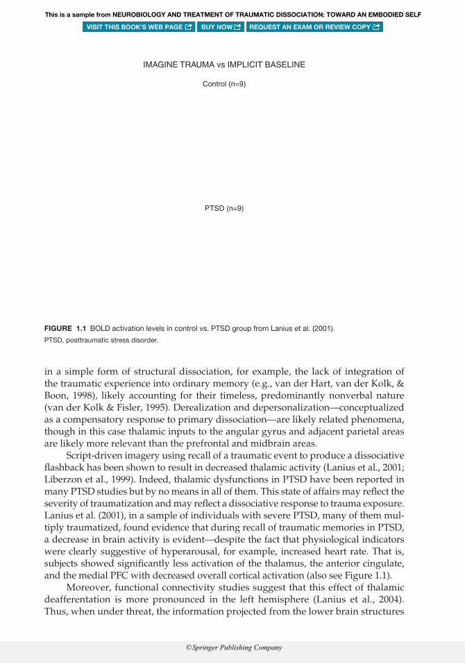
in a simple form of structural dissociation, for example, the lack of integration of the traumatic experience into ordinary memory (e.g., van der Hart, van der Kolk, & Boon, 1998), likely accounting for their timeless, predominantly nonverbal nature (van der Kolk & Fisler, 1995). Derealization and depersonalization—conceptualized as a compensatory response to primary dissociation—are likely related phenomena, though in this case thalamic inputs to the angular gyrus and adjacent parietal areas are likely more relevant than the prefrontal and midbrain areas.
Script-driven imagery using recall of a traumatic event to produce a dissociative flashback has been shown to result in decreased thalamic activity (Lanius et al., 2001; Liberzon et al., 1999). Indeed, thalamic dysfunctions in PTSD have been reported in many PTSD studies but by no means in all of them. This state of affairs may reflect the severity of traumatization and may reflect a dissociative response to trauma exposure. Lanius et al. (2001), in a sample of individuals with severe PTSD, many of them mul-tiply traumatized, found evidence that during recall of traumatic memories in PTSD, a decrease in brain activity is evident—despite the fact that physiological indicators were clearly suggestive of hyperarousal, for example, increased heart rate. That is, subjects showed significantly less activation of the thalamus, the anterior cingulate, and the medial PFC with decreased overall cortical activation (also see Figure 1.1).
Moreover, functional connectivity studies suggest that this effect of thalamic deafferentation is more pronounced in the left hemisphere (Lanius et al., 2004). Thus, when under threat, the information projected from the lower brain structures
IMAGINE TRAUMA vs IMPLICIT BASELINE
Control (n=9)
PTSD (n=9)
FIGURE 1.1 BOLD activation levels in control vs. PTSD group from Lanius et al. (2001).
PTSD, posttraumatic stress disorder.
This is a sample from NEUROBIOLOGY AND TREATMENT OF TRAUMATIC DISSOCIATION: TOWARD AN EMBODIED SELF VISIT THIS BOOK’S WEB PAGE BUY NOW REQUEST AN EXAM OR REVIEW COPY
© Springer Publishing Company

to right hemisphere—and there are a greater number of connections from the lower brain structures to the right hemisphere—is less likely to be integrated by the left brain (e.g., Schore, 2003b). Moreover, altered thalamic connectivity is not only evi-dent under threat, as in recall of traumatic memories, but also during resting states, for example, default network connectivity (Yin et al., 2011).
We hypothesize that the decrease in thalamic activity is a response to excessive arousal. Indeed, it may reflect the simultaneous increase of both sympathetic nervous system activity associated with noradrenaline, in conjunction with a concomitant increase in parasympathetic activity, particularly dorso-vagal activity that is associ-ated with increased endogenous opioid tone. In essence, the down-regulation of the thalamic relay can be construed as a circuit breaker or thalamocortical switch that will protect the neocortex from overactivation and the effects of excessive arousal.
The Effect of Endogenous Opioids on Thalamic Function
Indeed, the very areas identified by Lanius et al. (2001) as exhibiting decreased acti-vation, for example, PFC, anterior cingulate, and thalamus, are similar to the ones identified by Lieberzon et al. (2007) using PET neuroimaging and a mu-opioid selec-tive radiotracer. Liberzon and colleagues suggested that the functional alteration in regional cerebral blood flow was attributable to alterations in opioid receptor bind-ing after psychological trauma.
The very areas identified in these studies also happen to be the very areas that happen to exhibit the highest densities of opiate receptors in the cortex (Kling et al., 2000). Moreover, opioid activation has been associated with decreased local cerebral glucose utilization, particularly in the thalamus, limbic system, and forebrain regions (e.g., Fanelli, Szikszay, Jasinski, & London, 1987), suggesting a possible underlying mechanism. Endogenous opioids commonly affect specific thalamic nuclei depend-ing on the origin of the presynaptic input, but they can cause inhibition of the entire thalamus (Brunton & Charpak, 1998). High opioid receptor densities occur also in the superior colliculi. As suggested earlier, both the thalamus and the colliculi have been associated with the presence of gamma oscillations that may play an essen-tial role in information processing and cognitive temporal binding (see Chapter 11). Indeed, gamma oscillations decrease in response to opioids, a phenomenon that can be reversed by the administration of an opioid antagonist (Whittington, Traub, Faulkner, Jefferys, & Chettiar, 1998).
At the same time, through the down-regulation of the thalamic relay and the resulting diminished sensory input to the cortical areas of the brain the capacity of the organism to respond to environmental input in any mindful manner is reduced. The nervous system has reverted to the quick and reflexive survival actions for which the brainstem, derived from the reptilian brain, is most adaptive. Thus, the disrup-tion of the thalamic relay may be at the core of dissociative responses to overwhelm-ing traumatic experience. High levels of arousal are associated with a disruption of the thalamic relay. There is a loss of sensory input to cortical areas, interfering with exteroceptive awareness, and an impaired capacity to integrate information at the cortical level. Moreover, this is associated with a decreased ability to allow cortical regulation of affective responses arising from the midbrain at the level of the PAG.
This is a sample from NEUROBIOLOGY AND TREATMENT OF TRAUMATIC DISSOCIATION: TOWARD AN EMBODIED SELF VISIT THIS BOOK’S WEB PAGE BUY NOW REQUEST AN EXAM OR REVIEW COPY
© Springer Publishing Company
Indeed, even the response to eye contact in individuals with complex PTSD is reflec-tive of greater brain stem activation, as compared to an increased cortical response in individuals without PTSD (Steuwe, Lanius, & Frewen, 2012). With the disruption of the thalamic relay, the brain essentially reverts to dominance of brainstem function-ing principally directed to survival, losing the very qualities that distinguish humans from lower animals, for example, the ability to self-reflect and the capacity to feel.
The Role of the Thalamic Nuclei in Integrative Functioning of the Brain
As suggested earlier, the involvement of the thalamus in state switching likely involves thalamic inputs to the ACC. The pregenual ACC is relatively inactive in response to trauma-related stimuli in PTSD (Vogt, Aston-Jones & Vogt 2009). There is also reduced connectivity of the thalamus with the anterior cingulate in the resting state in PTSD (Yin et al., 2011b), specifically implicating thalamocortical projections from the mediodorsal, medial pulvinar, intralaminar, and midline nuclei of the thala-mus (Shibata & Yukie, 2009).
The medial pulvinar provides multimodal sensory and visual memory infor-mation to the anterior cingulate areas 25 and 32 and to the posterior cingulate area 23. It receives much of this sensory information from the superior colliculi (Bentivoglio et al., 1993). The intralaminar nuclei also transfer information from spinothalamic tracts and from intermediate and deep layers of the superior colliculi to all areas of cingulate cortex. With cholinergic inputs from the mesopontine tegmentum, nora-drenergic inputs from the LC, and serotoninergic inputs from the raphe nuclei, these thalamic nuclei contribute to sensorimotor integration (Bentivoglio et al., 1993).
The midline nuclei, which process inputs from the hypothalamus, the bed nucleus of the stria terminalis (BNST), the spinal cord, and the midbrain areas of PAG, parabrachial nucleus, nucleus of the solitary tract (NTS), and LC also project to area 32 of the ACC.
The thalamus is a composite of nuclei that receives input from all the senses and disperses information throughout the cortex. The part of the ACC that is functioning abnormally in PTSD has major inputs from subcortical structures for sensorimotor integration, emotion, defense, and autonomic regulation mediated by mediodorsal, intralaminar, and midline nuclei and the medial pulvinar of the thalamus. Thus, dis-rupting the thalamus means disrupting widespread activation throughout the brain, and thus connectivity between different brain regions. This includes both vertical and horizontal integration of brain functioning, ultimately contributing to the break-down of integrative functioning of the brain under threat.
Opioid Activation, Deafferentation, and Symptom Specificity
Deafferentation commonly refers to central pain phenomena that are associated with a partial or complete loss of sensory input from a portion of the body following lesions in somatosensory pathways (e.g. phantom limb pain, nerve injury-associated pain). This disruption of sensory pathways commonly occurs at the level of the thalamus
This is a sample from NEUROBIOLOGY AND TREATMENT OF TRAUMATIC DISSOCIATION: TOWARD AN EMBODIED SELF VISIT THIS BOOK’S WEB PAGE BUY NOW REQUEST AN EXAM OR REVIEW COPY
© Springer Publishing Company
with concomitant changes in the somatosensory cortex (Flor & Birbaumer, 2000). We hypothesize that opioid activation is a probable mechanism that results in decreased thalamic activation. This results in lowering of the level of consciousness through deafferentation of cortex from the lower brain structures.
Moreover, as suggested by Brunton and Charpak (1998), endogenous opioids commonly affect specific thalamic nuclei depending on the origin of the presynaptic input. Therefore, excessive stimulation in a specific sensory modality may inhibit the associated sensory nuclei in the thalamus. For instance, overwhelming visual input would lead to an inhibition of the lateral geniculate and medial pulvinar nuclei of the thalamus. Such inhibition is likely to result in deafferentation, or partial deafferenta-tion, of the visual input into the visual cortex, which would result in that very visual input not being integrated with other sensory input, for example, touch and smell, to produce an integrated experience. Thus, the person is likely to continue to experience visual flashbacks.
Overwhelming arousal resulting in excessive endogenous opioid activation impairs information processing. Limited sensory information is conveyed to the areas of the cortex where integration of different senses, emotions, and thoughts can occur. This may be the functional mechanism that maintains the sensory memory as a fragment, rather than the integrated experience that is common for ordinary mem-ories. Further, as such sensory input remains unintegrated, but also is associated with activation of a specific emotional circuit, a fragmented, trauma-related self-state with distinct sensory experience is created.
Pierre Janet—Field of Consciousness, Partial Catalepsy, and Deafferentation
Janet (1889), in conjunction with dissociation, refers to a narrowing or a retraction of the field of consciousness, a reduction in the number of phenomena that are held in conscious awareness. This notion bears resemblance to the changes in thalamic activity that are related to arousal as reported by Portas et al. (1998). That is, with low arousal the field of consciousness normally widens, whereas high arousal is accom-panied by a narrowing of the field of consciousness. As van der Hart, Nijenhuis, and Steele (2006) point out, such a narrowing of consciousness alone is insufficient to result in structural dissociation or pathological dissociation.
However, Janet also refers to “Les catalepsies partielles,” the partial catalepsies. Catalepsy is characterized by immobilization and muscular rigidity, as well as a lack of responsivity to external stimuli and decreased sensitivity to pain. It is further asso-ciated with a slowing down of bodily functions, such as breathing, and can ultimately result in a loss of consciousness. A number of substances have been associated with the induction of catalepsy, one of them being opioids. This condition specifically is referred to as opioid or opiate catalepsy (e.g., Ling & Pasternak, 1982). Opioid cata-lepsy can be reversed by the administration of opioid antagonists, particularly in the PAG and thalamus (e.g., Wilcox, Bozarth, & Levitt, 1983).
Specifically, we suggest that the narrowing of the field of consciousness that occurs in conjunction with an opioid-mediated cataleptic state is at the core of “dis-aggregation” or dissociation (Janet, 1919/1976), the breaking up or breaking apart
This is a sample from NEUROBIOLOGY AND TREATMENT OF TRAUMATIC DISSOCIATION: TOWARD AN EMBODIED SELF VISIT THIS BOOK’S WEB PAGE BUY NOW REQUEST AN EXAM OR REVIEW COPY
© Springer Publishing Company

of the self. This phenomenon was depicted in Janet’s seminal work, L’Automatisme Psychologique (1889): He conceptualizes and visually depicts the lack of horizontal inte-gration between different types of sensory input. That is, in the figure below, the lack of integration between one part (P) and another (P′) is illustrated (also see Figure 1.2). P refers to the association of two types of auditory input (A and A′) and two types of visual input (V and V′). P′ refers to auditory (A″), muscular (M′), and tactile (T′) input. Assuming they represent different aspects of an experience, one would expect the individual to fluidly conjoin those experiences into a narrative. Where a retraction of consciousness attributable to high arousal occurs in conjunction with an opioid-mediated cataleptic response, one would expect the state change to be no longer fluid. That is, state change will be accompanied by alterations in consciousness that result in a lack of horizontal integration and in more extreme cases in amnesia.
The Nature of Affective Circuits and Structural Dissociation
We suggest that separate self-states are likely a direct result of the breakdown of inte-grative functioning and deafferentation. Those that arise in direct response to threat, for example, EPs (van der Hart et al., 2006), likely reflect truncated defense responses separated at a subcortical level. Others that arise in response to facilitating adaptive functioning, often with little or no emotional charge, may be based on relatively inde-pendent corticothalamic loops.
The thalamus conveys information from the body—relayed in spinothalamic tracts—to the insular and cingulate cortices (Craig, 2006). Ego states that have specific physiological components must have interoceptive circuits through the thalamus. Also, those that do not have marked autonomic accompaniments will have cortical loops through the basal ganglia and thalamus. Therefore the thalamus may contain nodes for circuit switching—from one ego state to another parallel loop—although it is suggested in a later chapter that it is the insula that is key: responding to changes in body state.
These circuits can be functioning relatively independently at different levels of the brain. For example, cortical loops take sensory input into the cortex, process through the basal ganglia, and return through the thalamus to the cortex for motor output (Redgrave et al., 2010). Subcortical loops, in contrast, have the thalamic relay
T T′ M′
P′
T″ M″ V V″ A A′ A″V′M
P
FIGURE 1.2 Dissociation between Parts P and P′, as described by Pierre Janet (1889).
This is a sample from NEUROBIOLOGY AND TREATMENT OF TRAUMATIC DISSOCIATION: TOWARD AN EMBODIED SELF VISIT THIS BOOK’S WEB PAGE BUY NOW REQUEST AN EXAM OR REVIEW COPY
© Springer Publishing Company

on the input to the striatum. There is a return to the subcortical structures via the basal ganglia for motor output. Sensorimotor loops can thus form two distinct cir-cuits in the brain, which have the capacity for horizontal disconnection: reflexive defensive behaviors and goal-directed actions employing different neural pathways. Vertical loops for defense responses could have inputs from different brain levels—for example, from the right insular cortex, the right amygdala, and the brain stem—but be insufficiently integrated across the corpus callosum and throughout the PFC.
Analgesic Response and Separate Self-States: ANPs and EPs
At times of severe threat, the brain needs a range of analgesic responses to facilitate survival. We suggest that the separateness of these self-states is a consequence of acti-vation of endogenous neurochemical analgesics that prevent overload during high-arousal states. These analgesics are part of a dorso-vagal parasympathetic response, resulting in sensory and somatic fragments of traumatic experiences escaping inte-gration. This may occur through a variety of mechanisms that include unblended colliculus-thalamus-cortex loops linked by the basolateral amygdala.
In some instances the hyperarousal of brainstem survival functioning leads to a shutdown of the thalamic relay and a relative deafferentation of specific cortical areas. Then the emotional expression of behavior is not integrated in the larger con-text, as the person can no longer bring neocortical resources to bear on it. The basic affective circuits reverberate in an ongoing manner, reactive to outside stimuli, but relatively impervious to top-down modulation and horizontal integration at the level of the neocortex.
Separate self-states can be complex emotional states based in truncated defense responses and have relatively independent interoceptive loops through the brain-stem, the body, the spinothalamic tracts, and the cortex. At the other extreme are separate body states that have circuits through the brainstem and body with little involvement even of thalamic structures. They resemble what van der Hart et al. refer to as EP’s.
Other self-states are stored in cortico-striato-thalamo-cortical loops that have little affective or defensive loading. These different states resemble what Van der Hart et al. (2006) describe as ANPs. These self-states are more likely separated at a subcortical level—thalamocortical loops through the basal ganglia.
Truncated Affective Circuits, Structural Dissociation, and Self-States
Nijenhuis et al. (1999) relate discrete traumatized self-states, including specific soma-toform dissociative phenomena, to animal defensive responses and recuperative states that develop in response to predatory attack. That is, van der Hart et al. (2006) suggest that the structural dissociation model reflects “emotional personalities” that involve different animal defense-like systems.
Stimulation of the dorsal and lateral aspects of the PAG provokes defensive responses of fight or flight. Stimulation of the caudal ventrolateral PAG results in
This is a sample from NEUROBIOLOGY AND TREATMENT OF TRAUMATIC DISSOCIATION: TOWARD AN EMBODIED SELF VISIT THIS BOOK’S WEB PAGE BUY NOW REQUEST AN EXAM OR REVIEW COPY
© Springer Publishing Company

quiescence or freeze. Whereas lesions of the caudal ventrolateral PAG reduce condi-tioned freeze, lesions of the dorsal aspect reduce fight-or-flight behavior. That is, dif-ferent animal defensive responses such as fight and flight and surrender and collapse likely become the core of self-states or EPs, where fight corresponds to RAGE and flight to FEAR and intense separation distress leads to extreme states of PANIC.
Panksepp (1998) links the PANIC system with attachment, where unmet attach-ment needs in the face of separation will ultimately induce states of panic. The term panic is often used in a less-specific sense to indicate the terror or acute intense fear based in the dorsolateral PAG. Panksepp does not have a separate system for the ani-mal equivalent of the submissive, surrendered, dissociative collapse, which is impor-tant in human victims of severe trauma. The dorsal vagal freeze state of immobility and bradycardia is accompanied by opioid-mediated analgesia, which may contrib-ute to the structural dissociation of this state. For active defense states generated in the dorsolateral PAG, the endogenous analgesia is mediated by endogenous can-nabinoids. These may contribute to structural dissociation by deafferentation of thal-amocortical projections and contribute to memory disturbance through their impact on the basolateral amygdala and hippocampus. Defense response states can become structurally dissociated in subthalamic loops through the body, in spinothalamic interoceptive loops through the ventromedial PFC, and in subcortical loops through the basal ganglia.
Loss of Higher Cortical Functioning—Positive and Negative Symptoms
Van der Hart et al. (2006), as did Janet (1893/1901/1977) and Myers (1940) before them, differentiate between positive and negative symptoms of dissociation. Negative symptoms of dissociation are generally held to refer to losses of function, such as memory (i.e., amnesia), higher cortical functions, loss of feeling, loss of motor con-trol, as well as loss of somatosensory perceptions, for example, numbness. Positive symptoms may include intrusive traumatic memories, flashbacks, intrusive voices, as well as intentions, emotions, cognitions, and behaviors, including complex pat-terns such as reenactments.
It is easy to see how opioid activation is responsible for negative symptoms of dissociation. Similarly, we suggest amnesia and anesthesia are opioid-mediated phenomena that either relate to the lack of sensory transmission at the thalamic level due to shutting down of particular thalamic nuclei (e.g., symptoms of anesthesia) and a more general amnestic process in the brain that again relates to a conditioned response. Alternatively, negative dissociative responses may also reflect the effects of opioid activation of the ventrolateral PAG.
That is, negative dissociative symptoms likely not only relate to passive defense responses evoked at the level of the PAG but also due to higher centers being inhibited at the level of the cortex. Both of these likely result in an absence of func-tion or “negative” symptoms. “Positive” symptoms, on the other hand, are likely caused by the functional release of the lower centers due to lack of effective cortical down-regulation, contributing to decreased modulation of affective responses at the level of the PAG.
This is a sample from NEUROBIOLOGY AND TREATMENT OF TRAUMATIC DISSOCIATION: TOWARD AN EMBODIED SELF VISIT THIS BOOK’S WEB PAGE BUY NOW REQUEST AN EXAM OR REVIEW COPY
© Springer Publishing Company

Somatoform Dissociation and Deafferentation
Clinical observations indicate that dissociation can manifest in somatoform ways (e.g., Nemiah, 1991). Somatoform dissociation includes many somatic and senso-rimotor phenomena and can present in a variety of ways that include sensory distor-tions, motor weakness, freezing, numbing, paralysis, and tremors (Nijenhuis, 1999). Shaking and convulsions are also common, as are sleepiness, attentional impairment, and headaches and other pain sensations that may be much less obvious.
That is, van der Hart and colleagues (2006) specifically refer negative and positive dissociative symptoms and suggest that negative dissociative symptoms likely relate to passive defensive responses and associated predominant dorsova-gal activation and associated opioid activation in the ventrolateral PAG. Positive dissociative symptoms on the other hand are likely believed to relate to active defensive responses. While among the latter sympathetic tone predominates, excessive associated arousal will likely result in significant endogenous opioid release and resulting levels of deafferentation. In the case of symptoms of disso-ciative collapse, these EPs may be further defined by an increasing opioid innerva-tion of the ventrolateral PAG that results in states of immobilization and catatonia. Opioid activation is also related to alterations in eating behavior, where excessive opioid tone is associated with both starvation as well as binging behavior (e.g., Bodnar, 2007).
Pain phenomena can be conceptualized in a similar way, as being attributable to deafferentation resulting in somatic reorganization similar to central pain phe-nomena as they occur in deafferentation and phantom limb pain (e.g., Giummarra & Moseley, 2011). Similarly, if during sexual abuse a person experiences overwhelming tactile stimulation in the genital area, deafferentation of the ventral posterior nucleus may ensue. This may account not only for somato-sensory flashbacks of the experi-ence but also an inability to experience sexual pleasure through sensory stimulation of that area and even possibly account for genital pain. This has implications for conversion symptoms.
We suggest this type of urogenital pain, in particular, is likely attributable to deafferentation of body regions to trauma related to sexual abuse. Other pain disor-ders common in individuals with trauma-related disorders are likely attributable to a similar kind of functional mechanism.
We further suggest that opioid activation is responsible for what van der Hart et al. (2006) describe as negative symptoms of dissociation. Stress and threats to life induce analgesia and numbness, a phenomenon that has been described as learned helplessness (e.g., Van der Kolk, Greenberg, Orr, & Pitman, 1989). Nijenhuis, Spinhoven, van Dyck, van der Hart, and Vanderlinden (1998) found that dissociative symptoms reminiscent to analgesia and numbness in particular, for example, freez-ing, anesthesia, analgesia, disturbed eating, and urogenital pain, were predictive of a diagnosis of dissociative disorder, with anesthesia–analgesia being the best pre-dictor, thus supporting our notion of the relevance of opioid activation to the phe-nomenology of dissociative disorders.
We suggest amnesia and anesthesia are opioid-mediated phenomena that either relate to the lack of sensory transmission at the thalamic level due to shutting down of
This is a sample from NEUROBIOLOGY AND TREATMENT OF TRAUMATIC DISSOCIATION: TOWARD AN EMBODIED SELF VISIT THIS BOOK’S WEB PAGE BUY NOW REQUEST AN EXAM OR REVIEW COPY
© Springer Publishing Company

particular thalamic nuclei (e.g., symptoms of anesthesia) and a more general amnes-tic process in the brain that again relates to a conditioned response. Alternatively, negative dissociative responses may also reflect the effects of opioid activation of the ventrolateral PAG.
Summary and Future Directions
Events experienced as traumatic evoke the release of the brain’s own analgesic com-pounds that include opioid, cannabinoid, and other agents. This leads to thalamic deafferentation, including reduction of input to the ACC and other cortical areas. Reduced connectivity of the anterior and posterior cingulate cortices limits informa-tion processing, especially when it is self-related. Anesthetic neurochemicals released in response to stress also induce functional activation or deactivation in areas that mediate the affective responses. These peritraumatic chemical changes in the mid-brain and cortex can mimic or induce the diminished activation of relevant areas of thalamus releasing the cortical imbalance—some areas relatively more active, others less so.
We therefore argue that clinically relevant dissociation is not on a continuum of compartmentalized attentional focus. Dissociation, which results from overwhelm-ing experience, has immediate neurochemical mediators that promote the creation of neural networks that can be evoked to function relatively independently. The primary purpose of the chemical release is the reduction of pain, fear, rage, and/or separation distress. The brain produces its own equivalents of morphine and cannabis to attenu-ate fear and pain. This produces changes in the plasticity of the connections between neurons creating new networks. When these networks are later functioning as inde-pendent ego states, the contribution of the analgesic neurochemicals may be much less than when the states came into being. Nevertheless, they arose through a failure of integrative capacity—when the brain’s strategies for overload were urgently acti-vated. The morphine and cannabis equivalents dull the pain immediately but affect long-term functioning in a way that ultimately ceases to be adaptive.
When the cortex shuts down subcortical generation of painful affects, this top-down control can be described as overmodulated dissociation: it may become habitual and relatively independent of endogenous analgesic agents. When the sub-cortically generated affects are overwhelming and the cortical modulators are unable to diminish the distressing impact, this dissociation may be described as undermod-ulated: the cortex gives up and goes “offline.” The prototypical dissociative states arise in infancy when needs are not being met to such an extent that endogenous opioids and cannabinoids are released to dull the pain of protest and despair. The later detachment phase may not have the same intensity of neurochemical mediation but may arise from the new neural networks created to cope with the demands of the environment. When fuses in an electrical circuit keep melting, they can be replaced by ones of lesser sensitivity. While that saves on fuses in the short term, the result may be damage to the appliance and a risk of fire. The sensors need to be adjusted to the right setting for maximal resilience and long-term functioning when the envi-ronment is not hostile.
This is a sample from NEUROBIOLOGY AND TREATMENT OF TRAUMATIC DISSOCIATION: TOWARD AN EMBODIED SELF VISIT THIS BOOK’S WEB PAGE BUY NOW REQUEST AN EXAM OR REVIEW COPY
© Springer Publishing Company

Emotional experiences that need brain analgesics to blunt their intensity under-lie clinical dissociation. Finally, there may be additional effects of opioids that are relevant to our understanding of altered thalamo-cortical connectivity that may be relevant to our understanding of dissociation that involve decreases as well as increases in brain activation in response to opioids (Su, Huang, Wang, Wang, & Luo, 2012). These are not yet elaborated in our model but may yield some relevance to our understanding of the development of nontraumatized self-states.
REFERENCES
Adams, J. H., Graham, D. I., & Jennett, B. (2000). The neuropathology of the vegetative state after an acute brain insult. Brain, 123(7), 1327–1338.
Alkire, M. T., Haier, R. J., & Fallon, J. H. (2000). Toward a unified theory of narcosis: Brain imaging evidence for a thalamocortical switch as the neurophysiologic basis of anesthet-ic-induced unconsciousness. Consciousness and Cognition, 9(3), 370–386.
Bentivoglio, M., Kultas-Ilinsky, K., & Ilinsky, I. (1993). Limbic thalamus: Structure, intrinsic organization, and connections. In B. A. Vogt, & M. Gabriel (Eds.), Neurobiology of cingu-late cortex and limbic thalamus (pp. 71–122). Boston, MA: Birkhauser.
Bittencourt, A. S., Carobrez, A. P., Zamprogno, L. P., Tufik, S., & Schenberg, L. C. (2004). Organization of single components of defensive behaviors within distinct columns of periaqueductal gray matter of the rat: Role of N-methyl-D-aspartic acid glutamate receptors. Neuroscience, 125(1), 71–89.
Bodnar, R. J. (2007). Endogenous opiates and behavior: 2006. Peptides, 28(12), 2435–2513. doi:10.1016/j.peptides.2007.09.002
Brandão, M. L., Melo, L. L., & Cardoso, S. H. (1993). Mechanisms of defense in the inferior colliculus. Behavioral Brain Research, 58(1–2), 49–55.
Brecht, M., Goebel, R., Singer, W., & Engel, A. K. (2001). Synchronization of visual responses in the superior colliculus of awake cats. Neuroreport, 12, 43–47.
Brunton, J., & Charpak, S. (1998). mu-Opioid peptides inhibit thalamic neurons. Journal of Neuroscience,18(5), 1671–1678.
Coull, J. T., Jones, M. E., Egan, T. D., Frith, C. D., & Maze, M. (2004). Attentional effects of nora-drenaline vary with arousal level: Selective activation of thalamic pulvinar in humans. Neuroimage, 22(1), 315–322.
Craig, A. D. (2006). Evolution of pain pathways. In J. H. Kaas (Ed.), Evolution of nervous systems (Vol. 3, pp. 227–236). Oxford, UK: Academic press.
Dasilva, M. A., Grieve, K. L., Cudeiro, J., & Rivadulla, C. (2012). Endocannabinoid CB1 recep-tors modulate visual output from the thalamus. Psychopharmacology (Berl), 219(3), 835–845. doi:10.1007/s00213–011-2412–3
De Bellis, M. D., Keshavan, M. S., Clark, D. B., Casey, B. J., Giedd, J. N., Boring, A. M., . . . Ryan, N. D. (1999). A.E. Bennett Research Award. Developmental traumatology. Part II: Brain development. Biological Psychiatry, 45(10), 1271–1284.
Dringenberg, H. C., Dennis, K. E., Tomaszek, S., & Martin, J. (2003). Orienting and defensive behaviors elicited by superior colliculus stimulation in rats: Effects of 5-HT depletion, uptake inhibition, and direct midbrain or frontal cortex application. Behavioral Brain Research, 144(1–2), 95–103.
Fanelli, R. J., Szikszay, M., Jasinski, D. R., & London, E. D. (1987). Differential effects of mu and kappa opioid analgesics on cerebral glucose utilization in the rat. Brain Research, 22(2), 257–266.
This is a sample from NEUROBIOLOGY AND TREATMENT OF TRAUMATIC DISSOCIATION: TOWARD AN EMBODIED SELF VISIT THIS BOOK’S WEB PAGE BUY NOW REQUEST AN EXAM OR REVIEW COPY
© Springer Publishing Company

Flor, H., & Birbaumer, N. (2000). Phantom limb pain: Cortical plasticity and novel therapeutic approaches. Current Opinion in Anaesthesiology, 13(5), 561–564.
Giummarra, M. J., & Moseley, G. L. (2011). Phantom limb pain and bodily awareness: Current concepts and future directions. Current Opinion in Anaesthesiology, 24(5), 524–531. doi:10.1097/ACO.0b013e32834a105f
Haines, D. E. (2003). Neuroanatomy: Atlas of structures, sections, and systems (6th ed.). Philadelphia, PA: Lippincott Williams & Wilkins.
Hughlings-Jackson, J. (1958). Evolution and dissolution of the nervous system (Croonian Lectures). In J. Taylor (Ed.), The selected writings of John Hughlings Jackson (Vol 2., pp. 45–75). New York, NY: Basic Books.
Janet, P. (1889). L’automatisme psychologique. Paris, France: Félix Alcan.Janet, P. (1893/1901/1977). The mental state of hystericals: A study of mental stigmata and mental
accidents. Washington, DC: University Publications of America.Janet, P. (1976). Psychological healing: A historical and clinical study (2 vols. E. Paul & C. Paul,
Trans.). New York, NY: Arne. (Original work published in 1919)John, E. R. (2001). A field theory of consciousness. Consciousness and Cognition, 10(2), 184–213.Kling, M. A., Carson, R. E., Borg, L., Zametkin, A., Matochik, J. A., Schluger, J., . . . Kreek, M. J.
(2000). Opioid receptor imaging with positron emission tomography and [(18)F]cyclo-foxy in long-term, methadone-treated former heroin addicts. Journal of Pharmacology and Experimental Therapeutics, 295(3), 1070–1076.
Krystal, J. H., Bennett, A. L., Bremner, J. D., Southwick, S. M., & Charney, D. S. (1995). Toward a cognitive neuroscience of dissociation and altered memory functions in post-traumatic stress disorder. In M. J. Friedman, D. S. Charney, & A. Y. Deutch (Eds.), Neurobiological and clinical consequences of stress: From normal adaptions to PTSD (pp. 239–268). New York, NY: Raven Press.
Långsjö, J. W., Alkire, M. T., Kaskinoro, K., Hayama, H., Maksimow, A., Kaisti, K. K., . . . Scheinin H. (2012). Returning from oblivion: Imaging the neural core of consciousness. Journal of Neuroscience, 32(14), 4935–4943. doi:10.1523/JNEUROSCI.4962–11.2012
Lanius, R. A., Williamson, P. C., Densmore, M., Boksman, K., Gupta, M. A., Neufeld, R. W., . . . Menon, R. S. (2001). Neural correlates of traumatic memories in posttraumatic stress disorder: A functional MRI investigation. The American Journal of Psychiatry, 158, 1920–1922.
Lanius, R. A., Williamson, P. C., Densmore, M., Boksman, K., Neufeld, R. W., Gati, J. S., & Menon, R. S. (2004). The nature of traumatic memories: A 4-T FMRI functional connec-tivity analysis. The American Journal of Psychiatry, 161, 36–44.
Liberzon, I., Taylor, S. F., Amdur, R., Jung, T. D., Chamberlain, K. R., Minoshima, S., . . . Fig, L. M. (1999). Brain activation in PTSD in response to trauma-related stimuli. Biological Psychiatry, 45, 817–826.
Liberzon, I., Taylor, S. F., Phan, K. L., Britton, J. C., Fig, L. M., Bueller, J. A., . . . Zubieta, J. K. (2007). Altered central micro-opioid receptor binding after psychological trauma. Biological Psychiatry, 61(9), 1030–1038.
Ling, G. S., & Pasternak, G. W. (1982). Morphine catalepsy in the rat: Involvement of μ1 (high affinity) opioid binding sites. Neuroscience Letters, 32, 193–196.
MacLean, P. D. (1990). The triune brain in evolution: Role in paleocerebral functions. New York, NY: Plenum Press.
MacLean, P. D. (1993). Introduction: Perspectives on cingulate cortex in the limbic system. In B. Vogt, & M. Gabriel (Eds.), Neurobiology of cingulate cortex and limbic thalamus (pp. 1–18). Boston, MA: Birkhauser.
Mashour, G. A. (2005). Cognitive unbinding in sleep and anesthesia. Science, 310(5755), 1768–1769.
Mashour, G. A. (2006). Integrating the science of consciousness and anesthesia. Anesthesia and Analgesia, 103(4), 975–982.
This is a sample from NEUROBIOLOGY AND TREATMENT OF TRAUMATIC DISSOCIATION: TOWARD AN EMBODIED SELF VISIT THIS BOOK’S WEB PAGE BUY NOW REQUEST AN EXAM OR REVIEW COPY
© Springer Publishing Company

Masterton, R. A., Carney, P. W., & Jackson, G. D. (2012). Cortical and thalamic resting-state functional connectivity is altered in childhood absence epilepsy. Epilepsy Research, 99(3), 327–334. doi:10.1016/j.eplepsyres.2011.12.014
Maxwell, W. L., Pennington, K., MacKinnon, M. A., Smith, D. H., McIntosh, T. K., Wilson, J. T., & Graham, D. I. (2004). Differential responses in three thalamic nuclei in moder-ately disabled, severely disabled and vegetative patients after blunt head injury. Brain, 127(11), 2470–2478.
Meares, R. (1999). The contribution of Hughlings Jackson to an understanding of dissociation. American Journal of Psychiatry, 156, 1850–1855.
Merker, B. (2007). Consciousness without a cerebral cortex: A challenge for neuroscience and medicine. Behavioral and Brain Sciences, 30, 63–134.
Morgan, M. M., & Clayton, C. C. (2005). Defensive behaviors evoked from the ventrolat-eral periaqueductal gray of the rat: Comparison of opioid and GABA disinhibition. Behavioural Brain Research, 164(1), 61–66.
Mountcastle, V. B. (1997). The columnar organization of the neocortex. Brain, 120(4), 701–722.Myers, C. S. (1940). Shell shock in France 1914–18. Cambridge, UK: Cambridge University
Press.Nemiah, J. C. (1991). Dissociation, conversion and somatization. In A. Tasman & S. Goldfinger
(Eds.). Review of psychiatry (Vol. 10, pp. 248–260). Washington, DC: American Psychiatric Press.
Nijenhuis, E. R., Spinhoven, P., van Dyck, R., van der Hart, O., & Vanderlinden, J. (1998). Psychometric characteristics of the Somatoform Dissociation Questionnaire: A replica-tion study. Psychotherapy and Psychosomatics, 67, 17–23.
Nijenhuis, E. R. S., van Dyck, R., Spinhoven, P., van der Hart, O., Chatrou, M., Vanderlinden, J., & Moene F. (1999). Somatoform dissociation discriminates between diagnostic cat-egories over and above general psychopathology. Australian and New Zealand Journal of Psychiatry, 33, 512–520.
Panksepp, J. (1998). Affective neuroscience: The foundations of human and animal emotions. New York, NY: Oxford University Press.
Panksepp, J. (2003). Feeling the pain of social loss. Science, 302, 237–239.Pfeiffer, A., Brantl, V., Herz, A., & Emrich, H. M. (1986). Psychotomimesis mediated by kappa
opiate receptors. Science, 233(4765), 774–776.Porges, S. W. (2011). The polyvagal theory: Neurophysiological foundations of emotions, attachment,
communication, and self-regulation. New York, NY: W. W. Norton.Portas, C. M., Rees, G., Howseman, A. M., Josephs, O., Turner, R., & Frith, C.D. (1998). A
specific role for the thalamus in mediating the interaction of attention and arousal in humans. Journal of Neurosciences, 18, 8979–8989.
Redgrave, P., Coizet, V., Comoli, E., McHaffie, J. G., Leriche, M., Vautrelle, N., . . . Overton, P. (2010). Interactions between the Midbrain Superior Colliculus and the Basal Ganglia. Frontiers in Neuroanatomy, 4(132). doi:10.3389/fnana.2010.00132
Sakurada, O., Sokoloff, L., & Jacquet, Y. F. (1978). Local cerebral glucose utilization following injection of beta-endorphin into periaqueductal gray matter in the rat. Brain Research, 153(2), 403–407.
Schore, A. N. (2003). Affect dysregulation and the repair of the self. New York, NY: W. W. Norton.Sergent, J. (1990). Furtive incursions into bicameral minds. Integrative and coordinating role
of subcortical structures. Brain, 113(2), 537–568.Shibata, H., & Yukie, M. (2009). Thalamocingulate connections in the monkey. In B. A. Vogt (Ed.),
Cingulate neurobiology and disease (pp. 95–112). Oxford, UK: Oxford University Press.Steuwe, C., Lanius, R. A., & Frewen, P. A. (2012). The role of dissociation in civilian post-
traumatic stress disorder—evidence for a dissociative subtype by latent class and confir-matory factor analysis. Depression and Anxiety, 29 (8), 689–700.
This is a sample from NEUROBIOLOGY AND TREATMENT OF TRAUMATIC DISSOCIATION: TOWARD AN EMBODIED SELF VISIT THIS BOOK’S WEB PAGE BUY NOW REQUEST AN EXAM OR REVIEW COPY
© Springer Publishing Company

Su, Y.-L., Huang, J., Wang, N., Wang, J.-Y., & Luo, F. (2012). The effects of morphine on basal neuronal activities in the lateral and medial pain pathways. Neuroscience Letters, 525, 173–178.
Uzan, M., Albayram, S., Dashti, S. G., Aydin, S., Hanci, M., & Kuday, C. (2003). Thalamic proton magnetic resonance spectroscopy in vegetative state induced by traumatic brain injury. Journal of Neurology, Neurosurgery, and Psychiatry, 74(1), 33–38.
van der Hart, O., Nijenhuis, E. R., & Steele, K. (2006). The haunted self: Structural dissociation and the treatment of chronic traumatization. New York, NY: W. W. Norton.
van der Hart, O., van der Kolk, B. A., & Boon, S. (1998). Treatment of dissociative disorders. In J. D. Bremner & C. R. Marmar (Eds.), Trauma, memory, and dissociation (pp. 253–283). Washington, DC: American Psychiatric Press.
van der Kolk, B. A., & Fisler, R. (1995). Dissociation and the fragmentary nature of traumatic memories: Overview and exploratory study. Journal of Traumatic Stress, 8, 505–525.
van der Kolk, B. A., Greenberg, M. S., Orr, S. P., & Pitman, R. K. (1989). Endogenous opioids, stress induced analgesia, and posttraumatic stress disorder. Psychopharmacology Bulletin, 25, 417–421.
van der Kolk, B. A., McFarlane, A. C., & Weisaeth, L. (Eds.). (1996). Traumatic stress: The effects of overwhelming experience on mind, body and society. New York, NY: Guilford Press.
Vogt, B. A., Aston-Jones, G., & Vogt, L. J. (2009). Shared norepinephrinergic and cingulate circuits, nociceptive and allostatic interactions, and models of functional pain and stress disorders. In B. A. Vogt (Ed.), Cingulate neurobiology and disease (pp. 467–498). Oxford, UK: Oxford University Press.
Vogt, B. A., & Laureys, S. (2009). The primate posterior cingulate gyrus: Connections, senso-rimotor orientation, gateway to limbic processing. In B. A. Vogt (Ed.), Cingulate neurobi-ology and disease (pp. 275–308). Oxford, UK: Oxford University Press.
Vogt, B. A., & Sikes, R. W. (2009). Cingulate nociceptive circuitry and roles in pain processing: The cingulate premotor pain model. In B. A. Vogt, Cingulate neurobiology and disease (pp. 311–338). New York, NY: Oxford University Press.
White, N. S., & Alkire, M. T. (2003). Impaired thalamocortical connectivity in humans during general-anesthetic-induced unconsciousness. Neuroimaging, 19(2), 402–411.
Whittington, M. A., Traub, R. D., Faulkner, H. J., Jefferys, J. G., & Chettiar, K. (1998). Morphine disrupts long-range synchrony of gamma oscillations in hippocampal slices. Proceedings of the National Academy of Sciences of the United States of America, 95, 5807–5811.
Wilcox, R. E., Bozarth, M., & Levitt, R. A. (1983). Reversal of morphine-induced catalepsy by naloxone microinjections into brain regions with high opiate receptor binding: A pre-liminary report. Pharmacology Biochemistry and Behavior, 18, 51–54.
Yin, Y., Jin, C., Hu, X., Duan, L., Li, Z., Song, M., . . . Li, L. (2011). Altered resting-state functional connectivity of thalamus in earthquake-induced posttraumatic stress disorder: A func-tional magnetic resonance imaging study. Brain Research, 1411, 98–107.
This is a sample from NEUROBIOLOGY AND TREATMENT OF TRAUMATIC DISSOCIATION: TOWARD AN EMBODIED SELF VISIT THIS BOOK’S WEB PAGE BUY NOW REQUEST AN EXAM OR REVIEW COPY
© Springer Publishing Company