neurogenesis in the mossy chiton, mopalia muscosa (gould) (polyplacophora): evidence against...
TRANSCRIPT

Neurogenesis in the Mossy Chiton, Mopalia muscosa(Gould) (Polyplacophora): Evidence Against MolluscanMetamerismStefan Friedrich, Andreas Wanninger, Martin Bruckner, and Gerhard Haszprunar*
Zoologische Staatssammlung Munchen and Zoologisches Institut der LMU Munchen, Munchen, Germany
ABSTRACT Neurogenesis in the chiton Mopalia mus-cosa (Gould, 1846) was investigated by applying differen-tial interference contrast microscopy, semithin serial sec-tioning combined with reconstruction techniques, as wellas confocal laser scanning microscopy for the detection offluorescence-conjugated antibodies against serotonin andFMRFamide. The ontogeny of serotonergic nervous struc-tures starts with cells of the apical organ followed by thoseof the cerebral commissure, whereas the serotonergic pro-totroch innervation, pedal system, and the lateral cordsdevelop later. In addition, there are eight symmetricallyarranged serotonergic sensory cells in the dorsal pretro-chal area of the larva. FMRFamide-positive neural ele-ments include the cerebral commissure, specific “ampul-lary” sensory cells in the pretrochal region, as well as thelarval lateral and pedal system. In the early juvenile thecerebral system no longer stains with either of the two
antibodies and the pedal system lacks anti-FMRFamideimmunoreactivity. Outgroup comparison with all othermolluscan classes and related phyla suggests that thecord-like, nonganglionized cerebral system in the Polypla-cophora is a reduced condition rather than a primitivemolluscan condition. The immunosensitivity of the pedalcommissures develops from posterior to anterior, suggest-ing independent serial repetition rather than annelid-likeconditions and there is no trace of true segmentationduring nervous system development. Polyplacophoranneurogenesis and all other available data on the subjectcontradict the idea of a segmented molluscan stem spe-cies. J. Morphol. 253:109–117, 2002.© 2002 Wiley-Liss, Inc.
KEY WORDS: Mollusca; Polyplacophora; neurogenesis;immunocytochemistry; segmentation; phylogeny
During the last decade the application of antibody-conjugated fluorescent dyes combined with improvedimaging techniques, e.g., by means of confocal laserscanning microscopy (CLSM), has brought majorprogress in our understanding of invertebrate neuro-genesis. Most studies of Mollusca dealt with euthyneu-ran gastropods, a few bivalves, and, up to now, noamphineuran (aculiferan) taxa (i.e., Solenogastres orNeomeniomorpha, Caudofoveata or Chaetodermomor-pha, and Polyplacophora or Neoloricata). The last formthe basic stem line of the Mollusca (Salvini-Plawenand Steiner, 1996; Waller, 1998; Haszprunar, 2000).Accordingly, the plesio–morphic type of the molluscannervous system is still debatable. Most authors con-sider the overall amphi-tetraneuran, cord-like (i.e.,lacking true ganglia) nervous system of Polypla-cophora as most primitive for Mollusca (e.g., Salvini-Plawen, 1991; Haszprunar, 1992). In contrast, Page(1994) regards the conditions of nudibranch larvae(with true ganglia) as plesiomorphic for gastropodsand molluscs. Based on investigation of the neurogen-esis of Mytilus edulis and Patella caerulea, Raineri(1995, 2000) considered the Mollusca to represent a“bent (annelid-like) trochophore” and a two-cord,ladder-like nervous system as its basic condition.
Although the ontogeny of polyplacophoran mol-luscs has been studied by several authors through-
out the last 120 years (see review by Eernisse andReynolds, 1994; Salvini-Plawen and Bartolomaeus,1995), only one recent study has applied immunocy-tochemical methods (Haszprunar et al., 2002). In thecourse of a general study on the ontogeny of molluscswith lecithotrophic mode of development, particu-larly of Polyplacophora (see also Haszprunar andWanninger, 2000; Wanninger and Haszprunar,2002), we investigated neurogenesis of the mossychiton, Mopalia muscosa (Gould), in order to shedlight on the plesiomorphic neural conditions of theMollusca. Since the Polyplacophora show the mostsignificant signs of seriality (“metamerism”) of allmolluscan classes, traces of an annelid-like, seg-mented nervous system—if present—should be mostobvious during neurogenesis of the chiton larva.
Contract grant sponsor: the DFG (German Science Foundation);Contract grant number: HA 2598/1-3,1-4 (to G.H.).
*Correspondence to: Prof. Dr. Gerhard Haszprunar, ZoologischeStaatssammlung Munchen, Munchhausenstrasse 21, D-81247Munchen, Germany. E-mail: [email protected]
Published online 00 Month 2002 inWiley InterScience (www.interscience.wiley.com)DOI: 10.1002/jmor.10010
JOURNAL OF MORPHOLOGY 253:109–117 (2002)
© 2002 WILEY-LISS, INC.

MATERIALS AND METHODS
In spring 1999 adult Mopalia muscosa (Gould, 1846)were collected near Argyle Creek, San Juan Island,WA, USA. Several individuals immediately spawnedin the laboratory (Friday Harbor Laboratories/WA,USA). The embryos and larvae were maintained insmall custard dishes in millipore-filtered seawater(MFSW), 10–12°C. In order to avoid bacterial, infec-tion 50 mg streptomycin and 60 mg penicillin per literMFSW were added. To induce metamorphosis, smallrocks covered with encrusting coralline red algae orstones, where adult specimens were removed from,were put into the culture dishes with metamorphiccompetent larvae. Juveniles were cultured until 10days after the beginning of metamorphosis.
Later larvae and juveniles were relaxed by addingdrops of 7.14% or 3.57% MgCl2 solution to theMFSW. For semithin sectioning (and parallel SEMand TEM work; see Haszprunar et al., 2001), larvaewere fixed in 4% glutaraldehyde in 0.2 M sodium-cacodylate buffer; for osmolarity 0.1 M NaCl and0.35 M sucrose were added. Larvae were postfixed in1% OsO4 in 0.2 M sodium-cacodylate buffer with 0.3M NaCl. The fixed specimens were dehydrated in anethanol series and embedded in low-viscosity resin(Spurr, 1969). Ribbons of serial sections (1.5 �m)were prepared with “Ralph” glass knives and contactcement (“Pattex”) at the lower cutting edge (Henry,1977) and were stained with methylene-blue-azureII (Richardson et al., 1960). The section slides havebeen deposited in the Mollusca Section of the Zoo-logical State Collection Munich (ZSM: 20006641/1;20006643/1).
Two ontogenetic stages, larvae aged about 180 hpostfertilization (hpf) and early juveniles about 10days postmetamorphosis (dpm), were additionallystudied by differential interference contrast (DIC)light microscopy.
Table 1 lists all stages to which we applied immu-nocytochemical dyes. Specimens were fixed in 4%paraformaldehyde (PFA) in 0.1 M phosphate buffer(pH 7.3) (PBS) with 10% sucrose added for osmolar-ity. After rinsing with 0.1 M PBS the samples were
stored in a solution of 0.1% NaN3 in 0.1 M PBS.After treatment for 60 min with a solution of 0.15%Triton X-100 in 0.1 M PBS plus 0.1% NaN3 (PTA),the specimens were incubated in a solution of 6%goat-serum (Jackson ImmunoResearch LaboratoriesWest Grove, PA, USA) in PTA for 18 h at 4°C.Anti-FMRF-amid (DiaSorin, Stillwater, MN, USA)in a concentration of 1:400 and anti-serotonin (Cal-biochem, Cambridge, MA, USA) in a concentrationof 1:800, both in PTA, plus 6% goat-serum were usedas primary antibodies. The specimens were incu-bated for 24 h at 4°C, followed by multiple rinsingfor 10 h in PTA. Under red-light, goat-antirabbit-immunoglobulin G conjugated with a fluorescentdye was applied as secondary antibody in a concen-tration of 1:100 in PTA for 20 h. Both rhodamine-(TRITC-) conjugated antibody (Jackson ImmunoRe-search Laboratories) and Oregon green- (FITC)-conjugated antibody (Molecular Probes, Eugene,OR, USA) provided the same quality of signal. Aftermultiple rinsing in 0.1 M PBS for 20 h the specimenswere mounted in Vectashield (Vector Laboratories,Burlingame, CA, USA) and stored at �20°C. Obser-vations were done by application of confocal laser-scanning microscopy (CLSM: Leica DM IRBE) sup-plied with a Leica TCS laser scanning unit withLeica TCS NT 4D-software. TRITC-stained speci-mens were observed at 518 nm, FITC-stained spec-imens were studied at 488 nm wavelength. Stackprojections of Z-series (1 �m depth distance) andred-green stereoimages were created. Rotated pro-jections of stacks were created with Scion Image(Scion Corp., Frederick, MD, USA) software.
RESULTS
A general description of the ontogeny of Mopaliamuscosa with special reference to shell-plate forma-tion and myogenesis has been provided most re-cently by Wanninger and Haszprunar (2001) and istherefore not repeated here.
TABLE 1. Immunoreactivity against serotonin and FMRFamide during neurogenesis of Mopalia muscosa*
Neural structure Stages (hpf) 25 35 45 55 65 85 95 120 140 200 Juvenile
Apical Organ Serotonin(cellnumber)
� (1) �� (3) �� (3) �� (5) �� (5) � (8) � (8) � (8) � (8) � x
FMRFamide � � � � � � � � � � xCerebral system Serotonin � � � �� �� � � � � � �
FMRFamide � (�) � �� �� �� � � � � �Prototroch
systemSerotoninFMRFamide
��
��
� ��
��
���
���
���
���
��
xx
Pedal system Serotonin � � � � � � � � � � ��FMRFamide � � � � � �� �� � � � �
Lateral system Serotonin � � � � � � � � � � �FMRFamide � � � � � � � (�) � � ��
*(�) No signal, (�) weak signal, � strong signal, �� very strong signal, x structure not present.
110 S. FRIEDRICH ET AL.

Fig. 1. Mopalia muscosa. A,B: Dorsal view by interference contrast microscopy. A: Larva at 181 hpf showing apical ciliary ruft,prototroch, and larval ocelli. Lateral marks (C–E) indicate section planes. B: Early juvenile at 10 days after metamorphosis. The larvalocelli are still present. C–E: Subsequent semithin cross sections through larva at 181 hpf. For section planes see A. C: The three apicalcells enclose the bases of the ciliated tuft. The mucous ring is dorsally placed. D: The cerebral commissure is situated ventrally.E: Immediately behind the prototroch are the anterior pedal commissure and the larval ocelli. act, apical ciliary tuft; ao, apical organ;cc, cerebral commissure; fmo, frontal mucous organ; fg, foregut; fs, foot sole; lnc, lateral nerve cord; oc, ocelli; pc, pedal commissure;pnc, pedal nerve cord; pt, cilia of prototroch.

Gross Anatomy of Neurogenesis
Semithin sectioning and a reconstruction of thenervous system of a larva with the age of 181 hpfrevealed the following features (Figs. 1, 2): The api-cal pole of the larva shows a distinct apical senseorgan with a prominent ciliary tuft. In dorsal posi-tion there is a ring of mucous cells that are histolog-ically distinct from those of the remaining apicalarea (Fig. 1C). Further epithelial cells of the pretro-chal region include ciliated cells and the so-calledampullary cells, which have been described in detailelsewhere (Haszprunar et al., 2001). The bases ofthe cells of the apical organ contact the central partof the cerebral commissure, which is situated ven-trally (Fig. 1D). The distance between cerebral andanterior pedal commissure is large and equalsnearly half of the length of the larva (Fig. 2A). Also,the lateral (visceral) cords with a posterior commis-sure, as well as the pedal ring with anterior andposterior pedal commissures, are already developed.In contrast, we could not detect any trace of a buccalnervous system and neither is the posterior gut yet
differentiated. The cerebropedal connective is situ-ated immediately posterior to the prototroch and isin close association with the larval ocelli (Figs.1A,E, 2A) and the paired protonephridium (omit-ted in Fig. 2).
The nervous system of an early juvenile specimen(10 dpm: Figs. 1B, 2B) already resembles the adultpolyplacophoran condition (for details of the lattersee Gantner, 1989; summarized and reviewed byEernisse and Reynolds, 1994). The apical complex isentirely lost during metamorphosis, the distance be-tween cerebral and anterior pedal commissure isshorter than in the larva, and there are more pedalcommissures detectable than in the earlier stage.The position of these commissures is neither corre-lated with the position of the developing shell platesnor with the bundles of the shell muscles (Wan-ninger and Haszprunar, 2002). Protonephridia(omitted in Fig. 2) and larval ocelli are still present.A buccal nervous system could not be detected, al-though the radula is already present (see also Fig.3H).
Fig. 2. Mopalia muscosa. Reconstruction of the nervous system based on serial semithin sections. Ciliary structures are schemat-ically added. A: Larva at 181 hpf (corresponds with Fig. 1A). B: Early juvenile 10 days after metamorphosis (corresponds with Fig. 1B).act, apical ciliary tuft; am, ampullary cells; ao, apical organ; lnc, lateral nerve cord; oc, larval ocellus; pnc, pedal nerve cord; pt,prototroch; asterisk, cerebral commissure; arrowheads, pedal commissures.
112 S. FRIEDRICH ET AL.
Ontogeny of Serotonin-Positive Elements
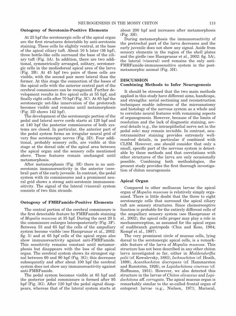
At 25 hpf the serotonergic cells of the apical organare the first structures detectable by anti-serotoninstaining. These cells lie slightly ventral, at the baseof the apical ciliary tuft. About 10 h later (36 hpf),three bottle-like cells surround the base of the cili-ary tuft (Fig. 3A). In addition, there are two addi-tional, symmetrically arranged, solitary, serotoner-gic cells in the mediodorsal apical area of the larva(Fig. 3B). At 45 hpf two pairs of these cells arevisible, with the second pair more lateral than theformer. At this stage the connection of the bases ofthe apical cells with the anterior central part of thecerebral commissure can be recognized. Further de-velopment results in five apical cells at 55 hpf, andfinally eight cells after 70 hpf (Fig. 3C). At 85 hpf theserotonergic net-like innervation of the prototrochbecomes visible and remains until metamorphosis(Fig. 3D shows 142 hpf).
The development of the serotonergic portion of thepedal and lateral nerve cords starts at 120 hpf andat 140 hpf the posterior commissures of both sys-tems are closed. In particular, the anterior part ofthe pedal system forms an irregular neural grid ofvery fine serotonergic nerves (Fig. 3D). Four addi-tional, probably sensory cells, are visible at thisstage at the dorsal side of the apical area betweenthe apical organ and the sensory cells mentionedabove. These features remain unchanged untilmetamorphosis.
After metamorphosis (Fig. 3E) there is no anti-serotonin immunoreactivity in the anterior (cere-bral) part of the early juvenile. In contrast, the pedalsystem with its commissures and a prominent neu-ral grid shows a strong anti-serotonin immunosen-sitivity. The signal of the lateral (visceral) systemconsists of two thin strands.
Ontogeny of FMRFamide-Positive Elements
The central portion of the cerebral commissure isthe first detectable feature by FMRFamide stainingof Mopalia muscosa at 35 hpf. During the next 20 hthe commissure enlarges lateroposteriorly (Fig. 3F).Between 55 and 65 hpf the cells of the ampullarysystem become visible (see Haszprunar et al., 2002:fig. 5) and at 65 hpf cells of the apical organ alsoshow immunoreactivity against anti-FMRFamide.This sensitivity remains constant until metamor-phosis but disappears with the loss of the apicalorgan. The cerebral system shows its strongest sig-nal between 60 and 90 hpf (Fig. 3G); this decreasessubsequently and after about 100 hpf the cerebralsystem does not show any immunoreactivity againstanti-FMRFamide.
The pedal system becomes visible at 85 hpf andthe posterior pedal commissure is formed after 95hpf (Fig. 3G). After 120 hpf the pedal signal disap-pears, whereas that of the lateral system starts at
about 200 hpf and increases after metamorphosis(Fig. 3H).
During metamorphosis the immunoreactivity ofthe pretrochal part of the larva decreases and theearly juvenile does not show any signal. Aside fromsensory elements in the region of the shell platesand the girdle (see Haszprunar et al., 2002: fig. 5A),the lateral (visceral) cord remains the only anti-FMRFamide-immunosensitive system in the post-metamorphic animal (Fig. 3H).
DISCUSSIONCombining Methods to Infer Neurogenesis
It should be stressed that the two main methodsapplied in this study have different aims, handicaps,and strengths: serial sectioning and reconstructiontechniques enable inference of the microanatomyand histology of the nervous system as a whole andto correlate neural features with remaining aspectsof organogenesis. However, because of the limits ofresolution and the lack of diagnostic staining, sev-eral details (e.g., the intraepithelial nerve net in thepedal sole) may remain invisible. In contrast, neu-rotransmitter staining provides extremely well-resolved details, in particular if combined withCLSM. However, one should consider that only asmall, specific part of the nervous system is detect-able by these methods and that correlations withother structures of the larva are only occasionallypossible. Combining both methodologies, thepresent study provides the first thorough investiga-tion of chiton neurogenesis.
Apical Organ
Compared to other molluscan larvae the apicalorgan of Mopalia muscosa is relatively simply orga-nized. There is little doubt that the three to eightserotonergic cells that surround the apical ciliarytuft are sensory structures. Since chemoreceptivefunction is probable for the entirely different cells ofthe ampullary sensory system (see Haszprunar etal., 2002), the apical cells proper may play a role inmechanoreception, as assumed for the apical organof nudibranch gastropods (Chia and Koss, 1984;Kempf et al., 1997).
The very prominent circle of mucous cells, lyingdorsal to the serotonergic apical cells, is a remark-able feature of the larva of Mopalia muscosa. Thisstructure has not been described in any other chitonlarva investigated so far, either in Middendorffiapolii (cf. Kowalevsky, 1883), Ischnochiton (cf. Heath,1899), Acanthochiton discrepans (cf. Hammarstenand Runstrom, 1926), or Lepidochitona cinereus (cf.Hoffmann, 1931). However, we also detected thisstructure in the larvae of Chiton olivaceus and Lepi-dochitona aff. corrugata. The apical mucous organ isremarkably similar to the so-called frontal organ ofentoproct larvae (e.g., Nielsen, 1971; Mariscal,
113NEUROGENESIS IN THE MOSSY CHITON

Figure 3

1975). Further studies, in particular on lepidopleu-ridan and aplacophoran larvae, are necessary to elu-cidate possible homologies of this feature.
Apical Sensory Systems
The fine structure and immunocytochemistry ofthe so-called “ampullary system,” which is part ofthe apical complex in other molluscs (see Schaeferand Ruthensteiner, 2001, for detailed review), isoutlined and discussed in detail by Haszprunar et al.(2002). This system may be regarded as an apomor-phic feature of the Polyplacophora (although data onaplacophoran larvae are missing) and probably hasa chemoreceptive function.
Due to their very strong anti-serotonin immuno-reactivity, a sensory role of the two sets of four cellsin the apical area of the larva appears very probable.Since all these cells are lost during metamorphosis,their role must be limited to larval life. Future in-vestigations by means of combined immunoelectronmicroscopy are necessary for identification and forultrastructural and physiological data to infer amore specific function of these cells.
Larval Ocelli
Larval ocelli have been reported in all cases ofontogenetic studies on chitons. Rosen et al. (1979)and Bartolomaeus (1992) provide fine-structural de-tails and regard the larval ocelli as homologous tocephalic eyes of gastropods and cephalopods. How-ever, the larval ocelli of chitons are situated poste-rior to the prototroch and are innervated by thelateral (visceral) nerve cord, whereas the eyes ofgastropods are located anterior to the prototroch andare cerebrally innervated. According to the posi-
tional criterion of homology (e.g., Remane, 1956;Ruppert, 1982; Haszprunar, 1998), the larval ocelliof the Polyplacophora (as organs) are probably anautapomorphic character of this class regardless oftheir epigenetic background (Nielsen, 1996; see alsoGehring and Ikeo, 1999, vs. Meyer-Rochow, 2000, vs.Gehring, 2000). However, this does not rule out apossible homology of the photoreceptive cellsthroughout the Mollusca or even beyond.
Immunoreactivity of the Nervous System
Whereas the early cerebral system is sensitiveagainst both anti-serotonin and anti-FMRFamide,the postmetamorphic cerebral commissure lacks anytrace of immunoreactivity. In addition, this is theonly class within the Mollusca where no distinctcerebral ganglion is present. Within the Annelidasimilar conditions occur by parallelism in Pogono-phora and certain paedomorphic polychaetes (e.g.,Oweniidae). Therefore, it is likely that the postmeta-morphic cord-like, nonganglionated, cerebral systemof the Polyplacophora is generally reduced ratherthan representing a plesiomorphic stage within theMollusca, as usually assumed.
One of the most significant and unexpected re-sults of the present study is the fact that the lateral(visceral) and pedal cord of the developing “am-phineuran” (i.e., equally elaborated) nervous systemexhibit very different reactivity against the two an-tibodies applied. Only the early pedal cords showanti-FMRFamide immunoreactivity; later there isreactivity against anti-serotonin alone. In contrast,the lateral (visceral) cords show strong and constantreactivity against both antibodies.
The present results somewhat disagree with thoseprovided by Moroz et al. (1994) on adult Lepidopleu-rus asellus (Lepidopleurida), which showed anti-FMRFamide immunoreactivity of the pedal nervecords and of the cerebrobuccal system. These dis-crepancies might be explained in two ways: 1) Thespecies studied belong to each of the two main cladesof the Polyplacophora, namely, the Lepidopleuridaand the Chitonida. However, since the gross anat-omy of the nervous system remains largely constantthroughout the class, we regard this explanation asimprobable. 2) We studied early juveniles, whereasadult animals were investigated by Moroz et al.(1994). Indeed, the developmental stage certainlyhas a strong influence on the appearance of thebuccal nervous system, which could not be found byany method in the present study. Also, the mode oflocomotion changes from ciliary gliding in early ju-veniles (pers. obs. of living animals and suggested bythe densely ciliated foot sole) to creeping by muscu-lar activity in later stages, which might cause thesignificant differences in the immunoreactivity, par-ticularly of the pedal nerve cord against anti-FMRFamide.
Fig. 3. Mopalia muscosa. CLSM. A–E: Immunocytochemicalstaining of serotonergic cells. A: Larva at 36 hpf (ventral view)with three cells of apical organ. B: Same larva (dorsal view) withtwo anterior serotonergic cells. C: Larva at 70 hpf (ventral view)with apical organ consisting of eight cells and the cerebral com-missure. D: Larva at 142 hpf (dorsal view) with apical organ, twogroups of serotonergic sensory cells, the prototrochal plexus, thepedal system, and (in part and weak) the lateral system. E: Earlyjuvenile 10 days after metamorphosis (ventral view) with well-stained pedal and lateral system, whereas the cerebral systemlacks immunosensitivity. F–H: Immunocytochemical staining byanti-FMRFamide (see also Haszprunar et al., 2001, for additionalpreparations). F: Larva at 55 hpf. (ventral view) showing thecerebral commissure alone. G: Larva at 96 hpf (ventral view)showing the cerebral and pedal system just before the formationof the posterior pedal commissure. The spicule cells of the girdleshow autofluorescence. H: Early juvenile 10 days after metamor-phosis (ventral view) with well-stained lateral system, whereasthe cerebral and pedal system lack immunosensitivity. The an-lage of the radula shows autofluorescence. ao, apical organ; asc,anterior serotonergic sensory cells; cc, cerebral commissure; ls,lateral system; pg, prototrochal grid; ps, pedal system; psc, pos-terior serotonergic sensory cells; ra, radula; sp, spicule cells of thegirdle.
115NEUROGENESIS IN THE MOSSY CHITON
Neurogenesis and the Idea of Segmentationin Chitons (Molluscs)
The present results on the neurogenesis of chitonsonce more provide evidence against the still remain-ing ideas (e.g., Sutton et al., 2001; Stokstad, 2001)that various kinds of serial repetition (often called“metamerism”) in the Mollusca are signs or relics ofannelid-like segmentation. In particular, the differ-entiation of the serotonergic part of the pedal systemwith its serial commissures from posterior to ante-rior and via an irregular grid of neural fibers (Fig.3E) contradicts the assumption of annelid-like fea-tures, since true segmentation always develops fromanterior to posterior. Hammarsten and Runnstrom(1926: 312) originally stressed that “the nervous sys-tem of chitons cannot be regarded as being seg-mented” (translated from the original German). Inaddition, the anti-FMRFamide neural elements ofthe developing polyplacophoran nervous system donot show any trace of serial repetition. Moreover,there is no correlation of any neural seriality withthe pattern of repetition of any other organ systems(e.g., shell plates), except where this is functionallynecessary (e.g., neural supply of ctenidia or mus-cles).
The significance of polyplacophoran neurogenesisas a counter-argument for possible segmentation en-tirely agrees with all other data on the subject.Russel-Hunter and Brown (1965) and Russel-Hunter (1988) provide evidence that multiplicationof the ctenidia is variable (backwards in Lepidopleu-rida, forwards and often asymmetrically developedin Chitonida) and is correlated with size rather thanwith body partitioning. The anlagen of (seven ofeight) shell plates occur simultaneously in ontogeny(Haas et al., 1979; Kniprath, 1980; Jacobs et al.,2000), not sequentially, as one would expect in asegmented animal. There is a single pair of gonads,a single pair of nephridia, and only the most derived(acanthochitonidan) Polyplacophora show multipli-cation of atria of the heart. Finally, most recentstudies on polyplacophoran myogenesis (Wanningerand Haszprunar, 2002) revealed simultaneous oc-currence of the shell-plate musculature, a primarilymultiple seriality of these, and a later, secondaryconcentration to seven (eight) dorsoventral shellmuscle bundles. With the present data, which con-firm a mode of neurogenesis that is incompatiblewith annelid-like segmentation, there remains notrace of evidence for true segmentation in chitons.The same conclusion has been reached by thoroughstudies on monoplacophoran (tryblidian) anatomy,particularly on the partly pedomorphic Micropilinaarntzi (cf. Haszprunar and Schaefer, 1997) and byseveral recent cladistic analyses of the Mollusca as awhole (Haszprunar, 1996, 2000; Salvini-Plawen andSteiner, 1996; Waller, 1998). Silurian fossils (Suttonet al., 2001a,b), which may or may not be molluscs atall (Steiner and Salvini–Plawen, 2001), cannot be
directly applied to reconstruct the design of the mol-luscan stem species, which certainly lived in thePrecambrian. In accordance with Salvini-Plawen(1981, 1985), there remains no doubt that Molluscaare nonsegmented lophotrochozoans.
ACKNOWLEDGMENTS
Andreas Wanninger is grateful for hospitality ofthe Friday Harbour Laboratories. We thank Prof.Dr. Charles David (Zoological Institute of the LMUMunich) for access to Confocal Microscopy. Dr. Bern-hard Ruthensteiner (ZSM) provided very valuablehelp concerning serial sectioning, reconstructiontechniques, and comments on the draft of the type-script.
LITERATURE CITED
Bartolomaeus T. 1992. Ultrastructure of the photoreceptor in thelarvae of Lepidochiton cinereus (Polyplacophora) and Lacunadivariacta (Mollusca, Gastropoda). Microfauna Marina 7:215–236.
Chia F-S, Koss R. 1984. Fine structure of the cephalic sensoryorgan in the larva of the nudibranch Rostanga pulchra (Mol-lusca, Opisthobranchia, Nudibranchia). Zoomorphology 104:131–139.
Eernisse DJ, Reynolds PD. 1994. Polyplacophora. In: HarrisonFW, Kohn A, editors. Microscopic anatomy of invertebrates, vol.5. Mollusca I. New York: Wiley-Liss. p 55–110.
Gantner R. 1989. Morphologie und Nervensystem der Kafer-schneckenart Lepidochitona monterasatoi (Mollusca, Polypla-cophora). Diploma Thesis, Zoological Institute of the TechnicalUniversity of Munich.
Gehring WJ. 2000. Reply to Meyer-Rochow. Trends Genet 16:245.Gehring WJ, Ikeo K. 1999. Pax6 mastering eye morphogenesis
and eye evolution. Trends Genet 15:371–377.Haas W, Kriesten K, Watabe N. 1979. Notes on the shell forma-
tion in the larvae of the Placophora (Mollusca). Biomineralisa-tion 10:1–8.
Hammarsten OD, Runnstrom J. 1926. Zur Embryologie von Ac-anthochiton discrepans (Brown). Zool Jb Anat 47:261–318.
Haszprunar G. 1992. The first molluscs-small animals. Boll Zool59:1–16.
Haszprunar G. 1996. The Mollusca: coelomate turbellarians ormesenchymate annelids? In: Taylor, JD., editor. Origin andevolutionary radiation of the Mollusca. Oxford: Oxford Univer-sity Press. p 1–28.
Haszprunar G. 1998. Parsimony analysis as a specific kind ofhomology estimation, and the implications for characterweighting. Molec Phylog Evol 9:333–339.
Haszprunar G. 2000. Is the Aplacophora monophyletic? A cladis-tic point of view. Am Malac Bull 15:115–130.
Haszprunar G, Schaefer K. 1997. Anatomy and phylogenetic sig-nificance of Micropilina arntzi (Mollusca, Monoplacophora, Mi-cropilinidae fam. nov.). Acta Zool (Stockh) 77:315–334.
Haszprunar G, Wanninger A. 2000. Molluscan muscle systems indevelopment and evolution. J Zool Syst Evol Res 38:157–163.
Haszprunar G, Friedrich S, Wanninger A, Ruthensteiner B. 2002.Fine structure and immunocytochemistry of a new chemosen-sory system in the chiton larva (Mollusca: Polyplacophora). JMorphol 252:210–218.
Heath H. 1899. The development of Ischnochiton. Zool Jb Anat12:567-656, pls. 31–35.
Henry E.C. 1977. A method for obtaining ribbons of serial sec-tions of plastic embedded specimens. Stain Technol 52:59–60.
Hoffmann H. 1931. Beitrage zur Kenntnis der Chitonen. 1. Uberdie Fortpflanzung und Entwicklung von Trachydermon cine-reus L. Z Morph Okol Tiere 20:719–732.
116 S. FRIEDRICH ET AL.

Jacobs DK, Wray CG, Wedeen CJ, Kostriken R, De Salle R,Staton JL, Gates RD, Lindberg DR. 2000. Molluscan engrailedexpression, serial organization, and shell evolution. Evol Dev2:340–347.
Kempf SC, Page LR, Pires A. 1997. Development of serotonin-likeimmunoreactivity in the embryos and larvae of nudibranchmolluscs with emphasis on the structure and possible functionof the apical sensory organ. J Comp Neurol 386:507–528.
Kniprath E. 1980. Ontogenetic plate and plate field developmentin two chitons, Middendorffia and Ischnochiton. Roux’s ArchDev Biol 189:97–106.
Kowalevsky MA. 1883. Embryogenie du Chiton polii (Philippi)avec quelques remarques sur le developpement des autres chi-tons. Ann Mus Hist nat Marseille Zool 1:1–46.
Mariscal RN. 1975. Entoprocta. In: Giese PC, Pearse JS, editors.Reproduction of marine invertebrates, vol. 2. London: AcademicPress. p 1–40.
Meyer-Rochow VB. 2000. The eye: monophyletic, polyphyletic orperhaps biphyletic? Trends Genet 16:244–245.
Moroz LL, Nezlin L, Elofsson R, Sakhorarov D. 1994. Serotonin-and FMRFamide-immuno-reactive nerve elements in the chitonLepidopleurus asellus (Mollusca, Polyplacophora). Cell TissueRes 275:277–282.
Nielsen C. 1971. Entoproct life-cycles and the entoproct/ectoproctrelationship. Ophelia 9: 209–341.
Nielsen DE. 1996. Eye-ancestry: old genes for new eyes. Curr Biol6:39–42.
Page LR. 1994. The ancestral gastropod larval form is best ap-proximated by hatching-stage opisthobranch larvae: evidencefrom comparative developmental studies. In: Wilson WH,Stricker SA, Shinn GL., editors. Reproduction and developmentof marine invertebrates. Baltimore: Johns Hopkins Univ Press.p 206–222.
Raineri M. 1995. Is a mollusc an evolved bent metatrochophore?A histochemical investigation of neurogenesis in Mytilus (Mol-lusca: Bivalvia). J Mar Biol Assoc UK 75:571–592.
Raineri M. 2000. Early neurogenesis pattern in Patella coerulea(Patellogastropoda) and its possible phylogenetic implications.Malacologia 42:131–148.
Remane A. 1956. Die Grundlagen des naturlichen Systems, dervergleichenden Anatomie und der Phylogenetik, 2nd ed.Leipzig: Akademische Verlagsgesellshaft, Geest & Portig.
Richardson KC, Jarett L, Finke EH. 1960. Embedding in epoxyresins for ultrathin sectioning in electron microscopy. StainTechnol 35:313–323.
Rosen MD, Stasek CR, Hermans CO. 1979. The ultrastructureand evolutionary significance of the ocelli in the larva of Katha-rina tunicata (Mollusca, Polyplacophora). Veliger 22:173–178.
Ruppert EE. 1982. Homology recognition as a basis for compar-ative biology. In: Nelson DR, editor. Proceedings of the ThirdInternational Symposium on the Tardigrada. East TennesseeState University Press. p 31–54.
Russel-Hunter WD. 1988. The gills of chitons (Polyplacophora) andtheir significance in molluscan phylogeny. Am Malac Bull 6:69–78.
Russel-Hunter WD, Brown S. 1965. Ctenidial number in relationto size in certain chitons, with a discussion of its phyleticsignificance. Biol Bull 128:508–521.
Salvini-Plawen Lv. 1981. On the origin and evolution of theMollusca. In: Origine dei Grande Phyla dei Metazoi. Rome: Attidei Convegni Lincei. 49:235–293.
Salvini-Plawen Lv. 1985. Early evolution and the primitivegroups. In: Trueman ER, Clarke MR, editors. The Mollusca, vol.10. Evolution. London: Academic Press. p 59–150.
Salvini-Plawen Lv. 1991. Origin, phylogeny and classification ofthe phylum Mollusca. Iberus 9:1–33.
Salvini-Plawen Lv, Bartolomaeus T. 1995. Mollusca: mesenchy-mata with a “coelom.” In: Lanzavecchia G, Valvassori R,Candia-Carnevali MD, editors. Body cavities: function and phy-logeny. Selected symposia and monographs 8. Modena: Mucchi.p 75–92.
Salvini-Plawen Lv, Steiner G. 1996. Synapomorphies and plesi-omorphies in higher classification of the Mollusca. In: TaylorJD., editor. Origin and evolutionary radiation of the Mollusca.Oxford: Oxford University Press. p 29–51.
Schaefer K, Ruthensteiner B. 2001. The cephalic sensory organin pelagic and intracapsular larvae of the primitive opistho-branch genus Haminoea (Mollusca: Gastropoda). Zool Anz240:69 – 82.
Spurr AR. 1969. A low-viscosity epoxy resin embedding mediumfor electron microscopy. J Ultrastruct Res 26:31–43.
Steiner G, Salvini–Plawen LV. 2001. Acaenoplax–polychaete ormollusc? Nature 414:601–602.
Stokstad E. 2001. New fossil may change idea of first mollusc.Science 291:2292–2293.
Sutton MD, Briggs DEG, Siveter DaJ, Siveter DeJ. 2001a. Anexceptionally preserved vermiform mollusc from the Silurian ofEngland. Nature 410:461–463.
Sutton MD, Briggs DEG, Siveter DaJ, Siveter DeJ. 2001b.Acaenoplax–polychaete or mollusc?–reply. Nature 414:602.
Waller TR. 1998. Origin of the molluscan class Bivalvia and aphylogeny of major groups. In: Johnston PA, Haggard JW,editors. Bivalves: an eon of evolution. Calgary: Univ CalgaryPress. p 1–45.
Wanninger A, Haszprunar G. 2002. Chiton myogenesis: perspec-tives for the development of larval and adult muscle systems inmolluscs. J Morphol (in press).
117NEUROGENESIS IN THE MOSSY CHITON