neuroimaging in childhood
TRANSCRIPT

1/16/2012
1
Pediatric Neuroimaging inNeurometabolic-degenerative disorder
and Epilepsy
Bhagwan Moorjani, MD, FAAP, FAAN
Neuroimaging in Childhood
• Neuroimaging issues are distinct from adults
• Sedation/anesthesia
• Motion artifacts
• Requires knowledge of normal CNS developmental (i.e. myelin maturation)
• Contrast media
• Parental anxiety
Diagnostic Approach
• Age of onset
• Static vs Progressive– Look for treatable causes
– Do not overlook abuse, Manchausen if all is negative
• Phenotype presence (syndromic, HC, NCS, systemic involvement)
• Predominant symptom (epilepsy, DD, weakness/motor, psychomotor regression, cognitive/dementia)
Neuroimaging in Epilepsy
• Peak incidence in childhood
• Occurs as a co-morbid condition in many pediatric disorders (birth injury, dysmorphism, chromosomal anomalies, developmental delays/regression)
• Many neurologic disorders in children have the same chief complaint

1/16/2012
2
Congenital Malformation
• Characterized by their anatomic features
• Broad categories: based on embryogenesis– Stage 1: Dorsal Induction: Formation and closure
of the neural tube. (Weeks 3-4)
– Stage 2: Ventral Induction: Formation of the brain segments and face. (Weeks 5-10)
– Stage 3: Migration and Histogenesis: (Months 2-5)
– Stage 4: Myelination: (5-15 months; matures by 3 years)
Dandy Walker Malformation
• Criteria:– high position of tentorium– dysgenesis/agenesis of vermis– cystic dilatation of fourth ventricle
• commonly associated features: – hypoplasia of cerebellum– scalloping of inner table of occipital bone
• associated abnormalities:– hydrocephalus 75%– dysgenesis of corpus callosum 25%– heterotropia 10%
Dandy walker

1/16/2012
3
Etiology of Epilepsy: Developmental and Genetic Classification of
Cortical Dysplasia
1. Secondary to abnormal neuronal and glial proliferation/apoptosis
2. Secondary to abnormal neuronal migration
3. Secondary to abnormal cortical organization or late migration
4. Not otherwise classified• IEM• Other unclassified dysplasia
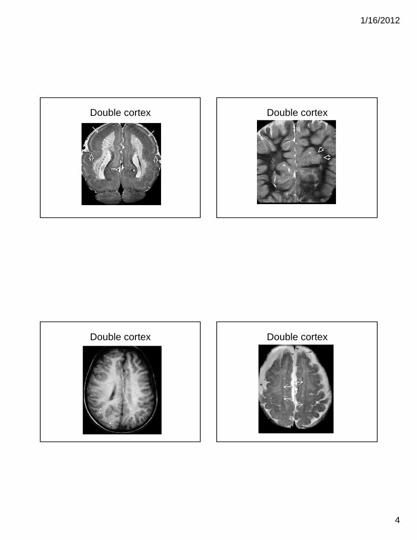
Gray Matter Heterotropia
• displaced masses of nerve cells (gray matter)
• most common: small nest adjacent to lateral ventricles
• Range from nodular to band heterotropia to schizencephaly, lissencephaly and polymicrogyria
• clinical: seizures
• MRI: isointense with gray matter in all sequence
• Subependymal heterotropia (most common)
• Band heterotropia (double cortex)
• Lissencephaly
• Cobblestone cortex (lis 2)
• Subcortical heterotropia
Subependymal heterotropia Subcortical heterotropia
1/16/2012
4
Double cortex Double cortex
Double cortex Double cortex

1/16/2012
5
Polymicrogyria
• multiple abnormal tiny indentation along brain surface (5-7mm)
• abnormal cortical histology
• can be unilateral
• MRI: decreased number of broad, thick, smooth gyri
polymicrogyria
Polymicrogyria Pachygyria
• thick and more completely developed gyri• commonly diffused with relative sparing of
temporal lobes• associated with: agenesis of CC and
heterotopias• clinical: microcephaly, seizures, MR,
developmental delay• MRI: circumferential band of high signal on T2
within the cortex

1/16/2012
6
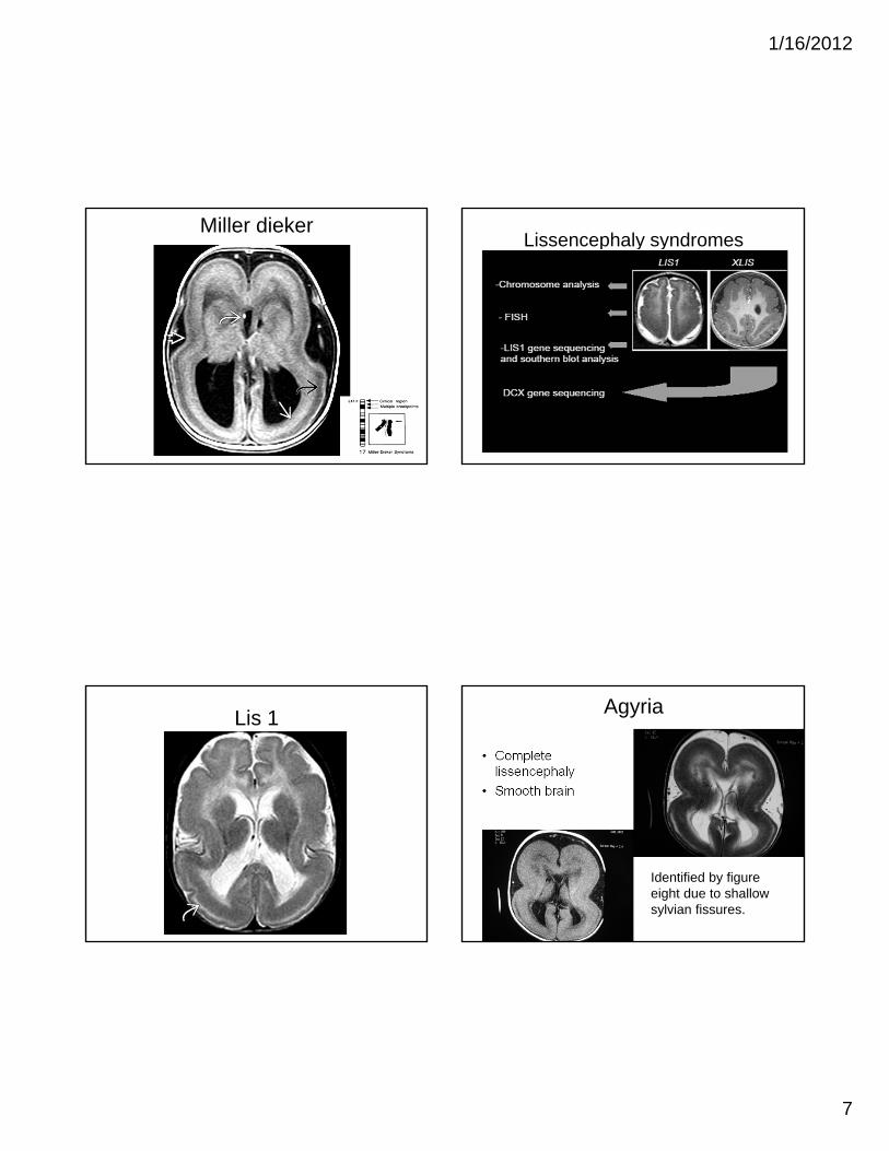
Lissencephaly
• Severe form of neuronal migration Disorder
• Can be seen in:– Isolation
– Miller Dieker Syndrome
– TORCH infection (CMV)
• Clinical S/S:– Mental Retardation
– Intractable Epilepsy
– Microcephaly
Lissencephaly Imaging
• Hourglass or figure of 8– Shallow sylvian fissure
• LIS 1: parietal-occipital
• X-LIS: Subfrontal/temporal
• 3 layers ay be seen in neonates on T2W
– Outer layer – thin, smooth
– Intervening cell sparse layer
– Deeper thick layer – mimicking band heterotropia
• Posterior > Anterior involvement in LIS 1
lissencephaly Lissencephaly Variants
1/16/2012
7
Miller diekerLissencephaly syndromes
Lis 1 Agyria
• Complete lissencephaly
• Smooth brain
Identified by figure eight due to shallow sylvian fissures.

1/16/2012
8
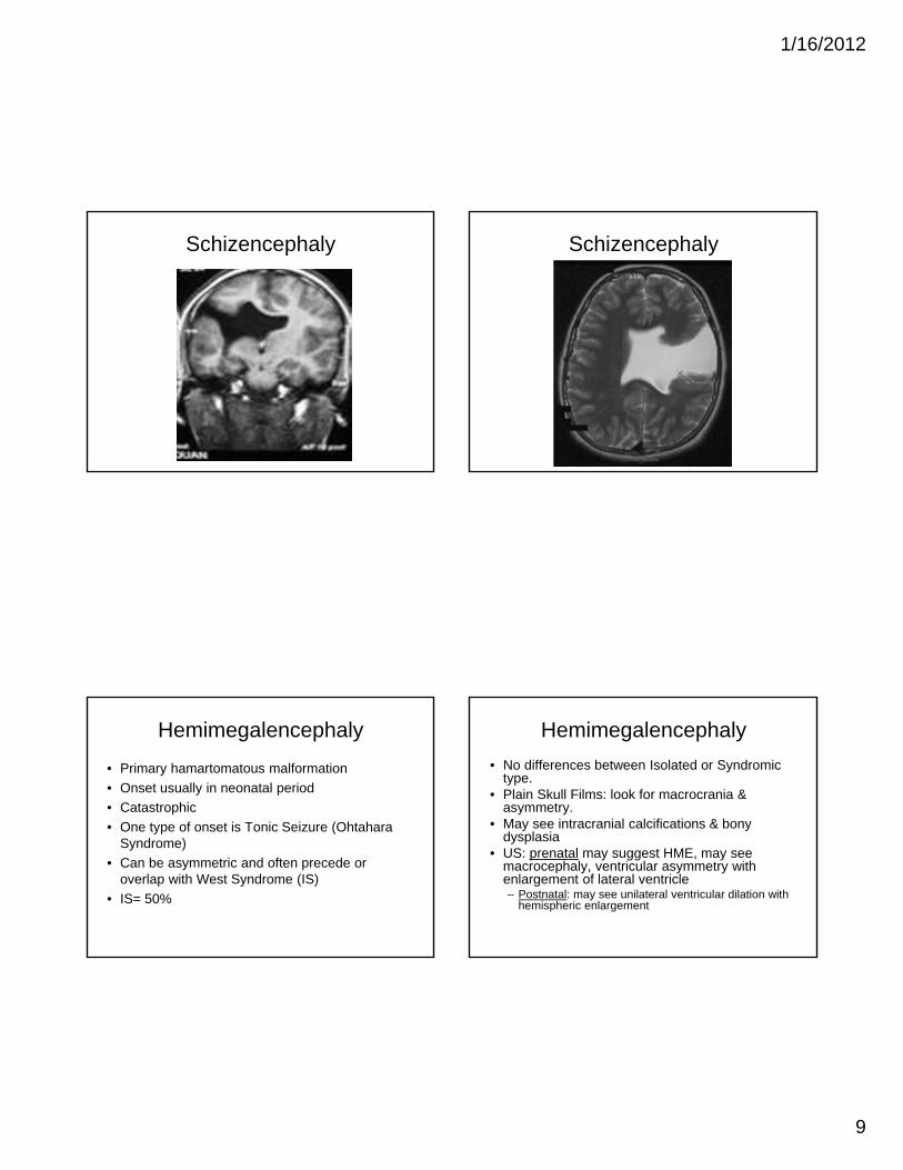
Schizencephaly
• Gray matter- lined cleft that extends from the ventricular ependyma to the pia.
• Unilateral or bilateral
• Two types: – Closed lip (type I)
– Open lip (type II)
Schizencephaly Imaging
• Transmantle gray matter lining clefts– Dimple in wall of ventricle if closed or narrow
• Frontal and Parietal lobes near central sulcus
• Distinction of GM lining cleft can be difficult prior to myelination
• DVA overlie cleft seen on MRV
SchizencephalyDifferential Diagnosis
• Porencephaly– Lined by gliotic white matter
– No dysplastic gray matter
• Semilobar Holoprocencephaly– Can mimic bilateral open lip schizencephaly
Schizencephaly
1/16/2012
9
Schizencephaly Schizencephaly
Hemimegalencephaly
• Primary hamartomatous malformation
• Onset usually in neonatal period
• Catastrophic
• One type of onset is Tonic Seizure (Ohtahara Syndrome)
• Can be asymmetric and often precede or overlap with West Syndrome (IS)
• IS= 50%
Hemimegalencephaly
• No differences between Isolated or Syndromic type.
• Plain Skull Films: look for macrocrania & asymmetry.
• May see intracranial calcifications & bony dysplasia
• US: prenatal may suggest HME, may see macrocephaly, ventricular asymmetry with enlargement of lateral ventricle– Postnatal: may see unilateral ventricular dilation with
hemispheric enlargement

1/16/2012
10
Hemimegalencephaly Imaging
• CT & MRI:– Gross asymmetry– 1 hemisphere enlarged
• Posterior falx and occipital pole displaced to contralateral side
– Dysplastic Cortex– Asymmetry of Ventricular System -
• 4 points of abnormal ventricle.– 1. Straightened frontal horns (pointed)– 2. mild-extreme dilation of lateral ventricle– 3. reverse of contralateral horn (mass effect appearance)– 4. colpocephaly (disproportionate developmental dilation of
occipital horn of lateral ventricle) in all grades of HME
Hemimegalencephaly
Hemimegalencephaly

1/16/2012
11
FCD with Balloon Cells
• Abnormal gyral pattern when large
• Blurring of gray-white junction
• Abnormal tissue: cortex to border of lateral ventricle
• Typically associated with TS
• Solitary – no other TS features
• T2 hyperintense “comet tail” from cortex to ventricle– Best seen on flair
FCD, Balloon Cell
FCD with balloon cell FCD with balloon cell

1/16/2012
12
DNET
• Intractable epilepsy• Focal deficits• More common 2nd and 3rd decade• Considered part of abnormal neuronal/glial
proliferation - neoplastic• MRI Findings:
– T1 hypointense– T2 hyperintense– Modest to no enhancement– Scalloping– Lack of mass effect, edema
DNET
DNET DNET

1/16/2012
13
Neurometabolic-Degenerative Disorders
• Knowledge of normal myelination pattern is essential
• General rules:– Caudal to cranial
– Posterior to anterior
• MRI provides the best imaging modality– T1 matures at 12-14 months
– T2 matures at 24-26 months
Neurometabolic-Degenerative Disorders• Type of myelination involvement
– Delayed myelination– Demyelination– Dysmyelination
• Nervous system involvement– Brainstem involvement– Cerebellar involvement– Spinal Cord involvement
• Tissue Involvement– Gray matter involvement– White matter involvement
1 month
9 months
36 months
From Alberico
Factors to Consider
• Age of Onset
• Degree of derangement
• Abnormal metabolite– Deficiency or excess
• Stage of the disease process
• Phenotype

1/16/2012
14
Symptoms of Progressive CNS DiseaseEarly Onset (Newborn or young infant)
• Intermittent lethargy, decreased responsiveness associated with emesis
• Irritability, excessive or absent startle response
• Decreasing interest in surroundings, excessive sleepiness, decreased smiling
• Poor or absent eye fixation, nystagmus
• Poor head control, weak suck
• Feeding difficulties, FTT
• Excessive tongue trusting, asymmetric movements
• Floppy or spastic
• Seizures
Symptoms of Progressive CNS DiseaseLater Onset ( older infant or child)
• Change in temperament, behavior or activity level
• Disturbance in gait (progressive ataxia), clumsiness
• Unusual movements, dystonia, chorea
• Progressive muscle weakness, change in tone
• Progressive loss of speech, vision or hearing
• Headaches
• Change in bowel/bladder function
• Seizures
White Matter versus Gray MatterEarly Disease
White Matter
• Gait disturbance
• Ataxia
• Brisk reflexes
• Spasticity
• Babinski sign
Gray Matter
• Progressive cognitive impairment
• Seizures
• Blindness
White Matter versus Gray MatterAdvanced Disease
White Matter
• Progressive cognitive impairment
• Blindness
• Deafness
• Seizures
Gray Matter
• Ataxia
• Hyperreflexia
• Spasticity
• Babinski sign

1/16/2012
15
Leukodystrophies• Abnormal signal in white matter
• Symetric usually
• Periventricular, deep or subcortical in location
• Failure to achieve myelination milestones
• MRS abnormalities reflect neuronal loss and increased cellular turnover
• Some have contrast enhancement– ALD: zone of active inflammation
– Alexander: ventricular lining, periventricular rim, frontal WM, optic chiasm, fornix, BG, thalamus, dentate nucleus
LeukodystrophiesDifferential Diagnosis
• Radiation and Chemotherapy injury
• Viral encephalitis
• ADEM
• MS
• In neonates: HIE – Periventricular pattern
Head Circumference
Macrocephalic
• Canavan
• Alexander
• Tay Sachs (GM2 gangliosidosis)
• Vanishing White Matter
• Van der Knaap Disease
• L-2-hydroxyglutaric aciduria
Microcephalic/Normal
• ALD
• MLD
• Cockayne Disease
• Pelizaeus Merzbacher disease
• Zellweger Disease
• Krabbe
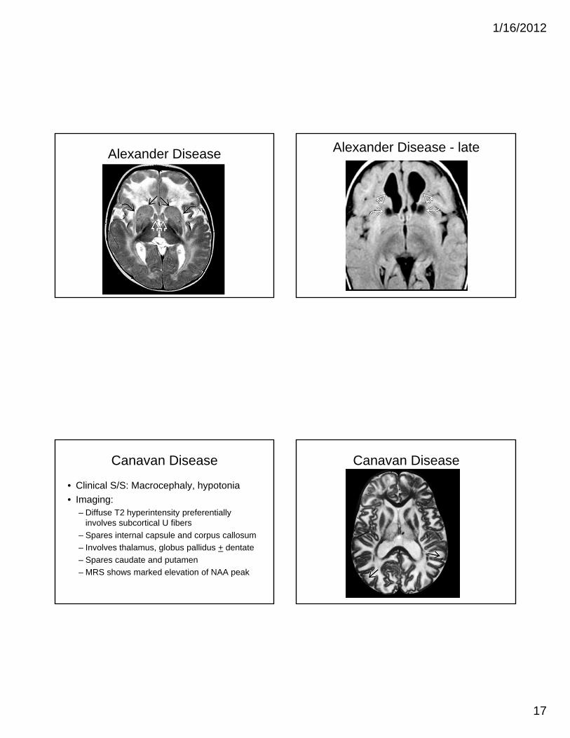
Alexander Disease
• Clinical S/S: macrocephaly, seizures• Mutation: GFAP, Chromosome 17q21• Imaging
– Extensive WM changes with frontal predominance– Abnormal signal in BG and thalami– Enhancement: ventricular lining, periventricular rim,
frontal WM, optic chiasm, fornix, BG, thalamus, dentate nucleus
• Give contrast to all unknown cases of hydrocephalus and abnormal WM

1/16/2012
16
Alexander Disease Alexander Disease
Alexander Disease Alexander Disease
1/16/2012
17
Alexander Disease Alexander Disease - late
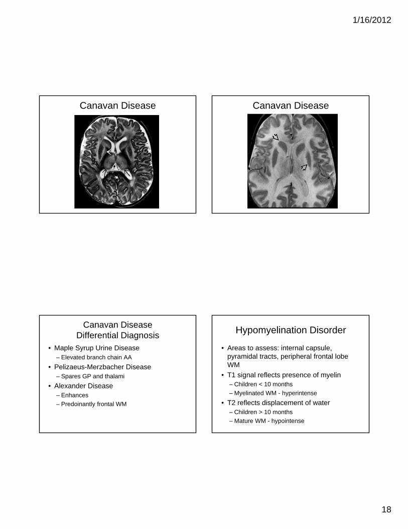
Canavan Disease
• Clinical S/S: Macrocephaly, hypotonia
• Imaging:– Diffuse T2 hyperintensity preferentially
involves subcortical U fibers
– Spares internal capsule and corpus callosum
– Involves thalamus, globus pallidus + dentate
– Spares caudate and putamen
– MRS shows marked elevation of NAA peak
Canavan Disease
1/16/2012
18
Canavan Disease Canavan Disease
Canavan DiseaseDifferential Diagnosis
• Maple Syrup Urine Disease– Elevated branch chain AA
• Pelizaeus-Merzbacher Disease– Spares GP and thalami
• Alexander Disease– Enhances
– Predoinantly frontal WM
Hypomyelination Disorder
• Areas to assess: internal capsule, pyramidal tracts, peripheral frontal lobe WM
• T1 signal reflects presence of myelin– Children < 10 months
– Myelinated WM - hyperintense
• T2 reflects displacement of water– Children > 10 months
– Mature WM - hypointense

1/16/2012
19
Pelizaeus-Merzbacher Disease
• Clinical S/S: microcephaly, hypertonia, stridor
• Deficiency of proteolipid protein (PLP)• Hypomyelination disorder• Imaging
– Variable – Nonspecific and symmetrical abnormality of
WM– Lack of myelin
Pelizaeus-Merzbacher Disease
Vanishing white matter disease Hypomyelination

1/16/2012
20
Metachromatic Leukodystrophy
• Decreased arylsulfatase A – Central and peripheral demyelination
• Imaging– Confluent butterfly-shaped increased T2 signal deep
cerebral WM• Spares U fibers in early disease• Involves U fibers in late disease
– Sparing of perivenular myelin producing the tigroid appearance
– No enhancement of WM– May have enhancement of cranial nerves and cauda
equina
MLD
MLD Krabbe Disease
• aka Globoid cell leukodystrophy• Clinical S/S: irritability• CT: hyperdensity in thalamus, BG• MRI Imaging:
– Faint hyperintensities in thalamus and BG (T1W)– Ring like appearance around dentate nucleus (T2W)– PV WM hyperintensities (T2W)
• Initially spares U fibers
– Enlarged optic nerves and cranial nerves (T1W)• MRS: increased choline,myoinositol, decreased
NAA, lactate accumulation

1/16/2012
21
Krabbe DiseaseCT Scan
Krabbe Disease
Krabbe Disease Krabbe Disease

1/16/2012
22
Adrenoleukodystrophy (ALD)
• Rare, genetic disorder characterized by the breakdown or loss of the myelin sheath surrounding nerve cells in the brain
• Progressive dysfunction of the adrenal gland
• X linked recessive
Adrenoleukodystrophy
• slow mentation, lack of interest or hyperactivity
• dysphagia, dysarthria, • occasional psychotic
behavior• visual loss and hearing
loss• progressive hemiplegia
may appear• seizures in 1/3 of patients• vegetative in 1 to 2 years
Chromosome Xq28
ALD
• Always abnormal in neurologically symptomatic males
• Often provides first lead
• Predominately posterior white matter –80%
• Splenium of corpus callosum usually involved
ALD

1/16/2012
23
SSPE
• Nonspecific leukoencephalopathy
• MRS: – decreased NAA/Cr
– Increased Cho/Cr; Ins/Cr and Lac-Lip
SSPE
Gray Matter Metabolic Disorder
• Wilson Disease
• Leigh Disease
• PKAN
• 3-methylglutaconic aciduria
• Biotin dependent encephalopathy
• Methylmalonic acidemia
Leigh Syndrome
• Progressive neurodegenration• Respiratory chain disorder• Clinical S/S: psychomotor delay/regression• Imaging:
– Bilateral symmetric increased T2/FLAIR signal• Putamen>caudate>GP, periaqueductal gray,
SN/STN, dorsal pons, cerebellar nuclei
– Restrictive diffusion in areas of acute disease
• MRS: increased lactate; decrease NAA, increase choline

1/16/2012
24
Leigh Syndrome Leigh Syndrome
Leigh SyndromePantothenate kinase-associated
neurodegeneration (PKAN)
• Neurodegeneration with brain iron accumulation (NBIA)
• Clinical S/S: progressive dystonia, dysarthria, rigidity, and pigmentary retinopathy
• Approximately half of the individuals have identifiable mutations in the PANK2 gene
• 'eye-of-the-tiger' sign found to have at least one mutation in PANK2
• Differential Diagnosis: signal changes in GP

1/16/2012
25
PKAN PKAN
Differential diagnosis
• Abnormalities of plasma ceruloplasmin concentration or copper metabolism (Wilson Disease)
• Evidence of neuronal ceroid lipofuscinosis
• Family history of Huntington disease or other dominantly inherited movement disorder
• Caudate atrophy
• ß-hexosaminidase A deficiency or GM1-galactosidase deficiency
Wilson Disease
• Clinical S/S: hepatic abnormality; psychiatric• Imaging:
– T1 shortening of GP– Symmetrical T2 hyperintensity or mixed signal intensity in
putamen, GP, CN, thalami– BG: initially enlarged due to edema than atrophy– ‘face of the giant panda’ – midbrain level (axial)
• Hyperintensity tegmentum except RN• Hypointensity superior colliculi• Normal signal lateral portion of pars reticularis of SN
– Increased signal intensity in cerebral and cerebellar WM (T2W)
– DWI + - acute/active

1/16/2012
26
Wilson disease Wilson Disease
Summary
• Neuroimaging has a role in the determining the etiology of pediatric neurologic conditions
• Serial MRI may provide for prognostication and progression of disease
• Knowledge of normal myelination pattern will decrease the chances of misinterpretation
• To obtain the most complete data set for pattern recognition, a systematic and comprehensive evaluation of brain structures is mandatory