neurological disorders chapter 13 pathology. brain anatomy cerebrum ◦reasoning ◦judgment...
TRANSCRIPT
Neurological DisordersNeurological Disorders
Chapter 13Pathology

Brain AnatomyBrain AnatomyCerebrum
◦ Reasoning◦ Judgment◦ Concentration,◦ Motor, sensory, speech
Cerebellum◦ Coordination
Brainstem◦ Cranial nerves◦ Respiratory center◦ Cardiovascular center
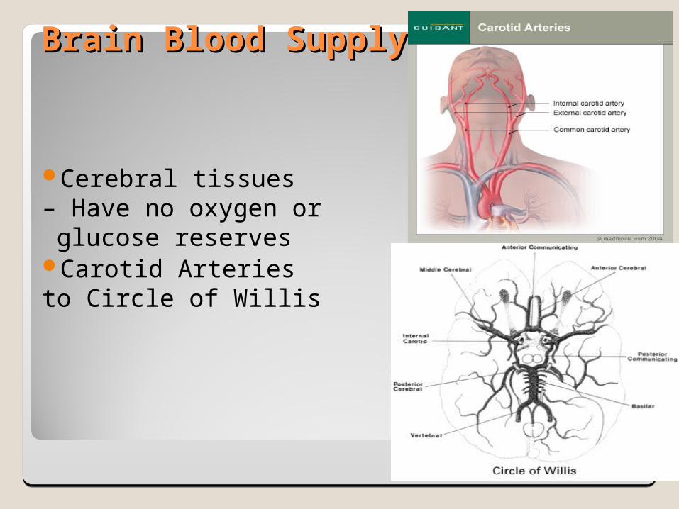
Brain Blood SupplyBrain Blood Supply
Cerebral tissues – Have no oxygen or glucose reservesCarotid Arteries to Circle of Willis
Intracranial Pressure (ICP)Intracranial Pressure (ICP)
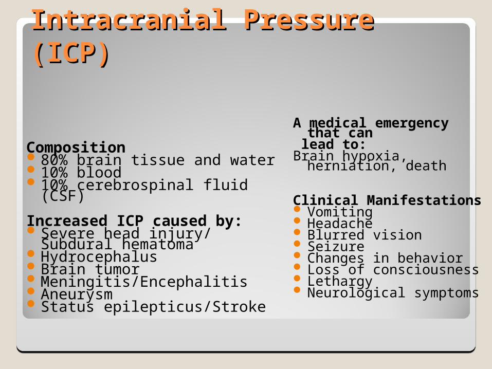
Composition80% brain tissue and water 10% blood 10% cerebrospinal fluid (CSF)
Increased ICP caused by:Severe head injury/ Subdural
hematomaHydrocephalusBrain tumorMeningitis/EncephalitisAneurysmStatus epilepticus/Stroke
A medical emergency that can
lead to:Brain hypoxia, herniation,
death
Clinical Manifestations Vomiting Headache Blurred vision Seizure Changes in behavior Loss of consciousness Lethargy Neurological symptoms
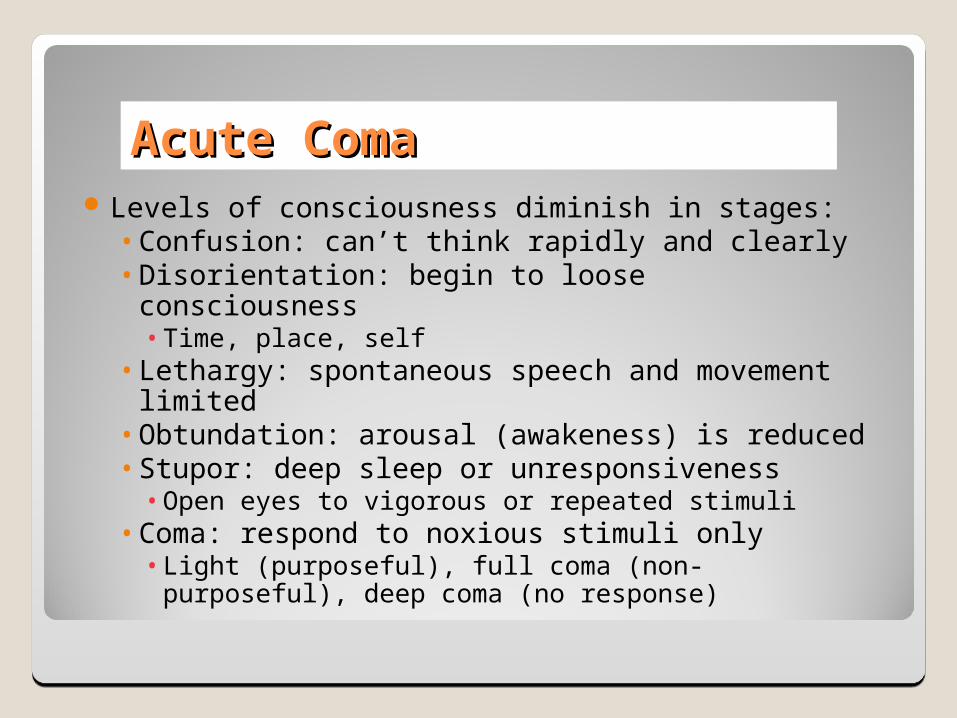
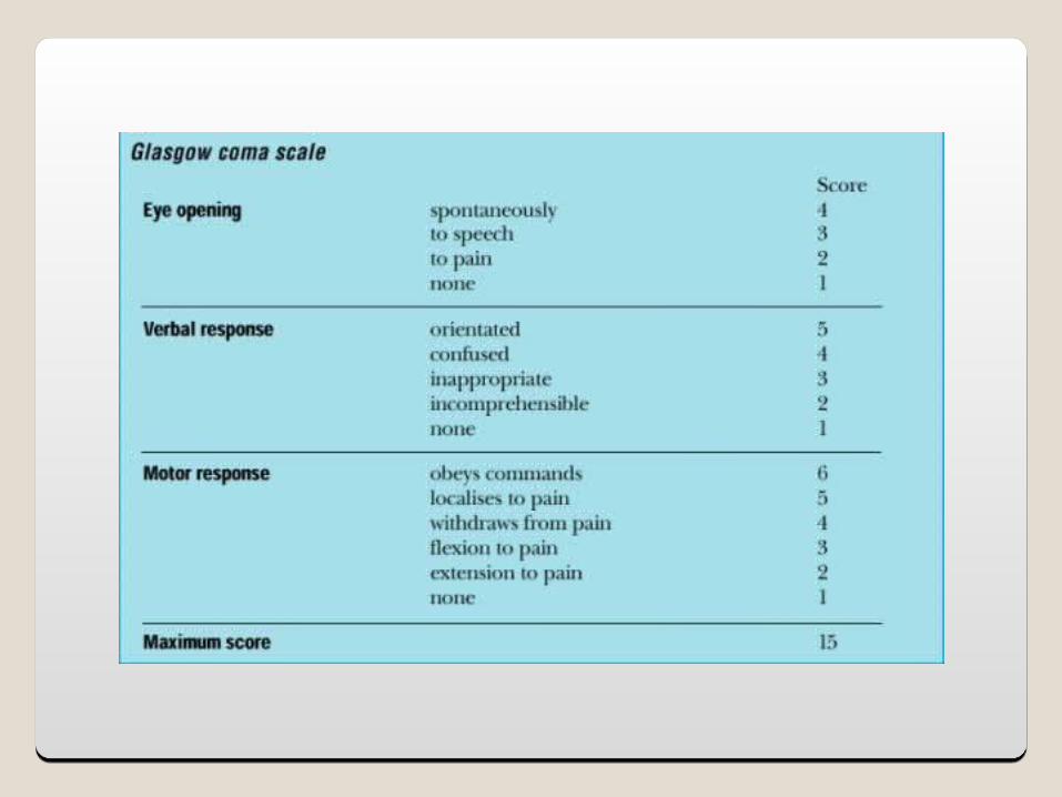
Acute ComaAcute ComaLevels of consciousness diminish in stages:• Confusion: can’t think rapidly and clearly• Disorientation: begin to loose consciousness• Time, place, self
• Lethargy: spontaneous speech and movement limited• Obtundation: arousal (awakeness) is reduced• Stupor: deep sleep or unresponsiveness • Open eyes to vigorous or repeated stimuli
• Coma: respond to noxious stimuli only• Light (purposeful), full coma (non-purposeful), deep
coma (no response)
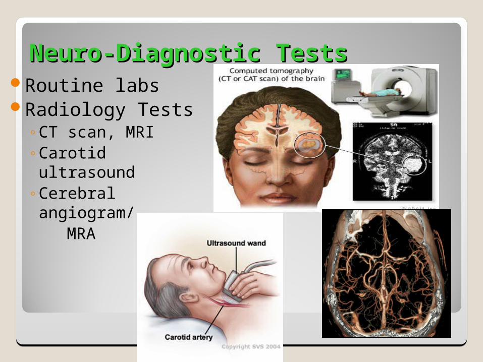
Neuro-Diagnostic TestsNeuro-Diagnostic TestsRoutine labsRadiology Tests
◦CT scan, MRI◦Carotid ultrasound◦Cerebral angiogram/ MRA
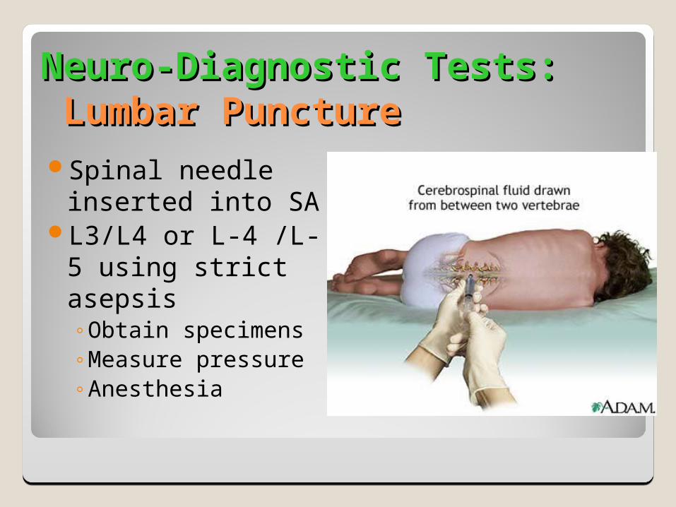
Neuro-Diagnostic Tests:Neuro-Diagnostic Tests: Lumbar Puncture Lumbar Puncture Spinal needle
inserted into SAL3/L4 or L-4 /L-5
using strict asepsis◦Obtain specimens◦Measure pressure◦Anesthesia
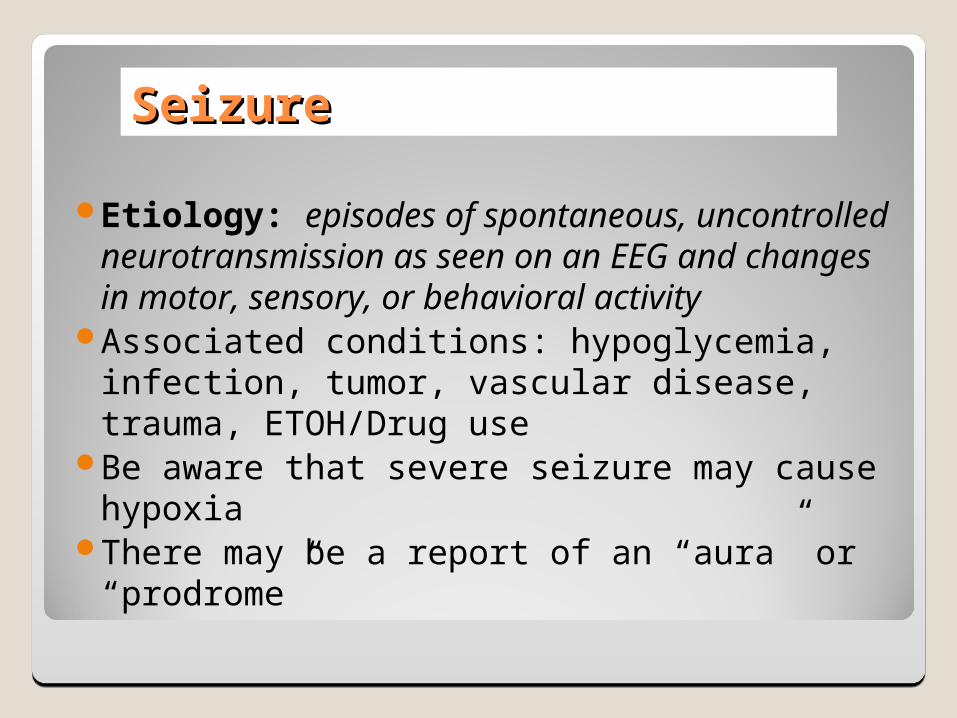
SeizureSeizure
Etiology: episodes of spontaneous, uncontrolled neurotransmission as seen on an EEG and changes in motor, sensory, or behavioral activity
Associated conditions: hypoglycemia, infection, tumor, vascular disease, trauma, ETOH/Drug use
Be aware that severe seizure may cause hypoxiaThere may be a report of an “aura” or “prodrome”
Generalized SeizureGeneralized Seizure
30% of the seizures Stem from the “deep brain”Impaired consciousness will always be
presentExamples:• Tonic, Clonic, or Clonic-tonic (Grand mal)• Absence seizures (Petit mal)• Simple vs. complex
Clinical evaluation tool: EEGhttp://www.vh.org/adult/patient/neurology/
electroencephalogramtest/index.html
Partial SeizurePartial Seizure
Also termed “focal seizures”Rise from the cortex part of the brainSimple: no impairment of consciousnessComplex: with impairment of
consciousness◦60%
DementiaDementia
A clinical syndrome that can be caused by various illnesses.• It is progressive failure of cerebral functions• e.g. mental abilities are affected• Orientation, recent memory, remote memory, language,
and behavior alterations• Etiological factors;• Tumors, trauma, infections, vascular disorders
• http://www.vh.org/adult/provider/neurology/alzheimers/index.html#TOC
Alzheimer’s DiseaseAlzheimer’s Disease
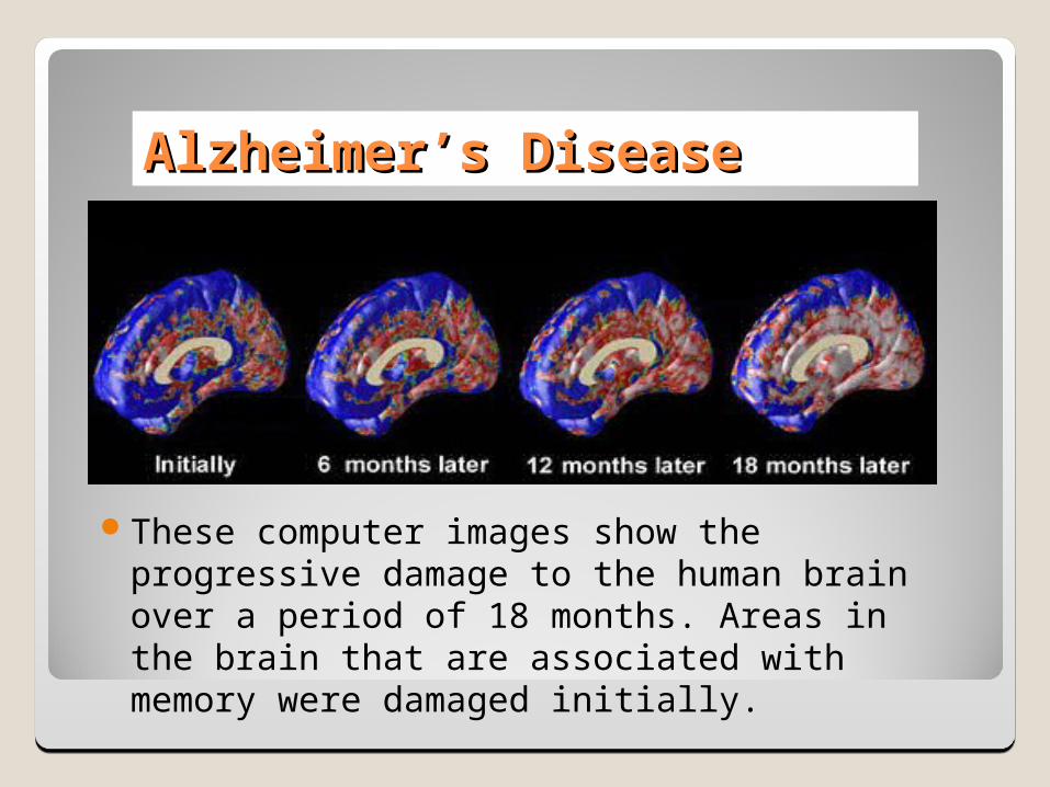
These computer images show the progressive damage to the human brain over a period of 18 months. Areas in the brain that are associated with memory were damaged initially.
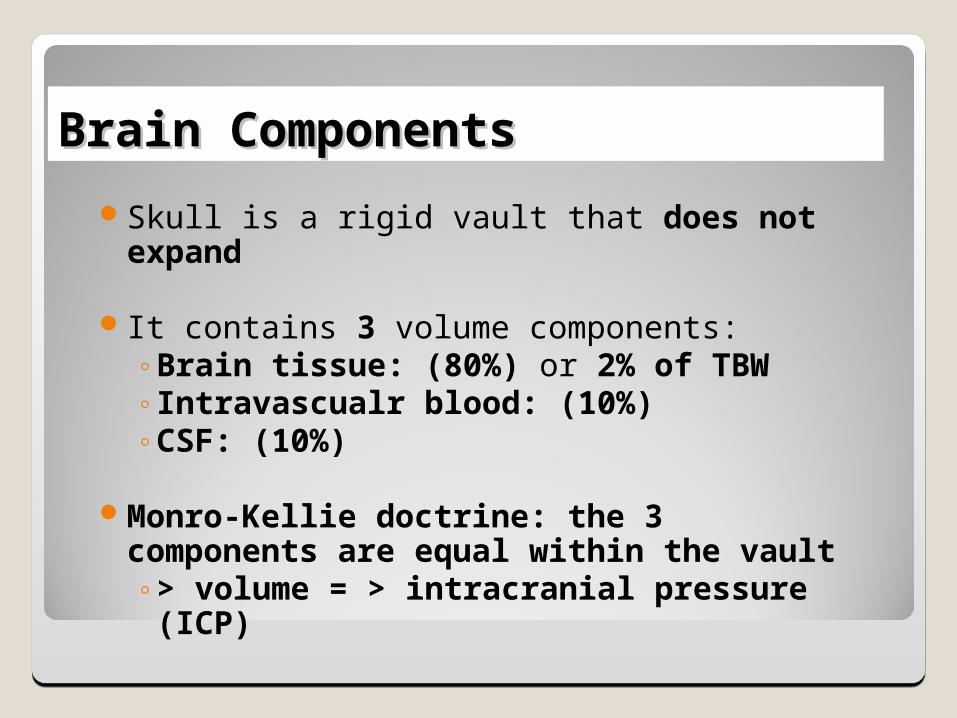
Brain ComponentsBrain Components
Skull is a rigid vault that does not expand
It contains 3 volume components:◦Brain tissue: (80%) or 2% of TBW◦Intravascualr blood: (10%) ◦CSF: (10%)
Monro-Kellie doctrine: the 3 components are equal within the vault◦> volume = > intracranial pressure (ICP)
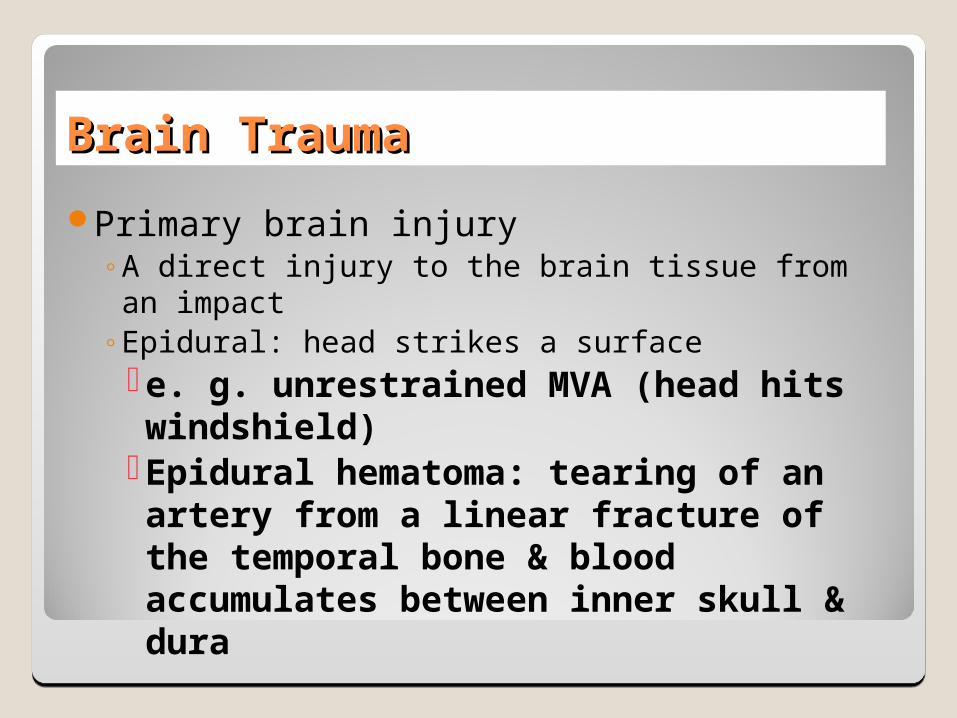
Brain TraumaBrain Trauma
Primary brain injury◦A direct injury to the brain tissue from an impact◦Epidural: head strikes a surface e. g. unrestrained MVA (head hits windshield)Epidural hematoma: tearing of an artery from a
linear fracture of the temporal bone & blood accumulates between inner skull & dura
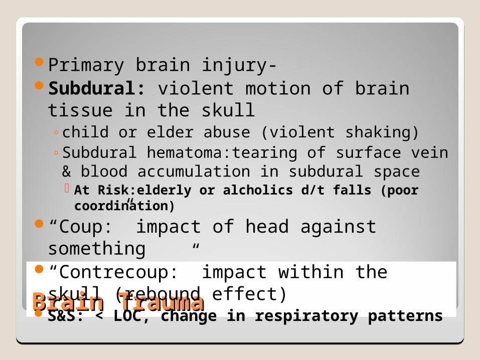
Brain TraumaBrain Trauma
Primary brain injury-Subdural: violent motion of brain tissue in the skull
◦child or elder abuse (violent shaking)◦Subdural hematoma:tearing of surface vein & blood accumulation
in subdural space At Risk:elderly or alcholics d/t falls (poor coordination)
“Coup:” impact of head against something“Contrecoup:” impact within the skull (rebound effect)S&S: < LOC, change in respiratory patterns
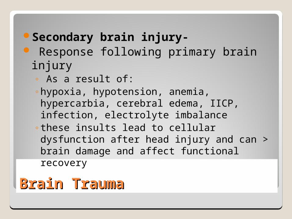
Brain TraumaBrain Trauma
Secondary brain injury- Response following primary brain injury
◦ As a result of:◦hypoxia, hypotension, anemia, hypercarbia, cerebral edema, IICP,
infection, electrolyte imbalance◦these insults lead to cellular dysfunction after head injury and can
> brain damage and affect functional recovery
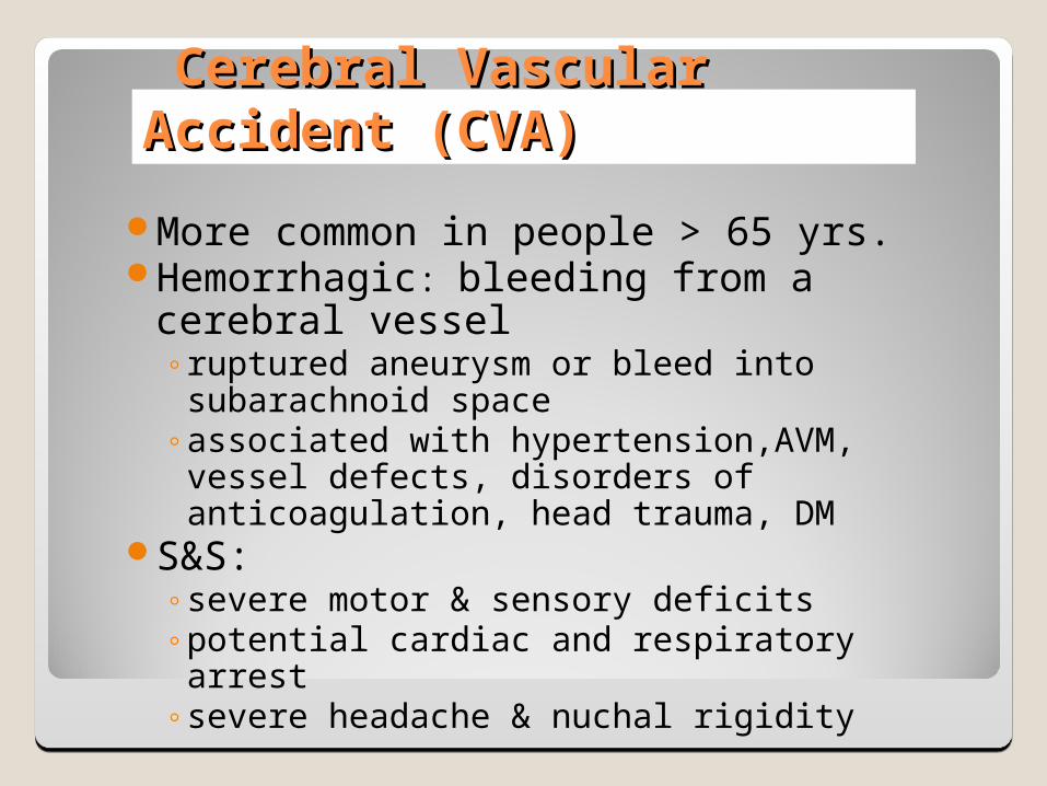
Cerebral Vascular Accident (CVA)Cerebral Vascular Accident (CVA)
More common in people > 65 yrs.Hemorrhagic: bleeding from a cerebral vessel
◦ruptured aneurysm or bleed into subarachnoid space◦associated with hypertension,AVM, vessel defects,
disorders of anticoagulation, head trauma, DMS&S:
◦severe motor & sensory deficits◦potential cardiac and respiratory arrest ◦severe headache & nuchal rigidity
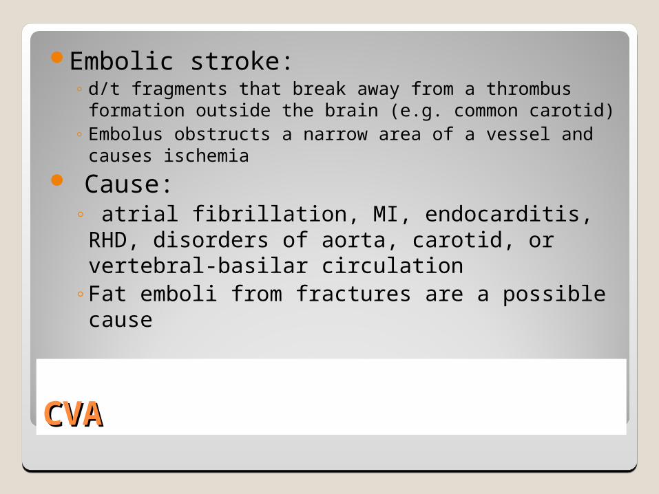
CVACVA
Embolic stroke:◦ d/t fragments that break away from a thrombus
formation outside the brain (e.g. common carotid)◦ Embolus obstructs a narrow area of a vessel and causes
ischemia Cause:
◦ atrial fibrillation, MI, endocarditis, RHD, disorders of aorta, carotid, or vertebral-basilar circulation
◦Fat emboli from fractures are a possible cause
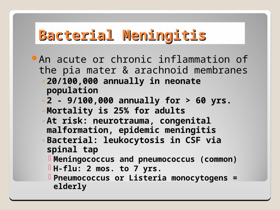
Bacterial MeningitisBacterial Meningitis
An acute or chronic inflammation of the pia mater & arachnoid membranes◦20/100,000 annually in neonate population◦2 - 9/100,000 annually for > 60 yrs.◦Mortality is 25% for adults◦At risk: neurotrauma, congenital malformation, epidemic
meningitis◦Bacterial: leukocytosis in CSF via spinal tap
Meningococcus and pneumococcus (common) H-flu: 2 mos. to 7 yrs. Pneumococcus or Listeria monocytogens = elderly
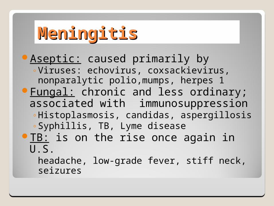
MeningitisMeningitisAseptic: caused primarily by
◦Viruses: echovirus, coxsackievirus, nonparalytic polio,mumps, herpes 1
Fungal: chronic and less ordinary; associated with immunosuppression ◦Histoplasmosis, candidas, aspergillosis◦Syphillis, TB, Lyme disease
TB: is on the rise once again in U.S.headache, low-grade fever, stiff neck, seizures
Clinical PresentationsClinical Presentations
Bacterial:◦ Systemic: fever, tachycardia, chills, petechial rash◦ Irritation: general throbbing h/a, photophobia, nuchal
rigidity◦ Neurological: cranial nerve damage and irritation◦ CN II: papilledema (> ICP), blindness◦ CN III, IV, VI: ptosis, diplopia, visual field problems◦ CN V: photophobia◦ CN VII: facial paresis◦ CN VIII: deafness, tinnitus, vertigo
Signs of MeningitisSigns of Meningitis
Brudzinski’s: passive flexion of the neck produces pain & increased rigidity
Kernig’s: Flex hip and knee and then straighten the knee…pain or resistance?
Opisthotonos: back & extremities arch backward in a spasm & the body rests on head & heels
Current FindingsCurrent Findings
Meningococcal Disease◦ Risk: crowded living quarters, cold or flu, active or
passive tobacco use, deficient immune system, alcohol consumption
Meningococcemia◦ More deadly disease; symptoms mimic flu;
Telltale “purple rash”◦Size of a pinhead or as a large as a quarter◦Medical attention is imperative
Future improvement in current vaccine Conjugate vaccine: sets off a stronger immune response
http://www.nytimes.com/2003/02/11/health/11MENI.html?ex=1046023735&ei=1&en=73abb2d0332e82f3
Major DepressionMajor Depression
Etiology: precise cause is unknownHypothesis: A neurochemical deficiency
◦monoamine deficiency ( serotonin or norepinephrine)◦a depressed mood or anhedonia (lack of passion) for at least 2
consecutive weeks and having 3 symptoms change in appetite or weight, change in sleep pattern, agitation,
fatigue, feelings of worthlessness or guilt > loss of work…more than other chronic disorders
Major DepressionMajor Depression
Clinical S &S:◦dysphoria, < activity, <libido, wt. loss or gain, anxiety,
pessimism, hopelessness, lack of energyPrevention & Tx: < risk factors may reduce
episodes; antidepressant drugs; regular exercise (> release of endorphins)
60 % of suicides d/t depression ( 18,000/ yr. in USA)
SchizophreniaSchizophrenia
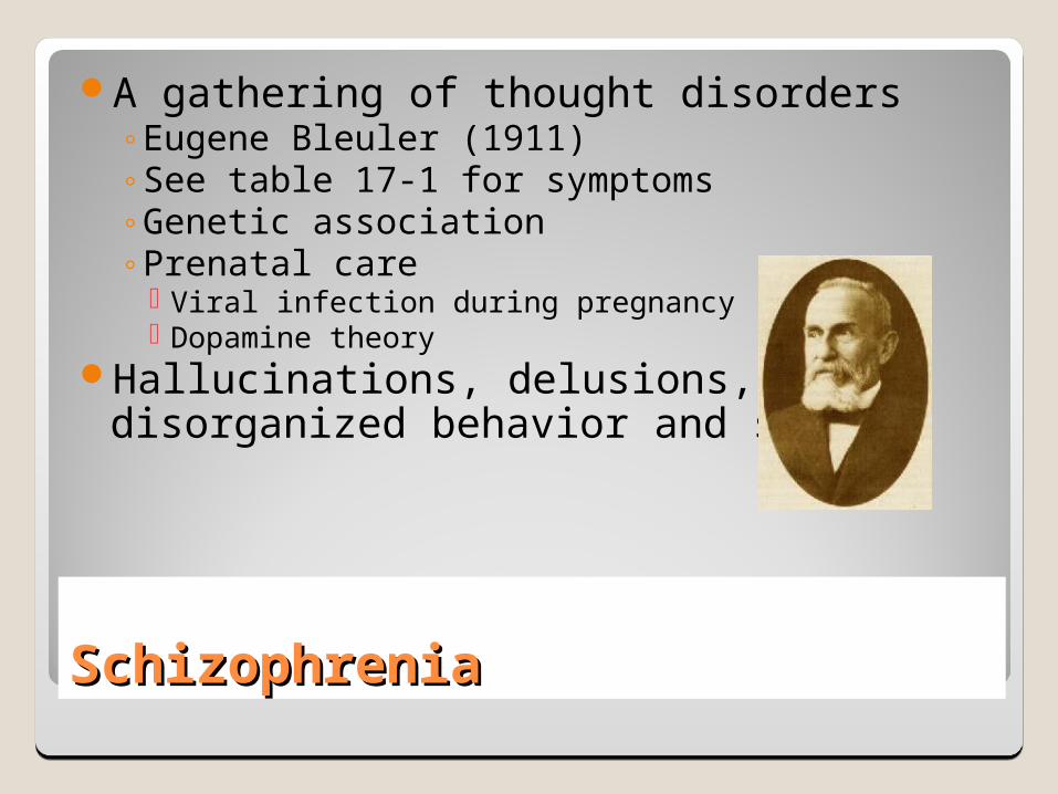
A gathering of thought disorders◦Eugene Bleuler (1911) ◦See table 17-1 for symptoms◦Genetic association◦Prenatal care
Viral infection during pregnancy Dopamine theory
Hallucinations, delusions, disorganized behavior and speech