neurological emergencies -...
TRANSCRIPT
NEUROLOGICAL
EMERGENCIES WILLIAM J. FREIBERG, DO
AKRON NEUROLOGY, INC.
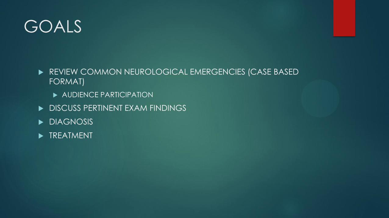
GOALS
REVIEW COMMON NEUROLOGICAL EMERGENCIES (CASE BASED
FORMAT)
AUDIENCE PARTICIPATION
DISCUSS PERTINENT EXAM FINDINGS
DIAGNOSIS
TREATMENT
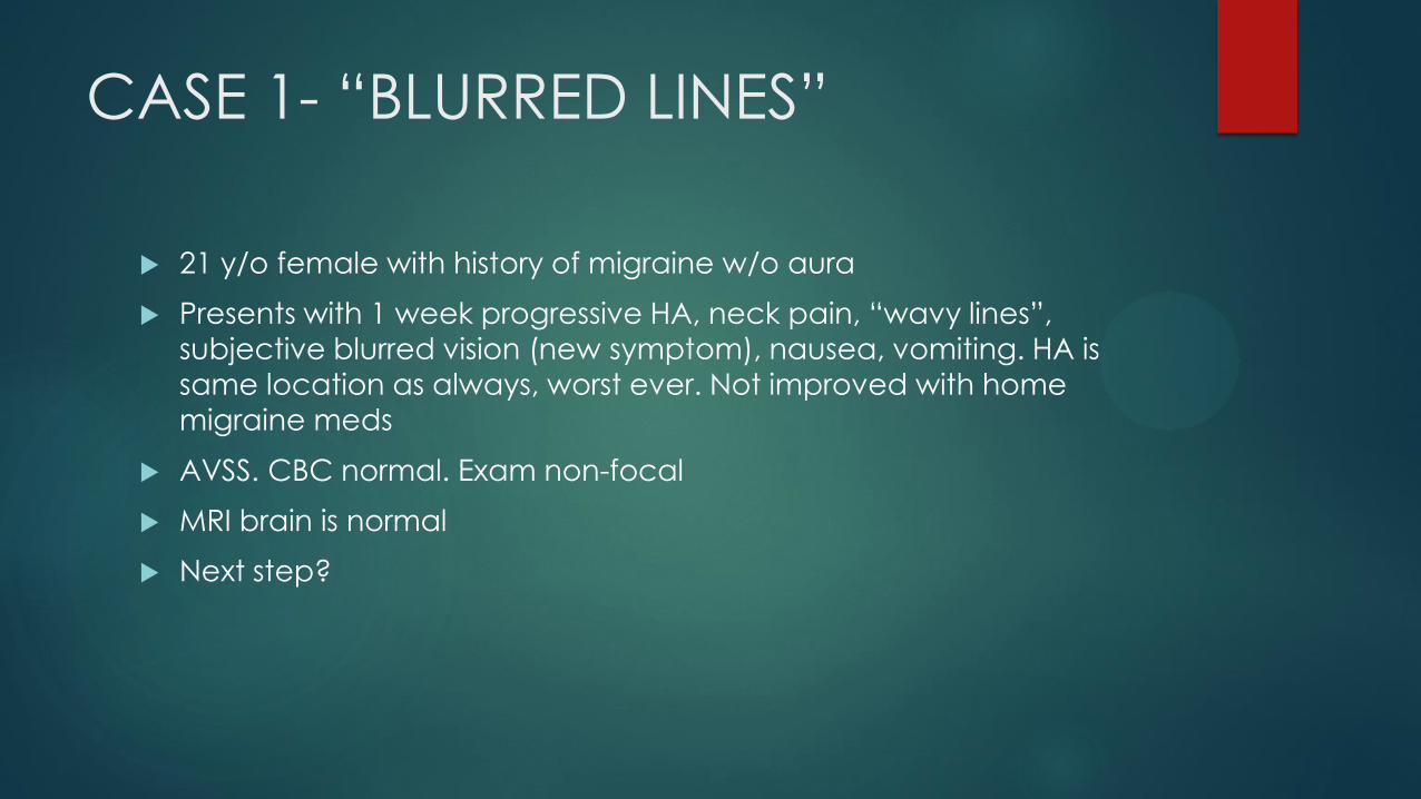
CASE 1- “BLURRED LINES”
21 y/o female with history of migraine w/o aura
Presents with 1 week progressive HA, neck pain, “wavy lines”,
subjective blurred vision (new symptom), nausea, vomiting. HA is
same location as always, worst ever. Not improved with home
migraine meds
AVSS. CBC normal. Exam non-focal
MRI brain is normal
Next step?
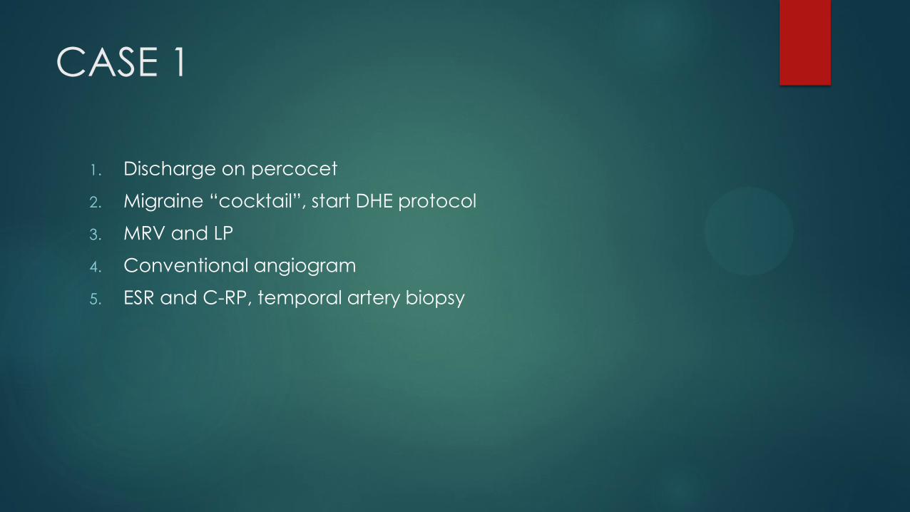
CASE 1
1. Discharge on percocet
2. Migraine “cocktail”, start DHE protocol
3. MRV and LP
4. Conventional angiogram
5. ESR and C-RP, temporal artery biopsy
CASE 1- (Idiopathic Intracranial
Hypertension)
Etiology Unknown
More common in young, obese females. Associations: hypothyroid,
Vitamin A, Tetracycline, steroids, prior trauma.
DX: MRI/ MRV imaging: always rule out sinus thrombosis. Inquire
about OCP, miscarriage, DVTs
RX:
LP (also therapeutic). Opening pressure > 20 cm water
Diamox
Vision loss: consider optic nerve sheath fenestration/ ophthalmology
consult
Shunt/drain placement
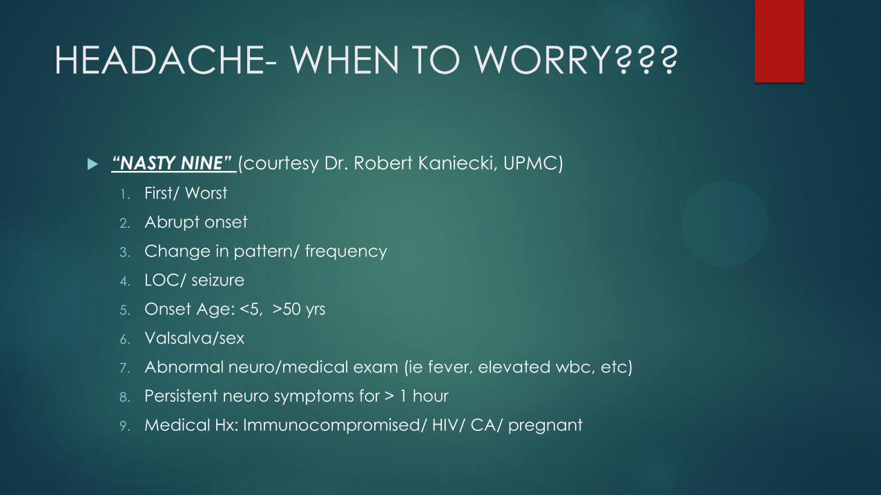
HEADACHE- WHEN TO WORRY???
“NASTY NINE” (courtesy Dr. Robert Kaniecki, UPMC)
1. First/ Worst
2. Abrupt onset
3. Change in pattern/ frequency
4. LOC/ seizure
5. Onset Age: <5, >50 yrs
6. Valsalva/sex
7. Abnormal neuro/medical exam (ie fever, elevated wbc, etc)
8. Persistent neuro symptoms for > 1 hour
9. Medical Hx: Immunocompromised/ HIV/ CA/ pregnant
CASE 2: HEAD DROP
65 y/o man with history HTN, HPL
Presents with 3 months progressive dysarthia, dysphagia, diplopia,
difficulty climbing stairs. Occasionally improved in AM. Recent URI.
Worsening for 3 days.
EXAM: severe dysarthria, diffuse limb weakness, neck flexion
markedly weak with head drop, b/l ptosis. Sensory exam normal. Reflexes depressed/ symmetric
MRI brain/ C spine normal as outpatient 10 days prior
Next step?
CASE 2
1. Discharge to home. Arrange outpatient EMG/ NCS
2. LP- look for albuminocytologic dissociation
3. STAT EMG/ NCS
4. Tell the patient he likely has ALS. Consult Hospice.
5. Admit to ICU, Prepare for intubation
6. CT angiogram head/neck
CASE 2- Myasthenic Crisis
Def: Myasthenia Gravis exacerbation requiring intubation
Etiology: AchR antibodies compete with Ach at N-M junction.
EXAM: fatigable motor weakness. Normal sensory.
DX: exam/hx, AchR Ab (most specific), Rep Stim on Nerve conduction studies (80% sensitive for generalized), Single fiber EMG (90% sensitive). MusK antibody- rare cases
REMEMBER 3 ANTIBODIES: Binding (most common), blocking, modulating
RX:
Exacerbation: steroids, eventually steroid sparing agent (cellcept, immuran). Mestinon (does not modulate disease)
Acute Crisis: IVIG or plasmapheresis. Start steroids/long term immunosuppression. Mestinon for symptoms
CASE 3: “UNCONSCIOUS”
58 y/o female. HTN, HPL, DM
Presents to ED. Normal 5 hours prior. Progressive confusion, gait
difficulty, diplopia, dizziness, diffuse weakness, drowsy.
ED: AVSS. irregular breathing. EXAM: does not follow commands,
dysconjugate eyes, no movement, hyperreflexia. BMP/CBC/INR
normal
EKG: a-fib. NCCTH: “no acute process”
Next Step?
CASE 3
1. STAT MRI Brain
2. STAT EEG, give ativan
3. Get labs: HFP, CPK, NH3, ESR. Prepare for LP.
4. STAT CT angiogram head/neck
5. Administer Dantrolene
CASE 3: Acute Basilar Occlusion/
basilar syndrome
Any combination of the following:
Diplopia/ ophthalmoparesis/ dysconjugate gaze
Dizziness
Weakness: unilateral OR bilateral face/ limbs
Dysarthria
Ataxia
Confusion/Depressed consciousness/ coma
Vision loss
CASE 3: Acute Basilar Occlusion
Some Etiologies
cardioembolus (a-fib in this case)
atheroembolus from posterior vessel disease: basilar, vertebral, aortic
arch
Vertebral artery dissection- stroke
HC state
CASE 3: Acute Basilar Occlusion
Treatment
IV tPA if candidate (which this patient is not)
NOTIFY ANGIO INTERVENTION
If TPA given, repeat vessel imaging immediately- usually conventional
angio. Mechanical intervention if no recannulization.
Investigate etiologies
MRI later-already know diagnosis
STROKE- KEY POINTS
1. Suspect stroke- STAT CTH to rule out hemorrhage
2. Next immediate question: “ARE VESSELS OPEN?”
3. Don’t delay vessel imaging if possible. GET HEAD and NECK.
- CTA head/neck
4. Always Remember- anterior AND posterior circulation.
CASE 4: “SPEECHLESS”
73 y/o female. Hx a-fib on Coumadin, HTN, HPL, posterior L MCA
stroke, mild residual R side weakness.
acute language loss at dinner, R side weakness.
ED 2 hours from onset. VSS, basic labs normal. Glucose normal. INR
2.5.
EXAM: Following no commands, global aphasia, mild R hemiparesis, eyes transiently deviate to the Right.
STAT CTH and CTA head neck: “no acute process”, “large vessels all
patent. Chronic infarct left posterior MCA”.
Next step?
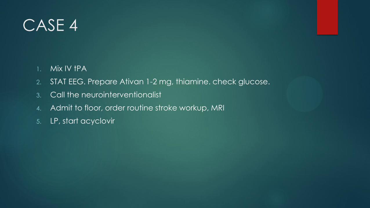
CASE 4
1. Mix IV tPA
2. STAT EEG. Prepare Ativan 1-2 mg, thiamine. check glucose.
3. Call the neurointerventionalist
4. Admit to floor, order routine stroke workup, MRI
5. LP, start acyclovir
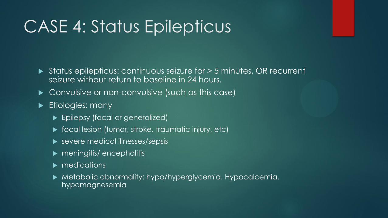
CASE 4: Status Epilepticus
Status epilepticus: continuous seizure for > 5 minutes, OR recurrent seizure without return to baseline in 24 hours.
Convulsive or non-convulsive (such as this case)
Etiologies: many
Epilepsy (focal or generalized)
focal lesion (tumor, stroke, traumatic injury, etc)
severe medical illnesses/sepsis
meningitis/ encephalitis
medications
Metabolic abnormality: hypo/hyperglycemia. Hypocalcemia. hypomagnesemia
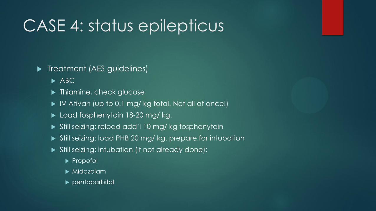
CASE 4: status epilepticus
Treatment (AES guidelines)
ABC
Thiamine, check glucose
IV Ativan (up to 0.1 mg/ kg total. Not all at once!)
Load fosphenytoin 18-20 mg/ kg.
Still seizing: reload add’l 10 mg/ kg fosphenytoin
Still seizing: load PHB 20 mg/ kg. prepare for intubation
Still seizing: intubation (if not already done):
Propofol
Midazolam
pentobarbital
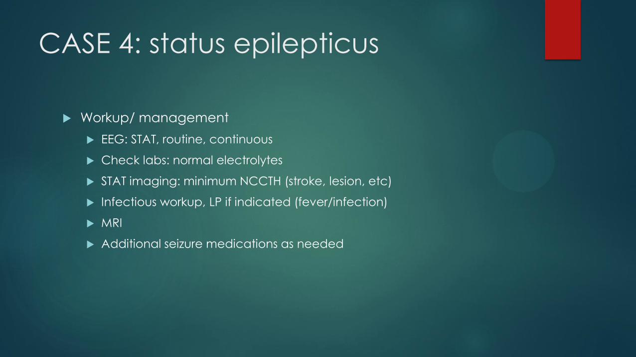
CASE 4: status epilepticus
Workup/ management
EEG: STAT, routine, continuous
Check labs: normal electrolytes
STAT imaging: minimum NCCTH (stroke, lesion, etc)
Infectious workup, LP if indicated (fever/infection)
MRI
Additional seizure medications as needed
CASE 5: “On pins and needles”
29 y/o male. No prior PMHx. 5 days progressive paresthesias- toes
followed by finger tips. Back pain. Difficulty walking- legs are
“heavy”
Afebrile. HR in 120s. Neuro exam: proximal arm and leg weakness.
Length-dependent vibration and sensory loss to the knees, mid
forearms. Cannot touch nose with eyes closed. Absent brachioradialis, Achilles reflexes.
Basic labs normal
Immediate next step?
CASE 5
1. STAT CTA head/ neck
2. Administer IVIG
3. STAT EMG/ NCS
4. NCCTH- rule out bleed
5. STAT MRI C-spine
CASE 5: AIDP (Guillain-Barre
syndrome)
Etiology: ?? Presumed autoimmune reaction against myelin of
peripheral nerves
Result: acute sensory-motor demyelinating peripheral neuropathy
(most peripheral neuropathies are axonal)
Exam: Acute, length dependent neuropathy. Areflexia.
Back pain, dysautonomia, constipation/urinary retention not uncommon (demyelinating thoracic nerves)
AXONAL VARIANTS: AMAN, AMSAN, ASAN
MILLER-FISCHER VARIANT: ataxia, ophthalmoplegia, areflexia: GQ1B Ab
CASE 5: AIDP (Guillain-Barre
syndrome)
DX:
NEURO HX/EXAM!!!
CSF: albuminocytologic dissociation (elevated protein, normal/ mild
elevation WBC). Don’t wait for it.
EMG/NCS: 11-14 days post-onset.
r/o: lyme, CMV, neoplastic/lymphoma
CASE 5: AIDP (Guillain-Barre
syndrome)
RX:
IVIG or plasmapheresis. NO STEROIDS
Supportive:
Check regular NIF/ VC.
Treat autonomic dysfunction (risk of death)
bowel regimen
treat back pain/ neuropathic pain
DVT prophylaxis
CASE 6: “SCARED STIFF”
1st day on new service. Called to bedside for decreased
responsiveness, pt. “not moving”.
70 y/o man hospital day 6. PMHx HTN, HPL. community acquired
Pneumonia. Day 2-delirium.
Next 3 nights: delirium, severe agitation.
Febrile 100.8, HR 110, BP stable. BUN 30, Cr 1.5 low urine output
Exam: stupor, non-verbal, not following commands, very rigid tone
NCCTH negative. CXR: improving PNA. UA normal. WBC normal
CASE 6
1. STAT EEG
2. STAT MRI
3. STAT CT angiogram head/ neck
4. Prepare for LP ASAP, give vanco/ceftriaxone/acyclovir
5. Ask the medical student to get his medication administration record
6. Intubate
CASE 6
Student reports he has received 10 mg Haldol total each night for
the past 3 nights, due to severe agitation. Reports: “less bright”
throughout the morning.
Next step?
CASE 6
1. STAT EEG
2. LP
3. STAT CPK, HFP. Prepare IV ativan, bromocriptine
4. Load with fosphenytoin
5. Give Provigil for hypoactive delirium
CASE 6: Neuroleptic Malignant
Syndrome Acute/subacute onset: fever, vital sign abnormalities, altered mental
status, parkinsonism/ rigidity
Etiologies:
Neuroleptics: Haldol, atypical antipsychotics. also lithium, VPA
Withdrawal of dopaminergic agents (i.e. sinemet)
Diagnosis: FEVERS
Fever
Elevated LFTs
Vital signs
Elevated CPK
Renal failure
Stiff
CASE 6
Diagnosis (cont)
Rule out infectious/medical causes
Medication review!!!
Treatment/mgmt (American journal of psychiatry,2000)
D/C neuroleptics
IV Ativan
Bromocriptine (DA agonist)
amantadine
Severe: Dantrolene, ECT
CASE 6
Treatment (cont.)
Supportive care, consider ICU
IVF
Monitor renal, liver function, CPK
Restart dopaminergic meds
CASE 7 “Quadraparesis”
19 y/o female. No PMHx. No Meds. Awoke with severe neck pain.
5 hours later: gait difficulty, arm weakness, diffuse numbness,
difficulty breathing
ED: respiratory failure. Intubated
Exam: AVSS. Wide awake, intubated. Blinks and moves eyes to
commands. Moves mouth to command. L eyelid appears drooped.
L pupil is asymmetrically small. No other CN findings. Quadroplegic.
No response to painful stimuli below tops of shoulders. Diffuse
hyperreflexia. Toes upgoing.
NCCTH: normal.
NEXT TEST???
CASE 7
1. STAT CTA head/ neck: attn. to basilar
2. STAT MRI brain: attention to Pons
3. STAT MRI C and T spine
4. Spinal angiogram urgently
CASE 7
DX?
1. Basilar occlusion
2. “locked in syndrome”
3. Vertebral dissection
4. AIDP
5. Transverse myelitis
FINAL THOUGHT
Always ask yourself:
1. Is there neurologic disease?
2. Does it localize?
3. If so, to where?
No substitute for the neuro exam
Essentials of stroke
r/o hemorrhage
vessels open?
Neurological emergencies are treatable!! Recognize them quickly!!

THANKS!!