neuromodulation workshop - …d3nqfeqdtaoni.cloudfront.net/images/nm_workshop... · fusf...
TRANSCRIPT

FUSF Neuromodulation Workshop - March 3-4, 2014 1
Neuromodulation Workshop
March 3-4, 2014, Charlottesville, VA
Introduction Neuromodulation – reversible stimulation or suppression of neural activity – can be induced by a
range of energies and technologies, including electrical (e.g. deep brain stimulation), chemical,
thermal, cryogenic, mechanical and magnetic (e.g. transcranial magnetic stimulation).
Neuromodulation could potentially enable a range of therapeutic benefits including: targeting of
regions in the brain for ablative procedures; suppressing epileptic seizures or symptoms of
psychiatric disorders; reversible nerve blocks to treat pain; and brain mapping. Although less
widely used, focused ultrasound can also induce neuromodulation, depending on the parameters of
the energy applied to neural tissue. This is achieved through either pulsing of focused ultrasound
using various sequences, or by subtly raising the temperature of the tissue.
Studies have shown that the mechanical effects of pulsed focused ultrasound can reversibly
decrease the functionality of targeted neurons. This allows for the temporary blocking of neural
signals from targeted locations within the brain or spinal/peripheral nerves. Such techniques hold
promise in the treatment of epilepsy or chronic pain.
Conversely, pulsed focused ultrasound can also be used to stimulate targeted neurons. Ultrasound
energy with specific pulse parameters can trigger the activation and propagation of neural signals
that could excite muscle contractions or stimulate specific areas of the brain; thus, focused
ultrasound may potentially be used for precise brain mapping, to enable a better understanding of
how the brain works by identifying how individual cells and complex neural circuits interact (also a
primary focus of the President’s recent BRAIN Initiative).
Finally, the thermal effects of focused ultrasound can also induce neuromodulation. When brain
tissue is raised to a slightly elevated temperature—lower than that required for thermal ablation—
neural signals may be reversibly suppressed in that area. This technique can be used to confirm the
precise target in the brain during neurofunctional treatments (e.g. essential tremor), before
delivering the therapeutic dose of ultrasound energy to permanently ablate the targeted neural
tissue.
The field of neuromodulation using focused ultrasound is growing, with many academic sites
directing their research towards a wide range of clinical applications. The Focused Ultrasound
Foundation has recognized the promise of this field and the need for collaboration to most
effectively drive the field towards clinical utility. To this end, the Foundation convened a workshop
on March 2-3, 2014 which included participation from several luminary investigators within the
field. This document presents the goals and outputs of the workshop, including a detailed roadmap
to achieve the first clinical use of focused ultrasound neuromodulation for targeting prior to
thermal ablation.

FUSF Neuromodulation Workshop - March 3-4, 2014 2
Goals
Collect an inventory of current state of the field. Identify important clinical indications for focused ultrasound neuromodulation. Develop a roadmap which will achieve the first clinical use of focused ultrasound
neuromodulation.
Participants
Luminaries in transcranial focused ultrasound, ultrasound physics, MR imaging, medical device manufacture, neurology, and neurosurgery
Academia, industry, FDA and FUSF represented.
Presenters
Jeff Elias (University of Virginia)
Jeff Elias provided a clinical background for the workshop by describing some of the historical uses
of neuromodulation and by suggesting some near term clinical applications. He described
neuromodulation as the reversible inhibition or excitation of neurons or neuronal circuits.
Historically this has been achieved by a variety of means, including electrical, chemical, thermal,
cryogenic, mechanical and magnetic [Dallapiazza 2014].
Neuromodulation (NM) with focused ultrasound has a number of potentially important indications
both acute and chronic, including pre-ablation brain mapping, diagnostic mapping of deep circuits,
acute seizure interruption and chronic treatment for psychiatric disorders.
Stereotactic ablation targets of immediate interest are the ventralis intermedius nucleus of the
thalamus (Vim) for tremor, the globus pallidus for dystonia/dyskinesia associated with Parkinson’s
disease and the subthalamic nucleus for Parkinson’s disease tremor and dyskinesia. These nuclei
are not defined on MR and require clinical guidance. These targets are surrounded by regions that
can be tested with NM to verify the target location prior to ablation. Heating to approximately 50°C
with FUS produces NM and is used in the context of ongoing movement disorder trials for clinical
targeting guidance. However, it was discussed that heating to this temperature approaches the

FUSF Neuromodulation Workshop - March 3-4, 2014 3
threshold for damage. Therefore the relatively low energy , non-thermal regime of FUS NM is highly
interesting as a safer, more repeatable physiological target verification tool. Even with
improvements in imaging, it will likely always be the case that clinical guidance via NM will be
required for targets within the thalamus. Anatomical mapping is insufficient and functional
mapping will be required for a number of indications, particularly those without immediate
therapeutic effects. NM could play an important role clinically and scientifically as a functional brain
mapping tool.
Yoav Levy (Insightec)
Yoav Levy discussed the current Insightec technology and future plans. Insightec’s immediate
priorities beyond neurofunctional lesioning are drug delivery and neuromodulation. Insightec is
part of the Brain Monitoring and Stimulation Toolkit (BMST) consortium working to integrate brain
neuromodulation and monitoring platforms. Consortium members include commercial, academic
and clinical partners. These include Brainsway, ElMindA, InSightec, Ornim, Alpha Omega, Technion,
Ben Gurion, and Bar Ilan. Insightec has academic partnerhship for neuromodulation with Technion
and Sheba.
Levy stated that the state of the field is immature, but with a
lot of activity. Currently, clinical requirements and the
technical specification of sonication parameters are not well
defined. The most important early application of FUS
neuromodulation is the verification of neurofunctional lesion
targeting, beginning with movement disorders (Essential
tremor and Parkinson’s disease) and eventually addressing
other indications including neuropathic pain, obsessive
compulsive disorder, depression and epilepsy. Interesting
targets are deep brain targets for now.
The current goal is to determine parameters for safe
neuromodulation. Small animal models have and are being
used to investigate FUS NM, with transition to large animal
models in progress. The group discussed anesthesia and
monitoring methods for conducting preclinical investigation of
NM. Anesthesia remains a challenge for animal studies. Better
monitoring methods need to be developed. The group discussed the use of EEG and fMRI in
addition to evoked potential monitoring.
Mark Schafer (Sonic Tech) – Dosimetry and Standards
Mark Schafer introduced Sonic Tech, which works with companies to bring US products to the
market. They do transducer design, work with the FDA and participate in standards work. He
discussed the issues involved with dosimetry and standards with FUS NM. There are several
challenges including the wide range of US frequency and exposure levels and the lack of a unified

FUSF Neuromodulation Workshop - March 3-4, 2014 4
standard for characterization and
reporting. It is important to understand
how standards are developed and who
approves them.
Characterization and reporting are
critical. Mechanisms need to be
identified so that experiments can be
controlled and replicated. Mechanical
Index (MI) is one of the most misapplied
parameters and was intended for
characterizing diagnostic US pulses only.
“Derating” to determine safe exposure is
complicated. The derating scheme is based on an average of tissues and does not provide actual
pressures and intensities:
where F is frequency and d is depth in tissue.
FDA limits are regulatory limits, not safety limits. Standards are developed to promote commerce
and should always follow technology development. Conformance to a standard is easier than
conducting a clinical trial. The FDA and EU work with the IEC standards body. IEC TC87 in
ultrasonics covers a range of US
applications. Working group 7
specializes in US surgical and
therapeutic equipment and meets
annually. High Intensity Therapeutic
Ultrasound (HITU) standards were
published in July 2013.
The concept of dose is not yet
defined for FUS NM. We don’t yet
understand which mechanical factor
is important. Dose also needs to be
defined for cavitation.
Jeff Anderson (University of Utah) – Neurodiagnostics
Jeff Anderson discussed work toward the neurodiagnostic uses of NM. His group is using a preterm
lamb model to investigate whether it might be possible to develop functional biomarkers ventilator
induced brain damage. In order to determine functional connectivity it would be valuable to be able
to stimulate at a point in the brain while measuring function everywhere. This would go beyond the
concept of a connectome by describing a causal network. In a mouse model it was possible to cause
activation and see the propagation of activity. This has not yet been successfully demonstrated in
the lamb model. BOLD effect was seen throughout the brain which was correlated with the FUS

FUSF Neuromodulation Workshop - March 3-4, 2014 5
pulsing scheme, but without evidence of activated functional networks. Might be attributed to MR
artifact due to water motion, but additional experiments are needed. Anesthesia is also a potential
issue contributing to the lack of definitive NM results. The successful combination of targeted FUS
NM with fMRI could have powerful scientific and clinical applications.
Jean-Francois Aubry (Institut Langevin, Paris)– Neuomodulation in Rats
A recent rat stimulation study [Medical physics 40:082902, 2013] was presented and discussed. A
250 kHz, 64 mm diameter transducer operating at 320 kHz was used. The animals were lightly
anesthetized with Ketamine (66 mg/kg) and Xylazine (13 mg/kg). The anesthesia allowed only a
limited working time and results proved variable from one experiment to the next on the same
animal. It was possible to stimulate discrete gross motor movements (eyes, whiskers, etc.) but the
effects were unpredictable. Heterodyne interferometer measurements of the pressure field inside
the skull showed a complex pattern. It was mentioned that Pierre Mourad’s group (University of
Washington) recently published good results at 2 MHz.
Pierre Pouget (ICM, Paris) – Neuromodulation in awake behaving monkeys
Pierre Pouget summarized a recent paper on NM in alert macaque monkeys. The monkeys were
trained to look away from a moving visual target (antisaccade) using juice reinforcement. The FUS
NM (320 kHz, 0.6 MPa, 100 ms) was targeted to the frontal eye field (FEF). The latency of eye
movement was measured with and without ultrasound stimulation. Latency was reduced with
ultrasound, similar to previous optogenetics results and superior to a transcranial magnetic
stimulation (TMS) study by the same group. Ipsilateral sonication reduced latency and contralateral
sonication showed no effect. The group is currently investigating the superior colliculus as a target.
Alexander Korb (UCLA) – Preparing for Low Intensity Focused Ultrasound Pulsation
(LIFUP) first in human trials
Alexander Korb described work ongoing at UCLA to develop a non-invasive clinical
neuromodulation system for reaching deep cortical structures. Intended therapeutic indications for
the device include epilepsy, depression and anxiety. There may also be brain mapping applications
for presurgical planning and scientific inquiry. The device incorporates a single element 650 kHz
ultrasound transducer. Sonication parameters for stimulation and excitation build on the work of
Seung-Schik Yoo (Brigham and Women’s Hospital). The effects of transmission through bone were
examined finding the expected strong attenuation. However, the focus appeared to be maintained,
but with a 2-3 mm shift in focus location. Bone heating is not significant at expected exposure
levels. Experience with transcranial magnetic stimulation (TMS) shows NM effects only last for
milliseconds beyond the exposure, but it is hoped that chronic application of low intensity, pulsed
focused ultrasound may produce longer lasting effects. UCLA is nearing FDA IDE approval for an
initial clinical study on epilepsy patients.
Kim Butts-Pauly (Stanford) – Neuromodulation group
Kim Butts-Pauly reviewed a number of related research projects within her group at Stanford. They
are investigating retinal stimulation. A FUS NM study in a mouse model showed specificity of motor
stimulation via FUS NM between neck and tail muscles, but not between left and right sides of the

FUSF Neuromodulation Workshop - March 3-4, 2014 6
animal. Acoustic radiation force imaging (ARFI) is being developed to maximize signal to noise ratio
(SNR). The group discussed application to non-thermal localization of the focal spot for NM
applications. The clinical scenario for lesion making would be to verify the prescribed focal spot
location with ARFI, followed by neurophysiological verification of the target with NM, concluding
with thermal ablation to create a permanent lesion. A study was suggested to investigate potential
long-term effects of FUS NM exposure. Ultra short TE (UTE) MRI could be used to take skull bone in
to account. ARFI capability will be available soon on the GE/Insightec platform.
Matthew Meyers (FDA) – Neuromodulation and traumatic brain injury
Mattew Meyers discussed the FDA’s goals for internal research: understanding mechanisms and
parameters for different bioeffects of ultrasound in order to give insight for the review of devices
related to ultrasound ablation, neuromodulation and traumatic brain injury (TBI).
Specific work toward understanding
non-thermal, non-cavitational effects of
pressure waves on blast-induced TBI
was discussed. A HIFU pulse train can be
modulated to produce an envelope
similar to the shape of blast
overpressure. T radiation force profile is
also similar to blast overpressure
exposure. In a mouse model mild TBI
(mTBI) produced BBB disruption,
inflammation, immune responses and
behavioral disturbance, including
disrupted sleep patterns. The HIFU
model looks promising as a simulation of blast-induced TBI. His group is currently studying the use
of electrophysiological sensors for monitoring effects on mice. These include miniature implanted
electrode arrays. Going forward, monitoring devices will be used in concert with FUS to study the
effects of mTBI on mice.
An earthworm model for NM was presented. Suppression of action potential seemed to correlate
with cumulative radiation force impulse across multiple parameters and the number of pulses.
Eitan Kimmel (Technion) – Cell membrane dynamics
Eitan Kimmel presented 20 year old data from fish skin studies where spaces in cell membranes
were observed after US exposure. Other in vitro studies observed similar effects. These bilayer
spaces or sonophores could not be explained with radiation force, cavitation or streaming. Eitan
proposed that the pressure difference during US exposure between membrane components can
result in a variety of effects related to non-thermal mechanisms. A model of neuronal bilayer
sonophores (NBLS) has been developed which suggests that US stimulates neurons by changing
membrane capacitance.

FUSF Neuromodulation Workshop - March 3-4, 2014 7
Shy Shoham (Technion) – neuronal bilayer sonophore model
Shy Shoham continued the previous presentation with additional discussion of the NBLS model
(recently published in Phys Rev. X) and some applications. The NBLS model appeared to predict
results presented by King et al. The model suggests that with specific parameters, one can stimulate
inhibitory neurons . The Technion group has built an interface between Matlab and an NBLS
simulation they have created.
The group is working to develop methods to excite continuous patterns with an US phased array.
An application of this project is US image generation via retinal stimulation.
Zion Zibly (Sheba) – Clinical translation
Zion Zibly discussed clinical projects being planned at Sheba Medical Center. They are developing a
large animal model for showing feasibility and safety of MR guided HIFU neuromodulation. The first
indication will be the treatment of pain in end-stage cancer, with the ventral caudal (Vc) nucleus as
the target. Other indications to follow include dystonia, OCD, epilepsy and Alzheimers disease.
Outcomes
Neurofunctional ablation target verification was selected as the first application for development.
o Initial indication is thalamotomy for Essential and Parkinsonian tremor, or neuropathic pain.
o Roadmap created with timeline, milestones and responsibilities. o A goal was set to demonstrate neuromodulation successfully during a patient
treatment before the end of 2014.

FUSF Neuromodulation Workshop - March 3-4, 2014 8
An inventory of potential high impact clinical indications was created. A preliminary list of current research sites and investigators was drafted.
Next Steps
Create and publish a whitepaper detailing the workshop (draft and final). Collect and publish an inventory of neuromodulation sonication parameters. Follow up on first preclinical roadmap steps in two animal models (BWH: monkey,
UVa: pig) Define and announce specific details on a clinical neuromodulation prize, given to
the first investigator to illicit transient sensory symptoms or tremor suppression using focused ultrasound neuromodulation during a patient treatment (ET, PD or Pain).
Establish a neuromodulation investigator group email list. Schedule breakfast at the FUS symposium.

FUSF Neuromodulation Workshop - March 3-4, 2014 9
Neuromodulation Parameter Inventory
Authors Model Neural Response Target Intensity frequency total duration PRF pulse duration duty cycle
Yoo et al. 2011 rabbit muscle contraction motor cortex
12.6 W/cm^2 SPPA 6.3 W/cm^2 SPTA 690kHz >=1 s 10 Hz 50ms 50%
fMRI activation motor cortex
3.3 W/cm^2 SPPA 1.6 W/cm^2 SPTA 690kHz >=1 s 10 Hz 50ms 50%
p30 VEP component visual cortex
3.3 and 6.4 W/cm^2 SPPA 690kHz >=7-8 s 100 Hz 0.5 ms 5%
fMRI suppression visual cortex
3.3 W/cm^2 SPPA 0.160 W/cm^2 SPTA 690kHz >=7-8 s 100 Hz 0.5 ms 5%
Yoo et al. 2011 rat waking from anesthesia thalamus
6 W/cm^2 SPPA 650kHz 20 mn 100 Hz 0.5 ms 5%
Min et al. 2011 rat suppressing seizures thalamus
2.6 W/cm^2 SPPA 0.130 W/cm^2 SPTA 690kHz 3 min 100 Hz 0.5 ms 5%
Min et al. 2011 rat
increased dopamine increased serotonin decreased GABA (unpub) thalamus
3.5 W/cm^2 SPPA 0.175 W/cm^2 SPTA 650kHz 20 min 100 Hz 0.5 ms 5%
Kim et al. 2012 rat eye abduction abducens nerve
8.6 W/cm^2 SPPA 4.6 W/cm^2 SPTA 350kHz 200 ms 1.5 kHz 0.36 ms 54%
10 sets @ 1 Hz
King et al. 2013 mouse muscle contractions motor cortex
0.01-10 W/cm^2 SPTP 250-600kHz 80 ms CW 1 Figure 10
parameters
0.1-16.8 W/cm^2 SPTP 500kHz 80 ms CW 1 Figure 5
4.2 W/cm^2 SPTP 500kHz 20-320 ms CW 1 Figure 7
0.1-100 500kHz 80 ms 1.5 kHz 0.2 ms 30% FIgure 11

FUSF Neuromodulation Workshop - March 3-4, 2014 10
W/cm^2 SPPA 0.1-30 W/cm^2 SPTA
4.2, 16.8, 29.8 W/cm^2 SPPA 500kHz
40 ms - 10.9 s 120 pulses 11-3000 Hz 0.2 ms 0.22-60% Figure 12
Menz et al. 2013 salamander retinal stimulation retina
10-30 W/cm^2 SPTA 43MHz 1-5 min 0.5 Hz 500 ms 0.5
King et al. mouse muscle contractions motor cortex 500kHz 80 ms CW 1
localization
Deffieux et al. 2013 monkey antisaccade latency FEF
4 W/cm^2 SPPA 320kHz 100 ms CW
1
Legon et al. 2014 human
refined 2 point discrimination
somatosensory cortex
5.90 W/cm^2 SPPA 500kHz 500 ms 1 kHz 360 us 0.36
Fry et al., 1958 cat partial VEP suppression
lateral geniculate nuclueus not shown not shown 20-120 sec CW
Figure 1
Ballantine et al.1960 cat
functional respond of eye pupil
Edinger-Wesrpal nucleus
1700 W/cm^2 peak intensity 2.7 MHz 1-13 pulses
3 s pulse period 0.14 sec
Figures 7-11
Ballantine et al.1960 cat reversible enhancement spinal cord reflex
350 W/cm^2 peak intensity 2.7 MHz 3 pulses
1 s pulse period 0.3 s
Figure 13
"
following by depression
17-185 pulses 3 s pulse period 0.3 s
Younan et al.
2013 rat muscle contractions motor cortex
7.5 W/cm^2
=> 17.5
W/cm^2 SPPA
corrected w/
standing
waves 320kHz 250 ms 2 KHz 0.23 ms 50%
Deffieux et al.
2013 monkey antisaccade latency Frontal Eye Field
4 W/cm^2
SPPA 320kHz 100 ms CW 1
Legon et al.
2014 human
refined 2 point
discrimination
somatosensory
cortex
5.90 W/cm^2
SPPA 500kHz 500 ms 1 kHz 360 µs 0.36
FUSF Neuromodulation Workshop - March 3-4, 2014 11
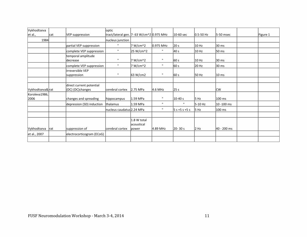
Vykhodtseva et al., cat VEP suppression
optic tract/lateral gen. 7- 63 W/cm^2 0.975 MHz 10-60 sec 0.5-50 Hz 5-50 msec
Figure 1
1984
nucleus junction
partial VEP suppression " 7 W/cm^2 0.975 MHz 20 s 10 Hz 30 ms
complete VEP suppression " 25 W/cm^2 " 40 s 10 Hz 50 ms
temporal amplitude decrease " 7 W/cm^2 " 60 s 10 Hz 30 ms
complete VEP suppression " 7 W/cm^2 " 60 s 20 Hz 30 ms
irreversible VEP suppression " 63 W/cm2 " 60 s 50 Hz 10 ms
Vykhodtseva& rat direct current potential (DC) (DC)changes cerebral cortex 2.75 MPa 4.6 MHz 25 s
CW
Koroleva1986, 2006
changes and spreading hippocampus 1.59 MPa " 10-40 s 5 Hz 100 ms
depression (SD) induction thalamus 1.59 MPa " " 5-10 Hz 10 -100 ms
nucleus caudatus 2.24 MPa " 5 s +5 s +5 s 5 Hz 100 ms
Vykhodtseva rat suppression of cerebral cortex
1.8 W total acoustical power 4.89 MHz 20- 30 s 2 Hz 40 - 200 ms
et al., 2007
electrocorticogram (ECoG)

FUSF Neuromodulation Workshop - March 3-4, 2014 12
Indications Functional target verification for lesioning (ET, PD, Pain) Brain Mapping
Neurodiagnostic biomarkers o Autism o Schizophrenia o Bipolar disorder o ADHD
Presurgical Mapping Replacement for invasive monitoring in epilepsy Eloquent area mapping for tumor or AVM resection
Epilepsy Therapy Lower excitability of tissue
Psychiatric Indications OCD Depression Obesity
Effectiveness of pharmacotherapy Wada test replacement
Memory localization Cancer pain treatment
Thalamic neuromodulaiton Stroke
Neuro rehab, plasticity stimulation Multimodal stimulation (TMS + FUS neuromodulation) Retinal Prosthetic
Research Sites Bolded sites have access to clinical transcranial focused ultrasound systems
Sheba Medical Center Technion – Iraeli Institute of Technology Brigham and Women’s Hospital ICM/Institut Langevin, Paris Stanford University Sunnybrook Health Sciences Center - University of Toronto Saint Mary’s Hospital, Korea University of Virginia Zurich University Children’s Hosptal University of California, Los Angeles Virginia Tech Carilion Research Institute University of Washington University of Arizona University of Utah Chang-Gung University, Taiwan FDA

FUSF Neuromodulation Workshop - March 3-4, 2014 13
Attendees
Name Organization
Eyal Zadicario InSightec
Yoav Levy InSightec
Mark Schafer Sonic Tech
Jeff Elias University of Virginia
Rob Dallapiazza University of Virginia
Jean-Francois Aubry Institut Langevin, Paris
Seung-Schik Yoo Brigham and Women’s Hospital
Nathan McDannold Brigham and Women’s Hospital
Natalia Vykhodtseva Brigham and Women’s Hospital
Dana Berneman Sheba Medical Center
Zion Zibly Sheba Medical Center
Eitan Kimmel Technion
Shy Shoham Technion
Matthew Myers FDA-OSEL
Kim Butts-Pauly Stanford
Patrick Ye Stanford
Jeff Anderson Utah
Pierre Pouget ICM, Paris
Dennis Parker University of Utah
Alex Korb UCLA
Neal Kassell Focused Ultrasound Foundation
John Snell Focused Ultrasound Foundation
Arik Hananel Focused Ultrasound Foundation
Jessica Foley Focused Ultrasound Foundation
FUSF Neuromodulation Workshop - March 3-4, 2014 14
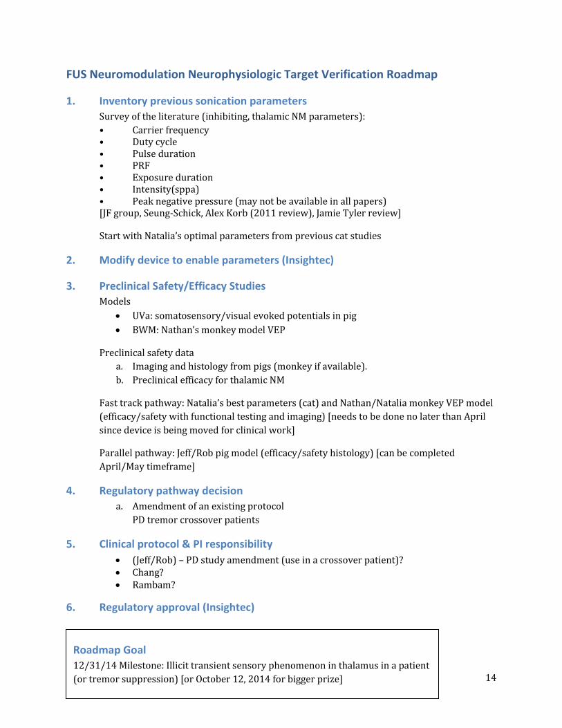
FUS Neuromodulation Neurophysiologic Target Verification Roadmap
1. Inventory previous sonication parameters
Survey of the literature (inhibiting, thalamic NM parameters):
• Carrier frequency • Duty cycle • Pulse duration • PRF • Exposure duration • Intensity(sppa) • Peak negative pressure (may not be available in all papers) [JF group, Seung-Schick, Alex Korb (2011 review), Jamie Tyler review]
Start with Natalia’s optimal parameters from previous cat studies
2. Modify device to enable parameters (Insightec)
3. Preclinical Safety/Efficacy Studies
Models
UVa: somatosensory/visual evoked potentials in pig
BWM: Nathan’s monkey model VEP
Preclinical safety data
a. Imaging and histology from pigs (monkey if available).
b. Preclinical efficacy for thalamic NM
Fast track pathway: Natalia’s best parameters (cat) and Nathan/Natalia monkey VEP model
(efficacy/safety with functional testing and imaging) [needs to be done no later than April
since device is being moved for clinical work]
Parallel pathway: Jeff/Rob pig model (efficacy/safety histology) [can be completed
April/May timeframe]
4. Regulatory pathway decision
a. Amendment of an existing protocol
PD tremor crossover patients
5. Clinical protocol & PI responsibility
(Jeff/Rob) – PD study amendment (use in a crossover patient)? Chang? Rambam?
6. Regulatory approval (Insightec)
Roadmap Goal
12/31/14 Milestone: Illicit transient sensory phenomenon in thalamus in a patient
(or tremor suppression) [or October 12, 2014 for bigger prize]

FUSF Neuromodulation Workshop - March 3-4, 2014 15