neuromuscular disease in children
TRANSCRIPT

NEURO-MUSCULAR DISEASE IN CHILDRENAllison Conravey, MD
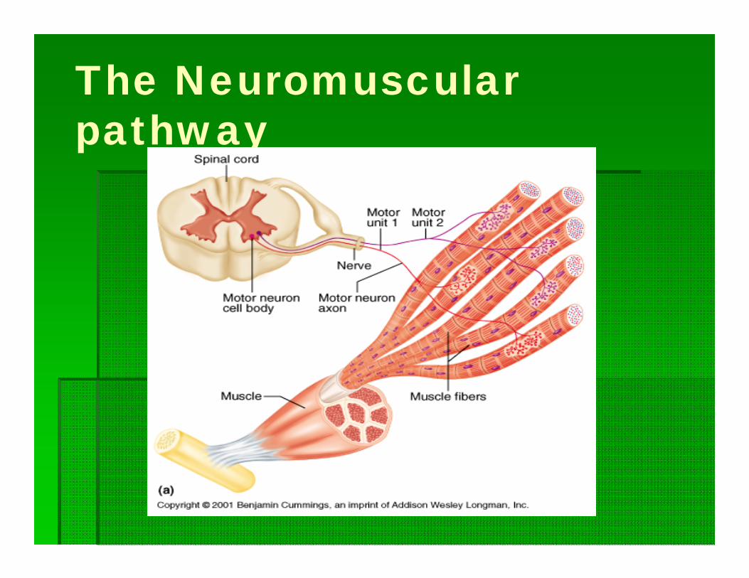
The Neuromuscular pathway
Signs and symptoms
Hypotonia- not improved with irritation Failure to reach milestones weakness Decreased reflexes
Muscle Diseases
Duchene's/ Becker's Muscular Dystrophy Congenital muscular dystrophy Congenital Myopathies Myotonic dystrophy Inflammatory myopathies Metabolic myopathies

Muscular dystrophies
4 criteria- 1. primary myopathies 2. genetically 3. progressive 4. myofiber degeneration at some stage
Duchene Becker Emery-Dreifess Facioscapulahumoral Limb Girdle Oculopharyngeal Distal congenital
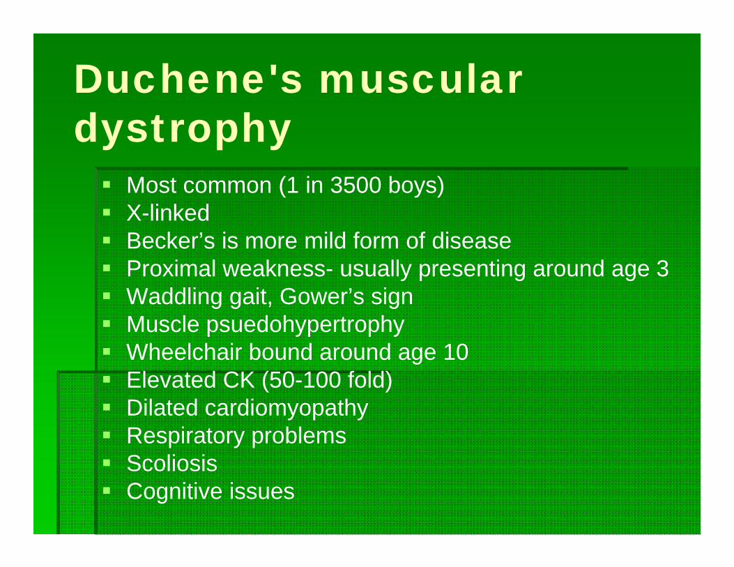
Duchene's muscular dystrophy Most common (1 in 3500 boys) X-linked Becker’s is more mild form of disease Proximal weakness- usually presenting around age 3 Waddling gait, Gower’s sign Muscle psuedohypertrophy Wheelchair bound around age 10 Elevated CK (50-100 fold) Dilated cardiomyopathy Respiratory problems Scoliosis Cognitive issues

Gower’s sign

Dystrophin Mutation in
dystrophin gene A cytoskeletal
protein, critical in stabilizing the link between the sarcolemmal membrane and the extracellular matrix
Without dystophin, there is sarcolemma breakdown and then muscle cell death

Duchene's Muscular dystrophy Diagnosed by clinical picture and elevated CK Molecular diagnosis available Muscle biopsy usually not needed Treatment consists of physical therapy,
orthopedics, cardiology and pulmonary therapies
Corticosteroids can help preserve ambulation Gene therapy in future

Congenital muscular dystrophy Heterogeneous group of disorders Most are automsomal recessive Weakness and hyporeflexia in first year of life Elevated CK (not as high as Duchene's) Muscle biopsy shows signs of myofibrillar necrosis and
regeneration, along with endomysial fibrosis and deposition of fat Some die in infancy and others may live into adulthood with
minimal disability Merosin (alpha-2 chain of laminin2) positive or merosin deficient Many are associated with CNS abnormalities (cobblestone
lissencephaly, MR, seizures, white matter changes) Fukyama type, Muscle-eye-brain disease, Walker-Warburg
syndrome, Ulrich form

Congenital myopathies Onset in early life with hypotonia, hyporeflexia, generalized weakness that is more often
proximal than distal, and poor muscle bulk Often with dysmorphic features that may be secondary to the weakness Relatively nonprogressive Hereditary Unique morphological features on histochemical or ultrastructural examination of the muscle
biopsy sample that originate within the myofiber Myopathies with protein accumulation
Nemaline myopathy Myosin storage myopathy Cap disease Reducing body myopathy
Myopathies with cores Central core disease Core-rod myopathy Multiminicore disease
Myopathies with central nuclei Myotubular myopathy Centronuclear myopathy
Myopathies with fiber size variation Congenital fiber type disproportion

Myotonic dystrophy I
Most common form of muscular dystrophy among whites
Prevalence 3-5/100,000 Usually appears in late adolescence or early
adult life Can present at birth (congenital myotonic
dystrophy) or during the first decade (juvenile myotonic dystrophy) (20%)
Myotonia with a dystrophic process of muscle with multisystem involvement
Caused by a CTG trinucleotide expansion on chromosome 19q13.3

Pathogenesis
Gene is DMPK which encodes a serine-threonine protein kinase (myotonia protein kinase [DMK])
The trinucleotide repeat does not affect the coding portion of the gene (transcribed to RNA but not to protein)
Explanation for clinical manifestations of DM1 is multifactorial
Study on mice showed abnormal accumulation of abnormally expanded RNA molecules resulted in defective splicing of the skeletal muscle chloride channel pre-mRNA (Mankodi et al, 2002)
The loss of chloride channel protein from the sarcolemma may lead to channel dysfunction and membrane hyperexcitability, resulting in myotonia
Genetics
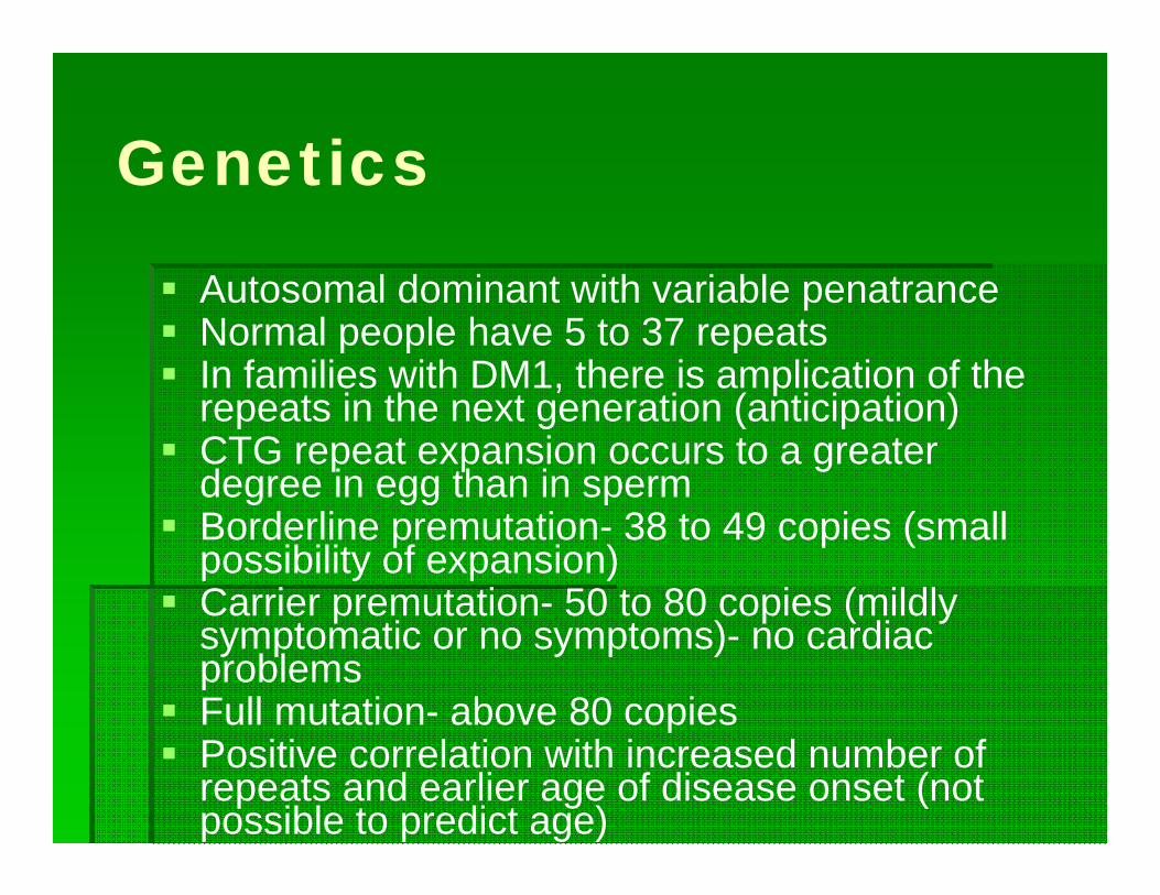
Autosomal dominant with variable penatrance Normal people have 5 to 37 repeats In families with DM1, there is amplication of the
repeats in the next generation (anticipation) CTG repeat expansion occurs to a greater
degree in egg than in sperm Borderline premutation- 38 to 49 copies (small
possibility of expansion) Carrier premutation- 50 to 80 copies (mildly
symptomatic or no symptoms)- no cardiac problems
Full mutation- above 80 copies Positive correlation with increased number of
repeats and earlier age of disease onset (not possible to predict age)
Clinical
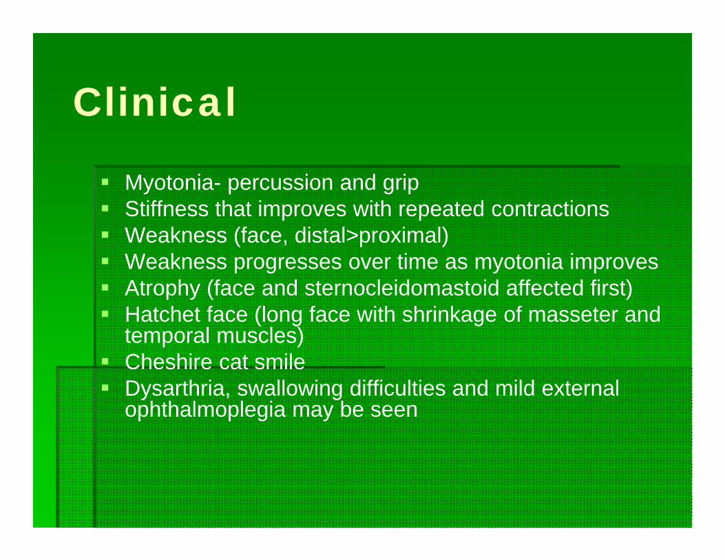
Myotonia- percussion and grip Stiffness that improves with repeated contractions Weakness (face, distal>proximal) Weakness progresses over time as myotonia improves Atrophy (face and sternocleidomastoid affected first) Hatchet face (long face with shrinkage of masseter and
temporal muscles) Cheshire cat smile Dysarthria, swallowing difficulties and mild external
ophthalmoplegia may be seen
Associated features
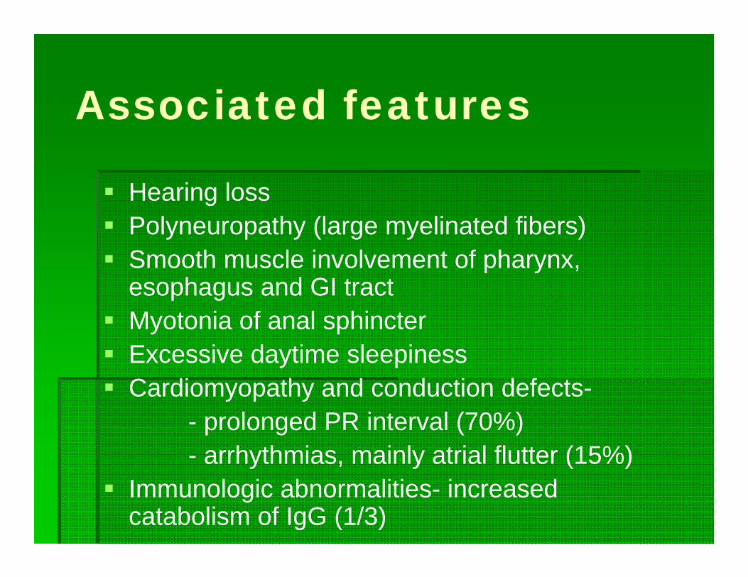
Hearing loss Polyneuropathy (large myelinated fibers) Smooth muscle involvement of pharynx,
esophagus and GI tract Myotonia of anal sphincter Excessive daytime sleepiness Cardiomyopathy and conduction defects-
- prolonged PR interval (70%)- arrhythmias, mainly atrial flutter (15%)
Immunologic abnormalities- increased catabolism of IgG (1/3)
Associated symptoms
Endocrinopathies-- frontal baldness- loss of body hair- testicular atrophy, infertility- hypothyroidism- growth hormone secretion disturbances- insulin-resistant diabetes (6.5-20%)
Ophthalmology- cataracts after age 10 Cognitive deficits in 80% Older adults have frontal and temporal lobe
cognitive decline

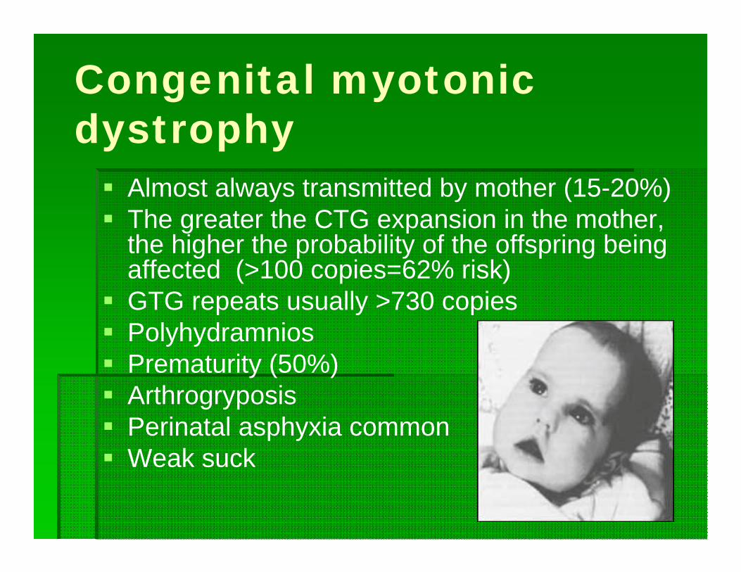
Congenital myotonic dystrophy Almost always transmitted by mother (15-20%) The greater the CTG expansion in the mother,
the higher the probability of the offspring being affected (>100 copies=62% risk)
GTG repeats usually >730 copies Polyhydramnios Prematurity (50%) Arthrogryposis Perinatal asphyxia common Weak suck

Congenital Myotonic Dystrophy Hypotonia, especially involving neck muscles Facial diplegia (87%) Delayed motor development (87%) Mental retardation (68%) Clinical myotonia is absent in 1st year- 12% of 1
to 5 year old children In 1st year- respiratory insufficiency, dysphagia
and aspiration, and problems with GI motility (16% mortality)
After 1st year- appear to improve for a few years, only to deteriorate gradually with features of the adult form of disease

Inflammatory Myopathies
Associated with a presumed immunologic attack
Polymyositis, Dermatomyositis and infectious myositis
Elevated CK with muscle weakness and pain

Dermatomyositis
Muscle pain and weakness associated with rash Can be acute or insidious Erythematous, scaly discoloration of eyelids which then
spreads to cheeks and extensor surfaces of joints (Grotton rash), elbows and knees
Increased ESR and WBC May have positive ANA MRI- increased signal in T2 with normal T1 Treatment- prednisone or other immunosuppressives

Polymyositis
Uncommon in children Chronic inflammatory process of muscles Cell mediated disease with abnormal T
lymphocytes Can begin as early as 1yo Can look like muscular dystrophy- more rapid
onset Antibodies to myosin in 90% Course is generally downhill
Metabolic myopathies
Muscle phosphorylase deficiency (McArdle’s)- onset in childhood with painful muscle cramps and weakness after exertion
Congenital defect of phosphofructokinase (Glycogenosis type VII)- easy fatigability and weakness of stiffness induced by exertion
Phosphoglyceromutase deficiency- life long history of muscle pain, cramps and weakness after exercise and MR
Aldolase A deficiency- unexplained episodes of jaundice and anemia and markedly elevated CK

Disorders of neuromuscular junction Myasthenia gravis Botulism Lambert-Eaton
Myasthenia Gravis
Autoimmune disease in which antibodies are directed against acetylcholine receptor antibodies which impair neuromuscular transmission and produce weakness

Types
Neonatal- transplacental passage of antibodies to baby Juvenile- <18yo, F>M Adult onset (early or late) Congenital- not autoimmune
-defects in proteins at NMJ- can be presynaptic, synaptic or postsynaptic

Generalized Myasthenia Gravis Eye findings (ptosis or extraocular muscle weakness)
most common- 50% at presentation and 90% at some time during illness
Double vision Pupil is always spared Bulbar symptoms are the next most common symptom Nasal speech Dysphagia Jaw weakness

Muscle weakness
Fluctuates and progressively worsens over the course of the day
Fatigability Painless Variable distribution and severity (may be asymmetric) Distal weakness less common Legs usually affected later Most common muscles- jaw closure, neck flexors,
deltoids, triceps Respiratory muscles may be affected Bowel and bladder spared

Physical exam
Pupils unaffected Fatigability- sustained upgaze, count to
100, repetitively testing proximal limb or neck flexors Subtle weakness- functional testing Ice bag test DTRs preserved No sensory findings
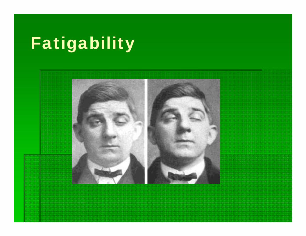
Fatigability

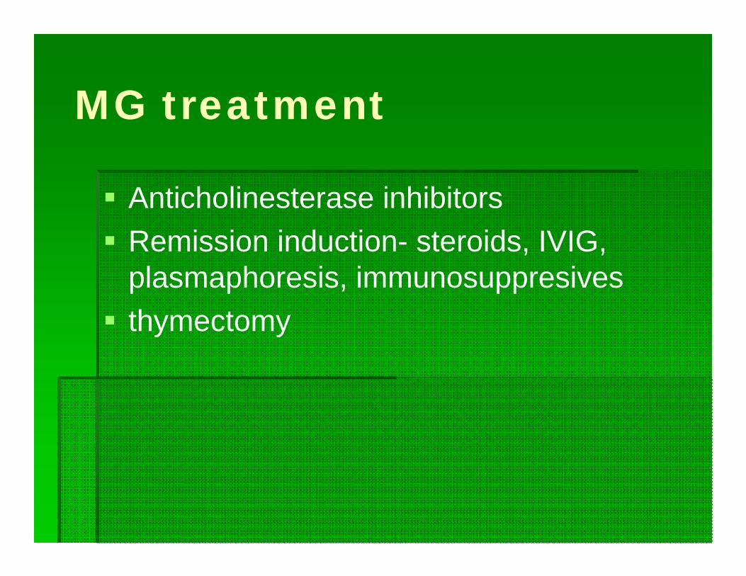
MG treatment
Anticholinesterase inhibitors Remission induction- steroids, IVIG,
plasmaphoresis, immunosuppresives thymectomy

Drugs and MG
A number of drugs have the potential to impair neuromuscular transmission and worsen MG
Antirheumatic: Penicillamine, chloroquine Antibiotics: amionglycosides> macrolides, floroquinolones Antiarrythmics: beta blockers, calcium channel blockers,
procainamide Anesthetic agents: Non-depolarizing agents (vecuronium,
pancuronium), succinylcholine Magnesium phenytoin Carnitine Ace inhibitors
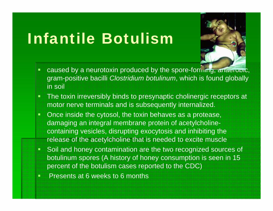
Infantile Botulism
caused by a neurotoxin produced by the spore-forming, anaerobic, gram-positive bacilli Clostridium botulinum, which is found globally in soil
The toxin irreversibly binds to presynaptic cholinergic receptors at motor nerve terminals and is subsequently internalized.
Once inside the cytosol, the toxin behaves as a protease, damaging an integral membrane protein of acetylcholine-containing vesicles, disrupting exocytosis and inhibiting the release of the acetylcholine that is needed to excite muscle
Soil and honey contamination are the two recognized sources of botulinum spores (A history of honey consumption is seen in 15 percent of the botulism cases reported to the CDC)
Presents at 6 weeks to 6 months
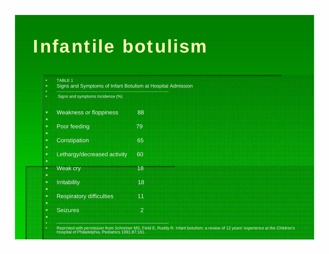
Infantile botulism TABLE 1 Signs and Symptoms of Infant Botulism at Hospital Admission -------------------------------------------------------------------------------- Signs and symptoms Incidence (%)
Weakness or floppiness 88 Poor feeding 79 Constipation 65 Lethargy/decreased activity 60 Weak cry 18 Irritability 18 Respiratory difficulties 11 Seizures 2 -------------------------------------------------------------------------------- Reprinted with permission from Schreiner MS, Field E, Ruddy R. Infant botulism: a review of 12 years' experience at the Children's
Hospital of Philadelphia. Pediatrics 1991;87:161.

Infantile Botulism
Treatment is supportive use of botulinum immune globulin in infants
has successfully reduced the time spent in the hospital and the need for mechanical ventilation and tube feeding
prognosis is excellent, with a case-fatality rate of less than 2 percent
Recovery results from the regeneration of nerve terminals and motor endplates.
Diaphragmatic function returns before peripheral muscle recovery.

Peripheral Neuropathies
Guillian- Barre syndrome (AIDP) CIDP Hereditary motor and sensory
neuropathy (CMT)
Guillian-Barre syndrome (AIDP) Acquired, immune mediated polyradiculoneuropathy
causing dysfunction, segmental demyelination and/or axonal degeneration is peripheral nerves, spinal sensory and motor nerve roots, and cranial nerves
1-2/100,000 per year Previous infection in 2/3 of children (URI or AGE) Most frequent between ages 4 and 9 Associated with pain and parasthesias Usually symmetric ascending paralysis Ataxia (44%) CN involvement
GBS
Can have papilledema Can have impaired vital capacity Dysautonomia Sphincter disturbances in 1/3 Decreases or absent reflexes Increased CSF protein- may have small increase in
WBC EMG- conduction block and prolonged F-waves Treatment- IVIG or plasma exchange Recovery usually is 2 months- may take up to 18
months

CIDP
Childhood incidence of 0.5/100,000 per year Like GBS but slower progression CSF- albumino-cytologic dissociation Treatment- steroids, IVIG, plasma
exchange, immunosuppresives
Hereditary motor and sensory neuropathy Also called Charcot-Marie-Tooth disease (CMT) Peroneal muscular atrophy CMT1- AD
- extensive segmental demyelination and remyelination of peripheral nerves
- thickening of peripheral nerves CMT2- less common axonal form CMT3 (Dejerine-Sottas syndrome)- most severe
demyelinating form
CMT 1
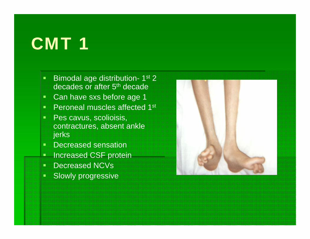
Bimodal age distribution- 1st 2 decades or after 5th decade
Can have sxs before age 1 Peroneal muscles affected 1st
Pes cavus, scolioisis, contractures, absent ankle jerks
Decreased sensation Increased CSF protein Decreased NCVs Slowly progressive

CMT 2
Genetically heterogeneous Axonal degeneration of peripheral nerves Clinical picture similar to CMT 1 with
slower progression

CMT 3 (Dejerine-Sottas Disease) Onset of sxs in infancy
or early childhood progressing to severe disability
AD or AR Hypotonia, muscle
atrophy and weakness Facial weakness Very slow NCVs

Anterior horn cell disease
Spinal Muscular Atrophy Infection (polio, enteroviruses, rabies)

Spinal Muscular Atrophy AR 1 in 10,000-25,000 Mutation in SMN1- 2 copies SMA 1 (Wednig-Hoffman)- ¼ SMA 2- ½ SMA 3 (Kugleburg Welander) Symmetric proximal weakness Atrophy Diaphragm spared until late Cardiac and smooth muscle
spared Tongue fasciculation Absent DTR’s Some sensory symptoms
Blood marker for SMN1 gene CK- normal or mild elevation EMG- fibs and fasics Biopsy- dennervation atrophy
SMA 1
Acute and rapidly progressive Almost always fatal by 3yo

SMA 2
Symptoms by 18 months Tremor in upper extremities CK increased up to 5x normal May have gastroc hypertrophy

SMA 3
Present 18 months to adult Proximal muscle weakness Impaired joint mobility
Poliomyelitis
Affects motor units of spinal cord and brain Mostly affects anterior horns cells Clinically ranges from non specific febrile
illness to a severe and potentially fatal paralytic disease
Paralytic polio follows aseptic meningitis Pain, fasciculations, twitching and decreased
DTR’s Polio-like syndrome- seen with west nile virus
and other viruses