neuromuscular effects of stun device...
TRANSCRIPT
Journal of Surgical Research 143, 78–87 (2007)
Neuromuscular Effects of Stun Device Discharges
Daniel J. Valentino, M.D.,*,† Robert J. Walter, Ph.D.,*,†,1 Andrew J. Dennis, D.O.,*,† Kimberly Nagy, M.D.,*,†Michele M. Loor, M.D.,*,† Jerry Winners, B.S.,* Faran Bokhari, M.D.,*,† Dorion Wiley, M.D.,*,†
Azher Merchant, B.S.,† Kimberly Joseph, M.D.,*,† and Roxanne Roberts, M.D.*,†
*Cook Country Trauma Unit, Chicago, Illinois; and †Department of General Surgery, Rush University Medical Center, Chicago, Illinois
Submitted for publication January 8, 2007
doi:10.1016/j.jss.2007.03.049
Background. Stun guns or electromuscular incapac-itation devices (EMIs) generate between 25,000 and250,000 V and can be discharged continuously for aslong as 5 to 10 min. In the United States, over 200,000individuals have been exposed to discharges from themost common type of device used. EMI devices arebeing used increasingly despite a lack of objectivelaboratory data describing the physiological effectsand safety of these devices. An increasing amount ofmorbidity, and even death, is associated with EMI de-vice use. To examine this type of electrical injury, wehypothesized that EMI discharges will induce acute ordelayed cardiac arrhythmia and neuromuscular in-jury in an animal model.
Methods. Using an IACUC approved protocol, fromMay 2005 through June 2006 in a teaching hospitalresearch setting, 30 Yucatan mini-pigs (24 experimen-tals and 6 sham controls) were deeply anesthetizedwith ketamine and xylazine without paralytics. Ex-perimentals were exposed to discharges from an EID(MK63; Aegis Industries, Bellevue, ID) over the femo-ral nerve on the anterior left hind limb for an 80 sexposure delivered as two 40 s discharges. EKGs,EMGs, troponin I, CK-MB, potassium, and myoglobinlevels were obtained pre-discharge and post-dischargeat 5, 15, 30, and 60 min, 24, 48, and 72 h (n � 6 animals)and 5, 15, and 30 d post-discharge (n � 6 animals ateach time point). Skin, skeletal muscle, and peripheralnerve biopsies were studied bilaterally. Data were com-pared using one-way analysis of variance and pairedt-tests. P-values <0.05 were considered significant.
Results. No cardiac arrhythmias or sudden deaths
1 To whom correspondence and reprint requests should be ad-dressed at Department of Trauma, Rm. 1300, Stroger Hospital ofCook County, 1900 West Polk St., Chicago, IL 60612. E-mail:
[email protected].780022-4804/07 $32.00© 2007 Elsevier Inc. All rights reserved.
were seen in any animals at any time point. No evi-dence of skeletal muscle damage was detected. No sig-nificant changes were seen in troponin I, myoglobin,CK-MB, potassium, or creatinine levels. There were nosignificant changes in compound muscle action poten-tials (CMAP). No evidence of conduction block, con-duction slowing, or axonal loss were detected on EMG.M-wave latency (Mlat, ms), amplitude (Mamp, mV), area(Marea, mV-ms), and duration (Mdur, ms) were not signif-icantly affected by MK63 discharge compared withcontralateral or sham controls. F-wave latency (Flat,ms), a sensitive indicator of retrograde nerve conduc-tion and function, was not significantly affected byMK63 discharge compared with contralateral or shamcontrols. No significant histological changes were seenat any time point in skeletal muscle or peripheralnerve biopsies although mild skin inflammation wasevident.
Conclusions. There was no evidence of acute ar-rhythmia from MK63 discharges. No clinically signifi-cant changes were seen in any of the physiologicalparameters measured here at any time point. Neu-romuscular function was not significantly altered bythe MK63 discharge. In this animal model, evenlengthy MK63 discharges did not induce muscle ornerve injury as seen using EMG, blood chemistry, orhistology. © 2007 Elsevier Inc. All rights reserved.
Key Words: electromuscular incapacitation; stun de-vice; EID; swine; metabolic acidosis; respiratory aci-dosis; dysrhythmia; ketamine; xylazine; EKG; EMG;electromyography; combined muscle action potential;troponin I; lactate; MK63; stun baton; heart; bloodpressure; CK-MB; pH; potassium; creatinine.
INTRODUCTION
Electrical discharges may produce a wide spec-
trum of injury [1]. The manifestations of electrical
79VALENTINO ET AL.: NEUROMUSCULAR EFFECTS OF STUN DEVICE DISCHARGES
injury depend on the waveform characteristics of thecurrent applied (AC, DC, mixed, strength, duration,frequency) and its anatomical location and paththrough the tissues of the body [2– 4]. Effects mayinclude skin burns, neuromuscular incapacitation,skeletal muscle death, cardiac arrhythmia, osteocyteand osteoblast death, and blood vessel endotheliumdysfunction [3– 8]. Electromuscular incapacitation(EMI) devices use high voltage (20 to 250 kV), lowfrequency (10 to 100 Hz), time-varying amperage (upto 18 A) DC current to produce pain and strongmuscle contractions resulting in the incapacitationof volitional movement. The utility of these effects inpersonal defense and law enforcement has led to theproliferation of EMI devices for use as an alternativeto lethal force. EMI devices have been shown to bevery effective when used to incapacitate combativeindividuals while reducing risk to officers, suspectsand bystanders [9]. All EMI devices generate timevarying DC current with waveforms that are similarbut distinctive to each specific device. However, theimmediate effects and safety profile of these dis-charges on living organisms are poorly understood[10 –12].
More than 100 fatalities have been reported to beassociated with EMI use in the United States for the2001–2004 period [9]. This growing list of fatalitieshas drawn a great deal of public attention and raisedquestions about the safety of EMI devices and theirpotential complications, especially their associationwith fatal ventricular arrhythmia [10 –13]. Morethan 100 types of EMI devices are marketed to lawenforcement agencies around the world to subduecombative subjects and are used increasingly by pri-vate citizens for personal protection. Despite the in-creasing usage of EMI devices there is no consensusin the medical literature regarding the safety or typeof injuries produced by EMI. Many of the initialstudies on stun devices used the much less powerfulfirst or second generation devices [14 –17]. The cur-rent peer-reviewed literature on fourth generationEMI devices is slowly emerging, but many of theresults are conflicting. Some studies show no evi-dence of acute arrhythmia [10, 18] in swine and noacidosis or hyperkalemia in healthy human volun-teers [17, 19], whereas others indicate the potentialfor the development of significant acidosis [20] orarrhythmia [21, 22].
As a result of the increasing use of EMI devices, agrowing number of individuals are presenting withinjuries related to their exposure to these devicesand a growing number of morbidities and mortalitiesare being observed. We hypothesize that the time-varying DC current used by certain EMI devices mayproduce significant neuromuscular and/or myocar-
dial injury which can result in neuromuscular dys-function, acute dysrhythmia and even death. Wehave developed a model system to study the effects ofEMI devices in anesthetized miniature swine andreport the neuromuscular effects of exposure to dis-charges from this device here.
MATERIALS AND METHODS
Animals and Groups
Three to 4-mo-old Yucatan mini-pigs (Sinclair Research, Co-lumbia, MO) weighing between 13 and 33 kg were used. Animalsin the experimental groups received 80 s discharges over theanterior thigh (n � 24). Animals in this group were divided intotwo groups, a short-term subgroup (up to 72 h post-discharge) anda long-term subgroup (5, 15, and 30 d post-discharge, n � 6 in eachgroup). Immediate effects from electroporation were expected dur-ing the 60 min period post-discharge. Acute effects from electro-poration and joule heating should be seen during the 5 d post-discharge period. In humans exposed to high voltage or lightningdischarges, neuromuscular symptoms continue to manifest overweeks to months after exposure [4]. If longer term effects occurwith this device, they might be seen within the subsequent 30 d.Since no previous short- or long-term follow-up studies had beenperformed on the neuromuscular effects of stun devices, we deter-mined that a 30 d follow-up would provide adequate opportunityfor the development of delayed injury. The negative or shamcontrol group was divided similarly and was comprised of sixanimals. This project was reviewed and approved by the IACUCfor the Hektoen Institute for Medical Research, LLC.
Animals were sedated with IM ketamine (Ketaset; Fort DodgeAnimal Health, Fort Dodge, IA) and xylazine (Anased; Lloyd,Shenandoah, IA) and respiratory secretions were inhibited usingglycopyrrolate (Robinul; Fort Dodge Animal Health) in the ratio:30/3/0.01 mg/kg. During EMI discharge and for all subsequentmonitoring, animals were anesthetized with ketamine and xyla-zine (5.6/0.8 mg/mL) in sterile saline instilled intravenously usingan infusion pump (Flogard 6200; Travenol, Deerfield, IL) througha 21G or 23G cannula placed into an ear vein at a rate of 3mL/h/kg (16.8/2.4 mg/kg). Animals were intubated using cuffedendotracheal tubes (5.0 to 6.5 mm; Rusch; Kernen, Germany) afteranesthetizing the larynx with 0.25 to 1.0 cc of sprayed 20% ben-zocaine (Hurricaine; Beutlich Pharmaceuticals, Waukegan, IL).Breathing was controlled (15 breaths per min; tidal volume � 10cc/kg; min volume � 150 cc/kg). Animals were maintained indorsal recumbency for all electrical discharges and monitoringprocedures. At the conclusion of each monitoring session fromwhich animals were to recover, intravenous yohimbine (0.05 to0.15 mg/kg Yobine; BenVenue Laboratories, Bedford, OH) wasused to reverse the effects of xylazine and to speed recovery fromanesthesia.
Instead of using inhaled halothane or isoflurane anesthesia,ketamine/xylazine was used throughout this study. The primarylocal electrical injury anticipated with these waveforms was mem-brane electroporation particularly of nerve and muscle. This effectis sensitive to the presence of lipids or highly lipid-soluble agentssuch as isoflurane [7], halothane, or barbiturates. These anesthet-ics may act to artifactually reverse electroporation effects gener-ated in this experimental system. Ketamine and xylazine havesome lipid character, but less than isoflurane or halothane, so theyare preferred anesthetics for this study. The ketamine/xylazinecombination used here has been shown to be an effective generalanesthetic in swine [23, 24] and our data confirm this (see below).
Test Device
The MK63 stun baton (Aegis Industries, Bellevue, ID) and
specifically the component containing the electronic circuitry for
80 JOURNAL OF SURGICAL RESEARCH: VOL. 143, NO. 1, NOVEMBER 2007
generating EMI discharges (e-pod) was studied. The MK63 wasstudied as a representative device, which causes electromuscularincapacitation using the usual principles of high voltage andtime-varying current to produce EMI. Whether different stundevices with different waveforms have unique or unusual physi-ological effects is not known. Our goal here was to study theneuromuscular effects of EMI, and then later, study the effects ofdifferent waveforms from different devices. The e-pod was incor-porated into a custom-made laboratory apparatus fashioned from1-1/4 in. and 2 in. diameter PVC pipe and weights (see Fig. 1). Thisapparatus was held vertically by clamping the 2 in. PVC pipe to aheavy stand and was assembled such that the 1-1/4 in. diameterPVC pipe contained the e-pod and this pipe could slide freely upand down within the larger pipe while maintaining uniform down-ward force resulting from a final total mass of 1.5 kg for thee-pod/pipe assembly. A 12.0 V DC, 800 mA power supply was usedas the power source.
Experimental Set-Up and EMI Discharge
EMI devices exert their main effects through the musculoskel-etal system potentially causing nerve or muscle damage. Theseneuromuscular effects have not been studied previously. Torsodischarges would make EMG and related studies very difficultwhereas limb discharges provide a much more useful anatomicallocation for studying neuromuscular effects. At present, the rela-tionship between the discharge vector and the proximity of thedischarge to the heart is not understood. So stun devices dis-charged at some distance from the heart, i.e., on extremities, mayhave cardiac consequences. In addition, some subjects exposed toelectrical discharges report subsequent lasting neuromusculardeficiencies. As a result, we examined cardiac function in allanimals studied here but focused on neuromuscular function.
While in dorsal recumbency, all four limbs of the animal wererestrained with moderate extension to the table. The e-pod wasplaced over the left anterior thigh inferior to the inguinal liga-ment and discharges were administered with the electrodes ori-ented parallel to the femur and thus to the muscle fibers of theunderlying quadratus muscle group. The e-pod was dischargedcontinuously for two 40 s intervals separated by a 10 s rest for theexperimental group. The ventilator was shut off during the dis-charges, but spontaneous breaths were permitted. Two ventilatedbreaths (during 10 s) were administered between the 40 s dis-charges.
Cardiac rhythm was evaluated and monitored continuously
FIG. 1. E-Pod and MK63 lab simulator. The distance betweenthe outer electrodes on the MK63 was 2.1 in. and the distancebetween the inner electrodes was 0.35 in. The total mass of experi-mental device was 1.5 kg.
during anesthesia using a 5-lead EKG and monitor (Datex Instru-
ments, Helsinki, Finland) at each experimental time point, 10 to15 s tracings were printed, and retained. EKGs were also recordedduring the discharge. There were 11 time points at which centralvenous blood was drawn from the pre-caval venous complex, vitalsigns (tissue oxygen saturation, heart rate, and blood pressure),and additional EKGs were recorded. The sampling time pointswere: Pre-discharge (time 0), 5, 10, 15, 30, 60 min, 24, 48, 72 h,and 5, 15, and 30 d post-discharge. Animals were humanely eu-thanized according to AVMA standards after the last monitoringtime point by switching the anesthesia to 5% inhaled isofluraneand subsequently injecting 3M KCl into the heart.
Immediately after drawing, each blood sample was placed intoheparinized and plain Vacutainer tubes. The heparinized bloodwas tested using an iSTAT analyzer (Abbott Point-of-Care, AbbottPark, IL) using CG8�, CG4�, creatinine, and troponin I (TnI)cartridges. These cartridges return data on a variety of parame-ters including pH, pCO2, bicarbonate, lactate, potassium, TnI, andcreatinine. Blood samples were stored on ice for a maximum of 2 h,centrifuged (3000 � g for 15 min at 4°C), plasma and serumaliquoted into 400 �l microcentrifuge tubes, and samples stored at�85°C until use. Serum from each time point was thawed andassayed for creatine kinase-MB isoform (CK-MB) and myoglobinusing microplate enzyme-linked immunosorbent assays (ELISAs).
Serum Myoglobin and CK-MB Determination
Plasma or serum myoglobin, TnI, and CK-MB have been shownto be useful in evaluating possible cardiac muscle damage usuallydue to myocardial infarction [25–31]. The time course for theappearance of each of these markers is known. Levels of cardiacTnI, the most specific marker for myocardial damage, peak at 12to 24 h, and may remain elevated for several days. Serum myo-globin becomes elevated within 2 to 4 h of myocardial injury.CK-MB is found in cardiac and skeletal muscle but is present inmuch higher quantities in cardiac muscle. CK-MB levels becomeelevated within 3 to 4 h of cardiac injury and remain elevated for60 to 70 h. Myoglobin and CK-MB can become elevated fromnon-cardiac related injuries such as chronic muscle disease, skel-etal muscle trauma, and renal failure [25, 28, 32]. As a result, allthree of these markers were studied to determine the extent ofcardiac and skeletal muscle injury.
Serum samples stored at �85°C were thawed once and testedfor myoglobin (20 �L/well) and CK-MB (25 �L/well) using solidphase microplate sandwich ELISA assays (Diagnostic Automa-tion, Calabasas, CA). All samples and standards for these assayswere performed in duplicate and averaged. Standard curves using4 to 7 reference standards of different concentrations were gener-ated for each run. Myoglobin and CK-MB concentrations for theexperimental serum samples were interpolated from these stan-dard curves using best-fit regression formulas generated by Excel(Microsoft, Redmond, WA).
Electromyography
Compound muscle action potential (CMAP) recordings were ob-tained using EMG (Dantec Instruments, Skovlunde, Denmark) withpediatric Ag/AgCl surface electrodes (Neotrode, Conmed Corp., Utica,NY). Electrodes were placed over the middle of the muscle belly (record-ing electrode) and on the quadratus tendon (reference electrode) at theknee (8 cm distal to the recording electrode). Stimulatory pulses weredelivered cutaneously over the femoral nerve using gold plated elec-trodes separated by 1 cm. The amplitude of the CMAP was maximizedby adjusting the position of the trigger electrodes and the amperage ofthe stimulating current. The positions of the trigger electrodes, refer-ence, and sensing electrodes were marked using indelible marker sothat electrodes could be placed in the same exact positions at eachsubsequent monitoring session. A grounding electrode was placednearby and the stimulation current used was 20 mA, which exceeded
the amperage needed to achieve a maximal CMAP by approximately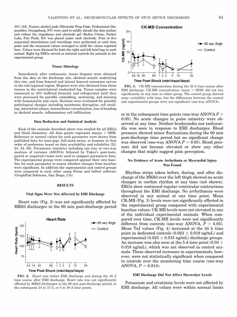
81VALENTINO ET AL.: NEUROMUSCULAR EFFECTS OF STUN DEVICE DISCHARGES
50% [33]. Pumice alcohol pads (Electrode Prep Pads, Professional Dis-posables, Orangeburg, NY) were used to mildly abrade the skin surfaceand reduce the impedance and electrode gel (Redux Crème, ParkerLabs, Fair Field, NJ) was placed under each electrode. Four or fivesequential stimulations and recordings were performed at each timepoint and the measured values averaged to yield the values reportedhere. Values were obtained for both the right and left hind legs in eachanimal. Right leg EMGs served as internal controls for animals in theexperimental group.
Tissue Histology
Immediately after euthanasia, tissue biopsies were obtainedfrom the skin at the discharge site, skeletal muscle underlyingthis site, and from femoral and lateral femoral cutaneous nervesin the sub-inguinal region. Biopsies were also obtained from thesetissues in the contralateral unshocked leg. Tissue samples wereimmersed in 10% buffered formalin and refrigerated until theywere processed for paraffin embedding, sectioning, and stainingwith hematoxylin and eosin. Sections were evaluated for possiblepathological changes including membrane disruption, cell swell-ing, interstitial edema, intracellular vacuolization, loss of bandingin skeletal muscle, inflammatory cell infiltration.
Data Reduction and Statistical Analysis
Each of the animals described above was studied for all EKGsand blood chemistry. All data points represent means � SEM.Reference or normal values for each parameter were drawn frompublished data for mini-pigs, full-sized swine, or humans in thatorder of preference based on data availability and reliability [23,24, 34 –38]. Parametric statistics including one-way or two-wayanalysis of variance (ANOVA) followed by Tukey’s post-tests,paired or unpaired t-tests were used to compare parametric data.The experimental groups were compared against their own base-line for each parameter to assess whether changes from baselinewere significant. In addition the experimental and control groupswere compared to each other using Prism and InStat software(GraphPad Software, San Diego, CA).
RESULTS
Vital Signs Were Not Affected by EMI Discharge
Heart rate (Fig. 2) was not significantly affected byMK63 discharges in the 60 min post-discharge period
FIG. 2. Heart rate before EMI discharge and during the 30 dtime course after EMI discharge. Heart rate was not significantlyaffected by MK63 discharges in the 60 min post-discharge period, in
the subsequent 24 to 72 h, or 5 to 30 d time points.or in the subsequent time points (one-way ANOVA P �0.05). No acute changes in pulse oximetry were ob-served at any time. Neither bradycardia nor tachycar-dia was seen in response to EMI discharges. Bloodpressure showed minor fluctuations during the 60 minpost-discharge time period but no significant changewas observed (one-way ANOVA P � 0.05). Blood pres-sure did not become elevated or show any otherchanges that might suggest pain perception.
No Evidence of Acute Arrhythmia or Myocardial InjuryWas Found
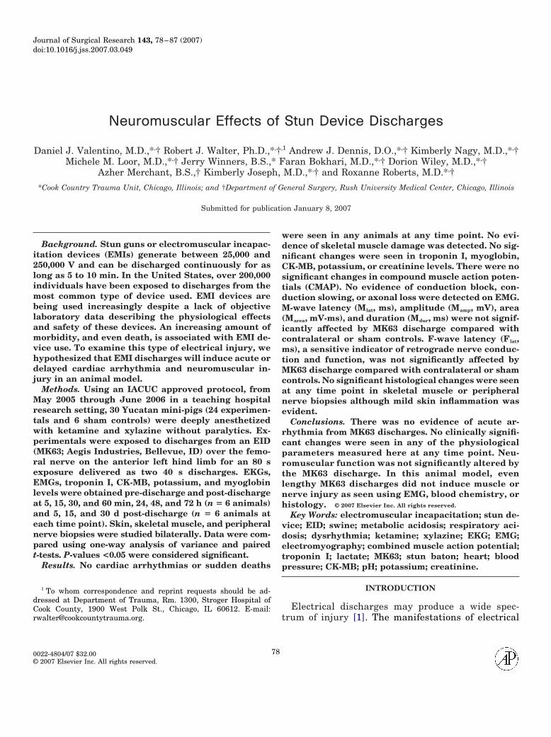
Rhythm strips taken before, during, and after dis-charge of the MK63 over the left thigh showed no acutechanges in cardiac rhythm at any time (not shown).EKGs show continued regular ventricular contractionsthroughout the EMI discharge. No arrhythmias wereobserved in any animal at any time point. MeanCK-MB (Fig. 3) levels were not significantly affected inthe experimental group compared with experimentalbaseline values. CK-MB levels were not elevated in anyof the individual experimental animals. When com-pared over time, CK-MB levels were not significantlydifferent from controls (one-way ANOVA, P � 0.05).Mean TnI values (Fig. 4) increased at the 24 h timepoint in dedicated controls (0.023 � 0.019 ng/mL) andexperimental (0.023 � 0.015 ng/mL) discharge groups.An increase was also seen at the 5 d time point (0.03 �0.018 ng/mL), which was not observed in control ani-mals. These observed increases in experimentals, how-ever, were not statistically significant when comparedto controls over the monitoring time course (one-wayANOVA, P � 0.814).
EMI Discharge Did Not Affect Electrolyte Levels
Potassium and creatinine levels were not affected by
FIG. 3. CK-MB concentration during the 30 d time course afterEMI discharge. CK-MB concentration (mean � SEM) did not risesignificantly at any time in either group. The control group showedsome variability with time, but the differences between the controland experimental groups were not significant (one-way ANOVA).
EMI discharge. All values were within normal limits

82 JOURNAL OF SURGICAL RESEARCH: VOL. 143, NO. 1, NOVEMBER 2007
for these parameters at all time points in all animals.Potassium values (Fig. 5) did not exceed normal limitsin any animal at any time point (range � 2.9 to 4.4mmol/L). Creatinine values (Fig. 6) did not changesignificantly following EMI discharge. At no time didcreatinine values exceed normal levels in any animal(range � 0.6 to 1.3 mg/dL).
EMI Discharge did not Significantly Affect Serum Myoglobin
Mean myoglobin levels (Fig. 7) in the experimentalgroup were increased when compared to the controlgroup at most time points, but these increases were notsignificantly different (one-way ANOVA, P � 0.05).Mean myoglobin levels did not exceed the upper limitof normal (54 ng/mL). The highest single myoglobinlevel seen was 131 ng/mL at 48 h after EMI exposure.
FIG. 4. Troponin-I (TnI) values during the 30 d time course afterEMI discharge. At the 24 h time point, the TnI value for the controlgroup rose to 0.023 � 0.019 ng/mL and for the experimental group to0.023 � 0.018 ng/mL (mean � SEM). TnI returned to baseline valuesfor the control group subsequently, but became elevated again in theexperimental group on day 5 (0.030 � 0.017 ng/mL). This elevationwas largely due to a high value in one animal (0.110 ng/mL). None-theless, the mean TnI values at day 5 were not significantly differentfor the experimental and control groups (one-way ANOVA, P � 0.05).
FIG. 5. Potassium ion concentrations during the 30 d timecourse. Potassium levels did not change significantly after EMI dis-charge. There were no significant differences between the experi-mental and control groups indicating a lack of significant muscle
injury.All elevated myoglobin values returned to normal lev-els by the next monitoring interval.
EMI Discharge had Limited Effects on EMG Responses
M-wave CMAP latency reflects nerve conductionrates from the site of stimulation to the muscle bellywhere the majority of motor end plates are located.Longer latencies indicate slower rates of nerve conduc-tion [33]. M-wave latency (Fig. 8) was not affected inthe shocked limb or the contralateral control limb.While an initial increase in values in the experimentalgroup was observed there was no statistical difference(one-way ANOVA, P � 0.05) between internal controls(right leg) and the experimental side (left leg). Addi-tionally, no difference was noted between the dedicatedcontrol animals and the experimental limbs (one-wayANOVA, P � 0.05).
M-wave CMAP amplitude reflects the vectorial sumof fields produced by individual action potential con-
FIG. 6. Creatinine concentrations seen during the 30 d timecourse. Creatinine levels did not change significantly after EMIdischarge. There were no statistically significant differences betweenthe experimental and control groups indicating a lack of muscleinjury.
FIG. 7. Myoglobin concentrations seen during the 30 d timecourse. Mean myoglobin levels in both groups fluctuated somewhatover the time course, but did not change significantly relative to the
baseline for each group.
83VALENTINO ET AL.: NEUROMUSCULAR EFFECTS OF STUN DEVICE DISCHARGES
ducting muscle cells. Only cells with intact membranesthat can actively produce ATP are capable of generat-ing action potentials. Therefore, changes in CMAP am-plitude can be used to quantify the extent of tissueinjury associated with damaged cell membranes.CMAP recordings have previously been used to esti-mate electroporation injury accumulation in the anes-thetized rat hind limb [39]. M-wave amplitude (Fig. 9)showed no significant changes during the 72 h post-discharge period but rose gradually from mean valuesof 15.3 � 1.3 and 16.4 � 1.5 mV to 22.1 and 20.8 mVduring the long-term follow-up for the control and ex-perimental limbs, respectively. No significant changeswere noted in M-wave CMAP amplitude when the val-ues for EMI exposed animals were compared to non-shocked controls (one-way ANOVA, P � 0.05).
M-wave CMAP area represents the total amount ofthe compound muscle action potential elicited over the
FIG. 8. M-wave CMAP latency for femoral nerve seen during the30 d time course. M-wave latency was not significantly affected in theshocked limb or the contralateral control limb.
FIG. 9. M-wave CMAP amplitude for femoral nerve seen duringthe 30 d time course. M-wave amplitude showed no significantchanges during the 72 h post-discharge period but rose graduallyfrom mean values of 15.3 � 1.3 and 16.4 � 1.5 mV to 22.1 and 20.8mV during the long-term follow-up for the control and experimental
limbs, respectively.several milliseconds after the CMAP depolarizationbegins until it ends. M-wave area (Fig. 10) showed nosignificant changes during the 72 h post-discharge pe-riod but rose gradually from mean values of 40.6 � 2.7and 39.4 � 3.1 mV-ms to 54.8 and 53.4 mV-ms duringthe long-term follow-up for the control and experimen-tal limbs, respectively. For internal and sham controls,M-wave area showed little variation during the entiremonitoring time course (one-way ANOVA, P � 0.05).
F-wave latency reflects the amount of time that isrequired for the EMG stimulus to be conducted anti-dromically or retrograde along the motor nerves untilit reaches the spinal cord, where it elicits firing ofanterior horn motor neurons. These action potentialssubsequently trigger distal muscle contractions in thesame muscle or muscle group innervated by the nerveoriginally stimulated by the EMG. The muscle re-
FIG. 10. M-wave CMAP area for femoral nerve seen during the30 d time course. M-wave area showed no significant changes duringthe 72 h post-discharge period but rose gradually from mean valuesof 40.6 � 2.7 and 39.4 � 3.1 mV-ms to 54.8 and 53.4 mV-ms duringthe long-term follow-up for the control and experimental limbs, re-spectively.
FIG. 11. F-wave latency for femoral nerve seen during the 30 dtime course. Mean F-wave latency decreased by 7% to 14% in theexperimental and contralateral control limbs at the 5 min time point,but this difference was not significant. F-wave latency was similar in
both limbs at all time points studied.
84 JOURNAL OF SURGICAL RESEARCH: VOL. 143, NO. 1, NOVEMBER 2007
sponse will be much weaker than the M-wave responseand will be seen at a later time as a result of the longerpath that the stimulus must travel. It will also reflectthe patency of the axons and synapses in the spinalcord proximal to the MK63 discharge. Mean F-wavelatency (Fig. 11) decreased by 7% to 14% in the exper-imental and contralateral control limbs at the 5 mintime point, but this difference was not significant (one-way ANOVA, P � 0.05). Additionally, no significantchanges were seen when comparing F-wave latency inEMI exposed animals to sham controls. F-wave latencywas similar in both limbs at all time points studied.This suggests that MK63 discharges caused no signif-icant damage to the proximal nerve pathways leadingto the spinal cord and then back to the quadratusfemoris muscle group.
M-wave CMAP duration represents the length oftime that the CMAP from the EMG stimulus contin-ued. This may provide further information aboutpossible tissue injury. There were no changes inM-wave duration seen at any time point here (datanot shown).
Minor Tissue Alterations Occurred After EMI Discharge
Normal cell and tissue histology (Fig. 12) was seen atall time points (3, 5, 15, 30, d) studied for skeletalmuscle and peripheral nerve. No evidence of muscle oraxonal cell membrane disruption or tissue necrosis wasseen. In some biopsies taken at 72 h post-discharge,minor highly localized inflammatory changes in skinwere seen at the discharge site.
DISCUSSION
Cardiac Effects
Case reports, autopsies, and retrospective analysishave found EMI discharge to be associated with fatalventricular fibrillation in humans although the fre-quency of this complication is extremely low [10 –12,40, 41] In the present study, no changes in cardiacrhythm were seen even after lengthy EMI exposures.At no time in the 72 h monitoring period did anyanimal expire prior to being intentionally eutha-nized. Other studies in anesthetized swine have re-ported that TASER X26 discharges resulted in acuteonset of tachycardia [20]. This effect was also re-ported in studies of healthy human volunteers wherethe response was ascribed to intense pain associatedwith the discharge [17, 19]. The absence of tachycar-dia in this experimental model suggests a deep planeof anesthesia that suppresses pain or may indicatethat the MK63 device evoked less pain in this modelthan did the TASER X26 in anesthetized swine orconscious humans. In any case, the MK63 device didnot appear to directly interrupt or capture cardiac
rhythm.Alternatively, sudden deaths associated with TA-SER discharges in humans may result from direct orindirect damage to the myocardium, which thenleads to delayed arrhythmia [10 –12]. Two cardiacmarkers, CK-MB and TnI, were assayed in thepresent study to assess myocardial injury. Therewere no elevations in CK-MB after 80 s MK63 dis-charges and TnI showed small but insignificantrises. TnI is released from cardiac myocytes [25–31,42, 43] when their cell membranes are damaged.Free TnI then diffuses into the interstitial space andeventually into the blood [44]. The high sensitivityand specificity of commercially-available assays havemade TnI the gold standard for detecting myocardialinjury. Release of TnI from both human and swinecardiac myocytes peaks 18 to 24 h after the injuryand then gradually decreases to normal over thecourse of the next several days [44, 45].
The time-related pattern and magnitude of TnI in-creases seen here was very different from that seen inhumans or swine with myocardial injury [44, 45]. Inmodels of myocardial ischemia, TnI values peak at 18to 24 h and remain elevated for days to weeks there-after. Since TnI values here decreased to zero within48 h, it is unlikely that these elevated TnI valuessignify myocardial injury such as that seen in humanheart attack or swine models of myocardial ischemia.
FIG. 12. Tissue biopsies obtained at 72 h post-discharge. Tissuefrom the dedicated control animals is shown on the left and fromanimals exposed to 80 s MK63 discharges is on the right. Normaltissue structure is seen for the skeletal (rectus femoris) muscle andperipheral (femoral) nerve biopsies. No evidence of muscle or axonalcell membrane disruption or tissue necrosis was seen. In some biop-sies, some inflammatory cell infiltrate is seen in the dermis of skinobtained from the discharge site.
The induction and prolonged anesthesia sessions (2 to

85VALENTINO ET AL.: NEUROMUSCULAR EFFECTS OF STUN DEVICE DISCHARGES
3 h) used on the first day of the experiment may havecaused some degree of cardiac stress that contributedto the TnI elevation seen here. Anesthesia, especiallyat induction, is a known cardiac stressor which resultsin an increased risk of adverse cardiac events [46].
Electrical injuries can cause severe muscle and deeptissue injury by a combination of joule heating andmembrane electroporation [1, 4]. Injury to skeletalmuscle can result in rises in serum myoglobin, creati-nine, and potassium concentrations. It had been previ-ously reported by Ordog et al. [14] that EMI exposuremay be associated with rhabdomyolysis, which willalso increase these values. Additionally, acute changesin potassium concentration may precipitate fatal dys-rhythmia. Jauchem et al. [20], in a swine model ofTASER X26 exposure, reported small increases in po-tassium concentration with no changes in myoglobinwith no evidence of acute dysrhythmia. Ho et al. [19]reported no significant changes in potassium or creat-inine concentration, with small increases in myoglobinafter exposure to the TASER X26 in humans. No sig-nificant changes in potassium, myoglobin, or creati-nine were observed in this study. Thus it appears fromthese data that the application of EMI electrical energydoes not result in any consistently reproducible bio-chemical evidence of muscle injury. This conclusion isfurther supported by the lack of structural changes inour histology specimens.
EMG is a sensitive indicator of neuromusculardysfunction. No EMG evidence of acute or delayedneuromuscular injury was seen here. This study isthe first to look at the effects of EMI electrical energyon neuromuscular function through EMG. Thepresent data show no significant changes in EMGparameters when shocked limbs are compared tocontralateral non-EMI exposed limbs. Additionally,no significant differences were seen in EMG param-eters between EMI-exposed animals and sham con-trol animals. Thus, it appears that the electricalenergy produced by the MK63 does not induce mea-surable neuromuscular dysfunction. The electricalenergy produced by other EMI devices, however, isnot uniform and varies from device to device. Otherdevices, such as the TASER X26, which have a dif-ferent waveform and energy output, may producedifferent results.
As a positive comparison, swine exposed to electri-cal discharges on the thigh from a cardiac defibrilla-tor showed signs of severe neuromuscular injury.Blood levels for myoglobin, creatinine, and potas-sium were significantly increased (data not re-ported). These animals also showed gross musclenecrosis and microscopic pathologic changes includ-ing severe cell swelling, interstitial edema, and vac-uolization were seen in skeletal muscle cells and
peripheral nerve biopsies (data not shown). EMGvalues from these animals showed marked increasesin F-wave and M-wave CMAP latency and large(�40%) decreases in M-wave area and amplitude.
The present study has examined the effects of theMK63 stun baton using anterior thigh discharges inanesthetized healthy swine. It does, however, havesome limitations: (1) The number of animals usedwas relatively small but was counter-balanced by thehigh inter-animal reproducibility of the results. (2)For ethical reasons, ketamine/xylazine anesthesiawas used in this swine model. Anesthesia precludespain perception, which is one of the two principaleffects of stun device discharges in conscious hu-mans. Pain perception would undoubtedly alter someof the responses reported here. (3) In the field, stundevices are used to subdue combative individualswho are usually in a state of greatly increased sym-pathetic activity and, in many cases, are under theinfluence of alcohol or other drugs that may alter thethresholds for dysrhythmia and for pain. Underthose conditions, the effects of MK63 dischargesmight deviate considerably from those seen here. Wehave, as yet, not studied the effects of stimulants inthis system. Now that we have characterized thesystem and established the types of responses thatoccur, we can proceed to further examine the effectsof stimulants. Lakkireddy et al. [18] were the first toreport on the effects of cocaine on TASER dischargesin swine and Nanthakumar et al. [21] have similarlyreported on the effects of epinephrine.
In summary, no evidence of acute or delayed ar-rhythmia, myocardial damage, electrolyte abnormal-ities, or neuromuscular dysfunction were seen in thepresent study. In this swine model, prolonged dis-charges from the MK63 device produced no signifi-cant or harmful physiological changes. Since previ-ous animal studies of the TASER X26 showed somedramatic physiological changes [20, 21], the presentfindings may be due to the unique waveform andpulse power generated by the MK63 device, to differ-ences in the electrode spacing for the MK63 com-pared with the TASER X26, or differences betweenthe model systems. Further studies are needed todistinguish among these possibilities and to eluci-date the mechanism by which these devices triggerEMI. This knowledge will be instrumental in devel-oping future guidelines and treatment protocolsfor the growing number of individuals exposed toEMI.
We conclude that, within the limitations of thisstudy, discharges from the MK63 administered onthe lower extremity do not appear to cause any mea-surable neuromuscular or cardiac injury in thismodel. Thus, it appears that EMI can be safelyachieved using this device even for lengthy periods
without causing significant injury.
86 JOURNAL OF SURGICAL RESEARCH: VOL. 143, NO. 1, NOVEMBER 2007
ACKNOWLEDGMENTS
The authors thank the staff of the Animal Facility for their assis-tance and Raphael Lee, M.D., D.Sc. for helpful discussions. They alsothank Avocet Polymer Technologies, Plainfield, IL, for their supportof these studies.
REFERENCES
1. Koumbourlis AC. Electrical injuries. Crit Care Med 2002;30:S424.
2. Fish RM. Electric injury, part I: Treatment priorities, subtlediagnostic factors, and burns. J Emerg Med 1999;17:977.
3. Oltman CL, Clark CB, Kane NL, et al. Coronary vascular dys-function associated with direct current shock injury. Basic ResCardiol 2003;98:406.
4. Lee RC. Injury by electrical forces: Pathophysiology, manifes-tations, and therapy. Curr Probl Surg 1997;34:677.
5. Kalkan T, Demir M, Ahmed AS, et al. A dynamic study of thethermal components in electrical injury mechanism for betterunderstanding and management of electric trauma: An animalmodel. Burns 2004;30:334.
6. Luce EA. Electrical burns. Clin Plast Surg. 2000;27:133.7. Lee RC, Zhang D, Hannig J. Biophysical injury mechanisms in
electrical shock trauma. Annu Rev Biomed Eng 2000;2:477.8. Danielson JR, Capelli-Schellpfeffer M, Lee RC. Upper extrem-
ity electrical injury. Hand Clin 2000;16:225.9. Battershill P, Naughton B, Laur D, et al. Taser technology
review and interim recommendations. Available at: www.cprc.org/docs/bcopcc_final.pdf, 1-59; 2005.
10. McDaniel WC, Stratbucker RA, Nerheim M, et al. Cardiacsafety of neuromuscular incapacitating defensive devices. Pac-ing Clin Electrophysiol 2005;28(Suppl 1):S284.
11. Bleetman A, Steyn R. The advanced Taser: A medical review.Available at: www.taser.com/documents/tasersubmit.pdf, 1-30.2003.
12. Bleetman A, Steyn R, Lee C. Introduction of the Taser intoBritish policing. Implications for UK emergency departments:An overview of electronic weaponry. Emerg Med J 2004;21:136.
13. Bozeman WP. Withdrawal of Taser electroshock devices: Toomuch, too soon. Ann Emerg Med 2005;46:300.
14. Ordog GJ, Wasserberger J, Schlater T, et al. Electronic gun(Taser) injuries. Ann Emerg Med 1987;16:73.
15. O’Brien DJ. Electronic weaponry—a question of safety. AnnEmerg Med 1991;20:583.
16. Koscove EM. The Taser weapon: A new emergency medicineproblem. Ann Emerg Med 1985;14:1205.
17. Levine SD, Sloane C, Chan T, et al. Cardiac monitoring ofsubjects exposed to the Taser. Acad Emerg Med 2005;12:71.
18. Lakkireddy D, Wallick D, Ryschon K, et al. Effects of cocaineintoxication on the threshold for stun gun induction of ventric-ular fibrillation. J Am Coll Cardiol 2006;48:805.
19. Ho JD, Miner JR, Lakireddy DR, et al. Cardiovascular andphysiologic effects of conducted electrical weapon discharge inresting adults. Acad Emerg Med 2006;13:589.
20. Jauchem JR, Sherry CJ, Fines DA, et al. Acidosis, lactate,electrolytes, muscle enzymes, and other factors in the blood ofSus Scrofa following repeated TASER (R) exposures. ForensicSci Int 2005;161:20.
21. Nanthakumar K, Billingsley IM, Masse S, et al. Cardiac elec-trophysiological consequences of neuromuscular incapacitating
device discharges. J Am Coll Cardiol 2006;48:798.22. Webster JG, Will JA, Sun H, et al. Can TASERS directly causeventricular fibrillation? IFBME Proc 2006;14:3307.
23. Swindle MM. Surgery, Anesthesia, and Experimental Tech-niques in Swine. Ames: Iowa State University Press, 1998:33–63, 299–311.
24. Bollen PJA, Hansen AK, Rasmussen HJ. Important BiologicalFeatures. In: Suckow MA, Ed. The Laboratory Swine. BocaRaton: CRC Press, 2000:1.
25. Apple FS, Christenson RH, Valdes R, Jr, et al. Simultaneousrapid measurement of whole blood myoglobin, creatine kinaseMB, and cardiac troponin I by the triage cardiac panel fordetection of myocardial infarction. Clin Chem 1999;45:199.
26. Apple FS, Murakami MM, Quist HH, et al. Prognostic value ofthe Ortho Vitros cardiac troponin I assay in patients withsymptoms of myocardial ischemia. Risk stratification using Eu-ropean Society of Cardiology/American College of Cardiologyrecommended cutoff values. Am J. Clin Pathol 2003;120:114.
27. Apple FS, Wu AH, Jaffe AS. European Society of Cardiologyand American College of Cardiology guidelines for redefinitionof myocardial infarction: How to use existing assays clinicallyand for clinical trials. Am Heart J 2002;144:981.
28. Apple FS, Murakami M, Panteghini M, et al. Internationalsurvey on the use of cardiac markers. Clin Chem 2001;47:587.
29. Apple FS, Wu AH. Myocardial infarction redefined: Role ofcardiac troponin testing. Clin Chem 2001;47:377.
30. Ng SM, Krishnaswamy P, Morissey R, et al. Ninety-minuteaccelerated critical pathway for chest pain evaluation. Am JCardiol 2001;88:611.
31. Ng SM, Krishnaswamy P, Morrisey R, et al. Mitigation of theclinical significance of spurious elevations of cardiac troponin Iin settings of coronary ischemia using serial testing of multiplecardiac markers. Am J Cardiol 2001;87:994.
32. Perry SV. The regulation of contractile activity in muscle. Bio-chem Soc Trans 1979;7:593.
33. Jabre JF, Hackett ER. EMG Manual. Jabre, J. F.: teleEMG.com2002;1:75.
34. Sinclair Research: Clinical Chemistry Values of Sinclair Pigs.1991. Columbia, MO.
35. Brechbuhler T, Kaeslin M, Wyler F. Reference values of variousblood constituents in young minipigs. J Clin Chem Biochem1984;22:301–304.
36. Waagstein LM, Wennberg E, Waagstein F, et al. Hypertonicsaline without or with dextran-70 in the treatment of experi-mental acute myocardial ischemia and reperfusion. Crit CareMed 1999;27:605.
37. Rixen D, Raum M, Holzgraefe B, et al. Local lactate and hista-mine changes in small bowel circulation measured by microdi-alysis in pig hemorrhagic shock. Shock 2002;18:355.
38. Hicks TA, McGlone JJ, Whisnant CS, et al. Behavioral, endo-crine, immune, and performance measures for pigs exposed toacute stress. J Anim Sci 1998;76:474.
39. Lee RC, River LP, Pan FS, et al. Surfactant-induced sealing ofelectropermeabilized skeletal muscle membranes in vivo. ProcNatl Acad Sci USA 1992;89:4524.
40. Roy OZ, Podgorski AS. Tests on a shocking device—the stungun. Med Biol Eng Comput 1989;27:445.
41. Schlosberg M, Levin J, Batliwalla S, et al. Stun gun fallacy:How the lack of Taser regulation endangers lives. Available at:http://www.aclunc.org/issues/criminal_justice/police_practices/special_report_stun_gun_fallacy.shtml. 2005.
42. Kost GJ, Kirk JD, Omand K. A strategy for the use of cardiacinjury markers (troponin I and T, creatine kinase-MB massand isoforms, and myoglobin) in the diagnosis of acute myo-
cardial infarction. Arch Pathol Lab Med 1998;122:245.
87VALENTINO ET AL.: NEUROMUSCULAR EFFECTS OF STUN DEVICE DISCHARGES
43. Apple FS, Ler R, Chung AY, et al. Point-of-care i-STAT car-diac troponin I for assessment of patients with symptomssuggestive of acute coronary syndrome. Clin Chem 2006;52:322.
44. Zipes DP, Libby P, Bonow RO, et al. Braunwald’s Heart Dis-ease: A Text Book of Cardiovascular Medicine. Philadelphia:Saunders, 2005:1134–1161.
45. Feng YJ, Chen C, Fallon JT, et al. Comparison of cardiac
troponin I, creatine kinase-MB, and myoglobin for detection ofacute ischemic myocardial injury in a swine model. Am J ClinPathol 1998;110:70.
46. Williams CG, Prough DS. Chap 17. Anesthesiology Princi-ples, Pain Management, and Conscious Sedation”. In:Townsend CM, Beauchamp DR, Evers MB, et al. Eds.Sabiston Textbook of Surgery. Philadelphia: Saunders, 2004:
426.