neuron sponsored document from mike mcmanus 2,6, and …
TRANSCRIPT
Members of the miRNA-200 FamilyRegulate Olfactory Neurogenesis
The Harvard community has made thisarticle openly available. Please share howthis access benefits you. Your story matters
Citation Choi, Philip S., Lisa Zakhary, Wen-Yee Choi, Sophie Caron, EzequielAlvarez-Saavedra, Eric A. Miska, Mike McManus, et al. 2008.Members of the miRNA-200 family regulate olfactory neurogenesis.Neuron 57(1): 41-55.
Published Version doi:10.1016/j.neuron.2007.11.018
Citable link http://nrs.harvard.edu/urn-3:HUL.InstRepos:4455260
Terms of Use This article was downloaded from Harvard University’s DASHrepository, and is made available under the terms and conditionsapplicable to Other Posted Material, as set forth at http://nrs.harvard.edu/urn-3:HUL.InstRepos:dash.current.terms-of-use#LAA
Members of the miRNA-200 Family Regulate OlfactoryNeurogenesis
Philip S. Choi1,8, Lisa Zakhary1,8, Wen-Yee Choi2, Sophie Caron2, Ezequiel Alvarez-Saavedra3, Eric A. Miska3,9, Mike McManus4, Brian Harfe5, Antonio J. Giraldez2,7, RobertH. Horvitz3, Alexander F. Schier2,6, and Catherine Dulac1∗1Howard Hughes Medical Institute, Department of Molecular and Cellular Biology, Harvard University,Cambridge, MA 02138, USA.
2Department of Molecular and Cellular Biology, Harvard University, Cambridge, MA 02138, USA.
3Howard Hughes Medical Institute, Department of Biology, Massachusetts Institute of Technology,Cambridge, MA 02139, USA.
4Diabetes Center, University of California, San Francisco, San Francisco, CA 94143, USA.
5Department of Molecular Genetics and Microbiology, University of Florida, Gainesville, FL 32611, USA.
6Division of Sleep Medicine, Center for Brain Science, Harvard Stem Cell Institute, Broad Institute,Cambridge, MA 02139, USA.
7Genetics Department, Yale University School of Medicine, New Haven, CT 06520, USA.
SummaryMicroRNAs (miRNAs) are highly expressed in vertebrate neural tissues, but the contribution ofspecific miRNAs to the development and function of different neuronal populations is still largelyunknown. We report that miRNAs are required for terminal differentiation of olfactory precursorsin both mouse and zebrafish but are dispensable for proper function of mature olfactory neurons. Therepertoire of miRNAs expressed in olfactory tissues contains over 100 distinct miRNAs. A subset,including the miR-200 family, shows high olfactory enrichment and expression patterns consistentwith a role during olfactory neurogenesis. Loss of function of the miR-200 family phenocopies theterminal differentiation defect observed in absence of all miRNA activity in olfactory progenitors.Our data support the notion that vertebrate tissue differentiation is controlled by conserved subsetsof organ-specific miRNAs in both mouse and zebrafish and provide insights into control mechanismsunderlying olfactory differentiation in vertebrates.
KeywordsDEVBIO; MOLNEURO
© 2008 Elsevier Inc.∗Corresponding author [email protected] authors contributed equally to this work.9Present address: Wellcome Trust/Cancer Research UK, Gurdon Institute, University of Cambridge, UK.This document was posted here by permission of the publisher. At the time of deposit, it included all changes made during peer review,copyediting, and publishing. The U.S. National Library of Medicine is responsible for all links within the document and for incorporatingany publisher-supplied amendments or retractions issued subsequently. The published journal article, guaranteed to be such by Elsevier,is available for free, on ScienceDirect.
Sponsored document fromNeuron
Published as: Neuron. 2008 January 10; 57(1): 41–55.
Sponsored Docum
ent Sponsored D
ocument
Sponsored Docum
ent
IntroductionMicroRNAs (miRNAs) constitute a large class of small noncoding RNAs that providemulticellular organisms with elaborate yet poorly understood strategies for posttranscriptionalgene regulation (Bartel, 2004). Hybridization to fully or partially complementary sequencesenables miRNAs to specifically direct degradation or translational inhibition of targettranscripts (Plasterk, 2006). Genetic analyses in invertebrate systems have identified essentialroles for miRNAs in the regulation of various developmental processes, including specificsteps of neuronal differentiation. In C. elegans, lsy-6 and miR-273 have been reported toparticipate in negative-feedback loops that ensure asymmetric expression of taste receptors inchemosensory neurons (Chang et al., 2004; Johnston and Hobert, 2003). In Drosophila, miR-7has been implicated in photoreceptor cell differentiation through regulation of local EGFreceptor signaling (Li and Carthew, 2005). The essential roles played by some miRNAs incontrolling invertebrate neurogenesis and the dynamic patterns of miRNA expression duringvertebrate development have raised the issue as to whether miRNAs might similarly regulateaspects of vertebrate neural development (Miska et al., 2004; Kosik and Krichevsky, 2006;Cao et al., 2006; Makeyev et al., 2007). This question has remained unanswered because loss-of-function studies of specific neural microRNAs in vertebrates have not yet been performed.
Thanks to its molecular and genetic tractability, the process of olfactory neurogenesis offers aunique opportunity to uncover regulatory networks underlying neuronal specification anddifferentiation. The main olfactory epithelium (MOE) of mammals is a pseudostratifiedepithelium, which extends from an underlying basal lamina to the lumen of the nasal cavity.Olfactory neurogenesis in rodents is initiated at midgestation with the thickening andinvagination of the bilaterally symmetric olfactory placodes. The posterodorsal recess of theplacodal epithelium differentiates into a mature, self-regenerating sensory epithelium thatcontains a highly heterogeneous and constantly renewing population of neurons and neuronalprecursors (reviewed in Dulac and Zakhary, 2004). Adult MOE contains three major cellgroups: basal cells, olfactory sensory neurons (OSN), and supporting cells. The basal cells area population of dividing cells located adjacent to the basal lamina that continuously generateolfactory progenitors, which in turn differentiate into olfactory neurons. In the mouse, eachmature olfactory sensory neuron expresses a unique olfactory receptor gene from a large familyof approximately 1000 genes such that all neurons expressing the same receptor transcript arerandomly dispersed within one of four broad zones of the olfactory epithelium (Buck and Axel,1991; Chess et al., 1994; Malnic et al., 1999; Ressler et al., 1993; Vassar et al., 1993).
What are the transcriptional regulators underlying such sensory diversity? Genetic analysis ofthe olfactory epithelium has pointed to the essential role played by basic helix-loop-helix(bHLH)-containing transcription factors related to the Drosophila proteins achaete-scute andatonal in controlling MOE development (reviewed in Bertrand et al., 2002). Mature olfactorysensory neurons do not develop in mice with a targeted deletion of the achaete-scute homolog,Mash1 (Guillemot et al., 1993). Expression of Mash1 in early olfactory progenitor cells (OPCs)controls expression of the bHLH-containing transcription factors Ngn1 and NeuroD, which inturn regulate olfactory differentiation (Cau et al., 1997). The larger process of morphogenesis,patterning, and differentiation of the nasal cavity into its various sensory and nonsensorycomponents is controlled by the spatially restricted release of various signaling molecules, suchas sonic hedgehog (Shh), retinoic acid (RA), bone morphogenetic proteins (BMPs), and thefibroblast growth factor FGF8 (LaMantia et al., 2000; Kawauchi et al., 2005).
What roles, if any, are played by miRNAs during this process? We describe here thecharacterization of the repertoire of miRNAs expressed in the adult and the developingolfactory system, which includes several miRNA families that appear highly enriched inolfactory tissues. The specific expression of miRNA subsets by distinct olfactory cell
Choi et al. Page 2
Published as: Neuron. 2008 January 10; 57(1): 41–55.
Sponsored Docum
ent Sponsored D
ocument
Sponsored Docum
ent
populations in the embryo and the adult is consistent with the idea that miRNAs may playspecific and significant roles in the mature and developing olfactory system. Analyses ofgenetically modified mice in which mature olfactory sensory neurons have been depleted ofDicer function, an enzyme required for the production of functional miRNAs (Bernstein et al.,2001), demonstrate that miRNAs are dispensable in terminally differentiated olfactoryneurons. By contrast, conditional knockout of Dicer in olfactory progenitor cells causesdevelopmental arrest and degeneration of the olfactory neuroepithelium, while the adjacent,nonneural respiratory epithelium persists. Antisense morpholino experiments in zebrafishreveal that the inhibition of expression of a single miRNA family, miR-200, largelyphenocopies the defect in terminal olfactory differentiation resulting from lack of Dicerfunction in mouse olfactory progenitor cells. Preliminary data suggest that lunatic fringe(lfng) and zinc-finger homeobox 1 (zfhx1), two key factors associated with Notch and BMPpathways, respectively, as well as foxg1, a transcription factor required for normal olfactorydevelopment, may be relevant miR-200 targets. Our data support the notion that vertebratetissue differentiation is controlled by subsets of organ-specific miRNAs.
ResultsThe Repertoire of miRNAs in the Mature and Developing Olfactory System
In order to understand the roles played by miRNAs during olfactory development, we aimedto identify the repertoire of miRNAs expressed in peripheral olfactory tissues. Reverse-transcribed and amplified cDNA generated from the 18–26 nucleotide small RNA fraction ofolfactory as well as from various neural and nonneural tissues dissected from newborn andadult rats were hybridized to microarrays capable of detecting the expression of 138 knownmammalian miRNAs (Miska et al., 2004). Ninety-four (68%) of these known miRNAs werepresent at detectable levels in the adult and newborn MOE, vomeronasal organ (VNO), orolfactory bulb (OB) (Figure 1A and see Table S1 available online). Forty-one miRNAs (30%),including many of the let-7 variants, show expression in all tissues examined, whether olfactoryderived or not (Table S1). By contrast, we identified 12 miRNAs corresponding to 9 families(miR-199, miR-140, miR-152, miR-214, miR-205, miR-200, miR-183, miR-182, miR-96) thatdisplayed highly enriched expression in the olfactory system (Figure 1A). Hierarchicalclustering confirmed that the miRNA repertoire from each primary olfactory tissue (i.e.,newborn and adult MOE and VNO) is more similar to each other than to any other neural ornonneural tissue tested. Data obtained by the microarray assay were subsequently validated bynorthern blot analyses (Figure 1B), which confirmed the enrichment of subsets of miRNAfamilies in the olfactory system.
In order to comprehensively characterize the repertoire of olfactory miRNAs, including speciesthat may not be included in the microarray described above, we systematically cloned smallRNAs between 18 and 26 nucleotides in length from adult VNO and adult and newborn MOEand sequenced 3600 clones. We obtained 643, 1036, and 883 small RNAs from rat postnatalday 1 (P1) MOE tissue, P60 MOE, and P60 VNO, respectively, of which 317 (49%), 595(57%), and 267 (30%) corresponded to known miRNAs (Table S2). Not surprisingly, miR-124and let-7 variants, known to be highly expressed in the brain (Lagos-Quintana et al., 2002),were among the most abundant miRNAs identified by direct cloning. In addition, we clonedmembers of eight of the nine miRNA families predicted by the microarray assay to be highlyenriched in the olfactory system. One of these families, miR-200 family comprising miR-200a,miR-200b, miR-200c, miR-429, and miR-141, also highly detected by microarray, was amongthe most frequently cloned species in all olfactory tissues examined (Table S2).
Excluding sequences corresponding to known miRNAs, ribosomal genes, and mRNAs, 100small RNA sequences not present in the microarray were identified. Among them, we used thefollowing criteria to identify genuine miRNAs: 18–24 nucleotides in length, prediction of a
Choi et al. Page 3
Published as: Neuron. 2008 January 10; 57(1): 41–55.
Sponsored Docum
ent Sponsored D
ocument
Sponsored Docum
ent
stem loop structure for the miRNA precursor (Zuker, 2003), and detection of an 18–24nucleotide band by northern hybridization analyses. To distinguish miRNAs from other smallRNAs or degradation products, we evaluated the probability of the ∼60 base pair genomicsequence immediately upstream and downstream of a candidate miRNA to form a hairpinstructure using Mfold, a program designed for analysis of RNA secondary structure (Zuker,2003). Thirty of the 100 clones passed the filters and were further tested for expression inolfactory tissues by northern hybridization analyses. Of these, 18 clones displayed the expected18–24 nucleotide bands and were subsequently listed in miRBase database, among which nineappeared highly enriched in the olfactory and vomeronasal epithelia (Figure 1C).
Cellular Distribution of microRNAs in the Mature and Developing Olfactory SystemIn order to gain cellular resolution of miRNA expression, we performed in situ hybridizationexperiments in mouse tissues using locked-nucleic-acid (LNA)-modified DNAoligonucleotide probes (Wienholds et al., 2005; Figure 2A). Experiments in zebrafish havepreviously established that LNA probes specifically recognize mature miRNA species and donot hybridize with precursor miRNAs. Moreover, LNA probes are highly specific and candiscriminate among members of the same miRNA family (Wienholds et al., 2005). We focusedour efforts on 24 miRNAs that displayed strong and preferential expression in the developingand mature olfactory system by northern blot analyses (list, sequence, and summary ofexpression patterns of the 24 miRNAs are found in Table S3). Although a subset of the LNAprobes (6 of 24) did not yield any signal, most probes generated detectable expression patterns.Five of 24 probes, including miR-449 and miR-205, displayed expression limited to thenonneural respiratory epithelium (Figure 2A, left column, and Table S3). Five of 24 miRNAs,including miR-199a∗ and miR-140∗ (Figure 2A, center column, and Table S3), showedexpression in the mesenchyme underlying or cartilage surrounding the MOE and VNO. Finally,8 of 24 miRNA probes, including miR-200a and miR-200b, as well as miR-96, miR-141,miR-182, miR-183, miR-191, and miR-429, revealed robust expression in the MOE and VNOneuroepithelium, with weaker expression in the adjacent respiratory epithelium (Figure 2A,right column, and Table S3). Expression was excluded from the supporting cell layer locatedadjacent to the nasal lumen and was detectable in both immature and mature MOE and VNOneuroepithelia (Figure 2A, right column, and 2B, lower panel). Across our study, we did notidentify any miRNA species that were differentially expressed between the VNO and the MOEneuroepithelium.
The intriguing specificity and intensity of expression of the miR-200 family members in theMOE prompted us to pursue an in-depth investigation of their distribution during embryonicdevelopment and in the adult. Expression of the miR-200 family can be detected in olfactoryplacodes as early as E9.5, which is the first identifiable stage of olfactory development, withcontinued expression within the MOE anlage in the posterodorsal aspect of the olfactory pit atE11.5 (Figure 2B). From E13.5 onward, miR-200b expression becomes evenly expressedthroughout the MOE at the exclusion of the supporting cell layer (Figure 2B). In the adult, theexpression pattern of all miR-200 family members is restricted to the immature and matureneuronal cell layers of the MOE and is excluded from the basal and sustentacular cell layers(Figure 2B). In mouse, the miR-200 family is composed of five family members (miR-141,-200a, -200b, -200c, -429) clustered into two loci of chromosomes 4 and 6 (Figure 2C). Allindividual members of the miR-200 family display similar expression patterns. However,miR-141 and -200a express different 5′ seed heptamers from miR- 200b, -200c, and -429 andare thus likely to form two functional subgroups within the miR-200 family (Figure 2C; Doenchand Sharp, 2004; Lewis et al., 2005). The strong, specific, and coordinated expression ofmiR-200 members in the MOE anlage and in the mature and immature MOE is consistent witha potential role of this miRNA family during MOE neurogenesis.
Choi et al. Page 4
Published as: Neuron. 2008 January 10; 57(1): 41–55.
Sponsored Docum
ent Sponsored D
ocument
Sponsored Docum
ent
Conditional Dicer Inactivation in Olfactory Progenitors and Mature NeuronsIn order to evaluate the potential roles played by miRNAs during olfactory development andin mature olfactory neurons, we used a previously established conditional null allele of Dicerto inactivate Dicer function within specific olfactory cell types (Figure 3A; Harfe et al.,2005).
In order to abolish Dicer function in mature olfactory neurons, we took advantage of the specificexpression of the olfactory marker protein (OMP) in fully differentiated MOE and VNOneurons. Mice harboring the conditional Dicer allele were crossed with a mouse line in whichCre recombinase is expressed under the control of the endogenous OMP promoter (Egganet al., 2004). To verify the efficiency of our genetic strategy, we monitored the expression ofmiRNAs in OMP+ cells of control and mutant animals. In wild-type animals, the expressionof OMP and miR-200b is partially overlapping, with OMP exclusively expressed bydifferentiated neurons located in the apical half of the neuroepithelium, while miR-200b isexpressed throughout the neuroepithelium in both mature and immature neurons (Figure 3B).In contrast, upon Cre-mediated deletion of Dicer in OMP-positive cells, miR-200b expressionis abolished from the apical portion of the neuroepithelium, while it is maintained within basalimmature neurons (Figure 3B). Northern blot analysis confirmed that the level of miR-200bexpression throughout the entire olfactory epithelium is reduced by ∼50%, due to the absenceof miRNA processing in OMP-expressing neurons, while it remains in immature precursorcells (Figure 3B).
In order to abolish miRNA processing in olfactory progenitors, we took advantage of the earlyexpression of Foxg1 in the developing olfactory placodes (Kawauchi et al., 2005). Miceharboring the conditional Dicer allele were crossed with a mouse line expressing Crerecombinase under the control of the endogenous Foxg1 promoter (Hebert and McConnell,2000). Cre activity has been detected in the olfactory placodes of Foxg1-Cre mouse embryosas early as E9.5 (Kawauchi et al., 2005), ensuring that Dicer function is abolished at a stageprior to, or concurrent with, the initiation of olfactory neurogenesis. As shown in Figure 3C,miR-200a is widely expressed throughout the developing MOE neuroepithelium in embryonicday 13.5 (E13.5) wild-type mice. In marked contrast, miR-200a expression is undetectable inthe MOE of E13.5 Foxg1-Cre+/−; DicerloxP/loxP mutants, despite the fact that the main olfactoryepithelium is still present at this stage, as revealed by Foxg1 staining in adjacent sections(Figure 3C). Similarly, expression of miRNAs from the respiratory epithelium, such asmiR-449, is abolished in E16.5 Foxg1-Cre+/−; DicerloxP/loxP mutants, confirming that Dicerfunction can be effectively knocked out in all structures originating from the olfactory placodes(Figure 3C). These experiments confirm that a dual genetic strategy can specifically preventgeneration of mature miRNAs in olfactory neurons or in their progenitors.
miRNAs Are Required for Maintenance but Not Initiation of Olfactory NeurogenesisFoxg1-Cre; DicerloxP/loxP animals die in utero, have small eyes and forebrains, and developsmall snouts. At E10.5, no gross morphological defect is detectable in the olfactory pits ofFoxg1-Cre+/−; DicerloxP/loxP mutant animals relative to wild-type controls. However, thenumber of cells positive for neuroD, a marker of committed progenitor cells of the neuronallineage (Cau et al., 1997), is reduced by 18% compared to mutant olfactory pits (Figure 4A,mean ± SEM, WT 41.71 ± 2.10, n = 5; mutant 34.33 ± 1.60, n = 4, p < 0.01, Student's t test).Quantification of postmitotic neurons, as assayed by Hu-C/D expression, showed a 28%reduction in olfactory pits of mutant embryos compared to wild-type controls (mean ± SEM,WT 45.82 ± 2.57, n = 3; mutant 31.32 ± 2.09, n = 3, < 0.01, Student's t test) (Figure 4A).
By E13.5, the reduced expression of olfactory progenitor markers, such as Mash1 and Ngn1,and the marked thinning of the neuroepithelium indicate a severe defect in neurogenesis in the
Choi et al. Page 5
Published as: Neuron. 2008 January 10; 57(1): 41–55.
Sponsored Docum
ent Sponsored D
ocument
Sponsored Docum
ent
mutant MOE (Figure S1). Moreover, expression of mature olfactory neuronal markers, suchas OMP (Figure 4B) and olfactory receptors (data not shown) is not detectable in Foxg1-Cre+/−; DicerloxP/loxP mutant MOE, suggesting that mutant olfactory progenitor cells do notterminally differentiate. At subsequent stages, we observe a specific loss of neuroepithelialcells that culminates in the total disappearance of markers of neuronal lineages, such as Mash1,Ngn1, Lhx2, and Foxg1 by E16.5 (Figure 5B). By contrast, development of the nonneuralrespiratory epithelial cells, as detected by the marker stratifin (Sfn) (Visel et al., 2004), ismaintained. Thus, miRNA function appears to be required for both the terminal differentiationof olfactory neuronal precursor cells as well as for the maintenance of olfactory progenitorcells.
From early embryonic stages onward, the nasal pit is spatially segregated into several neuronaland nonneuronal components. The vomeronasal organ is located in an antero-ventral portionof the nasal septum, and the respiratory nonneuronal epithelium is located immediately ventralto the main olfactory neuroepithelium. Moreover, the MOE neuroepithelium displays adorsoventral patterning according to which olfactory receptor gene expression is spatiallyrestricted to one of four circumscribed zones (Ressler et al., 1993; Vassar et al., 1993). In orderto evaluate whether the defect in neurogenesis described above coincides with changes inolfactory patterning, we performed in situ hybridization using markers that distinguish betweenthe various compartments of the embryonic olfactory cavity. At E11.5, the earliest knownmarkers of olfactory progenitor cells, Mash1 (Guillemot et al., 1993), Ngn1 (Cau et al.,1997), and Foxg1 (Kawauchi et al., 2005), as well as markers of immature neurons, such asLhx2 (Hirota and Mombaerts, 2004) (Figure 5A and Figure S2B), show similar expression inboth control and mutant animals. However, the olfactory neuroepithelium appears thinnerrelative to that of controls. At this stage, the expression pattern of OMACS-like, a marker ofthe two most dorsal MOE zones (Oka et al., 2003), is indistinguishable between wild-type andmutant MOE (Figure 5A). The zonal expression of OMACS-like is maintained at E13.5(Figure S1).
The segregation of the nonneural respiratory epithelium from the ventral aspect of thedeveloping main olfactory neuroepithelium was followed using Sfn as a marker. Sfn appearsrestricted to the ventral aspect of the developing olfactory pit at both E11.5 (Figure 5A) andE13.5 (Figure S1) in both control and mutant animals in a pattern that does not overlap withthe more dorsal MOE neuroepithelium. Sfn is expressed throughout the mutant olfactory tissueat E16.5, a time point by which all neural lineages of the main olfactory neuroepithelium havedegenerated and only respiratory epithelium remains (Figure 5B).
Finally, we investigated the specification of the vomeronasal placode from the medial wallsof the olfactory pits and the subsequent budding of the resulting VNO toward the midline. Thebudding vomeronasal placode was clearly identified in both wild-type and mutant olfactorypits at E11.5, along with the expression of neurogenesis markers, such as Ngn1, Mash1, Foxg1,and Lhx2 (Figure 5A). Taken together, these results indicate that MOE cells are specified andinitially maintained in Foxg1-Cre+/−; DicerloxP/loxP mutant MOE.
In order to determine the mechanism responsible for the reduction in olfactory neuroepithelialprogenitor cells, we performed immunohistochemical analyses for both proliferating andapoptotic cells. At E10.5, the earliest stage at which a reduction in olfactory markers wasobserved in mutant embryos, immunostaining for the M-phase-specific marker,phosphorylated histone H3, revealed no significant changes in the number of proliferating cellsbetween mutant and control olfactory epithelia at E10.5 (mean ± SEM, WT 23.95 ± 1.06, n =3; mutant 21.61 ± 1.09, n = 3, p = 0.13, Student's t test), the earliest stage at which a reductionin olfactory markers was observed in mutant embryos, nor at E12.5 (mean ± SEM, WT 13.02± 0.76 cells, n = 3; mutant 14.49 ± 0.77 cells, n = 3, p = 0.19, Student's t test) (Figure 5C and
Choi et al. Page 6
Published as: Neuron. 2008 January 10; 57(1): 41–55.
Sponsored Docum
ent Sponsored D
ocument
Sponsored Docum
ent
Figure S2). By contrast, immunostaining for the apoptotic marker active caspase-3 revealedsignificantly increased numbers of apoptotic cells in mutant peripheral olfactory tissues at bothE10.5 (mean ± SEM, WT 7.76 ± 1.44, n = 3; mutant 41.97 ± 3.31, n = 3, p < 0.01, Student's ttest) and E12.5 (mean ± SEM, WT 5.18 ± 0.54, n = 3; mutant 83.42 ± 5.54, n = 3, p < 0.01,Student's t test) compared to control littermates (Figure 5C and Figure S2). Taken together,these results indicate that the loss of MOE cells is due to increased cell death rather thandecreased proliferation and that, although olfactory neuroepithelial progenitor cells and theirprogeny are initially specified and patterned correctly in the absence of miRNA processing,they are unable to undergo terminal differentiation.
miRNA Function Is Not Required in Mature Olfactory and Vomeronasal NeuronsIn order to evaluate the contribution of miRNA functions in mature olfactory neurons, weanalyzed adult OMP-Cre; DicerloxP/loxP mutant mice, in which Dicer function has beenspecifically abolished in fully differentiated olfactory neurons (Figure 3B). In striking contrastto the Foxg1-Cre+/−; DicerloxP/loxP mutants, OMP-Cre; DicerloxP/loxP mice are viable, shownormal weight and survival rates, and appear to maintain normal olfactory-related functions,such as suckling, feeding, and mating.
We further investigated the state of the adult neuroepithelium in mutant and control animals.Cells positive for various markers of olfactory cell differentiation, such as Ki67 (Ohta andIchimura, 2000) in dividing cells, Mash1 in basal progenitors, NCAM in immature and matureneurons, and OMP and olfactory receptors in terminally differentiated OSNs, appeared similarin wild-type and mutant MOE, both in terms of pattern and cell number (Figure 6A and datanot shown). We also performed olfactory behavioral assays in order to reveal differences thatmay arise from the integration of multiple, subtle changes. In a crude but classic assay forolfactory function, we monitored the time required for 6- to 10-week-old control and mutantmice to locate a hidden olfactory stimulus (Stowers et al., 2002). Control animals found ahidden cookie in 66.14 ± 27.91 s compared with 88.63 ± 19.83 s for OMP-Cre;DicerloxP/loxP mutants (Figure 6B; p = 0.53, Students t test), suggesting no statistical differencein the ability to sense and respond to olfactory cues. Moreover, no statistically significantdifferences were observed in the rate of proliferating (mean ± SEM, WT 5.79 ± 0.50, n = 3;mutant 5.05 ± 0.37, n = 3, p = 0.24, Student's t test) or apoptotic (mean ± SEM, WT 12.19 ±0.77, n = 3; mutant 11.76 ± 0.74, n = 3, p = 0.69, Student's t test) cells in the olfactory epitheliaof mutants relative to controls (Figure 6D). Thus, we could rule out an increase in Dicer-depleted OSN apoptosis compensated by a rapid replacement of OSNs, which would have ledto the absence of observable phenotypic defect in OMP-Cre; DicerloxP/loxP animals.
Similarly, we did not observe any detectable differences in marker expression between wild-type and mutant adult VNOs, including NCAM in immature and mature neurons, the V1Rclass of vomeronasal receptors in fully differentiated vomeronasal sensory neurons and Galphasignaling molecules that delineate zones of the VNO (Figure 6C). To test vomeronasal function,we performed a standard resident-intruder assay using 6- to 10-week-old male mice of eithermutant or wild-type genetic background that had been housed in isolation for several days priorto the assay. Resident males are expected to attack a male intruder if the vomeronasal systemis intact (Stowers et al., 2002). The number of aggressive attacks initiated by the resident OMP-Cre; DicerloxP/loxP mutants in every 15 min recording session appeared statisticallyindistinguishable from that of wild-type controls (mean ± SEM, WT 35.6 ± 13.65, n = 5; mutant35.75 ± 15.93, n = 4, p = 0.99, Student's t test) (Figure 6B).
Olfactory (OSNs) and vomeronasal (VSNs) sensory neurons send their axons to discreteglomeruli in the main olfactory bulb (MOB) and accessory olfactory bulb (AOB), respectively.OSNs expressing a given olfactory receptor gene project their axons to two bilaterallysymmetric glomeruli in the MOB (Ressler et al., 1993; Vassar et al., 1993), while VSNs
Choi et al. Page 7
Published as: Neuron. 2008 January 10; 57(1): 41–55.
Sponsored Docum
ent Sponsored D
ocument
Sponsored Docum
ent
expressing a given V1R or V2R receptor gene project their axons to multiple glomeruliclustered within the anterior or posterior half of the AOB, respectively (reviewed in Dulac andTorello, 2003). In order to visualize axon projections of OSNs and VSNs in the Dicer knockoutbackground, we crossed OMP-Cre; DicerloxP/loxP mice with genetically modified miceharboring either the olfactory receptor reporter allele P2-IRES-tauLacZ (Mombaerts et al.,1996) or the V1R receptor reporter allele VN12-IRES-tauLacZ (Belluscio et al., 1999). Ourdata show that in the absence of miRNA function, P2-expressing OSNs and VN12-expressingVSNs are able to correctly target the appropriate glomeruli within the olfactory bulb (Figure 6Eand data not shown).
Taken together, our results provide both molecular and behavioral evidence that miRNAs arelargely dispensable for the function of mature olfactory and vomeronasal neurons, while theyare required for olfactory differentiation in the embryo.
An In Vivo Strategy to Block Activity of Specific miRNAsAnalyses of conditional Dicer mutants in the mouse reveal that miRNAs play an essential roleduring olfactory development. In a subsequent step, we aimed at evaluating the contributionof specific miRNA species. Determination of specific miRNA families during olfactorydevelopment in mice is difficult because genetic loss-of-function analyses are hampered byredundancy within microRNA families. We reasoned that the zebrafish could provide a usefulmodel system due to the remarkable conservation in peripheral olfactory organization betweenfish and mouse at the genetic, molecular, and morphological levels (Figure 7A). For example,zonal olfactory receptor expression, signal transduction mechanisms, and olfactory bulbtargeting are all conserved (reviewed in Hansen and Zielinski, 2005).
We first investigated the requirement of Dicer for zebrafish olfactory development. Removalof Dicer in maternal-zygotic dicer mutants eliminates all mature microRNAs during zebrafishembryogenesis and results in morphogenesis defects (Giraldez et al., 2005). Injection ofmiR-430 into MZdicer mutants rescues early abnormalities, but does not restore the functionof microRNAs that are expressed at later stages of development. We therefore analyzedolfactory development in MZdicer mutants injected with miR-430 microRNAs. Earlypatterning of the nervous system is unperturbed in MZdicer+miR430 mutants, e.g., markers forspecified optic stalk, forebrain, midbrain-hindbrain boundary, otic vesicles, hindbrainrhombomeres, dorsal neural tube, and ventral neural tube are present (Giraldez et al., 2005).However, in contrast to control animals, the expression of markers of terminally differentiatedolfactory sensory neurons, such as OMP and olfactory receptors, is largely abolished inMZdicer+miR-430 mutants at 48 hpf (Figure 7B). In addition, the expression of foxg1, a markerfor early olfactory stages in mice (Kawauchi et al., 2005 and Figure S2B), is upregulated,suggesting an expansion of olfactory progenitors that might be unable to mature into OSNs inabsence of microRNAs (Figure 7B). These results indicate that miRNAs are critical for normalolfactory neurogenesis in both zebrafish and mouse.
To evaluate the contribution of specific miRNAs, we focused on the miR-200 family, whichis highly and specifically expressed in the developing olfactory system. The function ofmiR-200 during olfactory development is likely to be conserved throughout evolution, asjudged from the absolute conservation of miR-200 orthologs between mouse and zebrafishwith respect to the relative genomic clustering position, the conserved seed region sequences,the conserved size of the family, and the conserved arm of the hairpin that generates the maturemiRNA (Figure S3). Moreover, as shown in the mouse, miR-200 family members display earlyexpression in zebrafish (Wienholds et al., 2005) and appear highly enriched in olfactory tissuesby the time olfactory placodes arise at 26 hpf (Figure 7A). Antisense morpholinooligonucleotides complementary to microRNAs hairpin sequences have been shown tospecifically abolish mature miRNA activity (Flynt et al., 2007; Kloosterman et al., 2007). We
Choi et al. Page 8
Published as: Neuron. 2008 January 10; 57(1): 41–55.
Sponsored Docum
ent Sponsored D
ocument
Sponsored Docum
ent
designed three morpholino antisense oligonucleotides predicted to each target the maturesequence of one or a few members of the miR-200 family (Figure S4A). The morpholinosequences lacked any homology to other known zebrafish transcripts. To identify the minimalconcentration at which the morpholinos used in our experiments can inhibit the generation ofcognate miRNAs, we injected one-cell zebrafish embryos with a range of concentrations (1 ngto 6 ng per embryo) and incubated the morphants from 18 hpf to 48 hpf before analysis. In situhybridization analyses using LNA antisense probes to detect mature miRNAs indicated that 4ng per embryo per miR-200 family member was the minimal dose required to knock downmiRNA expression to threshold levels of detection (data not shown). Consequently, we used4 ng dosages in all proceeding experiments. In order to test the specificity of each morpholino(MO) sequence, we systematically injected one-cell zebrafish embryos with either miR-141MO, miR-200b MO, or miR-429 MO and performed in situ hybridization against all fivemiRNAs of the miR-200 family. As predicted from sequence analyses and thermal stabilitycalculations, miR-141 MO specifically inhibited miR-200a and miR-141, miR-200b MOspecifically inhibited miR-200b and miR-200c, and miR-429 MO specifically inhibitedmiR-429 (Figure S4B). In addition, in situ hybridization analyses (Figure 7C) show that amixture of all three morpholinos (Triple MO mix: miR-141 MO, miR-200b MO, and miR-429MO) was sufficient to simultaneously inhibit the expression of all five mature zebrafishmiR-200 family members to threshold levels of detection.
Antisense experiments can be plagued by nonspecific phenotypes, such as cell death in thehead, general neural degeneration, CNS necrosis, and general lethality, which are likely toresult from nonspecific interactions of MOs with inappropriate targets (reviewed in Sumanasand Larson, 2002). In order to test for such effects in our experiments, we performed in situhybridization with a number of genes widely expressed in the nervous system. Our data showthat expression patterns of genes expressed throughout the brain and in areas devoid of miR-200family expression were comparable between wild-type and triple MO morphants, indicatingthat widespread neural defects were absent in the morphant fish (Figure 7D). Furthermore,analyses of wild-type fish and fish injected with individual or mixtures of MOs did not displayany morphological signs of widespread cell death, necrosis, or lethality (Figure S4C). Weconclude that mature zebrafish miR-200 family members can be specifically and efficientlyknocked down in various combinations in the developing olfactory system using antisensemorpholinos without confounding “off-target” effects.
miR-200 Family Members Are Required for the Proper Differentiation of Olfactory ProgenitorCells
Embryos injected with individual antisense morpholinos showed knockdown of the expectedmiRNAs but did not display any visible olfactory phenotype, as visualized by a normal patternof OMP expression in morphant fish (data not shown). We next wished to determine whetherthe distinct 5′ seeds contributed differentially to the physiological function of the miRNA-200family. Embryos injected with either miR-141/miR-200a or miR-200b/miR-429 pairs ofantisense morpholinos showed lack of expression of the corresponding miR-200 members witha given 5′ seed but did not display any change in OMP expression relative to wild-type controls(data not shown). Finally, we eliminated the function of all miR-200 family members byinjecting embryos simultaneously with the Triple MO mix. Forty-eight hours after injection,triple MO morphants showed a reduction of OMP and olfactory receptor expression in thedeveloping olfactory epithelium relative to wild-type controls (Figure 7E). We also observeda concomitant increase in foxg1 expression in the presumptive area of the olfactory epithelium(Figure 7E). These results indicate that the functional loss of the miR-200 family precludesnormal differentiation of olfactory progenitor cells into mature olfactory neurons and thusphenocopies an important aspect of the Dicer knockout phenotype observed both in mice andzebrafish.
Choi et al. Page 9
Published as: Neuron. 2008 January 10; 57(1): 41–55.
Sponsored Docum
ent Sponsored D
ocument
Sponsored Docum
ent

We subsequently performed immunohistochemical identification of proliferating and apoptoticcells in order to determine whether miR-200 morphant olfactory phenotypes are accompaniedby increased cellular apoptosis, as observed in Dicer null mouse olfactory placodes.Immunostaining for the M-phase-specific marker, phosphorylated histone H3, at 72 hpfrevealed no significant changes in the number of proliferating cells between mutant and contrololfactory epithelia (mean ± SEM, WT 2.55 ± 0.45, n = 11; morphant 3.57 ± 0.67, n = 14, p =0.24, Student's t test) (Figure 7F). By contrast, miR-200 morphant olfactory epithelia presentedsignificantly increased numbers of apoptotic cells relative to wild-type controls (mean ± SEM,WT 12.55 ± 1.46, n = 11; mutant 30.67 ± 2.59, n = 12, p < 0.01, Student's t test) (Figure 7F),as detected by TUNEL staining. Taken together, these results indicate that in the absence ofmiR-200 family expression during olfactory placodal development, zebrafish olfactoryprogenitors are unable to undergo normal terminal differentiation and, instead, undergoapoptosis. This phenotype closely resembles the olfactory defect resulting from the lack ofDicer expression by mouse olfactory progenitors.
Notch and TGFβ Signaling Pathways and Foxg1 Are Candidate Targets of the miR-200 FamilyTo gain further insights into the role of the miR-200 family in mediating olfactorydifferentiation, we used a bioinformatic approach to predict and validate potential miR-200targets. We used the web-accessible miRNA target prediction algorithm, miRanda, which wascapable of conveniently analyzing zebrafish 3′UTRs at the time of inquiry (Enright et al.,2003). The olfactory phenotype observed in both Foxg1-Cre; DicerloxP/loxP mice and morphantfish prompted us to focus our attention on targets with known roles in the regulation of neuronaldifferentiation, and in particular on four genes: neuroD and foxg1, genes required for olfactoryprogenitor cell differentiation in mice, ranked in the top 40 and 220 hits out of 736 total hits,respectively (data not shown); and lfng, a modifier of the Notch signaling pathway; andzfhx1, an enhancer of TGFβ signaling, located within the top 20 hits. These genes are expressedin the basal cell layer and lamina propria of mouse MOE, respectively, and are associated withNotch and BMP signaling pathways shown to be essential for mouse MOE development(Beites et al., 2005; Cau et al., 2002). Due to the molecular and cellular similarity of mouseand zebrafish olfactory development processes and the high degree of conservation betweenthe miR-200 miRNAs in the respective organisms, we reasoned that physiologicallymeaningful targets were likely to be conserved between the zebrafish and mouse genomes. Weused the MicroCosm system that interfaces the miRanda prediction software with miRBase,the accepted database of miRNA classification, to confirm that mouse orthologs of zebrafishneuroD, foxg1, zfhx1, and lfng have conserved miR-200 seeds in their 3′UTRs (Griffiths-Joneset al., 2006) (Figure 8A). MicroRNA binding sites containing homology to 5′ seeds (8-mer,positions 1–8) represent the best indicator of likely miRNA targets (Grimson et al., 2007), andthis arrangement applies to all four predicted targets in zebrafish (Figure 8A). In addition, themouse orthologs of foxg1 and zfhx1 maintain strong 8-mer 5′ seeds while homology to 5′ seedheptamers (7-mer, position 2–8) also yields high signal-to-noise predictions in the mouse lfng3′UTR (Lewis et al., 2005) (Figure 8A). Moreover, increased foxg1 expression observed in thezebrafish morpholino experiments and in the mouse conditional Dicer microarray experiments(Table S3) also suggests that foxg1 may be a genuine miR-200 family target. We conclude thatfoxg1, zfhx1, and lfng are likely to be genuine targets for miR-200 family members in bothmouse and zebrafish olfactory systems, while neuroD might only be a target in the fish.
In order to further validate the physiological requirement for miR-200's action on these targets,we generated GFP reporters containing the full-length 3′ UTRs for zebrafish neuroD, foxg1,zfhx1, and lfng (Giraldez et al., 2006). Exogenous miR-200 duplex RNA was able to reduceexpression of the lfng and zfhx1 reporters, while miR-200 duplexes did not affect GFPexpression levels for the foxg1 and neuroD reporters (Figure 8B). These results argue thatlfng and zfhx1 can be efficiently downregulated by the miR-200 family alone, whereas foxg1
Choi et al. Page 10
Published as: Neuron. 2008 January 10; 57(1): 41–55.
Sponsored Docum
ent Sponsored D
ocument
Sponsored Docum
ent
and neuroD, although likely genuine targets, may require the combined action of severalmiRNA species in addition to miR-200 action, in order to be efficiently downregulated.
DiscussionThe exact roles played by miRNAs during biological processes and the precise mechanismsby which they exert a regulatory function are currently under intense experimental scrutiny.Potential regulatory functions of miRNAs in the developing and adult nervous system areparticularly intriguing. For example, more than half of the 115 zebrafish miRNAs for whichspatial and temporal expression patterns were obtained exhibited expression in specific regionsof the central nervous system (Wienholds et al., 2005), and the key contribution of miRNAsin invertebrate neurogenesis may suggest similar roles during vertebrate neural development(Kosik and Krichevsky, 2006; Cao et al., 2006). Our study took advantage of the molecularand genetic amenability of the olfactory system to gain insights into the specific contributionof miRNA-mediated regulation in vertebrate neurogenesis and in neuronal function.
We first aimed at identifying the repertoire of miRNAs expressed by olfactory sensory neuronsand by their embryonic progenitors. From the over 100 distinct miRNAs identified in olfactorytissues, the most abundant miRNAs isolated from our study include species that are widelyexpressed in many neural tissues (miR-124a and let-7 variants), as well as a highly restrictedfamily of miRNAs (miR-200). Subsequent northern and in situ hybridization analysesconfirmed that around 20 miRNA species are enriched in olfactory tissues.
In order to determine whether miRNAs are required during olfactory neuronal development,we analyzed embryonic tissues in which Dicer function had been specifically ablated inolfactory progenitor cells. Our data show that loss of miRNA function from olfactoryprogenitor cells produced no detectable alterations in patterning, such as main olfactoryepithelial zonal patterning, initial cell fate specification, or induction of nonneural respiratoryepithelium. Similarly, loss of Dicer function in several other developing tissues has been shownto leave early patterning events relatively unperturbed. For example, conditional Dicer ablationin skin epithelial progenitors does not preclude initial perinatal epidermal cell differentiation,and loss of Dicer function in developing limb mesoderm does not affect digit number orcartilage patterning (Andl et al., 2006; Harfe et al., 2005; Yi et al., 2006). In contrast, we findthat terminal differentiation of the olfactory progenitor pool into mature olfactory neurons doesnot occur and that the olfactory precursor cell population is not maintained. In addition, theMOE epithelial cells selectively degenerate due to increased apoptosis, while the nonneuralrespiratory epithelium appears to develop relatively unperturbed despite the loss of miRNAfunction in these cells. This supports the idea that phenotypes resulting from conditional Dicerablation are mostly manifested during the terminal differentiation phase of progenitordevelopment. Accordingly, in the absence of miRNA activity, skin epidermal cells have beenshown to develop into deformed cysts rather than invaginating, and limb buds undergo growtharrest due to global apoptosis (Andl et al., 2006; Harfe et al., 2005; Yi et al., 2006). It is alsounlikely that the observed phenotypes are due to non-cell-autonomous effects (e.g., defects inolfactory bulb-derived signals) because respiratory epithelial identity is maintained and OSNsare able to terminally differentiate despite the complete absence of an olfactory bulb (Sullivanet al., 1995). Although recent reports suggest the possibility that Cre recombinase toxicity mayat least in part be responsible for the observed increase in cell death (Lee et al. 2006; Schmidt-Supprian and Rajewsky, 2007), this reason is unlikely to be the cause of the observed apoptosisphenotype, given that no perturbations were observed in either Foxg1-Cre+/−; Dicer+/loxP
control animals, which are viable, or OMP-Cre; Dicer animals in which OMP represents 0.5%of the total RNA per olfactory neuron (Rogers et al., 1985).
Choi et al. Page 11
Published as: Neuron. 2008 January 10; 57(1): 41–55.
Sponsored Docum
ent Sponsored D
ocument
Sponsored Docum
ent
A unique aspect of our study was the phenotypic comparison of conditional Dicer ablation attwo different stages of olfactory development—olfactory progenitor cells and terminallydifferentiated olfactory sensory neurons. In marked contrast to the severe phenotype observedin Foxg1-Cre; DicerloxP/loxP olfactory placodes, specific ablation of Dicer function in matureolfactory neurons produced no observable abnormal phenotype, as assessed by molecular,behavioral, and axon guidance assays. Although miRNA-mediated regulation has beenproposed to be physiologically relevant to mature neuron function (Schratt et al., 2006), ourresults suggest that miRNA activity in mature olfactory neurons is dispensable in vivo.
We next addressed the issue of the specific contribution of discrete miRNA species in mediatingolfactory development. It is widely assumed that miRNA redundancy may greatly challengethe analysis of specific miRNA function (Plasterk, 2006). Indeed, very few miRNA mutantshave been identified in traditional forward screens using such genetically tractable systems asthe fruit fly Drosophila or the nematode C. elegans. Successful identification of individualmiRNA functions has been accomplished in experimental systems in which the miRNA speciesof interest constituted a substantial fraction of the total miRNA population (Giraldez et al.,2005; Zhao et al., 2005). Accordingly, we decided to focus our efforts on potential functionsmediated by the miR-200 family, which is among the most highly and most specifically miRNAsubset expressed in the developing olfactory system. The similarity in the cellular andmolecular process of olfactory development in zebrafish and mouse and the parallel olfactorydefects observed in MZdicer+miR-430 zebrafish embryos and in Foxg1-Cre; DicerloxP/loxP
mouse embryos allowed us to use an antisense morpholino-mediated strategy (Flynt et al.,2007; Kloosterman et al., 2007). Knocking down the expression of mature miR-200 familymembers led to impairment of mature olfactory marker expression and expansion of the earlymarker, foxg1, in the olfactory primordium. These results suggest that the loss of miR-200family function disrupts terminal differentiation of olfactory progenitor cells, thusphenocopying an important aspect of the defects observed in mouse Foxg1-Cre;DicerloxP/loxP mutant MOE. The miR-200 family is therefore among the first neuronal miRNAfamilies in vertebrates with a loss-of-function phenotype.
How does the miR-200 family mediate its control of olfactory neurogenesis? Intriguingly,miR-200 family members are coordinately expressed from different loci, yet members expressdifferent 5′ seed heptamers, changes in which are thought to alter the binding specificity totarget mRNA (Doench and Sharp, 2004; Lewis et al., 2005). The morpholino knockdownexperiments show that miR-200 family members are likely to act redundantly, even thoughthey display different 5′ seed regions. In addition, our preliminary microarray and GFP-sensorexperiments suggest that foxg1 itself, as well as lunatic fringe (lfng) and zinc-finger homeobox1 (zfhx1), two key factors associated with Notch and BMP pathways, respectively, may begenuine miR-200 targets. Further experiments must be conducted to determine thephysiological relevance of these targets. In addition, other predicted miR-200 family targetsmay also contribute to the olfactory phenotypes observed in morphant fish and the Foxg1-Cre;DicerloxP/loxP mutant mice.
Recently, independent reports have demonstrated that the miR-200 family is highly expressedin skin epidermal cells (Yi et al., 2006). The progenitors of this epidermal cell population arethought to share many common mechanisms of progenitor cell development with olfactoryprogenitors. For example, cytokeratin 14, a marker of skin epithelial progenitor cells, is alsoexpressed in olfactory basal progenitors (Holbrook et al., 1995; Vaioukhin et al., 1999), andboth cell types regenerate throughout life. Moreover, lfng is expressed in the basal layer of theepidermis containing the progenitor cells (Thélu et al., 1998), and both Notch and BMPsignaling are important regulators of epidermal progenitor differentiation (Botchkarev andSharov, 2004; Nicolas et al., 2003). Thus, the regulatory step involving the miR-200 family,and shown here to be essential for olfactory neurogenesis, may be employed by other systems
Choi et al. Page 12
Published as: Neuron. 2008 January 10; 57(1): 41–55.
Sponsored Docum
ent Sponsored D
ocument
Sponsored Docum
ent
of epithelial origin to ensure the proper mediation of critical signaling cascades duringdevelopment.
Experimental ProceduresmiRNA Isolation, miRNA Microarray, and Small RNA Cloning
Total RNA was isolated as described in Supplemental Data. 300 μg of total RNAs for eachtissue were size fractionated on denaturing PAGE gels. MiRNA printing was exactly asdescribed previously (Miska et al., 2004), and microarrays were hybridized and analyzed asdescribed in Supplemental Data. Small RNA cloning experiments were conducted in a similarmanner as described in Supplemental Data. Expression of identified miRNAs was confirmedby northern hybridization analyses as described in Supplemental data.
Immunostaining and Cell CountingImmunostaining and cell counting of mouse tissues were performed as described inSupplemental Data using the following primary antibodies: sc-1084 (1:500, anti-neuroD, SantaCruz Biotech), anti-Hu-C/D (1:200, Molecular Probes), anti-phospho-histone H3 (PH3, 1:200,Upstate Biotechnology), and rabbit anti-active-caspase-3 (AC3, 1:250, Promega) primaryantibody.
Immunostaining and cell counting of zebrafish tissues were performed as described inSupplemental Data using anti-HuC (1:1000, Molecular Probes) in combination with either anti-PH3 (1:500, Upstate Biotechnology) or TUNEL staining using the Apoptag FluoresceinApoptosis Detection Kit (Chemicon).
In Situ Hybridization and RNA ProbesLNA probes were purchased from Exiqon SA, labeled using a DIG 3′ end labeling kit (Roche),and purified using Sephadex G25 MicroSpin columns (Amersham). Whole-mount in situhybridizations were performed essentially as described in Schier et al. (1997) and Wienholdset al. (2005). RNA in situ hybridization on mouse sections was performed as described(Schaeren-Wiemers and Gerfin-Moser, 1993). MOE tissue was dissected and freshly frozenin Tissue-Tek OCT compound (Sakura Finetek).
Olfactory Behavior and Resident-Intruder AssaysThe time required for 6- to 10-week-old mice to unearth an olfactory stimulus (cookie) hiddenwithin the pine bedding of a large cage at the opposite corner was measured. The resident-intruder assay was performed essentially as described in Stowers et al. (2002). Behaviors fromboth assays were recorded using Protech video equipment and software.
Zebrafish Microinjection ExperimentsMorpholinos targeting the miR-200 family were generated as described in Supplemental Data.Morpholinos, either alone or in combination, were diluted in phenol red to a final concentrationof 2 ng/nl each. For 3′UTR sensor assays, 3′UTR sensor constructs were generated as describedin Supplemental Data and were microinjected into one-cell zebrafish embryos according to themethods described in Supplemental Data.
Supplemental DataRefer to Web version on PubMed Central for supplementary material.
Choi et al. Page 13
Published as: Neuron. 2008 January 10; 57(1): 41–55.
Sponsored Docum
ent Sponsored D
ocument
Sponsored Docum
ent
Acknowledgments
We wish to acknowledge R. Hellmiss for artistic work and F. Meale for assistance with editing the manuscript. Wethank S. McConnell for providing the Foxg1-cre mouse line and R. Axel for providing the OMP-cre mouse line. Weare grateful to members of the Dulac lab and to S. Kraves for discussions and help with experiments. This researchwas supported by an NSF predoctoral fellowship (P.S.C.), HFSP postdoctoral fellowship (A.G.), NIH grantsNS049319 and GM56211 (A.F.S.), and Wellcome Trust grant 066790/B/02/Z (C.D. and P.S.C.).
ReferencesAndl T. Murchison E.P. Liu F. Zhang Y. Yunta-Gonzalez M. Tobias J.W. Andl C.D. Seykora J.T. Hannon
G.J. Millar S.E. The miRNA-processing enzyme Dicer is essential for the morphogenesis andmaintenance of hair follicles. Curr. Biol. 2006;16:1041–1049. [PubMed: 16682203]
Bartel D.P. MicroRNAs: genomics, biogenesis, mechanism, and function. Cell 2004;116:281–297.[PubMed: 14744438]
Beites C.L. Kawauchi S. Crocker C.E. Calof A.L. Identification and molecular regulation of neural stemcells in the olfactory epithelium. Exp. Cell Res. 2005;306:309–316. [PubMed: 15925585]
Belluscio L. Koentges G. Axel R. Dulac C. A map of pheromone receptor activation in the mammalianbrain. Cell 1999;97:209–220. [PubMed: 10219242]
Bernstein E. Caudy A.A. Hammond S.M. Hannon G.J. Role for a bidentate ribonuclease in the initiationstep of RNA interference. Nature 2001;409:363–366. [PubMed: 11201747]
Bertrand N. Castro D.S. Guillemot F. Proneural genes and the specification of neural cell types. Nat. Rev.Neurosci. 2002;3:517–530. [PubMed: 12094208]
Botchkarev V.A. Sharov A.A. BMP signaling in the control of skin development and hair follicle growth.Differentiation 2004;72:512–526. [PubMed: 15617562]
Buck L. Axel R. A novel multigene family may encode odorant receptors: a molecular basis for odorrecognition. Cell 1991;65:175–187. [PubMed: 1840504]
Cao X. Yeo G. Muotri A.R. Kuwabara T. Gage F.H. Noncoding RNAs in the mammalian central nervoussystem. Annu. Rev. Neurosci. 2006;29:77–103. [PubMed: 16776580]
Cau E. Gradwohl G. Fode C. Guillemot F. Mash1 activates a cascade of bHLH regulators in olfactoryneuron progenitors. Development 1997;124:1611–1621. [PubMed: 9108377]
Cau E. Casarosa S. Guillemot F. Mash1 and Ngn1 control distinct steps of determination anddifferentiation in the olfactory sensory neuron lineage. Development 2002;129:1871–1880.[PubMed: 11934853]
Chang S. Johnston R.J. Frokjaer-Jensen C. Lockery S. Hobert O. MicroRNAs act sequentially andasymmetrically to control chemosensory laterality in the nematode. Nature 2004;430:785–789.[PubMed: 15306811]
Chess A. Simon I. Cedar H. Axel R. Allelic inactivation regulates olfactory receptor gene expression.Cell 1994;78:823–834. [PubMed: 8087849]
Doench J.G. Sharp P.A. Specificity of microRNA target selection in translational repression. Genes Dev.2004;18:504–511. [PubMed: 15014042]
Dulac C. Torello A.T. Molecular detection of pheromone signals in mammals: from genes to behaviour.Nat. Rev. Neurosci. 2003;4:551–562. [PubMed: 12838330]
Dulac, C.; Zakhary, L. Stem cells of the olfactory epithelium. In: Lanza, R., editor. Handbook of StemCells. Volume 2. Academic Press; Maryland Heights: 2004. p. 233-244.
Eggan K. Baldwin K. Tackett M. Osborne J. Gogos J. Chess A. Axel R. Jaenisch R. Mice cloned fromolfactory sensory neurons. Nature 2004;428:44–49. [PubMed: 14990966]
Enright A.J. John B. Gaul U. Tuschl T. Sander C. Marks D.S. MicroRNA targets in Drosophila. GenomeBiol. 2003;5:R1. [PubMed: 14709173]
Flynt A.S. Li N. Thatcher E.J. Solnica-Krezel L. Patton J.G. Zebrafish miR-214 modulates Hedgehogsignaling to specify muscle cell fate. Nat. Genet. 2007;39:259–263. [PubMed: 17220889]
Giraldez A.J. Cinalli R.M. Glasner M.E. Enright A.J. Thomson J.M. Basckerville S. Hammond S.M.Bartel D.P. Schier A.F. MicroRNAs regulate brain morphogenesis in zebrafish. Science2005;308:833–838. [PubMed: 15774722]
Choi et al. Page 14
Published as: Neuron. 2008 January 10; 57(1): 41–55.
Sponsored Docum
ent Sponsored D
ocument
Sponsored Docum
ent
Giraldez A.J. Mishima Y. Rihel J. Grocock R.J. Van Dongen S. Inoue K. Enright A.J. Schier A.F.Zebrafish miR-430 promotes deadenylation and clearance of maternal mRNAs. Science2006;312:75–79. [PubMed: 16484454]
Griffiths-Jones S. Grocock R.J. van Dongen S. Bateman A. Enright A.J. miRBase: microRNA sequences,targets and gene nomenclature. Nucleic Acids Res. 2006;34:D140–D144. [PubMed: 16381832]
Grimson S. Farh K.K. Johnston W.K. Garrett-Engele P. Lim L.P. Bartel D.P. MicroRNA targetingspecificity in mammals: determinants beyond seed pairing. Mol. Cell 2007;27:91–105. [PubMed:17612493]
Guillemot F. Lo L.C. Johnson J.E. Auerbach A. Anderson D.J. Joyner A.L. Mammalian achaete-scutehomolog 1 is required for the early development of olfactory and autonomic neurons. Cell1993;75:463–476. [PubMed: 8221886]
Hansen A. Zielinski B.S. Diversity in the olfactory epithelium of bony fishes: development, lamellararrangement, sensory neuron cell types and transduction components. J. Neurocytol. 2005;34:183–208. [PubMed: 16841163]
Harfe B.D. McManus M.T. Mansfield J.H. Hornstein E. Tabin C.J. The RNaseIII enzyme Dicer isrequired for morphogenesis but not patterning of the vertebrate limb. Proc. Natl. Acad. Sci. USA2005;102:10898–10903. [PubMed: 16040801]
Hebert J.M. McConnell S.K. Targeting of cre to the Foxg1 (BF-1) locus mediates loxP recombination inthe telencephalon and other developing head structures. Dev. Biol. 2000;222:296–306. [PubMed:10837119]
Hirota J. Mombaerts P. The LIM-homeodomain protein Lhx2 is required for complete development ofmouse olfactory sensory neurons. Proc. Natl. Acad. Sci. USA 2004;101:8751–8755. [PubMed:15173589]
Holbrook E.H. Szumowski K.E. Schwob J.E. An immunochemical, ultrastructural, and developmentalcharacterization of the horizontal basal cells of rat olfactory epithelium. J. Comp. Neurol.1995;363:129–146. [PubMed: 8682932]
Johnston R.J. Hobert O. A microRNA controlling left/right neuronal asymmetry in Caenorhabditiselegans. Nature 2003;426:845–849. [PubMed: 14685240]
Kawauchi S. Shou J. Santos R. Hebert J.M. McConnell S.K. Mason I. Calof A.L. Fgf8 expression definesa morphogenetic center required for olfactory neurogenesis and nasal cavity development in themouse. Development 2005;132:5211–5223. [PubMed: 16267092]
Kloosterman W.P. Lagendijk A.K. Ketting R.F. Moulton J.D. Plasterk R.H. Targeted inhibition ofmiRNA maturation with morpholinos reveals a role for miR-375 in pancreatic islet development.PLoS Biol. 2007;5:1738–1749.
Kosik K.S. Krichevsky A.M. The elegance of the microRNAs: A neuronal perspective. Neuron2006;47:779–782. [PubMed: 16157272]
Lagos-Quintana M. Rauhut R. Yalcin A. Meyer J. Lendeckel W. Tuschl T. Identification of tissue-specificmicroRNAs from mouse. Curr. Biol. 2002;12:735–739. [PubMed: 12007417]
LaMantia A.S. Bhasin N. Rhodes K. Heemskerk J. Mesenchymal/epithelial induction mediates olfactorypathway formation. Neuron 2000;28:411–425. [PubMed: 11144352]
Lee J.-Y. Ristow M. Lin X. White M.F. Magnuson M.A. Hennighausen L. RIP-Cre revisited, evidencefor impairments of pancreatic β-cell function. J. Biol. Chem. 2006;281:2649–2653. [PubMed:16326700]
Lewis B.P. Burge C.B. Bartel D.P. Conserved seed pairing, often flanked by adenosines, indicates thatthousands of human genes are microRNA targets. Cell 2005;120:15–20. [PubMed: 15652477]
Li X. Carthew R.W. A microRNA mediates EGF receptor signaling and promotes photoreceptordifferentiation in the Drosophila eye. Cell 2005;123:1267–1277. [PubMed: 16377567]
Makeyev E.V. Zhang J. Carrasco M.A. Maniatis T. The microRNA miR-124 promotes neuronaldifferentiation by triggering brain-specific alternative pre-mRNA splicing. Mol. Cell 2007;27:435–448. [PubMed: 17679093]
Malnic B. Hirono J. Sato T. Buck L.B. Combinatorial receptor codes for odors. Cell 1999;96:713–723.[PubMed: 10089886]
Choi et al. Page 15
Published as: Neuron. 2008 January 10; 57(1): 41–55.
Sponsored Docum
ent Sponsored D
ocument
Sponsored Docum
ent
Miska E.A. Alvarez-Saavedra E. Townsend M. Yoshii A. Šestan N. Rakic P. Constantine-Paton M.Horvitz H.R. Microarray analysis of microRNA expression in the developing mammalian brain.Genome Biol. 2004;5:R68–R80. [PubMed: 15345052]
Mombaerts P. Wang F. Dulac C. Chao S.K. Nemes A. Mendelsohn M. Edmondson J. Axel R. Visualizingan olfactory sensory map. Cell 1996;87:675–686. [PubMed: 8929536]
Nicolas M. Wolfer A. Raj K. Kummer J.A. Mill P. van Noort M. Hui C.C. Clevers H. Dotto G.P. RadtkeF. Notch1 functions as a tumor suppressor in mouse skin. Nat. Genet. 2003;33:416–421. [PubMed:12590261]
Ohta Y. Ichimura K. Proliferation markers, proliferating cell nuclear antigen, Ki67, 5-Bromo-2′-Deoxyuridine, and cyclin D1 in mouse olfactory epithelium. Ann. Otol. Rhinol. Laryngol.2000;109:1046–1048. [PubMed: 11089996]
Oka Y. Kobayakawa K. Nishizumi H. Miyamichi K. Hirose S. Tsuboi A. Sakano H. O-MACS, a novelmember of the medium-chain acyl-CoA synthetase family, specifically expressed in the olfactoryepithelium in a zone-specific manner. Eur. J. Biochem. 2003;270:1995–2004. [PubMed: 12709059]
Plasterk R.H. MicroRNAs in animal development. Cell 2006;124:877–881. [PubMed: 16530032]Ressler K.J. Sullivan S.L. Buck L.B. A zonal organization of odorant receptor gene expression in the
olfactory epithelium. Cell 1993;73:597–609. [PubMed: 7683976]Rogers K.E. Grillo M. Sydor W. Poonian M. Margolis F.L. Olfactory neuron-specific protein is translated
from a large poly(A)+ mRNA. Proc. Natl. Acad. Sci. USA 1985;82:5218–5222. [PubMed: 2410916]Schaeren-Wiemers N. Gerfin-Moser A. A single protocol to detect transcripts of various types and
expression levels in neural tissue and culture cells: in situ hybridization using digoxigenin-labeledcRNA probes. Histochemistry 1993;100:431–440. [PubMed: 7512949]
Schier A.F. Neuhauss S.C. Helde K.A. Talbot W.S. Driever W. The one-eyed pinhead gene functions inmesoderm and endoderm formation in zebrafish and interacts with no tail. Development1997;124:327–342. [PubMed: 9053309]
Schmidt-Supprian M. Rajewsky K. Vagaries of conditional gene targeting. Nat. Immunol. 2007;8:665–668. [PubMed: 17579640]
Schratt G.M. Tuebing F. Nigh E.A. Kane C.G. Sabatini M.E. Kiebler M. Greenberg M.E. A brain-specificmicroRNA regulates dendritic spine development. Nature 2006;439:283–289. [PubMed: 16421561]
Stowers L. Holy T.E. Meister M. Dulac C. Koentges G. Loss of sex discrimination and male-maleaggression in mice deficient for TRP2. Science 2002;295:1493–1500. [PubMed: 11823606]
Sullivan S.L. Bohm S. Ressler K.J. Horowitz L.F. Buck L.B. Target-independent pattern specificationin the olfactory epithelium. Neuron 1995;15:779–789. [PubMed: 7576628]
Sumanas S. Larson J.D. Morpholino phosphorodiamidate oligonucletides in zebrafish: a recipe forfunctional genomics? Brief. Funct. Genomics Proteomics 2002;1:239–256.
Thélu J. Viallet J.P. Dhouailly D. Differential expression pattern of the three Fringe genes is associatedwith epidermal differentiation. J. Invest. Dermatol. 1998;111:903–906. [PubMed: 9804358]
Vaioukhin V. Degenstein L. Wise B. Fuchs E. The magical touch: Genome targeting in epidermal stemcells induced by tamoxifen application to mouse skin. Proc. Natl. Acad. Sci. USA 1999;96:8551–8556. [PubMed: 10411913]
Vassar R. Ngai J. Axel R. Spatial segregation of odorant receptor expression in the mammalian olfactoryepithelium. Cell 1993;74:309–318. [PubMed: 8343958]
Visel A. Thaller C. Eichele G. GenePaint.org: an atlas of gene expression patterns in the mouse embryo.Nucleic Acids Res. 2004;32:D552–D556. [PubMed: 14681479]
Wienholds E. Kloosterman W.P. Miska E. Alvarez-Saavedra E. Berezikov E. de Bruijn E. Horvitz H.R.Kauppinen S. Plasterk R.H. MicroRNA expression in zebrafish embryonic development. Science2005;209:310–311. [PubMed: 15919954]
Yi R. O'Carroll D. Pasolli H.A. Zhang Z. Dietrich F.S. Tarakhovsky A. Fuchs E. Morphogenesis in skinis governed by discrete sets of differentially expressed microRNAs. Nat. Genet. 2006;38:356–362.[PubMed: 16462742]
Zhao Y. Samal E. Srivastava D. Serum response factor regulates a muscle-specific microRNA that targetsHand2 during cardiogenesis. Nature 2005;436:214–220. [PubMed: 15951802]
Choi et al. Page 16
Published as: Neuron. 2008 January 10; 57(1): 41–55.
Sponsored Docum
ent Sponsored D
ocument
Sponsored Docum
ent

Zuker M. Mfold web server for nucleic acid folding and hybridization prediction. Nucleic Acids Res.2003;31:3406–3415. [PubMed: 12824337]
Choi et al. Page 17
Published as: Neuron. 2008 January 10; 57(1): 41–55.
Sponsored Docum
ent Sponsored D
ocument
Sponsored Docum
ent
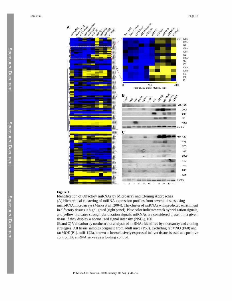
Figure 1.Identification of Olfactory miRNAs by Microarray and Cloning Approaches(A) Hierarchical clustering of miRNA expression profiles from several tissues usingmicroRNA microarrays (Miska et al., 2004). The cluster of miRNAs with predicted enrichmentin olfactory tissues is highlighted (right panel). Blue color indicates weak hybridization signals,and yellow indicates strong hybridization signals. miRNAs are considered present in a giventissue if they display a normalized signal intensity (NSI) ≥ 100.(B and C) Validation by northern blot analysis of miRNAs identified by microarray and cloningstrategies. All tissue samples originate from adult mice (P60), excluding rat VNO (P60) andrat MOE (P1). miR-122a, known to be exclusively expressed in liver tissue, is used as a positivecontrol. U6 snRNA serves as a loading control.
Choi et al. Page 18
Published as: Neuron. 2008 January 10; 57(1): 41–55.
Sponsored Docum
ent Sponsored D
ocument
Sponsored Docum
ent
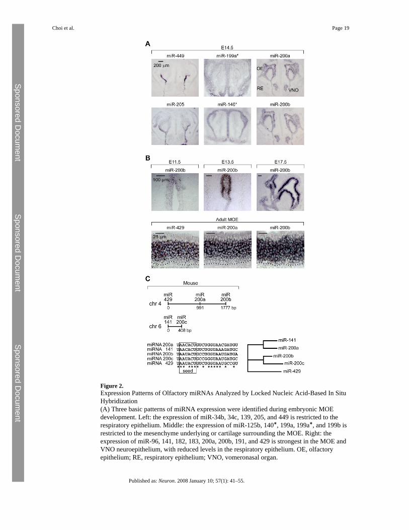
Figure 2.Expression Patterns of Olfactory miRNAs Analyzed by Locked Nucleic Acid-Based In SituHybridization(A) Three basic patterns of miRNA expression were identified during embryonic MOEdevelopment. Left: the expression of miR-34b, 34c, 139, 205, and 449 is restricted to therespiratory epithelium. Middle: the expression of miR-125b, 140∗, 199a, 199a∗, and 199b isrestricted to the mesenchyme underlying or cartilage surrounding the MOE. Right: theexpression of miR-96, 141, 182, 183, 200a, 200b, 191, and 429 is strongest in the MOE andVNO neuroepithelium, with reduced levels in the respiratory epithelium. OE, olfactoryepithelium; RE, respiratory epithelium; VNO, vomeronasal organ.
Choi et al. Page 19
Published as: Neuron. 2008 January 10; 57(1): 41–55.
Sponsored Docum
ent Sponsored D
ocument
Sponsored Docum
ent

(B) Developmental time course analysis of miR-200 family member expression.(C) Genomic organization of mouse miR-200 family members.
Choi et al. Page 20
Published as: Neuron. 2008 January 10; 57(1): 41–55.
Sponsored Docum
ent Sponsored D
ocument
Sponsored Docum
ent
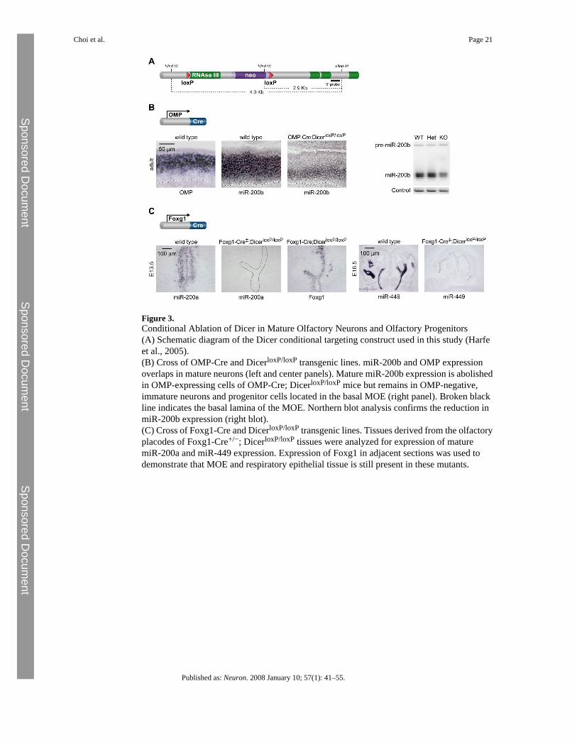
Figure 3.Conditional Ablation of Dicer in Mature Olfactory Neurons and Olfactory Progenitors(A) Schematic diagram of the Dicer conditional targeting construct used in this study (Harfeet al., 2005).(B) Cross of OMP-Cre and DicerloxP/loxP transgenic lines. miR-200b and OMP expressionoverlaps in mature neurons (left and center panels). Mature miR-200b expression is abolishedin OMP-expressing cells of OMP-Cre; DicerloxP/loxP mice but remains in OMP-negative,immature neurons and progenitor cells located in the basal MOE (right panel). Broken blackline indicates the basal lamina of the MOE. Northern blot analysis confirms the reduction inmiR-200b expression (right blot).(C) Cross of Foxg1-Cre and DicerloxP/loxP transgenic lines. Tissues derived from the olfactoryplacodes of Foxg1-Cre+/−; DicerloxP/loxP tissues were analyzed for expression of maturemiR-200a and miR-449 expression. Expression of Foxg1 in adjacent sections was used todemonstrate that MOE and respiratory epithelial tissue is still present in these mutants.
Choi et al. Page 21
Published as: Neuron. 2008 January 10; 57(1): 41–55.
Sponsored Docum
ent Sponsored D
ocument
Sponsored Docum
ent
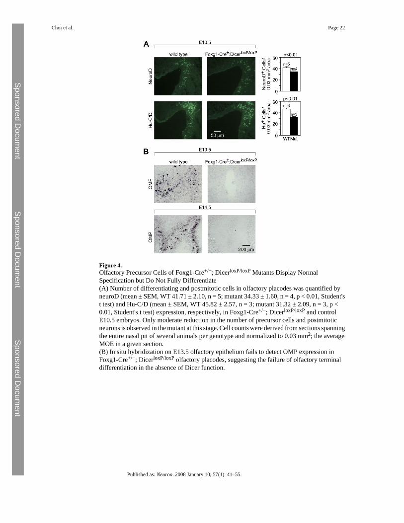
Figure 4.Olfactory Precursor Cells of Foxg1-Cre+/−; DicerloxP/loxP Mutants Display NormalSpecification but Do Not Fully Differentiate(A) Number of differentiating and postmitotic cells in olfactory placodes was quantified byneuroD (mean ± SEM, WT 41.71 ± 2.10, n = 5; mutant 34.33 ± 1.60, n = 4, p < 0.01, Student'st test) and Hu-C/D (mean ± SEM, WT 45.82 ± 2.57, n = 3; mutant 31.32 ± 2.09, n = 3, p <0.01, Student's t test) expression, respectively, in Foxg1-Cre+/−; DicerloxP/loxP and controlE10.5 embryos. Only moderate reduction in the number of precursor cells and postmitoticneurons is observed in the mutant at this stage. Cell counts were derived from sections spanningthe entire nasal pit of several animals per genotype and normalized to 0.03 mm2; the averageMOE in a given section.(B) In situ hybridization on E13.5 olfactory epithelium fails to detect OMP expression inFoxg1-Cre+/−; DicerloxP/loxP olfactory placodes, suggesting the failure of olfactory terminaldifferentiation in the absence of Dicer function.
Choi et al. Page 22
Published as: Neuron. 2008 January 10; 57(1): 41–55.
Sponsored Docum
ent Sponsored D
ocument
Sponsored Docum
ent
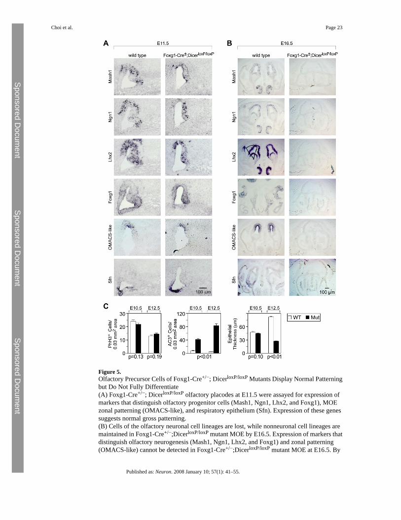
Figure 5.Olfactory Precursor Cells of Foxg1-Cre+/−; DicerloxP/loxP Mutants Display Normal Patterningbut Do Not Fully Differentiate(A) Foxg1-Cre+/−; DicerloxP/loxP olfactory placodes at E11.5 were assayed for expression ofmarkers that distinguish olfactory progenitor cells (Mash1, Ngn1, Lhx2, and Foxg1), MOEzonal patterning (OMACS-like), and respiratory epithelium (Sfn). Expression of these genessuggests normal gross patterning.(B) Cells of the olfactory neuronal cell lineages are lost, while nonneuronal cell lineages aremaintained in Foxg1-Cre+/−;DicerloxP/loxP mutant MOE by E16.5. Expression of markers thatdistinguish olfactory neurogenesis (Mash1, Ngn1, Lhx2, and Foxg1) and zonal patterning(OMACS-like) cannot be detected in Foxg1-Cre+/−;DicerloxP/loxP mutant MOE at E16.5. By
Choi et al. Page 23
Published as: Neuron. 2008 January 10; 57(1): 41–55.
Sponsored Docum
ent Sponsored D
ocument
Sponsored Docum
ent
contrast, expression of respiratory epithelium (Sfn) persists in mutant MOE. In addition, thenormally convoluted structure of the MOE is reduced to a simple epithelium comprised solelyof nonneural respiratory epithelium.(C) Quantification of phospho-histone H3 and active caspase-3 immunoreactive cells inembryonic MOE of Foxg1-Cre+/−; DicerloxP/loxP mutants and controls at E10.5 (mean ± SEM,WT 23.95 ± 1.06, n = 3; mutant 21.61 ± 1.09, n = 3, p = 0.13, Student's t test) and E12.5 (mean± SEM, WT 13.02 ± 0.76 cells, n = 3; mutant 14.49 ± 0.77 cells, n = 3, p = 0.19, Student's ttest) and active caspase-3 at E10.5 (mean ± SEM, WT 7.76 ± 1.44, n = 3; mutant 41.97 ± 3.31,n = 3, p < 0.01, Student's t test) and E12.5 (mean ± SEM, WT 5.18 ± 0.54, n = 3; mutant 83.42± 5.54, n = 3, p < 0.01, Student's t test) indicate that loss of Dicer function results in increasedcellular apoptosis and unchanged cellular proliferation in the olfactory epithelium.
Choi et al. Page 24
Published as: Neuron. 2008 January 10; 57(1): 41–55.
Sponsored Docum
ent Sponsored D
ocument
Sponsored Docum
ent
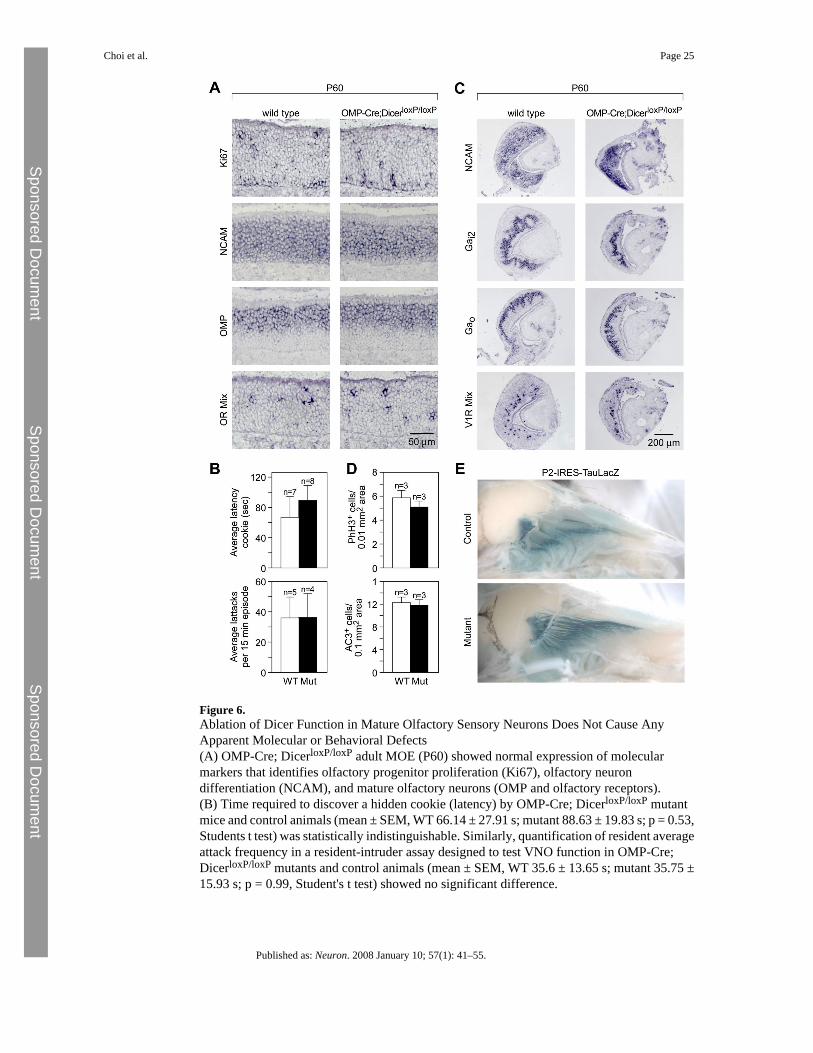
Figure 6.Ablation of Dicer Function in Mature Olfactory Sensory Neurons Does Not Cause AnyApparent Molecular or Behavioral Defects(A) OMP-Cre; DicerloxP/loxP adult MOE (P60) showed normal expression of molecularmarkers that identifies olfactory progenitor proliferation (Ki67), olfactory neurondifferentiation (NCAM), and mature olfactory neurons (OMP and olfactory receptors).(B) Time required to discover a hidden cookie (latency) by OMP-Cre; DicerloxP/loxP mutantmice and control animals (mean ± SEM, WT 66.14 ± 27.91 s; mutant 88.63 ± 19.83 s; p = 0.53,Students t test) was statistically indistinguishable. Similarly, quantification of resident averageattack frequency in a resident-intruder assay designed to test VNO function in OMP-Cre;DicerloxP/loxP mutants and control animals (mean ± SEM, WT 35.6 ± 13.65 s; mutant 35.75 ±15.93 s; p = 0.99, Student's t test) showed no significant difference.
Choi et al. Page 25
Published as: Neuron. 2008 January 10; 57(1): 41–55.
Sponsored Docum
ent Sponsored D
ocument
Sponsored Docum
ent
(C) OMP-Cre; DicerloxP/loxP adult MOE (P60) showed normal expression of molecularmarkers for vomeronasal neuronal differentiation (NCAM), zonal patterning (G proteinsubunits) and mature function (V1 receptors).(D) Quantification of phospho-histone H3 immunoreactive cells (mean ± SEM, WT 5.79 ±0.50, n = 3; mutant 5.05 ± 0.37, n = 3, p = 0.24, Student's t test) and active caspase-3immunoreactive cells (mean ± SEM, WT 12.19 ± 0.77, n = 3; mutant 11.76 ± 0.74, n = 3, p =0.69, Student's t test) in adult MOE of OMP-Cre; DicerloxP/loxP mutants and controls revealsno statistically significant differences in proliferation or apoptosis rates.(E) OMP-Cre; DicerloxP/loxP; P2-IRES-TauLacZ triple-transgenic mice (P45) showed normalexpression and axon targeting of LacZ in P2-expressing olfactory neurons.
Choi et al. Page 26
Published as: Neuron. 2008 January 10; 57(1): 41–55.
Sponsored Docum
ent Sponsored D
ocument
Sponsored Docum
ent
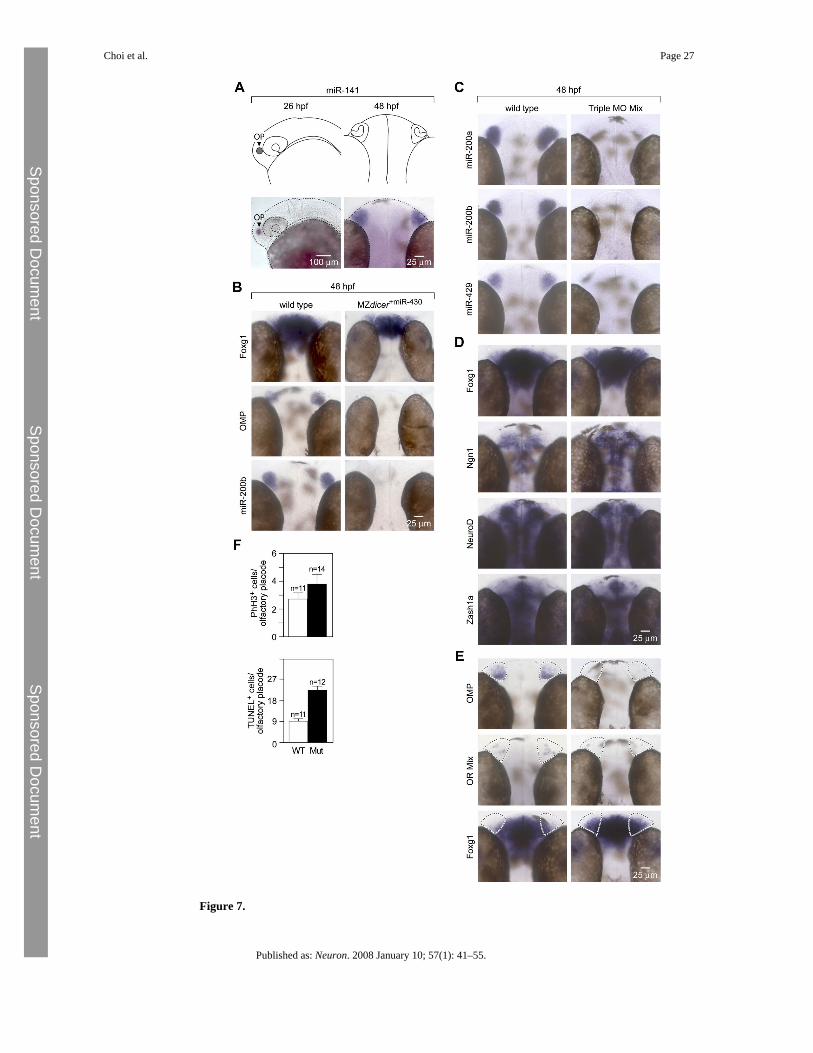
Figure 7.
Choi et al. Page 27
Published as: Neuron. 2008 January 10; 57(1): 41–55.
Sponsored Docum
ent Sponsored D
ocument
Sponsored Docum
ent
Zebrafish miR-200 Family Members Are Required for Terminal Differentiation of OlfactoryProgenitor Cells(A) Schematic diagram of the zebrafish olfactory placode and olfactory organ at 26 hpf and48 hpf, respectively, and corresponding expression pattern of miR-141, a member of themiR-200 family.(B) MZdicer embryos were injected with miR-430 (MZdicer+miR-430) to substantially rescuegeneral neuronal and other phenotypic defects observed in MZdicer mutants by supplying thecritical miRNA expressed during the earliest stages of development (Giraldez et al., 2005).MZdicer+miR-430 embryos assayed for expression of olfactory progenitor (foxg1), matureneuron (OMP), and miRNA (miR-200b) markers show impaired terminal differentiation ofolfactory progenitors.(C) In situ hybridization staining of 48 hpf embryos for expression of miR-200a, miR-200b,and miR-420 that were injected at the one-cell stage with a combination of miR-141 MO,miR-200b MO, and miR-429 MO (4 ng each; Triple MO Mix) show complete loss of miR-200family expression.(D) Wild-type and fish injected with various morpholinos at 48 hpf are morphologicallyindistinguishable from each other with the exception of expanded Foxg1 expression (see panel[E]).(E) Triple MO morphants injected at the one-cell stage and assayed for expression of olfactoryprogenitor marker (foxg1) and mature neuronal markers (OMP and an olfactory receptor mix)at 48 hpf show impaired terminal differentiation of olfactory progenitors.(F) Quantification of phospho-histone H3 immunoreactive cells (mean ± SEM, WT 2.55 ±0.45, n = 11; morphant 3.57 ± 0.67, n = 14, p = 0.24, Student's t test) and TUNELimmunoreactive cells (mean ± SEM, WT 12.55 ± 1.46, n = 11; mutant 30.67 ± 2.59, n = 12, p< 0.01, Student's t test) in 72 hpf Triple MO morphant olfactory epithelia and controls revealsa statistically significant difference in apoptosis, but not proliferation.
Choi et al. Page 28
Published as: Neuron. 2008 January 10; 57(1): 41–55.
Sponsored Docum
ent Sponsored D
ocument
Sponsored Docum
ent
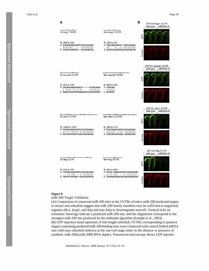
Figure 8.miR-200 Target Validation(A) Comparison of conserved miR-200 sites in the 3′UTRs of select miR-200 predicted targetsin mouse and zebrafish suggest that miR-200 family members may be sufficient to negativelyregulate zfhx1, foxg1, and lfng and may help to downregulate neuroD. Vertical ticks onschematic drawings indicate a predicted miR-200 site, and the alignments correspond to thestrongest miR-200 site produced by the miRanda algorithm (Enright et al., 2003).(B) GFP reporters fused upstream of full-length zebrafish 3′UTRs corresponding to putativetargets containing predicted miR-200 binding sites were coinjected with control DsRed mRNAinto wild-type zebrafish embryos at the one-cell stage either in the absence or presence ofsynthetic miR-200a/miR-200b RNA duplex. Fluorescent microscopy shows GFP reporter
Choi et al. Page 29
Published as: Neuron. 2008 January 10; 57(1): 41–55.
Sponsored Docum
ent Sponsored D
ocument
Sponsored Docum
ent

expression (green) and control DsRed expression (red) at 25–30 hpf, indicating that miR-200family members are sufficient to downregulate zebrafish zfhx1 and lfng.
Choi et al. Page 30
Published as: Neuron. 2008 January 10; 57(1): 41–55.
Sponsored Docum
ent Sponsored D
ocument
Sponsored Docum
ent