neuronal polarity axon / dendrite...
TRANSCRIPT

Neuronal polarity – axon / dendrite specification

Axon / dendrite specification in vivo
• postmitotic neurons leaving the germinative zone have uniform multiple processes (SVZ)
• radial / tangential migration requires polarized processes (leading / trailing process)

Axon / dendrite specification in vivo

• studies mostly in dissociated embryonic hippocampal cell cultures
• characteristic morphology – characteristic stages in development and maturation
Axon / dendrite specification in vitro
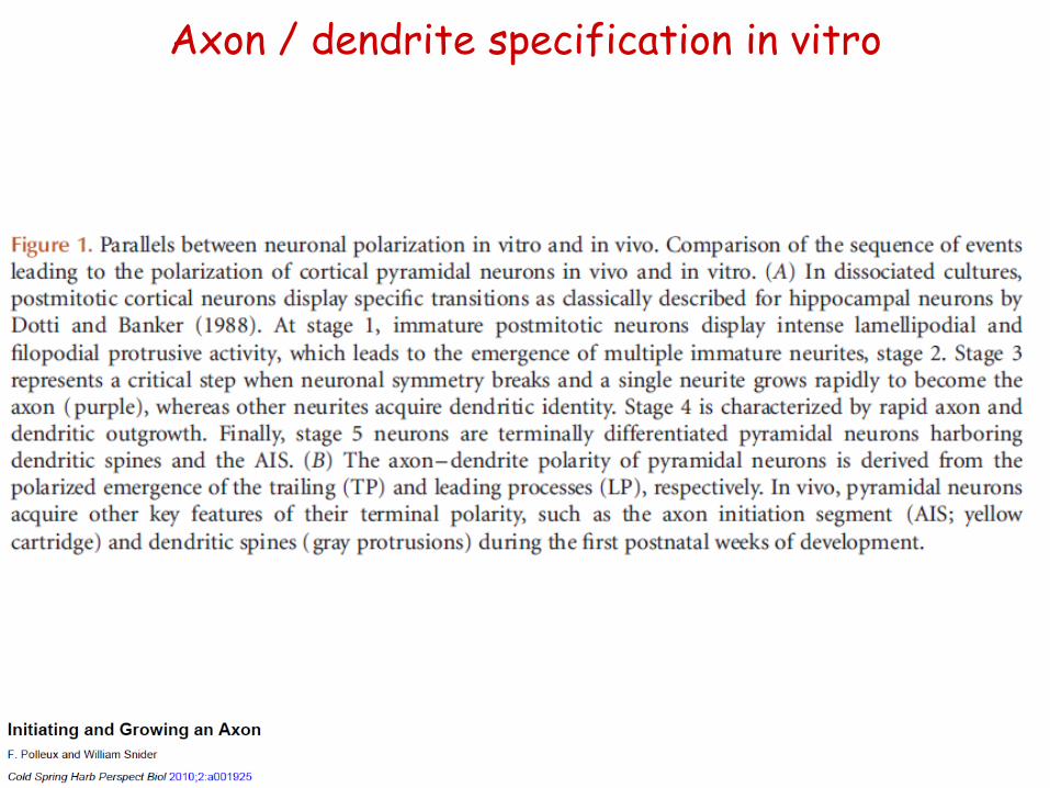
Axon / dendrite specification in vitro

Neuronal polarization

Neuronal polarization

Initial axon specification (in vitro)

General features of axons and dendrites
• number, appearence
• synaptic features
• membrane structure
• length, morphology, branching
• myelin
• cytoskeleton, MAPs
• protein synthesis, organelles
dendrit
axon
axon
axon
dendrit
dendrit
axon
axon
axon
dendrit
• electrical activity
• endosomes

Neuronal protein synthesis and sorting
• Nissl granules: within the perikaryon and proximal dendrites
free ribosomes (rosetta)
RER
• cytoplasmic, membrane-associated proteins:
- synthesis on free ribosomes or bound polysomes
• somatodendritic compartments, rarely in the axon (mainly during development)
- directly / indirectly attached to the cytoskeleton
- axon: mainly in resting state (RNA granulum) – activation upon injury -> local protein synthesis

• new results: presynaptic protein synthesis is required for long-term presynaptic plasticity
FUNCAT: fluorescent non-canonical amino
acid tagging
- no de novo protein synthesis during the fast presynaptic events, but it is required for long-term effects
- ribosomes can be detected by STORM superresolution within the presynaptic terminals
Neuronal protein synthesis and sorting

Neuronal membrane trafficking

Neuronal membrane trafficking

Polarized membrane trafficking kymographs
https://images.nature.com/f
ull/nature-
assets/nrn/journal/v17/n10/e
xtref/nrn.2016.100-sv1.mov

• transport of transmembrane proteins in the dendrites:
- ER, Golgi: also within the dendrites
• continuous ER-network
- local protein synthesis within the dendrites:
- bidirectional pre-Golgi transport
• Golgi outposts
- „basic” secretion: somatic ER -> Golgi, long path towards the plasma membrane
• specific cargo (eg. BDNF)
• dendritic secretion / release
• spine and PSD development
Emerging aspects of membrane traffic in
neuronal dendrite growth; Tang 2008
Neuronal protein synthesis and sorting

Neuronal protein synthesis and sorting
• transport of transmembrane proteins in the dendrites:
- AMPARs are transported in vesicles from the Golgi, but can be locally synthetised upon activation within the dendrites

Pathways of axonal transmembrane protein transport
1. axonally directed, polarised sorting from the TGN + selective binding to axon-specific delivery motors
2. indirect, polarised transport (transcytosis): endocytosis in dendrites, followed by selective anterograde axon-directed transport
3. non-polarised transport + selective retrieval / retention
L1/NgCAM
CB1R
APP
synaptophysin (p38)
EAA1/Rab5
dependent
endocytosis

Transcytosis
~ dendrite
~ axon
(YRSLE)
bazolaterális adaptor
klatrin adaptor
kináz

Transcytosis

Neuronal cytoskeleton • microtubules (MTs)
- a, b tubulin monomers, „dynamic stability”
- posttranlational modifications (Tyr, Ac, Pi)
- >10% of total brain protein; 25 nm diameter, > 100 mm length
- bundled structure (mainly in axons)
- intracellular transport, inner stabilisator of neurites: dynamic / stabile MTs
• axon, dist. dendrite: + end distally
• prox. dendrite: + / - ends mixed - diverse, specific MAPs
• nucleation: g-tubulin (centrosome); cleavage (katanin, spastin)
• +TIPs (+end tracking proteins): APC, DCX - axon

Microtubule organization and organelle distribution in axons and dendrites. Axons have tau-bound microtubules of uniform
orientation, whereas dendrites have microtubule-associated protein 2 (MAP2)-bound microtubules of mixed orientation. Dendrites also
contain organelles that are not found in axons, such as rough endoplasmic reticulum, polyribosomes and Golgi outposts.
Neuronal MTs

• microtubulus associated proteins (MAPs)
- HMW MAPs (>1300 KDa)
• MAP1: axon, dendrite; MAP2: dendrite
• sidebranches attached to microfilaments
• morphology – regulates neuronal plasticity
- tau Katanin
• [neurofibrillary tangles]
• MT stabilisation – Pi inactivates
• dephosphorylated in the distal axon (spec. marker)
Neuronal cytoskeleton

Microtubule organization in developing
axons. The organization and regulation of
microtubules in a stage 3 hippocampal
pyramidal neuron. Dynamic (blue) microtubules
predominate in minor processes (short,
unbranched neurites) and at the distal end of
the axon and collateral branches, whereas
stable (purple) microtubules are enriched in the
proximal part of the axon. Inset a shows katanin-
mediated release of microtubules nucleated at
the centrosome. Short polymers are transported
along microtubules by motors such as dynein.
Inset b shows the transport of tubulin dimers or
oligomers to the growth cone by a complex of
kinesin family member 5C (KIF5C) and collpasin
response mediator protein 2 (CRMP2; also
known as DPYSL2). On entering the axonal
growth cone microtubules splay, bend, loop,
bundle or get captured at the cell cortex. Inset c
illustrates the dynamic behaviour of splayed
microtubules. Proteins such as CRMP2 promote
microtubule assembly, whereas microtubule-
associated protein 1B (MAP1B) contributes to
the maintenance of microtubule dynamics and
KIF2 and stathmin induce microtubule
depolymerization. Inset d shows the protein
machinery that is involved in microtubule
capture (see also Supplementary information S1
(table)). Inset e shows how tau protects
microtubules from katanin-induced severing,
thereby contributing to microtubule stabilization
and preventing excessive collateral branching.
In the axon shaft, the formation of collateral
branches is regulated by the action of
microtubule severing proteins such as spastin.
APC, adenomatous polyposis coli; aPKC,
atypical protein kinase C (also known as
PRKCI); CLASP, cytoplasmic linker associated
protein; CLIP170, CAP-GLY domain containing
linker protein 170 (also known as CLIP1); EB,
end-binding protein; IQGAP1, IQ motif
containing GTPase activating protein 1.
Neuronal MTs

• microfilaments (actin)
- presynaptic terminal, dendritic spine, growth cone, cortical actin network (beneath the plasma membrane [PM])
- barbed-end capping (ezrin, radexin, moezin): Ranvier node
• localisation; also to ECM components
- indirect binding (spektrin, dystrophyin, a-actinin)
• cortical actin network, PSD, receptors, ion channels
- intensive, activity-dependent remodelling
- pre/postsynapse: barbed end towards the PM
Neuronal cytoskeleton
- 400-800 nm length, 4-6 nm diameter
- no major differences between axonal / dendritic functions or structure

• intermedier filaments
- few mm length, 8-12 nm diameter
- lamin (nuclear membrane; type V) + cytoplasm
• Type III: vimentin, GFAP
• Type IV: neurofilament (NF), nestin
- stabile structure: providing cellular morphology
- NFH ~180-200 KDa - NFM ~130-170 KDa - NFL ~60-70 KDa
• NF: side branches, highly phosphorylated (NFH, NFM mainly in axons)
• spatially extended -> regulates axon diameter
- mutations, injury: neuropathological changes
Neuronal cytoskeleton

• transport (cargo):
- vesicule
- multiprotein complex
- organelles
- RNA granules
- cytoskeletal elements
• anterograde or retrograde
• along MTs or actin
• kinesin / dynein / myosin
Neuronal motor proteins

• kinesin superfamily (KIFs)
• N-KIFs (N-term. motor domain): mainly towards MT+ end
kinezin-1
kinezin-3
kinezin-2
- > 45 genes, 14 classes
• C-KIFs (C-term. motor domain): mainly towards MT- end
• KIF2A, 2C: MT depolymerisation
- mainly indirect cargo binding: polarised transport
• Rab GTPases
• adaptor proteins
synaptic vesicule
precursor
axon Neuronal motor proteins
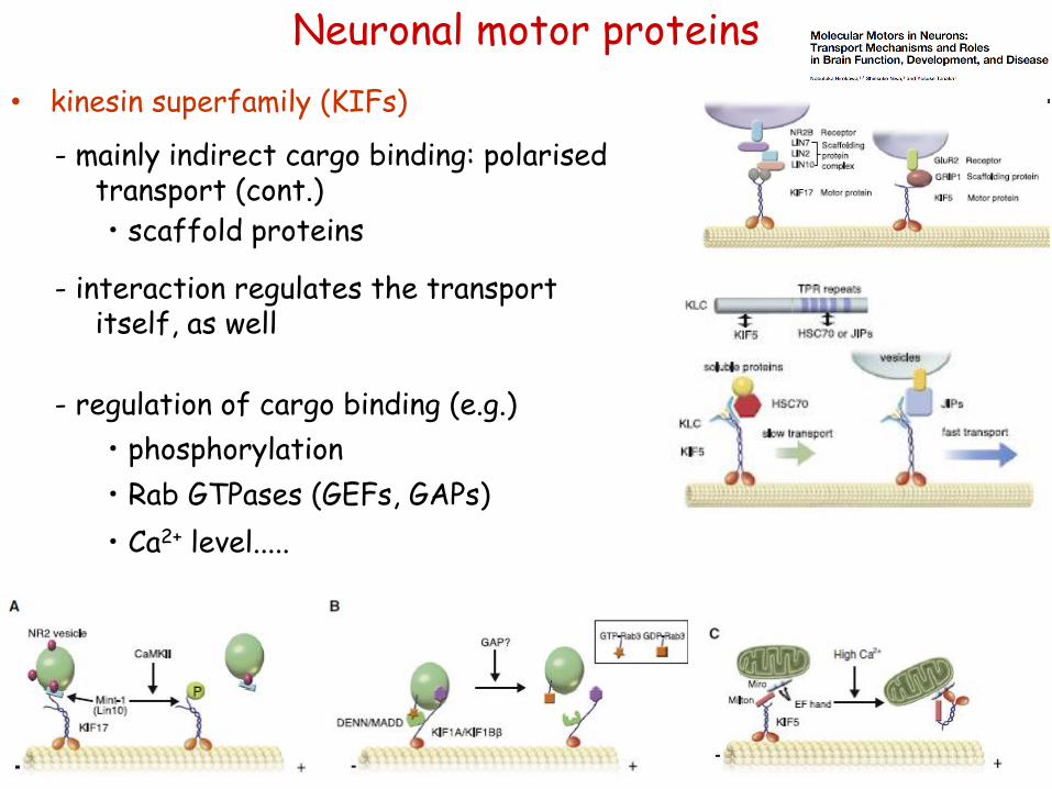
• scaffold proteins
- regulation of cargo binding (e.g.)
• phosphorylation
• Rab GTPases (GEFs, GAPs)
• Ca2+ level.....
- mainly indirect cargo binding: polarised transport (cont.)
- interaction regulates the transport itself, as well
Neuronal motor proteins
• kinesin superfamily (KIFs)
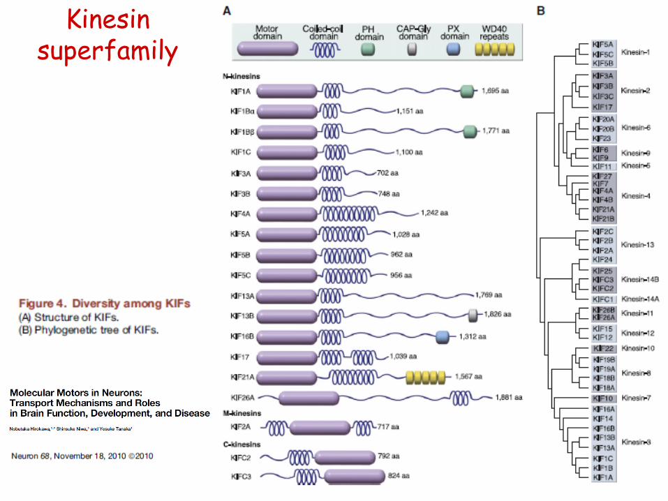
Kinesin superfamily

• dynein superfamily
• > 1,5 MDa protein complex
- axonemal (ciliary / flagellar) dynein
- citoplasmic dynein
- mainly towards MT- end; retrograde axonal transport
- cargo-binding via associated proteins (dynactin)
- competition for cargo binding: eg, BDNF vesicle-transport depends on the phosphorylation of the adaptor
Neuronal motor proteins

• myosin superfamily
- 18 classes; in neurons, myosin V (II, as well)
- cargo-binding via associated proteins / adaptors
dendritic spine
- cargo-binding normally depends on [Ca2+]
- actin-dependent movement
Neuronal motor proteins

https://www.youtube.com/watch?v=RRfH4ixgJwg
Transport inside the brain: The basic mechanisms of neuronal trafficking. Hoogenraad Lab
Transport within the neurons

• SCa (slow component a): cytoskeletal structures
• SCb (slow component b): cytoplasmic proteins
- NF, MT - 0.1 – 1 mm/day [0,001 – 0,01 mm/s]
- 2 – 4 mm/day [0,02 – 0,04 mm/s]
- actin, tubulin monomers, enzymes....
• normally stop-and-go movement
• KIF5: Hsc-70 adaptor myosin Va: NF-L dynein: MT (can bind NFs, as well)
• uni-directed, anterograde
• directs axonal elongation, regeneration
Slow axonal transport

Figure 2.8 Slow axonal transport represents the delivery of cytoskeletal and cytoplasmic constituents to the periphery. Cytoplasmic proteins are synthesized on free polysomes and organized for transport as cytoskeletal elements or macromolecular complexes (1). Microtubules are formed by nucleation at the microtubule-organizing center near the centriolar complex (2) and then released for migration into axons or dendrites. Slow transport appears to be unidirectional with no net retrograde component. Studies suggest that cytoplasmic dynein may move microtubules with their plus ends leading (3). Neurofilaments may move on their own or may hitchhike on microtubules (4). Once cytoplasmic structures reach their destinations, they are degraded by local proteases (5) at a rate that allows either growth (in the case of growth cones) or maintenance of steady-state levels. The different composition and organization of cytoplasmic elements in dendrites suggest that different pathways may be involved in delivery of cytoskeletal and cytoplasmic materials to dendrites (6). In addition, some mRNAs are transported into dendrites, but not into axons.
Slow axonal transport

• vesicular trafficking: mitochondrium, transmembrane receptors, synaptic vesicles, neuropeptides, neurotrophines, viruses...
• anterograde and retrograde
• 100 – 400 mm/day [1 – 4 mm/s] (retrograde transport is normally slower)
• KIF1, KIF5: mitochondrium myosin Va: synaptic vesicles, secretory
granules, mRNA granules dynein: signalling endosomes (eg. NGF-
TrkA); BDNF; Rab5 endosome, myosin V (retrograde)
Fast axonal transport

Figure 2.9 Fast axonal transport represents transport of membrane-associated materials, having both anterograde and retrograde components. For anterograde transport, most polypeptides are synthesized on membrane-bound polysomes, also known as rough endoplasmic reticulum (1), and then transferred to the Golgi for processing and packaging into specific classes of membrane-bound organelles (2). Proteins following this pathway include both integral membrane proteins and secretory polypeptides in the vesicle lumen. Cytoplasmic peripheral membrane proteins such as kinesins are synthesized on free polysomes. Once vesicles are assembled and appropriate motors associate with them, they move down the axon at a rate of 100–400 mm per day (3). Different membrane structures are delivered to different compartments and may be regulated independently. For example, dense core vesicles and synaptic vesicles are both targeted for presynaptic terminals (4), but release of vesicle contents involves distinct pathways. After vesicles merge with the plasma membrane, their protein constituents are taken up in coated vesicles via the receptor-mediated endocytic pathway and delivered to a sorting compartment (5). After proper sorting into appropriate compartments, membrane proteins are either committed to retrograde axonal transport or recycled (6). Retrograde moving organelles are morphologically and biochemically distinct from anterograde vesicles. These larger vesicles have an average velocity about half that of anterograde transport. The retrograde pathway is an important mechanism for delivery of neurotrophic factors to the cell body. Material delivered by retrograde transport typically fuses with cell body compartments to form mature lysosomes (7), where constituents are recycled or degraded. However, neurotrophic factors and neurotrophic viruses act at the level of the cell body and escape this pathway. Vesicle transport also occurs into dendrites (8), but less is known about this process.
Fast axonal transport
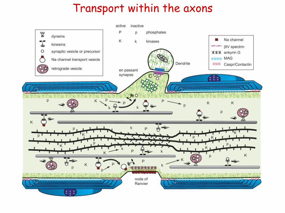
Transport within the axons

Figure 2.10 Axonal dynamics in a myelinated axon from the peripheral nervous system (PNS). Axons are in a constant flux with many concurrent dynamic processes. This diagram illustrates a few of the many dynamic events occurring at a node of Ranvier in a myelinated axon from the PNS. Axonal transport moves cytoskeletal structures, cytoplasmic proteins, and membrane-bound organelles from the cell body toward the periphery (from right to left). At the same time, other vesicles return to the cell body by retrograde transport (retrograde vesicle). Membrane-bound organelles are moved along microtubules by motor proteins such as the kinesins and cytoplasmic dyneins. Each class of organelles must be directed to the correct functional domain of the neuron. Synaptic vesicles must be delivered to a presynaptic terminal to maintain synaptic transmission. In contrast, organelles containing sodium channels must be targeted specifically to nodes of Ranvier for saltatory conduction to occur. Cytoskeletal transport is illustrated by microtubules (rods in the upper half of the axon) and neurofilaments (bundle of rope-like rods in the lower half of the axon) representing the cytoskeleton. They move in the anterograde direction as discrete elements and are degraded in the distal regions. Microtubules and neurofilaments interact with each other transiently during transport, but their distribution in axonal cross sections suggests that they are not stably cross-linked. In axonal segments without compact myelin, such as the node of Ranvier or following focal demyelination, a net dephosphorylation of neurofilament side arms allows the neurofilaments to pack more densely. Myelination is thought to alter the balance between kinase (K indicates an active kinase; k is an inactive kinase) and phosphatase (P indicates an active phosphatase; p is an inactive phosphatase) activity in the axon. Most kinases and phosphatases have multiple substrates, suggesting a mechanism for targeting vesicle proteins to specific axonal domains. Local changes in the phosphoryation of axonal proteins may alter the binding properties of proteins. The action of synapsin I in squid axoplasm suggests that dephosphorylated synapsin cross-links synaptic vesicles to microfilaments. When a synaptic vesicle encounters the dephosphorylated synapsin and actin-rich matrix of a presynaptic terminal, the vesicle is trapped at the terminal by inhibition of further axonal transport, effectively targeting the synaptic vesicle to a presynaptic terminal. Similarly, a sodium channel-binding protein may be present at nodes of Ranvier in a high-affinity state (i.e., dephosphorylated). Transport vesicles for nodal sodium channels (Na channel vesicle) would be captured upon encountering this domain, effectively targeting sodium channels to the nodal membrane. Interactions between cells could in this manner establish the functional architecture of the neuron.
Transport within the axons

• local protein synthesis + bidirectional transport for shorter distances
- KIF5: AMPA, GABA receptors; mRNA
• dendrite-specific motor:
- KIF17: NMDA receptors
- KIFC2 (C-KIF); MT- end: multivesicular bodies
• motors within the axons:
- dynein: Rab5, 7 endosome; glycine receptor
- myosin Va, Vb: AMPA receptor
• how can the axonal / dendritic membrane receptors be sorted specifically?
- „smart” and „dumb” motor proteins
- „dumb” motors are helped by selective endocytosis, too....
Transport within the dendrites
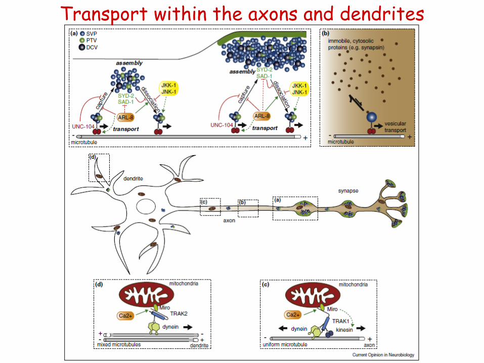
Transport within the axons and dendrites

Transport within the axons and dendrites

Essay questions (choose one) Describe the major steps and factors regulating early neuronal polarisation, thus, the
initial axon/dendrite specification! Ismertesse azokat a meghatározó szabályozási
lépéseket, amik a neuronális polarizációt, azaz a korai axon / dendrit elkülönülést
irányítják!
Compare the major axonal and dendritic features, highlighting similarities as well as
specific differences! / Hasonlítsa össze az axon, illetve a dendritek főbb jellemzőit!
Milyen tulajdonságaikban hasonlítanak vagy különböznek?
Explain the major axon-specific transport pathways! How do membrane or cytoplasmic
proteins get transported into the axons? / Milyen axon-specifikus transzportútvonalakat
ismer? Hogyan jutnak el az axonba a membrán- és a citplazmás fehérjék?
Characterise the major types of neuronal cytoskeletal proteins! Explain which features
are characteristic to the axons or to the dendrites! / Sorolja fel a neuronális vázfehérjék
fő típusait! Röviden jellemezze, hogy az axonokban és a dendritekben mely
komponensek jellemzőek és ezeknek milyen jellegzetességei figyelhetőek meg!
Characterise the major types of neuronal motor proteins! Explain which features are
characteristic to the axons or to the dendrites! / Sorolja fel a neuronális motorfehérjék fő
típusait! Röviden jellemezze, hogy az axonokban és a dendritekben melyek jellemzőek
és ezek milyen jellegzetességekkel bírnak!
Describe and compare the characteristics of fast and the slow axonal transport! /
Ismertesse és hasonlítsa össze a lassú és a gyors axonális transzport jellegzetességeit!

Recommended literature