neuropsychology of amnesia lecture (chapter 9) jaap murre [email protected]
TRANSCRIPT

In this lecture
• We will review basic aspects of amnesia
• We will try to locate memory in the brain and relate brain lesions to amnesia
• We will make a start with dementia, looking at progressive semantic dementia
Before we embark on our study of amnesia
• What types of memory are there?
• If amnesia is a form of memory loss, what is forgetting?

Forms of memory: Larry Squire’s memory taxonomy

Forgetting
• There is currently no theory that explains why we forget
• Forgetting seems to follow rather strict rules, but even these have not been fully explored
• It is postulated that very well rehearsed knowledge will never be forgotten (Harry Barrick’s ‘permastore’)
Before looking at the anatomy and clinical aspects of amnesia
• We will review a connectionist model of amnesia
• It will not be necessary to review the technical aspects of this model
• The model may help you to get an overall idea of what amnesia is

We will focus on some important characteristics
• Anterograde amnesia (AA)– Implicit memory preserved
• Retrograde amnesia (RA)– Ribot gradients
• Pattern of correlations between AA and RA– No perfect correlation between AA and RA

The French neurologist Ribot discovered more than 100 years ago that in retrograde amnesia one tends to loose recent memoriesMemory loss gradients in RA are called Ribot gradients
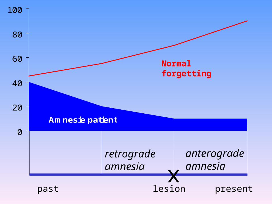
xretrograde amnesia
anterograde amnesia
lesion presentpast
0
20
40
60
80
100
Amnesie patient
Normal forgetting

An example of retrograde amnesia patient data
0
0.1
0.2
0.3
0.4
0.5
0.6
0.7
0.8
75-'8465-'7455-'6445-'5435-'44
Controls (n=16)
Korsakoff's (n=6)
Alzheimer's (n=8)
Kopelman (1989)News events test
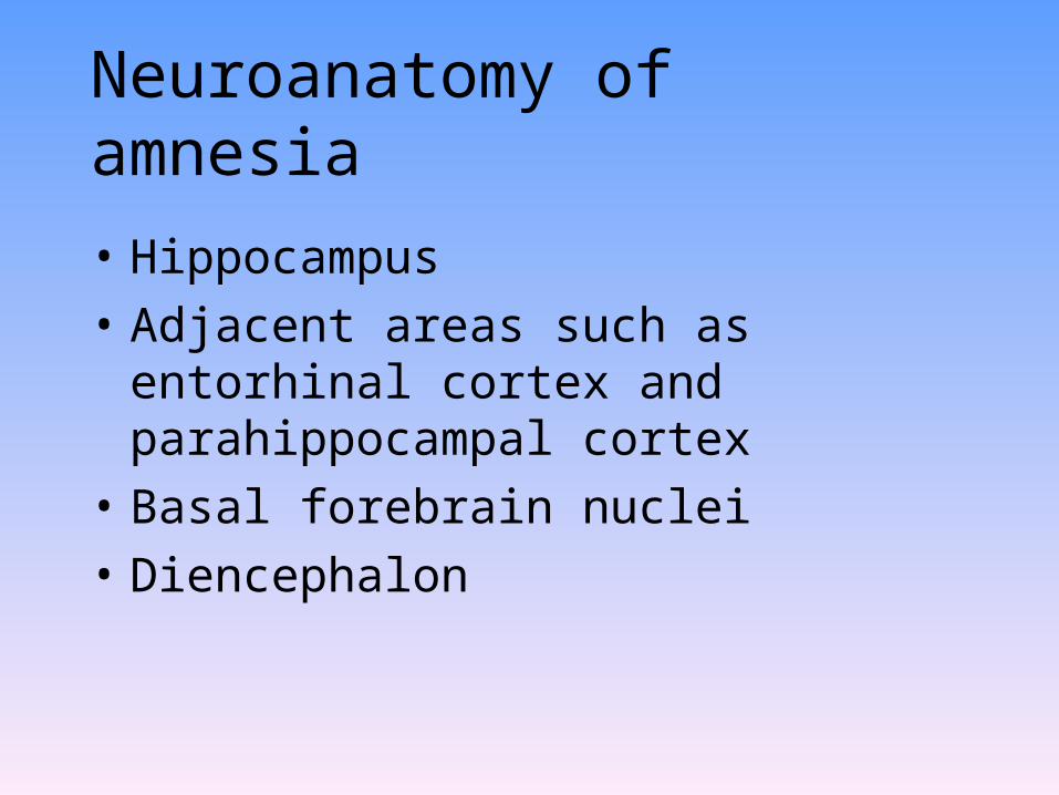
Neuroanatomy of amnesia
• Hippocampus
• Adjacent areas such as entorhinal cortex and parahippocampal cortex
• Basal forebrain nuclei
• Diencephalon
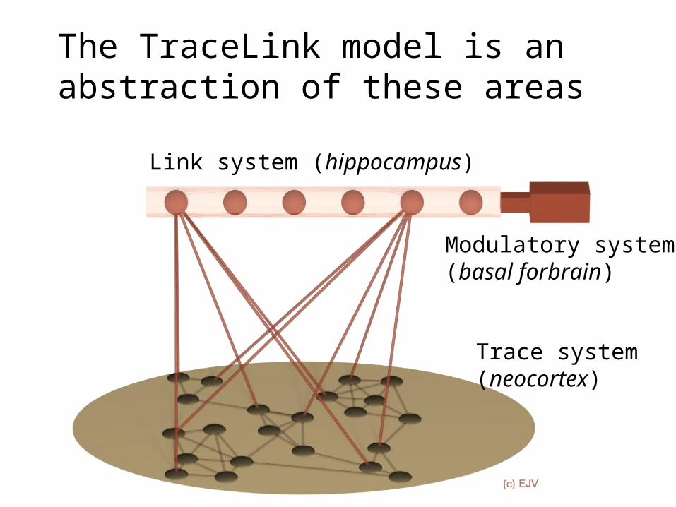
The TraceLink model is an abstraction of these areas
Link system (hippocampus)
Trace system (neocortex)
Modulatory system (basal forbrain)
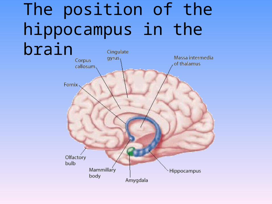
The position of the hippocampus in the brain
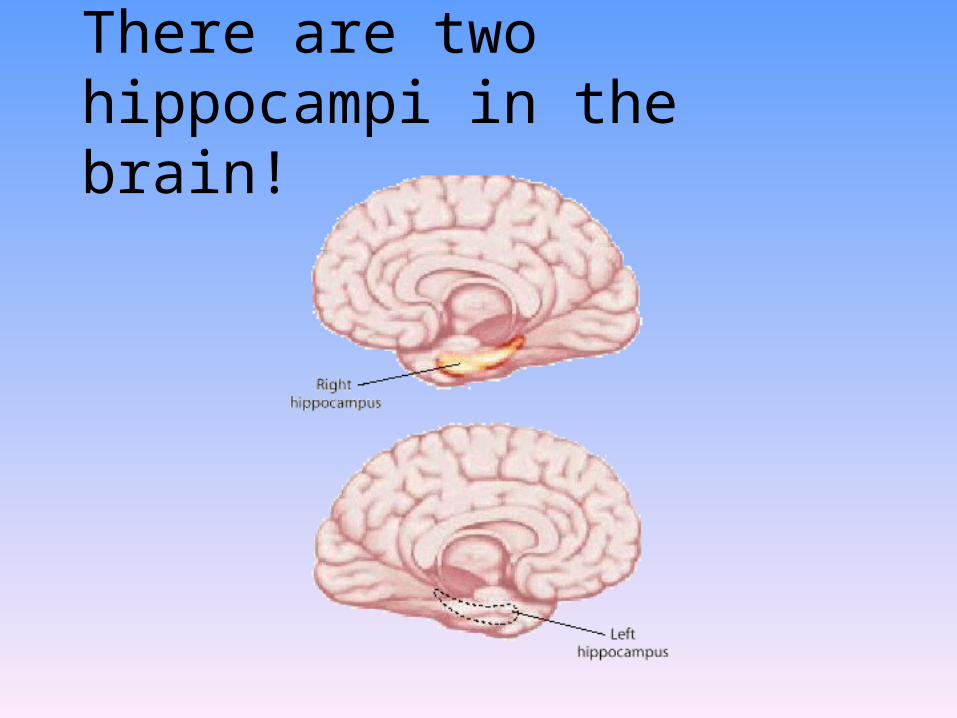
There are two hippocampi in the brain!

Connections to and from the hippocampus
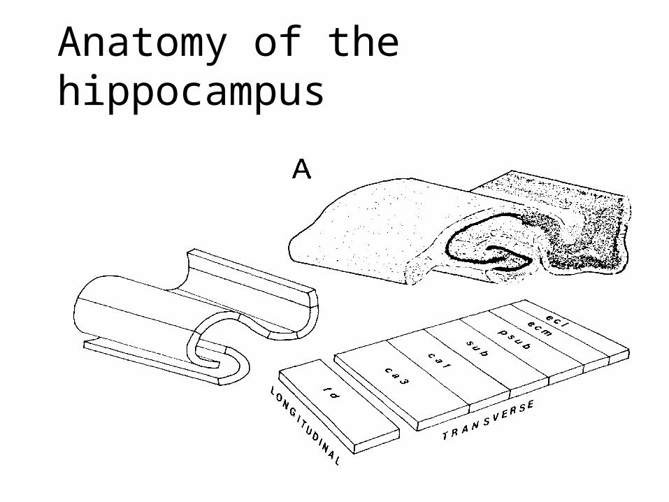
Anatomy of the hippocampus


Hippocampus
Entorhinal cortex
7a
36 TF TH 46
7b
3aP-IP-BV1M 3b
Visualareas
Somato-sensoryand motorareas
To and from sensory organs,via subcortical pathways
Hippocampus
Entorhinal cortex
Unimodal and polymodalassociation areas(frontal, temporal, and parietal lobes)
Parahippocampalcortex
Perirhinalcortex
(b)(a)
Hippocampus has anexcellent overview of the entire cortex

Diencephalon: dorsomedial nucleus and the mammillary bodies

Connectionist modelling
• Based on an abstraction of the brain
• Many simple processors (‘neurons’)
• Exchange of simple signals over connections (‘axons and dendrites’)
• Strength (‘synapse’) of the connections determines functioning of the network
• Such neural networks can be taught a certain range of behaviors
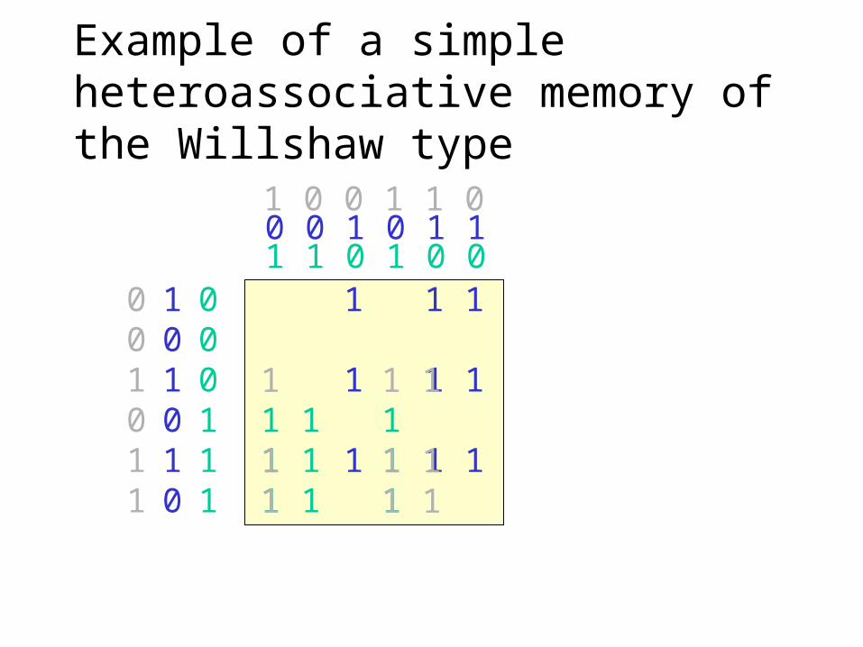
Example of a simple heteroassociative memory of the Willshaw type
1 1 0 1 0 0000111
1 1 1 1 1 1 1 1 1
101010
001011
0 0 1 0 1 11 0 0 1 1 0
1 1 1
1 1 1 1 1 1
1 1 1 1 1 11 1 1

Example of pattern retrieval
1 1 1 1 1 1 1 1 1
001011
1 1 1
1 1 1 1 1 1
1 1 1 1 1 11 1 13 2 2 3 3 21 0 0 1 1 0
Sum = 3Div by 3 =
(1 0 0 1 1 0)

Example of successful pattern completion using a subpattern
1 1 1 1 1 1 1 1 1
001001
1 1 1
1 1 1 1 1 1
1 1 1 1 1 11 1 12 1 1 2 2 11 0 0 1 1 0
Sum = 2Div by 2 =
(1 0 0 1 1 0)
1
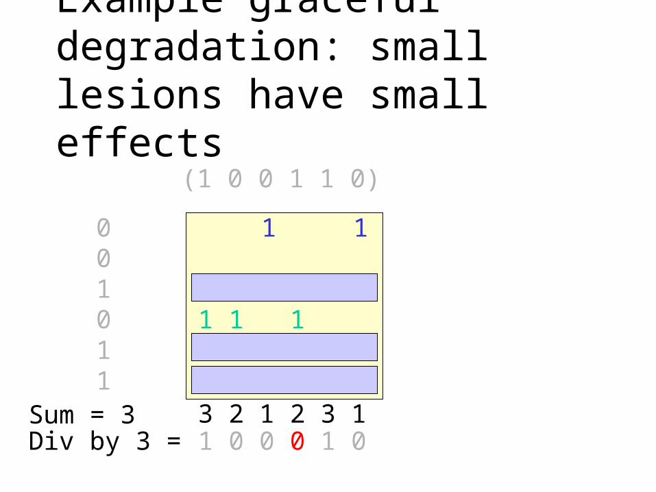
Example graceful degradation: small lesions have small effects
1 1 1 1 1 1 1 1 1
001011
1 1
1 1 1
1 1 1 1 11 1 13 2 1 2 3 11 0 0 0 1 0
Sum = 3Div by 3 =
(1 0 0 1 1 0)
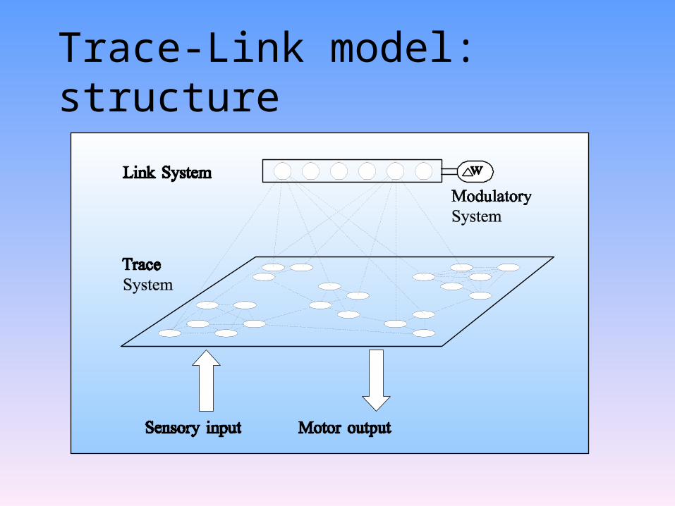
Trace-Link model: structure
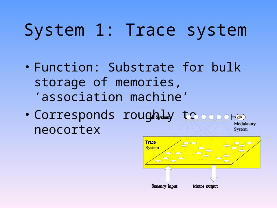
System 1: Trace system
• Function: Substrate for bulk storage of memories, ‘association machine’
• Corresponds roughly to neocortex
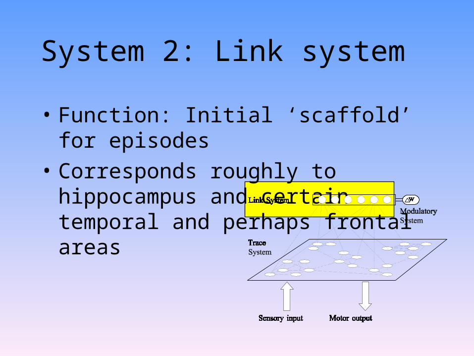
System 2: Link system
• Function: Initial ‘scaffold’ for episodes
• Corresponds roughly to hippocampus and certain temporal and perhaps frontal areas

System 3: Modulatory system
• Function: Control of plasticity• Involves at least parts of the hippocampus,
amygdala, fornix, and certain nuclei in the basal forebrain and in the brain stem
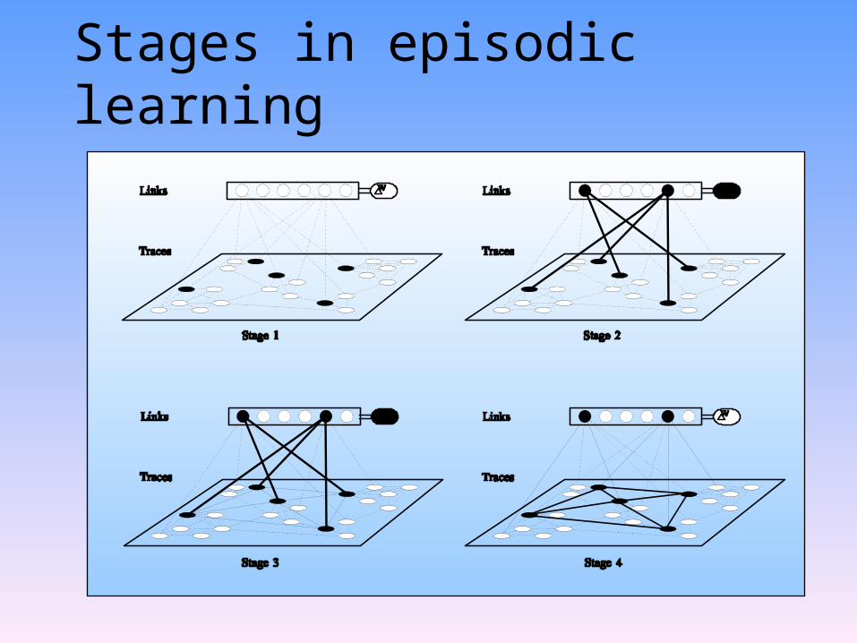
Stages in episodic learning

Retrograde amnesia
• Primary cause: loss of links
• Ribot gradients
• Shrinkage

Anterograde amnesia
• Primary cause: loss of modulatory system• Secondary cause: loss of links• Preserved implicit
memory

Semantic dementia
• The term was adopted recently to describe a new form of dementia, notably by Julie Snowden et al. (1989, 1994) and by John Hodges et al. (1992, 1994)
• Semantic dementia is almost a mirror-image of amnesia

Neuropsychology of semantic dementia
• Progressive loss of semantic knowledge
• Word-finding problems
• Comprehension difficulties
• No problems with new learning
• Lesions mainly located in the infero-lateral temporal cortex but (early in the disease) with sparing of the hippocampus
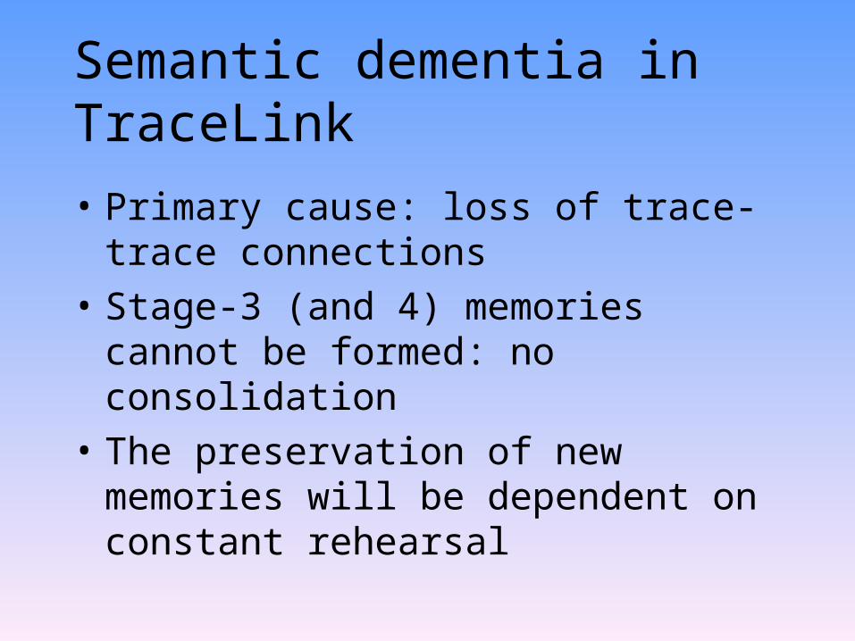
Semantic dementia in TraceLink
• Primary cause: loss of trace-trace connections
• Stage-3 (and 4) memories cannot be formed: no consolidation
• The preservation of new memories will be dependent on constant rehearsal

Severe loss of traceconnections
Stage-2 learning proceedsas normal
Stage 3 learning stronglyimpaired
Non-rehearsed memorieswill be lost
No consolidation in semantic dementia

Clinical presentation of amnesia
• Age
• Degenerative disorders
• Vascular disease
• Anoxia
• Korsakoff (vitamin B deficiency)
Clinical presentation of amnesia (con’d)
• Focal brain damage
• Closed-head injury
• Transient global amnesia (TGA)
• Electroconvulsive therapy
• Psychogenic (functional) amnesia
Rehabilitation of amnesia
• There is no known treatment
• Compensation will, thus, help the patient best: – ‘memory book’– electronic agenda
• Errorless learning is pioneered by Alan Baddeley and Barbara Wilson
Comments on the chapter
• Very few people now believe that the amygdala plays a role in episodic memory
• Most neurologists now accept the existence of focal retrograde amnesia (Kapur, 1993)
• Animal studies (rats, primates) show clear evidence of Ribot gradients in the range 30 to 100 days
Next lecture
• Implicit memory
• Dementia