neuroscience laboratory chu de québec research center, … · serge rivest neuroscience laboratory...
TRANSCRIPT

Serge Rivest Neuroscience Laboratory
CHU de Québec Research Center, Department of Molecular Medicine, Laval University, 2705 Laurier Blvd.,
Québec, Canada

The Amyloid Code!


Tg APP23 mouse!
Microglial Migration !


In 2013, Neuron was celebra4ng 25 years of publishing exci4ng neuroscience and this paper has been selected as the most influen4al paper for year 2006 (hCp://www.cell.com/neuron/twenty-‐five-‐years).


Microglia of bone marrow origin migrate towards endogenous β-amyloid of APPSwe/PS1 mice.
5 mo
6 mo
9 mo

Alzheimer Mouse
Mutation in the APP and PS1 genes


Defect in M1, not
M2!!

CCR2 KO!

Lentivirus-expressing CCR2 in BMSCs rescues!the cognitive deficit in APP/CCR2 KO mice!

CCR2-gene!Delivery
in!Cells of the bone
marrow in CCR2 KO!APP mice!!


The deposition of Aβ in APP/PS1/Cx3cr1 mice is age- and vessel-dependent

Different patterns of Aβ deposition in cortical arteries and veins

Monocytes are selectively attracted to small Aβ aggregates in veins
APP/PS1/Cx3cr1gfp/+ (5 mo) Cx3cr1gfp/+ APP/PS1/Cx3cr1gfp/+ (9 mo)

Monocytes are selectively attracted to small Aβ aggregates in veins

Crawling monocytes can internalize Aβ and circulate back to the bloodstream

Crawling monocytes can internalize Aβ and circulate back to the bloodstream

Deposition of vascular Aβ is dynamic in young mice and correlates with monocyte crawling

Crawling cells inside Aβ+ veins are mainly Ly6Clow monocytes

Reduction of Ly6Clow monocytes increases Aβ deposition


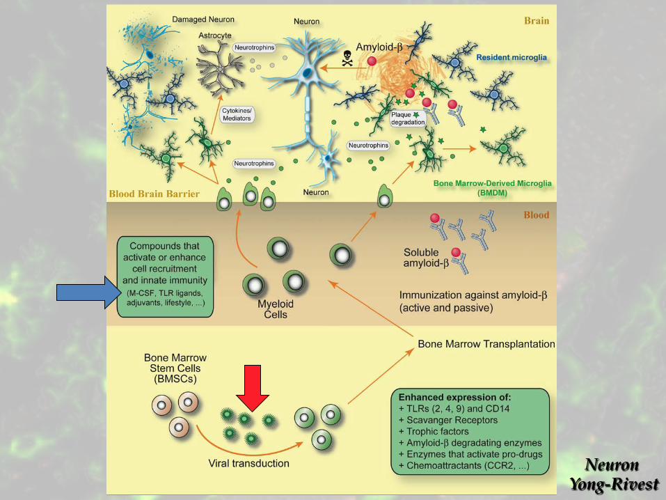
Conclusions
• Ly6Clo monocytes are a?racted to and crawl selecBvely onto Aβ-‐posiBve veins • Monocytes take up Aβ and circulate back to the bloodstream • Aβ deposiBon and eliminaBon by monocytes is dynamic in veins of young APP/PS1 mice • AblaBon of Ly6Clo monocytes in APP/PS1 mice increases Aβ load

Neuron! Yong-Rivest!

Toll-like receptor 4 stimulation with the detoxifiedligand monophosphoryl lipid A improves Alzheimer’sdisease-related pathologyJean-Philippe Michauda, Maxime Halléb, Antoine Lamprona, Peter Thériaulta, Paul Préfontainea, Mohammed Filalia,Pascale Tribout-Joverb, Anne-Marie Lanteigneb, Rachel Jodoinb, Christopher Cluffc, Vincent Brichardd, Rémi Palmantierd,Anthony Pilorgetb, Daniel Larocqueb,1, and Serge Rivesta,1
aNeuroscience Laboratory, Department of Molecular Medicine, Centre Hospitalier Universitaire de Québec Research Center, Laval University, Québec City, QC,Canada G1V 4G2; bGlaxoSmithKline Vaccines, Laval, QC, Canada H7V 3S8; cGlaxoSmithKline Vaccines, Hamilton, MT 59840; and dGlaxoSmithKline Vaccines,B-1330 Rixensart, Belgium
Edited by Shizuo Akira, Osaka University, Osaka, Japan, and approved December 19, 2012 (received for review September 1, 2012)
Alzheimer’s disease (AD) is the most common cause of dementiaworldwide. The pathogenesis of this neurodegenerative disease,currently without curative treatment, is associated with the accu-mulation of amyloid ! (A!) in brain parenchyma and cerebral vas-culature. AD patients are unable to clear this toxic peptide, leadingto A! accumulation in their brains and, presumably, the pathologyassociated with this devastating disease. Compounds that stimulatethe immune system to clear A! may therefore have great therapeu-tic potential in AD patients. Monophosphoryl lipid A (MPL) is anLPS-derived Toll-like receptor 4 agonist that exhibits unique immu-nomodulatory properties at doses that are nonpyrogenic. We showhere that repeated systemic injections of MPL, but not LPS, signif-icantly improved AD-related pathology in APPswe/PS1 mice. MPLtreatment led to a significant reduction in A! load in the brain ofthese mice, as well as enhanced cognitive function. MPL induceda potent phagocytic response by microglia while triggering a mod-erate inflammatory reaction. Our data suggest that the Toll-like re-ceptor 4 agonist MPL may be a treatment for AD.
innate immunity | microglial cells | monocytes | phagocytosis |inflammation
Alzheimer’s disease (AD) is a neurodegenerative pathologycharacterized by the accumulation of amyloid beta (Aβ) and
neurofibrillary tangles in the brain parenchyma (1). Inflammation,which occurs in parallel with the progression of the disease, isfeatured by the production of cytokines by activatedmicroglia. Therole of these cells in the pathogenesis of AD remains unclear andis an area of active investigation. Whereas chronic activation ofmicroglial cells by Aβ can trigger the exaggerated release of cyto-kines and neurotoxic mediators that could be detrimental to neu-rons, microglia can also clear Aβ via increased phagocytosis andproteolytic degradation, which may be neuroprotective (2).Toll-like receptors (TLRs) on the surface of microglial cells have
been shown to bind Aβ, which triggers downstream intracellularsignaling cascades (3, 4). Microglia deficient in TLR2, TLR4, orthe coreceptor CD14 are not activated by Aβ and do not exhibita phagocytic response (5). Transgenic AD mice lacking TLR4 havemarkedly elevated levels of diffuse and fibrillar Aβ (3). Furthermore,stimulation of microglial cells with TLR2-, TLR4-, or TLR9-specificagonists accelerates Aβ clearance both in vitro and in vivo (3, 6, 7).
Monophosphoryl lipid A (MPL) is a chemically detoxified lipidAmoiety derived from Salmonella minnesota R595 LPS (8). ThisTLR4 ligand is at least 100-fold less pyrogenic than LPS yet main-tains many of the immunomodulatory properties of LPS (9). Im-portantly, MPL is safe in humans and has been administered tomillions of patients as a component of several vaccine formulationssuch as the Cervarix vaccine (10). We investigated herein thechronic use of the nonpyrogenic TLR4 agonist MPL and comparedit with a strong TLR4 ligand (LPS) in a mouse model of AD.Although the therapeutic potential of innate immune activa-
tion for AD is being evaluated in preclinical models, this concept
has not been tested in humans. We propose that the age-relateddefects in immune cell function (11) commonly found in agingdiseases such as AD (12) can be reconciled with a prophagocyticphenotype, yet mildly proinflammatory, which may lead to animproved clearance of Aβ.Our data demonstrate that chronic, systemic administration of
MPL ameliorates AD-like pathology by decreasing the cerebralAβ load through the stimulation of the phagocytic capacity ofinnate immune cells.
ResultsMPL Drives a Distinct TLR4 Stimulation from LPS. MPL is derivedfrom the LPS of theGram-negative bacteria SalmonellaminnesotaR595 by three main chemical modifications: (i) elimination of thecore oligosaccharide, (ii) hydrolysis of the 1-phosphate from thereducing end glucosamine, and (iii) removal of the acyl chain fromthe 3-position of the disaccharide (Fig. 1A and B). The absence ofthe 1-phosphate on the MPL molecule was suggested to weakenthe dimerization of TLR4/MD2 (myeloid differentiation factor-2)(13). This presumably induces a structural change in the TLR4receptor complex that alters the recruitment of the adaptor pro-teins to the intracellular domain (13). Such a structural changemay account for the distinct signaling properties of MPL, whichpredominantly activates the TLR4-TRAM (TRIF-related adap-tor molecule)-TRIF (TIR-domain-containing adaptor proteininducing IFN-β) pathway over the more proinflammatory TLR4-MAL (MyD88-adaptor-like protein)-MyD88 (myeloid differen-tiation primary-response protein 88) signaling pathway (14). Thisdifferential use of intracellular adaptor proteins may be key toexplaining the distinct effects observed after exposure of cells toMPL or LPS.To characterize the ligand–receptor interaction of MPL with
the TLR4 receptor complex we used the HEK293 cell linetransfected with TLR4, MD2, and CD14 genes, as well as anNF-κB and AP-1 reporter system. At the highest concentrationof MPL tested (20 μg/mL), the activation of NF-κB and AP-1 wasat a level comparable to a 200-fold lower concentration of LPS(0.1 μg/mL) (Fig. 1C). A neutralizing antibody directed againstTLR4 inhibited the response to MPL. Incubating TLR2-trans-fected HEK293 cells with up to 2.5 μg/mL of MPL did not induceany activation of NF-κB and AP-1 (Fig. 1D), indicating that the
Author contributions: J.-P.M., A.L., A.P., D.L., and S.R. designed research; J.-P.M., M.H., A.L.,P.T., P.P., M.F., P.T.-J., A.-M.L., and R.J. performed research; J.-P.M., M.H., A.L., P.P., and D.L.analyzed data; and J.-P.M., M.H., A.L., C.C., V.B., R.P., A.P., D.L., and S.R. wrote the paper.
Conflict of interest statement: M.H., P.T.-J., A.-M.L., R.J., C.C., V.B., R.P., A.P., and D.L. areemployees of GlaxoSmithKline Vaccines. This research was supported in part byGlaxoSmithKline Vaccines.
This article is a PNAS Direct Submission.1To whom correspondence may be addressed. E-mail: [email protected] [email protected].
This article contains supporting information online at www.pnas.org/lookup/suppl/doi:10.1073/pnas.1215165110/-/DCSupplemental.
www.pnas.org/cgi/doi/10.1073/pnas.1215165110 PNAS Early Edition | 1 of 6
NEU
ROSC
IENCE

Comparison of LPS and MPL extracted from Salmonella minnesota R595.
(i) eliminaBon of the core oligosaccharide (ii) hydrolysis of the 1-‐phosphate from the reducing end glucosamine (iii) removal of the acyl chain from the 3-‐posiBon of the disaccharide

TLR4-‐specific ac=va=on of NF-‐κB and AP-‐1 in HEK 293 cells
TLR4-‐MD2-‐CD14 TLR2

Phosphoryla=on kine=cs of key TLR pathway of MPL vs LPS in microglia

MPL induces a low inflammatory response in microglia

MPL s=mulates ac=n reorganiza=on

Phagocytosis of fluorescent E.coli beads and Aβ oligomers

MPL upregulates Scavenger receptors-‐A in microglia

In vivo uptake of Aβ oligomers in monocytes

Low immune ac=va=on following MPL injec=on

MPL triggers only a weak TLR2 mRNA induc=on in the brain

MPL triggers the expansion of blood monocytes in mice

APPswe/PS1 mice treatment
-‐ 3 month-‐old APPswe/PS1 mice -‐ 12 consecu=ve i.p. injec=ons once a week -‐ MPL (50 μg, 130 μL) LPS (3,5 μg, 130 μL) PBS (130 μL) -‐ Pathology analysis at 6 month-‐old
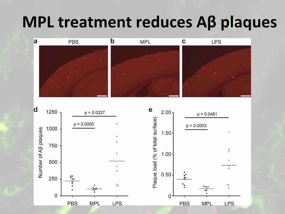
MPL treatment reduces Aβ plaques

MPL treatment reduces soluble Aβ

MPL treatment reduces cogni=ve deficits in APPswe/PS1 mice

Conclusions
-‐ MPL induces a potent phagocy=c response in microglia and monocytes (p38 pathway and SR-‐A)
-‐ MPL triggers a low inflammatory response -‐ MPL reduces Aβ loads and restricts cogni=ve deficits of APPswe/PS1 mice.

Neuron! Yong-Rivest!

Activation of systemic innate immune cells is a very efficient approach to clear vascular and brain amyloid load and prevent cognitive decline in Alzheimer's disease. This may also be the case for other brain diseases associated with the accumulation of toxic extracellular proteins (SOD1, alpha-Syn, …).

Who did the work? Marc-André Bellavance, M.Sc. Dr. Alain Simard Dr. Antoine Lampron Dr. Gaëlle Naert Jean-Philippe Michaud, M.Sc. Paul Préfontaine. M.Sc. Dr. Maxime Hallé (GSK) Dr. Daniel Larocque (GSK)

$$$ CIHR, CRC, Neuroscience Canada, Brain Repair Program (Dr. Wee Yong, PI), MS Canada Team Grant (Dr. Peter Stys, PI), Canadian Stroke Network, GSK-Vaccines.
14-02-26 17:45
Page 1 sur 1https://www.google.ca/blank.html
