neuroscience psychology: a concise introduction 2 nd edition richard griggs chapter 2 prepared by j....
TRANSCRIPT

Neuroscience
Psychology: A Concise Introduction
2nd Edition
Richard Griggs
Chapter 2
Prepared byJ. W. Taylor V

The Journey…
The Neuron
The Nervous System and the Endocrine System
The Brain
The Neuron
The Nervous System and the Endocrine System
The Brain

The Neuron
The Structure of a Neuron
How Neurons Communicate
Neurotransmitters, Drugs, and Poisons

Neurons and Glial Cells
Neurons are responsible for information transmission throughout the nervous system
Glial cells do not directly transmit information, but instead support neurons in their work by disposing of waste products of neurons, keeping their chemical environment stable, and insulating them
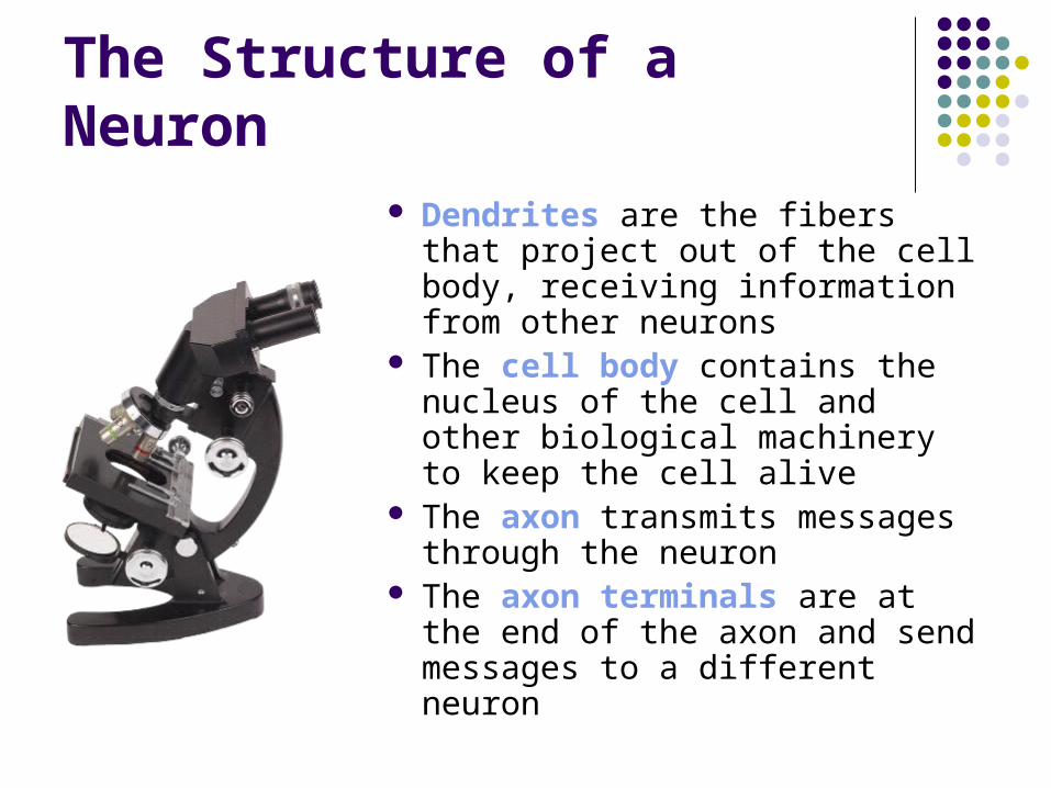
The Structure of a Neuron Dendrites are the fibers that
project out of the cell body, receiving information from other neurons
The cell body contains the nucleus of the cell and other biological machinery to keep the cell alive
The axon transmits messages through the neuron
The axon terminals are at the end of the axon and send messages to a different neuron

The Structure of a Neuron

How Neurons Communicate
Communication within
a neuron is electrical
Communication within
a neuron is electrical
Communication between
neurons is chemical
Communication between
neurons is chemical

The Electrical Impulse
Information from the dendrites is either excitatory (telling the neuron to generate an electrical impulse) or inhibitory (telling the neuron not to generate an electrical impulse) The impulse is an “all or nothing” event, meaning
that there either is or is not an electrical impulse Stimuli of varying intensities are encoded by the
quantity of neurons generating impulses and the number of impulses generated each second by the neurons

The Electrical Impulse
The myelin sheath is an insulating layer of fatty white substance that encases the axon, allowing electrical message to be transmitted faster within the neuron Damage to the myelin sheath will slow electrical
impulses, and can result in diseases like multiple sclerosis
Chemical Communication Between Neurons
Axon terminals contains sacs of neurotransmitters These neurotransmitters are naturally occurring
chemicals in the nervous system that specialize in transmitting information between neurons
Between the axon terminals of one neuron and the dendrites of another neuron is a small space called the synaptic gap, across which neurotransmitters are sent, allowing neurons to communicate


Brain Scans
Brain scans work because neurons require oxygen and other nutrients such as blood sugar Positron Emission Tomography (PET) scans
use a dose of radioactive glucose, which moves to the more-active areas of the brain
Functional Magnetic Resonance Imaging (MRI) detects active areas of the brain by highlighting those areas that require more oxygen

Neurotransmitters, Drugs, and Poisons
Key terms:
AgonistsAgonists AntagonistsAntagonists
Drugs and poisons that increase the activity of one or more neurotransmitters
Drugs and poisons that decrease the activity of one or more neurotransmitters

Neurotransmitters
1. Acetylcholine (ACh) is involved in both learning and memory and muscle movement
2. Dopmaine impacts our arousal and mood states, thought processes, and physical movement
3. Serotonin and norepinephrine are neurotransmitters involved in levels of arousal and mood, and play a major role in mood disorders such as depression
4. GABA is the main inhibitory neurotransmitter in the nervous system; glutamate is the main excitatory neurotransmitter
5. Endorphins are a group of neurotransmitters that are involved in pain perception and relief

Acetylcholine (ACh)
Botulinum poison (botulin) is an antagonist that blocks the release of ACh at muscle junctures, leading to paralysis and even death
Curare is an antagonist that paralyzes the body by occupying the receptor sites for ACh, thereby preventing ACh from getting in and carrying its message to a neuron
Black widow spider venom is an agonist for Ach, causing the continuous release of ACh, flooding the synapse

Dopamine
Low levels are associated with Parkinson’s disease, and excessively high levels are associated with schizophrenia
L-Dopa is an agonist that increases production of dopamine
Anti-psychotic drugs are antagonists that block the receptor sites for dopamine so that this neurotransmitter cannot send its messages
Amphetamine acts as an agonist by stimulating the release of dopamine from axon terminals
Cocaine is an agonist that blocks the re-uptake of dopamine

Serotonin and Norepinephrine
Some antidepressant drugs work by blocking the reuptake of serotonin and norepinephine
Anti-depressant drugs like Prozac, Paxil, and Zoloft are selective serotonin reuptake inhibitors
GABA and Glutamate
Anti-anxiety drugs are agonists for GABA Lack of GABA may contribute to epilepsy, a
brain disorder resulting in uncontrolled movement and convulsions
Glutamate is involved in memory storage and pain perception.
Excessive glutamate can lead to neuron death; deficient glutamate has been proposed to explain schizophrenia
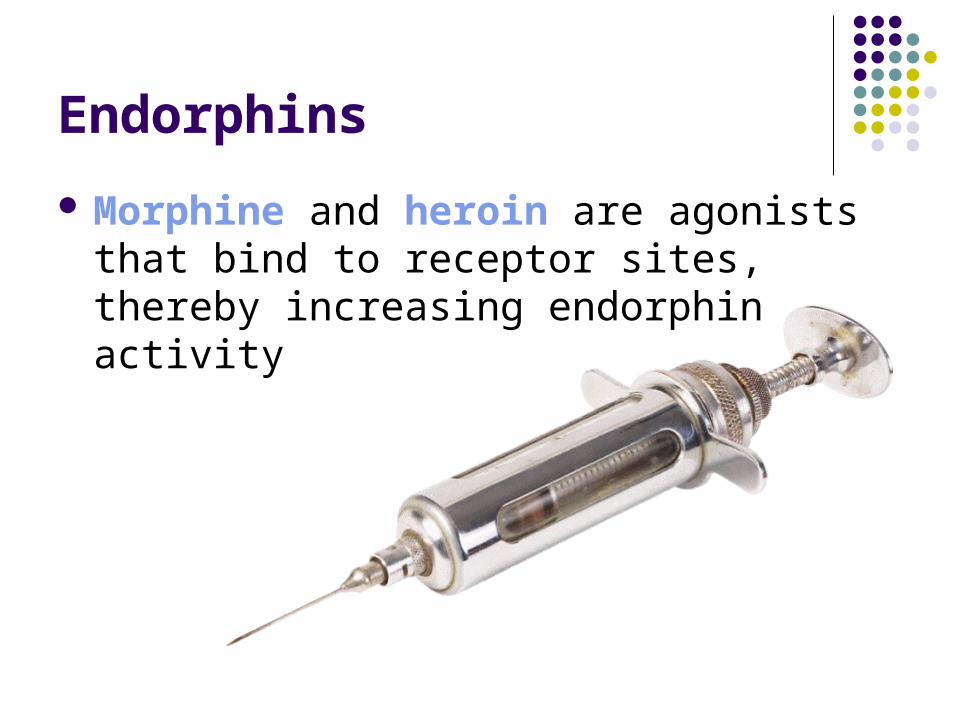
Endorphins
Morphine and heroin are agonists that bind to receptor sites, thereby increasing endorphin activity

The Nervous and Endocrine Systems
The Central Nervous System
The Peripheral Nervous System
The Endocrine Glandular System
Emotions and the Autonomic Nervous System
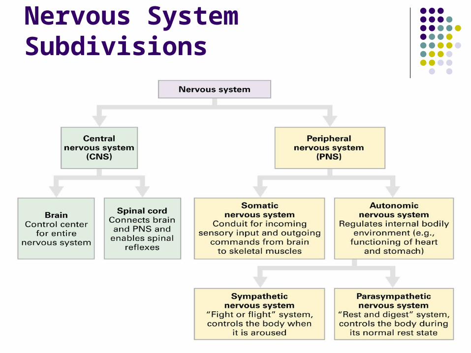
Nervous System Subdivisions

Types of Neurons
Interneurons exist only in the central nervous system
Sensory neurons carry information to the central nervous system from sensory receptors in the eyes, muscles, and glands
Motor neurons carry movement commands from the central nervous system to the rest of the body
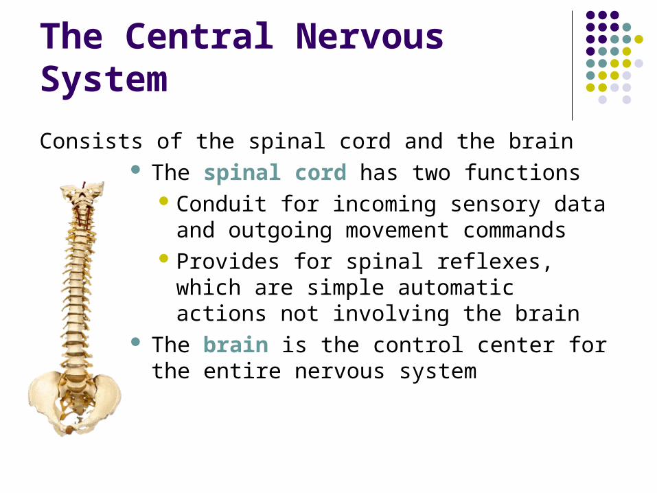
The Central Nervous System
Consists of the spinal cord and the brain The spinal cord has two functions
Conduit for incoming sensory data and outgoing movement commands
Provides for spinal reflexes, which are simple automatic actions not involving the brain
The brain is the control center for the entire nervous system
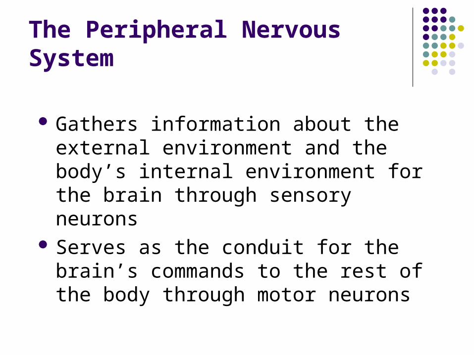
The Peripheral Nervous System
Gathers information about the external environment and the body’s internal environment for the brain through sensory neurons
Serves as the conduit for the brain’s commands to the rest of the body through motor neurons

The Peripheral Nervous System
Consists of two parts: The somatic (or skeletal) nervous system carries
sensory input from receptors to the CNS and relays commands from the CNS to the skeletal muscles to control their movement
The autonomic nervous system regulates our internal environment and consists of two parts The sympathetic nervous system is in control when we
are very aroused and prepares us for defensive action (such as running away or fighting)
The parasympathetic nervous system is in control when the aroused state ends to return our body to its normal resting state


The Endocrine Glandular System
Not part of the nervous system Works with the autonomic nervous system in
responding to stress, and plays a role in basic behaviors and bodily functions such as sex, eating, metabolism, reproduction, and growth
Endocrine glands secret hormones, which are chemicals carried by the bloodstream to target sites throughout the body

The Endocrine Glandular System
Endocrine glands are controlled by the hypothalamus, which controls the most influential gland, the pituitary Releases hormones essential for human growth and that
direct other glands to release their hormones
Some other glands: Thyroid gland affects our growth and maturation Adrenal glands are involved in metabolism and help
trigger the “fight or flight” response with commands from the autonomic nervous system
The pancreas is involved in digestion and maintaining blood-sugar levels

The Endocrine Glandular System

Components of Emotion
An emotion is a complex psychological state that involves three components:
An emotion is a complex psychological state that involves three components:
PhysicalPhysical BehavioralBehavioral CognitiveCognitive
A physiological state of arousal triggered by the
autonomic nervous system
An outward expression including facial expressions,
movements and gestures
An appraisal of the situation to determine which emotion we are experiencing and how
intensely
Components of Emotion
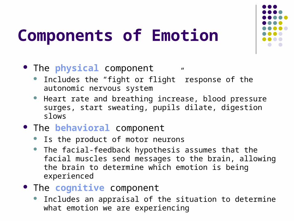
The physical component Includes the “fight or flight” response of the autonomic nervous
system Heart rate and breathing increase, blood pressure surges, start
sweating, pupils dilate, digestion slows The behavioral component
Is the product of motor neurons The facial-feedback hypothesis assumes that the facial muscles
send messages to the brain, allowing the brain to determine which emotion is being experienced
The cognitive component Includes an appraisal of the situation to determine what emotion
we are experiencing
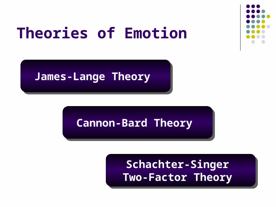
Theories of Emotion
James-Lange Theory James-Lange Theory
Cannon-Bard Theory Cannon-Bard Theory
Schachter-Singer Two-Factor Theory
Schachter-Singer Two-Factor Theory
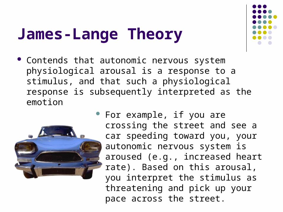
James-Lange Theory
Contends that autonomic nervous system physiological arousal is a response to a stimulus, and that such a physiological response is subsequently interpreted as the emotion
For example, if you are crossing the street and see a car speeding toward you, your autonomic nervous system is aroused (e.g., increased heart rate). Based on this arousal, you interpret the stimulus as threatening and pick up your pace across the street.

Cannon-Bard Theory
Contends that arousal patterns for different emotions are too physiologically alike to be able to determine what emotion is being experienced
Instead, an emotion-provoking stimulus sends messages to both the peripheral nervous system and the brain The brain produces the emotional feeling, the
autonomic nervous system produces the physiological response, and the motor neurons produce the behavioral response

Schachter-SingerTwo-Factor Theory
Contends that there are two important determinants of emotion: Physiological arousal tells us how intense the
emotion is The cognitive appraisal of the entire situation
allows us to identify the emotion, leading to the emotional feeling

Integrating the Theories
LeDoux (1996) contends that there are different brain systems for different emotions Fear, for example, does not require higher-level
cognitive processing and is generated almost instantaneously by the amygdala
More complex emotions, however, such as love or guilt, that do not require instantaneous responding for survival, may require higher-level processing

The Brain
Going up the Brain Stem
Processing in the Cerebral Cortex
Specializations of the Left and Right Hemispheres
Consciousness and the Sleeping Brain

Case 1: A Landscape Artist
Scenario Neuroanatomy Related Function
Anne the landscape artist is standing at her easel, painting with her right hand as she looks out the window at her garden. She’s listening to classical music as she paints.
Left motor cortex Left frontal lobe Visual cortex Both occipital lobes Auditory cortexes Both temporal lobes Right hemisphere Thalamus Frontal lobes Left sensory cortex Left parietal lobe Cerebellum
Controls right hand Contains motor cortex Used for vision Contain visual cortex Used to hear music Contain auditory cortexes Spatial ability for painting Relays sensory information Deciding what to paint Feeling the paintbrush Contains sensory cortex Coordinates moving arm
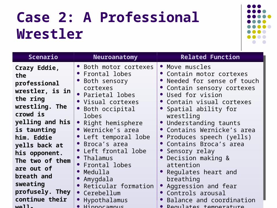
Case 2: A Professional Wrestler
Scenario Neuroanatomy Related Function
Crazy Eddie, the professional wrestler, is in the ring wrestling. The crowd is yelling and his is taunting him. Eddie yells back at his opponent. The two of them are out of breath and sweating profusely. They continue their well-orchestrated series of wrestling moves.
Both motor cortexes Frontal lobes Both sensory cortexes Parietal lobes Visual cortexes Both occipital lobes Right hemisphere Wernicke’s area Left temporal lobe Broca’s area Left frontal lobe Thalamus Frontal lobes Medulla Amygdala Reticular formation Cerebellum Hypothalamus Hippocampus
Move muscles Contain motor cortexes Needed for sense of touch Contain sensory cortexes Used for vision Contain visual cortexes Spatial ability for wrestling Understanding taunts Contains Wernicke’s area Produces speech (yells) Contains Broca’s area Sensory relay Decision making & attention Regulates heart and breathing Aggression and fear Controls arousal Balance and coordination Regulates temperature Memory for moves

Case 3: A Student
Scenario Neuroanatomy Related Function
Jill is a law student studying for her exam. She is reading about violent rape and murder cases. She is snacking on popcorn and drinking coffee.
Hippocampus Wernicke’s area Left temporal lobe Amygdala Frontal lobes Hypothalamus Angular gyrus
Remembering and learning Language comprehension Contains Wernicke’s area Anger and fear about cases Decision making & attention Regulates hunger and thirst Needed for reading
Source: Sheldon, J. P. (2000). A neuroanatomy teaching activity using case studies and collaboration. Teaching of Psychology, 27, 126-128.

The Central Core
The brain stem The medulla links the spinal cord to the brain and is involved in
regulating heartbeat, blood pressure, digestion, and swallowing The reticular formation is a network of neurons running up the
center of the brain stem and into the thalamus that is involved in controlling our different levels of arousal and awareness
The cerebellum is involved in the coordination of our movements, our sense of balance, and motor and procedural learning
The thalamus, located at the top of the brain stem, serves as a relay station for incoming sensory information (except smell) The basal ganglia are on the outer sides of the thamalus and are
concerned mainly with the initiation and execution of physical movements
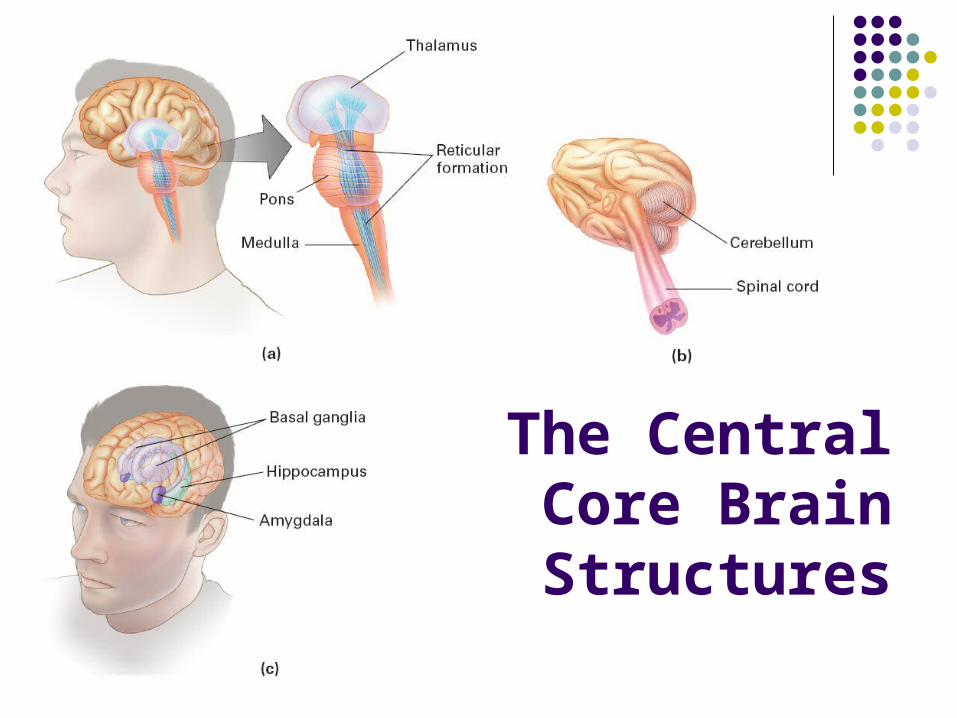
The Central Core Brain Structures

The Limbic System
Plays a role in our survival, memory, and emotions The hypothalamus control the pituitary gland, the
autonomic nervous system, and plays a major role in regulating basic drives such as eating, thirst, and sex
The hippocampus is involved in the formation of memories
The amygdala plays a major role in regulating our emotional experiences, especially fear, anger, and aggression

The Limbic System
Processing in the Cerebral Cortex
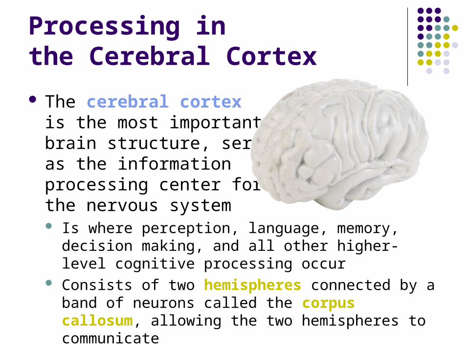
The cerebral cortex is the most important brain structure, serving as the information processing center for the nervous system Is where perception, language, memory, decision
making, and all other higher-level cognitive processing occur
Consists of two hemispheres connected by a band of neurons called the corpus callosum, allowing the two hemispheres to communicate
Brain Lobes
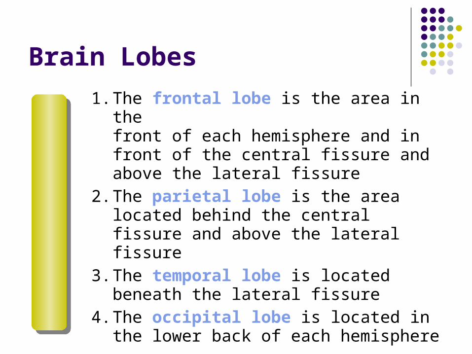
1. The frontal lobe is the area in the front of each hemisphere and in front of the central fissure and above the lateral fissure
2. The parietal lobe is the area located behind the central fissure and above the lateral fissure
3. The temporal lobe is located beneath the lateral fissure
4. The occipital lobe is located in the lower back of each hemisphere

The Four Lobes and the Sensory-Motor Processing Areas

The Motor Cortex
The frontal lobe strip of cortex, directly in front of the central fissure in each hemisphere, allows us to move different parts of our body
Each hemisphere controls the voluntary movement of the opposite side of the body (a contralateral relationship)
Amount of motor cortex devoted to a specific body part is related to the complexity and precision of movement of which that part is capable
The Somatosensory Cortex
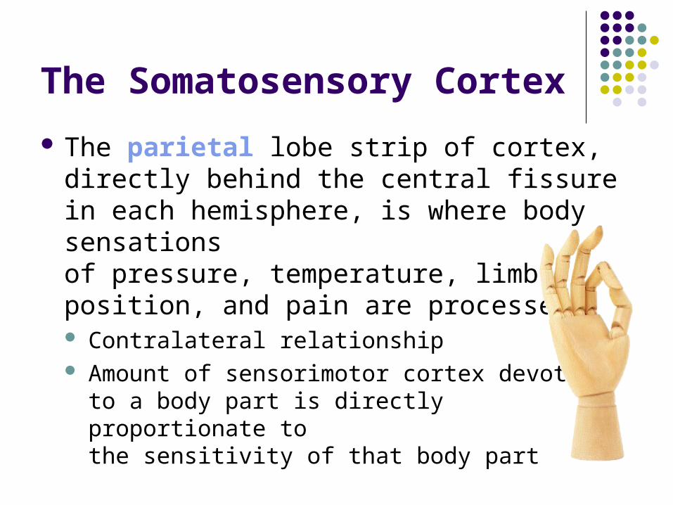
The parietal lobe strip of cortex, directly behind the central fissure in each hemisphere, is where body sensations of pressure, temperature, limb position, and pain are processed Contralateral relationship Amount of sensorimotor cortex devoted
to a body part is directly proportionate to the sensitivity of that body part

Homunculi for the Motor Cortex and the Somatosensory Cortex

Visual Cortexand Auditory Cortex
The visual cortex is located in the occipital lobes at the back of the hemispheres
The auditory cortex is in the temporal lobes These primary areas pass the
results of their analyses on to areas in the other lobes to complete the brain’s interpretation of the incoming visual or auditory information These secondary cortical processing
areas are part of what is termed the association cortex

Association Cortex
Consists of the other 70% of the cortex not in one of the previously mentioned areas
This is where the higher-level processing such as decision making, reasoning, perception, speech, and language occurs All of which require integration of various types
of information

The Case of Phineas Gage
Phineas Gage was railroad worker who survived when a metal tamping iron flew through his left cheek and head, exiting through his frontal lobes
He became irresponsible, impulsive, disorderly, indecisive, and cursed, leading neuroscientists to think the frontal lobes are important in such behaviors
Language
Broca’s area, in the left hemisphere’s temporal lobe, is responsible for fluent speech production When damaged, people cannot generate fluent speech,
but can still understand speech easily Singing and musical abilities seem to be housed in the
right hemisphere because damage to Broca’s area does not impair these abilities
Wernicke’s area is in the left temporal lobe and is responsible for the comprehension of speech and reading

Language
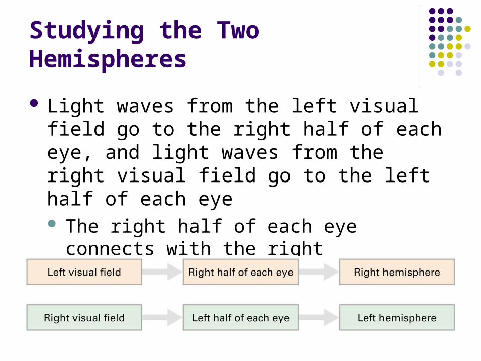
Studying the Two Hemispheres
Light waves from the left visual field go to the right half of each eye, and light waves from the right visual field go to the left half of each eye The right half of each eye connects with the
right hemisphere, and the left half of each eye connects with the left hemisphere

Pathways for Processing Information in the Left and Right Visual Fields

Studying the Two Hemispheres
With split-brained people, the information cannot transfer between hemispheres because the corpus callosum has been cut
Split-brain people can only identify information orally when it is presented briefly in the right visual field (and thus processing in the left hemisphere) If a spoon was flashed in the left visual
field, split-brained people could not say it was a spoon
If the person was blind-folded and told to find the object from a group of objects with the left hand, s/he can do this

What we know…
Left hemisphere Language Math and logic skills More analytical, analyzing wholes into pieces
Right hemisphere Spatial perception Solving spatial problems Drawing Face recognition
What we know…
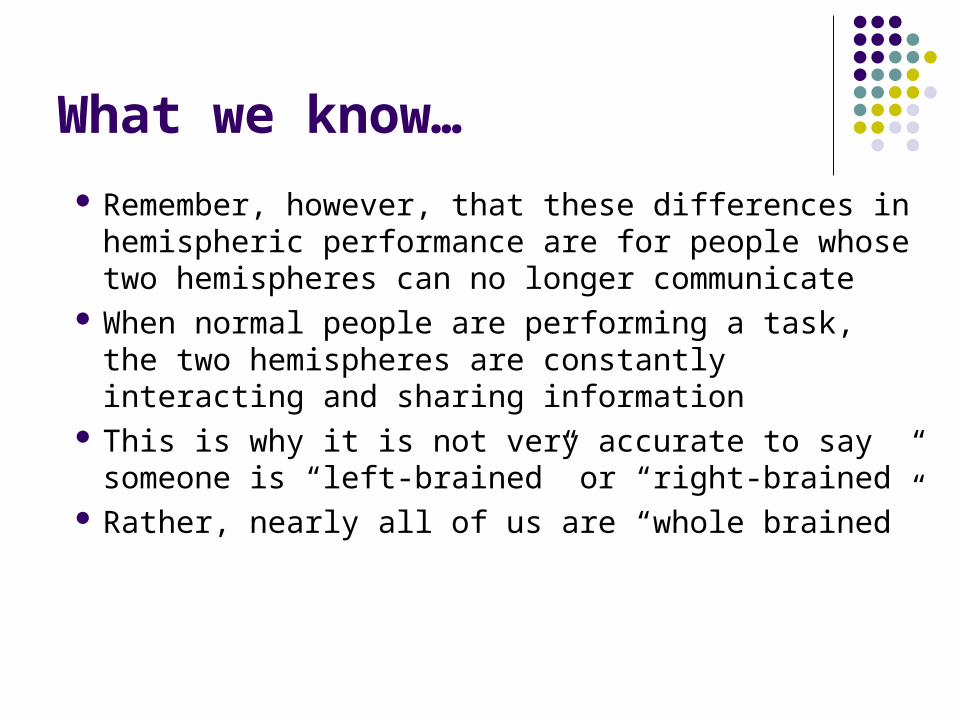
Remember, however, that these differences in hemispheric performance are for people whose two hemispheres can no longer communicate
When normal people are performing a task, the two hemispheres are constantly interacting and sharing information
This is why it is not very accurate to say someone is “left-brained” or “right-brained”
Rather, nearly all of us are “whole brained”

Consciousness and the Sleeping Brain
Consciousness is a person’s subjective awareness of both their inner thinking and feeling and their external environment
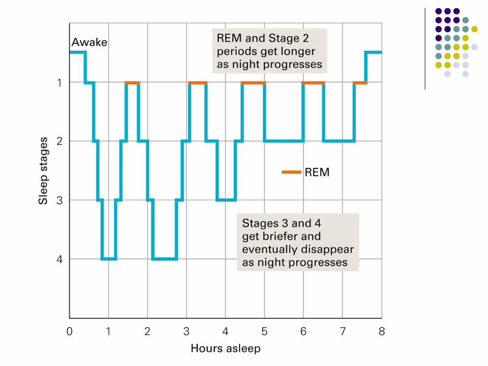
The five stages of sleep were determined by use of an electroencephalogram (EEG), which records a real-time graph of a person’s cortical electrical activity in the brain As we slip into sleep and pass through the first four
stages, our brain waves change, in general becoming progressively slower, larger, and more irregular, especially in Stages 3 and 4

Five Stages of Sleep
Stage 1: Lasts about 5 minutes Stage 2: Lasts about 20 minutes
Characterized by sleep spindles, rapid bursts of mental activity
Stage 3: Also known as transitional sleep and is characterized by delta waves, which are large, slow waves
Stage 4: Lasts about 30 minutes Parasympathetic nervous system is active, as
muscles relax, heartbeat slows, blood pressure declines, and digestion speeds up

Five Stages of Sleep
Stage 5: REM (rapid eye movement sleep) occurs after we leave stage 4 sleep and return through the earlier stages of sleep Called paradoxical sleep because your muscles are relaxed, but
other body systems, including the brain, are active, much like a waking pattern
Characterized by very rapid brain waves somewhat like those of Stage 1 sleep, but one is still sound asleep
If awakened during REM sleep, people often report having been dreaming
Most dreams are emotional and unpleasant, perhaps because the visual cortex and frontal lobe are inactive during REM sleep; the limbic system structures are active, however, creating irrational imagery and emotional experiences of our dream world
REM sleep accounts for 20–25% of total sleep time

Five Stages of Sleep

Five Stages of Sleep
These 5 stages (the sleep cycle) repeat themselves about every 90 minutes, with Stages 3 and 4 getting shorter with each cycle, and REM and Stage 2 getting longer with each cycle
REM sleep rebound effect is a significant increase in the proportion of REM sleep following deprivation of REM sleep

Why do we sleep and dream?
Sleep deprivation results in: Impaired concentration and a general bodily feeling of
weakness and discomfort Suppression of the immune system, lessening one’s
ability to fight off infection and disease Increased vulnerability to accidents Increased difficulty in concentrating, studying, and
taking exams

Why do we sleep and dream?
Explanations for dreaming : Sigmund Freud proposed that dreams were
disguised outlets for inner conflicts of our unconscious mind, a view not accepted by modern sleep researchers
The activation-synthesis hypothesis contends that dreams are merely the sleeping brain’s attempt to make sense of random neural activity without the rational interpretation of the frontal lobe