neutronpdf
TRANSCRIPT
*MT 4S1SGOO ^ «
ve Risø-M-2672
Neutron Radiography, Techniques and Applications J. C. Domanus
Risø National Laboratory, DK-4000 Roskilde, Denmark October 1987
RISØ-M-2672
NEUTRON RADIOGRAPHY, TECHNIQUES AND APPLICATIONS
J. C. Domanus
Abstract. After describing the principles of the "in pool" and
"dry" installations, techniques used in neutron radiography are
reviewed. Use of converter foils with silver halide films for
the direct and transfer methods is described. Advantages of the
use of nitrocellulose film for radiographying radioactive ob-
jacts are discussed. Dynamic imaging is shortly reviewed. Stan
dardization in the field of neutron radiography (ASTN and Eura-
tom Neutron Radiography Working Group) is described. The paper
reviews main fields o' use of neutron radiography. Possibi
lities of use of neutron radiography at research reactors in
various scientific, incjstrial and other fields are mentioned.
Examples are given of application of neutron radiography in
industry and the nuclear field.
October 1987 Risø National Laboratory, DK-4000 Roskilde, Denmark

»
This paper will be presented at the Seminar "Procedes de Con
trol Non Destructifs" in Liege, Belgium (4 & 5 February 1988)
and at the Danish-Israeli Symposium on Non-Destructive Eva
luation in Lyngby, Denmark (27 June - 1 July 1988).
ISBN 87-550-1371-6
ISSN 0418-6435
Grafisk Service, Risø, 1987
CONTENTS Page
1. PRINCIPLES OF NEUTRON RADIOGRAPHY 4
2. RADIATION SOURCES 6
3. NEUTRON RADIOGRAPHIC TECHNIQUES 6 3.1. Direct exposure technique 8 3.2. Transfer technique 9 3.3. Track-etch tecknique 10
4. VIEWING OF NEUTRON RADIOGRAPHS 12
5. DYNAMIC IMAGING 12
6. STANDARDIZATION 13
7. APPLICATIONS OF NEUTRON RADIOGRAPHY 14 7.1. Nuclear industry 16
Nuclear fuel 16 General 23
7.2. Industrial applications 23 Explosives and pyrotechnical devices 23 Turbine blades 23 Aerospace 24 Apollo 24 Ariane 24 Corrosion 24 Composite 25 Other industrial applications 25 Concrete 25 Soil and rock 25 Electric contacts 25 Electronic devices 26 Mechanical connectors and assemblies 26 Honeycomb 26 Flow in heat pipes 26 Metallurgy 26 Real-time 26 Engine fluids 26
7.3. Non-industrial applications 27 Biology, medicine and dentistry 27 Histopathology 27 Dentistry 27 Biology and medicine 27 Roots growing in soil 27 Forensic 27 Arts 28 Neutron autoradiography 28
ACKNOWLEDGEMENTS 28
REFERENCES 29

- 4 -
1. PRINCIPLES OF NEUTRON RADIOGRAPHY
All radiographic methods, whether making use of X-rays, gamma-
rays or neutrons are based on the same general principle: that
radiation is attenuated on passing through matter. The object
under examination is placed in the incident radiation beam.
After passing through, the beam that remains enters a detector
that registers the fraction of the initial radiation intensity
that has been attenuated, by each point in the object. Any
inhomogeneity in the object or an internal defect (such as e.g.
void, crack, porosity or inclusion) will show up as a change in
radiation intensity reaching the detector.
Thus detection of defects in radiography is based on the ob
servation of differences in radiation intensity after passing
through the object under examination. This occurs according to
the basic law of radiation attenuation:
J = J0 e""X
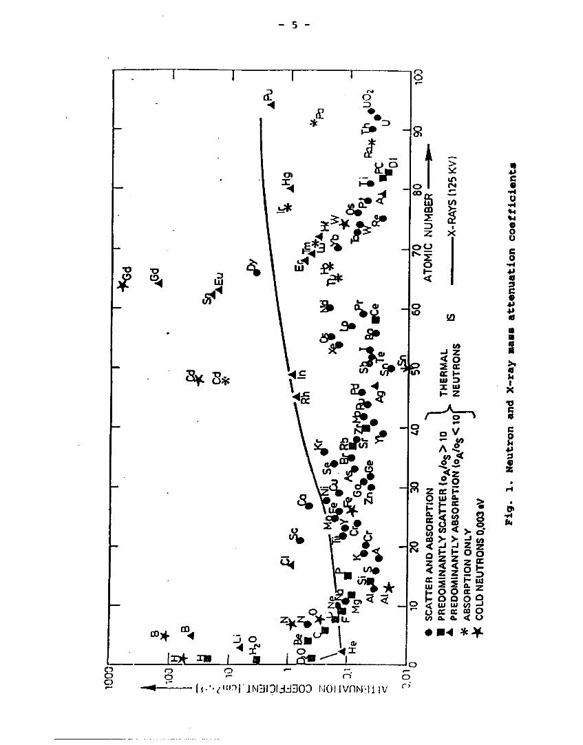
The radiation attenuation coefficient p shows a continous curve
for X-rays (over a wide range of wavelengths). This is not
true, however, for neutrons and it happens that adjacent atomic
number elements such as boron and carbon show for example
marked differences in neutron attenuation. Because of this it
is possible to detect hydrogen in zirconium. Conversly, dense
materials such as lead, tungsten, or uranium are relatively
easy to penetrate by neutrons.
A comparison of neutron and X-ray (125 kV) mass attenuation
coefficients for various elements is given in fig. 1.
Another impotant advantage of neutron radiography is the poss
ibility of examining radioactive objects such as spent fuel
element? directly.
- 5 -
C
o o o c o
•H 4* 18 9 C 0 •P +> ø n m 0 6 5s ø U I
X
•o c ø C o u *i 3 9 se
•H fa
<?"'~>rJNIOdJ303 NO).IVflM-IJ IV o o

- 6 r
2. RADIATION SOURCES
There are three sources of neutrons available for neutron ra
diography: accelerators, radioisotopes and nuclear reactors.
Only the latter will be reviewed below. At present nuclear re
actors provide the most intense neutron beams and therefore can
produce neutron radiographs of the highest quality.
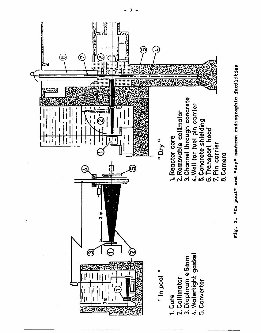
Two types of neutron radiographic facilities are used with
nuclear reactors. In the "in pool" facility the whole neutron
radiographic installation is immersed in the pool of the reac
tor. Here, irradiated reactor fuel rods, removed from the reac
tor core, are transferred to the neutron radiographic facility,
where they are examined without removing thei» from the reactor
pool. In the "dry" type facility a neutron beam taken out of
the core of the reactot is used outside the reactor for neutron
radiography.
The "in pool" and "dry" neutron radiographic facilities are
shown in fig. 2.
3. NEUTRON RADIOGRAPHIC TECHNIQUES
As in X- or gama-radiography, x-ray film is the medium for
producing neutron radiographs. For radiographying radioactive
materials the nitrocellulose film is also used.
Unfortunately, neutrons have very little direct effect on pho
tographic film. Thus an intensifying screen of some kind is
needed to improve the speed of the film. The nitrocellulose
film must also be used with a converter screen, as neutrons do
not directly affect this type of film.
Of the many existing methods of recording neutron images, only
those which are widely used in practice will be described here.
They are the following: the direct and transfer technique using
metal converter foils with X-ray film and the track-etch tech
nique using nitrocellulose film. The properties of metals used
as converter foils for the direct and transfe techniques are
- 7 -
^-rø^-^^gp
.•f.o.".«,..T~«:»-.s>»»i
SV
O O O)
C
o c
1° -•= *£».£ H5 TJ o g> SL*5 o 2 o g _ 2 o
" P " feo Q.5 •*> ^ "* w
v> > JS (/) O
O
£ o o c _ o _ O E O -r; C n *- -
. • • • • • » •
«-*<Njn«NjtntDt>.co
«
o
o •H
0 h
•H •O 0 h C O u 4J 9 0 S
s
•s 0
O 0 p. c H C
•H fa
»
o o a c i
a> o
C V)
•s g, •2|iU £ JT Jr, g* o .2.2 o
o o o ^ o • <\J n «** in

- 8 -
summarized as follows:
Material: gatGd; J^ln; gf^Dy. Thermal neutron absorption coef
ficient: 140.3; 0.73; 3.01 mm"1. Predomiant nuclear reaction:
(n,7). Half-life of radiation emitter: promt; 54 min; 2.5 h.
Type and energy of radiation: ic fiT, 71 fceV (main); p~, 1.28
MeV (max); fT, l.oo MeV (max).
Information about other techniques used in neutron radiography
can be found in numerous references on the subject.
3.1. Direct Exposure Technique
In the direct exposure technuque a metal converter foil is
placed in contact with X-ray film during the actual exposure
(fig. 3). Usually, a single gadolinium back screen is used.
This screen emits gamma-radiation on absorbing neutrons. The
gammas in the spectrum from gadolinium are suitable for pro
ducing electrons by internal conversion. Those low-energy elec
trons essentially expose only the emulsion facing the gado
linium. Single coated slow X-ray films are therefore used with
the direct technuque.
Irradiation
Q±^
Y-f
ilte
r
Direct \—|
nfc
r\ V
I 1 1 1 1
Dark - room processing
m •
Fig. 3. Direct exposure technique

- 9 -
At present a siagle, 25 Mm thick back screen is used. The
screen is usually laminated to aluminium to facilitate handl
ing. Vapour-phase deposited screens are also available, pro
tected with a sapphire coating l Mm thick. To assure good con
tact between the film and screen vacuum cassettes are used.
With the direct exposure technique, using Gd converter foil, a
very good spatial resolution can be achieved. A typical thermal
neutron exposure for a slow, single coated X-ray film and a
single 25 /an Gd screen is about 109 n.cm~2.
3.2. Transfer Technique
In the transfer technique (fig. 4) only the converter screen is
exposed directly to neutrons. The matal screen, placed in the
neutron beam, becomes radioactive in proportion to the intensi
ties in spatial neutron image. The screen is subsequently
transferred from the neutron beam to a dark room where it is
placed in close contact with the x-ray film using a vacuum
cassette. The radioactive emission from the screen then pro
duces an image on the film.
Irradiation Dark - room processing
E LL
I* t I
lo
n+Y (Dl n+Y i
Transfer 1
II 1 1 1 1
H II g ék
Fig. 4. Transfer technique
- 10 -
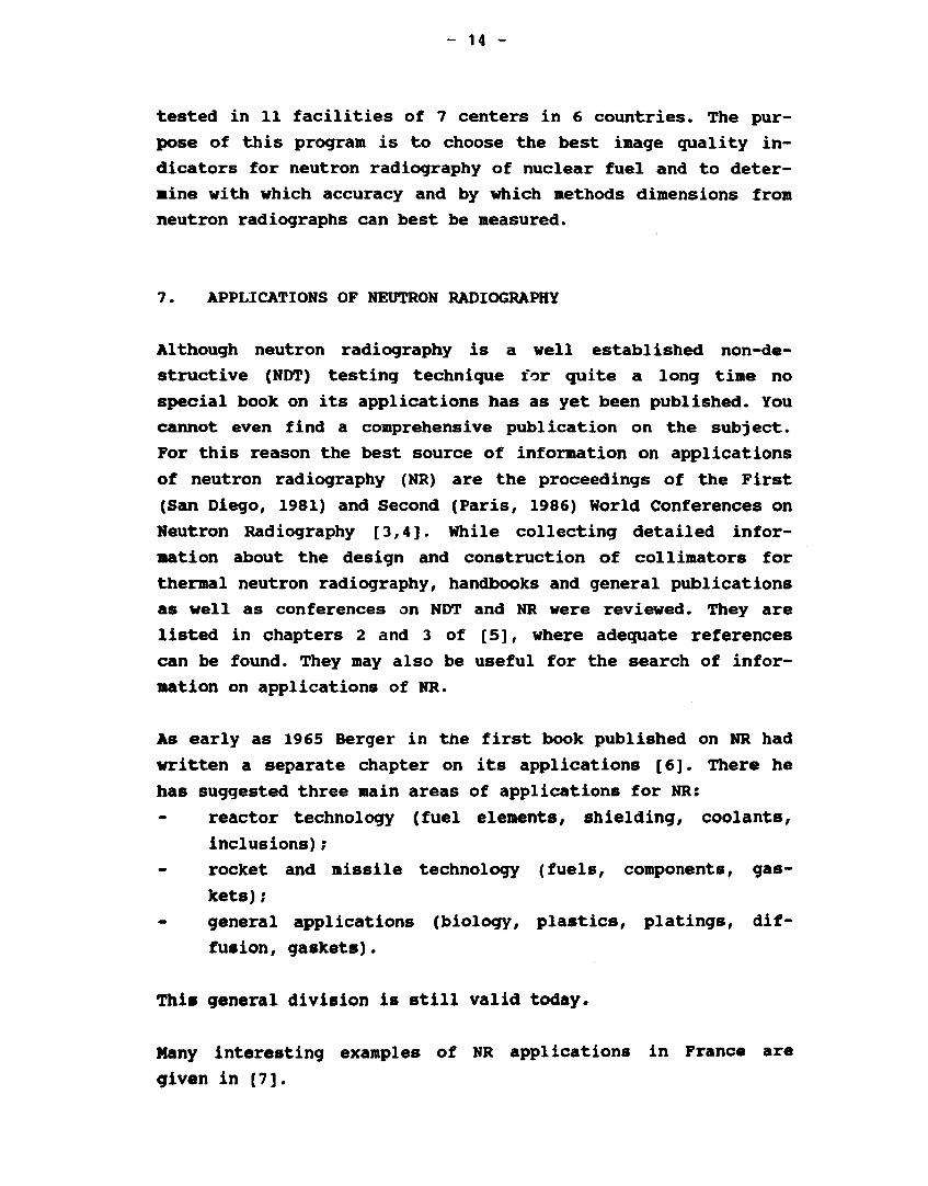
For the transfer texhnique a material must be chosen which is
rapidly activated and has a rapid decay, so that it can be used
again. Indium and dysprosium are the most commonly used mate
rials.
The transfer method offers the advantage that the film, which
is not present in the neutron beam, is not exposed to gamma-
radiation from a radioactive object or from gamma-rays in the
neutron beam itself.
The more energetic ^-particles emitted by the transfer screens
have sufficient energy to penetrate normal photosensitive film
completely. Thus both emulsions of a double coated X-ray film
will contribute appreciably to the film density and to the
unsharpness. To avoid the latter it is important to use single
coated films.
For the transfer technique mainly indium and dysprosium foils
are used. Dysprosium is a harder metal and therefore easier to
handle and maintain. It also has a higher thermal cross section
and longer half-life. Therefore, it can be used with weaker
neutron beams.
For a 100 fm Dy converter an exposure to neutrons of 3 x 10⁹
n.cm"2, followed by an exposure of several half-lives of the
converter to a medium speed X-ray film, will be needed. A good
spatial resolution can be reached whit a single coated film.
3.3. Track-etch Technique
For neutron radiography of radioactive objects (such as irra
diated nuclear fuel) nitrocellulose film is used as a neutron
detector. This is a dielectric material which can detect
charged particles by the radiation damage caused in it. Those
charged particles are produced by an u-emitting converter
screen. The radiation damage is made visible by etching in hot
sodium hydroxide solution (e.g., in 10% NaOH for 45 min at 50
C). The nitrocellulose film, sandwiched between two a-emitting
converter screens, is placed directly in the neutron beam (fig.

- 11 -
5) as it is insensitive to gamma-rays. As the nitrocellulose'
film is also insensitive to visible light the consecutive etch
ing need not be done in a dark room.
Irradiation
Etching
! Track-etch.
/
L
1 1 1 1
Fig. 5. Track-etch technique
Nitrocellulose film is available (from Kadak-Pathé, France) for
neutron radiography in two film/converter variations. The first
consists of a 100 pm thick sheet of cellulose nitrate coated on
both sides with lithium borate dispersed in a water-soluble
binder, which acts as a converter screen by means of the (n,a)
reaction (CN 85 Type B). After irradiation the lithium borate
coating is removed by washing and then film itself is etched.
The secona variation consists of the same CN 85 nitrocellulose
film (without coating) which is sandwiched between two con
verter screens (BN 1) made from natural boron, a (n,a) conver
ter. This converter is coated on a 100 »m thick, very stable,
polyester base and can be reused indefinitely. The efficeincy
of the BN 1 is heigher than that of the CN 85 Type B and there-

- 12 -
fore requires exposure times only slightly longer than those
for the transfer technique (with Dy converter and slow X-ray
film). To establish perfect contact between the converter and
nitrocellulose film the use of a vacuum cassette is essential.
When comparing nitrocellulose and X-ray films as midia for
neutron radiography one can see that although the contrast on
the nitrocellulose film is weaker the definition is better. A
very good spatial resolution can be obtained. In comparison to
the transfer technique there is no saturation of the converter;
this can be advantageous when using low-intensity neutron
sources. There is no handling of an active converter, as in the
transfer method. Finally, one shall mention the possibility of
stopping the etching processing at intermediate stages. After
evaluating the neutron radiograph at that stage the etching of
the same nitrocellulose film can be further continued. Thus
from a single exposure one can have several neutron radiographs
of different densities and contrast.
4. VIEWING OF NEUTRON RADIOGRAPHS
Viewing neutron radiographs produced by the direct or transfer
metod on X-ray film creates no special problems. Neutron radio
graphs produced by the track-etch technique on the other hand
are unsuited for direct viewing because of their low contrast.
The contrast can be significantly improved by printing the film
on a high contrast film, using a point source enlarger. How
ever, the nitrocellulose film can be directly examined by plac
ing it between two polarizing filters.
5. DYNAMIC IMAGING
Although methods of dynamic imaging could more appropriately be
called neutron fluoroscopy than neutron radiography, they will
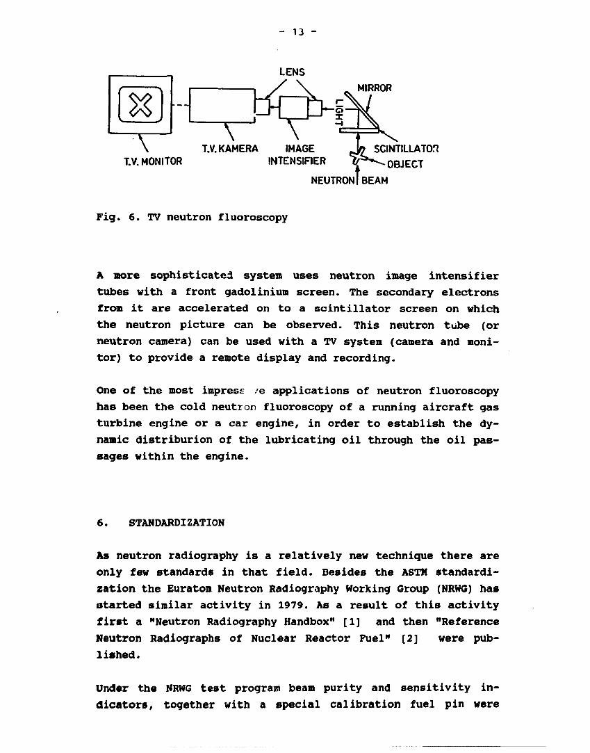
be shortly reviewed here. The principle of a TV system of neu
tron fluoroscopy is shown in fig. 6.
- 13 -
LENS
t ^ I 1 / \ ^ MIRROR
\ T.V. KAMERA IMAGE Jø SCINTILLATOR T.V. MONITOR INTENSIFIER ^ f * ^ OBJECT
NEUTRON! BEAM
Fig. 6. TV neutron fluoroscopy
A more sophisticated system uses neutron image intensifier
tubes with a front gadolinium screen. The secondary electrons
from it are accelerated on to a scintillator screen on which
the neutron picture can be observed. This neutron tube (or
neutron camera) can be used with a TV system (camera and moni
tor) to provide a remote display and recording.
One of the most impress /e applications of neutron fluoroscopy
has been the cold neutron fluoroscopy of a running aircraft gas
turbine engine or a car engine, in order to establish the dy
namic distriburion of the lubricating oil through the oil pas
sages within the engine.
6. STANDARDIZATION
As neutron radiography is a relatively new technique there are
only few standards in that field. Besides the ASTM standardi
zation the Euratom Neutron Radiography Working Group (NRWG) has
started similar activity in 1979. As a result of this activity
first a "Neutron Radiography Handbox" [1] and then "Reference
Neutron Radiographs of Nuclear Reactor Fuel" [2] were pub
lished.
Under the NRWG test program beam purity and sensitivity in
dicators, together with a special calibration fuel pin were
- 14 -
tested in 11 facilities of 7 centers in 6 countries. The pur
pose of this program is to choose the best image quality in
dicators for neutron radiography of nuclear fuel and to deter
mine with which accuracy and by which methods dimensions from
neutron radiographs can best be measured.
7. APPLICATIONS OF NEUTRON RADIOGRAPHY
Although neutron radiography is a well established non-de
structive (NDT) testing technique for quite a long time no
special book on its applications has as yet been published. You
cannot even find a comprehensive publication on the subject.
For this reason the best source of information on applications
of neutron radiography (NR) are the proceedings of the First
(San Diego, 1981) and Second (Paris, 1986) World Conferences on
Neutron Radiography [3,4]. While collecting detailed infor
mation about the design and construction of collimators for
thermal neutron radiography, handbooks and general publications
as well as conferences on NDT and NR were reviewed. They are
listed in chapters 2 and 3 of [5], where adequate references
can be found. They may also be useful for the search of infor
mation on applications of NR.
As early as 1965 Berger in the first book published on NR had
written a separate chapter on its applications [6]. There he
has suggested three main areas of applications for NR:
reactor technology (fuel elements, shielding, coolants,
inclusions);
rocket and missile technology (fuels, components, gas
kets) ;
general applications (biology, plastics, platings, dif
fusion, gaskets).
This general division is still valid today.
Many interesting examples of NR applications in France are
given in [7].

- 15 -
A more detailed review of NR applications in the following
fields can be found in [8]: nuclear, aerospace, explosives and
ordnance as well as biology.
A list of reports which describe NR applications up to 1977 is
given by Barton in [9].
A special issue of the IAEA Atomic Energy Review [10] deals
also with the applications of NR for the control of nuclear
fuel, studies of hydrogen transport, operations in industry
with specific reference to metallurgy.
R. Matfield in [1] gives also a short review of NR appli
cations, dividing them into nuclear, industrial, biomedical and
other areas.
In the Nondestructive Testing Handbook of the ASNDT [11] Berger
et al. [12] give a very short review of NR applications divid
ing them as follows:
general,
- explosives,
turbine blades,
electronic devices,
assemblies,
contrast agents,
metallurgy, and
nuclear industry.
Based on the literature described above an overview of appli
cations of NR will be given below.
As it is impossible to discuss in detail all the various appli
cations of NR, only a few examples were chosen, limited to
those that could be illustrated with appropriate neutron radio
graphs. For those who would like to know about some more appli
cations the review of literature on this subjects was given
above.
- 16 -
As is well known it is very difficult, or at times even imposs
ible, to reproduce radiographs by ordinary printing methods.
This is equally valid for neutron radiographs. Therefore in the
printed version of this paper only very few neutron radiographs
will be reproduced, whereas the oral presentation will make an
ample use of them. This is possible because many slides of
original neutron radiographs were kindly supplied by several
authors of papers on applications of NR. Their contribution is
kindly acknowledged herewith.
7.1. Nuclear industry
One of the main fields of application of NR is the nuclear
industry. Here NR is applied mainly to control irradiated nu
clear fuel as well as reactor control rods. Due to the high
radioactivity of the nuclear fuel X-ray radiography cannot be
used. Here the transfer and track-etch method of NR give the
solution.
The defects can be located in different parts of the nuclear
fuel. (The term "defect" is used to designate a change in ap
pearance shown on an original radiograph of a particular part
of the fuel as fabricated, to that shown on a subsequent radio
graph, usually post irradiation).
Some typical examples of nuclear fuel pins are given in fig. 7
where all the components of the pins are listed. They represent
pelletized, annual and vibro-compacted fuel.

- 17 -
-(A.g) Fuel column
/-(A.c) Pellet-to-pellet gap
Spacer
>R1 IH
Annular fuel
T ^ l ^ ^ f ^F*7TWWWHIWm
( C ^ D i s c - / (Ca) Spring S ) Spring sleeve J
(Ce) Top of fuel column to plug
^ e ) Vibro-compacted fuel -
Spring
E.b) Pressure transducer
Fig. 7. Examples of nuclear fuel pins

- 18 -
In other fields of industrial radiography defects that can be
revealed by radiography have been classified and reference
radiographs, showing typical defects (e.g. in weldings or cast
ings) have been completed and published long ago. No such clas
sification nor reference radiographs existed until recently in
the field of neutron radiography.
The assessment of neutron radiographs of nuclear fuel elements
can be much easier, faster and simpler if reference can be made
to typical defects that can be revealed by neutron radiography.
Therefore it was felt that a classification of such defects
will help speed up the assessment procedure. Therefore such a
classification was established by the NRWG, and "Reference
Neutron Radiographs of Nuclear Reactor Fuel" were published in
1984 [2]. In this publication classification of neutron radio
graphic findings (in light water and fast reactor) is given
together with 158 examples of defects in nuclear fuel as well
as its different parts as fabricated. The text of this collect
ion is produced both in English and French. Special terms used
throughout the collection as well as some useful ones in the
field of neutron radiography, are given in Danish, Dutch, Eng
lish, French, German and Italian.
From 158 neutron radiographic findings assembled in [2] some
typical examples are given below (as enlargements of original
radiographs on film).
On fig. 8 examples of pelletized fuel are given, whereas fig. 9
shows annular fuel, and fig. 10 vibro-compacted fuel. Cladding
is shown on fig. 11, plenum on fig. 12, Plug on fig. 13 and
Instrumentation on fig. 14. As in the collection [2] the exam
ples include also radiographs of nuclear fuel as fabricated
(before irradiation). Thus it is easier to identify the changes
that have occurred in the fuel during irradiation.

- 19 -
Fig. 8a. Pelletized fuel as fabricated
Fig. 8b. Random cracks in pellets
Fig. 8c. Longitudinal cracks in pellets
Fig. 8d. Central void in one pellet

- 20 -
Pig. 8e. Inclusions of Pu in pellets
i ^-T'ifir'rrfall'f̂ *: JlÉMÉltllltffiii åÉÉÉÉÉÉÉÉÉi ^ M t t t t H K ^^SB^^SJPWSJP^ ^ ^ ^ ^ ^ ^ ^ P ^ S w ^ ^||J?P^p!WjPi^!^^B!Jf ^ ^ ^ ^ ^ ^ P ^ e l f l ^ g
i gjtjto&rimm^^føgfc tevK^^^fåg^ijfåt ^ ^ H n ^ ^ g | ^ j | j ^ l^^jil j l^l^^ffiffi
! HBllllpSSi iWPKilBl wm$m$m®$$ WKHKKfm..
S20L6
Pig. 9a. Annual fuel as fabricated
Pig. 9b. Accumulation of Pu in central void
Pig. 10a. Vibro-coropacted fuel as fabricated

- 21 -
Fig. 10b. Missing chips in vibro-compacted fuel
R12L3
Fig. 11a. Deformed cladding
Fig. lib. Hydrides in cladding
Fig. 12a, Plenum. Spring as fabricated

- 22 -
Fig. 12b. Plenum. Dislocated insulating disc
Pig. 13. Bottom plug as fabricated
Pig. 14a.Melted thermocouple
Pig. 14b. Diameter gauge as fabricated

- 23 -
Many more examples of the application of NR to the control of
nuclear fuel can be found in [8] where a whole section was
devoted to that problem (pp. 181 to 237).
An extensive review of the above problem is also given in [10]
(pp. 221 to 247).
Part 5 of [3] (pp. 347 to 443) is also devoted to the appli
cations of NR to nuclear fuel (13 papers on that subject).
Part 6 of [3] (pp. 445 to 530) is devoted to general appli
cations of NR in the nuclear industry (10 papers). In this
group of applications NR of reactor control assemblies is worth
mentioning (described in [3] on pp. 447 to 459). Here an anti
mony-beryllium neutron source (pp. 453 to 459 and 367 to 374 in
[3]) as well as californium-252 radioisotopic source (pp. 359
to 366 in [4]) was used.
7.2. Industrial applications
One of the most important application of NR is the control of
explosives and pyrotechnical devices. Here NR can detect the
presence or absence of explosive in a metallic device, a break
in the explosive train, density changes, uniformity of explo
sives and inclusions of foreign materials. Whereas an X-ray
radiograph can show the metallic parts of those devices, the NR
radiograph shows the low atomic number materials (explosives,
plastic, adhesives). The two (X- and neutron radiographs) give
supplementary findings.
Many interesting applications are described in a chapter on
"Explosives and ordnance applications" (pp. 115 to 133) in [8],
in [7] (pp. 18 to 20) and in [3] (pp. 77 to 82).
NR provides an excellent method for the detection of residual
core material in cast turbine blades. The blades (mainly made
of a nickel alloy) are checked by NR to be sure that no re
sidual ceramic core material is left in the internal cooling
passages of the blades. For that purpose 1 to 2% of gadolinia
- 24 -
is added to the ceramic material, which thus attenuates neu
trons more than the relatively more transparent nickel alloy.
Here again reference can be made to a paper in [8] (pp. 152 to
157) as well as to a paper in [3] (pp. 77 to 82) and [4] (pp.
329 to 336).
NR is also widely applied in the aerospace industry, as de
scribed e.g. in "Aerospace applications" in [8] (pp. 135 to
180). NR played an important role in the Apollo missions, as
described in [3] (pp. 325 to 332) as well as in the control of
explosive chains of the Ariane launcher, described in [4] (pp.
263 to 270).
A general survey of industrial applications of NR in USA was
given in [10] (pp. 291 to 326) as well as in [4] (pp. 47 to
54).
A short description of various industrial applications of NR is
also given in [1] (pp. 62 to 64).
Low intensity, mobile neutron radiographic systems are used to
control corrosion in aircraft. As neutron source a small ac
celerator (as described in [4] on pp. 407 to 414 and 447 to
454) or a californium-252 radioisotopic source (as described in
[4] on pp. 431 to 438) is used.
Other NDT methods (ultrasonics, X-radiography, eddy-currents)
are also used for the detection of corrosion. The NR methods of
detection differ from the other NDT methods in that the primary
detecting mechanism does not involve the change in the thick
ness of the basic material, but rather the accumulation of the
corrosion product which contains hydrogen.
An investigation about the possibility of using real-time NR
(see pp. 565 to 570 in [4]) has shown that the thickness of
corrosion product that can be detected by that method is in the
order of 70 tm (corrosion product build-up in aluminium re
sulting from a metal loss as small as 25 pm).

- 25 -
Neutron radiography combined with low-voltage X-radiography is
used for inspecting of composite and composite-metal struc
tures. This was reported in [7] (pp. 24 to 29) for boron fibre
composites as well as in [4] (pp. 315 to 323) where NR has
provided imaging of resin porosity and X-radiography showing
the fiber distribution.
There are many other industrial applications of NR. They were
reviewed in the chapter "Other applications" in [8] and espe
cially in the paper on pp. 252 to 267 describing such applica
tions as rocket nozzle inspection, failure analysis (e.g. lack
of detonators or O-rings, parachute harness, electrical re
lays), explosives.
Part 4 of [3] is devoted to those other industrial appli
cations. It contains 8 papers on different subjects. In this
category of applications the NR control of concrete is worth
mentioning. In [4] (pp. 255 to 262) a method of studying the
water-permeability of concrete is described, whereas other
paper in [4] (pp. 321 to 328) illustrates the application of NR
to the study of the internal structure and microcracking of
concrete.
NR has also been used to the study of soil and rock. This was
reported in [8] (pp. 241 to 251), where the use of californium-
»52 for scanning inhomogeneities in moisture and density is
described.
In a paper in [4] (pp. 271 to 279) the investigation by NR of
mass transfer in a partially frozen soil is described. Here
neutrons from a research reactor were used for the study.
The internal structure of electric contacts can be studied by
NR, as described in [7] (pp. 34 to 37). This method consists of
applying a neutron absorbing grease (e.g. grease rich in hydro
gen, borated grease or one containing gadolinium sulphate) to
the surface of the contacts prior to their closing. After clos
ing the contacts the grease will be forced to leave the sur
faces of good contact and concentrate in the zones of bad con-
- 26 -
tact. Those zones will be then detected by NR.
Electronic devices such as relays are inspected by NR to detect
foreign materials that might interfere with their operation.
Mechanical connectors and assemblies can be usefully studied by
NR. The presence or absence or the displacement of sealing O-
rings can be revealed by NR (see e.g. pp. 281 to 393 in [4]).
Also honeycomb assemblies for aerospace or other applications
can be inspected by NR to show adhesives during manufacture or
repair, or to show the presence of water during service.
Two phase counter flow in heat pipes was studied by NR and is
described in [4] (pp. 609 to 616).
NR finds many applications in metallurgy. This was described in
detail in [10] (pp. 327 to 359). With NR one can observe the
distribution of high neutron cross sect.fon alloying agents
(such as cadmium), hydriding of materials (such as zirconium or
titanium), distribution of other agents (such as boron or li
thium) .
The real-time NR (or neutron fluoroscopy) is especially worth
mentioning as a special tool for industrial applications of NR.
That use was discussed in many papers during the first [3] as
well as the second [4] World Conference on NR. (See part 8 in
[3] containing 8 papers on pp. 599 to 669 and part VIII in [4]
with 13 papers on pp. 525 to 632).
The most spectacular application of neutron fluoroscopy is no
doubt in the study of engine fluids in aero engines and auto-
mative engines. This was described in [3] in two papers (pp.
625 to 642) as well as in [4] in the paper on pp. 579 to 586.
The equipment itself used for NR fluoroscopy is described in
[4] (pp. 527 to 535 and 595 to 600).
Neutron fluoroscopy was used also in other applications of NR

- 27 -
(as e.g. for corrosion detection described in [4], pp. 563 to
570).
7.3. Non-industrial applications
Although the non-industrial applications of NR are not so nu
merous as the industrial ones, many are worth mentioning here.
The papers presented on those subject at [3] were grouped in
part 7 (pp. 531 to 598 containing 7 papers) and in part VII of
[4] (pp. 471 to 524 containing 6 papers).
Most of those applications relate to biology, medicine and den
tistry.
The application of NR to histopatholoay was described in [8]
(pp. 77 to 86). This subject was further treated in [3] (pp.
533 to 553).
The problem of determining the presence of intraosseus tumor
tissues by NR (described in [8], pp. 77 to 86) was further
discussed in [4] (pp. 513 to 518) where it was also demon
strated how NR can examine nondestructively the interface be
tween metal implants and the host mandible.
These problems relate directly to dentistry. The applications
of NR in that field were described in [3] (pp. 555 to 563).
A more general study of the applications of NR in biology and
medicine were given in [3] (pp. 573 to 580) where e.g. changes
in the lumbar spine were detected by NR. This example was
further demonstrated in [4] (pp. 639 to 646).
Concluding the review of non-industrial applications of NR one
can mention two further interesting applications. The ^j.-t is
the study of young roots growing in soil ([3], pp. 5r.l tc 590)
and the second the forensic utilization of NR ([3], pn. 591 to
598).

- 28 -
In the group of special applications of NR ([4], pp. 471 to
524, 6 papers) the use of NR in the investigation of art ob
jects (metal objects and paintings) is described.
In [4] (pp. 489 to 496) examples are given of the use of NR for
examination of excavated ancient relics (as e.g. bronze and
iron objects).
The oil paintings of Rembrandt and other artists were invest
igated by a neutron autoradiography method. Although this can
not be described strictly as neutron radiography, it is worth
mentioning here, as it gives very interesting and valuable
results. This method, described in [4] (pp. 519 to 524) and in
[13,14] consists of activating the painting under investigation
by neutrons coming from a reactor. Thereafter a contact auto
radiography of the radioactive painting is made on an X-ray
film and at different spots of the painting (containing dif
ferent radioisotopes) a spectral analysis of the emitted gamma
radiation is made. Also half-lives of the particular radioiso
topes are measured. The autoradiographs make visible underlying
structures like prepai tory sketches, conceptional changes,
signatures and the individual characteristics of the artist's
brush-work to enable the work to be certified as genuine.
The technique described has the advantage of not being confined
to canvas paintings only.
Using the neutron autoradiographic technique it was ascertained
that the world-renowned painting "The man with the golden hel
met" attributed to Rembrandt was in fact not painted by him.
ACKNOWLEDGEMENTS
Several authors of papers published in the proceedings of the
II WCNR [4] have supplied slides illustrating their different
applications of neutron radiography. Those slides will be used
during the oral presentation of this paper. The contribution of
the following authors is gratefully acknowledged herewith: P.A.
Attwood (Shell Research Ltd.), E. Diihmke (Georg-August Univer-
- 29 -
sitat, Gottingen), D.H.C. Harris (UKAEA Harwell), G.-I. Macsu-
moto (Nagoya University), P.E. Underhill (Aerotest Operations
Inc.), W.L. Whittemore (GA Technologies Inc.).
REFERENCES
1. P. von der Hårdt, H. Rottger (editors). Neutron radiogra
phy handbook. D. Reidel Publishing Company, 1981, EUR
7622e, ISBN 90-227-1378-2.
2. J.C. Domanus (editor). Reference neutron radiographs of
nuclear reactor fuel. D. Reider Publishing Co., 1984. EUR
8916 EN EP ISBN 90-277-1717-6.
3. J.P. Baron, P. von der Hårdt (editors). Neutron radio
graphy. Proceedings of the First World Conference, San
Diego, California, U.S.A., 7-10.12.1981.
4. J.P. Barton, G. Farny, J.-L. Person, H. Rottger (editors).
Neutron radiography. Proceedings of the Second World Con
ference, Paris, France, 16-20.06.1986).
5. J.C. Domanus. Collimators for thermal neutron radiography.
An overview. D. Reidel Publishing Co., 1987. J.F.W. Mark-
graf, editor.
6. H. Berger. Neutron radiography. Methods, capabilities, and
applications. Elsevier Publishing Co., Amsterdam/London/
New York. 1965.
7. La neutronographie. Kodak-Pathé. 1974.
8. H. Berger (editor). Practical applications of neutron
radiography and gauging. ASTH STP 586. 1976.
9. J.P. Barton. Neutron radiography 1964-1977. The American
Society for Nondestructive Testing. Columbus, Ohio, USA
1977.

- 30 -
Atoaic Energy Review, Vol. 15, No 2, June 1977.
L.E. Bryant (technical editor), P. Mclntire (editor). Non
destructive testing handbook. Second Ed., Vol. 3. Radio
graphy and radiation testing. American Society for Non
destructive Testing. 1985.
H. Berger, D.C. Cut forth, D.A. Garrett, J. Haskins, F.
Iddings, R.L. Newacheck. Neutron radiography. Section 12
in [11].
J. Kelch (editor). Der Mann ait de« Goldhela. Geaalde-
galerie Staatiiche Museen Preussischer Kulturbesitz. Ber
lin 1986.
CO. Fischer, J. Kelch, C. Laureuze, K. Slusallek. Auto
radiography of paintings after neutron activation at a
cold neutron guide. Atonkernenergie. Kerntechnik, 51,
(1987) No. 1, 9-13.
Riso National Laboratory Riso - M - 2672
Titte *né mttlmr(s)
NEUTRON RADIOGRAPHY
Techniques and applications
J.C. Domanus
Dat. October 1987
Metallurgy
Grwvps *wa registration W
pMfvcl/cMilract IML
32 Tables IHntrat iMs 14 14 ISM 87-550-1371-6
Abstract. After describing the principles of the "in pool" and
"dry" installations, techniques used in neutron radiography are
reviewed. Use of converter foils with silver halide films for
the direct and transfer methods is described. Advantages of the
use of nitrocellulose film for radiographying radioactive ob
jects are discussed. Dynamic imaging is shortly reviewed. Stan
dardization in the field of neutron radiography (ASTM and Eura-
tom Neutron Radiography Working Group) is described. The paper
reviews main fields of use of neutron radiography. Possibi
lities of use of neutron radiography at research reactors in
various scientific, industrial and other fields are mentioned.
Examples are given of application of neutron radiography in
industry and the nuclear field.
This paper will be presented at the Seminar "Precedes de Control Non Destructifs" in Liége, Belgium (4 & 5 February 1988) and at the Danish-Israeli Symposium on Non-Destructive Evaluation in Lyngby, Denmark (27 June - 1 July 1988).
Dwcriptors - I N I S
IMAGE PROCESSING; NEUTRON RADIOGRAPHY; NITROCELLULOSE; NONDESTRUCTIVE
TESTING; PHOTOGRAPHIC FILMS; STANDARDIZATION; USES
P.O. •«« 41, DK-4000 RMMWt, Dmmwk. IWapfcom 02 371213, M l 2332. Tatea: 43111 T«MH: 02 33 Of 03

Available on request from. Ris« Library, Risø National Laboratory, P.O. Box 49, DK-4000 Roskilde, Denmark ISBN 87-550-1371-6 Phone (02) 371212 ext.2202 I S S N M | M 4 J 5