neutrophil-induced myocyte dysfunction: role of the a4
TRANSCRIPT

University of Calgary
PRISM: University of Calgary's Digital Repository
Graduate Studies Legacy Theses
1999
Neutrophil-induced myocyte dysfunction: Role of the
a4-integrin
Poon, Betty Y.
Poon, B. Y. (1999). Neutrophil-induced myocyte dysfunction: Role of the a4-integrin (Unpublished
master's thesis). University of Calgary, Calgary, AB. doi:10.11575/PRISM/23886
http://hdl.handle.net/1880/25252
master thesis
University of Calgary graduate students retain copyright ownership and moral rights for their
thesis. You may use this material in any way that is permitted by the Copyright Act or through
licensing that has been assigned to the document. For uses that are not allowable under
copyright legislation or licensing, you are required to seek permission.
Downloaded from PRISM: https://prism.ucalgary.ca

THE UNIVERSITY OF CALGARY
Neutrophil-Induced Myocyte Dysfunction: Role of the q-integrin
Betty Y. Poon
A THESIS
SUBMITTED TO THE FACULTY OF GRADUATE STUDIES
IN PARTIAL FULFILMENT OF THE REQUIREMENTS FOR THE DEGREE OF
MASTER OF SCIENCE
DEPARTMENT OF MEDICAL SCIENCE
CALGARY, ALBERTA
NOVEMBER, 1999
O Betty Y. Poon 1 999
National Library 1*1 of Canada Bibliothbque nationale du Canada
Acquisitions and Acquisitions et Bibliographic Services services bibliographiques 395 Wellington Streett 395, rue Wellington Ottawa ON KIA ON4 Ottawa ON K1A O N 4 Caneda Canada
The author has granted a non- exclusive licence allowing the National Library of Canada to reproduce, loan, distn'bute or sell copies of this thesis in microform, paper or electronic formats.
The author retains ownership of the copyright in this thesis. Neither the thesis nor substantial extracts kom it may be printed or othenvise reproduced without the author's permission.
L'auteur a accorde me licence non exclusive pennettant a la Bibliotheque nationale du Canada de reproduire, preter, distni'buer ou vendre des copies de cette these sous la fome de microfiche/film, de reproduction sur papier ou sur format electronique.
L'auteur conserve la propriete du droit d'auteur qui protege cette these. Ni la these ni des extraits substantieis de celle-ci ne doivent &re imprimes ou autrement reproduits sans son autorisation.

ABSTRACT
The aim of this thesis was to examine the role of the u-integrin in cardiac
myocyte dysfunction induced by emigrated neutrophils. Emigrated rat neutrophils
express the w-integrin and use this Iigand, in conjunction with Printegrin CD 18 to
adhere to isolated cardiac myocytes. We show that emigrated murine neutrophils also
used both a+- and p2-integrins to adhere to myocytes, however immunosuppression of the
Q-integrin alone was able to prevent neutrophil-induced myocyte dysfunction, as
measured by unloaded cell shortening. The myocyte injury was entirely dependent upon
neutrophil-derived free radicals. Single cell imaging techniques showed that neutrophil-
induced free radical generation in myocytes was coupled to the &-integrin. Myocytes
were not protected by over-expression of endogenous superoxide dismutase, but were
protected by exogenous superoxide dismutase added to the supefisate. Thus, emigrated
neutrophils generate free radicals upon engagement of the w-integrin, and cause
superoxide-dependent injury of the myocyte.

1 dedicate this work to my partner, Allan. Without your support and love, I could
not, and would not, have pushed myself to the next level.
Gramps, losing you this year was so difficult. Thank you for your loving
guidance. I hope I have made you proud.
I thank my graduate supervisor, Dr. Paul Kubes. You impressed me with your
vast array of scientific knowledge and instilled within me the need to always question
what I read. You pushed me to excel at a level beyond what I thought I could achieve.
I am indebted to Dr. Wayne R. Giles for inspiring me to following my own path
and for making me feel, without question, that I could succeed at anything I put my heart
into.
Thank you to my office-mate. Michael, whom I interrogated daily on all subjects
relating to science; and to my friends Lesley, Diane, Debbie, and Lena, whom I
interrogated daily on all subjects unrelated to science. Thank you all for making this time
hn.
Finally, I would like to express my most sincere thanks to the Alberta Heritage
Foundation for Medical Research, the University of Calgary, and the Medical Research
Council Group on Ion Channels for financial support.

TABLE OF CONTENTS . .
Approval Page.. . . . . . . . . . . . . . . . . . . . . . . . . . . . .. .... . . . . .. . . . . . .. .. .. . . . . . .. .. * . . . . . . . . . . . .. . . . . . . . . . . . -11
... Abstract ...... . ........................... ..... ......... . .... .C....... ..............................,... 111
Ac knowledgmeats . . . . . . . . . . . . . . . . . . . . . . . . . . . . . . . . . . . . . . . . . . . . . . . . . . . . . . . . . . . . . . . . . . . . . . . . . . . . . . . . . . . iv
Table of Contents. ..,. . .. . . . .. . . , , ,. . ... . . . . . . . . . . . . .. ... .. . . . . . . . . .. . . . . . .. .. . .. . . . . . . . . .. . . . . . . . . . ..v . .
List of Tables... ... .. .. .. . . .. . . . . . . . .. . . . . . . . . . . .. . . . . . .. .. .. . . . . . . ... .. .. . . . .. . . . . .. . . ............ .-VII
.**
List of Figures.. . . . . . . . . . . . . . . . . . . . . . . . . . . . . . . . . . . . . . . . . . . . . . . . . . . . . . . . . . . . . . . . . . . -. . . . . . - * - - . . ... . . -VLII
. *
List of Abbrev~atrons .... . .. ..-. . . ... . .. . .. . . .. .....*. ... ... . .. . .. .. . . ...... .. . .. . .. . . . . . . .. . ........ xi
CHAPTER 1: INTRODUCTION AND LITERATURE REVIEW.. .. . . . . . . . . . . . . . . . . . . . . . . . I 1.1 Introduction. . . . . . . . . . .. . . . . . . . . . .. . . . . . . . . . . . . . . . . . . . . . . . . . . . . . . . ..2
1.2 Mechanisms of Myocardial Injury in
Ischemia-Reperfusion.. . . . . . . . . . . . . . .. . . . . . . . .. . . . . . . . . . . . . . . . . . . .4
1.3 P M N Recruitment.. . , . ... . . . . .. - . . . . . . .. . . . . . . . .. . .. . . . . . . . . . . . . .7
1.4 PMN-Induced Endothelial vs Myocyte Damage.. . . . ... . . . . .9 1.5 PMN-Derived Free Radicals.. .. . . .. ....... ... .. + ... .. . .... .... 1 1
1.6 Statement of Hypothesis and Objectives.. . . . . . . . . . . . . . . . . ... 16
CHAPTER 2: METHODS AND MATERIALS ...... . . . . . .. . . . . . . .. . . .. . .. . . . . . . . . . . .. .. . . ... 18
2.1 Experimental Models.. . . . . . . . . . . . . . . . . . . . . . . . . . . . . . . . . . . . . . . . . .19
2.1 .1 PMN/Myocyte Adhesion Assay.. . . . . . . . . . . . . . . . . ...L 9
2.1.2 Unloaded Cell Shortening Assay . . . . . . . . . . . . . . . . . . ..2 1
2.1.3 Single Cell Imaging Assay.. . . . . . . . . . . . . . .. . . . . . . . .23
2.2 Experimental Protocols. ... ... ... ... .. .. ... .. . . .. .. . . ... . . . . . ... 24
2.2.1 Emigrated Murine PMN Adhesion to Cardiac
Myocytes via pr and m-integrins ................... 24
2.2.2 PMN-induced Myocyte DysfUnftion via
a-integrin.. . . . . . . . . . . . . . . . . . . . . . . . . . . . . . . . . . . . . . . . . . . . ..26
2.2.3 Free Radical Generation in Cardiac Myocytes
via the -integrin.. . . . . . . . .. , . . *. . ... -. . .. . . -. . . . +. . ... 26 . *
2.3 Sta~stfcs.... ..... .... ..,. .,. .,.... .,.., ....... . .......... ....C.C..27

CHAPTER 3: EMIGIRATED MURINE PMNS ADHERE TO CARDlAC MYOCYTES
VIA PT md a-INTEGRINS ................................................... 29
............................................................. 3.1 Results 3G
........................................................ 3.2 Discussion 32 . . . 3.3 Llrmtaoons ........................................................ 34
CHAPTER 4: PMN-INDUCED MYOCYTE DYSFUNCTION VIA THE
.................................................................. C L ~ - I N T E G ~ . . . 40
............................................................ 4.1 Results 41
....................................................... 4.2 Discussion 45 . . . 4.3 Llrmtabons. ..................................................... 48
CHAPTER 5: a-INTEGRIN MODULATES FREE RADICAL INJURY TO
......................... ........ CARDIAC MYOCYTES ....................,.,, 59
............................................................ 5.1 Results 60
5. 2 Discussion ....................................................... 61 . . . 5.3 Lrrmtahons ...................................................... 67
......................................... CHAPTER 6: SUMMAELY AND CONCLUSIONS -85
B B L I O G W W . . . . . . . . . .......................................................................... 90 ...................................................................................... APPENDICES 117

LIST OF TABLES
Table 3.1 Mean fluorescence of adhesion molecules CD 1 I b, CD 18, and Q-integrin
in circulating and emigrated neutrophils.. ................................... -39
Table 4.1 Functional observations of myocytes exposed to WT emigrated PMNs in
the presence and absence of CD 18 or a Abs.. .............................. -58
Table 5.1 Functional observations of myocytes exposed to WT or NADPH oxidase
KO emigrated PMNs.. ........................................................... -82
Table 5.2 Functional observations of WT and SOD over-expressing myocytes
........ exposed to WT e ~ g r a t ed PMNs.. ..................... .. -83
Table 5.3 Functional observations of myocytes exposed to emigrated PMNs in the
presence and absence of exogenous SOD treatment.. ............. .... 84

LJST OF FIGURES
Figure 3.1
Figure 3.2
Figure 3.3
Figure 3.4
Figure 4.1
Figure 4.2
Figure 4.3
Figure 4.4
Figure 4.5
Figure 4.6
Figure 4.7
Figure 4.8
Figure 5.1
Adhesion assay of tMLP pretreated PMNs to isolated cardiac myocytes.35
Adhesion assay of ZAP pretreated PMNs to isolated cardiac myocytes .. 3 6
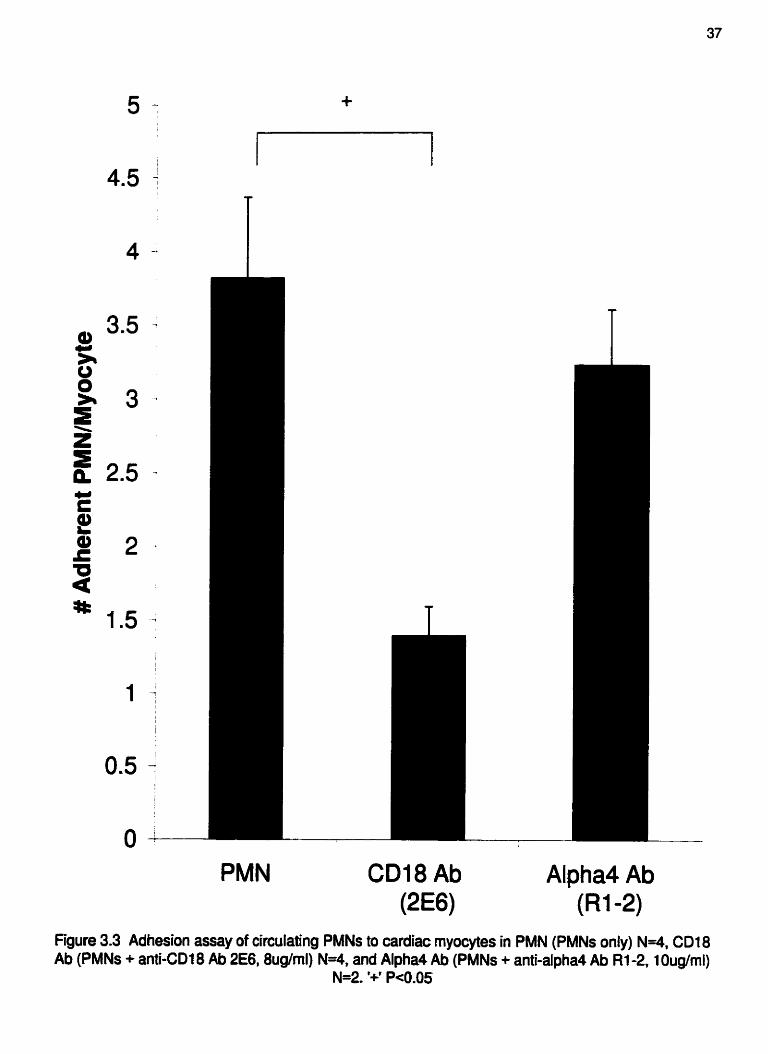
Adhesion assay of circulating PMNs to cardiac myocytes.. ............... .37
Adhesion assay of emigrated PMNs to cardiac myocytes .................. .3 8
Representative unloaded cell shortening record of a control rnyocyte
. challenged wth noproterenol.. ................................................ .SO
Representative unloaded cell shortening record of a WT myocyte exposed
to emigrated PMNs.. ............................................................ -5 1
Changes in unloaded cell shortening from baseline of myocytes due to
adherent and non-adherent emigrated PMNs.. ............................... .52
Cumulative unloaded cell shortening data obtained from myocytes in the
presence of emigrated PMNs at Smins.. ........................................ .53
Cumulative unloaded cell shortening data obtained from myocytes in the
presence of emigrated PMNs at IOmins.. ..................................... .54
Change in rates of contraction and relaxation of myocytes in the presence
of emigrated PMNs at l Omins.. ................................................ .55
Cumulative cell shortening data of myocytes in the presence of circulating
PMNs at 5 and l Omins.. ......................................................... .56
Change in rates of contraction and relaxation of rnyocytes in the presence
of circulating PMNs at i0mins.. .............................................. ..57
Cumulative unloaded cell shortening data of myocytes in the presence of
WT or NADPH oxidase KO emigrated PMNs at Smins.. .................. 69

Figure 5.2
Figure 5.3
Figure 5.4
Figure 5.5
Figure 5.6
Figure 5.7
Figure 5.8
Figure 5.9
Figure 5.10
Figure 5.1 L
Cumulative unloaded cell shortening data of myocytes in the presence of
WT or NADPH oxidase KO emigrated PMNs at lornins,. ........... .... -70
Change in rates of contraction and relaxation of myocytes in the presence
of WT or NADPH oxidase KO emigrated PMNs at 1Omins ................. 7 1
Cytochrome c reduction assay of WT and NADPH oxidase KO emigrated
PMNs .................................-............................................ .72
Representative fluorescence images of oxidant production in a myocyte
with an adherent emigrated PMN at baseline, 5, and 1 Omins.. .............. 73
Cumulative data for changes in fluorescence intensity of myocytes in the
presence of emigrated PMNs at Smins.. ..................................... ..74
Cumulative data for changes in fluorescence intensity of myocytes in the
presence of emigrated PMNs at 1 Omins.. .................................... -75
Cumulative unloaded cell shortening data of WT and SOD over-
... expressing myocytes in the presence of WT emigrated PMNs at 5mins 76
Cumulative unloaded cell shortening data of WT and SOD over-
expressing myocytes in the presence of WT emigrated PMNs at 10mins.77
Change in rates of contraction and relaxation of WT and SOD over-
... expressing myocytes in the presence of emigrated PMNs at 1 Omins.. .78
Cumulative unloaded cell shortening data of myocytes exposed to
emigrated PMNs in the presence and absence of exogenous SOD
treatment at Smins ................................................................. -79

Figure 5.12 Cumulative unloaded cell shortening data of myocytes exposed to
emigrated PMNs in the presence and absence of exogenous SOD
treatment at IOmins, . , . . , .. . . . . . . . . . . . . . . . . . , . . . , . . . , , . . . , , . . . . , , . . . , . , . . . . . . , . . . -80
Figure 5.13 Change in rates of contraction and relaxation of myocytes exposed to
emigrated PMNs in the presence and absence of exogenous SOD
treatment at IOmins..,,, .... ,.... ....................... . ..... . ............... ++,.,81

LIST OF ABBREVIATIONS
Ab
BS A
ca2+
CMClgro
CI'
DCFH
Fe
fMLP
H+
Ht02
HOCI
[CAM- I
IL
r/R
KO
LTB4
MPO
Na' NO
0 2
02'
OH-
ONOO'
PAF
PBS
PECAM-I
PKC
antibody
bovine serum aIbumin
calcium
cytokine-induced neutrophil chemoattractant/gro
chloride
6-carboxy-2', 7'-dichlorodihydrofluorescein diacetate
di(acetoxyrnethy1 ester)
iron
N-formy 1-Met-Leu-Phe
hydrogen
hydrogen peroxide
hypochlorous acid
intercellular adhesion molecule- 1
inter leukin
ischemia-reperfusion
knockout
Ieuko triene B4
myeloperoxidase
sodium
nitric oxide
molecular oxygen
superoxide radical
hydroxyl radical
peroxynitrite
platelet activating factor
phosphate buffered saline
platelet-endothelid cell adhesion molecule- 1
protein kinase C

PMN
SH3
SR
SOD
TNF-a
VCAM- 1
ZAP
polymorphonuclear leukocyte
Src homology 3
sarcoplasmic reticulum
superoxide dismutase
tumor necrosis factor-alpha
vascular adhesion molecule- 1
zymosan-activated plasma
xii

CHAPTER 1
lNTRODUCTION AND LITERATURE REVIEW

1.1 INTRODUCTION
A heart attack, or myocardial infarction, is triggered by an interruption of the
blood supply to tissue over a critical period, which can lead to tissue damage and
irreversible cell death I . Upon reperfusion, and the restoration of oxygen, myocardial
injury and inflammation is observed " 3. Since repefision is accompanied by a large
inflwc of polymorphonuclear leukocytes (PMNs) ", and depletion of PMNs from the
circulation reduced myocardial injury after ischemia-reperfusion (yR) '-lo, there is great
interest in the P M N as a target for therapeutic intervention. It is thought that after PMNs
infiltrate myocardium 11'14, they release cytotoxic factors like oxygen free radicals,
proteases, and arachidonic acid metabolites I I ; IS; I6 . Targeting these molecules can also
reduce the extent of myocardial injury after VR I"".
A large body of work has been dedicated to the malysis of PMN adhesion to
vascular endothelium, and the ensuing PMN-induced endothelial injury '"". Far less is
known about the interaction between PMNs and parenchymal cells like cardiac myocytes.
In the heart, a very important observation is that firm adhesion between cardiac myocytes
and PMNs is absolutely required for the release of toxic mediators " and subsequent
injury 3'26. Detailed reports conclude that the engagement of the Pz-integrin CD 18 is
essential for PMNs to release cytotoxic molecules " 27-29 . The tight seal between the
PMN and myocyte may exclude plasma, which contains important anti-oxidants and anti-
3 . 3 0 proteases . When PMN adbesion is disrupted with anti-CD 18 or anti-intercellular
adhesion molecule-I (ICAM-L ) molecules, plasma-derived anti-oxidants and anti-
proteases can prevent myocardial injury, highlighting the absolute requirement for PMN
adhesion through CD 18 in this pathology 5

AIthough these seminal studies have convincingly demonstrated the essential role
for adhesion between circulating PMNs and cardiac myocytes, the chosen experimental
conditions differed From the physiological situation since Ph4Ns must first emigrate out
of the vasculature before they interact with cardiac myocytes. The emigration process is
not trivial; emigrated PMNs have been shown to be far more responsive to inflammatory
31: 32 33-35 mediators , and to express novel adhesion molecules, including a~-integrin .
Indeed, the a-integrin has been shown to contribute significantly to emigrated PMN-
myocyte interactions. Following emigration, targeting only CD 18 with an anti-CD 1 8
antibody (Ab) no longer inhibited adhesion ". Rather, both anti-CD 18 and anti-a Abs
were required to prevent emigrated PMN-rnyocyte interactions in the rat model. This
data has raised many new questions about the importance of the m-integrin as a mediator
of emigrated PMN-dependent myocyte injury.
An activated PMN is able to produce a very high concentration of oxygen free
1 I; 16 radicals , and this level increases upon adhesion. Indeed, human PMNs adherent to
nylon fiber produced more superoxide radical (Or) and hydrogen peroxide (HzOz) than
the same cells in suspension 36. Once adherent to cardiac myocytes, circulating PMNs
were shown to generate CD lg-dependent oxygen free radicals, a likely mechanism of
PMN-dependent myocyte injury ". It is unknown whether emigrated PMNs will utilize
this same mechanism to injure cardiac myocytes. The present study was designed to
examine the mecharism of emigrated PMN-induced injury of cardiac myocytes through
an a-integrin-controlled free radical pathway.

1.2 MECHANISMS OF MYOCARDIAL INJURY IN ISCHEMIA-
REPERFUSION
Reperfhion of previously ischemic myocardium is crucial to patient recovery in
the clinical setting. Reperfi~i~ii however, may also paradoxically exacerbate myocardial
damage by causing morphological and metabolic de-arrangement and myocardial
necrosis 37. Although the pathogenesis of VR has attracted great interest, a full
understanding of the mechanism of myocardial injury in this pathology is incomplete.
Upon reperfusion of previously ischemic myocardium, there is a large influx of
PMNs ", and this PMN accumulation is associated with the areas of greatest injury in
the heart 11; 38; 39 . Reduction of circulating PMN numbers with anti-PMN Abs " PPMN
depletion filters or antimetabolites l4 all reduced infarct size in VR challenged hearts.
Limiting the recruitment of PMNs by immunosuppression of P M N adhesion molecules
4 1-44 also limited myocardial injury . Furthermore, experiments with complement
depletion '' and lypoxygenase inhibitors "6, aimed at reducing P M N chemotactic factors.
limited infarct size. Increased levels of PMN-derived proteolytic enzymes. including
elastases, P-glucosaminidases, P-glucuronidases, and myeloperoxidase (MPO), all of
which break down the barrier hc t ion of the endothelium and lead to impaired myocyte
hction, have been measured in reperfused myocardium '". Inhibition of known P M N
products, including oxygen free radicals, proteases, and arachidonic metabolites, also
reduced the extent of myocardial injury 18; 19; 1%48 . Although these data clearly show the
PMN is a key player in myocardial injury in I/R, alternate pathways of injury have been
proposed PMN-independent mechanisms of injury include the study of the pH paradox

49 , reperfbsion-induced calcium (ca2> overload ", and the generation of PMN-
51; S2 independent oxygen free radicals in the heart .
An association between altered pH levels and contractile function has been well
53; 54 documented . In fact, a decrease of just 0.22 pH units caused a 50% decrease in
contractile function of pefised rabbit hearts 49. It is evident therefore, that maintenance
of intracellular pH is crucial to proper cellular homeostasis. In the healthy myocardium,
an optimal intracellular pH of 7.3 is maintained by the constant extrusion of protons by
the sodiurn/hyd.ogen (~a+/'El?) exchanger. It is well established that ischemia produces
intracellular acidosis ", and increased intracellular Na + 49: 56. Theoretically, repefision
at physiologic pH would lead to a pH gradient across the sarcolemma and activation of
4% 57 the Na+/H+ exchanger to restore intracellular pH levels . H* is moved out of the cell
to increase intracellular alkalinity, while ~ a ' is taken into the cell. An increase in
intracellular ~ a + activates the Na+lca2+ exchanger, causing a large influx of ca2+ into the
cell and subsequent cellular injury. Although theoretically viable, experiments that
inhibited the N~'/H+ exchanger to limit the pH paradox have resulted in conflicting
reports. Treatment of hearts at the time of reperfitsion with amiloride, an inhibitor of the
58; 59 Na'/H+ exchanger , showed either broad ranged protection and enhanced ventricular
recovery 60, or no protection at all 6 ' . It is unclear, therefore if the pH paradox is the sole
mechanism of cellular injury in myocardial infarction.
Cyaolic ca2+ overload also occurs as a result of impaired generation of ATP 62, as
seen in ischemia where insufficient molecular oxygen (02) levels result in the depletion
of ATP and the formation of ADP, AMP, adenosine, inosine and finally hypoxanthine ".
The sarcoplasnic reticulum (SR) is therefore unable to take up ca2+ at a normal rate,

resulting in abnormally high cat+ levels within the cell. ~ a ' + overload activates
phospholipases that can destroy the cell membrane, leading to ca2+-mediated arrhythmia
and cell death ". Veraparnil, a non-dihydropyridine type ~ a " antagonist, showed
reductions in mortality when administered 2-3days after the initial infarct
demonstrating a possible role for ca2+ overload in late phase VR. Despite encouraging
data in animal studies. to date there is no convincing clinical data that humans benefit
from the blockade of ~ a ' + entry to the cell in the acute phase of myocardial infarction. In
fact, major clinical trials using nifedipine, a dihydropyridine type ~ a " antagonist,
showed no significant benefits in post-myocardial infarct 65. Although ~ a " overload
may contribute to myocardial tissue injury in I/R, there may be an alternative pathway of
injury independent of', or working in concert with, the uncontrolled influx of ca2+ to the
cell,
The role of oxygen free radicals in myocardial VR injury has been studied
extensively. Free radicals have been shown to cause lipid peroxidation 66, a disruption of
68; 69 myocardial cell membranes 67, an imbalance in ca2+ homeostasis . and cardiac
70: 71 contractile dysfunction . In animal studies, the addition of free radical scavengers
protected the heart fiom IIR-induced myocardial damage "' ". In fact, the addition of
exogenous fiee radical scavengers, superoxide dismutase (SOD) and catalase, protected
isolated rat hearts fiom VR-induced decrease in left ventricular pressure and increase in
left ventricular end-diastolic pressure; and partially inhibited the Winduced disruption
of SR ca2+ uptake and extrusion from the cytoso17'. During UR, there are many potential
sources of fiee radicals, including intracelluiar production trom the mitochondria 67,
conversion of xanthine oxidase to xanthine dehydrogenase 75, auto-oxidation of

catecholamines 76, and the arachidonic acid cascade n. It must be appreciated, however
that the PMN is a major source of free radicals in the inflamed heart ' I ' 16.
Although the proposed PMN-independent mechanisms may contribute to the
myocardial injury seen in yR, it is clear that we cannot exclude the potential role of the
PMN as a key player in the initiation and progression of these pathological pathways of
injury.
1.3 PMN RECRUITMENT
In order to interact with cardiac myocytes, PMNs must first leave the vasculature
to enter myocardial tissue. PMNs are recruited to areas of inflammation by a multi-step
recruitment paradigm. Initially, circulating PMNs slow down by making temporary
contacts with the endotheliurn, a process called tethering. After the initial contact, a
succession of contacts is made and the PMN begins to roll along the vascular wall. The
selectin family of adhesion molecules, including constitutively expressed L-selectin on
the PMN, and inducible P- and E-selectin on the endothelium, mediates this process of
78; 79 tethering and rolling .
To minimize steric interference, L-selectin is strategically located on the tips of
microvilli projections and is shed following activation and subsequent emigration ".
Possible ligands to L-selectin include P- and E-selectin on the endothelium " . P-selectin
is preformed and stored in Weibel Palade bodies in endothelial cells and is rapidly
mobilized to the endotheliai cell surface in response to inflammatory mediators P-
selectin plays an important role in the early phase of inflammation since P-selectin levels
are quickly decreased by 30-60min. P-selectin has been shown to bind primarily to P-

selectin glycoprotein ligand-l on most leukocytes 82. E-selectin is not preformed, but is
83; 84 synthesized in response to inflammatory cytokines . E-selectin may be more
important in late stage inflammation since maximal synthesis levels require 4-6 hours.
PSGL- I, E-selectin ligand- l , cutaneous lymphocyte antigen, and L-selectin are all
possible ligands for E-selectin HI: 8s: 84
The next step in PMN recruitment to areas of inflammation is firm adhesion.
Once the PMN has slowed down, it can then make more permanent interactions with the
endothelium. The p2-integrin (CD I I/CD 18) family of adhesion molecules is involved in
cell-cell interactions and thus mediates firm adhesion of PMNs to endothelium ". The
members of this adhesion molecule family include LFA- 1 (CD I 1 dCD 18). Mac- 1
(CD 1 1 b/CD 18), and p 150195 (CD 1 Ic/CD 18) 88. The Printegrins on PMNs adhere to
ICAM-I on cytokine stimulated endothelium 4, and on cells outside of the vasculature,
including fibroblasts, dendritic cells, and epithelial cells 85. Pz-integrins also have
extravascular ligands, including matrix proteins and complement fragments ", and these
may become important once the PMN leaves the vasculature.
Once PMNs have f d y adhered to the endothelium, they undergo a shape
change, which allows them to crawl between endothelial cells and move into tissue. This
emigration process may be mediated by platelet-endothelial cell adhesion molecule- l
(PECAM-I ), expressed along the border between endothelial cells and on PMNs 90. A
role for PECAM- L in PMN transmigration across endothelium has been shown in v i m
91;92 and is hypothesized to play a similar role in vivo .
Following emigration, PMNs migrate dong an increasing gradient of chemotactic
agents, incIuding cytokines (interleukin a)-1 and -8), complement cascade products

(CSa), bacterial products (N-formyl-Met-Leu-Phe (fMLP)), and products of phospholipid
metabolism (platelet activating factor (PAF) and leukotriene B4 (LTB4)) 4. PMNs are
now localized within myocardial tissue and can interact with cardiac rnyocytes in
pathophysiologies like VR.
1.4 PMN-INDUCED ENDOTHELIAL VS MYOCYTE DAMAGE
Although a role for the PMN in VR-induced myocardial injury seems clear, it is
unknown if the critical PMN-dependent injury is at the level of the endothelium or at the
level of the myocyte. It is well appreciated that the I/R damages the microvasculature,
and that the increased adhesiveness of PMNs to the endothelium contributes to the extent
of the tissue injury "; ". Indeed, adhesion of PMNs to the endothelium is a prerequisite
to emigration, and endothelial injury may be so severe that the myocardium is
irreversibly damaged and subsequent myocyte injury may play only a minimal role in
decreased myocardial function.
PMNs can directly injure endothelial cells through proteases and oxygen free
t 1; 15 radicals . PMN-derived oxygen free radicals have been shown to cause
disintegration of endothelial cell membranes, resulting in microvascular disorders arising
from cell dysfunction, edema, and cell death 94. Furthermore, oxygen free radicals
stimulate PAF release fiom the endothelium, which M e r exacerbates the locaI PMN
Mux by an amplifying feedback loop ". PMN-derived elastase hydrolyzes a variety of
96: 97 biological substrates , and its activity is increased in the blood of patients suffering
from myocardial infact 98. Furthermore, endotheIial cell monolayers exposed to anoxia
induced elastase release f?om PMNs upon reperfusion 99. These PMNs caused

endothelial cell detachment and resuited in a loss of cell-cell contact and exposure of the
underlying matrix, which was ameliorated by the addition of elastase inhibitors. One
consequence of endothelial detachment is the exposure of underlying smooth muscle to
the direct vasoconstricting effects of platelet-derived factors ".
The ability of the PMN to injure endothelium is well established, but whether this
mechanism of myocardial injury in I/R is the dominant pathway is unclear. It is evident
however, that patients may arrive at hospital after the myocardiai infarct and thus PMNs
have already been recruited and are in contact with cardiac myocytes. The adherence of
PMNs to vascular endothelium occurs within 2Omins post-reperfusion and these PMNs
begin to emigrate as early as i h post-reperfusion '". Many researchers have
acknowledged this time frame and have chosen to study the interaction of PMNs and
isolated cardiac myocytes arid have shown that PMNs can indeed directly injure cardiac
3; 26 myocytes . PMNs treated with chemoattractant phorbol 12-myristate 13-acetate
caused irregular contractions and subsequent contracture and blebbed formation in
murine embryo ventricular myocytes 26. Electron micrographs of these myocytes prior to
contracture revealed swollen rnitochondn*a, and ruptured plasma membranes and
vacuoles.
PMN adhesion is critical to PMN-induced myocyte injury since supernatant From
activated PMNs was unable to cause myocyte injury, and myocytes without adherent
PMNs were not injured 25.26; 101 - Researchers have, therefore begun to study the adhesion
molecules involved in PMN-myocyte interactions and found that PMNs adhered to
2% 101 myocytes through a CD 18-ICAM-1 mediated pathway . Furthermore, through
fluorescence imaging, these investigators have shown that PMNs caused oxidant

generation in the myocyte and that this increase in oxidants was CD 1 &mediated =. Interestingly, this research group also found that one adherent PMN alone was able to
cause myocyte injury, and that the magnitude of oxidant production was not increased
when multiple PMNs were adherent to the myocyte.
Although these studies show the importance of PMN adhesion through a CD L8-
mediated pathway to myocyte injury, these experiments involved circulating PMNs
isolated from whole blood. It is clear that PMNs must emigrate out of the vasculature
before they can interact with cardiac myocytes. Emigrated rat and human PMNs have
been shown to express the m-integrin, and this ligand is not expressed in the circulation
33-35 . Emigrated rat PMNs have also been shown to utilize this new adhesion molecule, in
conjunction with CD18, to adhere to isolated ventricular myocytes '). It is unknown
whether murine emigrated PMNs also express the a-integrin, and if this ligand plays a
role in emigrated PMN-myocyte interactions in this model. Furthermore, the ability of
emigrated PMNs to induce myocyte injury, and role of the m-integrin in mediating
injury, requires further study.
1.5 PMN-DERIVED FREE RADICALS
PMNs are known to produce oxygen free radicals and their reactive oxygen
intermediates have been shown to be toxic to many cell types 11: 16; 102 . Although cells
have natural protective free radical scavenging system, including SOD, catalase, and
glutathione peroxidase, many ischemic diseases of the heart, bowel, liver, kidney, and
brain have been Linked to free radical damage caused by oxidative stress ".

PMNs use the enzyme NADPH oxidase to mount a respiratory burst in response
to an inflammatory condition '03. NADPH oxidase transfers an electron fiom cytosolic
NADPH across the plasma membrane to 0 2 . 0; is formed fiom the acceptance of one
extra electron by 0 2 , and Oi or its secondary products can then be released and
accumulate in the extracellular space. In the myocardium, when there is a sudden rise in
intracellular ca2', the ''ca2* paradox" itself has also been shown to trigger the production
of Of ' W. In the vasculature, a major source of 02' is from the conversion of
hypoxanthine to Oi by the enzyme xanthine oxidase found in endothelial cells.
In healthy cells, SOD catalyzes the dismutation of 02' to H2O2, which is then
converted back to O2 and water by catalase and glutathione peroxidase. In
pathophysiological states like myocardial I/R, where free radical generation is increased,
these scavenging systems may become overwhelmed and oxidant-induced injury may
occur. Indeed, several studies with isolated heart preparations have reported a burst of
oxygen free radicals generated following repefision 105-109 . Furthermore, fiee radical
generation was measured up to 3 h post-reperfitsion using electron spin resonance and
spin-trapping techniques, providing direct evidence for the production of free radicals in
the setting of myocardial I/R lo.
02- has been shown to react with nitric oxide (NO) to produce peroxynitrite
(ON00-). ONOO- is a stronger oxidizing agent than either O< or NO alone, and quickly
68; 1 1 1 reacts with thiols, ascorbate, and lipids . Although ONOO' formation may be
beneficial to some cells, there is also a vast amount of data showing ONOO--induced
oxidative damage to bioiogicd tissues and subsequent pathogenic conditions ' "' ' I 3 In
the heart, ONOO- was shown to aggravate injury measured by depressed cardiac funftion

recovery, increased lactate dehydrogenase and creatine kiiase release, and enlarged
necrotic size 114; 115 . Researchers have confumed ONOO- production upon repefision of
previously ischemic myocardium 1 1 6 1 18 . The reaction rate for the formation of ONOO- is
6.7kO.9~ 1 0 ~ l ~ w s ' 19, which is approximately six times faster than the scavenging rate of
0; by SOD 12'. NO is the only known biological molecule produced in high enough
concentrations in pathophysiological states to successfblly out-compete SOD for Oi I I?.
Normally, a biological system generating 02* will produce H202 by the
disrnutation reaction, udess SOD levels are depressed or if Of is able to react
immediately with another molecule like NO. In several experiments where exogenous
free radical generating systems caused cell injury, H202 has been identified as the
specific free radical causing injury since cells were protected with exogenous catalase
and not SOD 12'. Furthermore, Hz02 not 02., is able to cross biological membranes,
allowing for intracellular cytotoxicity. In the heart, H202 caused contractile
abnormalities and injury to cardiac myocytes which were linked to an H&dependent
increase in ca2+ influx and subsequent ~ a " overload 69. H202 also damaged the SR,
causing reductions in ~ a " uptake and altered ca2+ homeostasis " . Moreover, H2O2 has
been shown to cause direct electrophysiological alterations to rat cardiac myocytes with a
slowing of the inactivation of ~ a + channels and prolongation of the action potential E3.
Increasing attention has been focussed on the role of hypochlorous acid (HOCI) in
tissue injury. Secretion of MPO into the phagocytic vacuole of the PMN can catalyze
the oxidation of chloride (C13 by H202 to yield HOCI. The local concentration of HOCI
produced by activated PMNs is estimated to be 60-90p.M '"' I". HOCl is highly reactive
and, at concentrations as Iow as 10-20pM, can quickly oxidize many biological
molecules, causing cellular injury 126; t27 HOCl is a weak acid at physiological pH, and
especially under acidic conditions of VR, remains mostly undissociated and permeable
'28. This may facilitate entry into the myocyte and allow HOCl to directly affect the
myofilaments. Indeed, the addition of exogenous HOCl caused an increase in ca2'
sensitivity, a decrease in maximal ca2+ force, and an increase in the resting tension of
skinned rat cardiac muscle 12'. Furthermore, HOCl has also been shown to mobilize
intracellular zinc in cardiac myocytes '26, and Free zinc has been shown to be a potent
inhibitor of cardiac contractility I z9 . Finally, HOCl from activated PMNs caused an 80-
90% inhibition of ca2' uptake. indicating severe SR damage ". This inhibition was
71; 130 completely restored by the addition of L-Methionine, a known scavenger of HOCl .
Oz- can also. however enter the Fenton reaction in the presence of free iron (Fe) to
produce the highly reactive hydroxyl radical (OK). The generation of OK by the Fenton
reaction has been demonstrated in many in vitro studies '31-'33, and many have proposed
that it is the true agent behind the toxic effects attributed to Of 131-134. Formation of OH*
is controversial however, since it is unclear whether or not the body generates high
enough free Fe levels to allow for this reaction to occur in vivo. Adult humans have 4g of
Fe, with two-thirds present as hemoglobin and 10% found in myoglobin The
remainder is present in intracellular storage proteins, femtin, and hemosiderin found
mainly in the liver, spleen, and bonemarrow I". Fe in the diet exists in the oxidized €om
Fe(I?I), and is generally tightfy bound to transferria, a carrier molecule glycoprotein with
two binding sites for Fe(III) '". Under normal conditions, the trmsferrin present in the
blood stream is only 30% loaded with Fe, so the amount of free Fe available in the blood
plasma would be virtually zero 135; 136
These reactive oxygen intermediates can affect a multitude of biological systems,
including lipid peroxidation ", modification of protein structure and function, and
ultimately cell death 13?. The relationship between free radicals and the functional state
of the myocardium has been studied extensively U: 138-140 . Free radical generating
systems administered exogenously have been shown to cause cardiac contractile
dysfunction and electrophysiological abnormalities 141; 112 . These fiee radicals were also
able to affect myocardial sarcolemmal membrane 143: 144 , SR and mitochondria1
functions Furthermore, it has been shown that free radicals depress the sarcolernrnal
C ~ ' + A T P ~ S ~ activity, resulting in reduced ca2+ extrusion from the cytosol I*. Free
radicals also promote ca2+ release from the SR and inhibit ca2+ sequestration to the SR
68 , leading to a disruption of ca2* homeostasis and subsequent ~ a " overload.

1.6 STATEMENT OF HYPOTHESIS AND OBJECTIVES
Avrothesis 1: Emigrated murine PMNs use both p2- and a-integrins to adhere to
isolated cardiac myocytes.
Objectives:
1) To determine whether the process of emigration alters the mechanism by
which murine PMNs adhere to cardiac myocytes.
2) To determine if murine PMNs express a new adhesion molecule profile
following emigration.
Hvrothesis 2: Emigrated PMNs cause injury to cardiac myocytes through the a(-
integrin.
Objectives:
1) To determine if emigrated PMNs cause myocyte dysfunction, and if so,
whether adherence of the PMN to the myocyte is necessary for injury to
ensue,
2) To determine whether injury is mediated through either CD 18, a-integrin, or
through both ligands.
3) To determine if circulating murine PMNs injure cardiac myocytes through the
same mechanism as emigrated PMNs.
Bv~othesis 3: PMN-induced myocyte injury is caused by the generation of free radicals.
0 bjec t ives:
I) To determine if PMNs require a respiratory burst to cause myocyte damage.

2) To visualize the production of fiee radicals upon PMN adhesion to the
rnyoc yte.
3) To determine whether fiee radical generation is mediated through either CD 18
or m-integrin.
4) To determine if the specific free radical responsible for myocyte injury is Oi.

CHAPTER 2
METHODS AND MATERIALS

2.1 EXPERIMENTAL MODELS
2.1.1 PMNlMyocyte Adhesion Assay
To examine the adhesion of murine PMNs to isolated cardiac myocytes, an
in vitro adhesion assay was employed 14'. Myocytes were coated onto a round glass
covenlip and mounted onto the inside of one side of a metal chamber. A second clean
coverslip was placed on top of the myocytes, separated by an O-ring gasket to form a
chamber space of approximately 700-800p1. The other side of the metal chamber was
then attached. PMN suspensions were injected into the chamber space between the
coverstips via a syringe and 23G needle. The PMNs then sealed by gravity onto the
myocyte layer. Once the chamber was inverted, all nonadherent PMNs fell away from
the rnyocyte layer, and those PMNs adherent to myocytes were counted with an inverted
microscope.
Ventricular myocytes were isolated as previously described for rat ventricular
myocytes '' with minor modifications for murine cells. Briefly, six-week old male
CS7BL6 mice were anaesthetized and the h e m removed and placed into Tyrode's buffer
(NaCl 140mM, KC1 5.4mM, Na2HP04 lmM, HEPES SmM, glucose IOmM, MgClz
I mM, pH adjusted to 7.4 with NaOH) containing ImM CaClz at 4OC. Hearts were then
cannulated via the aorta (within 3mins) for retrograde perfusion of the coronary arteries.
Initially, the hearts were perfused with Tyrode's buffer containing 1mM CaC12 at
2mVmin for Smins at 37OC and then with Tyrode's buffer containing no CaCIz at 2mVmin
for Smins. Perfusion was then switched to Tyrode's buffer containing 40p.M CaC12,
20pgIml collagenase, and 4pglmI protease and perfusion continued at 2mVrnin for Smins.
Digested hearts were then removed tiom the perfusion system and ventricles were

minced in Tyrode's buffer containing imM CaC12, 500pg/mI collagenase, 100pg/ml
protease, and 2.5% bovine senun albumin (BSA). Ventricular tissue segments were then
put into a shaking water bath for 10-20mins at 37OC to complete the dispersion and
obtain a suspension of individual myocytes. Myocytes were then placed in a KB-type
solution (K-glutamate 100mM, K-aspartate IOmM, KC1 ZSmM, KH2P04 IOmM. MgS04
Zm., taurine 20mM, creatine 5mM, EGTA O.5rnM. glucose 20mM, HEPES SmM, and
BSA I%, pH adjusted to 7.2 with KOH) at 4OC. and used within Shrs.
To obtain emigrated murine PMNs. six-week old male C57BL6 mice were
injected intraperitoneally with 1% oyster glycogen in saline After 4h, mice were
sacrificed and a peritoneal lavage performed with 3ml saline. Lavage fluid was placed on
ice for Smins then centrifbged at 1300rpm at 4OC for 6mins. Pellets were then
resuspended in Tyrode's buffer with 1mM CaCll at 4OC. This approach yielded a 99%
pure population of emigrated PMNs as analyzed with Wright-Giemsa staining. In all
experiments, PMNs were kept on ice and used within 2hrs of isolation.
Circulating murine leukocyte suspensions were isolated From whole blood by
lysis of the red blood cells. Briefly, blood (800-900~1) was collected by cardiac puncture
into a syringe with acid citrate dextrose (anticoagulant) ( 1 OOpl), added to ddH20 at 4OC,
and gently mixed. KC1 (0.6M), followed by phosphate buffered saline (PBS, NaCl
137mM. KC1 2.7rnM. Na2HP04 8. LmM, KH2P04 1.47mM), was added and the sample
centrihged at I300rpm at 4OC for 6mins. These steps were repeated to further isolate a
pure circulating Leukocyte population, and the pellet resuspended in Tyrode's buffer with
ImM CaC12 at 4OC. These leukocytes were initially exposed to myocytes and
histologicaI assessment revealed that all of the adherent cells were indeed PMNs.

Flow cytometry was used to measure the expression of CD 1 1 b, CD 18, and a-
integrins on circulating and emigrated PMNs. Circulating or emigrated murine PMNs
(1x10~ per tube) were stimulated with 1% zymosan-activated plasma (ZAP) ( IOmins at
room temp) and then washed. Red blood cells were lysed and PMNs were futed in 1%
formalin (15mins at room temp) and then washed. Primary Abs were then added to stain
for their respective adhesion molecules (CD I 1 b, MW 170,025pg per tube, Pharmingen;
CD18,2E6,0.8pg per tube, Endogen; and Q, Rl-2, lpg per tube, Pharmingen). After
30mins at room temperature, cells were washed and labeled with FITC-conjugated goat
anti-rat IgG (Cedar Lanes Laboratories LTD) for CD 1 1 b and Q, and FITC-conjugated
goat anti-hamster IgG (Caltag Laboratories) for CD 18. Afier 30mins at room
temperature, cells were washed and fluorescence was measured on a FACScan flow
cytometer (Becton Dickinson Imrnunocytochemistry Systems).
2.1.2 Unloaded Cell Shortening Assay
Isolated ventricular myocytes were allowed to adhere to a glass microscope stage
for Smins at room temperature I". Myocytes were then superfused at imI/min with
normal Tyrode's buffer containing 1m.M CaC12. Cells were field stimulated at 1 Hz using
a just threshold voltage level (Isolator U, Axon Instruments USA) to minimize production
of free radicals due to hydrolysis. Unloaded cell shortening was recorded using an edge
detection device (Solamere Technology Group) and the data acquired digitally at 1 O M z
sampling rate using customized software (Cellsoft V2.0, D. Bergman, University of
Calgary, Canada). The number of PMNs adherent per myocyte and the time of onset of
dysrythmia were recorded for each myocyte. For all experiments, cells were allowed to
equilibrate while being eIectrically stimulated continuousiy for 15mins. To ensure that

myocytes exhibited normal contractile behavior and inotropic capacity before PMN
treatment, the j3-adrenergic agonist isoproterenol(0.1 @i) was added and the resulting
positive inotropic response to electrical stimulation was monitored. Myocy-tes exhibiting
baseline shortening <5% of resting length, or those failing to respond to isoproterenol
were excluded fiom the study. A positive response to isoproterenol included a 2-fold
increase in extent of cell shortening, rate of contraction, and rate of relaxation from
baseline.
Afier isoproterenol was washed out ( l0mins). baseline measurements were taken
and then 1x10~ PMNs, pre-stimulated with 1% ZAP. were added to the superfusate.
Myocyte contractility was then recorded continuousiy for I Omins. Isoproterenol was
added again to the superfusate to reassess myocyte contractility. In all experiments,
myocyte contractility was recorded to the completion of the protocol unless cell death
occurred.
A cytochrome c reduction assay was utilized to measure the production of O<
from PMN suspensions Briefly, PMNs ( l~ l0~1rn i ) in PBS were added to PBS with
CaCh ( l.l9mM), MgC12 (0.54mM). and cytochrome c ( 1 .SmM, Sigma) for a paired
analysis. In one sample, SOD (from bovine erythrocytes, 264U/ml, Sigma) was added
and both samples read at the same time in a spectrophotometer (U-2000
Spectrophotometer, Hitachi) at 550nm. Optical density differences between the two
samples were recorded on an online chart recorder (Johns Scientific Inc). After Smins of
baseline measurements, 1% ZAP was added to both samples and optical density recorded
for an additional 10rnins.

2.13 Single Cell Imaging Assay
Isolated ventricular myocytes and emigrated PMNs were loaded with fluorescent
probe, 6-carboxy-2',7'-dichlorodihydro fluorescei diacetate di(acetoxymethy1 ester)
(DCFH, 1 j&l for myocytes and 1Op.M for PMNs, Molecular Probes) in Tyrode's buffer
with probenecid 0.5mM (Sigma) at room temperature for 15mins. DCFH is oxidized to
highly fluorescent 2',7'-dichIorofluorescein in the presence of free radicals. DCFH is not
specific for any one oxidant, and thus can be used only as an overall indicator of
oxidative stress within the cell 151-154 . Myocytes were allowed to adhere to a glass cover
slip sealed by vacuum grease to the bottom of a plastic stage chamber, for Smins at room
temperature. The chamber was clipped into a fitted stage platform on an Axiovert-135
inverted microscope (Zeiss) equipped with an oil immersion KUAR 100d 1.3 objective
for single cell imaging '55. A Delta-Ram High Speed Illuminator (Photon Technologies
International), consisting of a 75 Watt Xenon arc and a computer controlled random-
access wavelength monochromator, provided excitation light. Wavelengths were fbrther
selected prior to cell illumination by a dichroic filter (Chroma Technology Corporation)
mounted on a sliding apparatus under the objectives. [mageMaster v1.4 software (Photon
Technologies International) allowed for direct control of the camera, illumination, and
data acquisition. Digital images of em-ssions from selected fields were saved to
computer disk in sequential order for analysis. The cells were excited at 480nrn and
emission recorded at 5 10nm.
To ensure that morphologically viable myocytes were indeed healthy, single cell
fluorescence intensities for each myocyte prior to exposure to PMNs were recorded for
the first Smins. Cells exhibiting a rise of greater than LO raw intensity units were

assumed unhealthy and excluded from the study. Emigrated PMNs ( 1 x 1 06), pre-
stimulated with 1% ZAP, were added to the myocytes and fluorescence intensities
recorded every 1 Osecs for 10mins. Phase contrast photos of the myocytes with adherent
PMNs were recorded and stored digitally. At the end of each experiment, Hz02 (50mM,
BDH) was added to the cells to confum adequate loading of the cells with DCFH, and to
demonstrate that all cells had the ability to fluoresce upon reaction with HrOz. Upon
addition of the Hz02, all myocytes included in the study reached camera saturation
intensity levels (255 raw intensity units).
2.2 EXPERIMENTAL PROTOCOLS
2.2.1 Emigrated Murine PMN Adhesion to Cardiac Myocytes via pr and
Q-in tegrins.
Round glass coverslips (25mrn. Bellco Glass Inc) were pretreated with 1% filtered
gelatin and incubated for 1 hr at 37OC. The gelatin was then removed and a lml
suspension of isolated cardiac myocytes in Tyrode's buffer ( l~ 10'lml) was layered onto
the coverslips, and incubated for an additional hour at 37OC. One coverslip with
myocytes and one clean coverslip were then placed into the adherence chamber.
Suspensions of either circulating murine leukocytes or isolated emigrated murine PMNs
(SX 1 06/m.I) were pretreated with I % ZAP, injected into the chamber space, and allowed
to settle for 1Omins. The chamber was then inverted and all nonadherent ceIIs fell to the
bottom of the chamber, leaving only cells adherent to the myocyte layer. The number of
PMNs adherent per myocyte was counted at 200X magnification on an inverted
microscope (Zeiss) (a minimum of 20 myocytes per coverslip was counted).

This adhesion assay was used to study the effect of known PMN stimulants,
cytokines, and cell concentrations on P M N adhesion to myocytes to determine optimal
conditions for adhesion. PMN stimulants tested included M L P (5-20p.M). PAF (50-
1000ng/ml), cytokine-induced neutrophil chemoattractant/gro (CMC/gro. 5-20n.M).
LTB4 ( 10-'~-1 O-~M), KC (murine IL-8,5- 1 OOnM), and ZAP (0.1 - 10%). Since 1 % ZAP
optimally increased PMN adherence and is present in pathophysiological states of I/R lSb
I Z f l , this stimulant was used to activate PMNs in all subsequent experiments. Tumor
necrosis factor-a (TNF-a, 100-500U/ml) treatment of the myocytes showed little
difference in adhesion numbers as compared to untreated myocytes, therefore no myocyte
pretreatment was used for subsequent experiments. Finally, various PMN concentrations
( 1 x 1 0 ~ ~ 5 ~ lo6 /mi) were used in the assay and data showed optimal adhesion at the
highest concentration tested. PMNs at sx 106/m1 were used for all adhesion studies, but
for all other experiments (cell shortening and single cell imaging) the lower concentration
( 1 x 1 o6/rnl) was used to limit the number of mice required per experiment.
To examine the role of p2- and m-integrins in PMN-myocyte interactions,
hnctionally blocking Abs to CD 18 (anti-CD 18 Ab 2E6, Endogen) or to the a-integrin
(anti-a Ab Rl-2, Pharmingen) were added alone, or in combination to the suspension of
emigrated PMNs prior to injection into the adhesion chamber. Flow cytometry was used
to determine the saturating dose of each Ab. Doses of the anti-CD 18 and anti-% Abs at
2-20pg/ml were tested. For the anti-CD18 Ab, 8pg/ml and for the anti-cq Ab, lOp&ml
showed maxima1 fluorescent staining and these doses were used for all experiments. For
circulating leukocyte suspensions, addition of anti-CD 18 Ab done was sufficient to
inhibit adhesion, therefore, both Abs were not added in combination.

2.2.2 PMN-Induced Myocyte Dysfunction via the Q-integrin.
Following baseline cell shortening measurements and isoproterenol challenge,
emigrated PMNs or circulating leukocytes (1x10~) pretreated with 1% ZAP were added
to the pefision buffer. Unloaded cell shortening measurements, myocyte dysrythrma,
and contracture were recorded, and the number of adherent PMNs noted. Experiments
were conducted on myocytes alone, with emigrated PMNs or circulating leukocytes, and
with anti-CD 18 (2E6,8pg/ml) or anti-a Abs (Rl-2, 10pg/ml). Data was subsequently
analyzed for unloaded cell shortening, rate of contraction, and rate of relaxation for each
myocyte at Smin intervals.
Isoproterenol was used to assess the contractile properties of all myocytes before
PMN challenge. Myocytes were exposed to isoproterenol(0. IpM) for 5-20secs and the
time to maximal response and the time required to return to pre-isoproterenol levels were
recorded for each exposure time. For future experiments, all myocytes were exposed to
isoproterenol for lOsecs, with maximal response at Zmins, and returned to baseline cell
shortening levels by i Omins.
2.23 Free Radical Generation in Cardiac Myocytes via the a-integrin.
Unloaded cell shortening was used to assess the role of PMN-derived free
radicals on PMN-induced myocyte injury. Emigrated PMNs ( 1 x 1 06) were isolated from
mice lacking the ability to generate free radicals (NADPH oxidase knock out (KO) mice)
and added to myocytes from wild type (WT) mice (C57BL6). Cell shortening
measurements were recorded for WT myocytes alone, and after the addition of either WT
or NADPH oxidase deficient emigrated PMNs (pretreated with 1% ZAP).
As a control, the cytochrome c reduction assay was done comparing 0;

levels in PMNs from WT mice to those fiom NADPH oxidase deficient mice. As
expected, results showed O< levels below detection for the NADPH oxidase deficient
PMNs, confirming that NADPH oxidase was indeed lacking in our transgenic mice.
The single cell imaging technique was used to visualize and measure the changes
in oxidative stress in the myocyte upon adhesion of emigrated PMNs. Fluorescence
measurements of WT myocytes alone, with WT emigrated PMNs ( l x 1 06) pretreated with
1 % ZAP. and with anti-CD 18 (2E6,8pg/ml) or anti-at Abs (Rl-2, 1 Opglmi) were
recorded and images stored digitally. Images were analyzed for changes in fluorescence
intensity in raw intensity units at baseline, 5 and 10mins.
To determine if the cause of myocyte injury was O<, the unloaded cell shortening
assay was used on myocytes from mice over-expressing endogenous Cu/Zn-SOD I s 9 .
Myocytes isolated fiom these mice show a 10-fold increase in SOD expression 16* and as
a result, should be able to scavenge intracellular Oi. Cell shortening was recorded for
myocytes fiom WT (C57BL6) or SOD over-expressing mice in the presence and absence
of WT emigrated PMNs ( 1 x 1 06). Additional cell shortening experiments were done
with exogenous SOD (from bovine erythrocytes, 300U/rnl, Sigma) to determine if
exnacellular 9' (Oi released by the PMN) was responsible for the PMN-induced
myocyte injury.
23 STATISTICS
All data are expressed as the arithmetic mean f standard error of the mean. Data
were compared between treatment groups using an analysis of variance of raw data with

the Dtmnetts method for multiple comparisons to PMN only group, and the Student T-test
within groups. Values of PcO.05 are considered statistically significant.

EMIGRATED iMURINE PMNs ADHERE TO CARDIAC
MYOCYTES MA pr and wINTEGRINS

Hvwthesis: Emigrated murine PMNs use both P2- and m-integrins to adhere to isolated
cardiac myocytes.
Objectives:
I) To determine whether the process of emigration alters the mechanism by
which murine PMNs adhere to cardiac rnyocytes.
2) To determine if murine PMNs express a new adhesion molecule profile upon
emigration.
3.1 RESULTS
ZAP and fMLP increase adhesion of emigrated murine PMNs to cardiac
myocytes. To increase the number of adherent PMNs/myocyte, various P M N
stimulants and concentrations were tested. Of those tested, fMLP and ZAP both showed
consistent increases in adhesion over untreated cells. The addition of MLP increased
adhesion from 1.3 5k0.10 adherent PMNs/myocyte in untreated controls to 2.7kO .26
adherent PMNs at 10pM fMLP (Fig 3.1. N=l1, P<0.05). The addition of ZAP also
approximately doubled adhesion when administered at the I % ZAP dose (Fig 3.2, N*.
P<0.05). fMLP is a bacterial product present in inflammatory states like sepsis. ZAP,
however is present in pathophysiological states of VR 156; t57 and is readily available and
easy to use. Therefore, for subsequent experiments, 1% ZAP was used as a pretreatment
for all PMNs.
PMN concentration Pffects adhesion of emigrated PMNs to cardiac myocytes.
Preliminary studies showed that increasing the PMN concentration from 1 x 1 o6 to
5x10~ /ml resulted in a greater than 2-fold increase in adhesion, therefore for optimal

adhesion, PMNs at a concentration of 5x10⁶/ml, pretreated with I% ZAP, were used for
all subsequent adhesion studies.
Cytokine pretreatment of isolated cardiac myocytes does not affect adhesion
of emigrated PMNs. TNF-a pretreatment of the myocytes showed little difference in
adhesion throughout all TNF-a doses (100-500U) as compared to untreated myocytes,
consequently no myocyte pretreatment was used for firture adhesion experiments.
Circulating murine PMNs adhere to isolated cardiac myocytes via CD18.
Murine circulating PMNs, pretreated with I% ZAP, avidly adhered to isolated
cardiac myocytes (Figure 3.3). Addition of a fuactionally blocking Ab to CD 18 (2E6,
8pglml) inhibited adhesion by 63% (N4, Pc0.05). Immunosuppression of the a-
integrin with an anti-Q Ab (R 1-2, 1 Opgfml) however, did not inhibit adhesion (3.23k0.38
adherent PMNs/myocyte with anti-c~c Ab, and 3.48M.28 adherent PMNsImyocyte
without anti-a Ab, N=2). These data indicate that this adhesion pathway is CD 18-, and
not u-integrin-dependent.
Emigrated murine PMNs adhere to isolated cardiac myocytes via CD18 and
a-integrin. As previously shown, ZAP ( 1%) increased emigrated PMN adhesion to
cardiac myocytes (N=4, PcO.05, Figure 3.4). In contrast to circulating PMNs, addition of
the anti-CD 18 Ab (2E6,8pg/ml) to emigrated PMNs did not affect adhesion to cardiac
myocytes (N4, P=NS fiom PMN only group). Addition of the anti-& Ab (Rl-2,
lOpg/ml) aIso had no effect on adhesion (N=4, P=NS fiom PMN only group).
Immunosuppression of both integrins with anti-CD 18 and anti-- Abs, however inhibited
adhesion to pre-ZAP levels (N=4, P<O.O5 compared to 1% ZAP group).

Murine PMNs express CDllb, C D 9 and -integrin upon emigration.
Flow cytometry fluorescence data for CD 1 1 b, CD 18, and Q-integrin on
circulating and emigrated murine PMNs is summarized on Table 3.1. Mean fluorescence
increased upon emigration and subsequent stimulation with ZAP for all adhesion
molecules. Mean fluorescence increased from 74.6k4.3 to 3 1 1.6 1+3.57 for CD 1 1 b, from
89.36e5.6 to 277.34E26.5 for CD 18, and From 2.3 1M.06 to 17.96f 1.50 for the
integrin (N=4. P<0.05 for all adhesion molecules).
3.2 DISCUSSION
Human and rat PMNs have been shown to express new adhesion molecule
profiles upon emigration '3"5, and rat PMNs utilize the newly expressed ~ i n t e g r i n , in
conjunction with CD 18 to adhere to parenchymal cells like cardiac myocytes 33. The
purpose of the present study was to determine if this same paradigm exists in the murine
system. Indeed, our data showed that adhesion profiles of murine PMNs changed after
emigration. We found that circulating murine PMNs adhered to cardiac myocytes via
CD 18, c o n f d n g previous results showing the importance of CD 18 in circulating PMN-
myocyte interaction ? Application of PMN stimulant, ZAP increased adhesion of
emigrated m u ~ e PMNs by 2-fold. Anti-CD18 Ab however, was unable to inhibit
adhesion of these PMNs. Flow cytometry confumed the expression of the m-integrin
upon emigration, and co-administration of anti-CD 18 and anti-q Abs inhibited adhesion
of ZAP-pretreated emigrated PMNs to untreated control levels.
Interestingly, the addition of P M N stimulants increased adhesion of PMNs to
cardiac rnyocytes by 2-fold. In the rat system, Q-integrin expression in untreated

emigrated PMNs increased 5-fold after re-stimulation with fMLP 33. It is conceivable
that the increased level of a-integrin expression after stimulation allowed for greater
ligand-ligand interactions between the m-integrin on PMNs with the corresponding
ligand on the myocytes. The addition of PMN stimulants may have also re-mobilized
some of the a-integrin that was expressed after emigration. Furthermore, it has been
shown that both CD 18 161; 162 and the a-integrin 163; 165 can exist in high and low affinity
states and further stimulation is required for their activation. This parallels the
pathophysiological condition where P M N s are exposed to increasing gradients of
stimulants as they migrate out of the vasculature to areas of inflammation. Newly
mobilized Mac-I (CD 1 1 b/CD 18) required increased levels of stimulation to participate in
PMN adhesion 16', and the same may be true for the a-integrin.
It is possible that the expression of the Q-integrin on emigrated PMNs, and its
role in adhesion to parenchymal cells, is a result of the re-internalization or shedding of
Pz-integrins. Indeed, it has been proposed that Mac- l (CD 1 I b/CD 18) was shed upon
emigration of human PMNs "? Our flow cytometry data, showing increased expression
of both CD 1 i b and CD 18 after emigration, do not support this view. Furthermore, the
fact that both CD 18 and a-integrins were necessary for adhesion to myocytes further
supported our data suggesting that CD 18 is indeed expressed on, and plays a role in
adhesion of, emigrated PMNs.
The a-integrin, a 150-kD 999 amino acid protein subunit 16', can associate with
either the (VLA-4) or & (LPAM-1) subunit- VLA-4 can adherc to vascular adhesion
moIecuIe-1 (VCAM-1) Ibs or to the cell attachment domain (CS- 1) in an altemativeiy
spliced region of fibronectin LPAM-1 can adhere to mucosaf addressin cell adhesion

molecule, VCAM-I, and fibroaectin '7b1". It should be noted that the present study does
not address whether the a-integrin was bound to the PI or P7 subunit. Since LPAM- L is
predominately expressed on lymphocytes and not PMNs 172; 173 , and is expressed on
human and rat PMNs 1 74- I 76 , it is reasonable to suggest that the PI subunit may have
played a more dominant role in our study. Moreover, the present data does not elucidate
which ligand the w-integrin adhered to on the myocyte. It has been suggested that in
emigrated PMN-myocyte interactions in the rat system, the q-integrin adhered to the
myocyte via fibronectin 33. It is possible that this same ligand is used in the murine
system.
3.3 LIMITATIONS
Ideally, we should have used pure populations of isolated murine circulating
PMNs instead of isolating circulating leukocytes for these experiments. Unfortunately,
all protocols attempted resulted in very few PMNs isolated per mouse. These protocols
would have required at least 8- 10 mice (8- 1Oml of blood) per assay and we performed 3
assays per day, resuIting in a total of 25-3 1 mice per experiment day (24-30 for the PMNs
and 1 for myocytes). The isolation of circulating leukocytes was ethically necessary to
keep the number of mice used per experiment to a minimum (6 mice per experiment day
for 3 assays). Staining of the slides after the adhesion assay however, revealed that those
cells adherent to the rnyocytes were indeed PMNs and not other leukocytes.

control 5uM 1 OuM 15uM 20uM
fMLP Dose
Figure 3.1 Adhesion assay dose response of fMLP pretreated emigrated PMNs to cardiac rnyocytes. '+' P~0.05

control 0.1 0% 1%
ZAP Dose
Figure 3.2 Adhesion assay dose response for ZAP pretreated emigrated PMNs to cardiac myocytes. '+' PcO.05
PMN
Figure 3.3 Adhesion assay of circulating PMNs to cardiac myocytes in PMN (PMNs only) N-4, CD18 Ab (PMNs + anti -CD18 Ab 2€6,8ug/ml) N=4, and Alpha4 Ab (PMNs + anti-alpha4 Ab R1-2.1 Oug/ml)
N=2. '+' Pe0.05

no ZAP PMN + CD18 Ab Alpha4 CD18 + 1 % ZAP (2E6) Ab (R1-2) Alpha4
Abs
Figure 3.4 Adhesion assay of emigrated PMNs to cardiac myocytes in no ZAP (MI), PMN + 1 % ZAP (N=4). CD18 Ab (PMNs + anti-CD18 Ab 2€6,8ug/ml) N=4, Alpha4 Ab (PMNs + anti-alpha4 Ab R1-2, I Ouglml) N=4, and
CD18 + Alpha4 Abs (N=4). '+' Pe0.05

Table 3.1 : flow Cytometry For Adhesion Molecules on Circulating and Emigrated PMNs'
Mean Fluorescence
Adhesion Molecule Circulating PMNs Emiwated PMNs
*Mean fluorescence measurements for CDl 1 b, CD18, and Alpha4 in
circulating and emigrated rnurine PMNs (N=4). '+' ~ ~ 0 . 0 5 relative
to circulating PMNs.

CHAPTER 4
PMN-INDUCED MYOCYTE DYSFUNCTION VIA THE -1NTEGRIN

Evwthesis: Emigrated PMNs cause injury to cardiac myocytes through the m-integrin.
Objectives:
1) To determine if emigrated PMNs cause myocyte dysfunction, and if so, if
adherence of the PMN to the myocyte is necessary for the injury to ensue.
2) To determine whether injury is mediated through either CD 18, at-integrin, or
through both ligands.
3) To determine if circulating murine PMNs injure cardiac myocytes through the
same adhesive mechanism as emigrated PMNs.
4.1 RESULTS
Emigrated Murine PMNs Can Injure Cardiac Myocytes.
Figure 4.1 demonstrates a representative pattern of unloaded cell shortening
observed during the entire LOrnin protocol in myocytes that were not exposed to PMNs.
These cells were electrically stimulated at L Hz, and as expected, the unloaded cell
shortening at the beginning and end of each experimental protocol remained unchanged.
When the myocyte was exposed to isoproterenol, it showed the characteristic positive
inotropic responses to p-adrenergic stimulation; 1) marked increase in the extent of cell
shortening 2) faster rate of contraction and 3) increased rate of relaxation. These
responses were the same at the beginning and end of each experiment. In the next series
of experiments, the ceils were again first exposed to isoproterenol, and then emigrated
PMNs were added and dowed to adhere to the myocytes.
Figure 4.2 demonstrates a representative recording of cell shortening fiom this
experiment: following administration of emigrated PMNs, the unloaded cell shortening

decreased by approximately 50% (from 10% to 5% cell shortening) within Smins. This
represents a very profound alteration in myocyte function that was also observed after
l Omins of PMN exposure.
Adhesion of Emigrated PMNs to Myocytes is Required for the Ensuing
Injury. Analysis of the data within each group by considering only those
myocytes which had adherent PMNs, compared with those without adherent PMNs,
demonstrated the importance of adherence via the m-integrin (Figure 4.3). In the group
that received PMNs only (no Ab), only I myocyte out of 9 experiments had no adherent
PMNs, and this cell did not show any change in unloaded cell shortening (Figure 4.3A,
left panel). This confirms previous studies suggesting the absolute requirement of
adherence in PMN-mediated myocyte dysfunction ' 5 . Of the remaining cells, 5 myocytes
survived to the end of the experiment and these had adherent PMNs ranging From 1 to 8
per myocyte. A negative inotropic effect was measured in all of these cells at Smins and
all but one cell at lOmins (Figure 4.3A, right panel). It is noteworthy that there was no
correlation between the number of adherent PMNs and the amount of cellular dysbction
since a single PMN was apparently able to induce similar amounts of myocyte
dysfunction as 8 PMNs. Finally, three myocytes in this group went into contracture and
died within 5mins of PMN exposure (Table 4.1) and they had 1,2 and 4 adherent PMNs,
fkther emphasizing the ability of as few as one adherent PMN to induce myocyte
dysfunction.
In the anti-CD 18 group (Figure 4.3B, left panel), 2 myocytes did not have any
adherent PMNs and they showed no significant decrease in unloaded cell shortening.
One of these cells showed a 17.6% decrease from baseline at 5mins, but this cell

completely recovered by 10mins. However, in 6 of 7 cells that had adherent PMNs, there
was a decrease in unloaded cell shortening despite the presence of anti-CD 18 Ab. These
fmdings suggest, for the first time, that imrnunoneutralization of CD 1 8 is not sufficient to
completely prevent myocyte dysfunction in the presence of emigrated PMNs. Finally, 2
of the myocytes in this group, which had 1 and 3 adherent PMNs, went into contracture
within the first Smins and died (Table 4.1 ).
Importantly, in the anti-w Ab group, 6 of the 8 myocytes had no adherent PMNs,
and the majority of these myocytes showed no significant change in contractile activity
(unloaded cell shortening at Smins), although 2 of these cells showed a decline at 1 Omins.
In this group it was very difficult to find any myocytes that supported PMN adhesion. In
the two myocytes that did have adherent PMNs (1 and 4 PMNs), there was a 19%
decrease in unloaded cell shortening in the former at Smins but this cell completely
recovered by i0mins. In the myocyte with 4 adherent PMNs, there was no impairment in
unloaded cell shortening at either time point. In the group receiving a n t i - ~ Ab, no
myocytes went into contracture or failed to respond to the stimulus during the experiment
(Table 4.1).
Emigrated Murine PMNs injure cardiac myocytes via the Q-integrin.
Cumulative unloaded cell shortening data (data as a percentage of resting cell
length) are shown in Figure 4.4. Control unloaded cell shortening in myocytes (not
exposed to PMNs) was 10.06+1.16% (N=10). When PMNs were added to the myocytes,
a reduction of approximately 50% in unloaded cell shortening was observed (N=9,
PQ).OS). Addition of anti-CD 18 Ab did not protect the myocyte from the negative
inotropic effect of emigrated PMNs (N=9, P=NS compared to PMN ody group). The
anti-m Ab, however, greatly reduced PMN-induced impairment of cell shortening at
Smins (9.42M.94%, N=8, Pc0.05). A similar pattern of results was observed at LOmins
(Figure 4.5).
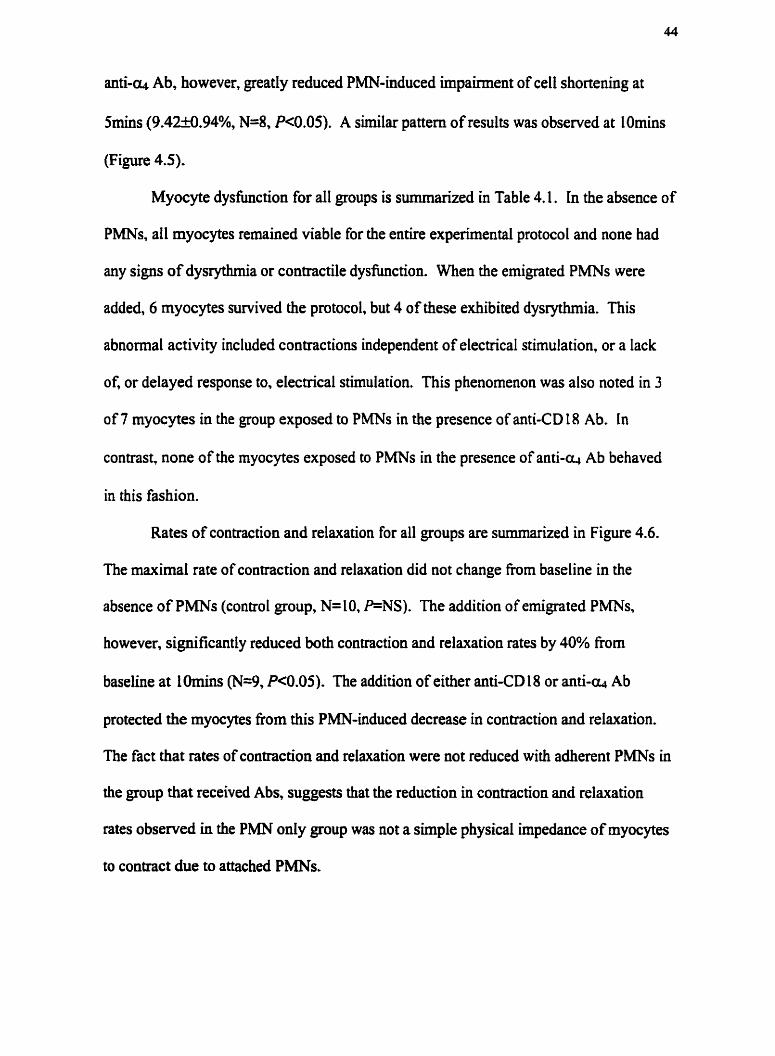
Myocyte dysfunction for all groups is summarized in Table 4.1. In the absence of
PMNs, all myocytes remained viable for the entire experimental protocol and none had
any signs of dysrythmia or contractile dysfunction. When the emigrated PMNs were
added, 6 myocytes survived the protocol, but 4 of these exhibited dysrythmia. This
abnormal activity included contractions independent of electrical stimulation, or a lack
of, or delayed response to, electrical stimulation. This phenomenon was also noted in 3
of 7 myocytes in the group exposed to PMNs in the presence of anti-CD 18 Ab. In
contrast, none of the myocytes exposed to PMNs in the presence of anti-Q Ab behaved
in this fashion.
Rates of contraction and relaxation for all groups are summarized in Figure 4.6.
The maximal rate of coatmction and relaxation did not change fiom baseline in the
absence of PMNs (control group, N=10, P=NS). The addition of emigrated PMNs.
however, significantly reduced both contraction and relaxation rates by 40% from
baseline at I Omins (N=9, P<0.05). The addition of either anti-CD 18 or anti-- Ab
protected the myocytes fiom this PMN-induced decrease in contraction and relaxation.
The fact that rates of contraction and relaxation were not reduced with adherent PMNs in
the group that received Abs, suggests that the reduction in contraction and relaxation
rates observed in the PMN only group was not a simple physical impedance of myocytes
to contract due to attached PMNs.

Circulating M u ~ e PMNs Injure Cardiac Myocytes via CD18.
Further experiments with circulating cells showed these cells could reduce
myocyte cell shortening by 35% from baseline at 5 and lOmins (N=2) (myocytes with 1
or 2 adherent PMNs) (Figure 4.7). Furthermore, the addition of anti-CD 1 8 Ab protected
the myocyte (N=2), primarily through inhibition of PMN adhesion (all experiments in the
presence of anti-CD 18 Ab showed myocytes with no adherent PMNs). Rates of
contraction and relaxation for circulating PMNs are shown in Figure 4.8.
4.2 DISCUSSION
Previous work from our laboratory has shown that both CD 18 and Q-integrin
were essential for emigrated PMN adherence to rat ventricular myocytes 33, and we have
now shown the same adhesion profile in the murine myocardium. The present results
extend this work and, for the first time, suggest that engagement of the a-integrin is
critical for the ensuing myocyte damage. In our study, the anti-CD 1 8 Ab was able to
protect the myocyte From damage to mechanisms controlling contraction and relaxation
rates, but was not able to protect against decreased cell shortening, myocyte dysrythmia,
or contractwe. This suggests that eintegrin, not CD18, is the dominate molecule in
PMN-induced myocyte dysfunction. These observations complement and significantly
extend previous studies wherein pretreatment with anti-CD 18 Ab prevented PMN
recruitment into tissues 4: In; 178 . Clinically, patients arrive at hospital after, not before, an
infarct at which point PMNs have already infiltrated the myocardium. Our data suggest
that one could therapeutically target the emigrated PMN to prevent ongoing myocardial
injury. Perhaps, both CDL8 and a-integrin pathways need to be inhibited to completely

prevent PMN-dependent injury in these pathophysiological states wherein the
endothelium is injured by circulating PMNs and myocytes are injured by emigrated
PMNs.
Previous reports have described the ability of integrins to receive signals from
outside the cell that can, in turn, signal the release of cytotoxic mediators from within the
cell I". Indeed, engagement of CD 18 on PMNs leads to reorganization of the
cytoskeleton, oscillating cystolic free ca2+ levels, shape change, and subsequent secretion
of granule proteins and oxidants 180: 181 . This type of outside-in signaling can also be
mediated by the m-integrin. Signal transduction through the m-integrin activates protein
tyrosine kinase activity in T cells '", and engagement of this fibronectin receptor induces
gene expression of enzymes, including collagenase and metalloproteinase stromelysin in
fibroblasts la'. The second messenger pathways regulated by the Q-integrin in PMNs
have yet to be explored. Since both adhesion pathways are involved in emigrated PMN
adhesion to myocytes, one might expect that inhibition of either CD L 8 or a-integrin
would lead to protection. Our study would suggest that adherence of emigrated PMNs
only minimally activated a CD 18-dependent pathway of injury.
CD 18 is upregulated on the PMN in response to stimulants in the vasculature,
which allows for firm adhesion to the endothelium and subsequent emigration '". Since
the CD 18 integrin has already been exposed to a stimulus prior to emigration, it has
already engaged its ligand It is conceivable that CD 18 can no longer respond to a
stimulus after emigration and therefore is much Iess effective in initiating the release of
specific cytotoxins h m the PMN. In this study, and in previous studies, the a-integrin
is expressed at onIy very low levels on circuIathg PMNs, and this expression level is

33; 185 increased following emigration and stimulation . Thereafter, a-integrin is ready to
engage its receptor and signaling via this ligand may be possible. The binding of the cl~-
integrin to its ligand on the myocyte may cause a release of proteases and oxidants from
the PMN, which can directly degrade the extracellular matrix. This may lead to changes
in membrane potential or integrity, thereby affecting the availability of cystolic ca2+ and
thus decrease the magnitude of cell shortening.
It is intriguing that the PMNs appear to injure myocytes in a time and site
specific manner. This is evidenced by the fact that global dysfunction did not occur in
individual myocytes at the same time periods. Although we observed a very profound
decrease in cell shortening at Smins after PMN exposure with no Ab, we did not see a
change in rate of contraction or relaxation until i0mins. These results suggest that the
emigrated PMN was able to reduce cell shortening, before impacting upon contraction or
relaxation mechanisms. It is well appreciated that the degree of contraction. the rate of
contraction, and the rate of relaxation are all mediated by different ionic events. This
raises the possibility that the myriad of molecules released by the PMN impacts on ion
channels through phosphorylation of proteins with differing degrees of efficiency.
Furthermore, it is possible that the PMN-induced damage is initially restricted to the
sarcolemma, affecting L-type ca2+ channels, which trigger contraction by initiating a
much larger ca2+ release from the SR. PMNs may subsequently cause membrane
depolarization, which could further reduce ca2+ influx via L-type ca2' channels. At later
times, intracellular organelles essential for excitation-contraction coupling and ca2+
homeostasis may be compromised. A decrease in ca2'-induced ca2' release fiom the SR

results in a decreased rate of contraction. Moreover, the ca2+ pump in the SR and the
rate of relaxation may also be significantly affected.
Our results demonstrate that unlike circulating PMNs, emigrated PMNs use a-
integrin to mediate the myocyte damage induced by PMNs. To date most studies have
focussed on the mechanisms by which PMNs adhere to the endothelium and infiltrate the
myocardium, with the goal of targeting this mechanism to reduce the injury associated
with pathophysiological conditions like myocardial infarction. The time window of
opportunity to intervene in the recruitment process may be so brief however, that therapy
may only work prophylactically (i.e. patients already have PMNs in the myocardium
upon amval at hospital). Our results provide a novel basis for therapeutic intervention on
the PMN that one may target even after this leukocyte has reached the myocardium. This
approach has the potential to reduce or prevent myocyte dysfunction without affecting
PMN hnction in the circulation.
4 3 LIMITATIONS
The adhesion chamber data from the previous chapter was obtained using a static
assay. PMNs were added to the chambers and allowed to settle by gravity onto the
myocytes. in contrast, the cell shortening assay is not a static model. The PMNs were
superfused over the myocytes and then subsequently washed away with buffer.
Furthermore, we used sx l o6 PMN/ml in the adhesion assay, and only 1 x 1 o6 PMNs/mi in
the cell shortening assay to limit the number of mice used per study. This difference in
PMN concentration explains why more PMNs adhered per myocyte in the adhesion assay
compared to the cell shortening assay.

Moreover, it was very difficult to find PMNs adherent to the myocytes in the cell
shortening assay when anti-= Ab was present. Data in the previous chapter clearly
showed that both anti-CD 18 and -a Abs were required to inhibit adhesion in the static
model. In the presence of anti-& Ab, PMNs adhere via a CD 18-[CAM- 1 pathway. It has
been shown that the adhesion of leukocytes to VCAM-I coated cover slips increased at
lower shear force (minimal adhesion at 20dynes/cm2 and maximal adhesion at
5dynes/cm2) '16. It is not possible to calculate shear forces in our cell shortening model,
but it is apparent that there is more shear stress in the cell shortening model where
myocytes are subjected to buffer flow rates of 1 d m i n as compared to the static state in
the adhesion chambers. It is possible that this same effect is seen with CD 18 and
ICAM- I .

Baseline
Figure 4.1 Representative cell shortening trace of a control cardiac myocyte before and after isoproterenol challenge.

Figure 4.2 Representative cell shortening trace of a cardiac myocyte + emigrated PMN at 5 and 1 Omins.

No Adherent PMNsJMyocyte
-60
Smin 1Omln
B) CDf 8 Ab
60
Smin
10 mln
Adherent PMNsJMyocyte
60
-60
Smin 1 Omin
Smin
Smin
Figure 4.3 Unloaded cell shortening expressed in terms of whether or not there were adherent PMNs for A) PMNs only. B) CD18 Ab (PMNs + anti-CD18 Ab 2€6,8ug/ml), and C) Alpha4 Ab
(PMNs + anti-alpha4 Ab R1-2.10uglml)

Control PMN CD18Ab Alpha4Ab (RI -2)
Figure 4.4 Unloaded cell shortening in control (no PMNs) N=10, PMN (PMN only) N=9, CD18 Ab (PMN + anti- Cot's Ab 2E6,bghl) N=9, and Alpha4 Ab (PMN + anti-alpha4 Ab R1-2, IOug/ml) N=8 at Smins. '+' Pc0.05

Control PMN CD18 Ab Alpha4 Ab (2E6) (R1-2)
Figure 4.5 Unloaded cell shortening in Contml (no PMN) N=10, PMN (PMN only) N=9, CD18 Ab (PMN + anti-CD18 Ab 2E6,8ug/ml) N=9, and Alpha4 Ab (PMN + anti-aplha4 Ab R1-2,t Oug/ml) N=8 at
IOmins. '+' "P<O-05

baseline
Control PMN CD18 Ab Alpha4 (2E6) Ab(R1-2)
baseline
T .1 Omin
CE Control PMN CD18 AB Alpha4 Ab (R1-2)
Figure 4.6 Change in rate of contraction and relaxation in Control (no PMNs) N-10, PMN (PMN only) N=9, CD18 Ab (PMNs + anti-CD18 A 6 2€6,8ug/ml) N=9, and Alpha4 Ab (PMNs +
anti-alpha4 Ab R1-2, 1 Oug/ml) N=8. '+' Pc0.05

Control PMN CD18 Ab (2E6)
Figure 4.7 Unloaded cell shortening with circulating PMNs in Control (no PMNs) N=2, PMN (PMN only) N=2, and CD18 Ab (PMNs + antLCD1 8 Ab 2E6,8ugm/rnl) N=2. Each experiment was completed on 2
seperate days

Control PMN
baseline H 1 Omin
baseline .1 Omin
400 aa
Figure 4.8 Change in rates of contraction and relaxation with circulating PMNs in Control (no PMNs) N=2, PMN (PMN only) N=2, and CD18 Ab (PMN + anti-CDI 8 Ab 2E6,8ug/ml) N=2.
Each experiment was completed on 2 seperate days
350 3 300 . C
L
- 5 250 -
- D m
+
- 'i;i 200 - x 1 150 -4
I00 - - 0 Q) 4-
50 - E O - --
Control PMN CD18 Ab (2E6)
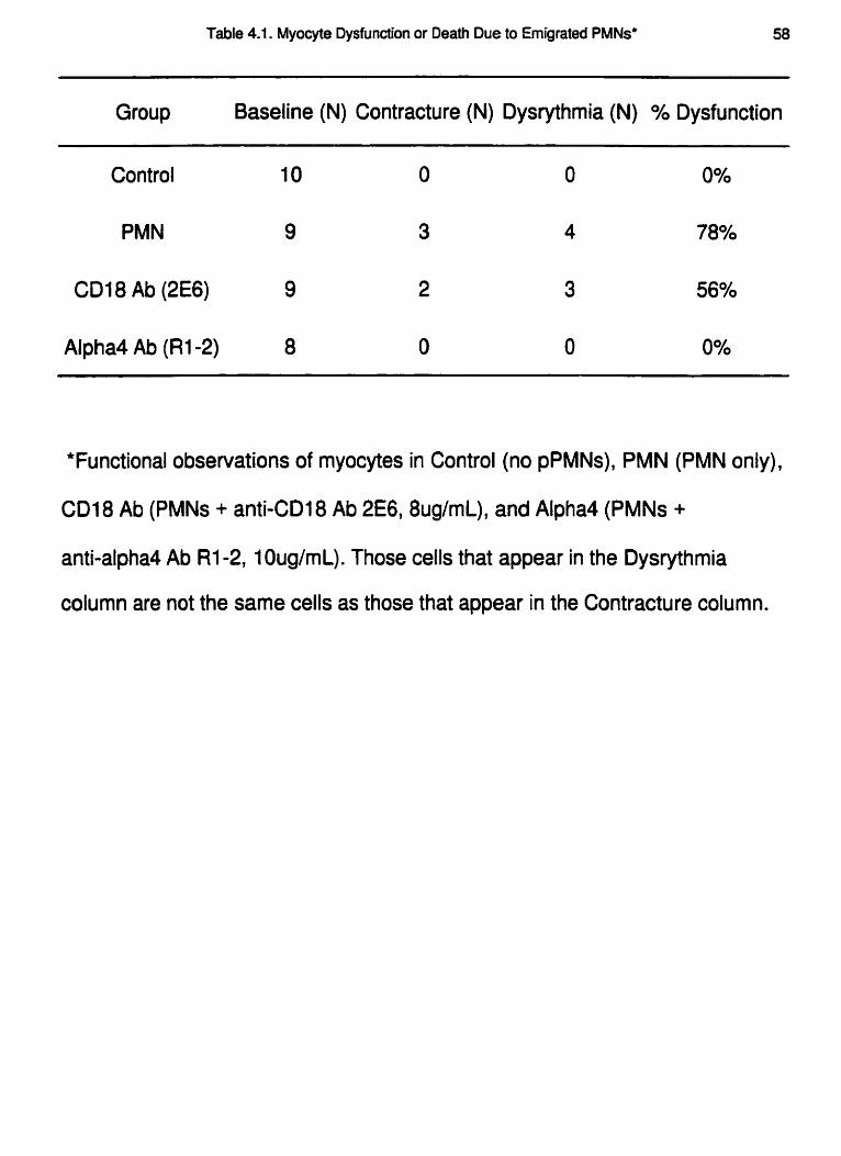
Table 4.1. Myocyte Dysfunction or Death Due to Emigrated PMNs* 58
Group Baseline (N) Contracture (N) Dysrythmia (N) % Dysfunction
Control 10 0 0 0%
PMN 9 3 4 78%
CD18 Ab (2E6) 9 2 3 56%
Alpha4 Ab (RI-2) 8 0 0 0%
*Functional observations of myocytes in Control (no pPMNs), PMN (PMN only),
CD18 Ab (PMNs + anti-CD18 Ab 2E6,8ug/mL), and Alpha4 (PMNs +
anti-alpha4 Ab R1-2, 1 OuglmL). Those cells that appear in the Dysrythmia
column are not the same cells as those that appear in the Contracture column.

CHAPTER 5
a-INTEGRIN MODULATES FREE RADICAL INJURY TO CARDIAC
MYOCYTES

Hmothesis: PMN-induced myocyte injury is caused by the generation of free radicals.
Objectives:
1) To determine if PMNs require a respiratory burst to cause myocyte damage.
2) To visualize the production of free radicals upon PMN adhesion to the
myocyte.
3) To determine whether free radical generation is mediated through either CD 18
4) To determine if the specific free radical responsible for myocyte injury is OF.
5.1 RESULTS
PMN-Derived Free Radical Injury to Cardiac Myocytes.
Figure 5.1 shows unloaded cell shortening (as a % of baseline) for all groups. Cell
shortening of myocytes alone did not change after Smins of electrical stimulation (N=6).
The addition of WT PMNs caused an approximately 40% reduction in cell shortening
within Srnins of PMN exposure (N=7, P<O.OS). PMNs deficient in NADPH oxidase (the
enzyme necessary to generate free radicals) were unable to cause a decrease in the
inotropic response of the myocytes (cell shortening levels at 96.8f 8.9% of baseline at
Srnins, N=6, PK0.05 compared to PMN only group). A similar pattern was observed
after 1 Omins of exposure to these PMNs (Figure 5.2).
Myocyte dysfunction is shown in Table 5.1. Control myocytes alone showed no
myocyte dysrythmia, but the addition of WT PMNs caused 5 of the 7 myocytes recorded
to become dysrythmic. When PMNs fiom NADPH oxidase deficient mice were added to
the myocytes, only I of the 6 myocytes recorded showed any dysrythmia.
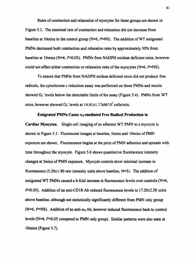
Rates of contraction and relaxation of myocytes for these groups are shown in
Figure 5.3. The maximal rate of contraction and relaxation did not decrease fiom
baseline at IOmins in the control group (N=6, P=NS). The addition of WT emigrated
PMNs decreased both contraction and relaxation rates by approximately 30% from
baseline at LOmins (N=6, R0.05). PMNs fiom NADPH oxidase deficient mice, however
could not affect either contraction or relaxation rates of the myocytes (N=6, P=NS).
To ensure that PMNs fiom NADPH oxidase deficient mice did not produce free
radicals, the cytochrome c reduction assay was performed on these PMNs and results
showed Of levels below the detectable limits of the assay (Figure 5.4). PMNs from WT
mice, however showed 0$ levels at 14.6 1~1.17n~/10~cells/min.
Emigrated PMNs Cause q-mediated Free Radical Production in
Cardiac Myocytes. Single cell imaging of an adherent WT PMN to a myocyte is
shown in Figure 5.5. Fluorescent images at baseline, Smins and lOmins of PMN
exposure are shown. Fluorescence begins at the point of PMN adhesion and spreads with
time throughout the myocyte. Figure 5.6 shows quantitative fluorescence intensity
changes at Smins of PMN exposure. Myocyte controls show minimal increase in
fluorescence (5.2Ok 1.80 raw intensity units above baseline, N=5). The addition of
emigrated WT PMNs caused a 6-fold increase in fluorescence levels over controls (N=6,
RO.05). Addition of an anti-CD 18 Ab reduced fluorescence levels to 1 720&2.50 units
above baseline, although not statistically significantly different fiom PMN only group
(N=6, FNS). Addition of an anti-- Ab, however reduced fluorescence back to control
levels (N4, R0.05 compared to PMN only group). Similar patterns were also seen at
lOmins (Figure 5.7).

Extracellular, but not Intracellular SOD Protects Myocytes From PMN-
Induced Injury. Unloaded cell shortening using myocytes isolated from SOD over-
expressing mice is shown in Figure 5.8. There was no difference in cell shortening
between WT and SOD myocytes alone (N=4, PNS) . The addition of WT emigrated
PMNs caused a reduction in cell shortening in both these rnyocyte groups. Cell
shortening decreased by approximately 30% in both the WT myocyte group (N=4,
Pc0.05 compared to WT control), and in the SOD myocyte group (N=5, P<0.05
compared to SOD control), at Smins of PMN exposure. A similar pattern was observed
for both WT and SOD myocytes at lOmins of PMN exposure (Figure 5.9). There was no
dysrythmia recorded for WT or SOD myocytes alone (Table 5.2). The addition of WT
PMNs caused dysrythmia in 3 of the 4 WT myocytes, but only 1 of the 5 SOD myocytes.
Rates of contraction and relaxation of SOD and WT myocytes are shown in
Figure 5.10. Myocytes alone, from either WT or SOD mice, were able to maintain rates
of contraction and relaxation at baseline levels throughout the 1 Omin experimental period
(N=4, P=NS). The addition of WT PMNs caused a 55% reduction in contraction rate in
both WT and SOD myocytes, and a 57% and 46% reduction in relaxation rates in WT
and SOD myocytes, respectively (N=4 for WT PMN group and N=5 for SOD Myocyte +
WT PMN group, P<O*OS).
Unloaded cell shortening with exogenous SOD treatment is shown in Figure 5.1 1.
Control myocytes (no PMNs) maintained cell shortening at baseline levels after 5 min
(N=4). The addition of PMNs caused a 45% reduction in cell shortening (N=4, P<0.05).
The addition of SOD to the PMNs protected the myocytes fiom injury (cell shortening at
96.7652.79% of baseline at Srnins, N=3, PQ).OS compared to PMN only group). A
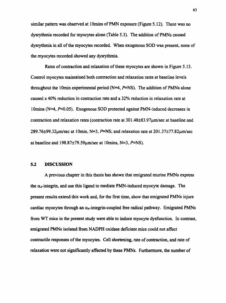
similar pattern was observed at lOmins of PMN exposure (Figure 5.12). There was no
dysrythrma recorded for myocytes alone (Table 5.3). The addition of PMNs caused
dysrythmia in all of the myocytes recorded. When exogenous SOD was present, none of
the myocytes recorded showed any dysrythrma.
Rates of contraction and relaxation of these myocytes are shown in Figure 5.13.
Control myocytes maintained both contraction and relaxation rates at baseline levels
throughout the lOmin experimental period (N=4, P=;NS). The addition of PMNs alone
caused a 40% reduction in contraction rate and a 32% reduction in relaxation rate at
lOmins (N=4, P<0.05). Exogenous SOD protected against PMN-induced decreases in
contraction and relaxation rates (contraction rate at 30 1.48f 83.97pdsec at baseline and
289.76k99.32pdsec at LOmin. N=3, P=NS; and relaxation rate at 20 1.37k77.82pdsec
at baseline and 198.87+79.59p/sec at l0mins. N=3, P=NS).
5.2 DISCUSSION
A previous chapter in this thesis has shown that emigrated murine PMNs express
the a-integrin, and use this ligand to mediate PMN-induced myocyte damage. The
present results extend this work and, for the first time, show that emigrated PMNs injure
cardiac myocytes through an a-integrin-coupled free radical pathway. Emigrated PMNs
from WT mice in the present study were able to induce myocyte dysfimction. In contrast,
emigrated PMNs isolated fiom NADPH oxidase deficient mice could not affect
contractile responses of the myocytes. Cell shortening, rate of contraction, and rate of
relaxation were not significantly affected by these PMNs. Furthermore, the number of

myocytes that displayed signs of dysrythmia was dramatically reduced when the PMNs
could not generate free radicals.
A previous study has shown the importance of free radical generation in the
ability of circulating PMNs to damage cardiac myocytes 25, suggesting that circulating
and emigrated PMNs use the same process to cause PMN-induced myocyte injury. Our
singie cell imaging measurements show that the adhesion of emigrated PMNs to isolated
cardiac myocytes caused a dramatic rise in the level of oxidants produced within the
myocyte. This observation is in direct agreement with the proposed role of free radicals
in circulating PMN-induced myocyte damage. The previous study showed that free
radical generation by circulating PMNs was CD 18 dependent ?5. Our work demonstrates,
for the first time, that the a-integrin was coupled to free radical production by emigrated
PMNs. The addition of an anti-as Ab inhibited oxidant production. In fact, oxidant
levels were at rnyocyte only control levels even though PMNs continued to adhere to the
myocyte via CD 18. These fmdings also suggest that as the P M N emigrates out of the
vasculature, the CD 18-NADPH oxidase becomes uncoupled or now requires the Q-
integrin. Immunosuppression of the a-integrin inhibited all parameters of emigrated
PMN-induced myocyte dysfunction, almost certainly by inhibiting oxidative stress.
These results provide important new evidence for the fimctional role of the a-integrin in
PMN-dependent myocyte injury.
It is well documented that integrins are involved in cell signaling events. Current
models postulate that when ligands are engaged, a multitude of different b a s e s bound to
the cytoplasmic tail of integrins, can initiate signaling cascades within the cell 18'.
Signaling through the ors-integrin resulted in protein b a s e C (PKC) activation in murine

T cells IB8, an event known to phosphorylate a key protein (p47ph") in the NADPH
oxidase complex. Indeed, p47ph0x was phosphorylated by purified PKC in a cell-free
system 189-191 . p47Ph0x is a cytosolic protein with a Src homology 3 (SH3) domain
structural motif involved in specific molecular interactions during signal transduction 192.
p47ph0x is critical for oxidase activation in intact cells, and the SH3 domain has been
implicated in the assembly and maintenance of the NADPH oxidase components in
PMNs Ig3. We hypothesize that the adhesion of emigrated PMNs to cardiac myocytes
causes PKC activation within the PMN and subsequent p47ph" phosphorylation. The
multi-component NADPH oxidase within the PMN is then assembled for activation, and
fiee radicals can be produced by the PMN. It is possible that this signaling pathway
could also be important for the ongoing activation of the NADPH oxidase complex.
Although unproven, theoretically this signaling mechanism could be playing a role in the
m-integrin dependent fiee radical generation observed in our model.
The addition of an anti-CD 18 Ab had only a weak inhibitory effect on oxidant
production in myocytes (not significant). These data are consistent with results in a
previous chapter where anti-CD 18 Ab only partially protected the myocyte from
emigrated PMNs by inhibiting the decrease in the rate of contraction and relaxation at
lOmins of PMN exposure. The inability of the anti-CD 18 Ab to affect the decrease in
cell shortening or dysrythmia could be related to the ability of this Ab to only partially
inhibit oxidant generation. It is feasible that a minimal level of extracelluIar fiee radicals
is necessary to affect ion channels (Like K+ or Na+ channels) on the myocyte surface and
thus indirectly affect ca2+-handling within the myocyte. These alterations may then
affect the rate of contraction and relaxation. Damage to mechanisms that control

myocyte contractility, like L-type caZ+ channels in the myocyte plasma membrane, may
require a lower level of fiee radical generation. The ability of extracellular and not
intracellular SOD to protect against PMN-induced injury implies that the PMN is causing
the injury fiom the outside of the myocyte and that the production of oxidants within the
myocyte is not the critical pathway of myocyte injury.
Although oxidant levels were measured, it is uncenain which molecule had the
predominant role in emigrated PMN-induced free radical injury. Over-expression of
endogenous Cu/Zn SOD in the myocytes was not able to protect the cell from emigrated
PMN-induced decreases in cell shortening, rates of contraction or relaxation, but was able
to inhibit the number of rnyocytes that were dysrythmic. It is possible that scavenging
Oi was only protective to the resting potential of the myocyte, enabling it to become
properly depolarized upon electrical stimulation. The data suggest that the free radical
predominantly responsible for the decrease in myocyte contractile properties is not
intmcellular 0;. The addition of exogenous SOD, however resulted in protection
against all parameters of myocyte dyshction induced by the PMNs. This data implies
that the P M N is releasing OF upon adhesion to the cardiac myocyte and that this O< then
causes detrimental changes to the ionic properties of the myocyte fiom the outside of the
cell.
We can conclude fiom the present study that emigrated PMN-induced myocyte
injury is an Of dependent event. The exact mechanism of fiee radical injury however,
remains unknown. It is conceivable that Oycaused direct damage to the myocyte, or
alternatively, 0< may have reacted with NO to produce ONOO-. Finally, OF may have
entered the Fenton reaction to form OK, MI of these fiee radical intermediates have the

potential to cause the myocyte dyshction observed in this thesis. Furthermore, we
cannot exclude the involvement of proteases in this PMN-induced myocyte dysfimction.
It is well recognized that fiee radicals can activate certain proteases by inactivating
proteiuase inhibitors 194,195 , and it has been proposed that proteases play an important
role in myocardial injury in myocardial infarction 196- 198
Our results demonstrate that emigrated PMNs, like circulating PMNs, use fiee
radicals to injure cardiac myocytes. However, the generation of Free radicals in myocytes
induced by emigrated PMNs is coupled in a more dominant manner by the a-integrin,
not by CD 18. This is an important finding as to date most studies I reference have
concentrated on the study of circulating PMN-myocyte interactions and have shown that
immunosuppression of CD 18 could protect the myocyte from free radical-induced injury.
Previous studies have not considered the potential role of the ~in tegr in . mainly because
the a-integrin was not thought to be expressed by PMNs. The interaction of circulating
PMNs and cardiac myocytes can never occur in the physiological or pathophysiological
state. PMNs must first leave the vasculature and these emigrated PMNs express a
different adhesion molecule profile, including the expression of the Q-integrin 33-35. ow
data show that immunosuppression of the m-integrin limits both the generation of fiee
radicals and the subsequent PMN-induced myocyte dysf'unction, and provides a rational
basis for potential therapies that could benefit patients suffering from pathophysiological
conditions like myocardial infarction.
53 LiMITATIONS
Since the fluorescent probe DCFH is not specific for 0; we could not confirm

that Of levels in the myocytes were indeed decreased when PMNs were added to the
SOD over-expressing myocytes. Although these myocytes have a t 0-fold increase in
endogenous Cu/Zn SOD 160, ideally we should conf'iirm that O< levels were indeed
affected.

Control WT PMN NADPH oxidase KO PMN
Figure 5.1 Unloaded cell shortening in Control (no PMNs) N=6, WT PMN (N=7), and NADPH oxidase KO PMN (N=6) at Smins. '+' Pe0.05

Control WT PMN NADPH oxidase KO PMN
Figure 5.2 Unloaded cell shortening in Control (no PMNs) N=6, WT PMN (N=7), NADPH oxidase KO PMN (N=6) at 10mins. '+' P9.05

baseline
1 Omin
a Control WT PMN NADPH oxidase KO
PMN
baseline
1 Omin
Control WT PMN NADPH oxidase KO
PMN
Figure 5.3 Change in rate of contraction and relaxation at lOmins in Control (no PMNs) N=6, WT PMN (N=7), and NADPH oxidase KO PMN (N=6). '+' Pc0.05

Figure 5.4 0* production in emigrated PMNs from WT (N=4) and NADPH oxidase KO (N=2) mice
Below Detection
r
NADPH oxidase KO

Figure 5.5 Representative fluorescence images of an adherent WT PMN to a WT myocyte. A) phase contrast photo (PMN marked wlfh arrow), 0) fluorescence image immediately following adherence of PMN to the myocyle (baseline], C) at 5mim, D) at 1 Omins, and E) color bar.

Control PMN CD18 Ab Alpha4 Ab w e ) (R1-2)
Figure 5.6 Change in fluorescence intensity for Control (no PMNs) N=5, PMN (PMN only) N=6, CDt 8 Ab (PMNs + anti-CD18 Ab 2E6,8ug/rnl) N=6, and Alpha4 Ab (PMNs + anti-alpha4 Ab R1-2,1 Ouglml)
N=4 at Srnins. '+' Pc0.05

Control PMN
Figure 5.7 Change in fluorescence intensity for Control (no PMNs) N=S, PMN (PMN only) N=6, CD18 Ab (PMNs + antCCD18 Ab 2€6,8ug/mi) N=6, and Alpha4 Ab (PMNs + anti-alpha4 Ab R1-2,1Oug/ml)
N=4 at 1 Omins. '+' Pc0.05

WT Control WT PMN SOD SOD Control Myocyte+
WT PMN
Figure 5.8 Unloaded cell shortening for WT Control (no PMNs) N 4 , WT PMN (WT myocyte + WT PMN) N=4, SOD Control (no PMNs) N 4 , and SOD Myocyte + WT PMN (N=5) at Srnins. '+' P<0.05

WT Control WT PMN SOD SOD Control Myocyte +
WT PMN
Figure 5.9 Unloaded cell shortening for WT Control (no PMNs) N=4, WT PMN (WT rnyocyte + WT PMN) N=4, SOD Control (no PMNs) N=4. and SOD Myocyte + WT PMN (N=5) at l0mins. '+' Pc0.05

baseline
WT Control
WTPMN SOD Control
SOD Myocyte
+ W T PMN
baseline H 1 Omin
WT Control
WTPMN SOD Control
SOD Myocyte
+WT PMN
Figure 5.1 0 Change in rate of contraction and relaxation in WT Control (no PMNs) N=4, WT PMN (WT myocyte + WT PMN) N 4 , SOD Control (no PMNs) N=4, and SOD Myocyte + WT
PMN (N=S) at l0mins. '+' Pe0.05

Control PMN SOD treatment
Figure 5.1 1 Unloaded cell shortening in Control (no PMNs) N4 , PMN (PMN only) N=4, and SOD treatment (PMNs + SOD, 300Uirnl) N=3 at Smins. '+' P<O.O5
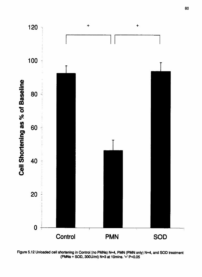
Figure
0 L
5.1 2 Unloaded
Control PMN SOD
cell shortening in Control (no PMNs) N 4 , PMN (PMN only) N 4 , and SOD treatment (PMNs + SOD, 300U/ml) N=3 at IOmins. '+' Pe0.05

Control PMN SOD treatment
Control PMN SOD treatment
baseline .1 Omin
baseline H I 0 min
Figure 5.1 3 Change in rate of contraction and relaxation in Control (no PMNs) N=4, PMN (PMN only) N=4, and SOD treatment (PMNs + SOD, 300Ulml) N=3 at 1Omins. '+' Pe0.05

Table 5.1. Myocyte Dysfunction Due to Emigrated PMNs* 82
- -
Group Baseline (N) Dysrythmia (N) % Dysfunction
Control 6
WT PMN 7
NADPH oxidase KO PMN 6
*Functional observations of myocytes in Control (no PMNs), WT PMN,
and NADPH oxidase KO PMN

Table 5.2 Myocyte Dysfunction Due to Emigrated PMNs* 83
Group Baseline (N) Dysrythmia (N) % Dysfunction
WT Control
WT PMN
SOD Control
SOD Mvocvte + WT PMN 5 1 20%
*Functional observations of myocytes in WT Control, WT PMN, SOD Control,
and SOD Myocyte + WT PMN.

Table 5.3. Myocyte Dysfunction Due to Emigrated PMNs* 84
Group Baseline(N) Dysrythrnia(N) %Dysfunction
Control 4
PMN 4
SOD treatment 3
'Functional observations of rnyocytes in Control (no PMNs), PMN (PMN only),
and SOD treatment (PMNs + SOD, 300Ulrnl).

CHAPTER 6
SUMMARY AND CONCLUSIONS

Only recently has the study of the a-kitegrin in PMN adhesion been investigated.
Since the first discovery of increased levels of this ligand on transmigrated human PMNs
in 1995 34, interest in d e p e n d e n t PMN adhesion has slowly increased. Although
information remains limited, Reinhart et a1 have previously shown that rat PMNs also
express the a-integrin after emigration 33. PMNs isolated from the circulation adhered
to cardiac myocytes via the P2-integrin CD 18, but once they emigrated, the PMNs
utilized the a-integrin, in conjunction with CD 18, to fvmly adhere. Prior to the series of
experiments in this thesis, it was unclear if w-integrin expression on PMNs occurred in
mice, and what biological significance could be attributed to this process in any species.
The work presented in the fust part of this thesis was conducted to determine if
the Q-integrin also played a role in P M N adhesion in the murine system. We found that
murine PMNs isolated from the cuctdation had very low surface expression of m-
integrin, and this level increased after emigration. Moreover, as was the case in the rat
system, murine circulating PMNs adhered avidly to cardiac myocytes via CD 1 8.
Following emigration however, the PMNs adhered via both CD 18 and Q-integrin.
This thesis was the first to show that upon adhesion, emigrated murine PMNs can
impair contractile responses of electrically stimulated cardiac myocytes through a
predominantly ~integrin-dependent pathway. We found that emigrated PMNs require a
respiratory burst to induce contractile impairment in cardiac myocytes and that the free
radical involved is most likely PMN-derived 02: Most importantly, we are the first to
show that emigrated PMN-induced oxidative injury to cardiac myocytes is coupled to the
engagement of the a-integrin.

Adhesion of PMNs to cardiac myocytes is necessary for PMN-induced injury
since myocytes in the presence of emigrated PMNs did not show impairment unless at
least one PMN was adherent to the myocyte. It is also interesting that only one PMN was
necessary to induce injury, highlighting the need for absolute inhibition of PMN adhesion
to limit myocyte injury. Previous studies have focussed on CD18 as the adhesion
molecule of interest in hopes of inhibiting circulating PMN-myocyte interactions,
however a circulating PMN will never interact with a cardiac myocyte. Only recently has
the importance of emigration in PMN-myocyte interactions been addressed j3. The
discovery of a new adhesion molecule on emigrated PMNs will prove vital to the design
of effective therapies for PMN-induced pathologies of the heart.
Although emigrated PMNs use both CD 18 and the as-integrin to adhere to
cardiac myocytes, immunosuppression of the a-integrin protected myocytes from
emigrated PMN-induced injury. Our data suggest that therapies involving CD 18
inhibition would only be effective at limiting PMN recruitment in the vasculature.
Indeed, if one could anticipate the onset of an inflammatory state, anti-CD 18 therapies
may prove effective. However, after PMNs have already infiltrated the myocardium anti-
CD 18 therapies will no longer protect against additional myocardial injury.
It appears that a PMN, either from the circulation or after emigration, will use the
same mechanism to injure cardiac myocytes. Although a PMN can produce a multitude
of toxins, it induces myocyte injury through a free radical-dependent pathway.
Emigrated PMNs that are deficient in NADPH oxidase, and thus unable to mount a
respiratory burst, cannot injure myocytes. Furthermore, through fluorescence
microscopy, we observed an elevated IeveI of oxidants in the myocyte within Smins of

PMN adhesion. Lnununosuppression of the m-integrin alone inhibited the oxidative
stress within the myocyte following adhesion of the emigrated PMN, while an anti-CD 18
Ab did not statistically reduce the free radical levels observed in the myocyte. These are
the fust data to imply a role for the a-integrin in controlling the production or release of
free radicals by the emigrated PMN, further emphasizing the vital role of the Q-integrin
in PMN-myocyte interactions.
There is extensive information on the destructive effects of free radicals on many
cell types, including cardiac myocytes. Exogenous fiee radicals affect ca2' handling, ion
channel fbnction, and cell viability. It remains unclear, based upon the data from this
thesis, which exact mechanism of injury was responsible for the impairment to myocyte
contractility observed. Additional studies involving simultaneous fluorescence
measurements of intracellular ~ a " and electrophysiologicd response of single cardiac
myocytes should be conducted in the presence of adherent emigrated PMNs to address
this question.
Our data suggest that the fiee radical predominantly responsible for the decrease
in myocyte contractile properties caused by emigrated PMNs is the 0;. Over-expression
of endogenous 0; scavenger SOD was unable to protect the myocyte from the emigrated
PMN, but exogenous SOD protected the myocytes fiom all of the PMN-induced injury
observed with PMNs alone. The data show that the emigrated PMNs injure the myocytes
fiom the outside of the cell, possibly altering ion channel activity or membrane integrity
which in turn results in myocyte dysfimction.
The importance of PMNs in myocardial pathologies like IR has been well
documented Since adhesion is critical to the ensuing injury, the development of anti-

adhesive therapies has been proposed as a strategy to limit myocardial injury. Before an
effective intervention can be developed however, we must have a complete understanding
of the adhesive properties of the PMN. Although CD 18 is important in PMN-
endothelium interactions, the study herein shows that we must acknowledge that the m-
integrin plays a vital role in emigrated PMN-myocyte interactions, and thus effective
therapies for myocardial VR must consider both of these ligands.

BIBLIOGRAPHY
Hakkert BC, Kuijpers TW, Leeuwengerg JFM, van M o d JA, Roos D: Neutrophil
and rnoaocyte adherence to and migration across monolayers of cytokine-
activated endothelial cells: the coatribution of CD 18, ELAM- 1 and VLA-4.
Blood 199 1;78:272 1-2726
Springer TA: Traffic signals for lymphocyte recirculation and leukocyte emigration:
the multistep paradigm. Cell 1994;76:301-3 14
Kubes P, Suzuki M, Granger DN: Nitric oxide: an endogenous modulator of
leukocyte adhesion. Proc Natl Acad Sci 199 1 ;88:465 1-4655
A!belda SM, Smith CW, Ward P: Adhesion molecules and inflammatory injury.
FASEB J 1 994; 8504-5 1 2
Balligand JL, Ungureanu-Longrois D, Simmons WW. Pimental D, Malinski TA,
Kapturczak M, Taha 2, Lowenstein CJ, Davidoff AJ, Kelly RA, Smith TW.
Michel T: Cytokine-inducible nitric oxide synthase (NOS) expression in
cardiac myocytes (characterization and regulation of I N S expression and
detection of iNOS activity in single cardiac myocytes in vitro). J Biof Chem
1994;269:27580-27588
Zhen Y , Quamme GA, McNeill JH: Depressed [CaZ+Ii responses to isoproterenol
and CAMP in isolated cardiomyocytes from experimental diabetic rats. Am
J Physiol1994;266:H2334-H2342
Goldhaber JI, Kim KH, Natterson PD, Lawerence T, Yang P, Weiss SN: Effects of
RJF-alpha on [CaZ+Ii and contractilif~ in isolated adult rabbit ventricdar
myocytes. Am J PhysolI996;27 1 B1449-HI455

8. Romson L, Hook BG, Kunkel SL, A b m GD, Schork MA, Lucchesi BR:
Reduction of the extent of ischemic myocardial injury by neutrophil
depletion in the dog. Circulatiofl 1983;67: 1 0 1 6- 1 023
9. Simpson PI, Fantone JC, Mickelson JK, Gallagher KP, Lucchesi BR: Identification
of a time window for therapy to reduce experimental canine myocardial
injury: suppression of neutrop hi1 activation during 72 hours of reperfusion.
Circ Res I988;63: 1070- 1079
10. Lia MR, Jeremy RW, Weisman HF, Winkelstein JA, Becker LC: Neutrophil
depletion limited to reperfusion reduces myocardial infarct size afler 90
minutes of ischemia: evidence for neutrophil-mediated reperfusion injury.
CircuIation 1989;80: 18 16- 1827
I 1. Rossen RD, Swain JL, Micheal LH, Weakly S, Giannini E, Entman ML: Selective
accumulation of the fmt component of complement and leukocytes in
ischemic canine heart muscle: a possible initiator of an extra myocardial
mechanism of ischemic injury. Circ Res 1985;57:119-130
12. Schrnid-Schonbein GW, Engler RL: Granulocytes as active participants in acute
myocardial ischemia and infarction. Am J Cardiovasc Path 1 986; 1 : 1 5-30
13. Engler RL, Dahlgren MD, Peterson MA, Dobbs A, Schmid-Schonbein GW:
Accumulation of polymorphonuclear leukocytes during 3-h experimental
myocardial ischem-a. Am J Physiol198 6;25 I :H93 -H 1 00
14. Mullane KM, Read N, Salmon JA, Moncada S: Role of leukocytes in acute
myocardial infaction in anaesthetized dogs: relationship to myocardial
salvage by anti-inflammatory drugs. J P h m Exp Ther 1984;228:5 10-522

15. Engler RL: Free radical and granulocyte-mediated injury during myocardial
ischemia and reperfusion. Am J Cardiol 1 989363 : 1 9E-23 E
16. Lucchesi BR, Wems SW, Fantone JC: The role of the neutrophil and Free radicals
in ischemic myocardial injury. J Mol Cell Cardiol 1 989;2 1 : 1 24 1 - 125 1
17. Ma X, Tsao PS, Lefer AM: Antibody to CD- 18 exerts endotheiial and cardiac
protective effects in myocardial ischemia and reperfitsion. J Clin Invest
1991;88:1237-1243
18. Bednar M, Smith B, Pinto A, Mullane KM: Nafazatrom-induced salvage of
ischemic myocardium in anaesthetized dogs is mediated through inhibition
of neutrophil function. Circ Res 1985;57: 13 1 - 14 1
19. Simpson PJ, Mitsos SE, Ventura A, Gallagher KP, Fantone JC, Abrams GD, Schork
MA, Lucchesi BR: Prostacyclin protects ischemic reperfbed myocardium
in the dog by inhibition of neutrophil activation. Am Heart J 1987; 1 13: 129-
137
20. Simpson PJ, Mickelson I, Fantone JC, Gallagher KP, Lucchesi BR: [loprost inhibits
neutrophil function in vitro and in vivo and limits experimental infarct size
in canine heart. Circ Res 1987;60:666-673
2 1. Lefer AM: Role of selectins in myocardial ischemia-reperfusion injury. Ann
7korac Surg 1995;60:773-777
22. Seccombe JF, Schaff HV: Coronary artery endothelid function after myocardial
ischemia and reperhion. Ann 77toruc Surg 1995;60:778-788
23. BoyIe EM, Pohlman TH, Cornejo CJ, Verrier ED: Endothelial cell injury in
cardiovascular surgery: ischemia-repefision. Ann Thorac Surg
1996;62: 1868- 1875
24. Entman ML, Youker K, Shappell SB, Siege1 C, Rothlein R, Dreyer WJ,
Schmalstieg FC, Smith CW: Neutrophil adherence to isolated adult canine
myocytes: evidence for a CD 18-dependent mechanism. J Ciin Invest
1990;85: 1497- 1 506
25. Entman ML, Youker K, Shoji T, Kukieka G, Shappell SB, Taylor AA, Smith CW:
Neutrophil induced oxidative injury of cardiac myocytes (a compartment
system requiring CD 1 i bICD 18-ICAM- 1 adherence). J Ciin Invest
1992;90: 1 335- 1345
26. Kuzuya T, Fuji H, Hoshida S, Nishida M. Goshha IS, Hori M. Kamada T. Tada M:
Polymorphonuclear leukocytes-induced injury in hypoxic cardiac myocytes.
Free Rad Biol Med 1994; 1750 1-5 I 0
27. Hansen PR, Stawski G: Neutrophil mediated damage to isolated myocytes after
anoxia and reoxygenation. Curdiovasc Res 1994;28:565-569
28. Kraemer R, Smith CW, Mullane KM: Activated human polymorphonuclear
leukocytes reduce rabbit papillary muscle function: role of the CD 18
glycoprotein adhesion complex. Cardiovasc Res 199 1 ;25: 1 72- 175
29. ShappelI SB, Toman C, Anderson DC, Taylor AA, Entman ML, Smith CW: Mac-I
(CD l 1 b/CD 18) mediates adherence-dependent hydrogen peroxide
production by human and canine neutrophils. J fmmunolI990; 1442702-

30. Rice WG, Weiss SJ: Regulation of proteolysis at the neutrophil-substrate interface
by secretory leukoprotease inhibitor. Science 1990;249: 178- 1 8 1
3 1. Zimmerli W, Seligmann B, Gallin JI: Exudation primes human and guinea pig
neutrophils for subsequent responsiveness to the chemotactic peptide N-
fonnylmethionylleucylphenylalainine and increases complement component
C3 bi receptor expression. J Clin Invest 1986;77:925-933
32. Yee J, Giannias B, Kapadia B, Chartrand L, Christou NV: Exudative neutrophils:
modulation of rnicrobicidal function in the inflammatory microenvironment.
Arch Surg 1994; 129:99- 1 05
33. Reinhart PH, Ward CA, Giles WR, Kubes P: Emigrated rat neutrophils adhere to
cardiac myocytes via alpha4 integrin. Circ Res 1997;8 1 : 196-20 1
34. Kubes P, Niu XF, Smith CW, Kehrli MEJr, Reinhart PH, Woodman RC: A novel
beta-ldependent adhesion pathway on neutrophils: a mechanism invoked by
dihydrocytochalasin B or endotheiial transmigration. FASEB J
1995;9:1103-1111
35. Werr I, Xie X, Hedqvist P, Ruoslahti E, Lindbom L: Beta-L integrin expression is
induced in extravasated granulocytes and supports granulocyte locomotion
in extravascular Qsue in vivo. J E*p Med 1 998; 1 87:209 1 -2096
36. Neumann M, Kownatzki E: The effect of adherence on the generation of reactive
oxygen species by human neutrophilic granulocytes. Agents & Actions
1989;26: 183- L 85
37. Ferrari R, Cecoai C, Curello S, AMeri 0, Visioli 0: Myocardial damage during
ischemia and reperfusion. Eur Heart J 1993;14:25-30

38. Dreyer WJ, Micheal LH, West MW, Smith CW, Rothlein R, Rossen RD, Anderson
DC, Entman ML: Neutrophil accumulation in ischemic canine myocardium:
insights into time course, distribution, and mechanism of localization during
early reperfusion. Circulation 199 1 ;84:400-4 1 1
39. Hawkins KK, Entman ML, Zhu N, Youker KA, Berens K, Dore M, Smith CW:
Acute inflammatory reaction after myocardial ischemic injury and
reperfusion. Am J Pathol 1996; 148: 1 957- 1 969
40. Engler RL, Dahigren MD, Morris DD, Peterson MA, Schmid-Schonbein GW: Role
of leukocytes in response to acute myocardial ischemia and reflow in dogs.
Am J Physiol 1986;25 1 :H3 14-H322
41. Lefer DJ, Flynn DM, Anderson DC, Buda N: Combined inhibition of P-selectin
and ICAM- I reduces myocardial injury following ischemia and reperfusion.
Am J Physiol1996;27 1 :I3242 1-H2429
42. Zhao 2-Q, Lefer DI, Sato H, Hart KK, Sefforda PR, Vinten-Johansen I: Monoclonal
antibody to ICAM-1 preserves postischemic blood flow and reduces infarct
size after ischemia-reperfhsion in rabbit. J Leukoc Biol 1997;62:292-300
43. Lefer DJ, Shandelya SML, S e m o CV. Becker LC, Kuppusamy P, Zweier JL:
Cardioprotective actions of a monoclonal antibody against CD-18 in
myocardial ischemia-reperfusion injury. Cirnratzon 1993;88: 1 779- 1 787
44. Tamiya Y, Yamamoto N, Uede T: Protective effect of monoclonal antibodies
against LFA-I and ICAM-I on myocardial repehion injury following
global ischemia in rat hearts. Immunopharmacotogy 1995;29:53-63

45. Maroko PR, Carpenter CB, Chiariello M, Fishbein MC, Radvany P, Knostman ID,
Hale SL: Reduction by cobra venom factor of myocardial necrosis after
coronary artery occlusion. J Clin Invest 1978;6 1166 1-670
46. Shappell SB, Taylor AA, Hughes H, Mitchell JR, Anderson DC, Smith CW:
Comparison of antioxidant and nonantioxidant lipoxygenase inhibitors on
neutrophil hct ion. Implications for pathogenesis of myocardial
reperfhion injury. J P hamacol Exp Ther 1990;252:53 1-53 8
47. Bell D, Jackson M, Nicoll JJ, Millar A, Dawes J, Muir, AL: hflammatory response,
neutrophil activation, and free radical production after acute myocardial
infarction: effect of thrombolytic treatment. British Hean Journal
1990;63:82-87
48. Ma X, Lefer AM, Rothlein R: Coronary endothelial and cardiac protective effects
of a monoclonal antibody to intercellular adhesion molecule- 1 in myocardial
ischemia and reperfusion. Circulation 1 992;86:937-946
49. Karmazyn M, Moffat MP: Role of N~+M+ exchange in cardiac physiology and
pathophysiology: mediation of myocardial reperfision injury by the pH
paradox. Cardiovarc Res 1993;27:9 15-924
50. Opie LH: Proposed role of calcium in reperfusion injury. [Review] [38 refs].
International Journai of Cardiology I989;23: 1 59- 164
5 1. Thompson JA, Hess ML: The oxygen free radical system: a hdamen td
mechanism in the production of myocardial necrosis. Progress in
Cardiovascular Diseme 1 986;28 :449-462

52. hasad K, Kalra J: Oxygen free radicals and heart failure. Angiology 1988; 1 :4 17-
420
53. Fabiato A, Fabiato F: Effects of pH on the myofilaments and the sarcoplasmic
reticulum of skinned cells from cardiac and skeletal muscles. J Physiof
1978;276:233-255
54. Kim D, Smith TW: Cellular mechanisms underlying calcium-proton interactions in
cultured chick ventricular cells. J P hysiof 1988;398:39 L-4 1 0
55. Cobbe SM, Poole-Wilson PA: The time of onset and severity of acidosis in
myocardial ischaemia. J Moi CeN Cardiol1980; 12:745-760
56. Piper HM, Siegmund B, Ladilov Y , Schluter KD: Calcium and sodium control in
hypoxic-reoxygenated cardiomyocytes. [Review] [4S refs]. Basic Res
Cardioi 1993;88:47 1-482
57. Levitsky J, Gurell D, Frishman WH: Sodium ionhydrogen ion exchange inhibition:
a new pharmacologic approach to myocardial ischemia and repetfusion
injury. [Review] [74 refs]. JCiin Pharmacol 1998;38:887-897
58. Benos DJ: Amiloride: a molecular probe of sodium mspon in tissues and cells.
[Review] [ 1 00 refs]. Am J PhysioL 1982;242:C 1 3 1 -C 145
59. Kleyman TR, Cragoe EJ, Ir.: Amiloride and its analogs as tools in the study of ion
transport. [Review] [86 refs]. J Membrane Biol1988; L 05: 1-2 1

Tani M, Neely JR: Role of intracellular Na+ in Ca2+ overload and depressed
recovery of ventricular function of reperfused ischemic rat hearts. Possible
involvement of H+-Na+ and Na+-Ca2+ exchange. Cir Res 1989;65: 1045-
1056
Karmazyn M: Amiloride enhances postischemic ventricular recovery: possible role
of Na+-H+ exchange. Am J Physiol1988;255:H608-H6 15
Opie L,H: The mechanism of myocyte death in ischemia. Eur Heart J 1993; 14:3 1-
33
McCord JM: Oxygen-derived free radicals in postischemic tissue injury. N Engl J
Med 1985;3 12: 159- 163
$
Anonymous: Effect of veraparnil on mortality and major events after acute
myocardial infarction (the Danish Verapamil Infarction Trial 11-DAVIT [I).
Am J Cardiol 1990:66:779-785
Wilcox RG, Hampton IR, Banks DC, Birkhead JS, Brooksby, IA, Bums-Cox CJ,
Hayes MJ, Joy MD, Malcolm AD, Mather HG: Trial of early nifedipine in
acute myocardial infarction: the Trent study. Br Med J Clin Res Ed 1986;.
293: 1 204- 1 208
Meerson FZ, Kagan VE, Kozlov YP, Belkina LM, Arkhipenko W: The role of
lipid peroxidation in pathogenesis of ischemic damage and the antioxidant
protection of the heart. Basic Res Cardiol 1982;77:465-485
Kako KJ: Free radical effects on membrane protein in myocardial
ischemia/reperfusion injury. J Mol Cell Cizrdioi 1987; 19:209-2 1 1

68. Kaneko M, Matsumoto Y, Hayashi H, Kobayashi A, Yarnazaki N: Oxygen fiee
radicals and calcium homeostasis in the heart. Mol Cell Biochem
1994;139:91-100
69. Josephson RA, Silverman HS, Lakatta EG, Stem MD, Zweier JL: Study of the
mechanisms of hydrogen peroxide and hydroxyl fiee radical-induced
cellular injury and calcium overload in cardiac myocytes. J Biol Chem
1990;266:2354-236 1
70. Kraemer R, Seligmann B, Mullane KM: Polymorphonuclear leukocytes reduce
cardiac function in vitro by release of H r 0 2 . Am J Physiol 1990;258:H 1847-
HI855
7 1. Kukreja RC, Weaver AB, Hess ML: Stimulated human neutrophils damage cardiac
sarcoplasmic reticulum hc t ion by generation of oxidants. Biochimica et
Biophysics Acta 1989;990: 198-205
72. Jolly Sit, Kane WJ, Bailie MB, Abrams GD, Lucchesi BR: Canine myocardial
reperfusion injury. Its reduction by the combined administration of
superoxide dismutase and catalase. Circ Res 1984;54:277-285
73. Kuzuya T, Hoshida S, Nishida M, Kim Y, Fuji H, Kitabatake A, Kamada T, Tada
M: Role of free radicals and neutrophils in canine myocardial reperfusion
injury: myocardial salvage by a novel free radical scavenger, 2-
octadec y lascorbic acid. Cardiovasc Res I 989;23 :323-3 30
74. Dhdla NS, Golfman L, Takeda S, Takeda N, Hagano M: Evidence for the role of
oxidative stress in acute ischemic heart disease: a brief review. Can J
Cardiol I 999; 15:587-593

75. Downey IM, Hearse DI, Yellon DM: The role of xanthine oxidase during
myocardial ischemia in several species including man. [Review] [53 refs]. J
Mul Cell Cardiol 1988;20 Suppl2:55-63
76. Rao PS, Cohen MV, Mueller HS: Production of free radicals and lipid peroxides in
early experimental myocardial ischemia. J Mol Cell Cardid 1983; 15:7 13-
716
77. Mullane KM, Salmon JA, Kraemer R: Leukocyte-derived metabolites of
arachidonic acid in ischemia-induced myocardial injury. [Review] [ 1 3 5
refs]. Federation Proceedings 1 987;46:2422-2433
78. Lasky LA: Selectins: interpreters of cell-specific carbohydrate information during
inflammation. [Review] [55 refs]. Science 1 992;258:964-969
79. Bevilacqua MP, Nelson RM: Selectins. [Review] [I79 refs]. J Cfin Invest
1993;9 1 1379-3 87
80. Kishimoto TK, Jutila MA, Berg EL, Butcher EC: Neutrophil Mac- l and MEL-14
adhesion proteins inversely regulated by chemotactic factors. Science
1 989;245: 123 8- 124 1
81. Picker LJ, Warnock RA, Burns AR, Doerschuk CM, Butcher EC: The neutrophil
selectin LECAM-I presents carbohydrate ligands to the vascular selectins
ELAM-1 and GMP-140. Cell 199 1;66:92 1-933
82. Larsen GR, Sako D, Ahern TJ, Shaffer M, Erban J, Sajer, SA, Gibson RM, Wagner
DD, Furie BC, Furie B: P-select. and E-selectin. Distinct but overlapping
leukocyte ligand specificities. J Biol Chem L992;267: I 1 104-1 I 1 10

83. Olofsson AM, Mors KE, Ramezani L, Wolitzky BA, Butcher, EC, von Andrian
UH: E-selectin mediates leukocyte rolling in interleukin- I-treated rabbit
mesentery venules. Blood 1994;84:2749-2758
84. Bevilacqua MP, Pober JS, Mendrick DL, Cotran RS, Gimbrone Jr MA:
Identification of an inducible endothelial-leukocyte adhesion mlecule. Proc
Natl Acad Sci 1987;84:9238-9242
85. Dustin ML, Rothlein Et, Bhan AK, Dinaretlo C A Springer, TA: Induction by L 1
and interferon-gamma: tissue distribution, biochemistry, and function of a
natural adherence molecule (ICAM-1). J Immunol 1986; 137:245-254
86. Patel KD, Moore KL, Nollert MU, McEver RP: Neutrophils use both shared and
distinct mechanisms to adhere to selectins under static and flow conditions.
J Cliri Invest I995;96: 1887- I 896
87. Hynes RO: Integrins: a family of cell surface receptors. Cell 1 987;48:549-554
88. Springer TA, Dustin ML, Kishimoto TK, Marlin SD: The lymphocyte function-
associated LFA- I, CD2, and LFA-3 molecules: cell adhesion receptors of
the immune system. Ann Rev Immunol1987;5:223-252
89. Diamond MS, Springer TA: The dynamic regulation of integrin adhesiveness.
[Review] [I 18 refs]. Cum Biology 1994;4:506-5 17
90. DeLisser HM, Newmaa PJ, Nbelda SM: Platelet endothelid cell adhesion
molecule (CD3 1). [Review] [37 refs]. Cum Topics Microbial Irnrnunoi
1993; I84:37-45

Muller WA, Weigl SA, Deng X, Phillips DM: PECAM-1 is required for
tmasendothelial migration of leukocytes. J Exp Med 1993; 1 78:449-460
Vaporciyan AA, DeLisser HM, Yan HC, Mendiguren II, Thorn, SR, Jones ML,
Ward PA, Albelda SM: Involvement of platelet-endothelial cell adhesion
molecule- 1 in neutrophil recruitment in vivo. Science L 993;262: 1 580- 1582
Granger DN, Kvietys PR Perry MA: Leukocyte-endothelial cell adhesion induced
by ischemia and repefision. [Review] [40 refs]. Can J Physiol Phannacol
1993;7 1 :67-75
Weiss SJ: Tissue destruction by neutrophils. N Engl J Med 1989;320:365-376
Lewis MS, Whatley RE. Cain P, McIntyre TM, Prescott SM. Zimrneman GA:
Hydrogen peroxide stimulates the synthesis of platelet-activating factor by
endothelium and induces endotheiial cell-dependent neutrophil adhesion. J
Clin lnvest 1988;82:2045-2055
Campbell EJ, Senior RM, McDonald JA, Cox DL: Proteolysis by neutrophils.
Relative importance of cell-substrate contact and oxidative inactivation of
proteinase inhibitors in vitro. J Clin Invest 1982;70:845-852
Weiss SJ, Curnutte JT, Regiani S: Neutrophil-mediated solubilization of the
subendothelial matrix: oxidative and nonoxidative mechanisms of
proteolysis used by normal and chronic granulomatous disease phagocytes.
JImrnunol1986; 136~636-641
Mehta J, Dinerman J, Mehta P, Saldeen TGP, Lawson D, Domelly WH, Wallin R:
Neutrophil function in ischemic heart disease. Cidatiorz 1989;79:549-
556

99. inauen W, Granger DN, Meininger CJ, Schelling ME, Granger HJ, Kvietys PR:
Anoxia-reoxygenation-induced, neutrophil-mediated endothelial cell injury:
role of elastase. Am J Physiol 1990;259:H925-H93 1
100. Ma XL, Weyrich AS, Lefer DJ, Buerke M, Albertine KH, Kishimoto TEC, Lefer
AM: Monocional antibody to L-selectin attenuates neutrophil accumulation
and protects ischemic reperfused cat myocardium. Circulation
1993;88:649-658
10 1. Smith CW, Entman ML, Lane CL, Beaudet AL, Ty TI, Youker K, Hawkins HK,
Anderson DC: Adherence of neutrophils to canine cardiac myocytes in vitro
is dependent on intracellular adhesion molecule- 1. J Clin Invest
1991;88:1216-1223
102. Nathan CF: Neutrophil activation on biological surfaces. Massive secretion of
hydrogen peroxide in response to products of macrophages and
lymphocytes. J CIin Invest 1987;80: 1550- 1560
103. Clark RA: Activation of the neutrophil respiratory burst oxidase. J In@ Disease
1999; 179:S309-S3 17
104. Schaffer SW, Roy RS, McMcord M: Possible role for cahodulin in caIcium
paradox-induced heart failure. Eur Heart J 1 98 3;4:8 1 -8 7
105. Zweier L, Rayburn BK, Flaherty JT, Weisfeldt ML: Recombinant superoxide
dismutase reduces oxygen fiee radical concentrations in reperfbed
myocardium. J C h lmest 1987;80: 1728- 1 734

L06. Zweier JL: Measurement of superoxide-derived free radicals in the reperfbsed heart.
Evidence for a free radical mechanism of repefision injury. J Bioi Chem
1988;263:1353-1357
107. Garlick PB, Davies MJ, Hearse DJ, Slater TF: Direct detection of fiee radicals in
the repefised rat heart using electron spin resonance spectroscopy. Circ
Res 1987;6 1 :757-760
108. Baker JE, Felix CC, Olinger GN, Kalyanaraman B: Myocardial ischemia and
reperfusion: direct evidence for free radical generation by electron spin
resonance spectroscopy. Proc Natl Acad Sci (USA) 1988;85:2786-2789
109. Knuner M, Arroyo CMT Dickens BF, Weglicki WB: Spin-trapping evidence that
graded myocardial ischemia alters post-ischemic superoxide production.
Free Rad Biol Med 1987;3: 1 53- 159
1 10. Bolli El, Patel BS, kroudi MO, Lai EK, McCay PB: Demonstration of fiee radical
generation in "stunned" myocardium of intact dogs with the use of the spin
trap alpha-phenyl N-tert-butyl nitmne. J Clin Invest 1988;82:476-485
1 1 1. Hughes MN: Relationships betweeen nitric oxide, nitroxyl ion, nitrosoniurn cation
and peroxynitrite. Biochimica et Biophysics Acta 1999; 14 1 1 :263-272
1 12. Beclanan JS, Koppenol WH: Nitric oxide, superoxide, and peroxynitrite: the good,
the bad, and the ugly. Am J P Irysol1996;27 1 :C 14244 143 7
1 13. Arteel GE, Briviba K, Sies H: Protection against peroxynitrite. [Review] [74 refs].
FEBS Letters 1999;445:226-230

1 14. Ma XL, Lopez BL, Liu GL, Christopher TA, fschiropoulos H: Peroxynitrite
aggravates myocardial reperfhion injury in the isolated perfused rat heart.
Cardiovasc Res 1 997;36: 195-204
115. Lopez BL, Liu GL, Christopher TA, Ma XL: Peroxynitrite, the product of nitric
oxide and superoxide, causes myocardial injury in the isolated pehsed rat
heart. Coronary Artery Disease 1997;8: 149- 153
1 16. Wang P, Zweier JL: Measurement of nitric oxide and peroxynitrite generation in the
postischemic heart. Evidence for peroxynitrite-mediated repefision injury.
J Bioi Chem 1996;27 1 29223-29230
1 17. Yasmin W, Strynadka KD, Schulz R: Generation of peroxynitrite contributes to
ischemia-reperfusion injury in isolated rat hearts. Cardiovasc Res
1 997;3 3 :422-432
118. Liu P, Hock CE, Nagele R, Wong PY: Formation of nitric oxide, superoxide, and
peroxynitrite in myocardial ischemia-reperfision injury in rats. Am J
Physiol1997;272:H2327-H2336
1 19. Huie RE, Padmaja S: The reaction of NO with superoxide. Free Rad Res Comm
1993;18:195-199
120. Cudd A, Fridovich I: Electrostatic interactions in the reaction mechanism of bovine
erythrocyte superoxide dismutase. J Biol Chem 1982;257: 1 1443- 1 1447
121. Rigo A, Viglino P, RotiIio G, Tornat R: Effect of ionic strength on the activity of
bovine superoxide dismutase. FEBS Letters 1 975;50:86-8 8

122. Halliwell B, Gutteridge JMC: Oxygen free radicals and iron in relation to biology
and medicine: some problems and concepts. Arch Biochem Biophys
1 986;246:50 1 -5 1 4
123. Ward CA, Giles WR: Ionic mechanism of the effects of hydrogen peroxide in rat
ventricular myocytes. J Physiol 1 997;500:63 1 -642
124. Kalyanaraman B, Sohnle PG: Generation of free radical intermediates from foreign
compounds by neutrophil-derived oxidants. J CIin Invest 1 985;75: 1 6 1 8-
1622
125. Wasil M, Halliwell B, Moorhouse CP, Hutchison DC, Baum: Biologically-
significant scavenging of the myeloperoxidase-derived oxidant
hypochlorous acid by some anti-inflammatory drugs. Biochem Phorrnacol
1987;36:3847-3850
126. Tatsumi T, Fliss H: Hypochlorus acid mobilizes intracellular zinc in isolated rat
heart myocytes. J Mol Cell Curdiol 1 994;26:47 1 -479
127. Schraufstatter IU, Browne K, Harris A, Hyslop PA, Jackson IH, Quehenberger 0,
Cochrane CG: Mechanisms of hypochlorite injury of target cells. J CIin
Invest t990;85:554562
128. MacFarlane NG, Miller DJ: Effects of the reactive oxygen species hypochlorous
acid and hydrogen peroxide on force production and calcium sensitivity of
rat cardiac myofilaments. Pjtltigeer Archiv t 994;428:56 1-568
129. Cunnane SC: Zinc:clinical and biochemica! signifcance, Boca Raton, Florida, CRC
Press, Inc, 1988,

130. Braunwald E, Kloner RA: Myocardial repefision: a double-edged sword? J Ciin
Invest 1985;76:1713-1719
13 1. McCord M, Day ED, Jr.: Superoxide-dependent production of hydroxyl radical
catalyzed by iron-EDTA complex. FEBS Letters 1978;86: 139- 142
1 32. Halliwell B: Superoxide-dependent formation of hydroxyl radicals in the presence
of iron chelates: is it a mechanism for hydroxyl radical production in
biochemical systems? FEBS Letters 1978;92:32 1-326
133. Beauchamp C, Fridovich I: A mechanism For the production of ethylene from
methional. The generation of the hydroxyl radical by xanthine oxidase. J
Biol Chem l970;245:464 1-4646
134. Girotti AW, Thomas JP: Damaging effects of oxygen radicals on resealed
erythrocyte ghosts. J Biol Chem 1984;259: 1 744- 1752
135. Halliwell B, Gutteridge IMC: Oxygen toxicity, oxygen radicals, transition metals
and disease. Biochem J 1984;2 19: 1 - 14
136. Gutteridge JM, Rowley DA, Halliwell B: Superoxide-dependent formation of
hydroxyl radicals in the presence of iron salts. Detection of 'Free' iron in
biological systems by using bleomycin-dependent degradation of DNA.
Biochem J I98 I;199:263-265
137. Freeman BA, Crapo JD: Biology of Disease: fiee radicals and tissue injury. Lab
lnvest 1982;47:4 12-426

138. Maupoil V, Rochette L: Evaluation of free radical and lipid peroxide fonnation
during global ischemia and repefision in isolated perfbed rat heart.
Cardiovasc Drugs ner 1988;2:6 1 5-62 1
139. Sarvazyan N: Visualization of doxorubicin-induced oxidative stress in isolated
cardiac myocytes. Am J Physiol 1996;27 1 :H2079-H2085
140. Courtois M, Maupoil V, Fantini E, Durot I, Javouhey-Donzel A, Athias P,
Grynberg A, Rochette L: Correlation between direct ESR spectroscopic
measurements and eIectromechanical and biochemical assessments of
exogenous free radical injury in isolated rat cardiac myocytes. Free Rad
Biol Med 1998;24:121-131
14 1. Pallandi RT, Perry MA, Campbell TJ: Proarrythmic effects of an oxygen-derived
fiee radical generating system on action potentials recorded from guinea pig
ventricular myocardium: a possible cause of reperfusion-induced
arrythmias. Circ Res 1987;6 1 :SO054
142. Barrington PL, Meier Jr. CF, Weglicki WB: Abnormal electrical activity induced
by free radical generating systems in isolated cardiomyocytes. J Mol Cell
Cardiol1988;20: 1 163- 1 178
143. Kaneko M, Chapman DC, Ganguly PEC, Beamish RE, Dhalla NS: Modification of
cardiac adrenergic receptors by oxygen fiee radicals. Am 1 Physiol
199 1 ;260:H82 1-H826
144. Kaneko M, Bemish RE, DhdIa NS: Depression of heart sarcolemrnal ca2+ -pump
activity by oxygen free radicals. Am JPhysiol1989;256fl368-H374

145. Okabe E, Odajima C, Taga R, Kulaeja RC, Hess ML, Ito H: The effect of oxygen
free radicals on calcium permeability and calcium loading at steady state in
cardiac sarcopIasmic reticulum. Mof Phann 198 8;34:3 88-394
146. Otani H, Tanaka H, Inoue T, Urnemoto M, Omoto K, Tanaka K, Sato T, Osaka T,
Masuda A, Nonoyama A, Kagawa T: In vitro study on contribution of
oxidative metabolism of isolated rabbit heart mitochondria to myocardial
reperfusion injury. Circ Res 1984;SS: 168- 175
147. Smith CW, Rothlein R, Hughes BJ, Mariscalco MM, Rudloff M, Schmalstieg FC.
Anderson DC: Recognition of an endothelial determinant for CD 18-
dependent human neutrophil adherence and transendothelial migration. J
CIin Invest 1988;82: 1746- I756
148. Baron U, Proctor RA: Elicitation of peritoneal poi ymorphonuclear neutrophils
from mice. J Irnrnunoi Methods 1982;49:305-3 1 3
149. Ward CA, Moffat MP: Modulation of sodium-hydrogen exchange activity in
cardiac myocytes during acidosis and realkalinisation: effects on calcium.
pHi, and cell shortening. Curdiovasc Res 1995;29:247-253
150. Markert M, Andrews PC, Babior BM: Measurement of 0; production by human
neutrophils, The preparation and assay of NADPH oxidase-containing
particles from human neutrophils. Methods Enzymol 1984; 105358-365
IS 1. LeBeI CP, IschiropouIos H, Bondy SC: Evaluation of the probe 2',7' - dichIorofluorescin as an indicator of reactive oxygen species formation and
oxidative stress. Chem Res Tom'col1992;5:227-23 1

152. Zhu H, Bannenberg GL, Moldeus P, Shertzer HG: Oxidation pathways for the
intracellular probe 2l.7'-dichlorofluorescein. Arch To-xicoi 1994;68:582-587
0
153. Carter WO, Narayanan PIC, Robinson JP: [ntracellular hydrogen peroxide and
superoxide anion detection in endothelial cells. JLeuk Biol 1994;55:253-
154. Murrant CL, Andrade FH, Reid MB: Exogenous reactive oxygen and nitric oxide
alter intracellular oxidant status of skeletal muscle fibres. Acta Physioiogica
Scandinavica 1999; L66: 1 1 1- 12 1
155. Murphy MP: Nitric oxide and cell death. Biochimica et Biophysica Acta
I999;1411:401-414
156. Yasojima EC, Kilgore KS, Washington RA, Lucchesi BR, McGeer PL: Complement
gene expression by rabbit heart: upregulation by ischemia and reperfusion.
Circulation Research 1998;82: 1 224- 1 230
157. Ivey CL, Williams FM, Collins PD, Jose PJ, Williams TJ: Neutrophil
chemoattractants generated in two phases during reperfusion of ischemic
myocardium in the rabbit. Evidence for a role for CSa and interleukin-8. J
Clin Invest 1 995;95:2720-2728
L 58. Yasojima K, Schwab C, McGeer EG, McGeer PL: Human heart generates
complement proteins that are upregulated and activated after myocardial
infarction. Circ Res 1998;83:860-869

159. Epstein CJ, Avraharn KB, Lovett M, Smith S, Elroy S, Rotman G, Bry C, Groner
Y: Transgenic mice with increased CdZn-superoxide dismutase activity:
animal model of dosage effects in Down syndrome. Proc NatI Acad Sci
(USA) 1987;84:8044-8048
160. Wang P, Chen H, Qin H, Sankarapandi S, Becher MW, Wong, PC. Zweier JL:
Overexpression of human copper, zinc-superoxide dismutase (SOD 1)
prevents postischemic injury. Proc Natl Acad Sci (USA) 1998;95:4556-4560
16 1. Dransfield I, Hogg N: Regulated expression of Mg2+ binding epitope on leukocyte
integrin alpha subunits. EMBO Journal 1989;8:3759-3765
162. McDowall A, Leitinger B, Stanley P, Bates PA, Rmdi AM, Hogg N: The I domain
of integrin Leukocyte function-associated antigen4 is involved in a
conformational change leading to high affinity binding to ligand
intercellular adhesion molecule 1 (ICAM- 1 ). J Biof Chem 1998;273:27396-
27403
163. Chen C, Mobley JL,, Dwir 0, Shimron F, Grabovsky V, Lobb, RR, Shimizu Y,
Aion R: High affinity very late antigen4 subsets expressed on T cells are
mandatory for spontaneous adhesion strengthening but not for roiling on
VCAM- 1 in shear flow. J Immune[ 1999; 162: 1084- 1095
164. Chen LL, Whitty A, Lobb RR, Adams SP, Pepinsky RB: Multiple activation states
of integrin detected through their different affit ies for a small
molecule ligand. J Biol Chem 1999;274 13 167- 13 175
165. Hughes BJ, Hollers JC, Crockett-Torabi E, Smith CW: Recruitment of
CD l l b/CD 18 to the neutrophil d a c e and adherence-dependent cell
locomotion. J Clin Imtest 1992;90: 1687- 1696

166. Davey PC, Aziz KA, Kamiguti AS, Hunt JA, Zuzel M: Degradation of CDI 1 b in
PMA-stimulated neutro phils. Blood 1 998;92: 1 8a(abstract)
167. Takada Y, Elices MJ, Crouse C, Hemler ME: The primary structure of the a4
subunit of VLA-4: homology to other integrins and a possible cell-cell
adhesion function. EMBO J 1989;8: 136 1- 1368
168. Elices MJ, Osbom L, Takada Y, Crouse C, Luhowsky S, Hemler ME, Lobb RR:
VC AM- 1 on activated endo the hum interacts with the leukocyte integrin
VLA-4 at a site distinct From the VLA-rUfibronectin binding site. CeU
1990;60:577-584
169. Wayner EA, Garcia-Pardo A, Humphries MJ, McDonald SM, Carter WG:
Identification and characterization of the T lymphocyte adhesion receptor
for an alternative cell attachment domain (CS- I) in plasma fibronectin. J
Cell Biol 1989; 109: I32 1 - 1330
170. Berlin C, Berg EL, Briskin MI, Andrew DP, Kilshaw PJ, Holmann B, Weissman
IL, Hamann A, Butcher EC: a4f37 integrin mediates lymphocyte binding to
the mucosal vascular addressin MAdCAM- 1. CeIl 1993;74: 1 85- 195
171. Chan BMC, Elices MJ, Murphy E, Hemler ME: Adhesion to vascular cell adhesion
molecule 1 and fibronectin: cornparision of a4pI (VLA-4) and a4p7 on the
human B cell line JY. JBiol Chem 1992;267:8366-8370
172. Butcher EC, Postigo AA, Sikorski EE, Pytela R, Erle DJ: Role of i n t e g ~
a4fWa4PP in lymphocyte adherence to fibronectin arid VCAM-I and in
homotypic cell clustering. J Cell BiolL992; 1 17: 179- 1 89
173. Lobb RR, Hemler ME: The pathophysiologic role of alpha4 integrins in vivo. J
CIin Invest 1 994;94: 1 722- 1728
174. Davenpeck KL, Stetbinsky SA, Bochner BS: Rat neutophils express a 4 and pl
integrins and bind to vascular cell adhesion molecule-1 (VCAM- 1) and
mucosal addressin cell adhesion molecule-1 (MadCAM-1). Blood
1998;9 1 ~234 1 -2346
175. Gao JX, Issekutz AC: The beta-1 integrin, very late activation antigen-4 on human
neutrophils can contribute to neutrophil migration through connective tissue
fibroblast barriers. Immunol 1997;90:448-454
176. Issekutz TB, Miyasaka M, Issekutz AC: Rat blood neutrophils express very late
antigen04 and it mediates migration to arthritic joint and dermal
inflammation. J Exp Med 1 996; 183:2 175-2 184
177. Smith CW: Endothelial adhesion molecules and their role in inflammation. Can J
Physiol Phannacol1993;7 1 :76-87
178. Harlan JM, Winn RK, Vedder NB, Doerschuk CM, Rice CL: Adhesion: its role in
inflammatory disease. New York, Freeman and Company, 1992, pp 1 17- 150
179. Damsky CH, Werb 2: Signal transduction by integrin receptors for extracellular
matrix: cooperative processing of extracellular information. Cum Opin CeN
Bi0&1992;4:772-78 1
180. Richter I, Ng-Sikorski J, OIsson I, Andersson T: Tumor necrosis factor-induced
degraoulation in adherent human neutrophils is dependent on CD 1 1/CD 18-
integrin-triggered oscillations of cytosolic free ca2+. Proc Natl Acod Sci
f 990; 8?9472-9476

18 1. Jaconi MEE, Theler JM, Schlegel W, Appel RD, Wright SD, Lew PD: Multiple
elevations of cystolic-free caZC in human neutrophils: initiation by
adherence receptors of the integrin family. J Cell Biol 1 99 1 ; 1 12: 1249- 1257
182. Nojima Y, Rothstein DM, Sugaita K, SchIossman SF, Morimoto C: Ligation of
VLA-4 on T cells stimulates tyrosine phosphorylaticn of a 105-kD protein.
JExp Med 1992;175:1045-1053
1 83. Werb 2, Tremble PM, Behrendtsen 0, Crowley E, Darnsky CH: Signal tranduction
through the fibronectin receptor induces collagenase and stromelysin gene
expression. J Cell Biol 1 989; 109:877-889
184. Carlos TM, Harlan JM: Leukocyte-endothelial adhesion molecules. Blood
1 994;84:2068-210 1
185. Wen I, Xie X, Hedqvist P, Ruoslahti E, Lindborn L: P 1 integrins are critically
involved in neutrophil locomotion in extravascular tissue in vivo. J Erp
Med 1998; 1 87:209 1-2096
186. Reinhardt PH, Kubes P: Differential leukocyte recruitment from whole blood via
endothefial adhesion molecules under shear conditions. Blood
1998;92:469 1 -4699
187. Yamada KM, Miyamoto S: Integrin transmembrane signaling and cytoskeletal
control. [Review] [84 refs]. Curr Opin Cell Biol I 995;7:68 1 -689
188. Tchilian EZ, Owen JJ, Jenknson U: Anti-alpha 4 integrin antibody induces
apoptosis in rnurine thymocytes and staphylococcal enterotoxin B-activated
Lymph node T cells. Immunol 1997;92:3210327

189. Majumdar S, Rossi MW, Fujiki T, Phillips WA, Disa S, Queen CF, Johnston RBI,
Rosen OM, Corkey BE, Korchak HM: Protein b a s e C isotypes and
signaling in neutrophils. Differential substrate specificities of a
translocatable calcium- and phospholipid-dependent beta-protein b a s e C
and a phospholipid-dependent protein kinase which is inhibited by long
chain fatty acyl coenzyme A. J Biol Chem 199 1 ;266:9285-9294
190. Pilloud-Dagher MC, Jouan A, Vignais PV: Purification and properties of a
functional 47-kilodalton cytosolic factor required for NADPH-oxidase
activation in bovine neutrop hils. Biochem Biophys Res Comm
1992; 186:73 1-738
19 1 . Kramer M, Verhoeven AJ, van der Bend RL, Weening RS, Roos D: Purified
protein b a s e C phosphorylates a 47-kDa protein in control neutrophil
cytoplasts but not in neutrophil cytoplasts From patients with the autosomal
form of chronic granulomatous disease. J Biol Chem L 988;263:2352-2357
192. de Mendez I, Homayounpour N, Leto TL: Specificity of p47phox SH3 domain
interactions in NADPH oxidase assembly and activation. Mol Cell Biol
1997; 17:2 177-2 1 85
193. de Mendez I, Adams AG, Sokolic RA, Malech HL, Leto TL: Multiple SH3 domain
interactions regulate NADPH oxidase assembly in whole cells. EMBO J
1996;15:I211-1220
194. Weiss SJ, Regiani S: Neutrophils degrade subendothelial matrices in the presence
of alpha-l-proteinase inhibitor. Cooperative use of lysosomal proteinases
and oxygen metabolites. J Clin Invest 19 84;73: 1 297- 1 303

195. Carp H, Janoff A: Potential mediator of inflammation. Phagocyte-derived oxidants
suppress the elastase-inhibitory capacity of alpha 1-proteinase inhibitor in
vitro. J Clin Invest 1980;66:987-995
196. Bolli R, Cannon RO, Speu E, Goldstein RE, Epstein SE: Role of cellular
proteinases in acute myocardial infarction. I. Proteolysis in nonischemic and
ischemic rat myocardium and the effects of antipain, leupeptin. pepstatin
and chymostatin administered in vivo. J Am CON CardioI 1983;2:67 1-680
197. Bolli R, Davenport NJ, Goldstein RE, Epstein SE: Myocardial proteolysis during
acute myocardial ischem0a. Cardiovasc Res 1983; 17:274-28 1
198. Bolli R Cannon RO, Speir E, Goldstein RE, Epstein SE: Role of cellular
proteinases in acute myocardial infarction. 11. Influence of in vivo
suppression of myocardial proteolysis by antipain, leupeptin and pepstatin
on myocardial infarct size in the rat. J Am Coil Cardiol 1983;2:68 1-688

APPENDICES
Appendix A: This thesis is partially based upon the following manuscript:
Betty Y. Poon, Chris A. Ward, Wayne R. Giles, and Paul Kubes. Emigrated neutrophils
regulate ventricular contractility via w-integrin. Circ. Res. 1 999; 84: 1245- 125 1.