new developments in magnetic resonance spectrocopy and
TRANSCRIPT

Els FieremansSteven Delputte Mahir Ozdemir
New developments in MagneticResonance Spectrocopy and DiffusionMRI

[email protected] – fMRI symposium – 24/01/2006
OverviewMagnetic Resonance Spectroscopy (MRS)‣ Basic physics of MRS‣ Quantitative MRS ‣ Pitfalls‣ MRS of the prostate
Diffusion MRI‣ Basic physics of diffusion MRI‣ Sequence development‣ Validation (hardware phantom)‣ Diffusion Tensor Tractography ‣ Validation (software phantom)
More MR research at UGent
MR SpectroscopyDTI: acquisition & validationDTI: tractography & validation

[email protected] – fMRI symposium – 24/01/2006
General Introduction: Nuclear spin
‣A nucleus with an odd atomic number or an odd mass number has a nuclear spin.‣ The spinning charged nucleus generates a magnetic field.
MR SpectroscopyDTI: acquisition & validationDTI: tractography & validation

[email protected] – fMRI symposium – 24/01/2006
General Introduction: Net magnetization M
‣ The magnetic fields of the spinning nuclei will align either parallel with the external field, or antiparallel to the field.
M
MR SpectroscopyDTI: acquisition & validationDTI: tractography & validation

[email protected] – fMRI symposium – 24/01/2006
General Introduction: Larmor frequency
‣ ν0 is the Larmor precessionfrequency‣ γ is the gyromagnetic ratio(42.58 MHz/T for hydrogen)‣ B0 is the main magnetic field(typically 1T to 3T)
00 Bγν =ν0
MR SpectroscopyDTI: acquisition & validationDTI: tractography & validation

[email protected] – fMRI symposium – 24/01/2006
General Introduction: RF excitation & FIDM is tilted from itsoriginal longitudinal z-axis orientation by B1 matching the larmor frequency of M.
The oscillation of Mxyproduces a fluctuating magnetic field that generates a current in the receiver coil: FID.
Spectrum: peak area is proportionalto proton concentration
ℑ
MR SpectroscopyDTI: acquisition & validationDTI: tractography & validation

[email protected] – fMRI symposium – 24/01/2006
Introduction MRS: real spectrum
‣ If all the proton nuclei in a mixture of molecules had the same Larmor frequency, spectra would be limited to a single peak!
Mahir
MR SpectroscopyDTI: acquisition & validationDTI: tractography & validation
IntroductionAbsolute quantificationPitfallsApplication

[email protected] – fMRI symposium – 24/01/2006
Introduction MRS: Magnetic shielding
↑ B0
a bare nucleus (H+)feels the full effect ofthe external field (B0)
electron density partiallyshields the nucleus from
B0 so it “feels” Blocal
electrons generate aninduced field (Bi)which opposes B0
↓ Bi
↑Blocal
locallocal B γν =
Mahir
MR SpectroscopyDTI: acquisition & validationDTI: tractography & validation
IntroductionAbsolute quantificationPitfallsApplication

[email protected] – fMRI symposium – 24/01/2006
Introduction MRS: chemical shift
‣ The difference between the resonance frequency and a standard reference frequency in Hz (chemical shift) is characteristic for each metabolite and is dependent on the magnetic field strength.‣ This difference, divided by that standard frequency is independent of the field strength:
δ (ppm)= shift (Hz) / frequency of excitation pulse (MHz)
Mahir
MR SpectroscopyDTI: acquisition & validationDTI: tractography & validation
IntroductionAbsolute quantificationPitfallsApplication

[email protected] – fMRI symposium – 24/01/2006
Introduction MRS: CSI versus SVS
Courtesy: Siemens
Chemical Shift ImagingSingle VoxelSpectroscopy
Mahir
MR SpectroscopyDTI: acquisition & validationDTI: tractography & validation
IntroductionAbsolute quantificationPitfallsApplication

[email protected] – fMRI symposium – 24/01/2006
Introduction MRS: Ratio based results
‣ Can generate mapsof certain metabolites‣ Maps of metaboliteratios such asNAA/Cre, or Cho/Cre
‣ The ratio based results can be used for the classification of tissues (eg. for discrimination between malignant versus benign tissues)
Ambiguity: is numerator or denominator changing?
Courtesy: GE
Mahir
MR SpectroscopyDTI: acquisition & validationDTI: tractography & validation
IntroductionAbsolute quantificationPitfallsApplication

[email protected] – fMRI symposium – 24/01/2006
Absolute quantification
‣ Resolves ambiguities caused by ratio based results.
‣ Correction for metabolite dependent values of T1, T2 and # of protons per molecule needed!!
‣ Choice of reference substance is of key importance:internal (creatine, water,…) versus external reference‣ Since last couple of years: internal water signal most popular as reference.
» Pathology related changes are relatively small compared to Cre» Concentration is very well known
ref
M
SS
xreferenceM ][][ =
Mahir
MR SpectroscopyDTI: acquisition & validationDTI: tractography & validation
IntroductionAbsolute quantificationPitfallsApplication

[email protected] – fMRI symposium – 24/01/2006
Absolute quantification
‣ Concentration of water:
‣ Concentration of metabolite is typically only in the order of 10mM!‣ Severe dynamic range problem (factor of 10000)!‣ But it is too time consuming to record both water unsuppressed and water suppressed data sets.
[ ]protonsMOmoleHHmoles
Mg
moleliter
g
=⇒
=×
1112
5.5518
11
1000
2
1
Mahir
MR SpectroscopyDTI: acquisition & validationDTI: tractography & validation
IntroductionAbsolute quantificationPitfallsApplication

[email protected] – fMRI symposium – 24/01/2006
Absolute quantification: Singular Value Decomposition
Unsuppressed waterspectrum SVD Metabolite spectrum
Mahir
107 104
MR SpectroscopyDTI: acquisition & validationDTI: tractography & validation
IntroductionAbsolute quantificationPitfallsApplication

[email protected] – fMRI symposium – 24/01/2006
Pitfall 1: Signal loss due to SVD
n (number of steps) Remaining signal loss (%)
5 11.7
20 3.9
50 1.3
100 0.98
Forward Problem
SVDP_true P_res
P_true>P_res
Inverse problem
Iterative
Algorithm
P_res P_trueIterativeAlgorithm
Mahir
18% signalloss
MR SpectroscopyDTI: acquisition & validationDTI: tractography & validation
IntroductionAbsolute quantificationPitfallsApplication

[email protected] – fMRI symposium – 24/01/2006
Pitfall 2: Sideband artifacts
Sitebands = gradient induced frequency modulations of the unsuppressed water signal
NAA
Residual water signal
Mahir
MR SpectroscopyDTI: acquisition & validationDTI: tractography & validation
IntroductionAbsolute quantificationPitfallsApplication

[email protected] – fMRI symposium – 24/01/2006
Pitfall 2: Corrected sideband artifacts
Mahir
MR SpectroscopyDTI: acquisition & validationDTI: tractography & validation
IntroductionAbsolute quantificationPitfallsApplication

[email protected] – fMRI symposium – 24/01/2006
Application: MRS of the prostate
Prostrate:2x4x3 cm, 20 g
Second cause of cancer related death in men (*)
*Imperial cancer research Fund, American Cancer Society Tumor
Reduced signal ratio between citrate & choline
Normal tissue
Mahir
MR SpectroscopyDTI: acquisition & validationDTI: tractography & validation
IntroductionAbsolute quantificationPitfallsApplication

[email protected] – fMRI symposium – 24/01/2006
Application: MRS of the prostate, validation with a pelvis phantom
ER coil
90 mM Citrate Solution
The phantom
m = 84.74 mM , std = 17.3 mMMahir
MR SpectroscopyDTI: acquisition & validationDTI: tractography & validation
IntroductionAbsolute quantificationPitfallsApplication

[email protected] – fMRI symposium – 24/01/2006

[email protected] – fMRI symposium – 24/01/2006
OverviewMagnetic Resonance Spectroscopy (MRS)‣ Basic physics of MRS‣ Quantitative MRS ‣ Pitfalls‣ MRS of the prostate
Diffusion MRI‣ Basic physics of diffusion MRI‣ Sequence development‣ Validation (hardware phantom)‣ Diffusion Tensor Tractography ‣ Validation (software phantom)
More MR research at UGent
MR SpectroscopyDTI: acquisition & validationDTI: tractography & validation

[email protected] – fMRI symposium – 24/01/2006
‣ DTI can disclose the 3D organization of fibrous tissue‣ DTT enables us to reconstruct non-invasively the white matter axonal pathways
General introduction to Diffusion MRI
MR SpectroscopyDTI: acquisition & validationDTI: tractography & validation Introduction

[email protected] – fMRI symposium – 24/01/2006
Basics of diffusion MRI
‣ The random movement of protons.
‣
Einstein equationD = diffusion coefficient
in free medium t = observation time
» Typically: 8μm in 35ms (D=1.0x10⁻³mm2s-1)
Dt2=step Mean
MR SpectroscopyDTI: acquisition & validationDTI: tractography & validation Introduction

[email protected] – fMRI symposium – 24/01/2006
Origin of diffusion signal in brain white matter
Myelin Sheath
Axon terminals
DendritesCell bodyNucleusAxon
10 μm
MR SpectroscopyDTI: acquisition & validationDTI: tractography & validation Introduction

[email protected] – fMRI symposium – 24/01/2006
IntroductionMR SpectroscopyDTI: acquisition & validationDTI: tractography & validation
Extra cellularFAST diffusion
Intra cellular SLOW diffusion
ExchangeIC / EC
Diffusion signal:

[email protected] – fMRI symposium – 24/01/2006
t
x
x
x
Bx
Bx
Δ
MR SpectroscopyDTI: acquisition & validationDTI: tractography & validation Introduction

[email protected] – fMRI symposium – 24/01/2006
Basics of diffusion MRI‣By applying diffusion gradients, the random movement of protons in the extra cellular space along a chosen direction is measured (DWI).‣Molecular mobility is not the same in all directions due to barriers (myelin and axon membranes) → anisotropy‣DTI: probing the three-dimensional architecture of brain white matter‣Diffusion Tensor Tractography (DTT): non-invasive tool for reconstructing thewhite matter axonal pathways of the human brain in vivo.
MR SpectroscopyDTI: acquisition & validationDTI: tractography & validation Introduction

[email protected] – fMRI symposium – 24/01/2006
Diffusion Tensor Imaging Sequences
‣ DTI in brain white matter: » Intra-voxel heterogeneity → a voxel may contain multiple fiber directions (eg. crossing fibers). » Low SNR
‣ Solution: Increase the number of acquisitions and angular resolution by applying diffusion gradients in many directions: 12-60 for DTI and 99-500 for High Angular Resolution Diffusion Imaging (HARDI, does not suppose any model for the diffusion).
‣ Speed of the sequencebecomes crucial!
Els
MR SpectroscopyDTI: acquisition & validationDTI: tractography & validation
Spiral acquisitionValidation: head phantom

[email protected] – fMRI symposium – 24/01/2006
MRI Imaging
Basic principle of magnetic resonance imaging: k-space formalism
ℑ)(kSr
==)(rI r
Image space Frequence space
Fourier space
k-space
Els
MR SpectroscopyDTI: acquisition & validationDTI: tractography & validation
Spiral acquisitionValidation: head phantom

[email protected] – fMRI symposium – 24/01/2006
K-space sampling strategies‣ Need for fast MR imaging sequences for fMRI, DTI, HARDI,…‣ 2 strategies for sampling the k-space rapidly:
Echo planar imaging (EPI) Spiral imaging
‣ Spiral sequences show some advantages/differences in comparison with cartesian EPI:
» Smoother trajectory → less demands on hardware performance» Less sensitive to motion artifacts.» Spirals (radial symmetric PSF) → blurring. » Cartesian EPI (anisotropic PSF)→ distortion artifacts
Els
MR SpectroscopyDTI: acquisition & validationDTI: tractography & validation
Spiral acquisitionValidation: head phantom

[email protected] – fMRI symposium – 24/01/2006
K-space sampling strategies
Comparison between Cartesian EPI and Spiral Imaging
Els
MR SpectroscopyDTI: acquisition & validationDTI: tractography & validation
Spiral acquisitionValidation: head phantom
[email protected] – fMRI symposium – 24/01/2006
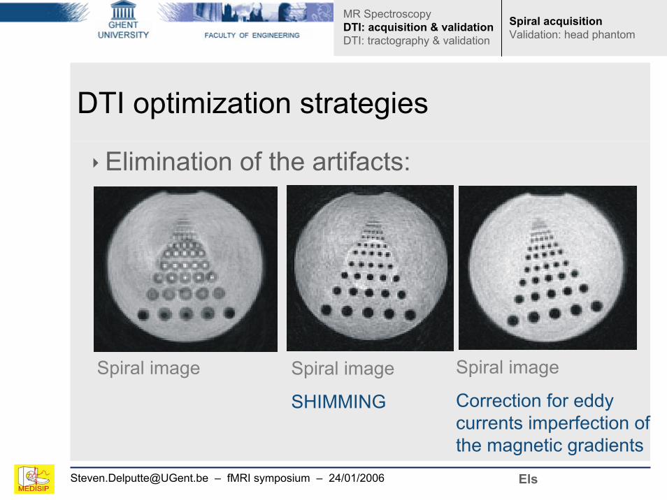
DTI optimization strategies
‣Elimination of the artifacts:
Spiral image Spiral image
SHIMMING
Spiral image
Correction for eddy currents imperfection of the magnetic gradients
MR SpectroscopyDTI: acquisition & validationDTI: tractography & validation
Spiral acquisitionValidation: head phantom
Els
[email protected] – fMRI symposium – 24/01/2006
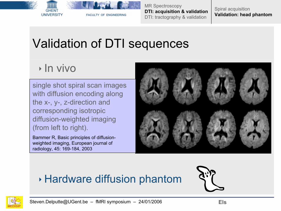
Validation of DTI sequences
‣ In vivo
‣Hardware diffusion phantom
single shot spiral scan images with diffusion encoding along the x-, y-, z-direction and corresponding isotropic diffusion-weighted imaging (from left to right). Bammer R, Basic principles of diffusion-weighted imaging, European journal of radiology, 45: 169-184, 2003
Els
MR SpectroscopyDTI: acquisition & validationDTI: tractography & validation
Spiral acquisitionValidation: head phantom

[email protected] – fMRI symposium – 24/01/2006
Validation: head diffusion phantom
‣Synthetic fibers to imitate the neural fascicle bundles.‣Anthropomorphic phantom of the major neural fiber tracts.‣MRI-compatibility: T1 and T2-relaxation times similar with brain white matter.‣DTI-compatibility: similar diffusion behavior as brain white matter (Monte Carlo diffusion simulations and quantitative measurements of DApp(t)-curves for different fiber materials).
Els
MR SpectroscopyDTI: acquisition & validationDTI: tractography & validation
Spiral acquisitionValidation: head phantom

[email protected] – fMRI symposium – 24/01/2006
Validation: a phantom bundle
• 400 parallel wires tightly held together by a shrinking tube• Wire = woven strand of Ultrahigh-Molecular Weight Polyethylene fibers (UHMWPE) (Dyneema®)
• FA = 0.45 (± σ = 0.15)
Els
MR SpectroscopyDTI: acquisition & validationDTI: tractography & validation
Spiral acquisitionValidation: head phantom

[email protected] – fMRI symposium – 24/01/2006
Validation: head diffusion phantom
MR SpectroscopyDTI: acquisition & validationDTI: tractography & validation
Spiral acquisitionValidation: head phantom
Els

[email protected] – fMRI symposium – 24/01/2006
Validation: head diffusion phantom
FA FA
Fractional Anisotropy3T, TE = 60ms, TR= 3sspin echo sequence with TRSE-diffusion preparation 12 directions, b-factors of 0 and 700 s/mm²
Tracking result of the corticospinal tract.
FA = 0.321 (± σ = 0.15)
Els
MR SpectroscopyDTI: acquisition & validationDTI: tractography & validation
Spiral acquisitionValidation: head phantom

[email protected] – fMRI symposium – 24/01/2006
OverviewMagnetic Resonance Spectroscopy (MRS)‣ Basic physics of MRS‣ Quantitative MRS ‣ Pitfalls‣ MRS of the prostate
Diffusion MRI‣ Basic physics of diffusion MRI‣ Sequence development‣ Validation (hardware phantom)‣ Diffusion Tensor Tractography ‣ Validation (software phantom)
More MR research at UGent
MR SpectroscopyDTI: acquisition & validationDTI: tractography & validation

[email protected] – fMRI symposium – 24/01/2006
DTT: reconstruction of axonal connections line propagation
Solve:
For each step, 2 decisions to make:‣ New direction?
» Principal diffusion direction» Tensor deflection» Tensor deflection with subpixel adaptive step size,…
‣ Integration method? » Euler (first order integration: )» Runge-Kutta (fourth order integration)» FACT (1999, Mori et al.),…
( ) eds
srd=
ecrr ii ⋅+=+1
Steven
MR SpectroscopyDTI: acquisition & validationDTI: tractography & validation
DTT: introductionDRFTValidation: software phantom

[email protected] – fMRI symposium – 24/01/2006
DTT: introductionDRFTValidation: software phantom
MR SpectroscopyDTI: acquisition & validationDTI: tractography & validation
DTT algorithms & visualization
Point to point rigid connections‣ Diagnostically valuable‣ Fast‣ Cumulative error propagation (spurious tracts)
“Likelihood of connectivity”maps
eg. Fast marching‣ More information‣ slower
Seedvoxel
Steven
[email protected] – fMRI symposium – 24/01/2006
Density Regularized Fiber Tracking (DRFT)Point to point connections+ pointwise estimate of probability + environmental architectural information
Based on the fact that the architectural environment plays a dominant role in the reproducibility of each tracking result
Steven
MR SpectroscopyDTI: acquisition & validationDTI: tractography & validation
DTT: introductionDRFTValidation: software phantom

[email protected] – fMRI symposium – 24/01/2006
Density Regularized Fiber Tracking (DRFT)
Temporary trackCM tract
di dd σ7.1+>
Stop temporarytrack
MR SpectroscopyDTI: acquisition & validationDTI: tractography & validation
DTT: introductionDRFTValidation: software phantom
Steven

[email protected] – fMRI symposium – 24/01/2006
DRFT results: visualization
Body of the corpus callosum
‣Color encodes directional information (A/P: green, I/S: blue, L/R: red)‣Transparency encodes estimate of probability‣Width encodes σd(dispersion)
MR SpectroscopyDTI: acquisition & validationDTI: tractography & validation
DTT: introductionDRFTValidation: software phantom
Steven

[email protected] – fMRI symposium – 24/01/2006
DTT validation: framework
‣ In vivo DWI acquisition‣ Anisotropic smoothing of DWIs‣ RESTORE (robust tensor estimation)‣ DRFT ground truth fibers
‣ Build the anatomically realistic phantom1 (using environmental architectural information)
‣ Add noise & try to reconstruct the ground truth fibers‣ Compute similarity measures
1extension of work by A. Leemans (MRM 2005, 53: 944-953)
A
B
C
MR SpectroscopyDTI: acquisition & validationDTI: tractography & validation
DTT: introductionDRFTValidation: software phantom

[email protected] – fMRI symposium – 24/01/2006
Phantom (b,d)
In vivo (a,c)
A good
between the colour codedsynthetic FA
images and the original in vivo
ones
correspondence is found
(a)
(b)
(c)
(d)
Steven
MR SpectroscopyDTI: acquisition & validationDTI: tractography & validation
DTT: introductionDRFTValidation: software phantom
[email protected] – fMRI symposium – 24/01/2006
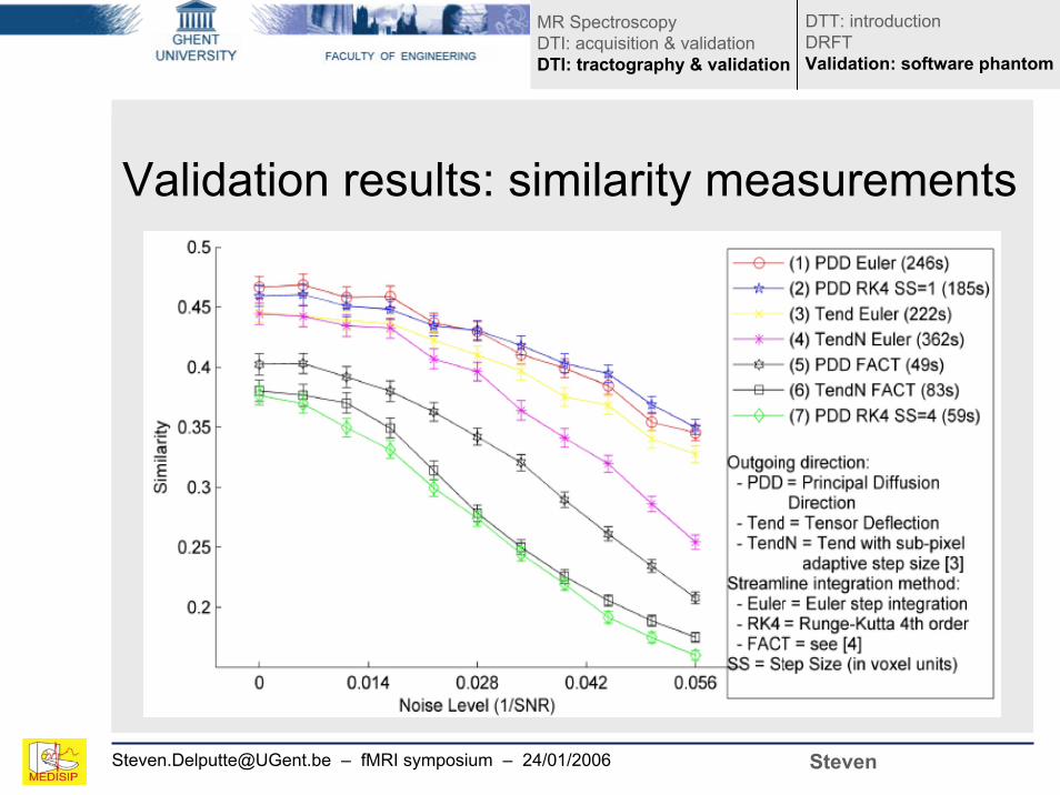
Validation results: similarity measurements
Steven
MR SpectroscopyDTI: acquisition & validationDTI: tractography & validation
DTT: introductionDRFTValidation: software phantom

[email protected] – fMRI symposium – 24/01/2006
DRFT and DTT validation
‣ DRFT results in diagnostically valuable 3D pathways AND at the same time gives an estimate of probability.‣ By using in vivo DRFT results, we were able to build a noise-free and anatomically realistic dataset.
» Noise and MRI acquisition artifacts can be incorporated in the synthetic phantom as well.
‣ With an anatomically realistic synthetic DT dataset we can:
» quantitatively predict how a (new) DTT algorithm will perform on real in vivo data (and not just for ad hoc cases such as helices etc.). » optimize internal and operator dependant tractography parameters.
Steven
MR SpectroscopyDTI: acquisition & validationDTI: tractography & validation
DTT: introductionDRFTValidation: software phantom

[email protected] – fMRI symposium – 24/01/2006
More MR research at Ugent (1.5T and 3T)‣ GifMi:
» fMRI studies of language and memory of epileptic patients, fMRI of stuttering» MRI techniques for measuring the biological malfunctioning of neurovascular units in migraine patients» Neuropsychology:
• Emotional disorders, mental rotation,…• cognitive dysfunctions ↔ structural brain damage in MS
patients
‣ Radiotherapy: » Quantitative T2-mapping for 3D geldosimetry & tissue classification» Molecular imaging
More

Els FieremansSteven Delputte Mahir Ozdemir
New developments in MagneticResonance Spectrocopy and DiffusionMRI