new information on hauffiosaurus (reptilia, …eprints.esc.cam.ac.uk/2331/1/benson_et_al_2011... ·...
TRANSCRIPT

NEW INFORMATION ON HAUFFIOSAURUS
(REPTILIA, PLESIOSAURIA) BASED ON A NEW
SPECIES FROM THE ALUM SHALE MEMBER (LOWER
TOARCIAN: LOWER JURASSIC) OF YORKSHIRE, UK
by ROGER B. J. BENSON* , HILARY F. KETCHUM� , LESLIE F. NOE�§ and
MARCELA GOMEZ-PEREZ–*Department of Earth Sciences, University of Cambridge, Downing Street, Cambridge CB2 3EQ, UK; e-mail: [email protected]
�Department of Zoology, University of Cambridge, Downing Street, Cambridge CB2 3EJ, UK; e-mail: [email protected]
�Thinktank Birmingham Science Museum, Millennium Point, Curzon Street, Birmingham B4 7XG, UK; e-mail: [email protected]
§College of Life and Environmental Sciences, School of Geography, Earth and Environmental Sciences, University of Birmingham, Edgbaston, Birmingham B15
2TT, UK
–CASP, West Building, 181A Huntingdon Road, Cambridge CB3 0DH, UK; e-mail: [email protected]
Typescript received 30 March 2010; accepted in revised form 6 July 2010
Abstract: An almost complete, three-dimensionally pre-
served plesiosaurian from the Hildoceras bifrons Zone of the
Alum Shale Member (Whitby Limestone Formation; Lower
Toarcian) of Yorkshire, UK, is described in detail. This repre-
sents a new species of Hauffiosaurus, H. tomistomimus, distin-
guished from H. zanoni (Harpoceras serpentinum Zone, Lower
Toarcian, Germany) by the proportionally shorter neck and
strongly concave preaxial margin of the tibia. It differs from
H. longirostris (previously ‘Macroplata’ longirostris; Har. ser-
pentinum Zone, Yorkshire) by the absence of prominent mid-
line ridges on the dorsal surface of the premaxillae and
ventral surface of the mandibular symphysis, and the absence
of midline pterygoid contact ventral to the basioccipital. Sev-
eral synapomorphies support a monophyletic Hauffiosaurus:
broad longitudinal troughs occupy the dorsolateral surface of
the maxilla and the posterior half of the lateral surface of the
dentary; basicranial fontanelle bounded laterally by postero-
laterally elongate projections of an undetermined ossification;
and the neural arch contacts the rib facet in all postaxial cer-
vical vertebrae. However, the systematic position of Hauffio-
saurus, as a pliosauroid or basal plesiosauroid, remains
uncertain. There is little evidence for geographic differentia-
tion of Lower Toarcian plesiosaurian faunas in the United
Kingdom and Germany as minor differences between abun-
dant taxa may arise from temporal offset of fossils from these
regions, and marked taxonomic differences are confined to
rare taxa whose absence in one or other area may be attribut-
able to incomplete sampling. Lack of consensus on the rela-
tionships of Lower Jurassic plesiosaurians requires further
detailed description of Lower Jurassic taxa.
Key words: Plesiosauria, Hauffiosaurus tomistomimus, Toar-
cian, Whitby Mudstone Formation, Alum Shale Member,
Posidonia Shale.
T he uppermost Triassic and Lower Jurassic marine
deposits of Western Europe have yielded numerous artic-
ulated plesiosaur specimens. Most are from the United
Kingdom, where numerous horizons yield abundant
remains from almost every stage of the interval. These
include the Rhaetian–Hettangian Pre-Planorbis and
Planorbis beds, which yield the stratigraphically earliest
plesiosaurians (Storrs and Taylor 1996), the Pliensbachian–
Sinemurian Lower Lias Group (e.g. Storrs 1997) and the
Toarcian Whitby Mudstone Formation of Yorkshire
(Benton and Taylor 1984). Together, these provide abun-
dant data on the early evolution of Plesiosauria (Benton
and Spencer 1995) and are central to understanding the
origins and early evolution of the group. Unfortunately,
the taxonomy and anatomy of Lower Jurassic plesiosauri-
ans are poorly understood, and detailed descriptive infor-
mation is available only for a few taxa (Storrs and Taylor
1996; Storrs 1997; Smith and Vincent 2010).
Here, we describe a plesiosaurian from the Alum Shale
Member of the Whitby Mudstone Formation (Hildoceras
bifrons Zone, lower Toarcian: Howarth 1980). Abundant
remains of marine reptiles have been collected from the
cliffs and alum quarries near Whitby over the past
200 years (Benton and Taylor 1984), including ichthyo-
saurs, thalattosuchian crocodylomorphs and the plesio-
saurians ‘Macroplata’ longirostris (Tate and Blake 1876;
[Palaeontology, Vol. 54, Part 3, 2011, pp. 547–571]
ª The Palaeontological Association doi: 10.1111/j.1475-4983.2011.01044.x 547

White 1940), Rhomaleosaurus cramptoni (Carte and Baily
1863; Smith and Dyke 2008), Rhomaleosaurus zetlandicus
(Phillips 1854; Taylor 1992; Vincent and Smith 2009),
Microcleidus homalospondylus and Microcleidus macropte-
rus (Owen 1881; Watson 1909, 1911) and Sthenarosaurus
dawkinsi (Watson 1909). The Alum Shale (Hildoceras
bifrons Zone) and underlying Jet Rock (Harpoceras
serpentinum Zone) members of the Whitby Mudstone
Formation are approximately coeval with the Posidonia
Shale (Harpoceras serpentinum Zone, lower Toarcian)
around Holzmaden, Germany, which has also yielded
abundant marine reptile fossils (Howarth 1980; Rohl
et al. 2001). As so many lower Toarcian fossils are
known, this interval has been the focus of Lower Jurassic
palaeobiogeographic hypotheses (Godefroit 1994; Maisch
and Ansorge 2004; O’Keefe 2004; Großmann 2007; Smith
and Vincent 2010).
The specimen described here was discovered in 1960
by an undergraduate geologic field party from Manches-
ter University (Broadhurst and Duffy 1970). The anterior
part of the skull was noticed projecting above a wave-
washed platform south of Robin Hood’s Bay, Yorkshire,
UK. The skull, neck, pectoral girdle and one forelimb
were covered by a thin layer of shale and collected
immediately, but the rest of the skeleton was more dee-
ply buried and had to be collected on a second expedi-
tion. The skeleton was transported to the Manchester
Museum, where its preparation was carried out using
standard mechanical techniques (MANCH unpublished
collections data). Subsequent acid preparation on the
specimen was undertaken by Roger Vaughan (BRSMG),
and the specimen is now on display at the Manchester
Museum, UK.
The specimen was previously referred to ‘Macroplata’
(or ‘Rhomaleosaurus’) longirostris and has been discussed
in the literature (Broadhurst and Duffy 1970; Benton and
Taylor 1984; Cruickshank 1996) and included in phyloge-
netic analyses (O’Keefe 2001, 2004; Ketchum and Benson
2010). However, it is described in detail here for the first
time. Several features indicate that it is distinct from ‘M.’
longirostris, although this taxon and the new specimen
form a closely related grouping with Hauffiosaurus zanoni
from the Posidonia Shale of Germany, and all three are
referred to the genus Hauffiosaurus herein.
In this article, use of specimen numbers denotes direct
observation of fossil specimens by one of the authors
unless otherwise noted.
GEOLOGIC SETTING
The Lower Jurassic Lias Group of Great Britain crops out
in a band across the United Kingdom from Devon and
Dorset in the south, to Yorkshire in the north-east, with
only small outcrops elsewhere (e.g. Howarth 1980). The
rocks consist predominantly of marine mudstones depos-
ited in a series of four interconnected basins separated by
shelf areas. The new specimen was found in the Cleveland
Basin of Yorkshire, which lies on the east coast of North
Yorkshire, north of the Market Weighton high. Coastal
exposures extend from Redcar in the north to Filey in the
south, subtending Whitby, Ravenscar and Scarborough.
Cleveland Basin deposits accumulated at the western mar-
gin of the North Sea Basinal system and comprise pre-
dominantly marine sediments (Rawson and Wright 1995).
The specimen was recovered from the ‘H. bifrons Zone
of the Alum Shale Series of the Upper Lias’ in the vicinity
of Ravenscar (Broadhurst and Duffy 1970, p. 30). This
equates to beds xvi–lvi of the Alum Shale Member of the
Whitby Mudstone Formation south-east of Peak (‘Old
Peak’ in some sources: Howarth 1962, 1980). The Whitby
Mudstone Formation has been divided into five members
(Rawson and Wright 1995), the middle of which is the
Alum Shale Member (formerly the Alum Shale Series;
Howarth 1962), consisting of medium to dark grey, fla-
key-weathering, nonlaminated, silty shale with numerous
bands of scattered nodules of calcareous and sideritic
concretions with a total thickness of 37.3 m near Raven-
scar (Howarth 1980; Rawson and Wright 1995). Unfortu-
nately, precise locality data for the new specimen are
unknown. However, detailed geologic data and maps of
the area can be found in Howarth (1962, pl. 27) and
Rawson and Wright (2000, fig. 15).
Anatomic abbreviations. aiv, anterior interpterygoid vacuity; ang,
angular; apr, anterior process of the articular; art, articular; at-
ax, atlas-axis complex; atc, atlantal centrum; atic, atlantal inter-
centrum; atna, atlantal neural arch; axi, axial intercentrum; axna,
axial neural arch; axncs?, possible axial neurocentral suture; axr,
axial rib; boc, basioccipital; bs, basisphenoid; ca1, first caudal
vertebra; ca20, twentieth caudal vertebra; ca35, thirty-fifth caudal
vertebra; ce3, third cervical vertebra; ce4, fourth cervical verte-
bra; ce34, thirty-fourth cervical vertebra; cl, clavicle; cor, coro-
noid; cora, coracoid; den, dentary; depr, depression; do1, first
dorsal vertebra; do7, seventh dorsal vertebra; do14, fourteenth
dorsal vertebra; do20; twentieth dorsal vertebra; do22 ⁄ sa1?, pos-
teriormost dorsal or first sacral vertebra; dob, dorsal blade of
scapula; en, external naris; exoc, exoccipital; fem, femur; for,
foramen; fr, frontal; frf, frontal facet on the parietal; gle, glenoid
(of pectoral girdle); hum, humerus; in, internal naris; isc,
ischium; lac, lacrimal; mx, maxilla; nc, neural canal; ncs, neuro-
central suture; ns, neural spine; pal, palatine; par, parietal; pifor,
pineal foramen; pmx, premaxilla; pofr, postfrontal; poz, postzy-
gapophysis; prfr, prefrontal; pro, prootic; pra, prearticular; prj,
projections of an unidentified ossification; prz, prezygapophysis;
ps, parasphenoid; pt, pterygoid; pub, pubis; qu, quadrate; rap,
retroarticular process; rug, rugosities; sa, surangular; sa1 ⁄ 2?, first
or second sacral vertebra; scap, scapula; scf, subcentral foramen;
soc, supraoccipital; spl, splenial; splf, splenial facet; sq, squamo-
sal; tro, trough; vom, vomer.
548 P A L A E O N T O L O G Y , V O L U M E 5 4

Institutional Abbreviations. BEDFM, Bedford Museum, Bedford;
NHMUK, Natural History Museum, London; BRSMG, Bristol
City Museum and Art Gallery, Bristol; CAMSM, Sedgwick
Museum of Earth Sciences, Cambridge; FHSM, the Sternberg
Museum of Natural History, Hays, Kansas; HAUF, Urwelt-
Museum Hauff, Holzmaden; LEICS, New Walk Museum and
Art Gallery, Leicester; MCZ, Museum of Comparative Zoology,
Harvard University, Cambridge, MA; MANCH, The Manchester
Museum, Manchester; MOR, Museum of the Rockies, Bozeman,
Montana; OXFUM, Oxford University Museum of Natural
History, Oxford; QM, Queensland Museum, Brisbane; SMNS,
Staatliches Museum fur Naturkunde Stuttgart, Stuttgart; USNM,
National Museum of Natural History, Washington, DC;
YORYM, The Yorkshire Museum, York.
SYSTEMATIC PALAEONTOLOGY
SAUROPTERYGIA Owen, 1860
PLESIOSAURIA de Blainville, 1835
Genus HAUFFIOSAURUS O’Keefe, 2001
Type species. Hauffiosaurus zanoni O’Keefe, 2001, from the Har-
poceras serpentinum Biozone (lower Toarcian) of Holzmaden,
Germany.
Additional included species. Hauffiosaurus longirostris (Blake in
Tate and Blake, 1876) [comb. nov.] from the Harpoceras serpent-
inum Biozone near Whitby, Yorkshire, UK; Hauffiosaurus tomi-
stomimus sp. nov.
Diagnosis. Longirostrine, long-necked (c. 34 cervical ver-
tebrae) plesiosaurians with 7–10 premaxillary teeth and
the following unique synapomorphies: broad longitudinal
troughs occupy the dorsolateral surface of the maxilla and
the posterior half of the lateral surface of the dentary;
basicranial fontanelle (midline opening in ventral sur-
face of braincase on basioccipital–basisphenoid suture)
bounded laterally by posterolaterally elongate projections
of an undetermined ossification; neural arch extends ven-
trally over lateral surface of centrum and contacts dorsal
portion of rib facet in all postaxial cervical vertebrae.
Remarks. Large foramina are present anteriorly and pos-
teriorly between the rib heads and cervical centra in
H. tomistomimus and postcranial material that may be
part of the holotype of H. longirostris, and this may also
be a synapomorphy of Hauffiosaurus (although the condi-
tion in H. zanoni was not determined during the present
study). H. zanoni possesses an elongate ilium, approxi-
mately two-thirds of the femoral length, and transversely
broad pubis with a subhexagonal outline resulting from
distinct anteriorly, anterolaterally and laterally facing
edges to the outline in ventral view. It is not possible to
determine whether these are autapomorphies of H. zanoni
or synapomorphies of Hauffiosaurus as the condition of
the pelvis cannot currently be determined in H. longiros-
tris or H. tomistomimus.
Hauffiosaurus tomistomimus sp. nov.
Text-figures 1–15
1970 Rhomaleosaurus longirostris Blake in Tate and
Blake; Halstead in Broadhurst and Duffy, p. 30,
fig. 28.
1996 Macroplata longirostris (Blake in Tate and Blake);
Cruickshank, p. 113.
2001 Macroplata longirostris (Blake in Tate and Blake);
O’Keefe, fig. 9.
Derivation of the name. Species epithet composed from Tomis-
toma, the generic name of the false gharial, a long-snouted croc-
odilian, and l�lo1, a Greek word meaning mimic.
Holotype. MANCH LL 8004 (Text-figs 1–15), an almost com-
plete skeleton.
Type locality. The bay between Old Peak and Blea Wyke Point,
south-east of Robin Hood’s Bay (National Grid Reference NZ 99
02: Ordnance Survey 1963).
Type horizon. Hildoceras bifrons Zone, Alum Shale Member,
Whitby Mudstone Formation, Lower Jurassic (lower Toarcian:
Broadhurst and Duffy 1970; Howarth 1980; Gradstein et al. 2005).
Diagnosis. Representative of the genus Hauffiosaurus in
which the preaxial margin of the tibia is strongly concave,
the middle cervical centra have a ratio of width to antero-
posterior length of approximately 1.2, the propodials are
shorter than the pelvis, the pterygoids do not contact
ventral to the basioccipital, and prominent longitudinal
midline ridges are absent from the dorsal surface of
the premaxillae or ventral surface of the mandibular
symphysis.
Remarks. Hauffiosaurus tomistomimus possesses three
additional features that may be autapomorphies, but their
presence or absence in H. longirostris and H. zanoni can-
not be determined: a long, distinct anterior process of the
parietals; a transverse constriction in the outline of the
pterygoid in ventral view at the base of the anterior pro-
cess; and a depression on the dorsolateral surface of the
third cervical neural arch.
DESCRIPTION
The skeleton of MANCH LL 8004 is almost complete and retains
a high degree of articulation (Text-fig. 1). As preserved, missing
B E N S O N E T A L . : H A U F F I O S A U R U S F R O M Y O R K S H I R E 549

the snout tip, the specimen is 4230 mm long, with the skull just
over one-tenth the length of the animal at 430 mm, the neck
approximately the same length as the trunk (1350 mm), and the
tail slightly shorter than the neck (1100 mm).
Cranium
Despite moderate dorsoventral crushing, bone surface preserva-
tion is good, allowing many of the cranial and mandibular
sutures to be confidently identified. The upper and lower jaws
are preserved tightly closed.
The skull has the shape of an elongate isosceles triangle in
dorsal view (Text-fig. 2). The distance between the quadrates is
265 mm, although this may have been exaggerated by dorsoven-
tral crushing, which has affected the skull medial and posterior
to the orbits. The external nares are small, anteroposteriorly ori-
ented oval openings located posteriorly on the snout, close to
the midline, anterior to the orbits. The postorbital bars are both
missing. However, in plesiosaurians the postorbital bars are usu-
ally located at the level of the parietal foramen (e.g. Andrews
1913; Druckenmiller and Russell 2008a), which is preserved.
In lateral view, the skull is long and dorsoventrally low (Text-
fig. 3); this has been accentuated by dorsoventral crushing poste-
riorly. Although it is incompletely preserved, the snout forms
greater than half the total skull length. H. tomistomimus is there-
fore a longirostrine taxon. By contrast, most other plesiosaurians
from the Toarcian of Europe have proportionally shorter rostra:
the plesiosauromorphs Microcleidus (NHMUK 36186), Hydrorion
and Seeleyosaurus (Maisch and Rucklin 2000; O’Keefe 2004;
Großmann 2007) and Occitanosaurus (Bardet et al. 1999) have
very short, rounded snouts; the pliosauromorphs Rhomaleosau-
rus (Watson 1910; Taylor 1992; Vincent and Smith 2009) and
Meyerasaurus (Smith and Vincent 2010) have relatively longer
snouts, but are brevirostrine compared to H. tomistomimus (the
snout is approximately 65 per cent of the length of the remain-
ing portion of the skull in R. zetlandicus; Taylor 1992). Only
Hauffiosaurus zanoni (HAUF 7; O’Keefe 2001) and H. longirostris
(MCZ 1033; White 1940) have longirostrine snouts comparable
to that of H. tomistomimus. In H. longirostris, the snout is
slightly longer than the remaining portion of the skull (White
1940). In Hauffiosaurus zanoni, the dorsal surface of the skull is
embedded in matrix (HAUF 7), but the approximate propor-
tions are similar.
Premaxilla. The preserved portions of the premaxillae are pri-
marily composed of their elongate posterior processes. These
are firmly joined along a straight midline suture and extend
posteriorly to the level of the anterior margin of the orbits,
where they terminate in a deeply interdigitating contact with
the frontals (Text-fig. 2). This is unlike the condition in many
other relatively long-snouted plesiosaurians such as Rhomaleo-
saurus zetlandicus (YORYM G503: Taylor 1992; Vincent and
Smith 2009), pliosaurids (e.g. Andrews 1913), polycotylids
(O’Keefe 2008) and some elasmosaurids (Sato 2002, 2003), in
which the premaxillae contact the parietals posteriorly (O’Keefe
2001; Druckenmiller and Russell 2008a; Ketchum and Benson
2010). The overall trend of the premaxilla–frontal contact is
posteromedial (Text-fig. 4). The parallel lateral margins of the
posterior processes contact the maxillae anterior to the external
naris, forming approximately straight sutures along most of
their length. However, these are slightly sinuous in places;
anterior to the external naris the suture undulates to form a
series of three peaks over approximately 15 mm (Text-fig. 3B).
These small-scale undulations of the suture between the maxilla
and posterior process of the premaxilla are also present in
Rhomaleosaurus megacephalus (LEICS G221.1851), but absent
in the pliosaurid Peloneustes, in which the suture is straight
(Ketchum 2007). Posterior to the external nares, the posterior
processes of the premaxillae are enclosed laterally by anterolat-
TEXT -F IG . 1 . Hauffiosaurus tomistomimus sp. nov. Skeleton MANCH LL 8004 in right anterodorsolateral view. Scale bar represents
1 m. Image is compressed by parallax towards the left.
550 P A L A E O N T O L O G Y , V O L U M E 5 4

eral processes of the frontals that contact the maxillae lateral
and medial to the external naris, excluding the premaxilla from
the narial margin. Although White (1940, fig. 2A) figured a
premaxilla–frontal contact that was restricted to the posterior
end of the premaxilla in MCZ 1033 (referred to Hauffiosaurus
longirostris herein), he also noted (p. 453) that preservation
was too poor to allow detection of most craniofacial sutures.
Our observations of MCZ 1033 confirm this. The dorsal sur-
faces of the premaxillae of H. tomistomimus are smoothly con-
vex and dorsoventrally low, lacking the prominent dorsal
midline crest that is present in H. longirostris (MCZ 1033:
White 1940).
The tooth-bearing anterior portion of the premaxilla is poorly
preserved and incomplete; the dorsal surface is broken. However,
the anterior section of the premaxilla–maxilla contact is visible
as a deeply interdigitating suture that curves anterolaterally,
C
A
D
B
E F
TEXT -F IG . 2 . Hauffiosaurus tomistomimus sp. nov. Skull of MANCH LL 8004 in dorsal view. A–B, complete skull, C–D
magnification (·1.75) of snout tip, E–F, magnification (·2) of skull roof anterior to pineal foramen. In line drawings (B, D, F), grey
tone indicates matrix, crossed-hatching indicates broken bone. Scale bar represents 100 mm. Abbreviations are given in the text.
B E N S O N E T A L . : H A U F F I O S A U R U S F R O M Y O R K S H I R E 551

A
B
TEXT -F IG . 3 . Hauffiosaurus tomistomimus sp. nov. Skull of MANCH LL 8004. A, complete skull in right lateral view with
magnification (·10) of dentary tooth showing possible apical wear facet, B–C, rostral portion of skull in left lateral view. B,
magnification (·2) showing sinuous premaxilla–maxilla suture. Scale bar represents 100 mm.
A
B
C
TEXT -F IG . 4 . Hauffiosaurus tomistomimus sp. nov. Skull of MANCH LL 8004 in left dorsolateral view. A, rostral portion of skull,
B–C, magnification (·1.5) showing sutures in the region of the external naris and anterior to the orbit. In line drawing (C), grey tone
indicates matrix and crossed-hatching indicates broken bone. Scale bar represents 50 mm.
552 P A L A E O N T O L O G Y , V O L U M E 5 4

delimiting the anterior end of the maxilla (Text-fig. 2D). It is
likely that the superficial exposure of this suture was less
strongly interdigitating than is the exposed, internal portion, as
in other plesiosaurians (e.g. Andrews 1910, 1913; Brown 1981;
O’Keefe 2001; Druckenmiller and Russell 2008a).
Maxilla. The maxillae enclose the anterior and lateral margins,
and the anterior half of the medial margin, of the external naris
(Text-figs 2, 4). Numerous small foramina are present on the
external surfaces of the maxilla. A broad, longitudinal trough
extends anteriorly from the naris along the dorsolateral surface
of the maxilla (Text-fig. 4A), a distinct feature also noted by
O’Keefe (2001). This trough is bounded laterally by a prominent
ridge immediately anterior to the naris. A similar trough and
ridge are present in Hauffiosaurus longirostris (MCZ 1033: White
1940), which is better preserved anteriorly, showing that the
ridge terminates on the posterior part of the lateral surface of
the premaxilla. It is unlikely that these well-defined structures
result from dorsoventral crushing. In MANCH LL 8004, crush-
ing is pronounced posteriorly, but does not seem to have
affected the snout. O’Keefe (2001, character 37) described a
trough on the maxilla in Macroplata tenuiceps (NHMUK
R5488), H. longirostris (‘Macroplata’) and rhomaleosaurids. In
Macroplata and rhomaleosaurids, this trough is shallow and does
not extend far anteriorly (NHMUK R5488, Ketchum and Smith
(2010); LEICS G221.1851, Cruickshank (1994a)), unlike the con-
dition in Hauffiosaurus (MCZ 1033, MANCH LL 8004).
The maxilla contacts the frontal both medial and lateral to the
external naris. The medial contact occurs approximately midway
along the external naris and is deeply interdigitating with a pos-
teromedial trend (Text-fig. 4B–C). Lateral contact between the
maxilla and frontal occurs at the posterolateral margin of the
external naris. It extends posteriorly as a weakly interdigitating
suture that terminates at the anterior border of the prefrontal.
The interdigitating maxilla-prefrontal suture curves posterolater-
ally and terminates at the anterodorsal margin of the lacrimal.
The posterior portion of the maxilla is poorly preserved. It
extends posteriorly, ventral to the lacrimal, which it contacts in
a weakly interdigitating, posteroventrally oriented suture.
Lacrimal. An anterodorsally elongate ossification forms the ante-
roventral margin of the orbit, dorsal to the posterior process of
the maxilla (Text-fig. 4). This forms interdigitating sutures with
the prefrontal dorsally and maxilla ventrally. The interdigitation is
weak, except for at the anterodorsal edge of the element. This
bone is identified as a lacrimal (sensu Williston (1907) in Bra-
chauchenius; Andrews (1913) in Liopleurodon; Linder (1913) in
Peloneustes), only otherwise identified in pliosaurids among
plesiosaurians (Druckenmiller and Russell 2008a). Other authors
have interpreted this ossification as a long anterior extension of
the jugal (e.g. Carpenter 1996; O’Keefe 2001), but our observa-
tions of well-preserved pliosaurid specimens indicate that the ‘lac-
rimal’ is separated from the jugal by an interdigitating suture
ventral to the orbit (Brachauchenius USNM 4989, Liopleurodon
NHMUK R2680, Peloneustes CAMSM X 50163, Pliosaurus brachy-
spondylus BRSMG Cc332). This region of the skull is poorly pre-
served in H. longirostris (MCZ 1033) and cannot be observed in
H. zanoni (O’Keefe 2001).
Prefrontal. The prefrontal forms the smooth, concave anterior
and anterodorsal margins of the orbit (Text-figs 2, 4). Medially,
the prefrontal overlaps the frontal. The posterodorsal process of
the prefrontal tapers posteriorly to its termination at the inter-
section of the prefrontal–frontal contact and the orbital margin.
Thus, the prefrontal extends approximately one-third of the way
along the dorsal margin of the orbit. A narrow longitudinal
ridge is present on the dorsal surface of the posterodorsal pro-
cess (Text-fig. 2B). Anterior to the orbit, the prefrontal contacts
the maxilla in a posterolaterally oriented, interdigitating suture
that extends to the prefrontal–lacrimal contact.
Frontal. The frontals are large, complex elements that extend
from the external nares to the posterior margins of the orbits, a
short distance anterior to the parietal foramen. The dorsal sur-
face of the frontal bears numerous small foramina (Text-figs 2,
4). The anterolateral processes of the frontals form the smooth
posterior margins of the external nares and are separated along
the midline by the posterior processes of the premaxillae. The
lateral margins of the anterolateral processes of the frontals con-
tact the maxillae anteriorly and the prefrontals posteriorly, form-
ing interdigitating sutures with both elements. The premaxilla is
excluded from the medial margin of the external naris by a small
anterior extension of the frontal, which contacts the maxilla
(Text-fig. 4B–C). There is no evidence for the presence of a sep-
arately ossified nasal in this region, which is well preserved. A
nasal has been identified in a range of plesiosaurians (reviewed
by Druckenmiller and Russell 2008a, p. 28) and more basal pis-
tosaurians (e.g. Meyer 1847–1855; Sues 1987; Cheng et al. 2006;
but see Rieppel et al. (2002) who suggested that Augustasaurus
and Pistosaurus lack nasals). However, these observations remain
tentative as little direct, photographic evidence has been pub-
lished, and different authors regard the ‘nasals’ of different taxa
as having highly variable proportions and morphology (e.g. Noe
2001; O’Keefe 2001; Cheng et al. 2006; Druckenmiller and Rus-
sell 2008a; Sato and Wu 2008; Gasparini 2009). Our observa-
tions suggest that the ‘nasals’ of at least some taxa may simply
be a narrow posteromedial extension of the maxilla similar to
that in H. tomistomimus (Text-fig. 4B–C), a broad anterior
extension of the prefrontal that contacts the posterior margin of
the naris (e.g. ‘Kronosaurus’, QM F51291), or are delimited by
cracks in the specimen that have been misidentified as sutures
(e.g. Peloneustes, NHMUK R8574: O’Keefe 2001). The variety of
means by which nasals may have been mistakenly identified
explains the highly variable apparent morphology, and we doubt
that they are widely present among adult plesiosaurians.
Although a separate ossification may be present in this region in
Liopleurodon (Noe 2001) and Nichollsaura (Druckenmiller and
Russell 2008b), we consider that it is absent in most other taxa.
The frontals contact each other posterior to the premaxillae,
forming a straight, midline butt joint. The central and posterior
portion of the left frontal is broken and displaced to the left,
and the right frontal has been displaced slightly to the right.
This reveals the longitudinally grooved medial contact surfaces
of the frontals (Text-fig. 2E–F). There is no evidence that a med-
ial aperture was present between the frontals prior to deforma-
tion. However, more posteriorly, narrow anterior processes of
the parietals divide the frontals across the midline. The medial
B E N S O N E T A L . : H A U F F I O S A U R U S F R O M Y O R K S H I R E 553
margins of the frontals slightly overlap the anterior processes of
the parietals. The posterolateral borders of both frontals are
broken. However, the preserved morphology suggests that the
frontal contributes to the dorsal margin of the orbit posterior to
the prefrontal and anterior to the postfrontal. The frontal enters
the dorsal margin of the orbit in most plesiosaurians, but are
excluded by contact between the prefrontal and the postfrontal
in some Lower Jurassic taxa [Macroplata tenuiceps, NHMUK
R5488, Ketchum and Smith (2010); unnamed taxon, NHMUK
49202], plesiosaurids (e.g. Storrs 1997), Cretaceous pliosaurids
such as Kronosaurus queenslandicus (QM F51291) and an
unnamed taxon (FHSM VP 321, referred to Brachauchenius
lucasi by Carpenter (1996), but see Ketchum and Benson
(2010)), and many Cretaceous plesiosauroids including leptoclei-
dians and elasmosaurids (see Ketchum and Benson (2010), char-
acter 19 for the distribution of this feature).
The exposure of the frontals on the ventral surface of the
skull roof differs from that on the dorsal surface. Paired, bar-
like processes extend posteriorly along the ventral surfaces of
the parietals adjacent to the midline, terminating immediately
anterior to the parietal foramen (Text-fig. 5C–D). Both pro-
cesses are broken posteriorly, revealing that they formed an
interdigitating sutural attachment to the parietal. The ventrome-
dial surfaces of these posterior processes bear deep troughs.
Together, these troughs form a midline channel in the ventral
surface of the frontals posteriorly. Anteriorly, this channel is
closed ventrally by contact between ventromedial flanges of the
frontals, forming a canal. This morphology was also described
in the pliosaurid Peloneustes by Andrews (1913, text-fig. 13),
who suggested that it accommodated the olfactory nerves. A
well-preserved ventral skull roof is also known in the plesiosau-
roid Seeleyosaurus (SMNS 16812). In Seeleyosaurus, the posterior
A
B
C
D
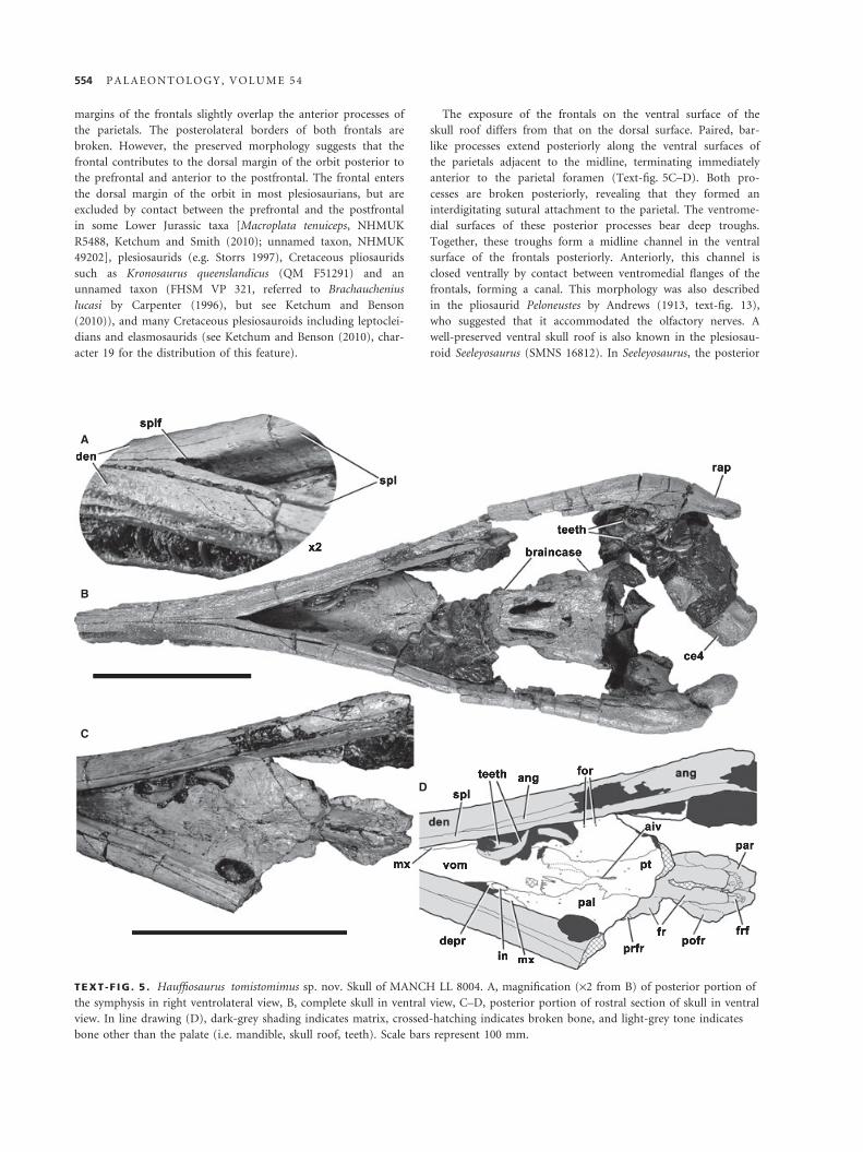
TEXT -F IG . 5 . Hauffiosaurus tomistomimus sp. nov. Skull of MANCH LL 8004. A, magnification (·2 from B) of posterior portion of
the symphysis in right ventrolateral view, B, complete skull in ventral view, C–D, posterior portion of rostral section of skull in ventral
view. In line drawing (D), dark-grey shading indicates matrix, crossed-hatching indicates broken bone, and light-grey tone indicates
bone other than the palate (i.e. mandible, skull roof, teeth). Scale bars represent 100 mm.
554 P A L A E O N T O L O G Y , V O L U M E 5 4

processes of the frontals are narrow and elongate and bear a
ventral channel that is not enclosed (Großmann 2007).
Postfrontal. A small part of each postfrontal is preserved antero-
lateral to the parietal foramen (Text-fig. 2). The exposure of the
postfrontal on the dorsal surface of the skull roof is small and
triangular and contacts the parietal posteromedially and the
frontal anteromedially. However, because the postfrontal slightly
underlaps the parietal and significantly underlaps the frontal,
exposure on the ventral surface of the skull roof is more exten-
sive (Text-fig. 5C–D).
Parietal. The parietals are large, unfused elements that contact
each other dorsally in a straight midline suture (Text-figs 2, 6A).
The parietal encloses the anteroposteriorly oriented, ovate parie-
tal foramen. Anterior to this foramen, the parietals form narrow,
prong-like anterior processes that separate the posterior portions
of the frontals along the midline. Short anterior processes of the
parietals are exposed on the dorsal surface of the skull in most
plesiosaurians other than cryptoclidids (Andrews 1910; Brown
1981; Maisch 1998), including Thalassiodracon (CAMSM
J.46986: Storrs and Taylor 1996). In pliosaurids, the parietals
extend far anteriorly to contact the posterior processes of the
premaxillae, but these anterior extensions are not distinct from
the body of the parietal as they are not transversely narrow (e.g.
Andrews 1913), unlike those of H. tomistomimus. In H. tomi-
stomimus, the anterior processes of the parietals are both distinct
and elongate (Text-fig. 2), even considering lateral displacement
of the frontals, which may have dorsally exposed an otherwise
concealed anterior portion of the processes. Long anterior pro-
cesses of the parietals may be an autapomorphy of H. tomistomi-
mus, although as the condition cannot be determined in
H. longirostris or H. zanoni this may represent a synapomorphy
of Hauffiosaurus. These processes are underlain by the posterior
processes of the frontals (Text-fig. 5C–D).
Posterior to the pineal foramen a low, sharp parietal crest
marks the apex of a robust vault over the braincase. This crest
divides the temporal fenestrae medially and has been displaced
slightly to the left and crushed ventrally over the supraoccipital.
Many other longirostrine plesiosaurians, such as the pliosaurids
Peloneustes (NHMUK R8574: Ketchum 2007) and an unnamed
taxon (FHSM VP321), some polycotylids such as Dolichorhync-
hops (O’Keefe 2004; Sato 2005) and Trinacromerum (O’Keefe
2008), and even some shorter-snouted taxa, including most el-
asmosaurids (e.g. Carpenter 1999; Sato 2002, 2003), have tall,
narrow parietal crests. The low crest of H. tomistomimus is com-
parable to those of more basal plesiosaurians such as Rhomaleo-
saurus megacephalus (LEICS G221.1851: Cruickshank 1994a),
Plesiosaurus (Storrs 1997), Thalassiodracon (CAMSM J.46986:
Storrs and Taylor 1996) and cryptoclidids (Andrews 1910;
Brown 1981).
Squamosal. The anterior processes of the squamosals, compris-
ing the lateral margins of the temporal fenestrae, are broken on
both sides (Text-figs 2–3). The dorsal rami of the squamosals
enclose the posterior margins of the temporal fenestrae (Text-
fig. 2). They contact across the midline posterior to the parietal
crest, forming an arch-like suspensorium over the braincase.
This has been crushed anteroventrally so that the concave
ventromedial margins of the dorsal rami of the squamosals are
visible in dorsal view (Text-fig. 2A–B). The dorsomedial inter-
squamosal contact interdigitates and is expanded posteriorly to
form a peaked squamosal bulb. The posterior surface of the left
squamosal is well preserved. It bears a series of rugose excresenc-
es, forming a mediolaterally oriented ridge (Text-fig. 7D–E). A
rugose ridge is also present in this location in many other ple-
siosaurians, including Thalassiodracon (CAMSM J.46986), Bore-
alonectes (Sato and Wu 2008) and the pliosaurid Peloneustes
(Ketchum 2007). However, it is generally a low, rugose, angular
ridge rather than a series of prominent rugosities.
The squamosal forms sheet-like ventral processes that overlap
the medial and lateral surfaces of the quadrate. Thus, the quad-
rate shaft is visible only in posterior view, as a subrectangular
surface (Text-fig. 7D–E), bounded medially and laterally by ver-
tically oriented sutures with the squamosal. The lateral squamo-
sal–quadrate suture is located at the apex of an angular,
dorsoventrally oriented crest on the posterolateral surface of the
suspensorium. A subcircular depression is present on the poster-
ior surface of the squamosal immediately dorsal to the quadrate
(Text-fig. 7D–E). This is widely present among plesiosaurians,
although in OXFUM J.28585 (Cruickshank (1994b), referred
therein to Eurycleidus but probably representing a distinct,
unnamed taxon), Rhomaleosaurus cramptoni (Smith and Dyke
2008) and Rhomaleosaurus zetlandicus (Taylor 1992), a large
foramen is present in this location (Smith and Dyke (2008),
character 31). It is possible therefore that this depression is a
vestige of fusion between the quadrate and squamosal, and its
presence may be ontogenetic. The medial surface of the squamo-
sal around midheight of the quadrate bears two small pits
(Text-fig. 7F–G). A single pit in this location was described in
Thalassiodracon as the ‘stapedial pit’ by Storrs and Taylor
(1996), but its function remains uncertain, as does the function
of the second pit in H. tomistomimus.
Palate
The palate of H. tomistomimus is almost planar. It is well pre-
served with little evidence of distortion or crushing. However, it
is broken posteriorly, and a large portion remains covered by
matrix or obscured by the articulated mandible (Text-fig. 5).
Vomer. The vomers form the anterior portion of the palate,
including the medial and anterior margins of the internal nares
(Text-fig. 5). The vomer is depressed immediately anterior to
the internal naris, although the deep groove extending anteriorly
from the naris described by Cruickshank et al. (1991) and Cru-
ickshank (1994a) in Rhomaleosaurus megacephalus, and observed
in Rhomaleosaurus cramptoni and Rhomaleosaurus thorntoni by
Smith and Dyke (2008), is absent in H. tomistomimus. The mid-
line suture between the vomers is fused and cannot be distin-
guished. The vomer is bounded laterally by contact with a
narrow palatal shelf of the maxilla. At the posterior margin of
the internal naris, the lateral edge of the vomer contacts the
anterior process of the palatine in a gently sinuous suture that
extends posteriorly for a short distance and terminates at the
B E N S O N E T A L . : H A U F F I O S A U R U S F R O M Y O R K S H I R E 555

A B C
D E
FG
H
I
TEXT -F IG . 6 . Hauffiosaurus tomistomimus sp. nov. Braincase of MANCH LL 8004 in A, dorsal, B–C, E, ventral, D, right
ventrolateral, F, left lateral, G, right lateral, and H–I, posterior views. D, magnification (·2) showing parasphenoid-basisphenoid suture
(indicated by arrows), E, magnification (·1.5) showing ventral surface of posterior basicranium. In line drawings (B, I), dark-grey tone
indicates matrix and crossed-hatching indicates broken bone. Scale bar represents 50 mm.
AC D
E
H
G
F
B
TEXT -F IG . 7 . Hauffiosaurus tomistomimus sp. nov. MANCH LL 8004, A–E, posterior portion of left mandible and anterior four
cervical vertebrae shown with mandible in A–B, medial, C, ventral and, D–E, posterolateral views, F–H, posterior portion of right
mandible in F–G, medial, and H, lateral views. In line drawings (B, G, E), dark-grey tone indicates matrix, light-grey tone indicates
teeth, and crossed-hatching indicates broken bone. Scale bar represents 50 mm.
556 P A L A E O N T O L O G Y , V O L U M E 5 4

transversely oriented, interdigitating vomer–pterygoid contact. A
short posterior projection of the vomers separates the anterior
processes of the pterygoids across the midline. Owing to poor
preservation in this region, the posterior extent of the vomers
cannot be precisely determined.
Palatine. The palatines form most of the palate posterior to the
vomers and lateral to the pterygoids. They bear numerous small
nutrient foramina on their ventral surfaces. The palate is incom-
pletely preserved, and the posterior portions of the palatines are
broken away. Transversely narrow, tapering anterior processes of
the palatines contact the posterior margins of the internal nares
(Text-fig. 5). Entry of the palatine into the internal narial mar-
gin is highly variable among plesiosaurians. For instance, the
palatine participates in the narial margin in some rhomaleosaur-
ids (e.g. Rhomaleosaurus megacephalus, LEICS G221.1851, Cru-
ickshank (1994a)), but is excluded in others (e.g. Maresaurus
coccai, Gasparini (1997); Meyerasaurus victor, SMNS 12478;
Smith and Vincent 2010); it participates in the margin in the
pliosaurid Peloneustes (Ketchum 2007), but is excluded in Liopl-
eurodon and Simolestes (Noe 2001). The right palatine is dis-
placed medially in MANCH LL 8004 so that it overlaps the
pterygoid. The left palatine is well preserved and apparently not
displaced. The medial contact with the pterygoid is interdigitat-
ing and forms a laterally concave curve.
Pterygoid. The anterior processes of the pterygoids extend ante-
riorly, lateral to the posterior process of the vomers and between
the anterior processes of the palatines. O’Keefe (2001, fig. 9) fig-
ured only a small anterior process in this position, and only on
the right side. However, the processes are transversely broad,
subrectangular, and present on both sides (Text-fig. 5C–D).
They are separated from more posterior portions of the pteryg-
oids by a transverse constriction. This constriction has not previ-
ously been observed in any other plesiosaurian palate (e.g.
Andrews 1910, 1913; O’Keefe 2001; Druckenmiller and Russell
2008a) and may be an autapomorphy of H. tomistomimus. As
the palate is not well preserved in H. longirostris or H. zanoni,
this may alternatively represent a synapomorphy of Hauffiosau-
rus. A narrow, slit-like anterior interpterygoid vacuity is present
between the pterygoids just posterior to the anterior processes
(Text-fig. 5). This slit-like morphology seems to arise from lack
of fusion along the midline interpterygoid suture; it is present in
smaller, likely ontogenetically immature, specimens of Liopleur-
odon (LEICS G418.1956 ⁄ 58a4: Noe 2001) and Peloneustes
(NHMUK R3803: Andrews 1913), but absent in larger individu-
als [Liopleurodon, NHMUK R3536, Noe (2001); Peloneustes,
NHMUK R5874]. However, it is distinct from the transversely
wide anterior interpterygoid vacuity of many leptoclidians, some
rhomaleosaurids and plesiosaurids (see Druckenmiller and Rus-
sell (2008a), character 49; Ketchum and Benson (2010), charac-
ter 59 for the distribution of this feature).
A large central portion of the pterygoids is not preserved or is
covered by matrix (Text-fig. 5B). The posterior parts of the
pterygoids form sheets that underlie the braincase, forming the
ventral and ventrolateral surfaces of the cranium (Text-fig. 6B–
G) and are separated along the midline by the parasphenoid,
posterior interpterygoid vacuity and ventral exposure of the
basioccipital. The medial margins of the pterygoids form inter-
digitating sutures with the parasphenoid, basisphenoid and the
ventral plate of the basioccipital (Text-fig. 6B–C). They also
form the smooth edges of the large, oval posterior interpterygoid
vacuity. The ventral surfaces of the pterygoids are smooth and
lack the posterolaterally oriented ridges that are present adjacent
to the posterior interpterygoid vacuity in pliosaurids (e.g.
Andrews (1913); Druckenmiller and Russell (2008a), character
51; Ketchum and Benson (2010), character 64) and Microcleidus
(NHMUK 36184).
Posterodorsally, the pterygoids contact the basal tubera of the
basioccipital. Posterior to the ventral exposure of the basioccipi-
tal, the posterior rami of the pterygoids diverge posterolaterally,
forming transversely narrow, dorsoventrally high quadrate
flanges. They are incompletely preserved, and the morphology of
the pterygoid–quadrate contact cannot be determined.
Braincase
Supraoccipital. The dorsolateral portions of the supraoccipital
(located anterolaterally owing to dorsoventral crushing of the
posterior part of the skull) are exposed in dorsal and lateral views
(Text-fig. 6A, F–G). The central portions of the bone are con-
cealed by the parietal. The supraoccipital was dorsoventrally low,
estimated as between two and three times as wide transversely as
it was high dorsoventrally. Its anteroposterior depth cannot be
determined. Typically in plesiosaurians, the supraoccipital con-
tacts the parietals dorsally and the exoccipital-opisthotics ventro-
laterally, forming the dorsal margin of the foramen magnum.
Exoccipital-opisthotic. The exoccipital-opisthotics have also been
displaced so that the paraoccipital processes are directed more
horizontally than they were in life. Hence, their original orienta-
tion cannot be precisely determined. The paired exoccipital-
opisthotics form the lateral margins of the foramen magnum.
The body (primarily comprising the opisthotic) forms a robust
pillar, and the paraoccipital process (comprising the lateral por-
tion of the exoccipital) is slender; the sutures between the two
elements are not clearly preserved. The distal ends of the para-
occipital processes are broken, and their morphology and
contacts cannot be determined.
Prootic. The prootics have convex lateral surfaces and approxi-
mately subcircular outlines in lateral view. The posterior and
dorsal surfaces are flattened and form contact surfaces for the
opisthotic and supraoccipital respectively.
Parasphenoid. Our interpretation of the identities and extent of
ventral braincase elements (Text-fig. 6B–E) differs from those
of O’Keefe (2001, fig. 9) and White (1940) for H. longirostris
(MCZ 1033). These differences are summarized in Table 1. The
parasphenoid forms interdigitating sutures with the pterygoids
anteriorly (Text-fig. 6D), although breakage and attached matrix
obscure the anterior extent of the parasphenoid. Its preserved
posterior portion extends a short distance posteriorly along the
ventral surface of the basisphenoid as a triangular process
B E N S O N E T A L . : H A U F F I O S A U R U S F R O M Y O R K S H I R E 557
between the interpterygoid vacuities (Text-fig. 6D). O’Keefe
(2001, fig. 9) interpreted the short, triangular element (which is
here interpreted as the entire parasphenoid) as a ventral keel
ornamenting a larger, robust parasphenoid body (interpreted
as the basisphenoid herein). However, interdigitating sutures
demarcate the boundary between this small triangular element
and the larger more robust element dorsal to it (Text-fig. 6D),
indicating that the ‘ventral keel’ of O’Keefe (2001) is a separate
ossification constituting the entire parasphenoid.
Basisphenoid. The basisphenoid is a robust element that bisects
the posterior interpterygoid vacuity (Text-fig. 6B–C). A ventral
longitudinal ridge on the basisphenoid extends a short distance
posterior to the parasphenoid. A single large foramen on the left
side and several, scattered, smaller foramina pierce the basisphe-
noid lateral to the midline ridge. A large ventrolateral foramen
on the left side of the basisphenoid is also present in a specimen
of Thalassiodracon (CAMSM J.46986), although other specimens
are too poorly preserved to determine whether the presence of
this foramen is polymorphic. The function of this opening is
uncertain, but it may simply be an enlarged nutrient foramen.
Posteriorly, the basisphenoid contacts the basioccipital in a
transversely oriented, interdigitating suture. A large midline
opening is present at this junction, similar to that of some cryp-
toclidids, such as Cryptoclidus (NHMUK R2860: Andrews 1910,
pl. 9, fig. 5) and Muraenosaurus (NHMUK R2422: Maisch 1998;
Ketchum and Benson 2010). This opening was noted by O’Keefe
(2001) as a possible autapomorphy of Hauffiosaurus longirostris
(‘Macroplata’), to which he referred MANCH LL 8004 (H. tomi-
stomimus herein). However, it is also present in H. zanoni
(HAUF 7) and was found as a synapomorphy of a clade com-
prising H. longirostris, H. tomistomimus (MANCH LL 8004) and
H. zanoni by Ketchum and Benson (2010, character 70). The
foramen may represent the basicranial fontanelle, an unossified
area at the basisphenoid–basioccipital contact of some tetrapods,
representing the embryonic fenestra basicranialis. Its location is
consistent with our interpretation of the anterior extent of the
basioccipital. The basicranial fontanelle is present in some extant
squamates and may close during ontogeny in some taxa (Conrad
(2004) and references therein), although this is unlikely in at
least some plesiosaurians as H. longirostris represents a large, and
possibly mature individual that retains the fontanelle (MCZ
1033). Posterolaterally elongate ventral projections from within
the braincase form the lateral margins of the basicranial fonta-
nelle and are clearly demarcated from the enclosing portions of
the basisphenoid and basioccipital. These are also present in
H. longirostris and were identified by White (1940) and O’Keefe
(2001) as the only visible portion of the basisphenoid (‘clivus’),
consistent with White’s (1940, fig. 4A) illustrations of the basi-
cranial complex of H. longirostris. Although our interpretation of
the braincase of Hauffiosaurus is different (Table 1), it is still
possible that these projections are part of a vertical notch in the
posterior surface of a dorsally located basisphenoid body. This
notch is primitively present in plesiosaurians (Druckenmiller
and Russell 2008a). The presence of these projections cannot be
determined owing to poor preservation in H. zanoni (HAUF 7).
Basioccipital. The basioccipital forms a robust palatal process
that projects ventral to the occipital condyle as a rectangular
plate (Text-fig. 6B–C, E). The posterior margin of this structure
was interpreted as a transversely oriented sutural connection
between the basisphenoid and the basioccipital by O’Keefe
(2001, fig. 9). However, close examination reveals no clear evi-
dence for a suture in this region.
Posteriorly the basioccipital forms the subcircular occipital con-
dyle (Text-fig. 6E, H, I). A small notochordal pit is located dorsally
on the posterior surface of the condyle. It is not possible to deter-
mine whether the exoccipital-opisthotic facets were separated from
the occipital condyle by a groove or neck as they are in some ple-
siosaurians (Druckenmiller and Russell 2008a), including H. longi-
rostris (White 1940, fig. 4A). However, a distinct groove encircles
the ventral and lateral surfaces of the neck of the condyle.
Mandible
The mandible is almost complete, measuring 420 mm long as
preserved. Only a small anterior portion of the symphysis is miss-
ing, and both mandibular rami are broken in the region between
the coronoid eminence and jaw articulation (Text-fig. 3). The
preorbital mandible is firmly attached to the cranium (Text-
figs 3, 5). Posterior to the orbit, however, the mandible is visible
from most angles (Text-fig. 7). The mandibular symphysis is
110 mm long. As it is incompletely preserved, it must have origi-
nally comprised more than 0.26 times the length of the mandible.
Dentary. The dentaries are the largest elements of the mandible.
They taper anteriorly until the lateral surfaces are approximately
parallel in the anterior half of the symphysial rostrum (Text-fig. 5).
Therefore, Hauffiosaurus tomistomimus has a narrow, unexpanded
snout tip, similar to that of H. longirostris (MCZ 1033: White
1940), H. zanoni (HAUF 7) and some polycotylids (e.g. Carpenter
1996; O’Keefe 2008), but unlike those of longirostrine pliosaurids,
in which the rostrum is transversely expanded anteriorly to form a
spatulate tip, separated from the remaining portion of the snout by
a rostral constriction (e.g. Peloneustes: Andrews 1913; O’Keefe
2001; Ketchum 2007). The ventral and lateral surfaces of the
dentary bear numerous anteroposteriorly elongate foramina; those
of the lateral surface are relatively large (Text-fig. 3). A shallow
longitudinal trough occupies the lateral surface of the dentary
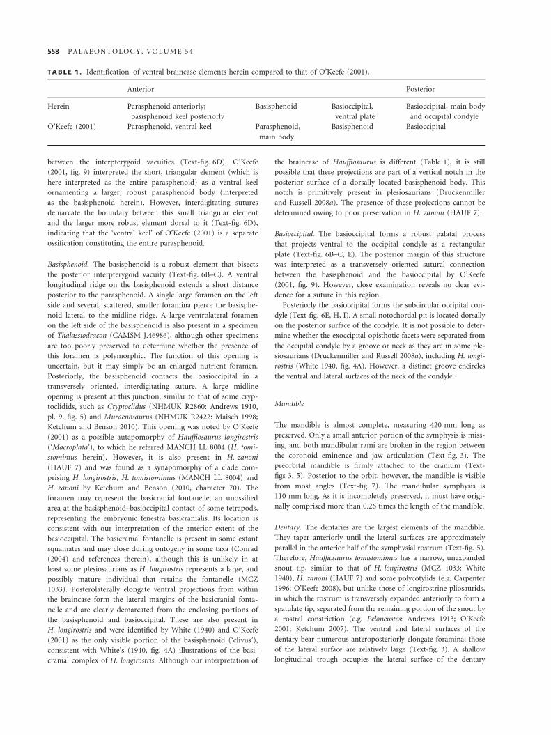
TABLE 1 . Identification of ventral braincase elements herein compared to that of O’Keefe (2001).
Anterior Posterior
Herein Parasphenoid anteriorly;
basisphenoid keel posteriorly
Basisphenoid Basioccipital,
ventral plate
Basioccipital, main body
and occipital condyle
O’Keefe (2001) Parasphenoid, ventral keel Parasphenoid,
main body
Basisphenoid Basioccipital
558 P A L A E O N T O L O G Y , V O L U M E 5 4
posterior to its midlength. This is also present in H. longirostris
(MCZ 1033: White 1940) and H. zanoni (HAUF 7).
Much of the medial surface of the dentary is covered by the
splenial, with which it forms a straight, longitudinal suture along
the ventral surface of the mandible (Text-fig. 5). This contact is
divided by the anterior process of the angular from approxi-
mately 40 mm posterior to the symphysis. The dentaries are sep-
arated ventrally on the midline by the splenials for much of the
length of the symphysis. Only in the anterior one-third of the
symphysis, as preserved, do the dentaries contact one another
along the midline in a straight butt joint that is visible in ventral
view (Text-fig. 5A–B). As the rostrum is incomplete, this pro-
portion would originally have been greater. The ventral surface
of the symphysis is smooth and gently convex. This is unlike the
condition in Hauffiosaurus longirostris, in which a prominent,
broad ventral keel is present (MCZ 1033); the ventral surface of
the symphysis of H. zanoni is unknown.
Splenial. In ventral view, the narrow anterior ends of the sple-
nials enter the mandibular symphysis medial to the dentaries
and are joined along a straight midline suture (Text-fig. 5). The
splenials have been separated by slight deformation and displace-
ment at the posterior end of the symphysis, revealing that their
medial contact surfaces are concave, perhaps because of incom-
plete ossification (Text-fig. 5A–B). A large, oval opening dorsal
to this contact represents the anterior opening of Meckel’s canal.
The splenial forms the smooth, ventromedial surface of the
mandible posterior to the mandibular symphysis; it is restricted
to the medial surface of the mandible from about midlength of
the postsymphysial mandibular rami by broadening of the ven-
tral exposure of the angular.
Coronoid. The coronoid covers the medial surface of the dentary
dorsal to the splenial anteriorly, but is not well exposed. It is
primarily visible in dorsal view, forming the dorsomedial surface
of the mandible ventral to the orbit (Text-fig. 2). It extends pos-
teriorly to an almost vertical, crenulated suture with the suran-
gular adjacent to the level of the parietal foramen. However, the
coronoid is incompletely preserved and may have overlapped the
surangular further posteriorly.
Angular. The angular forms a long anterior process that
separates the dentary and splenial on the ventral surface of the
mandible anteriorly and forms the ventral portion of the post-
symphysial mandible (Text-fig. 5), expanding posteriorly. It ter-
minates in an interdigitating suture with the articular that
transversely crosses the posterior surface of the retroarticular
process (Text-fig. 7).
A shallow longitudinal trough located ventrally on the medial
surface of the angular anterior to the glenoid is identified here
as the splenial facet (Text-fig. 7). A small sheet-like fragment of
bone covering this trough in the right angular is likely a poster-
ior fragment of the splenial. However, the posterior part of the
splenial has otherwise not been preserved.
Surangular. The surangular is visible immediately anterior to the
mandibular glenoid. More anteriorly it is broken, so it is not clear
whether it formed a transversely narrow plate of bone as in most
plesiosaurians, including the long-snouted polycotylids, or was
transversely broad, bearing a longitudinally elongate elliptical
depression as in pliosaurids (Druckenmiller and Russell 2008a;
Ketchum and Benson 2010). Druckenmiller and Russell (2008a, p.
47) also noted the broad condition in Rhomaleosaurus zetlandicus.
The surangular forms a straight, longitudinal contact with the
angular. This contact is visible on both the medial and lateral sur-
faces. The contact plane slants dorsolaterally so that the lateral
sutural exposure is higher dorsally than the medial exposure
(Text-fig. 7). The surangular contacts an anterior projection of
the articular posteroventrally on the medial surface of the mandi-
ble. However, other details of the surangular–articular contact
cannot be determined, and it is possible that the two were fused.
Prearticular. The prearticular is a dorsoventrally narrow, splint-
like element on the medial surface of the mandible (Text-fig. 7).
It lies within a shallow facet on the angular and is approximately
horizontal. The dorsomedial surface of the prearticular bears a
longitudinal groove around midlength. The ventral margin of
this groove is marked by a medially projecting longitudinal
ridge. Anterior to this, the mandible is poorly preserved, and the
morphology of the prearticular cannot be determined. The pos-
terior portion of the prearticular tapers dorsoventrally. It forms
a dorsally convex projection, housed in a groove between the
angular and articular (Text-fig. 7). This terminates ventral to
approximately glenoid midlength, whereas in pliosaurids such as
Peloneustes the prearticular terminates ventral to the anterior
margin of the glenoid (Ketchum 2007). In an unnamed Lower
Jurassic taxon (OXFUM J.28585: Cruickshank (1994b), referred
to Eurycleidus, but see Ketchum and Benson (2010)), the prear-
ticular terminates posterior to the glenoid and does not extend
far anteriorly. Unfortunately, this region of the mandible is often
poorly preserved, so the phylogenetic distribution of prearticular
morphology is unclear.
Articular. The articular forms the glenoid and dorsal portion of
the retroarticular process (Text-fig. 7). The glenoid is obscured
by the quadrate. It has a slightly thickened, sharp medial lip, but
its lateral surface is smooth and only slightly expanded. A rug-
ose, triangular process of the articular extends along the medial
surface of the mandible anterior to the glenoid. This is wide-
spread among plesiosaurians. Ventrally, at the base of this pro-
cess, a raised tuber projects medially (Text-fig. 7A).
The retroarticular process is transversely narrow and poster-
odorsally inclined. Its dorsal surface is weakly transversely con-
cave, and the ventral surface is highly convex, bearing a robust
longitudinal ridge of the angular. The retroarticular process of
the right mandible is heavily pitted, rugose and swollen in places
such that the angular–articular suture cannot be identified
(Text-fig. 7F–H). This is probably pathologic as the left retroar-
ticular process is smooth, well formed and clearly shows the
sutural line between the two constituent elements.
Dentition
The left dentary contains 33 alveoli (Text-fig. 4A). This is a min-
imum estimate, as the anterior end of the bone is not preserved,
B E N S O N E T A L . : H A U F F I O S A U R U S F R O M Y O R K S H I R E 559

and the maxilla obscures it posteriorly. We estimate that three
further alveoli may have been present in this region, and one or
two more were likely present anteriorly (based on comparison
with H. zanoni, HAUF 7). H. tomistomimus therefore had 37–38
dentary alveoli, the most posterior of which was located just
anterior to orbital midlength. By contrast, only 16 alveoli are
preserved in the right maxilla (Text-fig. 3A: 14 are preserved in
the left maxilla, which is less complete). The most posterior pre-
served alveolus is ventral to the anterior margin of the orbit. In
Hauffiosaurus zanoni, the maxillary tooth row continues posteri-
orly just anterior to the postorbital bar, and in this region the
alveoli are small and closely packed. Comparison suggests that
H. tomistomimus may have had approximately the same number
of maxillary alveoli as H. zanoni (there are at least 24 alveoli in
HAUF 7). Therefore, the maxilla contained approximately two-
thirds as many alveoli as the dentary in H. tomistomimus. This
discrepancy is partly attributable to the large size of the maxil-
lary alveoli, the largest of which are widely spaced and anteriorly
located. These enlarged anterior maxillary alveoli are also present
in H. zanoni (HAUF 7). They are preceded by a small anterior-
most maxillary tooth, which is likely present but not well pre-
served in MANCH LL 8004. This indicates the presence of a
‘heterodont’ dentition, which is also present in H. longirostris,
pliosaurids (Andrews 1913; O’Keefe 2001; Druckenmiller and
Russell 2008a), some polycotylids such as Edgarosaurus (MOR
751: Druckenmiller 2002), and a range of other plesiosaurians
(e.g. Druckenmiller and Russell 2008a). As the premaxilla is very
poorly preserved, the number of premaxillary alveoli cannot be
determined directly. However, comparison with the broken sec-
tion of the dentary anterior to the maxilla suggests that at least
five premaxillary teeth were present. Seven premaxillary teeth are
present in H. zanoni (HAUF 7; O’Keefe 2001), and nine and a
half are present in H. longirostris (MCZ 1033; White 1940; the
posteriormost premaxillary alveolus is bisected by the pre-
maxilla-maxilla suture).
Some broken teeth are preserved in their alveoli, and numer-
ous disarticulated teeth adhere to the palate and the posterior
end of the right mandible (Text-figs 5, 7). The teeth are taper-
ing, slender and recurved, with a circular cross-section. The
degree of curvature is more pronounced in more posterior teeth,
as in Pliosaurus (Taylor and Cruickshank 1993). Each tooth has
an enamelled crown and an unenamelled base (‘root’), approxi-
mately twice the length of the crown. The crown bears a series
of coarse enamel ridges, most of which extend the full apicobasal
length. A possible wear facet is located laterally on the apex of
one of the teeth of the left dentary (Text-fig. 3A). Apical wear
facets were also identified in Liopleurodon by Noe (2001, p. 145,
figs 22–23).
Axial skeleton
The holotype of H. tomistomimus (MANCH LL 8004) possesses
an articulated series of 34 cervical vertebrae (including the atlas
and axis) anterior to the pectoral girdle. This results in a long neck
compared to most other longirostrine plesiosaurians (O’Keefe
2002; O’Keefe and Carrano 2005). Thirty-three cervical vertebrae
could be removed from the exhibit and studied in detail (Text-
figs 8–11). Other vertebrae could not be studied in detail,
although the complete axial column is preserved (Text-fig. 12).
The centrum is firmly joined to the neural arch in all verte-
brae, although the neurocentral suture is still visible. The
sequence of closure of neurocentral sutures has yet to be studied
in detail for any plesiosaurian, and it is not clear what relation
this has to the termination of growth or the onset of sexual
maturity. However, this and other indicators suggest that
MANCH LL 8004 represents a subadult individual (see Discus-
sion, below).
Atlas-axis complex. The elements of the atlas-axis complex are
preserved in articulation, partly embedded in matrix and associ-
ated with the posterior part of the skull and the third and fourth
cervical vertebrae (Text-fig. 8). The sutures between the atlantal
elements are widely open, forming deep grooves on the anterior
surface of the atlantal cup (Text-fig. 8G–H). However, the
sutures between the atlantal and axial neural arches are not visi-
ble, and it is therefore difficult to interpret the dorsal surface of
the complex. The atlantal cup is concave and has a slightly rug-
ose surface texture. The anterior surface of the atlas centrum
forms the dorsal half of the cup medial to the atlantal neural
arches. It bears a small notochordal pit centrally on its anterior
surface (Text-fig. 8G–H). The dorsal surface of the centrum is
transversely concave, forming the ventral floor of the neural
canal.
The ventral and left lateral surfaces of the atlantal intercen-
trum are enclosed in matrix, and the right lateral surface is
highly and irregularly rugose, likely a pathologic condition
(Text-fig. 8). Because of these factors, it is not clear whether the
atlantal centrum is exposed on the ventral surface of the atlas-
axis complex, or whether it is excluded from the ventral surface
by contact between the atlantal and axial intercentra.
The atlantal neural arch forms the dorsal half of the atlantal
cup lateral to the centrum, unlike in cryptoclidids, in which the
centrum extends to the lateral surface (Andrews 1913; O’Keefe
2001). The neural arches have smooth, weakly convex lateral
surfaces. Two prong-like processes emerge posterodorsally from
the neural arch (Text-fig. 8). These have rugose, longitudinally
striated surfaces. The more ventral of these processes emerges
horizontally and overlaps a small anterior projection of the axial
neural arch. The more dorsal of the two processes of the atlantal
neural arch overlaps the dorsal surface of the axial neural arch.
Both of these processes could plausibly be identified as atlantal
postzygapophyses, and their homologies are unclear.
The axial rib is short and triangular with a proximal articula-
tion divided into two, anteroventrally elongate heads (Text-
fig. 8). The anterior head attaches to the axial intercentrum or
atlantal centrum ventrolaterally. The posterior head attaches to
the lateral surface of the axial centrum anteriorly, just ventral to
centrum midheight.
The body of the axis, which may represent the axial cen-
trum, is only visible in right lateral view. It has a weakly con-
vex, approximately rectangular lateral surface. The axial
neurocentral suture is difficult to identify, but may be repre-
sented by a thin horizontal groove a short distance ventral to
the axial postzygapophysis (Text-fig. 8A–B). If this interpreta-
tion is correct, then the axis is the only cervical vertebra in
560 P A L A E O N T O L O G Y , V O L U M E 5 4

which the neural arch does not contact the rib facet (see
below). Furthermore, a small, prezygapophysis-like anterolateral
projection from the axis underlies the more ventral posterior
process of the atlantal neural arch. If the axial neurocentral
suture has been identified correctly herein, then this prezygap-
ophysis-like structure is located on the axial centrum and not
on the neural arch (as might be expected of a true prezygap-
ophysis). As there is no other clear candidate for the axial neu-
rocentral suture, the only other possibility is that most or all of
the lateral surface of the axis seen in right lateral view repre-
sents the axial neural arch and the neurocentral suture, located
far ventrally, is mainly or entirely obscured by matrix and the
articulated axial rib.
The axial postzygapophyses project posterodorsolaterally. They
are comparable in morphology and relative size to those of more
posterior vertebrae: they are dorsoventrally low and have
approximately ventrally facing facets (Text-fig. 8). The axial neu-
ral spine is rugose, and a large central portion is broken. The
neural spine is transversely broad and dorsoventrally low, similar
to the condition in pliosaurids, but unlike those of other plesio-
saurians, in which the spine is taller dorsally (Andrews 1910,
1913).
Postaxial cervical vertebrae. The postaxial cervical centra have
gently concave articular surfaces, although in some cases these
are obscured by attached matrix including parts of the posterior
surface of the preceding centrum, thus giving the false impres-
sion of an opisthocoelous condition. All centra are slightly
shorter anteroposteriorly than they are high dorsoventrally and
slightly broader mediolaterally than dorsoventrally (Table 2).
The height and width steadily increase among more posterior
cervical vertebrae, whereas vertebral length reaches a maximum
among posterior cervical vertebrae and then decreases anterior
to the pectoral girdle (Table 2). A suboval depression is present
on the dorsolateral surface of the neural arch of the third cervi-
cal vertebra. This has not been described in any other plesiosau-
rian and may be an autapomorphy of H. tomistomimus.
The ventral surfaces of all cervical centra are rugose adjacent
to the anterior and posterior articular surfaces (Text-figs 9–11).
A rugose ventral longitudinal ridge is present on the ventral sur-
face of the anterior cervical centra (Text-fig. 9). This is less
prominent in the twelfth–sixteenth vertebrae and does not con-
tinue across the central portion of the centrum, instead forming
separate anterior and posterior ridges, separated by a smooth
area adjacent to the subcentral foramina (Text-fig. 10B). The
A B
C
E
D
G
HF
TEXT -F IG . 8 . Hauffiosaurus tomistomimus sp. nov. First four cervical vertebrae of MANCH LL 8004, including atlas and axis with
atlas-axis complex shown in A–B, right lateral, C–D, dorsal, and G–H anterior views and third–fourth cervical vertebrae shown in E–
F, right lateral view. In line drawings (B, D, F, H), dark-grey tone indicates matrix, and crossed-hatching indicates broken bone. Scale
bar represents 50 mm.
B E N S O N E T A L . : H A U F F I O S A U R U S F R O M Y O R K S H I R E 561

seventeenth–thirtieth cervical vertebrae lack a ventral ridge, and
in more posterior cervical vertebrae a broad, rounded ridge is
present (Text-fig. 11C). Small, paired nutrient foramina (subcen-
tral foramina) are located on the ventral surfaces of the centra
adjacent to the midline. The spacing between these foramina is
short, and in the twenty-fifth centrum the foramina are conflu-
ent, forming a single, bilobed opening (Text-fig. 10H).
The lateral surfaces of the cervical centra are rugose. This con-
trasts with the smooth neural arch pedicles. The neurocentral
suture extends ventrally around midlength so that it contacts the
diapophyses along the entire length of the neck, often forming a
small lappet that slightly overlaps the dorsal surface of the rib
(Text-figs 9–11). This contrasts with the situation in most ple-
siosaurians, in which the neural arch does not contact the ven-
trolaterally located rib facet in the cervical vertebrae and only
contacts the rib facet in anterior dorsal vertebrae (‘pectoral’ ver-
tebrae: Seeley 1874). However, both H. zanoni (HAUF 7) and
postcranial material that may represent the holotype individual
of H. longirostris (White 1940) shows the same condition as
H. tomistomimus (Text-figs 8–11), so this feature may be a syna-
pomorphy of Hauffiosaurus (Ketchum and Benson 2010).
Most cervical zygapophyses are broken. However, they are
large and emerge anterolaterally or posterolaterally from the
neural arch, rising dorsally. Most zygapophyseal facets face
dorsomedially or dorsolaterally at approximately 45 degrees, but
they are approximately horizontal in anterior cervical vertebrae
(Cruickshank 1996). The neural spines of the third and fourth
cervical vertebrae are very low and transversely broad, compara-
ble to that of the axis (Text-fig. 8). The neural arches of other
anterior–middle cervical vertebrae are poorly preserved. How-
ever, in the twenty-sixth and more posterior cervical vertebrae,
the neural spine is tall, sheet-like and angled posterodorsally
(Text-fig. 11). The cross-section of the spine is anteroposteriorly
long, approximately four-fifths of centrum length, and tapers to
a sharp edge anteriorly and posteriorly.
Cervical ribs. The cervical rib facets are located just ventral to
midheight on the lateral surfaces of the centra (Text-figs 8–11).
All are either broken, or preserved with articulated ribs, sug-
gesting that the ribs were firmly joined to the centrum,
although the sutures between the ribs and the centra are still
visible. The anterior–middle cervical rib facets are relatively
A
B
E
Fprz
L
K
przncs
rugoseforMribGscf
C
Dridge
H N
OJI P
TEXT -F IG . 9 . Hauffiosaurus tomistomimus sp. nov. Fifth to 15th cervical vertebrae of MANCH LL 8004. A–D, fifth–eighth, E–H,
9th–11th, I, ninth, J, 11th, K–N, 12th–15th, O, 12th, and P, 15th cervical vertebrae. A–B, E–F, K–L, left lateral, C, G, M, ventral, D, H,
N, dorsal, I, O, anterior, and J, P, posterior views. In line drawings (B, F, L), grey tone indicates matrix, and crossed-hatching
indicates broken bone. Scale bar represents 50 mm.
562 P A L A E O N T O L O G Y , V O L U M E 5 4

narrow dorsoventrally, but anteroposteriorly elongate, occupy-
ing most of the centrum length between the articular rims.
More posterior rib facets (from approximately the twenty-ninth
vertebra) are shorter, approximately half the length of the cen-
trum, but are higher dorsoventrally. The rib facets are paired,
constituting a dorsal diapophysis and a ventral parapophysis,
and support double-headed ribs, which are primitive for
Plesiosauria (e.g. O’Keefe 2001). The rib heads are divided
proximally by a channel. This is broad relative to that in other
plesiosaurians, thus forming large foramina anteriorly and
posteriorly, enclosed by the rib heads and lateral surface of the
centrum (Text-figs 8–11). This may also be present in H. longi-
rostris as White (1940, fig. 5) figured large foramina in the
cervical rib heads of postcranial material that may belong to the
holotype specimen. This morphology may be an autapomorphy
of Hauffiosaurus. However, the condition in H. zanoni was not
determined in the present study. Unfortunately, the cervical ribs
are all broken distally.
Postcervical axial column. As the postcervical series could not be
studied thoroughly, only a summary description is provided here
(Text-fig. 12). The dorsal vertebrae (including ‘pectoral’ verte-
brae) are preserved in articulation with the pectoral and pelvic
girdles, so they are visible in dorsal and lateral views (Text-
fig. 13). There are at least 21 dorsal vertebrae (if four sacral ver-
tebrae are present), and there may have been 22 if only three
sacral vertebrae are present. In the anterior three dorsal verte-
brae, the position of the rib facet is intermediate between that in
the cervical vertebrae (just ventral to centrum midheight) and in
Aprz ncs ns
ribforB
prz ns ribridgeC
TEXT -F IG . 11 . Hauffiosaurus tomistomimus sp. nov. 28th–
34th cervical vertebrae in A, right lateral, B, dorsal, and C,
ventral views (all images reversed). Scale bar represents 50 mm.
A ncs
B rib for E
D G
H
Cnc rib
F I
J Kpoz
rib
TEXT -F IG . 10 . Hauffiosaurus tomistomimus sp. nov. MANCH LL 8004, A–C, 16th–19th, D–F, 21st–23rd (20th not pictured), G–I
24th–27th, J, 24th, and K, 27th cervical vertebrae. A, D, G, left lateral, B, E, H, ventral, C, F, I, dorsal, J, anterior, and K, posterior
views. Scale bar represents 50 mm.
B E N S O N E T A L . : H A U F F I O S A U R U S F R O M Y O R K S H I R E 563

the more posterior dorsal vertebrae (on the transverse process of
the neural arch). Hence, the rib facet is successively more dor-
sally placed in each of these anterior three dorsal vertebrae, and
significant portions are located on both the centrum and neural
arch. They can thus be classified as ‘pectoral’ vertebrae (sensu
Seeley 1874). The neural arches of the anterior 15 dorsal verte-
brae form short, low and ventrally angled transverse processes,
with approximately circular diapophyses, located adjacent to
centrum midheight. In more posterior vertebrae, the transverse
processes extend laterally so the diapophysis is dorsal of the cen-
trum (Text-fig. 12). The zyagapophyseal facets of the dorsal ver-
tebrae emerge horizontally and are small compared to the neural
arch, relative to the proportions of cervical zygapophyses. The
zygapophyseal facets face dorsomedially or posterolaterally at a
low angle.
The anterior dorsal vertebrae have sheet-like, posterodorsally
inclined neural spines, comparable to those of the posterior cer-
vical vertebrae. In the middle of the series (fourteenth–nine-
teenth dorsal vertebrae), the neural spines are vertically
oriented and almost symmetrical in lateral view; weakly
expanded anteriorly and posteriorly. In more posterior dorsal
and sacral vertebrae, the neural spines are inclined anterodor-
sally (Text-fig. 12). This is unlike the situation in many Jurassic
plesiosaurians, such as Archaeonectrus (NHMUK 38525: Owen
1881), Macroplata (NHMUK R5488: Ketchum and Smith 2010),
Microcleidus (NHMUK 36184) and Thalassiodracon (NHMUK
14550), in which all dorsal neural spines are inclined poster-
odorsally. All of the dorsal neural spines of H. tomistomimus
are just less than 1.5 times the centrum and are between two
and three times as high as they are broad anteroposteriorly.
This is similar to the proportions in many plesiosaurians, but
unlike the condition in the Toarcian plesiosaurids Microcleidus
(NHMUK 36184: Watson 1909), Occitanosaurus (Bardet et al.
1999) and Seeleyosaurus (SMNS 16812), in which the neural
spines are exceptionally tall, more than twice the height of the
centrum. Three definite sacral vertebrae are present, identified
by the presence of a short rib with a transversely expanded dis-
tal portion that terminates adjacent to the partly disarticulated
pelvic girdle (Text-fig. 13B). The vertebra immediately anterior
to these three definite sacrals may also be a sacral vertebra or
may be the most posterior dorsal vertebra: the rib is short, but
longer than the other sacral ribs, and has only a slightly
expanded distal portion, although this converges posterolaterally
towards the positions of the distal ends of the other sacral ribs.
Thus, it is intermediate in morphology, and more detailed
information is required to determine its identity. The sacral
neural spines of H. tomistomimus are lower than the dorsal
neural spines (Text-fig. 12).
Thirty-five caudal vertebrae are present (Text-fig. 12). The
caudal centra are anteroposteriorly short. The anterior 23 of
these are relatively large; a pronounced size decrease, indicated
TEXT -F IG . 12 . Hauffiosaurus tomistomimus sp. nov. Composite image showing middle dorsal–distal caudal axial column of
MANCH LL 8004 in right lateral view. Scale bar represents 100 mm.
TABLE 2 . Selected measurements in millimetres of the cervical
vertebrae of MMUM LL 8004.
Length Posterior width Posterior height
Atlas – – –
Axis – – –
3 24 – –
4 25 – –
5 27 35 (anterior) 31 (anterior)
6 25 – –
7 28 – –
8 27 38 –
9 31 37 34
10 29 – –
11 30 40 35
12 33 – –
13 33 – –
14 34 – –
15 36 45 40
16 38 46 42
17 41 – –
18 39 – –
19 40 50 42
20 – – –
21 35* 54 –
22 43 – –
23 45 54 47
24 48 57 52
25 45 – –
26 38* – –
27 45 – –
28 47 64 54
29 44 – –
30 45 – –
31 45 – –
32 45 – –
33 45 – –
34 – – –
*Measurement of crushed specimen.
564 P A L A E O N T O L O G Y , V O L U M E 5 4

by a steady decline in estimated linear dimensions, occurs over
the following seven vertebrae, and the posteriormost five verte-
brae are reduced to small, cylindrical nubbins of bone that lack
neural arches. Large rib facets are present on the lateral surfaces
of anterior caudal centra. These diminish in size posteriorly and
are absent in the twentieth and more posterior caudal vertebrae.
The rib facets of the first nine caudal vertebrae are dorsoven-
trally oval, but in more posterior elements they are approxi-
mately circular. The caudal neural arches contact the dorsal
portion of the rib facet. Many proximal fragments of caudal ribs
are preserved attached to the centra by matrix. The well-devel-
oped zygapophyses of the anterior caudal vertebrae are approxi-
mately horizontal with slightly dorsomedially or ventrolaterally
inclined facets. In some more posterior caudal vertebrae, the fac-
ets are steeply angled. However, this may be the result of trans-
verse crushing. The caudal zygapophyses decrease in relative size
posteriorly and are absent in the fourteenth and more posterior
caudal vertebrae. The caudal neural spines are low, rectangular
and angled posterodorsally. Chevrons are not easily visible in the
specimen as displayed.
Appendicular skeleton
As with the postcervical axial column, the appendicular skeleton
was not thoroughly examined during the present study. There-
fore, for much of the material, we can only provide a summary
description in dorsal view (Text-fig. 13). Much of the pectoral
girdle is obscured by the articulated vertebral column. It is
therefore impossible to determine whether an interclavicle is
present. However, the lateral portions of the paired clavicles are
visible. They are triangular, with well-defined, rounded anterior
borders. The scapulae lie superficial to the clavicles, such that
the anterior part of the scapula underlaps the clavicle ventrally.
The glenoid lies on the scapulocoracoid contact and faces later-
ally. The right scapula preserves a partial dorsal process that
extends posterolaterally dorsal to the glenoid fossa. Only small
portions of the coracoids adjacent to the glenoid are preserved.
The left humerus is well preserved and is 400 mm long (Text-
fig. 14). It is thus slightly shorter than the pelvis (Table 3), unlike
in H. zanoni, in which the propodials are autapomorphically
longer than the pelvis (Table 3; O’Keefe 2001). The proximal
articular surface is large, as the combined dorsoventral depth of
the well-developed trochanter and capitulum is approximately
one-third of the length of the humerus. The preaxial (anterior)
margin of the humerus is almost straight. However, the postaxial
(posterior) border is concave, primarily because of the develop-
ment of a posterodistal expansion. The postaxial surface of the
right humerus is pierced by three large foramina; on the left
humerus, there is a single oval foramen at just over one-third of
the shaft length. The distal end of the humerus is convex and
coarsely pitted, indicating a cartilaginous covering in life (Robin-
son 1977). The radial and ulnar articular facets are not distinct as
only a slight angle separates the two. The radial facet is larger.
A partial ulna is preserved associated with the left forelimb
(Text-fig. 15A). It is subequal to the radius in length, but the
proximal end of the ulna is narrower. The preaxial margin is
broken. The radius is longer than wide and has concave preaxial
and postaxial borders (Text-fig. 15A). The proximal end of the
radius is straight with a rounded anteroproximal ‘corner’. The
distal end bears articular facets for the radiale (preaxial) and
A
B
TEXT -F IG . 13 . Hauffiosaurus tomistomimus sp. nov.
Interpretive drawings of A, pectoral, and B, Pelvic regions of
MANCH LL 8004 in dorsal view. Crossed-hatching indicates
broken bone or matrix. Scale bar represents 100 mm.
A B C D
TEXT -F IG . 14 . Hauffiosaurus tomistomimus sp. nov. Left
humerus of MANCH LL 8004 in A, anterior, B, ventral, C,
posterior, and D, dorsal views. Scale bar represents 100 mm.
B E N S O N E T A L . : H A U F F I O S A U R U S F R O M Y O R K S H I R E 565

intermedium (postaxial). The radiale is small and rounded. The
intermedium is subhexagonal and bears a larger ulnar contact
surface than radial contact surface. The ulnare is the largest of
the proximal carpals but is broken along its postaxial margin.
Distal carpal III is rounded and articulates with the intermedium
and ulnare. A second round distal carpal is present, but has
become disarticulated and is attached to a large block of matrix
containing nine partial phalanges. The metacarpals and pha-
langes are proximodistally elongate and hourglass-shaped, as in
other plesiosaurians (Text-fig. 15A).
Much of the pelvis is obscured by the articulated vertebrae
and ribs (Text-fig. 13B). The right pubis has an approximately
straight, anteromedially slanting anterior margin, which is
obscured by matrix medially. Posteriorly, the pubis contacts the
ischium to form the acetabulum. It is difficult to determine the
precise location of the puboischiadic suture on either side
because of poor preservation. A fragment of the right ilium is
preserved adjacent to the right ischium. Both ischia are broken
posteriorly.
The right femur is 370 mm long, only slightly shorter than
the humerus (Text-fig. 15B). Both preaxial and postaxial edges
are gently concave, resulting in an approximately symmetrical
outline. However, the postaxial edge is slightly more expanded
proximally and distally. The distal end of the femur is uniformly
convex and lacks distinct tibial and fibular facets. The epipodials
of the hind limb differ substantially from those of the forelimb
(Text-fig. 15B). The fibula has a rounded outline in dorsal view
and is as wide transversely as it is long proximodistally, with a
concave preaxial margin. The tibia is robust and approximately
the same length as the fibula. The preaxial margin of the tibia is
broken. Three ovate tarsals are preserved; definitive identification
is impossible because of partial disarticulation. A number of
elongate, hourglass-shaped metatarsals and phalanges are also
present (Text-fig. 15B). These are arranged as they were pre-
served, with portions of matrix remaining.
DISCUSSION
Ontogenetic stage
MANCH LL 8004 shares some ontogenetic features noted
by Brown (1981) in adult cryptoclidids. The ribs and neu-
ral arches are firmly joined to the cervical centra, as in
‘adults’, although the sutures between them are not closed
(i.e. are still visible), unlike in ‘old adults’. The cervical
centra are rugose, comparable to older individuals. How-
ever, other observations indicate incomplete ossification,
suggesting subadult status: the midline parietal suture is
open; atlantal sutures are widely open; the propodials lack
well-defined facets for the epipodials; and the phalanges
and metapodials have rounded edges. The nutrient
foramina in the vertebral centra (Ketchum and Smith
2010) and humeri are absent in older individuals, but are
present in MANCH LL 8004, so it is unlikely that
MANCH LL 8004 had reached maturity.
The affinities and validity of H. tomistomimus
MANCH LL 8004 was originally referred to the pliosau-
roid Rhomaleosaurus (as ‘Rhomaleosaurus’ longirostris,
considered herein as Hauffiosaurus longirostris) by Hal-
stead (in Broadhurst and Duffy 1970). However, recent
phylogenetic analyses have recovered MANCH LL 8004 as
a basal member of Pliosauridae (O’Keefe 2001, 2004) or
Plesiosauroidea (Ketchum and Benson 2010). In both
analyses, MANCH LL 8004 was phylogenetically proxi-
mate to Hauffiosaurus zanoni.
MANCH LL 8004 (‘Yorkshire taxon’) and Hauffio-
saurus zanoni formed successive sister taxa to a clade
TABLE 3 . Postcranial measurements (in metres) of MMUM
LL 8004 Hauffiosaurus tomistomimus species, gen. et sp. nov.
compared to those of H. longirostris and H. zanoni.
H. longirostris
MCZ 1033
H. tomistomimus
MMUM LL 8004
H. zanoni
HAUF 7
Skull length 0.68 >0.42 0.41
Skull width 0.27 0.19 0.17
Skeleton length – 4.3 3.4
Neck length – 1.35 1.00
Trunk length – 1.35 1.16
Tail length – 1.1 0.86
Humerus length – 0.40 0.32
Ulna length – 0.14 0.09
Radius length – 0.14 0.10
Femur length – 0.37 0.34
Tibia length – 0.11 0.09
Fibula length – 0.11 0.08
Pelvis length – 0.43 0.30
Measurements were taken to the nearest 10 mm and were esti-
mated from photographs for H. zanoni.
A
B
TEXT -F IG . 15 . Hauffiosaurus tomistomimus sp. nov.
Interpretive drawings of articulated limbs of MANCH LL 8004
in dorsal view. A, left forelimb, B, right hindlimb. Grey tone
indicates matrix, and crossed-hatching indicates broken bone.
Scale bar represents 100 mm.
566 P A L A E O N T O L O G Y , V O L U M E 5 4
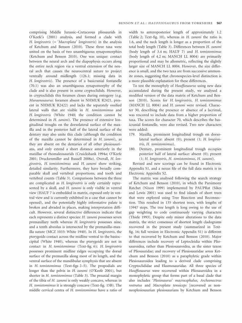
comprising Middle Jurassic–Cretaceous pliosaurids in
O’Keefe’s (2001) analysis, and formed a clade with
H. longirostris (= ‘Macroplata’ longirostris) in the analysis
of Ketchum and Benson (2010). These three taxa were
united on the basis of two unambiguous synapomorphies
(Ketchum and Benson 2010). One was unique: contact
between the neural arch and the diapophysis occurs along
the entire neck region via a ventral extension of the neu-
ral arch that causes the neurocentral suture to project
ventrally around midlength (126.1; missing data in
H. longirostris). The presence of a basicranial fontanelle
(70.1) was also an unambiguous synapomorphy of the
clade and is also present in some cryptoclidids. However,
in cryptoclidids this foramen closes during ontogeny (e.g.
Muraenosaurus: foramen absent in NHMUK R2421, pres-
ent in NHMUK R2422) and lacks the separately ossified
lateral walls that are visible in H. tomistomimus and
H. longirostris (White 1940; the condition cannot be
determined in H. zanoni). The presence of extensive lon-
gitudinal troughs on the dorsolateral surface of the max-
illa and in the posterior half of the lateral surface of the
dentary may also unite this clade (although the condition
of the maxilla cannot be determined in H. zanoni), as
they are absent on the dentaries of all other plesiosauri-
ans, and only extend a short distance anteriorly in the
maxillae of rhomaleosaurids (Cruickshank 1994a; O’Keefe
2001; Druckenmiller and Russell 2008a). Overall, H. lon-
girostris, H. tomistomimus and H. zanoni show striking,
detailed similarity. Furthermore, they have broadly com-
parable skull and vertebral proportions, and tooth and
vertebral counts (Table 3). Comparisons between the three
are complicated as H. longirostris is only certainly repre-
sented by a skull, and H. zanoni is only visible in ventral
view (HAUF 7 is embedded in matrix, exposed only in ven-
tral view and is currently exhibited in a case that cannot be
opened), and the potentially highly informative palate is
broken and abraded in places, making interpretation diffi-
cult. However, several distinctive differences indicate that
each represents a distinct species: H. zanoni possesses seven
premaxillary teeth whereas H. longirostris possesses nine,
and a tenth alveolus is intersected by the premaxilla–max-
illa suture (MCZ 1033: White 1940). In H. longirostris, the
pterygoids contact across the midline ventral to the basioc-
cipital (White 1940), whereas the pterygoids are not in
contact in H. tomistomimus (Text-fig. 6); H. longirostris
possesses prominent midline ridges occupying the dorsal
surface of the premaxilla along most of its length, and the
ventral surface of the mandibular symphysis that are absent
in H. tomistomimus (Text-figs 2–5). The propodials are
longer than the pelvis in H. zanoni (O’Keefe 2001), but
shorter in H. tomistomimus (Table 3). The preaxial margin
of the tibia of H. zanoni is only weakly concave, whereas in
H. tomistomimus it is strongly concave (Text-fig. 15B). The
middle cervical centra of H. tomistomimus have a ratio of
width to anteroposterior length of approximately 1.2
(Table 2; Text-fig. 10), whereas in H. zanoni the ratio is
1.6, and the neck length is longer as a proportion of the
total body length (Table 3). Differences between H. zanoni
(body length of 3.4 m; HAUF 7) and H. tomistomimus
(body length of 4.2 m; MANCH LL 8004) are primarily
proportional and may be allometric, reflecting the slightly
larger size of MANCH LL 8004. However, the size differ-
ence is small, and the two taxa are from successive ammon-
ite zones, suggesting that chronospecies-level distinction is
a more plausible explanation for these differences.
To test the monophyly of Hauffiosaurus using new data
accumulated during the present study, we analysed a
modified version of the data matrix of Ketchum and Ben-
son (2010). Scores for H. longirostris, H. tomistomimus
(MANCH LL 8004) and H. zanoni were revised. Charac-
ter 30, describing the presence or absence of a lacrimal,
was rescored to include data from a higher proportion of
taxa. The scores for character 70, which describes the bas-
icranial fontanelle, were also revised. Two new characters
were added:
179. Maxilla, prominent longitudinal trough on dorso-
lateral surface: absent (0); present (1; H. longiros-
tris, H. tomistomimus).
180. Dentary, prominent longitudinal trough occupies
posterior half of lateral surface: absent (0); present
(1; H. longirostris, H. tomistomimus, H. zanoni).
Revised and new scorings can be found in Electronic
Appendix S1, and a nexus file of the full data matrix is in
Electronic Appendix S2.
The matrix was analysed following the search strategy
of Ketchum and Benson (2010), in which the Parsimony
Ratchet (Nixon 1999) implemented by PAUPRat (Sikes
and Lewis 2001) was used to find islands of short trees
that were explored using Tree Bisection and Reconnec-
tion. This resulted in 135 shortest trees, with lengths of
15947 steps. The tree length is long owing to the use of
gap weighting to code continuously varying characters
(Thiele 1993). Despite only minor alterations to the data
matrix, the strict consensus of shortest length cladograms
recovered in the present study (summarized in Text-
fig. 16; full version in Electronic Appendix S1) is different
to that recovered by Ketchum and Benson (2010). Major
differences include recovery of Leptocleidia within Plio-
sauroidea, rather than Plesiosauroidea, as the sister taxon
of Pliosauridae; and recovery of Plesiosauridae sensu Ket-
chum and Benson (2010) as a paraphyletic grade within
Plesiosauroidea leading to a derived clade comprising
Cryptoclididae and Elasmosauridae. All three species of
Hauffiosaurus were recovered within Pliosauroidea in a
monophyletic group that forms part of a basal clade that
also includes ‘Plesiosaurus’ macrocephalus, Archeonectrus
rostratus and Macroplata tenuiceps [recovered as non-
neoplesiosaurian plesiosaurians by Ketchum and Benson
B E N S O N E T A L . : H A U F F I O S A U R U S F R O M Y O R K S H I R E 567

(2010)] and Rhomaleosaurus megacephalus (recovered as a
rhomaleosaurid by Ketchum and Benson (2010)).
This result indicates that relationships among Lower
Jurassic plesiosaurians are far from certain. In our view,
this is symptomatic of the sparse anatomical data cur-
rently available for these taxa. Plasticity in the phyloge-
netic position of Leptocleidia has been documented
previously (O’Keefe 2001; Druckenmiller and Russell
2008a), and the details of Jurassic plesiosaurian anatomy
will be central in forming a consensus on leptocleidian
affinities among pliosauroids or plesiosauroids.
Tree support metrics were generally low in the analysis
of Ketchum and Benson (2010), and as they are computa-
tionally expensive to calculate we do not present them
again here. We anticipate that future analyses will recover
a monophyletic Hauffiosaurus as representatives of the
genus are distinctly and strikingly similar to one another.
However, the precise phylogenetic position of Hauffiosau-
rus is considered uncertain, pending more detailed
description of other Lower Jurassic plesiosaurians. Fea-
tures shared uniquely with pliosaurids such as the pres-
ence of a lacrimal and the transversely broad, deeply
interdigitating morphology of the posterior termination
of the premaxilla suggest the possibility that Hauffiosaurus
may be a basal pliosaurid, as recovered by O’Keefe
(2001). Relationships among basal plesiosaurians are
uncertain (see the differing arrangements of O’Keefe
(2001), Druckenmiller and Russell (2008a) and Ketchum
and Benson (2010)), and further work is required to
establish synapomorphies of Plesiosauroidea, Pliosauroi-
dea and Plesiosauria as a whole. For instance, during this
study, it was difficult to make detailed comparisons with
rhomaleosaurids, for which published description is
almost limited to the craniofacial sutural anatomy (e.g.
Taylor 1992; Smith and Dyke 2008; Vincent and Smith
2009).
Toarcian plesiosaurian biogeography
Several authors have suggested that the distribution of
Toarcian marine reptiles, including thalattosuchians and
ichthyosaurs (Godefroit 1994; Maisch and Ansorge 2004)
or plesiosaurs considered in isolation (O’Keefe 2004;
Großmann 2007; Smith and Vincent 2010), indicates the
existence of discrete biogeographic provinces in Europe.
Maisch and Ansorge (2004) suggested that the London-
Brabant Massif acted as a barrier to dispersal between
hypothesized British and Germanic provinces, allowing
faunal differentiation. Plesiosaurians are a central compo-
nent of this hypothesis as they show the highest degree of
apparent endemism (Maisch and Ansorge 2004, p. 169),
based on the observation that British and German Toar-
cian taxa form nonoverlapping sets.
O’Keefe (2004), additionally posited that the German
Toarcian fauna was more primitive, based on a phyloge-
netic result in which the German Toarcian taxa Hydrorion,
Seeleyosaurus [both considered as ‘Plesiosaurus’ by O’Keefe
(2004)] and ‘Plesiopterys’ [considered as a subjective junior
synonym of Seeleyosaurus by Großmann (2007)] were
more basal than Mircocleidus among plesiosauroids and
Meyerasaurus victor (‘Rhomaleosaurus victor’) was consid-
ered more plesiomorphic than Rhomaleosaurus zetlandicus.
However, the more recent phylogenetic hypothesis of Ket-
chum and Benson (2010) recovered Hydrorion, Seeleyosau-
rus and Microcleidus within a monophyletic Plesiosauridae,
the youngest representatives of which are Toarcian, and
Rhomaleosaurus zetlandicus and Meyerasaurus victor in a
monophyletic Rhomaleosauridae. Strong support herein
for a monophyletic Hauffiosaurus further undermines sup-
port for a ‘plesiomorphic’ German Toarcian fauna.
Furthermore, we question the evidence for biogeo-
graphic provincialism among Toarcian plesiosaurians in
Europe. Plesiosauroids are abundant in both Germany
and the United Kingdom and represent separate, nomi-
nally valid taxa. However, detailed descriptions of the
British taxa Microcleidus homalospondylus, Microcleidus
macropterus and Sthenarosaurus dawkinsi have not yet
been published, and only future detailed comparative
work can establish whether the German taxa are genu-
inely distinct from the British taxa. Furthermore, minor
anatomic differences between the apparent regional vari-
ants may reflect temporal, rather than geographic, separa-
tion: the German taxa are from the Harpoceras
serpentinum ammonite Zone, whereas most of the UK
specimens are from the succeeding Hildoceras bifrons
Zone (Table 4). The plesiosauroid Occitanosaurus tourn-
emirensis from southern France is also of Toarcian age,
TEXT -F IG . 16 . Simplified cladogram representing the strict
consensus of 135 shortest length cladograms arising from
analysis of a modified version of the data matrix of Ketchum
and Benson (2010) showing the monophyly of Hauffiosaurus.
568 P A L A E O N T O L O G Y , V O L U M E 5 4
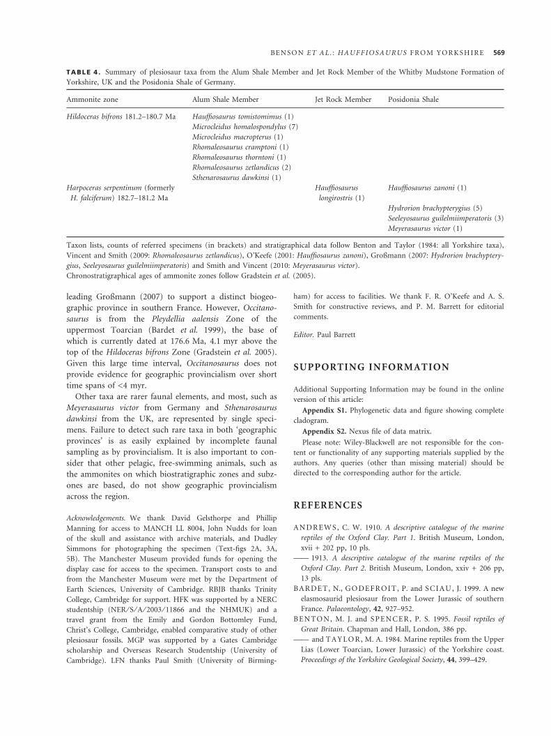
leading Großmann (2007) to support a distinct biogeo-
graphic province in southern France. However, Occitano-
saurus is from the Pleydellia aalensis Zone of the
uppermost Toarcian (Bardet et al. 1999), the base of
which is currently dated at 176.6 Ma, 4.1 myr above the
top of the Hildoceras bifrons Zone (Gradstein et al. 2005).
Given this large time interval, Occitanosaurus does not
provide evidence for geographic provincialism over short
time spans of <4 myr.
Other taxa are rarer faunal elements, and most, such as
Meyerasaurus victor from Germany and Sthenarosaurus
dawkinsi from the UK, are represented by single speci-
mens. Failure to detect such rare taxa in both ‘geographic
provinces’ is as easily explained by incomplete faunal
sampling as by provincialism. It is also important to con-
sider that other pelagic, free-swimming animals, such as
the ammonites on which biostratigraphic zones and subz-
ones are based, do not show geographic provincialism
across the region.
Acknowledgements. We thank David Gelsthorpe and Phillip
Manning for access to MANCH LL 8004, John Nudds for loan
of the skull and assistance with archive materials, and Dudley
Simmons for photographing the specimen (Text-figs 2A, 3A,
5B). The Manchester Museum provided funds for opening the
display case for access to the specimen. Transport costs to and
from the Manchester Museum were met by the Department of
Earth Sciences, University of Cambridge. RBJB thanks Trinity
College, Cambridge for support. HFK was supported by a NERC
studentship (NER ⁄ S ⁄ A ⁄ 2003 ⁄ 11866 and the NHMUK) and a
travel grant from the Emily and Gordon Bottomley Fund,
Christ’s College, Cambridge, enabled comparative study of other
plesiosaur fossils. MGP was supported by a Gates Cambridge
scholarship and Overseas Research Studentship (University of
Cambridge). LFN thanks Paul Smith (University of Birming-
ham) for access to facilities. We thank F. R. O’Keefe and A. S.
Smith for constructive reviews, and P. M. Barrett for editorial
comments.
Editor. Paul Barrett
SUPPORTING INFORMATION
Additional Supporting Information may be found in the online
version of this article:
Appendix S1. Phylogenetic data and figure showing complete
cladogram.
Appendix S2. Nexus file of data matrix.
Please note: Wiley-Blackwell are not responsible for the con-
tent or functionality of any supporting materials supplied by the
authors. Any queries (other than missing material) should be
directed to the corresponding author for the article.
REFERENCES
A N D R E W S , C. W. 1910. A descriptive catalogue of the marine
reptiles of the Oxford Clay. Part 1. British Museum, London,
xvii + 202 pp, 10 pls.
—— 1913. A descriptive catalogue of the marine reptiles of the
Oxford Clay. Part 2. British Museum, London, xxiv + 206 pp,
13 pls.
B A R DE T, N., G O DE F R O I T , P. and S C I A U , J. 1999. A new
elasmosaurid plesiosaur from the Lower Jurassic of southern
France. Palaeontology, 42, 927–952.
B E N T O N , M. J. and S PE N CE R , P. S. 1995. Fossil reptiles of
Great Britain. Chapman and Hall, London, 386 pp.
—— and T A Y L O R , M. A. 1984. Marine reptiles from the Upper
Lias (Lower Toarcian, Lower Jurassic) of the Yorkshire coast.
Proceedings of the Yorkshire Geological Society, 44, 399–429.
TABLE 4 . Summary of plesiosaur taxa from the Alum Shale Member and Jet Rock Member of the Whitby Mudstone Formation of
Yorkshire, UK and the Posidonia Shale of Germany.
Ammonite zone Alum Shale Member Jet Rock Member Posidonia Shale
Hildoceras bifrons 181.2–180.7 Ma Hauffiosaurus tomistomimus (1)
Microcleidus homalospondylus (7)
Microcleidus macropterus (1)
Rhomaleosaurus cramptoni (1)
Rhomaleosaurus thorntoni (1)
Rhomaleosaurus zetlandicus (2)
Sthenarosaurus dawkinsi (1)
Harpoceras serpentinum (formerly
H. falciferum) 182.7–181.2 Ma
Hauffiosaurus
longirostris (1)
Hauffiosaurus zanoni (1)
Hydrorion brachypterygius (5)
Seeleyosaurus guilelmiimperatoris (3)
Meyerasaurus victor (1)
Taxon lists, counts of referred specimens (in brackets) and stratigraphical data follow Benton and Taylor (1984: all Yorkshire taxa),
Vincent and Smith (2009: Rhomaleosaurus zetlandicus), O’Keefe (2001: Hauffiosaurus zanoni), Großmann (2007: Hydrorion brachyptery-
gius, Seeleyosaurus guilelmiimperatoris) and Smith and Vincent (2010: Meyerasaurus victor).
Chronostratigraphical ages of ammonite zones follow Gradstein et al. (2005).
B E N S O N E T A L . : H A U F F I O S A U R U S F R O M Y O R K S H I R E 569
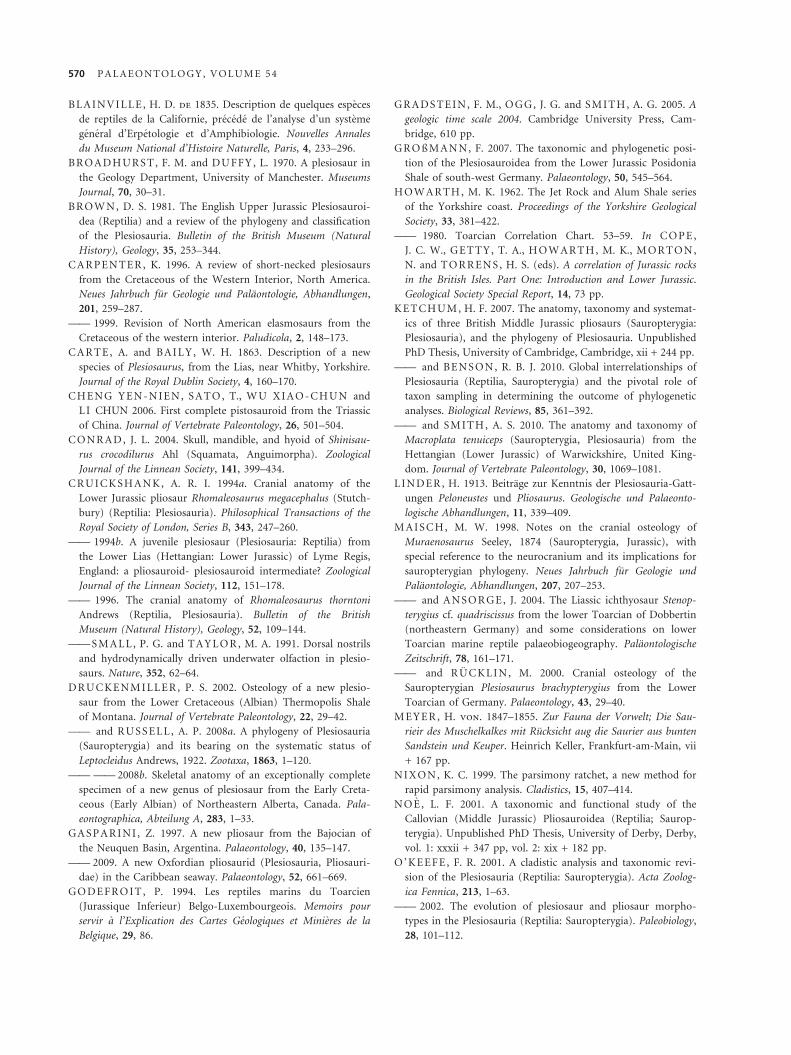
B L A I N V I L L E , H. D. de 1835. Description de quelques especes
de reptiles de la Californie, precede de l’analyse d’un systeme
general d’Erpetologie et d’Amphibiologie. Nouvelles Annales
du Museum National d’Histoire Naturelle, Paris, 4, 233–296.
B R OA D H UR S T , F. M. and DU FF Y , L. 1970. A plesiosaur in
the Geology Department, University of Manchester. Museums
Journal, 70, 30–31.
B R OW N , D. S. 1981. The English Upper Jurassic Plesiosauroi-
dea (Reptilia) and a review of the phylogeny and classification
of the Plesiosauria. Bulletin of the British Museum (Natural
History), Geology, 35, 253–344.
C A R PE N TE R , K. 1996. A review of short-necked plesiosaurs
from the Cretaceous of the Western Interior, North America.
Neues Jahrbuch fur Geologie und Palaontologie, Abhandlungen,
201, 259–287.
—— 1999. Revision of North American elasmosaurs from the
Cretaceous of the western interior. Paludicola, 2, 148–173.
C A R T E , A. and BA I L Y , W. H. 1863. Description of a new
species of Plesiosaurus, from the Lias, near Whitby, Yorkshire.
Journal of the Royal Dublin Society, 4, 160–170.
C H E N G Y E N - N I E N , S A T O , T., W U X I A O - C H U N and
L I CHUN 2006. First complete pistosauroid from the Triassic
of China. Journal of Vertebrate Paleontology, 26, 501–504.
C ON R A D , J. L. 2004. Skull, mandible, and hyoid of Shinisau-
rus crocodilurus Ahl (Squamata, Anguimorpha). Zoological
Journal of the Linnean Society, 141, 399–434.
C R UI CK S H A N K, A. R. I. 1994a. Cranial anatomy of the
Lower Jurassic pliosaur Rhomaleosaurus megacephalus (Stutch-
bury) (Reptilia: Plesiosauria). Philosophical Transactions of the
Royal Society of London, Series B, 343, 247–260.
—— 1994b. A juvenile plesiosaur (Plesiosauria: Reptilia) from
the Lower Lias (Hettangian: Lower Jurassic) of Lyme Regis,
England: a pliosauroid- plesiosauroid intermediate? Zoological
Journal of the Linnean Society, 112, 151–178.
—— 1996. The cranial anatomy of Rhomaleosaurus thorntoni
Andrews (Reptilia, Plesiosauria). Bulletin of the British
Museum (Natural History), Geology, 52, 109–144.
—— S M A L L , P. G. and T A Y L O R , M. A. 1991. Dorsal nostrils
and hydrodynamically driven underwater olfaction in plesio-
saurs. Nature, 352, 62–64.
D R UC KE N M I L L E R , P. S. 2002. Osteology of a new plesio-
saur from the Lower Cretaceous (Albian) Thermopolis Shale
of Montana. Journal of Vertebrate Paleontology, 22, 29–42.
—— and R U S S E L L , A. P. 2008a. A phylogeny of Plesiosauria
(Sauropterygia) and its bearing on the systematic status of
Leptocleidus Andrews, 1922. Zootaxa, 1863, 1–120.
—— —— 2008b. Skeletal anatomy of an exceptionally complete
specimen of a new genus of plesiosaur from the Early Creta-
ceous (Early Albian) of Northeastern Alberta, Canada. Pala-
eontographica, Abteilung A, 283, 1–33.
G A S P A R I N I , Z. 1997. A new pliosaur from the Bajocian of
the Neuquen Basin, Argentina. Palaeontology, 40, 135–147.
—— 2009. A new Oxfordian pliosaurid (Plesiosauria, Pliosauri-
dae) in the Caribbean seaway. Palaeontology, 52, 661–669.
G O DE F R O I T , P. 1994. Les reptiles marins du Toarcien
(Jurassique Inferieur) Belgo-Luxembourgeois. Memoirs pour
servir a l’Explication des Cartes Geologiques et Minieres de la
Belgique, 29, 86.
G R A DS TE I N , F. M., O G G, J. G. and S M I T H, A. G. 2005. A
geologic time scale 2004. Cambridge University Press, Cam-
bridge, 610 pp.
G R O ß M A N N , F. 2007. The taxonomic and phylogenetic posi-
tion of the Plesiosauroidea from the Lower Jurassic Posidonia
Shale of south-west Germany. Palaeontology, 50, 545–564.
H OW A R TH , M. K. 1962. The Jet Rock and Alum Shale series
of the Yorkshire coast. Proceedings of the Yorkshire Geological
Society, 33, 381–422.
—— 1980. Toarcian Correlation Chart. 53–59. In C OP E ,
J. C. W., G E T T Y , T. A., H O W A R T H, M. K., M OR T O N ,
N. and T O R R E N S , H. S. (eds). A correlation of Jurassic rocks
in the British Isles. Part One: Introduction and Lower Jurassic.
Geological Society Special Report, 14, 73 pp.
K E T CH UM , H. F. 2007. The anatomy, taxonomy and systemat-
ics of three British Middle Jurassic pliosaurs (Sauropterygia:
Plesiosauria), and the phylogeny of Plesiosauria. Unpublished
PhD Thesis, University of Cambridge, Cambridge, xii + 244 pp.
—— and BE N S ON , R. B. J. 2010. Global interrelationships of
Plesiosauria (Reptilia, Sauropterygia) and the pivotal role of
taxon sampling in determining the outcome of phylogenetic
analyses. Biological Reviews, 85, 361–392.
—— and S M I T H , A. S. 2010. The anatomy and taxonomy of
Macroplata tenuiceps (Sauropterygia, Plesiosauria) from the
Hettangian (Lower Jurassic) of Warwickshire, United King-
dom. Journal of Vertebrate Paleontology, 30, 1069–1081.
L I N D E R , H. 1913. Beitrage zur Kenntnis der Plesiosauria-Gatt-
ungen Peloneustes und Pliosaurus. Geologische und Palaeonto-
logische Abhandlungen, 11, 339–409.
M A I S C H, M. W. 1998. Notes on the cranial osteology of
Muraenosaurus Seeley, 1874 (Sauropterygia, Jurassic), with
special reference to the neurocranium and its implications for
sauropterygian phylogeny. Neues Jahrbuch fur Geologie und
Palaontologie, Abhandlungen, 207, 207–253.
—— and A N S O R G E , J. 2004. The Liassic ichthyosaur Stenop-
terygius cf. quadriscissus from the lower Toarcian of Dobbertin
(northeastern Germany) and some considerations on lower
Toarcian marine reptile palaeobiogeography. Palaontologische
Zeitschrift, 78, 161–171.
—— and R U C KL I N , M. 2000. Cranial osteology of the
Sauropterygian Plesiosaurus brachypterygius from the Lower
Toarcian of Germany. Palaeontology, 43, 29–40.
M E Y E R , H. von. 1847–1855. Zur Fauna der Vorwelt; Die Sau-
rieir des Muschelkalkes mit Rucksicht aug die Saurier aus bunten
Sandstein und Keuper. Heinrich Keller, Frankfurt-am-Main, vii
+ 167 pp.
N I X ON , K. C. 1999. The parsimony ratchet, a new method for
rapid parsimony analysis. Cladistics, 15, 407–414.
N O E , L. F. 2001. A taxonomic and functional study of the
Callovian (Middle Jurassic) Pliosauroidea (Reptilia; Saurop-
terygia). Unpublished PhD Thesis, University of Derby, Derby,
vol. 1: xxxii + 347 pp, vol. 2: xix + 182 pp.
O ’ KE E F E , F. R. 2001. A cladistic analysis and taxonomic revi-
sion of the Plesiosauria (Reptilia: Sauropterygia). Acta Zoolog-
ica Fennica, 213, 1–63.
—— 2002. The evolution of plesiosaur and pliosaur morpho-
types in the Plesiosauria (Reptilia: Sauropterygia). Paleobiology,
28, 101–112.
570 P A L A E O N T O L O G Y , V O L U M E 5 4

—— 2004. Preliminary description and phylogenetic position of
a new plesiosaur (Reptilia: Sauropterygia) from the Toarcian
of Holzmaden, Germany. Journal of Paleontology, 78, 973–988.
—— 2008. Cranial anatomy and taxonomy of Dolichorhynchops
bonneri new combination, a polycotylid (Sauropterygia: Plesi-
osauria) from the Pierre Shale of Wyoming and South Dakota.
Journal of Vertebrate Paleontology, 28, 664–676.
—— and CA R RA N O, M. T. 2005. Correlated trends in the
evolution of the plesiosaur locomotor system. Paleobiology, 31,
656–675.
ORDNANCE SURVEY 1963. Scarborough (Sheet 93). 1:63360.
Seventh Series. Director General of the Ordnance Survey,
Chessington.
O W E N , R. 1860. On the orders of fossil and Recent Reptilia,
and their distribution through time. Report of the British Asso-
ciation for the Advancement of Science, 1859, 153–166.
——1881. A monograph on the fossil Reptilia of the Liassic For-
mations. Part III: Plesiosaurus, Dimorphodon, and Ichthyo-
saurus. Monograph of the Palaeontographical Society, 35, 1–134,
pls 1–33.
P H I L L I P S , J. 1854. On a new Plesiosaurus in the York
Museum. Report of the British Association for the Advancement
of Science, 1853, 54.
R A W S ON , P. F. and W R I GH T , J. K. 1995. Jurassic of the
Cleveland Basin, North Yorkshire. 173–208. In T A Y LO R ,
P. D. (ed.). Field geology of the British Jurassic. Geological
Society, London, 286 pp.
—— —— 2000. The Yorkshire Coast. Geologists’ Association
Guide, 34, 130 pp.
R I E PP E L , O., S A N D E R , P. M. and S T OR R S , G. W. 2002.
The skull of the pistosaur Augustasaurus from the Middle Tri-
assic of Northwestern Nevada. Journal of Vertebrate Paleontol-
ogy, 22, 577–592.
R O B I N S O N , J. A. 1977. Intracorporal force transmission in
plesiosaurs. Neues Jahrbuch fur Geologie und Palaontologie,
Abhandlungen, 153, 86–128.
R O H L, H. J., S CH M I D- R O HL , A., O S C H M A N N , W.,
F R I M M E L , A. and S C H W A R K, L. 2001. The Posidonia
Shale (Lower Toarcian) of SW Germany: an oxygen-depleted
ecosystem controlled by sea level and paleoclimate. Palaeogeog-
raphy, Palaeoclimatology, Palaeoecology, 165, 27–52.
S A T O , T. 2002. Description of plesiosaurs (Reptilia: Sauroptery-
gia) from the Bearpaw Formation (Capanian-Maastrichtian)
and a phylogenetic analysis of the Elasmosauridae. Unpub-
lished PhD dissertation, University of Calgary, Calgary, 391 pp.
—— 2003. Terminonatator pontiexensis, a new elasmosaur (Rept-
ilia; Sauropterygia) from the Upper Cretaceous of Saskatche-
wan. Journal of Vertebrate Paleontology, 23, 89–103.
—— 2005. A new polycotylid plesiosaur (Reptilia: Sauropterygia)
from the Upper Cretaceous Bearpaw Formation in Saskatche-
wan, Canada. Journal of Paleontology, 79, 969–980.
—— and W U X I A O- C H UN 2008. A new Jurassic pliosaur
from the Melville Island, Canadian Arctic Archipelago. Cana-
dian Journal of Earth Sciences, 45, 303–320.
S E E LE Y , H. G. 1874. Note on some of the generic modifica-
tions of the plesiosaurian pectoral arch. Quarterly Journal of
the Geological Society of London, 30, 436–449.
S I KE S , D. S. and L E W I S , P. O. 2001. Beta software, Version 1.
PAUPRat: PAUP implementation of the parsimony ratchet. Dis-
tributed by the authors. Department of Ecology and Evolu-
tionary Biology, University of Connecticut, Storrs. http://
users.iab.uaf.edu/~derek_sikes/software2.htm.
S M I T H , A. S. and D Y K E , G. J. 2008. The skull of the giant
predatory pliosaur Rhomaleosaurus cramptoni: implications for
plesiosaur phylogenetics. Naturwissenschaften, 95, 975–980.
—— and V I N CE N T , P. 2010. A new genus of pliosaur (Rept-
ilia: Sauropterygia) from the Lower Jurassic of Holzmaden,
Germany. Palaeontology, 53, 1049–1063.
S T O R R S , G. W. 1997. Morphological and taxonomic clarifica-
tion of the genus Plesiosaurus. 145–190. In C A L L A W A Y ,
J. M. and N I CH O L L S , E. L. (eds). Ancient marine reptiles.
Academic Press, San Diego, 501 pp.
—— and T A Y L O R , M. A. 1996. Cranial anatomy of a new
plesiosaur genus from the lowermost Lias (Rhaetian ⁄ Hettan-
gian) of Street, Somerset, England. Journal of Vertebrate Pale-
ontology, 16, 403–420.
S U E S , H.-D. 1987. Postcranial skeleton of Pistosaurus and inter-
relationships of the Sauropterygia (Diapsida). Zoological
Journal of the Linnean Society, 90, 109–131.
T A T E , R. and B L A K E , J. F. 1876. The Yorkshire Lias. John
Van Voorst, London, ix + 475 pp + xii, pls 1–9.
T A Y L O R , M. A. 1992. Functional anatomy of the head of the
large aquatic predator Rhomaleosaurus zetlandicus (Plesio-
sauria, Reptilia) from the Toarcian (Lower Jurassic) of
Yorkshire, England. Philosophical Transactions of the Royal
Society of London, Series B, 335, 247–280.
—— and C R UI C K S H A N K, A. R. I. 1993. A plesiosaur from
the Linksfield erratic (Rhaetian, Upper Triassic near Elgin,
Morayshire). Scottish Journal of Geology, 29, 191–196.
T H I E L E , K. 1993. The Holy Grail of the perfect character: the
cladistic treatment of morphometric data. Cladistics, 9, 275–
304.
V I N CE N T, P. and S M I T H , A. S. 2009. A redescription of
Plesiosaurus propinquus Tate and Blake 1876 (Reptilia, Plesi-
osauria), from the Lower Jurassic (Toarcian) of Yorkshire,
England. Proceedings of the Yorkshire Geological Society, 57,
133–142.
W A T S O N , D. M. S. 1909. IV. A preliminary note on two
new genera of Upper Liassic plesiosaurs. Memoirs and Pro-
ceedings of the Manchester Literary and Philosophical Society,
54, 1–28.
—— 1910. Upper Liassic Reptilia, Part II. The Sauroptetygia of
the Whitby Museum. Memoirs and Proceedings of the Manches-
ter Literary and Philosophical Society, 54, 1–13.
—— 1911. XVII The Upper Liassic Reptilia. Part 3. Microcleidus
macropterus (Seeley) and the limbs of Microcleidus homalo-
spondylus (Owen). Memoirs and proceedings of the Manchester
Literary and Philosophical Society, 55 (17), 1–9.
W H I TE , T. E. 1940. The holotype of Plesiosaurus longirostris
Blake and classification of the plesiosaurs. Journal of Paleontol-
ogy, 14, 451–467.
W I L L I S T O N , S. W. 1907. The skull of Brachauchenius, with
observations on the relationships of the plesiosaurs. Proceed-
ings of the United States National Museum, 32, 477–489.
B E N S O N E T A L . : H A U F F I O S A U R U S F R O M Y O R K S H I R E 571