new limitation change to new limitation change to ... low technology method for the detection of...
TRANSCRIPT

UNCLASSIFIED
AD NUMBER
ADB189710
NEW LIMITATION CHANGE
TOApproved for public release, distributionunlimited
FROMDistribution authorized to DoD only;Specific Authority; 1 Aug 94. Otherrequests shall be referred to Commander,U.S. Army Medical Research and MaterielCommand, Attn: SGRD-RMI-S, Fort Detrick,Frederick, MD 21702-5012.
AUTHORITY
U.S. Army Medical Research and MaterielCommand ltr., dtd January 21, 2000.
THIS PAGE IS UNCLASSIFIED
AD-B 1 8 9 710
94 9 26 051
CONTRACTNO.: DAMD17-94-C-4038
TITLE: SYSTEMS TO DETECT BACTERIAL CONTAMINATION OF BANKED BLOOD
IN A RAPID, NON-INVASIVE LOW TECHNOLOGY MANNER
PRINCIPAL INVESTIGATOR: DANIEL A. KERSCHENSTEINER, Ph.D.
Cherrystone Corporation, Inc., P.O. Box 106, Southeastern, PA 19399-0106
REPORT DATE: AUGUST 25, 1994
TYPE OF REPORT: PHASE I FINAL D T ICI ELECTE
PREPARED FOR: EPE?19
P~SEP 2 7 1994U.S. Army Medical Research and MatericI Command W.,
Fort Detrick, Frederick, MD 21702-5011 6
DISTRIBUTION STATEMENT:
Distribution auicorized to DOD Components only, Specific Authority, August 1, 1994.
Other requests shall be referred to the Commander, U.S. Army Medical Research and
Materiel Command, ATTN: SGRD-RMI-S, Fort Detrick, Frederick, MD 21702-5012
The views, opinions and/or findings contained in this reprot are thoseof the author(s) and should not be construed as an official Departmentof the Army position, policy or decision unless so designated by otherdocumentation.
94-30800IIV III !111 IiI I
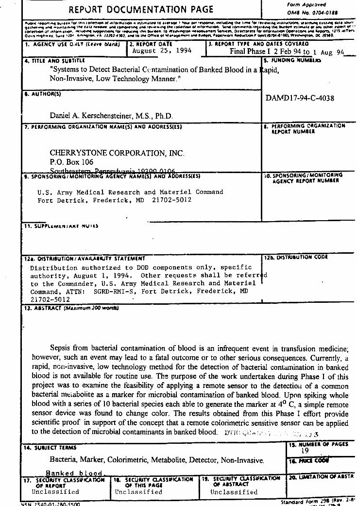
' I foron Appr •vedREPORT DOCUMENTATION PAGE IXI No. 0A0,.018
" -Coi 0t"'. 4vot 00Fq tr- toP..t *0I.¶ @4 tootPgOt .4 flt.'4dt" to a."&"q I d A" W. ft~hw" I M W"4 tfor fft,"0 WI%,.@wxsqq %@#cc"JO ,1c4.A .. tt * tI~qp4,J. &0%4 S.A&~4 1"4• 4 IP ViII A 4q*". On (of0sqtJ•4 Idd F" sA4, q IP. (C0i4Mtnp@ @4 0 M4iJOft. io* VA•A #E444d •4h D t Im r bft VIt|1•mu, W " Q~ rar of1C0iI*CJl0OIt ~ ,Alat--st-cm. ACI~dV.A %""Eiot.A for V@4XiW IPisEA toW nI *9U-.0*A "*"aOe Ed %Ifff .Crw J. OvctVotart# lta Infovmeoitn owscafot " Ato m 121% AtJIIOaw 0.9 04qdw$.41. IJ%04 1104, llqtOA. 44 IlflU .1 ~. &A'd to JJW 0"$hW of %f#A&.WP. &4 iVd Bt~de psefrwOftk 0t4,ddJOP 0 a (91044 Jul WeiV~Ws.W OC 24"J.
1. AGENCY USE OALY (Leavo 60ank) 2. REPORT DATE 13. REPORT TYPE AND DATES COVERED
August 25, 1994 Final Phase I 2 Feb 94 to 1 Aug 944. TITLE AND SUBTITLE S. FUNDING NUME[i
"Systems to Detect Bacterial Ccntamination of Banked Blood in a Iapid,Non-Invasive, Low Technology Manner."
4. AUTHOR(S) DAMD 17-94-C-403 8
Daniel A. Kerschensteiner, M.S., Ph.D.
7. PERFORMING ORGANIZATION NAME(S) AND AOORESS(ES) 8. PERFORMING ORGANIZATIONREPORT NUMBER
CHERRYSTONE CORPORATION, INC.P.O. Box 106
9. SPONSORING/ MONITORINGA-WtNCYKAI(S) A fMND ARESS(ES) '1O. SPONSORING I MONITORING
AGENCY REPORT NUMBER
U.S. Army Medical Research and Materiel Command
Fort Detrick, Frederick, MD 21702-5012
11. SUPPLcr.mviIAMr NUIit
12a. DISTRIBUTION/AVAILAaIUTY STATEMENT ... 12 S TITBUTION CODE
Distribution authorized to DOD components only, specificauthority, August 1, 1994. Other requests shall be referredto the Commander, U.S. Army Medical Research and MaterielCommand, ATTN: SGRD-RMI-S, Fort Detrick, Frederick, MD
21702-501213. ABSTRACT (Mazemum 200 words)
Sepsis from bacterial contamination of blood is an infrequent event in transfusion medicine;however, such an event may lead to a fatal outcome or to other serious consequences. Currently, arapid, nori-invasive, low technology method for the detection of bacterial contamination in bankedblood is not available for routine use. The purpose of the work undertaken during Phase I of thisproject was to examine the feasibility of applying a remote sensor to the detectiou of a commonbacterial mt'abolite as a marker for microbial contamination of banked blood. Upon spiking wholeblood with a series of 10 bacterial species each able to generate the marker at 40 C, a simple remotesensor device was found to change color. The results obtained from this Phase I effort providescientific proof in support of the concept that a remote colorimetric sensitive sensor can be appliedto the detection of microbial contaminants in banked blood. D' IC 3
14. SUIUECT TERMS 15. NUMBER Of PAGES
Bacteria, Marker, Colorimetric, Metabolite, Detector, Non-Invasive. 19 C cOOE
Banked bo -1 -O17. SE RJTY CLASSWICAflON 16. SECURITY CLASSIFICATIN 1. EUIT CLSSIFICATON 2.UTAOOAST
OF REPORT Of THIS PAGE OF ABSTRACT"Unclassified Unclassified Unclassified
-.ra, vrAA A, I • . . ...% Standard Form 28_(Re.. 22- 8
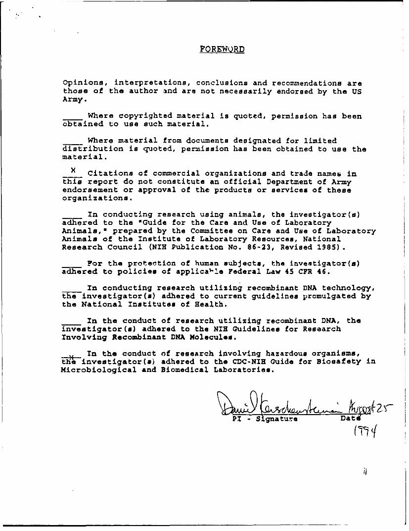
FOREWQRD
Opinions, interpretations, conclusions and recommendations arethose of the author and are not necessarily endorsed by the USArmy.
Where copyrighted material is quoted, permission has beenobtained to use such material.
Where material from documents designated for limiteddistribution is quoted, permission has been obtained to use thematerial.
X Citations of commercial organizations and trade names inthis report do not constitute an official Department of Armyendorsement or approval of the products or services of theseorganizations.
In conducting research using animals, the investigator(s)adhered to the "Guide for the Care and Use of LaboratoryAnimals," prepared by the Committee on Care and Use of LaboratoryAnimals of the Institute of Laboratory Resources, NationalResearch Council (NIH Publication No. 86-23, Revised 1985)
For the protection of human subjects, the investigator(s)adhered to policies of applical-!e Federal Law 45 CFR 46.
In conducting research utilizing recombinant DNA technology,the investigator(s) adhered to current guidelines promulgated bythe National Institutes of Health.
In the conduct of research utilizing recombinant DNA, theinvestigator(s) adhered to the NIH Guidelines for ResearchInvolving Recombinant DNA Molecules.
Tn the conduct of research involving hazardous organisms,the investigator(s) adhered to the CDC-NIH Guide for Biosafety inMicrobiological and Biomedical Laboratories.
PI - S gnature Date
I S gatu-
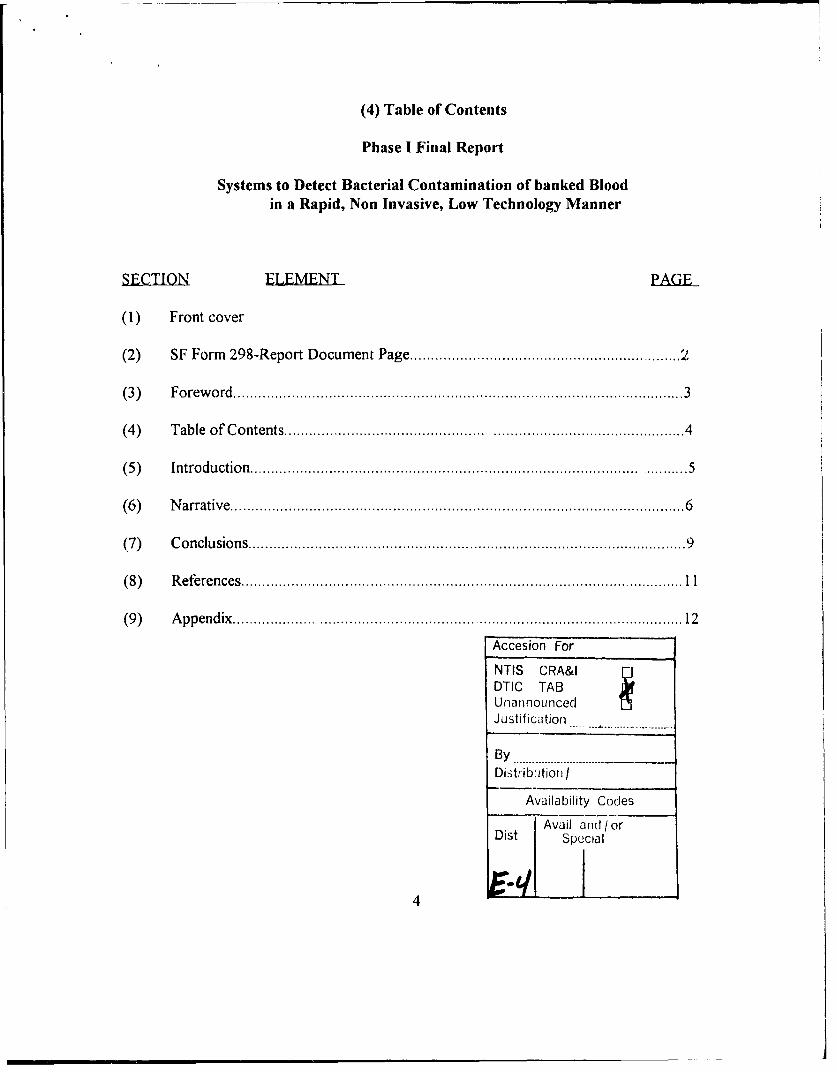
(4) Table of Contents
Phase I Final Report
Systems to Detect Bacterial Contamination of banked Bloodin a Rapid, Non Invasive, Low Technology Manner
SECTION FLEMENT PAGiE
(1) Front cover
(2) SF Form 298-Report Document Page ............................................................. 2
(3) F o rew o rd ................................................................................................ . . . . . 3
(4) Table of Contents ............................. .............. .............................................. 4
(5 ) In tro d u ctio n ............................................................................................. ........... 5
(6) N arrative ................................................................................................ . . . . . 6
(7) C o nclu sion s .................................................................................................. . . 9
(8 ) R e fe re n c e s .......................................................................................................... 1 1
(9 ) A p p e n d ix .......................................................................................................... 12Accesion For
NTIS CRA&IDTIC TABUnannouncedJustificatiori
By .............
DistribUtion I
Availability Codes
Avail and/ orDist Special
4
FINAL REPORT
(5) Introduction.
a. Nature of tProblem: Sepsis from bacterial contamination of blood is aninfrequent event in transfusion medicine; however, such an event may lead to, a fataloutcome or to other serious consequences (1). Currently, a rapid, non-invasive, lowtechnology method for the detection of bacterial contamination in banked blood is notavailable for routine use.
A major technical problem of detecting bacterial contamination in whole blood is avoidinginadvertent (additional) contamination through the analytical process or measurement--invasiveness of integrity; finding a marker or series of markers universally present innumerous potentially contaminating bacteria which is absent in blood and bloodcomponents--exclusivity; performing a candidate indicator test system rapidly orcontinuously with accumulating signaling--a tell-tale sensor; plus performing the detectionat low cost considering the large number of units collected--economical. Also, due to thelarge numbers of collected blood processed into components, inventoried and storagemaintained, it is advantageous to consider a signaling system which is capable ofautomated detection through unskilled operation--unattended operation.
b. Previous Work: Methods for the detection of microorganisms in liquid mediadesciibed in the literature involve the employment of costly equipment, inoculation ofsamples into a growth medium (invasiveness), significant time periods for incubation andoccasionally, the utilization of radioactive reagents. As one example, U.S. Patent No.5,232,839 (2), describes a method and apparatus that monitors the rate of change overtime in the pressure within the headspace above a sample in a closed vessel to provide anindication of microbial growth in the sample. Other methods include:
0 JMicroscopy. The microscopic evaluation of Gram-or acridine orange-stained bloodsmears has been considered a bacterial screening test to be performed just beforetransfusion. Because of the high. limits of reproducibly detecting bacteria and the laborintensity of the method, these have been considered poor screening techniques (8).
0 C'ulture methods. Instrumented blood culturing methods resulting in rapid detection ofbacteria by measuring increasing levels of carbon dioxide released by proliferating bacteriahave been tested. These methods are time consuming and may not be effective in detectingbacteria in freshly drawn units when contamination is low( 9).
C3 Nucleic acid hybridization. Has fast results using a "universally conserved" bacterialrRNA and detection limits of 104 CFU/ml. This test could potentially be performed justbefore transfusion( 10).
5
CI PCR. PCR-based methods have been developed but current problems inciude arduousnucleic acid preparation, extraneous contamination, and non-universal bacterial signaldetection( 11).
c. Purpose of Present Work: The purpose of the work undertaken during Phase Iof this project was to examine the feasibility of applying a remote sensor to the detectionof a common bacterial metabolite as a marker for microbial contamination of bankedblood.
d. Methods qf Approach: The remote sensor, ammonia-developing diazo paper orfilm, was affixed to the inner surfaces of the screw caps of sterile plastic vials. Thesematerials are sensitive to low levels of ammonia, a common microbial metabolite, andvisibly change color from light yellow to black when exposed to low levels of ammonia.Banked blood alone (controls) or blood spiked with ammonium carbonate; cell-freeammonia generating enzymes from bacteria and plant sources; or one of ten selectedbacterial species were incubated at 40 C for varying time periods. Sensor disks were thenexamined visually for a color change to black (yes/no) and then by Laser ScanningDensitometry for quantitation of the color changes.
(6) Narrative:
a. Eperimental Methods:
1. Bacteria: Ten bacterial species, representing a diversity of metabolicpathways for ammonia production, were obtained from the American Type CultureCollection (Appendix pAl). Among these species, some produce ammonia by action ofthe enzyme urease on urea (3, 4), others evolve ammonia by cleavage of arginine by theenzyme arginine dehydrolase, and the remainder by catabolic pathways involved in proteinand peptide degradation (5, 6). Lyophilized ATCC culture preparations were reconstitutedin Tryptone broth, streaked to agar media to check for purity, tested with API Diagnostickits to validate species identity and maintained as stocks on Tryptone agar slants stored at40 C. Working cultures for the experiments described below were initiated from irnoculafrom stocks to fresh Tryptone broth incubated overnight (18 to 20 hours) at 370 C.
2. Remote Sensor System: A variety of ammonia sensitive diazoprint papersand films were obtained commercially ( Azon Corporation, formerly Post & Company).Disks, approximately 1/4" in diameter, were cut from these materials and affixed to theinner surfaces of the screw caps to sterile plastic cryovials or cut into rectangular sheetsfor placement over microtiter platcs which had been charged with small volumes ofbacterial and substrate mixtures.
6
3. General Experimental Procedures:
a. Test Systeras__EmplQycd:
1. Preliminary Screening Experiments: Microtiter plates
containing 96 x 300 pA wells were utilized as a screening device in preliminary experimentsdesigned to detect ammonia evolution from bacterial-aqueous substrate mixtures. In these
screening experiments, 10 4l1 of the bacterial suspensions were added with mixing to 50 4liof aqueous substrate. The microtiter plates were then overlaid with rectangular sheets ofammonia-sensitive blueprint paper or film leaving a head space of approximately 10 mmbetween the paper or film and the liquid sample surfaces in the charged wells. The plateswere then incubated for periods of up to 7 days at ambient room temperature or at 40 C.Following incubation the ammonia-sensitive papers and films were removed and examinedfor the development of black spots above the sample wells.
2. Blood-Microbial Interactions: Sterile NUNC 5 ml screw-
capped plastic cryovials were used to evaluate the remote sensor system in experimentsdesigned to study ammonia evolution generated from bacterial contamination of sterilefetal calf serum or banked blood. In these experiments 0.2 ml of bacterial suspensionswere aseptically transferred to the bottom of the tubes and then 2.0 ml volumes of sterileserum or banked blood were added. Transparent screw caps to which 1/4" disks of diazoblack film had been attached to the inner surface were then tightly applied, leaving a headspace between the liquid sample surface and the inner surface of the cap of approximately3 cm. The sealed tubes were then incubated at 40 C for varying lengths of time up to 12days. The caps were then removed, the disks examined visually for a color change to blackand then removed from the caps and placed in rows on clear polystyrene plates. Densitiesof the disks were then quantitated by Laser Scanning Densitometry at 200 nm resolutionfrom 0 to 255 grey scale.
4. Results:
a Sg.eningExpriments in Microtiter Plates:
Bacterial suspensions in sterile saline, Tryptone broth, sterile fetal calf serum or 3 mMaqueous urea solutions were screened for their potential to evolve ammonia at levelssufficient for detection by a variety of commercially available ammonia-developing diazopapers or films. In these screens, uninoculated broth, sterile calf serum and sterile ureasolutions served as negative controls. Urea solutions containing Jack Bean urease and calfserum containing urease served as positive controls.
Qualitatively, all of the bacterial species screened in this manner producedammonia at levels sufficient to effect color changes of varying densities when mixed withcalf serum. The color change densities observed from the bacterial-serum mixtures weregenerally significantly greater than those observed from tne negative controls at bothambient room temperature and at 40 C. While all of the ammonia sensitive papers and
7
films screened by this method were observed to detect ammonia to varying degrees ofcolor density, Fast Speed Diazo Black film (FSDB) appeared to pmoduce the most rapidand consistent response to the ammonia evolved from these bacterial-serum mixtures. Forthis reason FSDB was selected for use as a remote sensor in the cryovial tcst system forstudies of Blood-bacterial interactions.
b. Cryovial Results.
1. Bood Spiked with Bacteria: A number of preliminaryexperiments utilizing 2.0 ml volumes of banked blood at bacterial contamination levels ofapproximately 103, 105 and 107 cfu/ml blood were completed to determine the validity ofthe test system. In general, the results observed during this preliminary work demonstratedthat
1) the FSDB disks were sensitive to the levels of ammonia produced by thecontaminated blood samples, both at ambient room temperate and at 40 C and
2) the degree of color change exhibited by the FSDB disks could be quantitated byLaser Densitometry.
We also observed that uncontaminated blood (negative controls) produced levels ofammonia lower than those observed with the experimentally contaminated blood, butnonetheless, detectable by the FSDB sensor system. Greenwalt, et al. (7) have shown thatlow levels of ammonia are a normal product of red blood cell metabolism.
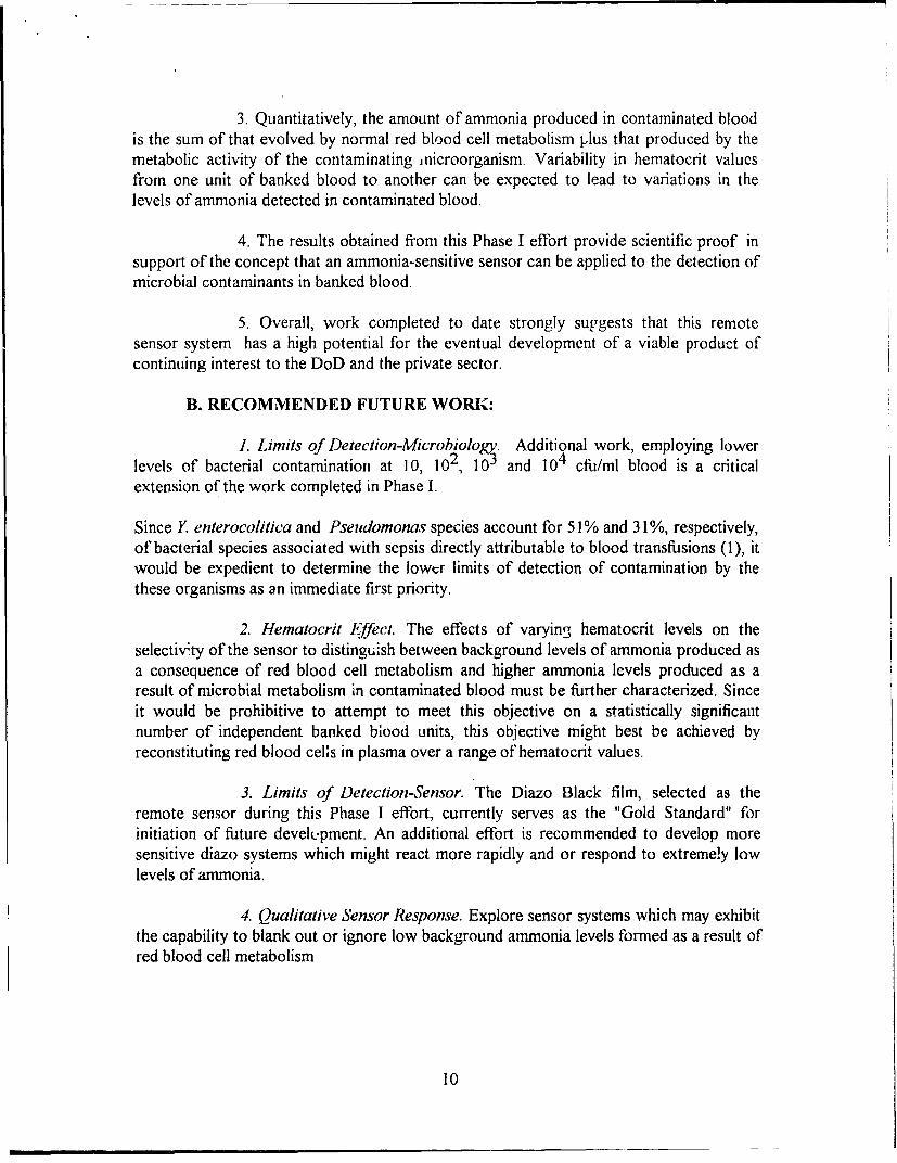
A major experiment was completed which involved deliberate contamination of bankedblood with ten different species of bacteria a. microbial levels of approximately Ito 5 x 10⁶ cfu/ml of lHood. These tubes, prepared in triplicate, were incubated in sets for 3,6, 9 or 12 days at 40 C and then examined visually and analyzed by Laser Densitometry.Because of the large number of tubes required, this study was carried out over twoconsecutive days and utilized blood from the same unit of blood on both days. Thus onDay 1 (series 1) blood was spiked with a set of 5 organisms and on Day 2 (series 2) bloodfrom the same unit was spiked with a different set of 5 organisms. The results obtainedfrom this experiment are as follows:
i. Eight of the ten selected bacterial species effected a color change in thediazo film which increased with time over 12 days aiid which in each casewas statistically significantly greater than those observed for uncontam-inated blood controls for all time periods (Table 2, Appendix pA2).
ii. Significant differences in red blood cell volumes were observed betweenthe series 1 and series 2 sets of tubes. Where the number of samplestotaled 54, the per cent settled red blood cells averaged 80% of totalattributable to settled red blood cells. These observed differences in redduring distribution of samples by gravity feed ( Table 3, Appendix pA2).
8
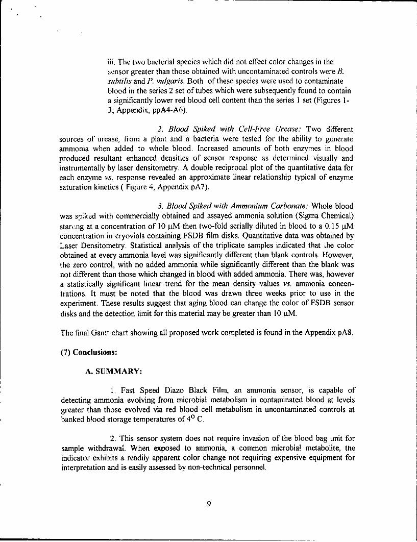
iii. The two bacterial species which did not effect color changes in the,ensor greater than those obtained with uncontaminated controls were B.suvbtilis and P. vulgaris. Both of these species were used to contaminateblood in the series 2 set of tubes which were subsequently found to containa significantly lower red blood cell content than the series I set (Figures 1-3, Appendix, ppA4-A6).
2. Blood Spiked with Cell-Free Urease: Two differentsources of urease, from a plant and a bacteria were tested for the ability to generateammonia when added to whole blood. Increased amounts of both enzymes in bloodproduced resultant enhanced densities of sensor response as determined visually andinstrumentally by laser densitometry. A double reciprocal plot of the quantitative data foreach enzyme vs. response revealed an approximate linear relationship typical of enzymesaturation kinetics ( Figure 4, Appendix pA7).
3. Blood Spiked with Ammonium Carbonate: Whole bloodwas s,ýpked with commercially obtained and assayed ammonia solution (Sigma Chemical)starcung at a concentration of 10 l.tM then two-fold serially diluted in blood to a 0.15 4Mconcentration in cryovials containing FSDB film disks. Quantitative data was obtained byLaser Densitometry. Statistical analysis of the triplicate samples indicated that &'he colorobtained at every ammonia level was significantly different than blank controls. However,the zero control, with no added ammonia while significantly different than the blank wasnot different than those which changed in blood with added ammonia. There was, howevera statistically significant linear trend for the mean density values vs. ammonia concen-trations. It must be noted that the blood was drawn three weeks prior to use in theexperiment. These results suggest that aging blood can change the color of FSDB sensordisks and the detection limit for this material may be greater than 10 4iM.
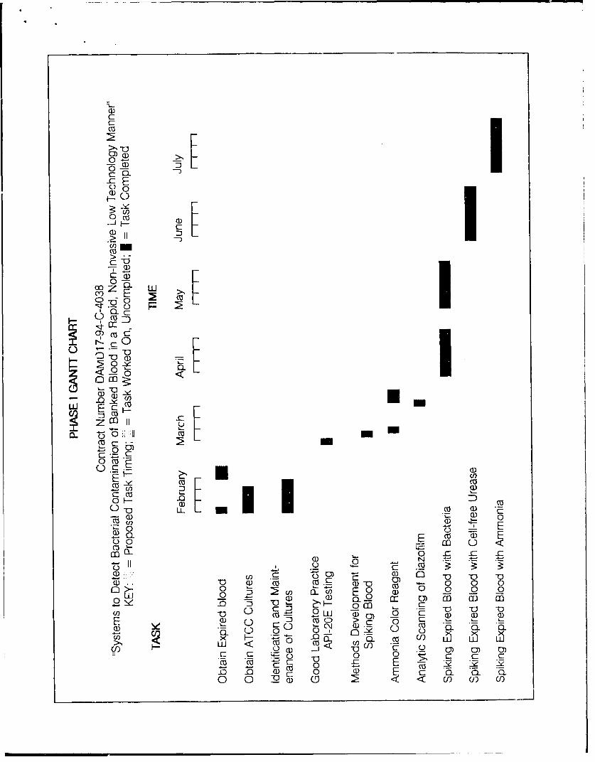
The final Gantt chart showing all proposed work completed is found in the Appendix pA8.
(7) Conclusions:
A. SUMMARY:
1. Fast Speed Diazo Black Film, an ammonia sensor, is capable ofdetecting ammonia evolving from microbial metabolism in contaminated blood at levelsgreater than those evolved via red blood cell metabolism in uncontaminated controls atbanked blood storage temperatures of 40 C.
2. This sensor system does not require invasion of the blood bag unit forsample withdrawal. When exposed to ammonia, a common microbial metabolite, theindicator exhibits a readily apparent color change not requiring expensive equipment forinterpretation and is easily assessed by non-technical personnel.
9
3. Quantitatively, the amount of ammonia produced in contaminated bloodis the sum of that evolved by normal red blood cell metabolism plus that produced by themetabolic activity of the contaminating ,nicroorganism. Variability in hematocrit valuesfrom one unit of banked blood to another can be expected to lead to variations in thelevels of ammonia detected in contaminated blood.
4. The results obtained from this Phase I effort provide scientific proof insupport of the concept that an ammonia-sensitive sensor can be applied to the detection ofmicrobial contaminants in banked blood.
5. Overall, work completed to date strongly suggests that this remotesensor system has a high potential for the eventual development of a viable product ofcontinuing interest to the DoD and the private sector.
B. RECOMMENDED FUTURE WORK:
1. Limits of Detection-Microbiology. Additional work, employing lowerlevels of bacterial contamination at 10, 102, 1"0 and 104 chu/ml blood is a criticalextension of the work completed in Phase I.
Since Y enterocolitica and Pseudomonas species account for 51% and 31%, respectively,of bacterial species associated with sepsis directly attributable to blood transfusions (1), itwould be expedient to determine the lower limits of detection of contamination by thethese organisms as an immediate first priority.
2. Hematocrit Effect. The effects of varying hematocrit levels on theselectivity of the sensor to distinguish between background levels of ammonia produced asa consequence of red blood cell metabolism and higher ammonia levels produced as aresult of microbial metabolism in contaminated blood must be further characterized. Sinceit would be prohibitive to attempt to meet this objective on a statistically significantnumber of independent banked btood units, this objective might best be achieved byreconstituting red blood cells in plasma over a range of hematocrit values.
3. Limits of Detection-Sensor. The Diazo Black film, selected as theremote sensor during this Phase I effort, currently serves as the "Gold Standard" forinitiation of future develupment. An additional effort is recommended to develop moresensitive diazo systems which might react more rapidly and or respond to extremely lowlevels of ammonia.
4. Qualitative Sensor Response. Explore sensor systems which may exhibitthe capability to blank out or ignore low background ammonia levels formed as a result ofred blood cell metabolism
10

5. Blood Bag Engineering. Finaiiy, efforts should be initiated towardsengineering blood bags to which the sensor is affixed to the exterior of the bag, or issensitive to gaseous ammonia that permeates the wall of the bag, and is protected againstlight destabilization and exhibits long shelf life stability.
(8) References:
1. Wagner, S.J., et al. Transfusion-associated bacterial sepsis. Clin. Microbiol.Rev. 7:290-303 (1994).
2. Gideon, E. and Sullivan, N.M. Detecting microbial growth. 1993. U.S.PatentNo. 5,232,839.
3. Mobley, H. and Hausinger, R.P. Microbial ureases: Significance, regulation andmolecular characterization. 1989. Microbiol. Rev. 53: 85-108 (1989).
4. Vince, A.G. Metabolism of ammonia, urea and amino acids and theirsignificance in liver disease. in Microbial Metabolism in the Digestive Tract. Hill, M.J (ed).CRC Press, Boca Raton. Florida 1987, pp 83-106.
5. Gottschalk, G. Bacterial Metabolism. Springer-Verlag, New York 1970.6. Dagley, S. and Nicholson, D. E. An Introduction to Metabolic Pathways. John
Wiley & Sons, New York, 1970.7. Greenwalt, T.J., et al. Studies in red blood cell preservation. 3. A phosphate-
ammonium-adenine addition solution. Vox Sang. 58: 94.99(1990).8. Reikin H. and Rubin, S.J. Evaluation of the buffy-coat smear for rapid detection
of bacteremia. :JAMA 245: 357-359 (1981).9. Arnow, P.M., el al. Eschericia coli sepsis from contaminated platelet
transfusion.Arch Intern. Med 146: 321-324 (1986).10. Brecher, M.E. ez al. The use of a chemiluminescence-link universal bacterial
ribosomal RNA gene probe and blood gas analysis for the rapid detection of bacterialcontamination in white cell-reduced and non-reduced platelets. Transfusion 33: 450-457(1993)
11. Feng, P. et al. Direct identification of Yersinia enterocolitica in blood ofpolymerase chain reaction amplification. Tranqfusion 32: 850-854 (1992).
11

(9) Appendix:
PAGEA TITLE
IA Table 1- Selected Bacteria
2A Table 2- Statistical Analysis
Table 3- Red Blood Cell Volumes
3A Figure 1-Sensor Response to S.epidermis in whole blood over 12days at 40 C.
4A Figure 2- Net Sensor DensityChanges-Series 1
5A Figure 3-Net Sensor DensityChanges-Series 2
6A Figure 4- Sensor Density Changes vs.Urease Activity in Whole Blood
7A Phase I Final Gantt Chart
12

* + * + * + + I
cl..
+ + S + I
+4 +n -4 +-* I
n f4I- e
zz z
- . ~ I- -0% 0% sf
L40 w- V)0- U2 f2 t ~
0~ O - 'O 0 ~ ' 0
I Hlgd

TABLE 2-STATISTICAL ANALYSIS
Bonferroni Multiple Comparison Test of Film Color IntensityGenerated by Bacteria in Blood Compared to Control
INCUBATION PERIOD
ORGANISM Day3 Day 6 Day 9 Day 12
SERIES I
Pseudomonas aeruginosa ns ns ns
Staphylococcus epiderm ns ** ** **
Klebsiella pneumouiae ns **
Streptococcus faecalis ns ns ns *
Escherichia coli ns ns *
SERIES 2
Staphylococcus aureus ns ns ns **
Bacillus subtilis ns ns ns ns
Serratia rubidea ns ns *** ns
Proteus vulgaris ns ns ns ns
Yersinia enterocolitica ns ns *** ns
KEY: ns = not significant; * = p< 0.05; ** = p< 0.01; * = p< 0.001
TABLE 3-RED BLOOD CELL VOLUMES
Percentage of Settled Red Blood Cells per Tube Volumefor Two Series of Experiments and their Net Mean Color Inensities
Percent of Volumej, ni=54 Pooled Net Chnogg + SEM
Bacteria Series 1 80 22925 + 3832
Bacteria Series 2 34 6346 ± 1617
2A
FIGURE 1SElSOR RESPONSE TO S. epidermis IN WHOLE BLOOD OVER 12 DAYS AT 4-C.
100000
60000-L
c
40000-
10
A c E 0 H
Columns
KEY:
A=DAY 3 EXPERIMENTALB=DAY 3 CONTROL
C=DAY 6 EXPERIMENTALD =DAY 6 CONTROL
E=DAY 9 EXPERIMENTALF= DAY 9 CONTROL
G=DAY 12 EXPERIMENTALH=DAY 12 CONTROL
I= BLANK VALUE
3A
FIGURE 2
NET SENSOR DENSITY CHANGFS-SERIES I
Time Course of Net Density Changefoi Series 1 Bacteria in Blood
Density (xW000)
50
40 - -
30 \
20--
10-
-10 -- I I I i I 1 _ _
0 2 4 6 8 10 12 14
Time, days
Pseudomonas--I- Staph epider _,$ Klebsiella
- Streptococcus-÷- Escherichia
4A
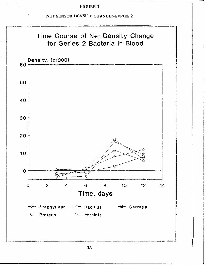
FIGURE 3
NET SENSOR DENSITY CHANGES-SERIES 2
Time Course of Net Density Changefor Series 2 Bacteria in Blood
Density, (x1000)60
50
40
30
20-
10
0-
0 2 4 6 8 10 12 14
Time, days
SStaphyl aur A Bacillus ---- Serratia
-- Proteus - Yersinia
5A
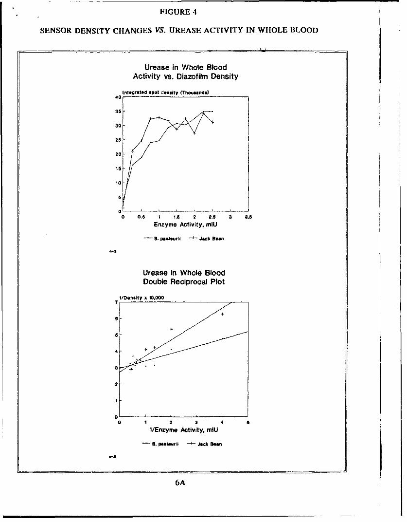
FIGURE 4
SENSOR DENSITY CHANGES VS. UREASE ACTIVITY IN WHOLE BLOOD
Urease in Whole BloodActivity vs. Diazofilm Density
Integrated spot iunsity (Thousands)
30
25
20
16
10
0 0.6 1 1.6 2 2.6 3 3.5
Enzyme Activity, miU
B- . peateuril -4- Jack Bean
Urease in Whole BloodDouble Reciprocal Plot
l/DenlJty x 10,000
4--
2
1
0 1 2 3 4 a
i/Enzyme Activity, mIU
6. patsmali - Jack Been
wa
6A
031*1
c
C- DO
TC:
co - I-
OCl~
0 In
- C
-ci
00
E coU) D CD r
cCIO ) U _ ~ gtQ) (z- m
6'0 c0~ ~ O ~ ~ ~ C0DC EC0 C s-
Cl) 0~ 0E o ~WJ <
0 0_r_ C Ca) 0 AJ E :z_-
Z5 .~ 0 CD: co0 a) w~ (b Q: _0 0) (j 0

MCMR-RMI-S (70-1y) 21 Jan 00
MEMORANDUM FOR Administrator, Defense Technical InformationCenter, ATTN: DTIC-OCA, 8725 John J. KingmanRoad, Fort Belvoir, VA 22060-6218
SUBJECT: Request Change in Distribution Statement
1. The U.S. Army Medical Research and Materiel Command hasreexamined the need for the limitation assigned to technicalreports written for the attached Awards. Request the limiteddistribution statements for Accession Document Numbers listed bechanged to "Approved for public release; distribution unlimited."These reports should be released to the National TechnicalInformation Service.
2. Point of contact for this request is Ms. Virginia Miller atDSN 343-7327 or by email at [email protected].
FOR THE COMMANDER:
En PHYLlSM. RINEHARTas Deputy Chief of Staff for
Information Management