newborn findings. 42 week infant with diffuse peeling and cracking
TRANSCRIPT

Newborn findings


• 42 week infant with diffuse peeling and cracking


• Lanugo – Fine body hair resembling peach fuzz present in
infants 24-32 weeks gestation


Brachial plexus injury
• A, Traction injury to C5, C6, and C7 spinal cord segments produces this (Erb) palsy. This infant demonstrates the characteristic posture of the limply adducted and internally rotated arm
• B and C, Infant with a Klumpke palsy involving lower segments of C7 and T1


• Supernumerary digit– Thin pedicle distinguishes this anomaly from true
polydactyl


• Bilateral polydactyly of the fifth toe


• Syndactyl– Bilateral fusion of soft tissue between first and
second toes– Surgical correction ~ 3 yrs of age– Sooner if cartilage or bony union


• Multiple pre-auricular skin tags• Remnants of the first branchial arch


• Ear pit or congenital aural fistula– Familial– More common in girls and african-americans– Can become infected


• Umbilical hernia– Defect of the central fascia– Common finding– Differentiate from a small omphalocele (more like
a fixed hernia)– True umbilical hernias require no therapy– If remain >3 yrs, consider surgical repair


• “Mongolian spot”– Patchy areas of hyperpigmentation– Epithelial cells with increased amounts of melanin


Transient pustular melanosis
• Self-limited dermatosis of unknown etiology• Present at birth• 1 to 2 mm vesiculopustules that disappear in
24-48 hrs• Lesions more prominent on neck, forehead,
lower back and legs• Self-limiting• Wright stain shows multiple neutrophils


Erythema toxicum
• Numerous yellow papules and pustules surrounded by intense erythematous rings
• Benign, self-limited• Lesions appears in 24-48 hrs of life up to 10 days• Can range from a few to several hundred lesions• Smear shows eosinophil predominence• Self resolves in 5-7 days


Sebaceous gland hyperplasia
• Common entity• 1-2 cm yellowish-white papules over the
nose/cheeks of full term babies• Normal physiologic response to maternal
androgenic stimulation of sebaceous gland growth
• Self-resolve in 4-6 months


Milia
• White papules on this baby's chin and cheeks.• Keratin filled epithethial cysts which occur in
up to 40% of newborns. • Spontaneous exfoliation and resolution is
expected within a few weeks. • Present at birth and have no inflammatory
component.


Cutis marmorata
• Transient net-like reddish-blue mottling of the skin
• Due to variable vascular constriction and dilatation
• Usually abates by 6 months of age

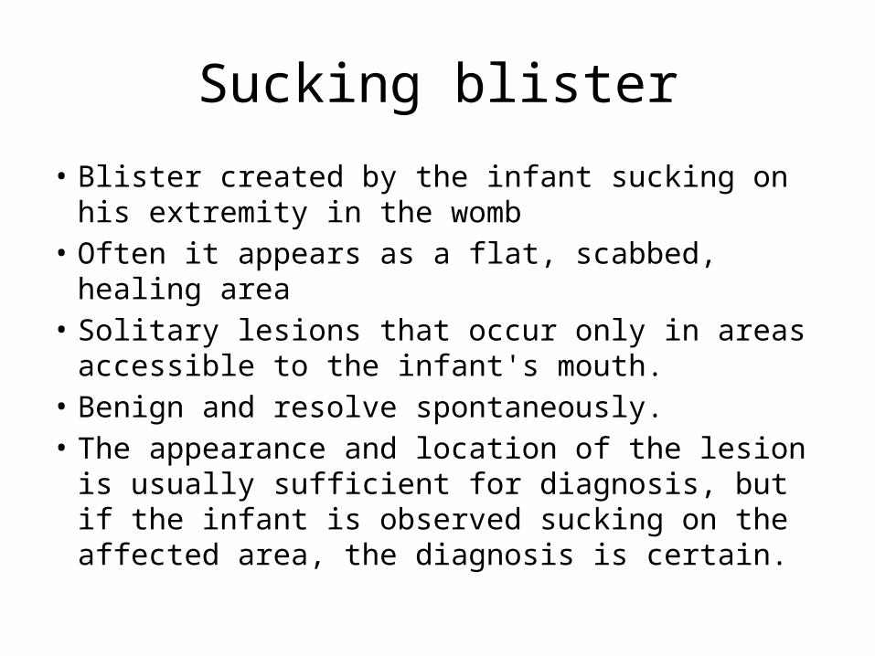
Sucking blister
• Blister created by the infant sucking on his extremity in the womb
• Often it appears as a flat, scabbed, healing area • Solitary lesions that occur only in areas accessible to
the infant's mouth. • Benign and resolve spontaneously. • The appearance and location of the lesion is usually
sufficient for diagnosis, but if the infant is observed sucking on the affected area, the diagnosis is certain.


Bruise from vaccum extraction
• Bruising is similar to that which occurs normally during the process of delivery except for the fact that it is well circumscribed.
• Bruising can be more severe with associated blisters or sloughing of the skin, or with underlying cephalohematoma or subglaeal hemorrhage.
• This finding spontaneously resolved.


Subconjunctival hemorrhage
• Frequent finding in normal newborns• Breakage of small vessels during the pressure
of delivery. • The red area may be large or small but is
always confined to the limits of the sclera. • It is asymptomatic, does not affect vision, and
spontaneously resolves in several days


Epstein pearl
• White papule seen in the midline of the palate.
• It represents epithelial tissue that becomes trapped during the palatal fusion.
• It is a very common and benign finding.

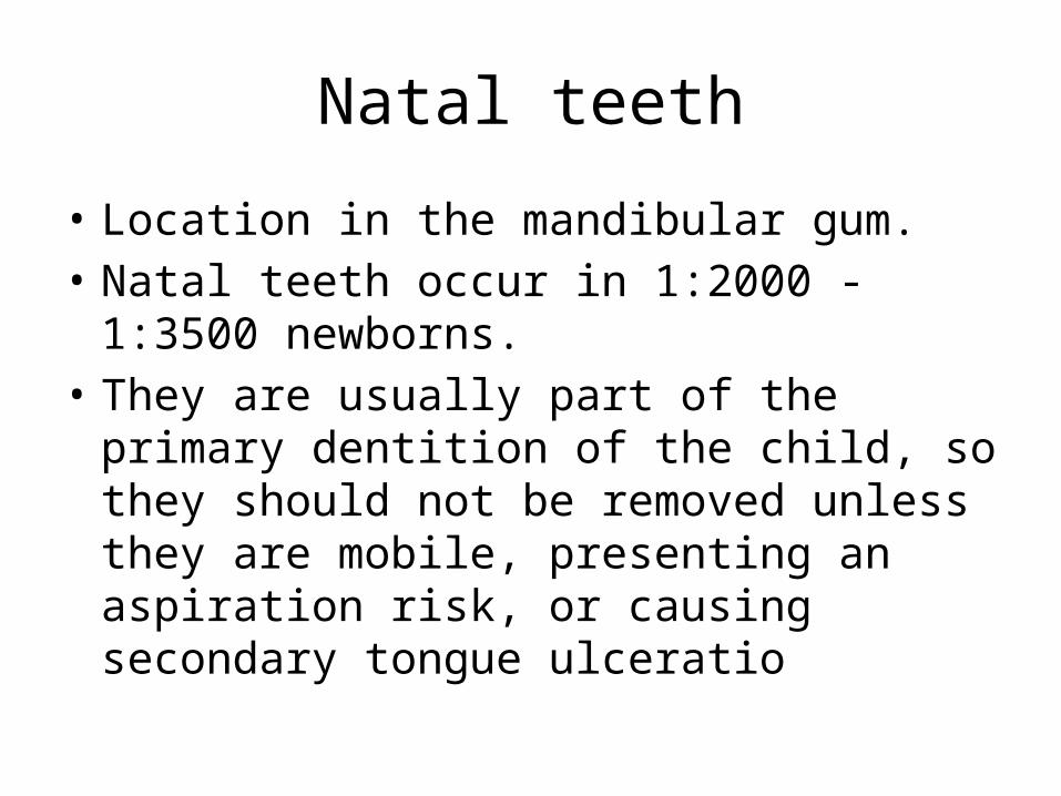
Natal teeth
• Location in the mandibular gum. • Natal teeth occur in 1:2000 - 1:3500
newborns. • They are usually part of the primary dentition
of the child, so they should not be removed unless they are mobile, presenting an aspiration risk, or causing secondary tongue ulceratio

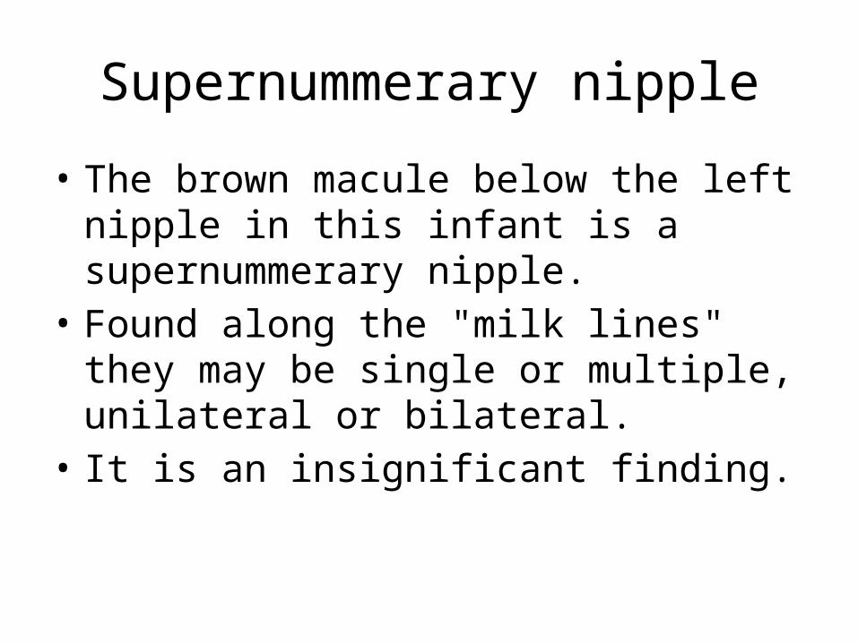
Supernummerary nipple
• The brown macule below the left nipple in this infant is a supernummerary nipple.
• Found along the "milk lines" they may be single or multiple, unilateral or bilateral.
• It is an insignificant finding.
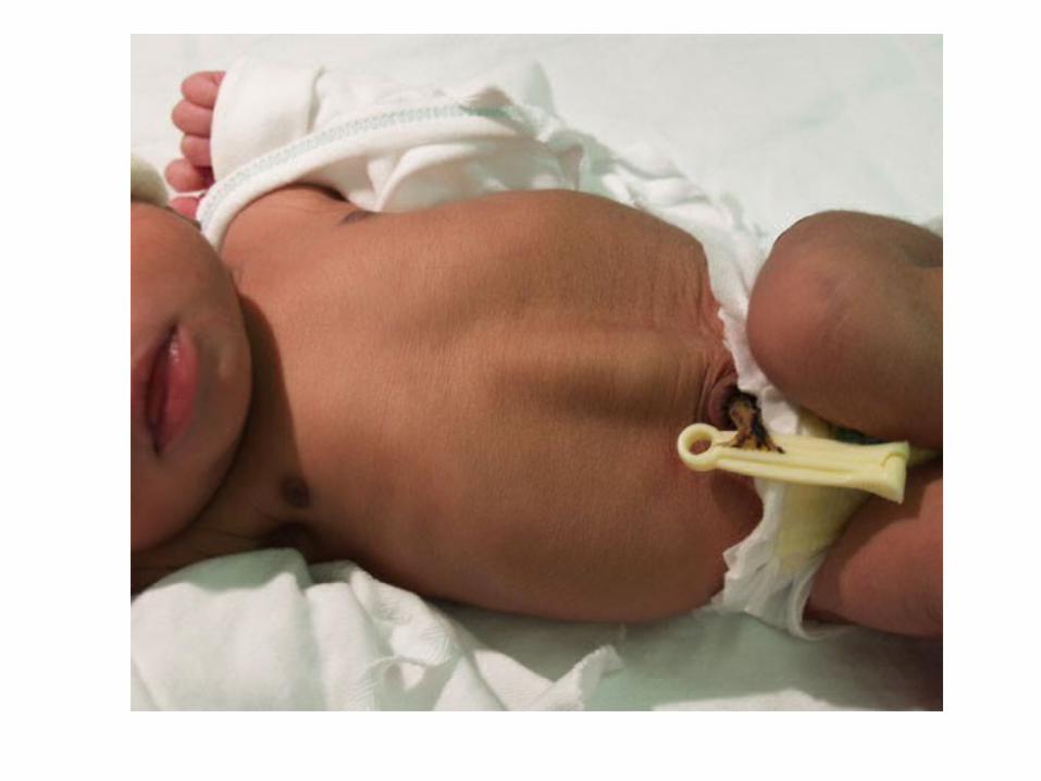

Diastasis recti
• Vertical bulge down the midline of the abdomen can be seen in many newborns when intra-abdominal pressure increases.
• Diastasis recti is caused by a relative weakness of the fascia between the two rectus abdominus muscles.
• It is not a herniation• Self-resolves


Epidermolysis bullosa
• Group of inherited mechanobullous disorders• Characterized by the development of blisters
after the skin is subjected to mild friction or trauma
• 3 types: epidermolytic, junctional and dermolytic


Incontinentia pigmenti
• A, Linearly distributed vesicles on an erythematous base are seen on the legs of this neonate.
• B and C, Subsequently, lesions evolve into warty papules, which can have thick overlying crusts.
• D, Splotchy hyperpigmented patches replaced the warty lesions by 8 months of age.
• E, In many cases the hyperpigmentation appears in swirls and streaks.
• F, These hypopigmented reticulated lesions on the leg of an affected child's mother represent old scars in areas of prior hyperpigmentation.

IP
• X-linked dominant disorder affecting skin, CNS, eyes and skeletal system
• Up to 30% have seizures, developmental delay, spasticity
• Opthalmic complications: strabismus, cataracts, blindness, micropthalmia
• 65% have pegged teeth and delayed dentition


Juvenile xanthogranuloma
• Results from infiltration and proliferation of histiocytes