niche-derived soluble dlk1 promotes glioma growth
TRANSCRIPT

Volume 22 Number 12 December 2020 pp. 689–701 689
Original Research
Niche-derived soluble DLK1 promotes
glioma growth
✩ , ✩✩ , ★
Elisa S. Grassi 1 ; Pauline Jeannot;
Vasiliki Pantazopoulou; Tracy J. Berg;
Alexander Pietras
∗
Division of Translational Cancer Research, Department of Laboratory Medicine, Lund University, Lund, Sweden
Abstract
Tumor cell behaviors associated with aggressive tumor growth such as proliferation, therapeutic resistance, and stem cell characteristics are regulated in part by soluble factors derived from the tumor microenvironment. Tumor-associated astrocytes represent a major component of the glioma tumor microenvironment, and astrocytes have an active role in maintenance of normal neural stem cells in the stem cell niche, in part via secretion of soluble delta-like noncanonical Notch ligand 1 (DLK1). We found that astrocytes, when exposed to stresses of the tumor microenvironment such as hypoxia or ionizing radiation, increased secretion of soluble DLK1. Tumor-associated astrocytes in a glioma mouse model expressed DLK1 in perinecrotic and perivascular tumor areas. Glioma cells exposed to recombinant DLK1 displayed increased proliferation, enhanced self-renewal and colony formation abilities, and increased
levels of stem cell marker genes. Mechanistically, DLK1-mediated effects on glioma cells involved increased and prolonged stabilization
of hypoxia-inducible factor 2alpha, and inhibition of hypoxia-inducible factor 2alpha activity abolished effects of DLK1 in hypoxia. Forced expression of soluble DLK1 resulted in more aggressive tumor growth and shortened survival in a genetically engineered
mouse model of glioma. Together, our data support DLK1 as a soluble mediator of glioma aggressiveness derived from the tumor microenvironment.
Neoplasia (2020) 22, 689–701
✩
Keywords: DLK1, Hypoxia, HIF-2a, Glioma, Tumor-associated astrocytes, Stem
∗ Corresponding author. E-mail address: [email protected] (A. Pietras).
✩ Abbreviations: ACM, astrocyte-conditioned media, CCGA, Chinese Glioma Genome Atlas, DLK1, delta like noncanonical Notch ligand 1, GBM, glioblastoma multiforme, GFAP, glial fibrillary acidic protein, HIF, hypoxia-inducible factor, HREs, hypoxia- responsive elements, IR, ionizing radiation, OCT, optimal cutting temperature compound, PDGF, platelet-derived growth factor, PFA, paraformaldehyde, PIGPC, Pdgfb-induced glioma primary culture, RCAS, replication-competent avian sarcoma-leukosis virus long- terminal repeat (LTR) with splice acceptor, RIPA, radioimmunoprecipitation assay, SVZ, subventricular zone, TCGA, The Cancer Genome Atlas. ✩ Funding: This work was supported by grants to AP from the Ragnar Söderberg Foundation (M20_16), the Swedish Cancer Society (19 0283; CAN 2016/328; CAN
2017/975), the Swedish Research Council (2014-02406; 2018-02864), the Swedish Childhood Cancer Fund (PR2017-0040; PR2014-0157; TJ2014-0016), Ollie & Elof Ericssons foundation, Jeanssons stiftelser, the Crafoord foundation, Gösta Miltons donationsfond, and Stiftelsen Cancera. ★ Declaration of Competing Interest: The authors declare no conflicts of interest.
1 Present address: Department of Clinical Sciences and Community Health (DISCCO), University of Milan, Milan, Italy.
Social media: @pietraslab (A. Pietras) Received 21 August 2020; received in revised form 30 September 2020; accepted 4 October 2020; Available online xxx
© 2020 The Authors. Published by Elsevier Inc. This is an open access article under the CC
BY license ( http://creativecommons.org/licenses/by/4.0/ ) https://doi.org/10.1016/j.neo.2020.10.005
I
rl
m
a
m
a
s
rn
og
t
ss
[
p
N
cell niche
ntroduction
Glioblastoma recurrence following standard-of-care treatment with adiotherapy, surgery, and chemotherapy invariably gives rise to incurable esions, and the median survival following diagnosis remains at 12 to 20
onths despite recent advances in our understanding of glioblastoma at molecular level [1] . Evidence from human patient samples and murineodels of brain tumors suggest that inherent therapeutic resistance within
subset of tumor cells with stem cell characteristics may be the primaryource of recurrent tumors [2 , 3] . While the origin and fate of such cellsemain controversial, glioblastoma cell phenotypes are highly plastic, and on −stem-like cells can acquire characteristics of stem cells as a resultf microenvironmental interactions with the extracellular matrix, with rowth factors, or as a result of altered oxygenation or pH [3–6] . Indeed,umor cells with stem cell properties appear to be spatially restricted topecific microenvironments such as the perinecrotic and perivascular niches, uggesting that these niches may control residing tumor cell phenotypes4 , 5 , 7] .
Delta Like noncanonical Notch ligand 1 (DLK1) is a transmembranerotein in the Notch family of ligands, that is capable of signaling in aotch-dependent and -independent manner depending on cellular context

690 Niche-derived DLK1 supports glioma growth E.S. Grassi et al. Neoplasia Vol. 22, No. 12, 2020
Figure 1. DLK1 expression in murine glioma. (A-C) Representative images of immunofluorescent stainings showing tumor-associated astrocytes and DLK1 localization in bulk tumor (A) and perinecrotic (N) and perivascular (V) areas (B, D, respectively) of shp53-induced murine gliomas. Scalebars represent 25 μm. (D, E) Representative images of immunofluorescent stainings showing tumor-associated astrocytes and N-terminal, secreted, DLK1 localization in bulk tumor (D) and perinecrotic areas (N) (E) of shp53-induced murine gliomas. (F-G) Colocalization analysis of immunofluorescent staining showing DLK1 and GFAP
expression in bulk tumor vs perinecrotic (F) and perivascular (G) niches. (H) Colocalization analysis of immunofluorescent staining showing secreted DLK1 and GFAP expression in bulk tumor vs perinecrotic niche. Scalebars represent 25 μm. Statistical analysis: (A-H) n = 3. Statistical significance was determined with t test with Welch’s correction for unequal variances applied to Pearson’s coefficients (F-H). In the whole figure significance is represented as ∗P < 0.05 and ∗∗P < 0.01 vs bulk tumor. DLK1, delta-like noncanonical Notch ligand 1; GFAP, glial fibrillary acidic protein.
wn
big
[8–13] . Expression of DLK1 is increased with tumor grade in glioma, andits signaling has been associated with various properties of aggressive tumorcells [14 , 15] . The mechanisms underlying these effects on tumor cell behaviorremain poorly understood, but likely include signaling from the extracellular,soluble domain of DLK1 [16] . Indeed, soluble DLK1 secreted from astrocytes
as recently shown to be a critical component of the subventricular zone eural stem cell niche [11] . Astrocytes represent a prominent cell type in therain tumor microenvironment [17] , and recent studies revealed that DLK1 s one of the top upregulated genes in tumor-associated astrocytes of high- rade vs low-grade gliomas [18] . The regulation of DLK1 expression is poorly

Neoplasia Vol. 22, No. 12, 2020 Niche-derived DLK1 supports glioma growth E.S. Grassi et al. 691
Figure 2. Effects of soluble DLK1 and astrocyte-derived factors on glioma cells. (A) ELISA assay data showing DLK1 secretion in human fetal astrocytes exposed to 1-time 10 Gy irradiation or 1% O 2 for up to 9 days. (B) Media transfer experiments showing the effects of normoxic and hypoxic Astrocyte- Conditioned media (ACM) on human glioma cell lines. (C) Representative images and densitometric analysis of western blots showing His-tagged soluble DLK1 expression in U3082MG, U3084, and U3065 cells transient transfection. (D) ELISA assay data showing DLK1 secretion in human glioblastoma cells transfected with soluble DLK1. (E) Media transfer experiments showing the effects of media from glioblastoma cells overexpressing soluble DLK1 on human glioma cell lines themselves. F: Proliferation assays of glioma cells treated with recombinant DLK1 for 72 hours. Statistical analysis: (A, B) n = 4 (C, D, E) n = 3, (F) n = 6. Statistical significance was determined by 2-way ANOVA (A, B) and t test (C-F), in E and F Welch’s correction for unequal variances was applied. In the whole figure significance is represented as ∗P < 0.05, ∗∗P < 0.01, and ∗∗∗P < 0.001 vs untreated controls. DLK1, delta-like noncanonical Notch ligand 1.
M
G
Rccaa
understood, but some elements of the brain tumor microenvironment suchas hypoxia have been shown to drive DLK1 expression [19 , 20] .
Here, we sought to investigate the role of soluble DLK1 in the high-grade glioma tumor microenvironment. We found increased secretion ofDLK1 from tumor-associated astrocytes subjected to stresses of the tumormicroenvironment, such as hypoxia and ionizing radiation. Soluble DLK1increased proliferation and stem cell characteristics of glioma cells, andpromoted tumor growth in a genetically engineered mouse model of glioma.Together, our findings suggest that soluble DLK1 is a niche-derived mediatorof aggressive tumor growth in brain tumors.
aterials and methods
lioma mouse models
Nestin-tv-a Ink4a/Arf −/ − mice (IMSR Cat# NCIMR:01XH4, RID:IMSR_NCIMR:01XH4) were intracranially injected with DF1 ells (ATCC Cat# CRL-12203, RRID:CVCL_0570) expressing replication- ompetent avian sarcoma-leukosis virus long-terminal repeat with splice cceptor (RCAS) encoding human platelet-derived growth factor B (PDGFB) nd RCAS-short hairpin p53 (RCAS-shp53), as indicated, in the neonatal

692 Niche-derived DLK1 supports glioma growth E.S. Grassi et al. Neoplasia Vol. 22, No. 12, 2020
Figure 3. Effects of soluble DLK1 on glioma cells. (A) Proliferation curves of U3082MG, U3084MG, U3065MG, and PIGPC cells grown at increasing concentrations of recombinant DLK1 for 72 hours. Data are expressed as fold change of untreated control. (B, C) Representative images and quantification of colony forming ability of U3082MG, U3084MG, U3065MG, and PIGPC cells grown at increasing concentrations of recombinant DLK1. Data are expressed as fold change of untreated control. Statistical analysis: independent experimental replicates are as follow, (A) n = 6, except U3065MG where n = 4, (C) n = 4, Statistical significance was determined by 1-way ANOVA, followed by Bonferroni post hoc test. In the whole figure significance is represented as ∗P <
0.05 and ∗∗P < 0.01 vs untreated controls. DLK1, delta-like noncanonical Notch ligand 1; PIGPC, primary murine glioma cell.
a(S
((
Ra5
DR
po
Ra
Ae
P
css
C
i
brain with a 10- μL gas-tight Hamilton syringe, as described previously[21 , 22] .
Soluble DLK-S was cloned into the RCAS vector and mice wereco-injected with a 1:1 mix of DF1 cells expressing RCAS-PDGFB andRCAS-DLK-S or empty RCAS as indicated. Each litter was allocated to1 experimental group. Mice were monitored daily and sacrificed upondisplaying brain tumor symptoms. All procedures were approved by theSwedish Board of Agriculture through the Malmö-Lund Regional Committee(permit M186–14). The sample number was determined based on the lawof diminishing returns with the resource equation method (total number ofanimals - total number of groups > 10). A total of 6 pups were excluded dueto nontumor symptoms during week 0 to 3, the final numbers were n = 26for PDGFB and n = 24 for DLK-S.
Immunofluorescence
Whole brains were embedded in optimal cutting temperaturecompound (OCT) (ThermoFisher) and frozen in precooled isopentane.Five micrometers thick cryosections were air-dried for 30 minutes, thenfixed in ice-cold acetone or 4% paraformaldehyde and permeabilizedusing 0.3% Triton X-100 in phosphate buffered saline (PBS) (Sigma).Blocking was performed using serum-free protein block (DAKO), thensections were incubated overnight with primary antibodies at 4 °Cwith background reducing components (DAKO). Alexa Fluor secondaryantibodies (Abcam) were used, and Vectashield Mounting medium withDAPI (Vector Laboratories) was used for mounting.
Primary antibodies: Pref-1/DLK1/FA1 antibody (Novus, Cat# NBP2-33697), DLK1 polyclonal antibody (Thermo Fisher Scientific Cat# PA5–72199, RRID:AB_2718,053), Mouse Pref-1/DLK1/FA1 Antibody (R and DSystems, Cat# AF8277), hypoxia-inducible factor 2 alpha (HIF-2a) antibody(Abcam Cat# ab199, RRID:AB_302739), Goat Anti-Human Olig2 (R
nd D Systems Cat# AF2418, RRID:AB_2157554), Chicken Anti-GFAP
Abcam Cat# ab4674, RRID:AB_304558), Ki-67 antibody (Thermo Fisher cientific Cat# RM-9106-S0, RRID:AB_2341197).
Secondary antibodies: Donkey Anti-Goat IgG H&L Alexa Fluor 555 Abcam Cat# ab150134, RRID:AB_2715537), Donkey anti-Rabbit IgG
H + L ) Alexa Fluor 488 (Thermo Fisher Scientific Cat# A-21206,RID:AB_2535792), Donkey anti-Chicken IgY H&L FITC (Abcam Cat# b63507, RRID:AB_1139472) Goat anti-Rabbit IgG ( H + L ) Alexa Fluor 68 (Thermo Fisher Scientific Cat# A-11011, RRID:AB_143157).
Images were acquired using an Olympus BX63 microscope and P80 camera and CellSens software (Olympus CellSens Software, RID:SCR_016238).
For DLK1 and GFAP localization images ( Figure 1 ), minimal ostproduction consisting of background subtraction and automated level ptimization was equally applied with ImageJ (Fiji, RRID:SCR_002285).
Colocalization analysis was performed with ImageJ (Fiji, RID:SCR_002285) Coloc2 plugin on the selected Regions of Interest of t least 3 independent experiments.
Ki67 quantification was performed with CellProfiler (CellProfiler Image nalysis Software, RRID:SCR_007358), at least 3 fields were analyzed for ach tumor, for a total of 101,717 nuclei analyzed, with n = 45,466 forDGFB tumors and n = 56,251 for DLK-S tumors.
Areas of necrosis were identified by 2 independent researchers based on ellularity and the presence of pseudopalisading nuclei, based on the DAPI tain. Vessels were identified by morphological inspection of DAPI and GFAP
tains.
ell culture and treatments
Primary murine glioma cells (PIGPCs [23] ) were cultured as described n DMEM (Life Technologies) + 10% fetal bovine serum and 1%
Neoplasia Vol. 22, No. 12, 2020 Niche-derived DLK1 supports glioma growth E.S. Grassi et al. 693
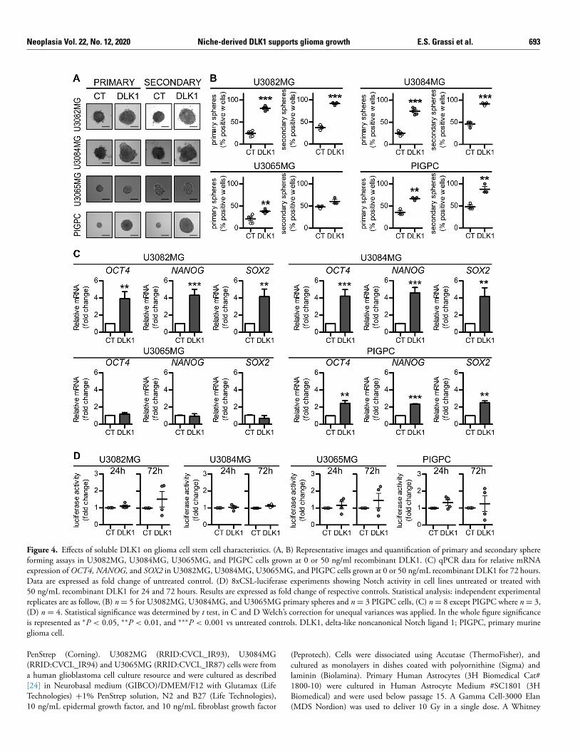
Figure 4. Effects of soluble DLK1 on glioma cell stem cell characteristics. (A, B) Representative images and quantification of primary and secondary sphere forming assays in U3082MG, U3084MG, U3065MG, and PIGPC cells grown at 0 or 50 ng/ml recombinant DLK1. (C) qPCR data for relative mRNA
expression of OCT4, NANOG, and SOX2 in U3082MG, U3084MG, U3065MG, and PIGPC cells grown at 0 or 50 ng/mL recombinant DLK1 for 72 hours. Data are expressed as fold change of untreated control. (D) 8xCSL-luciferase experiments showing Notch activity in cell lines untreated or treated with 50 ng/mL recombinant DLK1 for 24 and 72 hours. Results are expressed as fold change of respective controls. Statistical analysis: independent experimental replicates are as follow, (B) n = 5 for U3082MG, U3084MG, and U3065MG primary spheres and n = 3 PIGPC cells, (C) n = 8 except PIGPC where n = 3, (D) n = 4. Statistical significance was determined by t test, in C and D Welch’s correction for unequal variances was applied. In the whole figure significance is represented as ∗P < 0.05, ∗∗P < 0.01, and ∗∗∗P < 0.001 vs untreated controls. DLK1, delta-like noncanonical Notch ligand 1; PIGPC, primary murine glioma cell.
(c
l1B
(
PenStrep (Corning). U3082MG (RRID:CVCL_IR93), U3084MG(RRID:CVCL_IR94) and U3065MG (RRID:CVCL_IR87) cells were froma human glioblastoma cell culture resource and were cultured as described[24] in Neurobasal medium (GIBCO)/DMEM/F12 with Glutamax (LifeTechnologies) + 1% PenStrep solution, N2 and B27 (Life Technologies),10 ng/mL epidermal growth factor, and 10 ng/mL fibroblast growth factor
Peprotech). Cells were dissociated using Accutase (ThermoFisher), and ultured as monolayers in dishes coated with polyornithine (Sigma) andaminin (Biolamina). Primary Human Astrocytes (3H Biomedical Cat# 800-10) were cultured in Human Astrocyte Medium #SC1801 (3H
iomedical) and were used below passage 15. A Gamma Cell-3000 ElanMDS Nordion) was used to deliver 10 Gy in a single dose. A Whitney

694 Niche-derived DLK1 supports glioma growth E.S. Grassi et al. Neoplasia Vol. 22, No. 12, 2020
Figure 5. Effects of soluble DLK1 on HIF-2alpha activity under hypoxia. (A) Representative images and densitometric analysis of western blots showing HIF- 1alpha (HIF-1a) and HIF-2alpha (HIF-2a) expression in U3082MG cells after treatment with 50 ng/mL recombinant DLK1 or hypoxia exposure as indicated in the figure. (B) Representative images and densitometric analysis of western blots showing HIF-1alpha (HIF-1a) and HIF-2alpha (HIF-2a) expression in U3084MG cells after treatment with 50 ng/mL recombinant DLK1 or hypoxia exposure as indicated in the figure. (C) HRE-luciferase time course experiments showing hypoxia response in cell lines untreated or treated with 50 ng/mL recombinant DLK1 and grown in 1% O 2 for up to 72 hours. Results are expressed as fold changes of respective normoxic controls. (D) Representative images and colocalization analysis of immunofluorescent staining showing DLK1 and HIF2-alpha (HIF2) expression in perivascular and hypoxic niches. Scale bars represent 50 μm. Statistical analysis: independent experimental replicates are as follow, (A, B) n = 3, (C) n = 5 for U3082MG and n = 4 for the other cell lines, (D) n = 3. Statistical significance was determined by 1-way ANOVA (A, B) followed by Bonferroni post hoc test, 2-way ANOVA (C), 1-way ANOVA of Pearson’s coefficients (D). In the whole figure significance is represented as ∗P < 0.05, ∗∗P < 0.01, and ∗∗∗P < 0.001 vs control or as indicated by straight lines. DLK1, delta-like noncanonical Notch ligand 1; HIF, hypoxia-inducible factor; HRE, hypoxia-responsive element; N, necrosis; V, vessel.

Neoplasia Vol. 22, No. 12, 2020 Niche-derived DLK1 supports glioma growth E.S. Grassi et al. 695
Figure 6. Effects of HIF-2alpha inhibition on DLK1-mediated effects under hypoxia. (A) HRE-luciferase experiments showing hypoxia response in cell lines untreated or treated with 50 ng/mL recombinant DLK1, 10 μM PT2385 and grown in 21% or 1% O 2 for 72 hours. Results are expressed as fold changes of respective normoxic controls. (B) qPCR data for relative mRNA expression of NANOG, OCT4, and SOX2 in U3082MG, U3084MG and PIGPC cells pretreated with 50 ng/mL recombinant DLK1, 10 μM PT2385 and exposed to hypoxia for 72 hours as indicated in the figure. Data are expressed as fold change of untreated normoxic control. (C) Representative images and quantification of colony forming ability of U3082MG, U3084MG and PIGPC cells pretreated with 50 ng/mL recombinant DLK1, 10 μM PT2385 and exposed to hypoxia as indicated in the figure. Data are expressed as fold change of untreated normoxic control. Statistical analysis: independent experimental replicates are as follow, (A) n = 4, (B) n = 6 for U3082MG and U3084MG and n = 3 for PIGPC, (C) n = 4. All data are expressed as mean ± SEM. Statistical significance was determined by 1-way ANOVA followed by Bonferroni post hoc test. In the whole figure significance is represented as ∗P < 0.05, ∗∗P < 0.01, and ∗∗∗P < 0.001 as indicated by straight lines. DLK1, delta-like noncanonical Notch ligand 1; HIF, hypoxia-inducible factor; HRE, hypoxia-responsive element; PIGPC, primary murine glioma cell.
696 Niche-derived DLK1 supports glioma growth E.S. Grassi et al. Neoplasia Vol. 22, No. 12, 2020
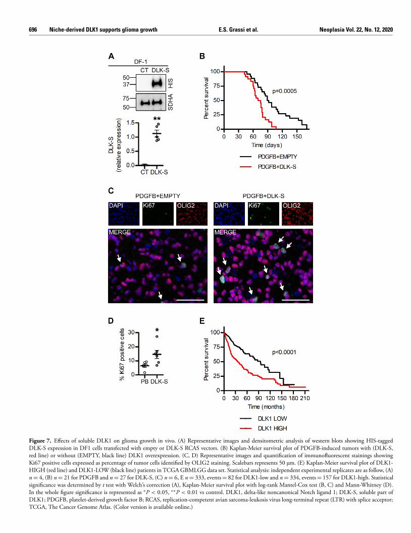
Figure 7. Effects of soluble DLK1 on glioma growth in vivo. (A) Representative images and densitometric analysis of western blots showing HIS-tagged DLK-S expression in DF1 cells transfected with empty or DLK-S RCAS vectors. (B) Kaplan-Meier survival plot of PDGFB-induced tumors with (DLK-S, red line) or without (EMPTY, black line) DLK1 overexpression. (C, D) Representative images and quantification of immunofluorescent stainings showing Ki67 positive cells expressed as percentage of tumor cells identified by OLIG2 staining. Scalebars represents 50 μm. (E) Kaplan-Meier survival plot of DLK1- HIGH (red line) and DLK1-LOW (black line) patients in TCGA GBMLGG data set. Statistical analysis: independent experimental replicates are as follow, (A) n = 4, (B) n = 21 for PDGFB and n = 27 for DLK-S, (C) n = 6, E n = 333, events = 82 for DLK1-low and n = 334, events = 157 for DLK1-high. Statistical significance was determined by t test with Welch’s correction (A), Kaplan-Meier survival plot with log-rank Mantel-Cox test (B, C) and Mann-Whitney (D). In the whole figure significance is represented as ∗P < 0.05, ∗∗P < 0.01 vs control. DLK1, delta-like noncanonical Notch ligand 1; DLK-S, soluble part of DLK1; PDGFB, platelet-derived growth factor B; RCAS, replication-competent avian sarcoma-leukosis virus long-terminal repeat (LTR) with splice acceptor; TCGA, The Cancer Genome Atlas. (Color version is available online.)

Neoplasia Vol. 22, No. 12, 2020 Niche-derived DLK1 supports glioma growth E.S. Grassi et al. 697
SR
C
p
F
cU
P
P
0
d
I
1
a
sA
m
m
R
t
s(B
f
o
�
TCOTAAC
T
S
a
t8ao
wt
S
Tg
an
m
H35 Hypoxystation (Don Whitley Scientific) was used to generate hypoxicconditions.
Cells were treated with the indicated concentrations of DLK1 human(Sigma Cat# SRP8006) or Recombinant Human DLK-1 protein (AbcamCat# ab151926). For HIF-2a inhibition, cells were treated with 10 μMPT2385 (MedChemExpress Cat# HY-12867) or equivalent amount ofDMSO 24 hours prior hypoxia exposure and kept in the dark for thewhole experiment. Transient transfection was performed with X-tremeGENE9 DNA Transfection Reagent (Sigma Cat# 06 365 809 001) followingmanufacturer’s recommendations. Human DLK1 ectodomain expressionvector was obtained from Addgene (DLK1-bio-His, RRID:Addgene_51876)[25] .
DLK1 ELISA assay
DLK1 secretion was measured with Human Pref-1 enzyme-linkedimmunosorbent assay (ELISA) kit for cell culture supernatants, plasma andserum samples RAB 1076 (Sigma-Aldrich). Medium from control and treatedastrocytes was collected every 2 to 3 days while medium from DLK-Stransfected cells was collected 72 hours hours post transfection, immediatelysnap frozen in dry ice and stored at −80 °C until the day of the assay.The enzyme-linked immunosorbent assay (ELISA) was performed followingmanufacturer’s instructions and 450 nm absorbance was read on a Synergy 2plate reader (BioTek).
Proliferation assay
Thousand cells/well (PIGPC) or 2500 cells/well (U3082MG, U3084MGand U3065MG) were seeded in 96 well plates. For astrocyte conditionedmedia (ACM) transfer experiments, 24 hours after seeding, media fromAstrocyte and transfected cells was filter sterilized and used to replace culturemedia. For astrocyte experiments, media was replaced every 2 to 3 days andproliferation was assessed after 9 days. For transfected cells, proliferation wasassessed after 72 hours.
For recombinant protein experiments, 24 hours after seeding cells weretreated with serial dilutions of recombinant DLK1 (0–200 ng/mL range) andgrown for 72 hours. At the moment of the assay, 10 μL of WST-1 solution(Roche) were added to each well and after 2 hours of incubation at 37 °C and5% CO2 450 nm absorbance was read on a Synergy 2 plate reader (BioTek).
Western blot
Cells were lysed in radioimmunoprecipitation assay buffer supplementedwith Complete Phosphatase and Complete Protease inhibitor cocktails(Roche). After dilution in Laemmli buffer with DTT and boiled for 5minutes, samples were loaded on 4% to 20% Mini-PROTEAN TGX PrecastProtein Gels (Biorad). Proteins were transferred on polyvinylidene fluoride(PVDF) membranes using a Transblot Turbo System (Biorad), blocked in5% nonfat dry milk/PBS, and incubated overnight at 4 °C with primaryantibodies. After washing, membranes were incubated for 1 hour withsecondary antibodies (Abcam). Images were acquired using a Fujifilm LAS3000 Imager. Densitometric analysis were performed with ImageJ software(Fiji, RRID:SCR_002285). Band signal intensity was normalized for therespective loading control values (actin or SDHA).
Primary antibodies: hypoxia-inducible factor 1 alpha HIF-1a antibody(Novus Cat# NB100-479SS, RRID:AB_790147), HIF-2a antibody(Abcam Cat# ab199, RRID:AB_302739), SDHA antibody (AbcamCat# ab14715, RRID:AB_301433), 6X His-tag antibody (Abcam Cat#ab9108, RRID:AB_307016), beta Actin antibody (Abcam Cat# ab75186,RRID:AB_1280759). Secondary antibodies: Goat anti-Rabbit IgG( H + L ) Secondary Antibody, HRP (Thermo Fisher ScientificCat# 31460, RRID:AB_228341), Goat anti-Mouse IgG ( H + L )
econdary Antibody, HRP (Thermo Fisher Scientific Cat# 31430, RID:AB_228307).
olony and sphere formation assays
Mechanical dissociation with Accutase (ThermoFisher) was used to repare single cell suspensions. Cells were counted using a hemocytometer.or colony formation assay, 350 cells were seeded in 5 cm dishesoated with polyornithine (Sigma) and laminin (Biolamina). U3082MG, 3084MG and U3065MG cells were cultured for 14 days whileIGPCs for 8 days, under the indicated conditions, then washed inBS and fixed using 4% paraformaldehyde. Cells were stained using,01% crystal violet/H2O. Wells were washed gently in water, then air-ried for 24 hours. Images were acquired with a Fujifilm LAS 3000mager.
Sphere formation assay was performed with the hanging-drop method. 0 cells in 35 μL drops were seeded on the lid of a 48 well platend grown under the indicated conditions for 2 weeks. For secondaryphere assay, primary spheres were pooled, pelleted, dissociated with ccutase and reseeded at the indicated conditions. Wells with spheres wereanually counted and images were acquired with a Zeiss AX10 invertedicroscope.
eal-time quantitative PCR
The RNeasy Mini Kit was used with Qiashredder (QIAGEN) accordingo the manufacturer’s instructions for RNA isolation, and cDNA wasynthesized using random primers and Multiscribe reverse transcriptase Applied Biosystems). A QuantStudio 7 real-time PCR system (Applied iosystems) with SYBR Green Master Mix (Applied Biosystems) was used
or amplification. Gene expression levels were normalized to the expressionf 3 housekeeping genes (UBC, SDHA, and YWHAZ) using the comparative�CT method.
The following primers were used: NANOG (GCTGGTTGCCTCA
GTTATTATGC; CCATGGAGGAAGGAAGAGGAGAGA), SOX2 (GC
TGGGCGCCGAGTGGA; GGGCGAGCCGTTCATGTAGGTCTG), CT4 (GCCTGGGCGCCGAGTGGA; CCACATCGGCCTGTGTATA
C), UBC (ATTTGGGTCGCGGTTCTT; TGCCTTGACATTCTCG
TGGT), SDHA (TGGGAACAAGAGGGCATCTG; CCACCACTGC
TCAAATTCATG), YWHAZ (ACTTTTGGTACATTGTGGCTTCAA; CGCCAGGACAAACCAGTAT)
ransfections and luciferase reporter assay
DLK-S expression plasmid was kindly provided by prof. Anne Ferguson-mith [11] , cloned into RCAS vector by classic restriction enzyme techniquend transfected into DF-1 cells. For luciferase reporter assay, cells were co-ransfected with hypoxia-responsive element (HRE)-luc (Addgene) [26] or xCSL-luc (gift from Håkan Axelson) and pCMV-renilla (Promega) and nalyzed using the Dual-Luciferase Reporter Assay System (Promega) n a Synergy 2 platereader (BioTek). Xtreme gene 9 (Roche) reagentas used according to manufacturer’s recommendations for transient
ransfections.
tatistical analyses
For DLK1 expression and patients survival, data from 620 patients fromhe Cancer Genome Atlas (TCGA, RRID:SCR_003193, https://portal. dc.cancer.gov/ ) [27] and the Chinese Glioma Genome Atlas [28] , werenalyzed using GlioVis (( http://gliovis.bioinfo.cnio.es/ ) [29] ) (DLK1-LOW
= 333, events = 82, median = 87.5; DLK1-HIGH n = 334, events = 157,edian = 34.9).
698 Niche-derived DLK1 supports glioma growth E.S. Grassi et al. Neoplasia Vol. 22, No. 12, 2020
Ss
gca
lpfttt
l
b
s(
htstp
Dg
PmUa
3
iiD(
r(
iS
p
ea
D
caia
s
1iD
adw
Mbc
For proliferation experiments, the EC50 and span were estimated witha nonlinear regression curve, using a log. agonist vs normalized response(variable slope) equation fitted in Graphpad Prism 5 (GraphPad Prism,RRID:SCR_002798). For immunofluorescence experiments, colocalizationwas measure by Pearson’s R coefficient in 3 independent experiments withImageJ Coloc2 plugin. For Ki67 quantification, at least 3 fields were analyzedfor each tumor, for a total of 101,717 nuclei analyzed, with n = 45,466 forPDGFB tumors and n = 56,251 for DLK-S tumors.
After normal distribution and variance similarity evaluation, 2-sidedunpaired t test (eventual Welch’s correction for groups with differentvariances), Mann-Whitney for nonparametric data, 1-way ANOVA withBonferroni post hoc test and 2-way ANOVA (timelines only) tests were usedto determine statistical significance, as indicated in respective figure legends.For survival evaluation, the Kaplan–Meier method was used to investigatevariables and overall survival correlation, while a log-rank test was employedto compare survival curves. In all figures data are shown as mean ±SEM,analyzed using GraphPad Prism 5 software and significance expressed as Pvalues ( ∗P < 0.05, ∗∗P < 0.01, ∗∗∗ P < 0.001).
Data availability
The data that support the findings of this study are available from thecorresponding author upon reasonable request.
Results
DLK1 is expressed and secreted by tumor-associated astrocytes in the glioma microenvironment
To test whether tumor-associated astrocytes could be a source of DLK1 inthe glioma tumor microenvironment, we generated PDGFB/shp53-inducedmurine gliomas using the RCAS/tv-a system as previously described [14] ,then co-stained tumors for the astrocyte marker glial fibrillary acidic protein(GFAP) and DLK1. While the bulk of the tumor cells appeared negativefor DLK1 expression, DLK1 signal was detected in areas of GFAP stainingboth in perinecrotic and perivascular tumor areas, and both in GFAP positiveand negative cells, suggesting DLK1 expression in astrocytes, tumor cells, andpotentially other cell types present in these areas ( Figure 1 A −C, F −G andSupplementary Figure 1 A −B).
The use of an independent antibody directed against the N-terminal,soluble domain of DLK1 also showed specific signal in the perinecrotictumor areas (Supplementary Figure 2 ) and co-staining of GFAP and DLK1( Figure 1 D −E, H and Supplementary Figure 1 C).
DLK1 gene expression was previously reported to be upregulated inisolated GFAP + tumor-associated astrocytes of high-grade glioma comparedto those of lower-grade tumors [18] , further confirming DLK1 expression inastrocytes in this model system. We speculated that this DLK1 inductioncould be mediated by microenvironmental factors. As hypoxia is 1 majormicroenvironmental reality of high-grade glioma compared to low-gradeglioma [5 , 30] , we cultured human fetal astrocytes under normoxic or hypoxicconditions for up to 10 days. We also subjected astrocytes to a single doseof irradiation to mimic another physiological response to a therapeuticintervention relevant to high-grade glioma. Both astrocytes subjected togrowth in hypoxia and those subjected to irradiation displayed increasedDLK1 levels secreted into the culture media, as measured by an ELISA assay( Figure 2 A). The higher levels of DLK1 in the media were sustained for theentire 10 day period in the case of astrocytes cultured in hypoxia, whereasirradiated astrocytes displayed a peak secretion at 2 days post-treatment withlevels returning to baseline after 9 days ( Figure 2 A). Together, these datasupport that astrocytes may secrete DLK1 into the tumor microenvironmentin glioma.
oluble DLK1 promotes glioma cell proliferation, survival and elf-renewal
We next examined what effect soluble DLK1 may have on glioma cells. We first performed a media transfer experiment by treating human
lioblastoma cell lines maintained in serum-free, stem cell-promoting onditions with media from astrocytes cultured in normoxic (ACM CTRL) nd hypoxic (ACM 1% O 2 ) conditions for up to 9 days. In 2 out of 3 cellines, media from hypoxic astrocytes induced a significant increase in the roliferation rate ( Figure 2 B). Since ACM contains many other different actors that may influence cancer cell growth, we then directly investigated he effects of soluble DLK1 with 2 different approaches. First, we transiently ransfected human glioblastoma cell lines with a plasmid containing the N- erminal soluble part of DLK1 (DLK-S, His-tagged). All the 3 transfected cellines overexpressed and secreted similar levels of DLK-S, as verified by westernlot and ELISA experiments ( Figure 2 C −D). A media transfer experimenthowed that 2 out of 3 cell lines significantly increased their proliferation Figure 2 E) when grown in DLK-S conditioned media. We then treated theuman glioblastoma cell lines with a recombinant protein corresponding to he DLK1 secreted part, and once again, the 2 DLK-responding cell lines howed significant increase in their proliferation ( Figure 2 F). Taken together, hese data demonstrate that soluble DLK1 is able to induce glioma cell roliferation, irrespectively of its origin.
As the use of the recombinant protein allows for better control of LK1 concentrations, we then moved forward with this approach. We first
enerated a dose-response curve in all 3 human glioblastoma cell lines and inDGFB-induced glioma primary cultures (PIGPCs) derived from the glioma ouse model. All the cell lines, with the exception of the nonresponding 3065MG, showed a dose dependent increase in cell proliferation, with plateau obtained at 200 ng/mL DLK1 and EC50s in between 25 and5 ng/mL ( Fig 3 A).
In line with these findings, all cell lines that responded to DLK1 n the proliferation assay also increased their colony formation ability n a dose-dependent manner when exposed to sub-maximal soluble
LK1 concentrations similar to those obtained in hypoxic astrocytes Figure 3 B −C).
Furthermore, soluble DLK1 strongly enhanced the self-renewal ability of esponsive glioma cell lines, as measured by the serial sphere-formation assay Figure 4 A −B) performed at clonal density (Supplementary Figure 3), andnduced a significant increase in the stem cell markers OCT4, NANOG, and OX2 ( Figure 4 C). Since DLK1 has been reported to influence the Notchathway [8 , 12] , we tested if these effects were Notch-dependent. Luciferasexperiments performed at different time points revealed no significant lterations in Notch activity in any tested cell lines ( Figure 4 D).
LK1-effects are mediated in part by HIF-2a
Because DLK1 secretion was increased by astrocytes under hypoxic onditions, and because of known previous links between DLK1 expression nd function to hypoxia [20] , we asked whether soluble DLK1 could nfluence the hypoxic response of glioma cells. We cultured U3082MG
nd U3084MG human glioblastoma cells for 24 or 72 hours in 1% O 2 ,timulated or not with soluble DLK1. While there was no difference in HIF-a stabilization with DLK1 treatment, Western blots showed significantly ncreased HIF-2a protein levels at 72 hours in cells cultured with soluble
LK1 ( Figure 5 A −B). This increased HIF-2a expression was reflected in stronger hypoxic response, as 2/3 human glioblastoma lines and PIGPCs isplayed increased activation of HREs at 72 hours of culture in hypoxia ith DLK1 stimulation, as measured in an HRE-luciferase assay ( Figure 5 C).oreover, analysis of PDGFB/shp53-induced murine gliomas revealed that
oth DLK1 and HIF-2a were strongly expressed and showed a significant o-localization only in the perivascular and perinecrotic niches ( Fig. 5 D).

Neoplasia Vol. 22, No. 12, 2020 Niche-derived DLK1 supports glioma growth E.S. Grassi et al. 699
t
w
w
i
s
t
w
s
c
r
t
i
2o
t
c
I
h
H
e
Imt
D
i
c
a
e
o
oi
w
d
sa
l
C
p
i
e
A
G
WS
A
MWVI&V
As HIF-2a is a known driver of stem cell characteristics in glioma andother tumor forms [31 , 23 , 32–34 ], we next tested whether effects of DLK1on glioma cell behavior were mediated by HIF-2a. The treatment with thespecific HIF-2a inhibitor PT2385 at concentrations with significant effectson HIF-2a protein [35 , 36] (Supplementary Fig. 4) was able to revert theDLK1-induced increase in hypoxia response ( Figure 6 A). Similarly, whilestimulation of glioma cells with soluble DLK1 boosted the increase in theexpression of the stem cell marker genes NANOG, OCT4, and SOX2 inhypoxic cells, addition of PT2385 blocked this specific DLK1 effect in all celllines tested ( Figure 6 B). Moreover, PT2385 decreased the colony formationability of glioma cells exposed to soluble DLK1 in hypoxia ( Figure 6 C).Notably however, PT2385 treatment did not significantly affect DLK1-induced gene expression in normoxia, and showed surprisingly modest effectsin DLK1-untreated cells at hypoxia. Together, these data suggest that DLK1promotes the glioma stem cell character in part via HIF-2a stabilization.
DLK1 promotes aggressive glioma growth in vivo
To test the effects of soluble DLK1 on glioma growth in vivo, we generateda mouse model for the overexpression of soluble DLK1 together with PDGFBusing the RCAS/tv-a system ( Figure 7 A). Co-injection of RCAS-PDGFBwith RCAS-DLK-S (soluble) resulted in more aggressive tumors as comparedto RCAS-PDGFB with empty vector control, as measured by survival timefollowing injections ( Figure 7 B). Evaluation of Ki67 expression revealed asignificant increase in cell proliferation in murine DLK-S tumors as comparedto controls ( Figure 7 C, D), thus confirming the in vitro data ( Figure 2 E −F,3A). In agreement with our in vivo data using a mouse model that givesrise to a range of low-to-high-grade gliomas, analysis of the human TCGALGGGBM data set [27] revealed that tumors expressing high levels ofDLK1 were significantly more aggressive than those with low levels of DLK1( Figure 7 E), presumably as a result of the higher DLK1 levels reported inhigh-grade glioma. These findings were replicated in an independent data set(Supplementary Figure 5 A −B). Notably, in data sets comprising GBM only,DLK1 expression was either associated with slightly longer survival, or notassociated with any significant survival difference at all, suggesting that thelink between DLK1 and shorter survival in the LGGGBM data set is related toincreased expression in higher-grade tumors (Supplementary Figure 5 A −B).
Discussion
An increasing focus on cancer stem cell characteristics has revealedparallels between normal neural stem cell regulation and cancer stem cellcharacteristics in brain tumors [37 , 38] . Control over tumor cell phenotypesby specific, local microenvironments within a tumor, for example, isreminiscent of the way that normal tissue stem cells reside within and relyon their niche to maintain the stem cell character [3 , 4] . It is likely thatsome of the same mechanisms involved in neural stem cell maintenancein the vascular niche of the subventricular zone may also be involved inmaintaining the cancer stem cell character of brain tumor cells located in aperivascular or periarteriolar [39] niche. We describe one such example here:soluble DLK1 secreted from astrocytes appears to be involved in stem cellmaintenance both of normal neural stem cells and glioma cells, as shownhere. An association between DLK1 expression and aggressive tumor growthin glioblastoma has previously been established [20] . By generating a mousemodel for testing effects of soluble DLK1 overexpression specifically in thecontext of glioblastoma, we show that the previously reported associationbetween DLK1 expression and tumor grade in glioma [14 , 15] may at least inpart be caused by DLK1 itself, as soluble DLK1-overexpressing tumors had ahigher proliferation rate and significantly decreased mice survival as comparedto controls. It is important to note that DLK1 expression is not limited totumor-associated astrocytes, and soluble DLK1 may be derived both fromother stromal cell types and tumor cells themselves. Our experiments indicate
hat soluble DLK1 affects tumor cell proliferation similarly regardless ofhether it was produced by astrocytes or tumor cells. In a previous study,e described release of and signaling from the intracellular domain of DLK1
n glioma cells [14] . Data presented here do not link signaling from theoluble DLK1 to that of the intracellular fragment, however, both appearo be regulated by the hypoxic tumor microenvironment. It is yet unclearhether or not DLK1 expression is required in tumor cells to be affected by
oluble DLK1 secreted into the niche [11] . As with astrocyte-derived DLK1 in regulation of normal neural stem
ells, the exact mechanism(s) by which soluble DLK1 signals to glioma cellsemains to be investigated. We show here that soluble DLK1 can contributeo a stronger and more prolonged response to hypoxia, as mediated byncreased HIF-2a stabilization in DLK1 treated cells. This effect on HIF-a stabilization indeed seemed important for the tumor-promoting effects f DLK1 signaling as inhibition of HIF-2a transcriptional activity by use ofhe specific HIF-2a inhibitor PT2385 abolished all effects of DLK1 on stemell marker gene expression and colony formation under hypoxic conditions.nterestingly, DLK1 expression itself has been shown to be regulated byypoxia in other cell systems [19 , 20] , suggesting that there may be a DLK1-IF feedback loop in hypoxic tumor cells. In the present investigation,
ffects of DLK1 treatment were enhanced by hypoxic culture conditions.mportantly, however, HIF-2a inhibition did not significantly affect DLK1- ediated stem cell marker expression under normoxic conditions, suggesting
hat there are other mediators downstream of DLK1 that can contribute toLK1 signaling in glioma.
Several questions remain regarding signaling mediated by soluble DLK1, ncluding that of potential receptors for DLK1. Among the human gliomaell lines and genetically engineered glioma mouse model tested in this study,ll but one cell line (U3065MG) responded to soluble DLK1. Based on thexperiments presented here, it is difficult to determine the reason for the lackf response in this cell line. It is possible that U3065MG cells lack expressionf necessary components downstream of soluble DLK1. Further investigation nto potential receptors and downstream mediators of DLK1 signaling isarranted, to better infer the applicability of the DLK1-mediated effectsescribed in this study. Furthermore, it is somewhat counterintuitive thatoluble DLK1 appears to simultaneously promote glioma cell proliferation nd stem cell characteristics, as it is widely assumed that stem-like cells displayower proliferation rates than more differentiated, non −stem-like cells.
onclusions
Taken together, our data support a role for soluble DLK1 as a tumor-romoting stem cell niche factor in glioma. Further research is warranted to
nvestigate whether or not signaling by DLK1 can be therapeutically targeted,ither via HIF2-a inhibition or by targeting upstream signaling.
cknowledgments
The authors thank Christina Möller for technical assistance and A Humanlioblastoma Cell Culture Resource ( www.hgcc.se ) (Lene Uhrbom, Bengtestermark, Karin Forsberg Nilsson and Sven Nelander, Uppsala University,
weden) for human GBM cultures.
uthor s ’ Contributions
ESG: Conceptualization; Data curation; Formal analysis; Investigation; ethodology; Validation; Visualization; Roles/Writing – original draft; riting – review & editing. PJ: Formal analysis; Investigation; Methodology;
alidation; Visualization; Writing – review & editing. VP: Formal analysis nvestigation; Methodology; Validation; Visualization; Writing – review
editing. TJB: Formal analysis; Investigation; Methodology; Validation; isualization; Writing – review & editing. AP: Conceptualization; Data

700 Niche-derived DLK1 supports glioma growth E.S. Grassi et al. Neoplasia Vol. 22, No. 12, 2020
[
[
[
[
[
[
[
[
[
[
[
[
[
[
[
[
[
[
curation; Formal analysis; Funding acquisition; Project administration;Resources; Supervision; Roles/Writing – original draft; Writing – review &editing.
Supplementary materials
Supplementary material associated with this article can be found, in theonline version, at doi:10.1016/j.neo.2020.10.005 .
References
[1] Huse JT, Holland EC. Targeting brain cancer: advances in the molecularpathology of malignant glioma and medulloblastoma. Nat Rev Cancer2010; 10 (5):319–31. doi: 10.1038/nrc2818 .
[2] Bernstock JD, Mooney JH, Ilyas A, Chagoya G, Estevez-Ordonez D, Ibrahim A,Nakano I. Molecular and cellular intratumoral heterogeneity in primaryglioblastoma: clinical and translational implications. J Neurosurg 2019:1–9.doi: 10.3171/2019.5.JNS19364 .
[3] Dirkse A, Golebiewska A, Buder T, Nazarov PV, Muller A, Poovathingal S,Niclou SP. Stem cell-associated heterogeneity in Glioblastoma results fromintrinsic tumor plasticity shaped by the microenvironment. Nat Commun2019; 10 (1):1787. doi: 10.1038/s41467- 019- 09853- z .
[4] Hambardzumyan D, Bergers G. Glioblastoma: defining tumor niches. TrendsCancer 2015; 1 (4):252–65. doi: 10.1016/j.trecan.2015.10.009 .
[5] Jawhari S, Ratinaud M-H, Verdier M. Glioblastoma, hypoxia and autophagy: asurvival-prone “ménage-à-trois”. Cell Death Dis 2016; 7 (10):e2434. doi: 10.1038/cddis.2016.318 .
[6] Ryskalin L, Gaglione A, Limanaqi F, Biagioni F, Familiari P, Frati A, Fornai F.The autophagy status of cancer stem cells in gliobastoma multiforme: from cancerpromotion to therapeutic strategies. Int J Mol Sci 2019 MDPI AG. doi: 10.3390/ijms20153824 .
[7] Majmundar AJ, Wong WJ, Simon MC. Hypoxia-inducible factors and theresponse to hypoxic stress. Mol Cell 2010; 40 (2):294–309. doi: 10.1016/j.molcel.2010.09.022 .
[8] Baladrón V, Ruiz-Hidalgo MJ, Nueda ML, Díaz-Guerra MJM, García-Ramírez JJ, Bonvini E, Laborda J. dlk acts as a negative regulator of Notch1activation through interactions with specific EGF-like repeats. Exp Cell Res2005; 303 (2):343–59. doi: 10.1016/j.yexcr.2004.10.001 .
[9] Ceder JA, Jansson L, Helczynski L, Abrahamsson P-A. Delta-like 1 (Dlk-1), a novel marker of prostate basal and candidate epithelial stem cells, isdownregulated by notch signalling in intermediate/transit amplifying cells of thehuman prostate. Eur Urol 2008; 54 (6):1344–53. doi: 10.1016/j.eururo.2008.03.006 .
[10] Falix FA, Aronson DC, Lamers WH, Gaemers IC. Possible roles of DLK1in the Notch pathway during development and disease. Biochim Biophys Acta2012; 1822 (6):988–95. doi: 10.1016/j.bbadis.2012.02.003 .
[11] Ferrón SR, Charalambous M, Radford E, McEwen K, Wildner H, Hind E,Ferguson-Smith AC. Postnatal loss of Dlk1 imprinting in stem cells and nicheastrocytes regulates neurogenesis. Nature 2011; 475 (7356):381–5. doi: 10.1038/nature10229 .
[12] Huang C-C, Cheng S-H, Wu C-H, Li W-Y, Wang J-S, Kung M-L, Tai M-H. Delta-like 1 homologue promotes tumorigenesis and epithelial-mesenchymaltransition of ovarian high-grade serous carcinoma through activation of Notchsignaling. Oncogene 2019; 38 (17):3201–15. doi: 10.1038/s41388- 018- 0658- 5 .
[13] Li L, Tan J, Zhang Y, Han N, Di X, Xiao T, Liu Y. DLK1 promotes lung cancercell invasion through upregulation of MMP9 expression depending on Notchsignaling. PLoS One 2014; 9 (3):e91509. doi: 10.1371/journal.pone.0091509 .
[14] Grassi ES, Pantazopoulou V, Pietras A. Hypoxia-induced release, nucleartranslocation, and signaling activity of a DLK1 intracellular fragment in glioma.Oncogene 2020:1–17. doi: 10.1038/s41388- 020- 1273- 9 .
[15] Yin D, Xie D, Sakajiri S, Miller CW, Zhu H, Popoviciu ML, Koeffler HP. DLK1:increased expression in gliomas and associated with oncogenic activities. Oncogene2006; 25 (13):1852–61. doi: 10.1038/sj.onc.1209219 .
16] Wang Y, Sul HS. Ectodomain shedding of preadipocyte factor 1 (Pref-1) by tumor necrosis factor alpha converting enzyme (TACE) and inhibition of adipocyte differentiation. Mol Cell Biol 2006; 26 (14):5421–35. doi: 10.1128/ MCB.02437-05 .
17] Mega A, Hartmark Nilsen M, Leiss LW, Tobin NP, Miletic H, Sleire L, Östman A.Astrocytes enhance glioblastoma growth. Glia 2020; 68 (2):316–27. doi: 10.1002/ glia.23718 .
18] Katz AM, Amankulor NM, Pitter K, Helmy K, Squatrito M, Holland EC. Astrocyte-specific expression patterns associated with the PDGF-induced glioma microenvironment. PLoS One 2012; 7 (2):e32453. doi: 10.1371/journal.pone. 0032453 .
19] Begum A, Kim Y, Lin Q, Yun Z. DLK1, delta-like 1 homolog (Drosophila), regulates tumor cell differentiation in vivo. Cancer Lett 2012; 318 (1):26–33. doi: 10.1016/j.canlet.2011.11.032 .
20] Kim Y, Lin Q, Zelterman D, Yun Z. Hypoxia-regulated delta-like 1 homologue enhances cancer cell stemness and tumorigenicity. Cancer Res 2009; 69 (24):9271–80. doi: 10.1158/0008- 5472.CAN- 09- 1605 .
21] Holland EC, Hively WP, DePinho RA, Varmus HE. A constitutively active epidermal growth factor receptor cooperates with disruption of G1 cell- cycle arrest pathways to induce glioma-like lesions in mice. Genes Dev 1998; 12 (23):3675–85. doi: 10.1101/gad.12.23.3675 .
22] Ozawa T, Riester M, Cheng YK, Huse JT, Squatrito M, Helmy K, Charles N,Michor F, Holland EC. Most human non-GCIMP glioblastoma subtypes evolve from a common proneural-like precursor glioma. Cancer Cell 2014; 26 (2):288–300. doi: 10.1016/j.ccr.2014.06.005 .
23] Johansson E, Grassi ES, Pantazopoulou V, Tong B, Lindgren D, Berg TJ, Pietras A. CD44 Interacts with HIF-2 α to modulate the hypoxic phenotype of perinecrotic and perivascular glioma cells. Cell Rep 2017; 20 (7). doi: 10.1016/j. celrep.2017.07.049 .
24] Xie Y, Bergström T, Jiang Y, Johansson P, Marinescu VD, Lindberg N, Uhrbom L. The human glioblastoma cell culture resource: validated cell models representing all molecular subtypes. EBioMedicine 2015; 2 (10):1351–63. doi: 10. 1016/j.ebiom.2015.08.026 .
25] Sun Y, Vandenbriele C, Kauskot A, Verhamme P, Hoylaerts MF, Wright GJ. A human platelet receptor protein microarray identifies FcepsilonR1alpha as an activating PEAR1 ligand. Mol Cell Proteomics 2015. doi: 10.1074/mcp.M114. 046946 .
26] Emerling BM, Weinberg F, Liu J-L, Mak TW, Chandel NS. PTEN regulates p300-dependent hypoxia-inducible factor 1 transcriptional activity through Forkhead transcription factor 3a (FOXO3a). Proc Natl Acad Sci USA
2008; 105 (7):2622–7. doi: 10.1073/pnas.0706790105 . 27] Ceccarelli M, Barthel FP, Malta TM, Sabedot TS, Salama SR, Murray BA,
Verhaak RGW. Molecular profiling reveals biologically discrete subsets and pathways of progression in diffuse glioma. Cell 2016; 164 (3):550–63. doi: 10. 1016/j.cell.2015.12.028 .
28] Zhao Z, Zhang K, Wang Q, Li G, Zeng F, Zhang Y, et al. Chinese gliomagenome atlas (CGGA): a comprehensive resource with functional genomic data for Chinese glioma patients. 2020. bioRxiv. Cold Spring Harbor Laboratory; 2020.01.20.911982. doi: 10.1101/2020.01.20.911982 .
29] Bowman RL, Wang Q, Carro A, Verhaak RGW, Squatrito M. GlioVis data portal for visualization and analysis of brain tumor expression datasets. Neuro Oncol 2017; 19 (1):139–41. doi: 10.1093/neuonc/now247 .
30] Jensen RL. Brain tumor hypoxia: tumorigenesis, angiogenesis, imaging, pseudoprogression, and as a therapeutic target. Journal of Neuro-Oncology . Kluwer Academic Publishers; 2009. doi: 10.1007/s11060- 009- 9827- 2 .
31] Das B, Pal B, Bhuyan R, Li H, Sarma A, Gayan S, Felsher DW. MYC regulatesthe HIF2 α stemness pathway via Nanog and Sox2 to maintain self-renewal in cancer stem cells versus non-stem cancer cells. Cancer Res 2019; 79 (16):4015–25. doi: 10.1158/0008- 5472.CAN- 18- 2847 .
32] Pietras A, Gisselsson D, Ora I, Noguera R, Beckman S, Navarro S, Påhlman S.High levels of HIF-2alpha highlight an immature neural crest-like neuroblastoma cell cohort located in a perivascular niche. J Pathol 2008; 214 (4):482–8. doi: 10.1002/path.2304 .
33] Pietras A, Johnsson AS, Påhlman S. The HIF-2 α-driven pseudo- hypoxic phenotype in tumor aggressiveness, differentiation, and
Neoplasia Vol. 22, No. 12, 2020 Niche-derived DLK1 supports glioma growth E.S. Grassi et al. 701
[
[
[
vascularization. Curr Top Microbiol Immunol 2010; 345 :1–20. doi: 10.1007/82 _2010 _ 72 .
[34] Yan Y, Liu F, Han L, Zhao L, Chen J, Olopade OI, Wei M. HIF-2 α promotesconversion to a stem cell phenotype and induces chemoresistance in breastcancer cells by activating Wnt and Notch pathways. J Exp Clin Cancer Res2018; 37 (1):256. doi: 10.1186/s13046- 018- 0925- x.
[35] Persson CU, von Stedingk K, Fredlund E, Bexell D, Påhlman S, Wigerup C,Mohlin S. ARNT-dependent HIF-2 transcriptional activity is not sufficientto regulate downstream target genes in neuroblastoma. Exp Cell Res2020; 388 (2):111845. doi: 10.1016/j.yexcr.2020.111845 .
[36] Wallace EM, Rizzi JP, Han G, Wehn PM, Cao Z, Du X, Josey JA. A small-molecule antagonist of HIF2 α is efficacious in preclinical models of renalcell carcinoma. Cancer Res 2016; 76 (18):5491–500. doi: 10.1158/0008-5472.CAN- 16- 0473 .
37] Dirks PB. Brain tumor stem cells: the cancer stem cell hypothesis writ large.Molecular Oncology . John Wiley and Sons Ltd; 2010. doi: 10.1016/j.molonc.2010.08.001 .
38] Lathia JD, Mack SC, Mulkearns-Hubert EE, Valentim CLL, Rich JN. Cancerstem cells in glioblastoma. Genes and Development . Cold Spring HarborLaboratory Press; 2015. doi: 10.1101/gad.261982.115 .
39] Hira VVV, Breznik B, Vittori M, Loncq de Jong A, Mlakar J, Oostra RJ,Khurshed M, Molenaar RJ, Lah T, Van Noorden CJF. Similarities betweenstem cell niches in glioblastoma and bone marrow: rays of hope for noveltreatment strategies. J Histochem Cytochem 2020; 68 (1):33–57. doi: 10.1369/0022155419878416 .