nickel effects on two maize (zea mays)...
TRANSCRIPT
8’
1547
Nickel effects on two maize (Zea mays) cultivars: growth, structure, Ni concentration, and localization
Laurent L’Huillier, Jean d’Auzac, Monique Durand, and Nicole Michaud-Ferrière
Abstract: The toxic effects of nickel on maize growth and structure and the accumulation and distribution of Ni in the plant were investigated. Two cultivars of maize, each with a different sensitivity to Ni, were grown on nutrient solution with different nickel concentrations for a period of 8 days. The sensitive and tolerant cultivars exhibited reduction in root and shoot growth with Ni concentrations greater than 20 pM and 40 pM, respectively. The toxic effects of nickel on the structure and ultrastructure of maize roots and leaves were studied by light microscopy and transmission and scanning electron microscopy. With 60 pM Ni, the sensitive cultivar showed a strong reduction in root mitotic activity (80%). Statoliths were absent in cells of the root cap, while leaves contained large amounts of starch in the chloroplasts of their bundle sheath cells. This suggests a decrease in carbohydrate transport between the leaves and the roots. The tissue localization of Ni by the dimethylglyoxim and silver sulphide methods showed important accumulation in the root apex and in the chloroplasts of the bundle sheath cells. These results suggest that Ni reduces maize growth by a reduction in root mitotic activity, probably because of direct action on the meristem. Starch accumulation in the leaves could come directly from a decrease in the root sink effect caused by the reduced mitotic activity or indirectly from an inhibition of carbohydrate transport.
Key words: maize, Ni toxicity, growth, Ni accumulation, Ni distribution, mitotic activity.
Résumé : Les effets toxiques du nickel sur la croissance et l’anatomie du maïs ainsi que l’accumulation et la répartition de Ni dans la plante ont été étudiés. Deux cultivars de maïs, de sensibilité différente au nickel, ont été cultivés pendant 8 jours sur solution nutritive contenant différentes concentrations de nickel. Le cultivar sensible et le cultivar tolérant montrent une diminution de la croissance des racines et des parties aériennes pour des concentrations en Ni supérieures à 20 pM et 40 pM, respectivement. Les effets toxiques du nickel sur la structure et l’ultrastructure des feuilles et des racines du maïs ont été étudiés par microscopie photonique, et microscopie électronique à balayage et à transmission. Avec 60 pM de Ni, on constate chez le cultivar sensible une forte diminution de l’activité mitotique des racines (80%). Les Statolithes sont absents dans les cellules de la coiffe racinaire, alors que les feuilles contiennent des quantités importantes d’amidon dans les chloroplaste,s des cellules de la gaine périvasculaire. Cela suggère une diminution du transport des carbohydrates depuis les feuilles vers les racines. La localisation de Ni dans les tissus par les méthodes au diméthylglyoxime et au sulfure d’argent montrent des accumulations importantes au niveau des apex racinaires et des chloroplastes des cellules de la gaine périvasculaire. Ces résultats laissent supposer que Ni diminue la croissance du maïs principalement par une diminution de l’activité mitotique des racines, probablement due à une action directe sur le méristème. L’accumulation d’amidon dans les feuilles pourrait découler directement de la réduction de l’effet sink racinaire consécutive à la diminution des mitoses, ou indirectement d’une inhibition du transport des carbohydrates.
Mots clés : maïs, toxicité du Ni, croissance, accumulation du Ni, répartition du Ni, activité mitotique.
Introduction
Heavy metals such as Ni, Cd, Zn, Cu, and Pb are major environmental pollutants. Known symptoms of their toxic
?
L
Received December 6, 1995.
L. L’Huillier. Laboratoire d’Agropédologie, ORSTOM, B.P. A5, 98 848 Nouméa cédex, Nouvelle-Calédonie. J. d’Auzac. Laboratoire de Biotechnologie et de Physiologie Végétale Appliquée, Université de Montpellier 2 , 34 095 Montpellier Cédex 5, France. M. Durand. Laboratoire de Recherche sur les Substances Naturelles, Université de Montpellier 2, 34 095 Montpellier Cédex 5, France. N. Michaud-Ferriere. Laboratoire Histologie-BIOTROP, CIRAD, B.P. 5035, 34032 Montpellier Cédex 1, France. ’ Author to whom all correspondence should be addressed.
e-mail: [email protected]
Can. J. Bot. 74: 1547-1554 (1996). Printed in Canada / Imprimé au Canada
effects on plants include reduced growth and production. Among heavy metals, only Zn, Cu, and Ni phytotoxicities occur frequently (12). Most agricultural soils contain an average 25 mg/kg soil dry wt. of Ni (16). However, certain soils, such as those derived from serpentinite and peridotite, possess concentrations that may exceed 7000 mg kg-’ (4). Many plants that naturally occur on these soils contain dry matter Ni concentrations in excess of 1000 pg * g-l (18, 32), but they generally possess mechanisms that permit them to tolerate Ni and hence develop without phytotoxic prob- lems (14, 21). On the other hand, cultivated plants may rapidly exhibit toxicity symptoms (13). They generally con- tain less than 5 pg of Ni/g dry matter (33, and symptoms of phytotoxicity often become apparent at Ni concentrations as low as 25-30 pg . g-l (9, 19).
Very little research has been conducted on the mechan- isms of Ni phytotoxicity. In general, heavy metals severely inhibit root growth (2, 26), and this is the case for Ni (28).
1548
Robertson and Meakin (29) are among the few who have shown that Ni inhibits root growth in part by inhibiting cellu- lar division in the root apex, yet they were unable to determine whether the effect was primary or secondary. Furthermore, several studies have indicated that Ni inhibits photosynthesis (3, 7, 20). However, toxic amounts of Ni have been shown to increase the amount of carbohydrates in the leaves (24, 30), which may appear contradictory to the previous studies. The effect of Ni on photosynthesis and the distribution of carbohydrates in plants needs further examination, as does Ni distribution in plants.
The aim of this study was to determine how Ni reduces maize growth. We examined Ni accumulation and distribu- tion in maize plants and the effects of toxic concentrations on growth, morphology, histology, and cytology of two culti- vars, each with a different level of sensitivity to Ni.
Materials and methods Maize seeds (Zea mays L. cvs. Hycorn 80 and XL 94) were treated for 10 min with 1 % NaOC1, rinsed with distilled water, and then germinated at 27°C on cotton moistened with distilled water. After a period of 40 h, seedlings with radicles measuring 20 f 2 mm were transferred to 5-L beakers (12 seedlings per beaker) contain- ing one-quarter strength modified Hoagland’s solution at pH 5.3 (Ca, 1 mM; Mg, 0.25 mM; K, 1.5 mM; NH,, 0.5 mM; NO,, 3.5 mM; PO,, 0.5 mM; SO,, 0.25 mM; C1, 12.5 pM; B, 6.25 pM; Mn, 0.5 pM; Zn, 0.5 pM; Cu, 0.12 pM; Mo, O. 12 pM; Fe-EDTA, 5 pM). The nutrient solutions were supplemented with O, 20, 40, 60, or 80 pM NiCl, . 6 H,O. They were continuously aerated and renewed every 3 days. Growth conditions were 25:21 f 1°C (light (L) : dark (D)), 70:80 f 5% (L:D) relative humidity, 14 h L : 10 h D photoperiod, with 200 2 10 pmol * m-2. s-’ photon flux density at leaf level. A randomized block factorial design with two cultivars, five nickel (Ni) concentrations, and four replicates was used.
For histological and histochemical observations, the following plant organs were collected after 8 days on uniform plants: the mid- blade section of the first mature leaf (above the cotyledon), the radicle apex (primary root apex), and the base of the radicle (sec- tion 1 cm below the root-shoot junction). A minimum of six samples were examined from each treatment.
For light microscopy (LM), samples collected at the same period were fixed in 0.1 M phosphate buffer (pH 7.2) containing 2% glutaraldehyde and 1 % paraformaldehyde. Once dehydrated with graded ethanol and embedded in resin (Kulzer 7100, Labonord), samples were cut in 3-pm sections using a microtome (LKB, Historange). Slices were stained with Schiff reagent (periodic acid 5 min, Schiff 10 min) and naphthol blue-black (7 min). These color- ing agents stained glycosyl derivatives red and soluble and insoluble proteins blue (1 1). Longitudinal median sections of root meristems magnified 400-fold were used to count the number of cells in kario- kinesis. The surface of the observed area was 0.25 ”2 at the radicle meristem level.
For the histochemical localization of Ni, the silver sulphide (AgS) (8) and dimethylglyoxim (DMG) methods were used. For the first one, samples were fixed in 2 % glutaraldehyde containing O. 1 % Na# in 0.1 M phosphate buffer (pH 7.2). Once dehydrated and embedded as mentioned above, 3-pm sections were collected on slides and placed in jars filled with the freshly prepared physical developer with the following composition: 60 mL gum arabic (500 g * L-’), 10 mL citrate buffer (2 M pH 5.3), 15 mL hydro- quinone (56.6 g * L-I), and 15 mL silver lactate (7.3 g . L-I). After 90 min at 26°C in the dark, the slides were washed for 30 min with running tap water, followed by distilled water. Observed by LM, a dark color indicated the location of heavy metals. For the
Can. J. Bot. Vol. 74, 1996
Table 1. Effects of Ni on dry matter yield and Ni concentration in roots and shoots.
Root Shoot
Ni Yield Ni concn. Yield Ni concn. (PMI (mdplant) (pdg DW) (mghlant) (pgk DW)
‘XL 94’ O 2 0 . l f 1 . 9 ~ 1724 71 .7 f5 .9~ 4 f l
20 19 .421 .5~ 890295 72.3+3.8u 54 f5 40 12.4f1.3b 1120290 53.125.2b 68f6 60 4 . 1 f 0 . 4 ~ 12852120 3 1 . 3 2 2 . 5 ~ 94f8 80 0.7fO.ld 14802115 24.4f2.8d 8 l f 8
‘Hycorn 80’
O 18 .8f1 .4~ 1623 48 .5 f4 .1~ 3 2 1 20 1 9 . 0 f 2 . l ~ 465+38 4 8 . 8 f 5 . 3 ~ 3724 40 17 .0 f1 .4~ 902f84 47 .7 f4 .4~ 4826 60 13.1fl . lb 13352105 46 .0 f3 .6~ 6426 80 6 . 2 f 0 . 7 ~ 1510f130 37.4f3.0b 88210
9
1
Note: Maize plants were grown on nutrient solution for 8 days, with different concentrations of NiCl,. Values are means f SE (n = 4). Different letters in a column indicate a significant difference at the 0.05 confidence level.
DMG method, fresh samples were gently squashed between two slides, stained with DMG (disodium salt octahydrate, Merck) at O. 1 % in water, then air dried on a hot plate (45°C). This specific chelating agent stained Ni red (6).
For transmission electron microscopy (TEM), samples were fixed using the photon microscopy method described previously and postfixed with 1% Oso4. The samples were then dehydrated in ascending series of acetone followed by propylene oxide and embedded in Spurr’s resin (33). Ultrathin sections (80 nm), coun- terstained with lead citrate and uranyl acetate, were observed and photographed using a Hitachi H-7100 transmission electron micro- scope.
For scanning electron microscopy (SEM), samples were fixed using the same procedure as for LM and dehydrated in an ethanol series and then by critical CO2 point drying (17). Samples were coated with a thin layer of gold, stuck on a slide by an adhesive, and viewed in a JEOL JSM-6300 F scanning electron microscope.
After 8 days of treatment, plants were harvested, divided into roots and shoots, washed twice in distilled water, dried at 105”C, and weighed. For analysis of Ni, the dried tissues were ground and then digested in concentrated HC1. Ni was determined by atomic absorption spectrophotometry (Varian AA 300).
Data were analysed by using ANOVA after being checked for homogeneity of variance. Only the root and shoot weight data reached this criteria after log transformation. Significance of differ- ences between means was performed using t test at the 95 % confi- dence limit.
?
Results Ni effects on growth Root and shoot growth of maize was significantly reduced for Ni concentrations in solution greater than 20 pM for ‘XL 94’ and 40 pM for ‘Hycorn 80’ (Table 1 and Fig. 1). Therefore, it follows that ‘XL 94’ is more sensitive to excess Ni than ‘Hycorn 80’; for instance, root length increase was inhibited by 56 and 13%, respectively, after 8 days of exposure to 60 pM Ni (Fig. 1). The tolerant ‘Hycorn 80’ showed a growth inhibition with 60 ,UM Ni from the 8th day,
-

L’Huillier et al. 1549
Fig. 1. Nickel effects on root length increase of maize ‘XL 94’ and ‘Hycorn 80’. Plants were grown in nutrient solution for 4 (m), 8 (A), and 12 (0) days, with different concentrations of NiCl,. The initial root length of 20 mm before transfer in solution was subtracted for the calculation of root length increase. Values are means SE (12 = 12).
30 t 1
O 20 40 60 80
Ni concentration in solution (pM)
while the sensitive ‘XL 94’ exhibited inhibition with 40 pM Ni after only 4 days (Fig. 1). As a general rule, Ni has a greater effect on root growth than on shoot growth (Table 1). Most histological observations and comparisons between the two cultivars presented below correspond to plants exposed to Ni for 8 days. This time was chosen because ‘Hycorn 80’ only then began to show slight growth reduction and growth of ‘XL 94’ was strongly inhibited (Fig. 1).
Ni effects on roots In the Ni-sensitive ‘XL 94’, numerous structural and physio- logical disorders were observed in the root apex (Figs. 2-6). Note that after 8 days of exposure to 60 pM Ni, the sensitive ‘XL 94’ continued to grow (Fig. 1).
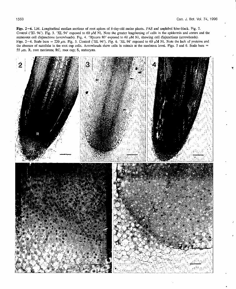
Seedlings exposed to 60 pM Ni for 8 days exhibited pro- nounced lengthening of cells in the zone of root elongation (Figs. 2,3). Between 1 and 2 mm above the apical meristem, cells of the cortical parenchyma measured 50 f 10 pm lengthwise in the treated plants and 20 f 5 pm in the control (means of four roots). A greater difference in cellular length was measured in the root epidermis: between 70 and 160 pm for root epidermis cells in treated plants and 20 & 5 pm for control plants. The lengthening of the cells was associated with an increase in the number and volume of the vacuoles. These variations were already observable at the meristem level, especially in the elongation zone.
At a concentration of 60 pM, nickel inhibited approxi- mately 80% of the mitotic activity in ‘XL 94’ (-46% in ‘Hycorn 80’; Table 2). Inhibition of mitotic activity was notable at 40 pM in ‘XL 94’ (-11 %). In the control meri- stem, numerous cell divisions were clearly visible (Fig. 5), whereas they were rare in roots grown in the presence of Ni (Fig. 6). Furthermore, in this case, the cell plate in the binucleate cells was not visible. In general, root meristem cell walls in seedlings exposed to Ni were less apparent and were stained less by the Schiff reagent. The numerous cellu- lar disjunctions exhibited by Ni-treated roots (Figs. 3, 4) might be associated with fragility and malformation of the cell walls. In addition, cells were more vacuolated and con- tained less dense cytoplasm and therefore fewer proteins.
30 [ I
O O 20 40 60 80
Ni concentration in solution (pM)
Table 2. Effects of Ni on mitotic activity in the root meristem.
Cells out Cells in Mitotic Ni Radicle length of mitosis mitosis Mitotic inhibition
(pM) reduction (%) (no.) (no.) index (%) ~~~ ~
‘XL 94’
40 14 342+15 32f3 8.651.2 -11 60 56 360f17 7 5 2 1.950.6 -80
‘Hycorn 80’
O O 335510 3654 9.7f1.3
O O 340f15 34f4 9.1f1.2 40 O 363f15 35f5 8.921.4 -2 60 13 350k12 1854 4.9*1.’2 -46 -~~ ~ ~
Note: Maize plants were grown on nutrient solution for 8 days, with different concentrations of NiCl,. Values are means SE (n = 3).
While statoliths were numerous in the root caps of the control plants (Fig. 5), their total absence in plants grown on 60 pM Ni was noteworthy (Fig. 6). The difference in stato- lith abundance was already apparent between plants grown on 40 pM Ni and control plants, but it requires further quan- titative determination.
In the same manner as for the radicle, 60 pM Ni reduced the growth of lateral roots and decreased their density (observed by SEM): 8 f 2/cm compared with 18 f 51cm for the control (between 3 and 4 cm below the junction where lateral roots were the most abundant). Moreover, in contrast with control plants, root hairs were absent between 3 and 10 mm from the root apex in treated plants. The root hairs appeared only about 10 mm above the root apex.
Between both maize cultivars exposed to Ni, root differ- ences exist mainly in mitotic activity: at 60 pM Ni, root cells in ‘Hycorn 80’ exhibited more meristematic activity than in ‘XL 94’ (Figs. 3, 4 and Table 2). The greater metabolic activity of ‘Hycorn 80’ could be associated with the lower inhibition of its root growth (Fig. 1 and Table 1). The only apparent disorders in ‘Hycorn 80’ were the absence of statoliths in the root cap cells and cell disjunctions (Fig. 4).
1550 Can. J. Bot. Vol. 74, 1996
Figs. 2-6. LM. Longitudinal median sections of root apices of 8-day-old maize plants. PAS and naphthol blue-black. Fig. 2. Control (‘XL 94’). Fig. 3. ‘XL 94’ exposed to 60 pM Ni. Note the greater lengthening of cells in the epidermis and cortex and the numerous cell disjunctions (arrowheads). Fig. 4. ‘Hycorn 80’ exposed to 60 pM Ni, showing cell disjunctions (arrowheads). Figs. 2-4. Scale bars = 220 pm. Fig. 5. Control (‘XL 94’). Fig. 6. ‘XL 94’ exposed to 60 pM Nì. Note the lack of proteins and the absence of statoliths in the root cap cells. Arrowheads show cells in mitosis at the meristem level. Figs. 5 and 6. Scale bars = 55 pm. R, root meristem; RC, root cap; S, statocysts.
4
L’Huillier et al. 1551
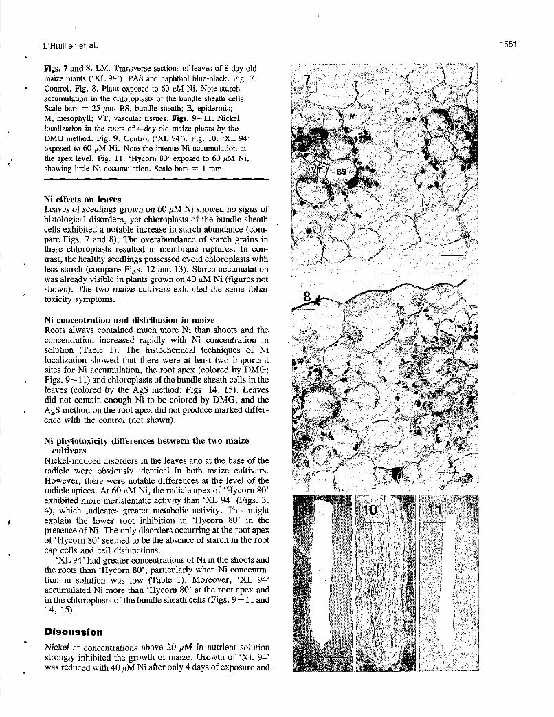
Figs. 7 and 8. LM. Transverse sections of leaves of 8-day-old maize plants (‘XL 94’). PAS and naphthol blue-black. Fig. 7. Control. Fig. 8. Plant exposed to 60 pM Ni. Note starch accumulation in the chloroplasts of the bundle sheath cells. Scale bars = 25 pm. BS, bundle sheath; E, epidermis; M, mesophyll; VT, vascular tissues. Figs. 9-11. Nickel localization in the roots of 4-day-old maize plants by the DMG method. Fig. 9. Control (‘XL 94’). Fig. 10. ‘XL 94’ exposed to 60 pM Ni. Note the intense Ni accumulation at the apex level. Fig. 11. ‘Hycom 80’ exposed to 60 p M Ni, showing little Ni accumulation. Scale bars = 1 mm.
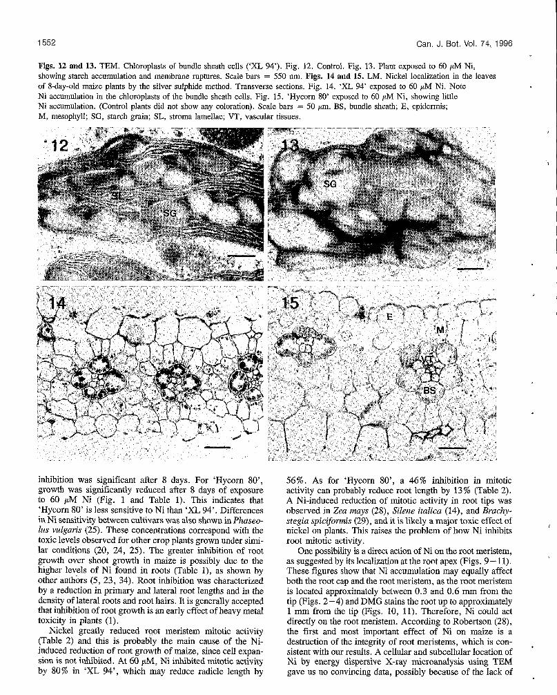
Ni effects on leaves Leaves of seedlings grown on 60 CJM Ni showed no signs of histological disorders, yet chloroplasts of the bundle sheath cells exhibited a notable increase in starch abundance (com- pare Figs. 7 and 8). The overabundance of starch grains in these chloroplasts resulted in membrane ruptures. In con- trast, the healthy seedlings possessed ovoid chloroplasts with less starch (compare Figs. 12 and 13). Starch accumulation was already visible in plants grown on 40 pM Ni (figures not shown). The two maize cultivars exhibited the same foliar toxicity symptoms.
Ni concentration and distribution in maize Roots always contained much more Ni than shoots and the concentration increased rapidly with Ni concentration in solution (Table 1). The histochemical techniques of Ni localization showed that there were at least two important sites for Ni accumulation, the root apex (colored by DMG; Figs, 9 - 11) and chloroplasts of the bundle sheath cells in the leaves (colored by the AgS method; Figs. 14, 15). Leaves did not contain enough Ni to be colored by DMG, and the AgS method on the root apex did not produce marked differ- ence with the control (not shown).
Ni phytotoxicity differences between the two maize
Nickel-induced disorders in the leaves and at the base of the radicle were obviously identical in both maize cultivars. However, there were notable differences at the level of the radicle apices. At 60 pM Ni, the radicle apex of ‘Hycorn 80’ exhibited more meristematic activity than ‘XL 94’ (Figs. 3, 4), which indicates greater metabolic activity. This might explain the lower root inhibition in ‘Hycorn 80’ in the presence of Ni, The only disorders occurring at the root apex of ‘Hycorn SO’ seemed to be the absence of starch in the root cap cells and cell disjunctions.
‘XL 94’ had greater concentrations of Ni in the shoots and the roots than ‘Hycorn SO’, particularly when Ni concentra- tion in solution was low (Table 1). Moreover, ‘XL 94’ accumulated Ni more than ‘Hycorn 80’ at the root apex and in the chloroplasts of the bundle sheath cells (Figs. 9 - 1 1 and 14, 15).
cultivars
Discussion Nickel at concentrations above 20 pM in nutrient solution strongly inhibited the growth of maize. Growth of ‘XL 94’ was reduced with 40 pM Ni after only 4 days of exposure and
1552
Figs. 12 and 13. TEM. Chloroplasts of bundle sheath cells (‘XL 94’). Fig. 12. Control. Fig. 13. Plant exposed to 60 p M Ni, showing starch accumulation and membrane ruptures. Scale bars = 550 nm. Figs. 14 and 15. LM. Nickel localization in the leaves of 8-day-old maize plants by the silver sulphide method. Transverse sections. Fig. 14. ‘XL 94’ exposed to 60 pM Ni. Note Ni accumulation in the chloroplasts of the bundle sheath cells. Fig. 15. ‘Hycorn 80’ exposed to 60 pM Ni, showing little Ni accumulation. (Control plants did not show any coloration). Scale bars = 50 pm. BS, bundle sheath; E, epidermis; M, mesophyll; SG, starch grain; SL, stroma lamellae; VT, vascular tissues.
Can. J. Bot. Vol. 74, 1996
inhibition was significant after 8 days. For ‘Hycorn 80’, growth was significantly reduced after 8 days of exposure to 60 pM Ni (Fig. 1 and Table 1). This indicates that ‘Hycorn 80’ is less sensitive to Ni than ‘XL 94’. Differences in Ni sensitivity between cultivars was also shown in Phaseo- lus vulgaris (25). These concentrations correspond with the toxic levels observed for other crop plants grown under simi- lar conditions (20, 24, 25). The greater inhibition of root growth over shoot growth in maize is possibly due to the higher levels of Ni found in roots (Table l), as shown by other authors (5, 23, 34). Root inhibition was characterized by a reduction in primary and lateral root lengths and in the density of lateral roots and root hairs. It is generally accepted that inhibition of root growth is an early effect of heavy metal toxicity in plants (1).
Nickel greatly reduced root meristem mitotic activity (Table 2) and this is probably the main cause of the Ni- induced reduction of root growth of maize, since cell expan- sion is not inhibited. At 60 pM, Ni inhibited mitotic activity by 80% in ‘XL 94’, which may reduce radicle length by
56%. As for ‘Hycorn SO’, a 46% inhibition in mitotic activity can probably reduce root length by 13% (Table 2). A Ni-induced reduction of mitotic activity in root tips was observed in Zea mays (28), Silene italica (14), and Brachy- stegia spic~omis (29), and it is likely a major toxic effect of nickel on plants. This raises the problem of how Ni inhibits root mitotic activity.
One possibility is a direct action,of Ni on the root meristem, as suggested by its localization at the root apex (Figs. 9- 11). These figures show that Ni accumulation may equally affect both the root cap and the root meristem, as the root meristem is located approximately between 0.3 and 0.6 mm from the tip (Figs. 2 -4) and DMG stains the root up to approximately 1 mm from the tip (Figs. 10, 11). Therefore, Ni could act directly on the root meristem. According to Robertson (28), the first and most important effect of Ni on maize is a destruction of the integrity of root meristems, which is con- sistent with our results. A cellular and subcellular location of Ni by energy dispersive X-ray microanalysis using TEM gave us no convincing data, possibly because of the lack of
I
,

L’Huillier et al. 1553
c
s
sensitivity of the technique and a loss of nickel during sample fixation (22). Nickel seemed to accumulate in roots of ‘XL 94’ more than ‘Hycorn SO’, particularly at low Ni con- centrations in solution (Table 1) and particularly at the apex (Figs. 10, 11). This probably plays a significant role in the stronger growth inhibition of ‘XL 94’ over ‘Hycorn 80’. We have no explanation for the differences between the two cul- tivars in Ni accumulation at the root apex. To our know- ledge, localization of Ni at the root apex had never been shown before, and it seems important to pursue this direc- tion. Furthermore, this is probably the first example of the DMG method used for Ni localization in plant material.
Another possible mechanism of Ni toxicity in maize is an inhibition of carbohydrate transport from leaves to roots, as suggested by the accumulation of starch in the leaves. This could be due to an increase in photosynthetic activity; how- ever, we observed no significant variation (22). Greger et al. (15) proposed a hypothesis whereby heavy metals have a greater effect in reducing carbohydrate transport than reduc- ing photosynthesis, thereby causing a carbohydrate accumu- lation in leaves and a decrease in roots, as already observed in other plants (24, 27, 30). The relatively high concentra- tions of Ni found in the shoots (Table 1) and especially in the chloroplasts (Figs. 14, 15) of the sensitive cultivar, as shown by others (36), suggest that Ni may have a toxic action at this level, resulting in a decrease in carbohydrate transport between leaves and roots. Several results presented here are consistent with this hypothesis: (i) starch statoliths, originat- ing from sucrose coming from leaves, were absent in the root cap; (ii) root growth, dependent on leaves for carbohydrates, was inhibited more than shoot growth; (iii) weak staining of cell walls at the root apex level, the absence of a cell plate in dividing cells, cell disjunctions, and increased lengthening of cells in the expansion zone suggest a deficiency in poly- saccharides and pectocellulosic compounds at the level of root apex cell walls; and (iv) poor protein density in the root apex, which might be explained by the phenomenon observed by Dieuaide et al. (lo) in which proteins replaced carbo- hydrates as major respiratory substrates after glucose star- vation.
As suggested by its accumulation in the chloroplasts of the bundle sheath cells, Ni could reduce carbohydrate transport by an inhibition of starch degradation into sucrose in the bundle sheath, and (or) by an inhibition of the active loading of sucrose into the phloem and subsequently of sucrose trans- port to the roots.
A decrease in carbohydrate supply for roots can then be part of the origin of the reduced mitotic activity in root tips, in particular by an inhibition of primary cell wall formation in dividing cells, as previously observed in B. spicifomis (29).
However, if the first effect of Ni is a direct inhibition of the mitotic activity, it must result in a decrease in the root sink effect for sugars and this probably contributes to the reduction in carbohydrate supply for roots. In this case, a decrease in carbohydrate transport may therefore be a secon- dary consequence of Ni-induced reduction in mitotic activity.
The absence or reduced number of statoliths in the root cap is surprising and has probably never been shown before. It seems important to examine how early the absence of statoliths is an observable effect and how Ni acts on the root cap cells.
Nickel staining in the leaves by the AgS method shows an accumulation in specific sites, such as the chloroplasts of the bundle sheath of Ni-treated plants. DMG failed to reveal Ni in the leaves because they did not contain enough Ni. In the roots, AgS did not produce marked differences between the control and the roots exposed to Ni (22), unlike DMG. It is probably due to the nonspecificity of AgS compared with DMG. Roots are generally the first site of accumulation of heavy metals (22,23,31), which probably masks Ni staining by AgS. The fact that AgS can stain Ni in the chloroplasts of the bundle sheath in plants exposed to Ni is probably due to a low concentration of other metals in this specific site that could interfere with Ni.
The reasons why the two maize cultivars are different in their sensitivity to Ni are not clear. When grown in 60 pM Ni, both cultivars showed an absence of statoliths in the root cap cells and an accumulation of starch in the leaves. How- ever, ‘Hycorn 80’, less sensitive to Ni than ‘XL 94’, exhibited greater root meristematic activity. This suggests that there is less inhibition of carbohydrate transport in ‘Hycorn 80’ than in ‘XL 94’, which is supported by the lower Ni concentration in the chloroplasts of ‘Hycorn SO’ (Figs. 14, 15). However, the higher root mitotic activity of this cultivar could also come from a smaller Ni uptake, espe- cially at low Ni concentrations in solution (Table 1) and from weaker action of Ni on the root meristem, as suggested by Figs. 10 and 11.
We may conclude from our results that in maize, Ni-induced reduction of growth is mainly the consequence of depressed mitotic activity in the root meristem. This could be due to at least two mechanisms that are not exclusive. The first one, the primary effect, could be a direct toxic action of Ni on the root meristem. The second one, which could only occur after, could result in a reduced carbohydrate supply for roots. This reduction could come either from a decrease in
. the sink root effect or from an inhibition in the leaves of starch degradation into sucrose and then of the transport of this later to the roots. Further studies on the cellular and sub- cellular location of Ni in roots and leaves, and particularly in root apex and cells of the bundle sheath, are necessary to clarify the mechanisms of Ni toxicity.
Acknowledgement The authors thank Southern Province of New Caledonia for financial aid.
References 1. Barceló, J., and Poschenrieder, C. 1990. Plant water relations
as affected by heavy metals stress: a review. J. Plant Nutr. 13:
2. Bennet, R.J., Breen, C.M., and Fey, M.V. 1987. The effects of alumipium on root cap function and root development in Zea mays L. Environ. Exp. Bot. 27: 91-104.
3. Bishnoi, N.R., Sheoran, I.S., and Singh, R. 1993. Influence of cadmium and nickel on photosynthesis and water relations in wheat leaves of different insertion level. Photosynthetica, 28: 473 -479.
4. Brooks, R.R. 1987. Serpentine and its vegetation. A multi- disciplinary approach. Dioscorides Press, Portland, Oreg.
5. Cataldo, D.A., Garland, T.R., Wildung , R.E., and Drucker, H.
1-37.
1554 Can. J. Bot. Vol. 74, 1996
1978. Nickel in plants. II. Distribution and chemical form in soybean plants. Plant Physiol. 62: 566-570.
6. Charlot, G. 1964. Colorimetric determination of elements. Elsevier Scientific Publishing Co., Amsterdam, The Nether- lands.
7. Clijsters, H., and Van Assche, F. 1985. Inhibition of photo- synthesis by heavy metals. Photosynth. Res. 7: 31-40.
8. Danscher, G. 1981. Histochemical demonstration of heavy metals. A revised version of the sulphide silver method suitable for both light and electromicroscopy . Histochemistry, 71:
9. Davis, R.D., and Beckett, P.H.T. 1978. Upper critical levels of toxic elements in plants. II. Critical levels of copper in young barley, wheat, rape, lettuce and ryegrass, and of nickel and zinc in young barley and ryegrass. New Phytol. 80: 23-32.
10. Dieuaide, M., Brouquisse, R., Pradet, A., and Raymond, P. 1992. Increased fatty acid @oxidation after glucose starvation in maize root tips. Plant Physiol. 99: 595-600.
11. Fisher, D.B. 1968. Protein staining of ribboned epon sections for light microscopy. Histochemie, 16: 92-96.
12. Foy, C.D., Chaney, R.L., and White, M.C. 1978. The phys- iology of metal toxicity in plants. Annu. Rev. Plant Physiol. 29:
13. Frank, R., Stonefield, K.I., Suda, P., and Potter, J.W. 1982. Impact of nickel contamination.on the production of vegetables on an organic soil, Ontario, Canada. Sci. Total Environ. 26:
14. Gabbrielli, R., Pandolfini, T., Vergnano, O., and Palandri, M.R. 1990. Comparison of two species with different nickel tolerance strategies. Plant Soil, 122: 271 -277.
15. Greger, M., Brammer, E., Lindberg, S., Larsson, G., and Idestam-Almquist, J. 1991. Uptake and physiological effects of cadmium in sugar beet (Beta vulgaris) related to mineral provi- sion. J. Exp. Bot. 42: 729-737.
16. Holmgren, G.G.S., Meyer, M.W., Chaney, R.L., and Daniels, R.B. 1993. Cadmium, lead, zinc, copper and nickel in agricultural soils of the United States of America. J. Environ. Qual. 22: 335-348.
17. Homès, J. 1975. La préparation des tissus végétaux pour l’observation au microscope électronique à balayage. Bull. Soc. R. Bot. Belg. 108: 219-231.
18. Jaffré, T., Brooks, R.R., Lee, J., and Reeves, R.D. 1976. Sebertia acuminata: a hyperaccumulator of nickel from New Caledonia. Science (Washington, D.C.), 193: 579-580.
19. Khalid, B.Y., and Tinsley, J. 1980. Some effects of nickel toxicity on rye grass. Plant Soil, 55: 139-144.
20. Krupa, Z., Siedlecka, A., Maksymiec, W., and Baszynski, T. 1993. In vivo response of photosynthetic apparatus of Phaseo- lus vulgaris L. to nickel toxicity. J. Plant Physiol. 142: 664- 668.
1-16.
511-566.
41 -65.
21. Lee, J., Reeves, R.D., Brooks, R.R., and Jaffré, T. 1978. The relation between nickel and citric acid in some nickel- accumulating plants. Phytochemistry, 17: 1033 - 1035.
22. L’Huillier, L. 1994. Biodisponibilité du nickel dans les sols ferrallitiques ferritiques de Nouvelle-Calédonie. Effets toxiques de Ni sur le développement et la physiologie du maïs. M.Sc. thesis, University of Montpellier II, Montpellier, France.
23. Liibben, S., and Sauerbeck, D. 1991. The uptake and distribu- tion of heavy metals by spring wheat. Water Air Soil Pollut. 57-58: 239-247.
24. Moya, J.L., Ros, R., and Picazo, I. 1993. Influence of cad- mium and nickel on growth, net photosynthesis and carbohy- drate distribution in rice plants. Photosynth. Res. 36: 75-80.
25. Piccini, D.F., and Malavolta, E. 1992. Effect of nickel on two common bean cultivars. J. Plant Nutr. 15: 2343 -2350.
26. Punz, W.F., and Sieghardt, H. 1993. The response of roots of herbaceous plant species to heavy metals. Environ. Exp. Bot.
27. Rauser, W.E. 1978. Early effects of phytotoxic burdens of cad- mium, cobalt, nickel and zinc in white beans. Can. J. Bot. 56:
28. Robertson, A.I. 1985. The poisoning of roots of Zea mays by nickel ions, and the protection afforded by magnesium and cal- cium. New Phytol. 100: 173-189.
29. Robertson, A.I., and Meakin, M.E.R. 1980. The effect of nickel on cell division and growth of Brachystegia spiciformis seedlings. Kirkia, 12: 115- 125.
30. Samarakoon, A.B., and Rauser, W.E. 1979. Carbohydrate levels and photoassimilate export from leaves of Phaseohs vulgaris exposed to excess cobalt, nickel and zinc. Plant Physiol. 63: 1165-1169.
31. Sauerbeck, D.R., and Hein, A. 1991. The nickel uptake from different soils and its prediction by chemical extractions. Water Air Soil Pollut. 57-58: 861-871.
32. Severne, B.C. 1974. Nickel accumulation by Hybanthus flori- bundus. Nature (London), 248: 807-808.
33. Spurr, A.R. 1969. A low-viscosity epoxy-resin embedding medium for electron microscopy. J. Ultrastruct. Res. 26:
34. Taylor, G.J. 1989. Multiple metal stress in Triticum aestivum, differentiation between additive, multiplicative, antagonistic and synergistic effects. Can. J. Bot. 67: 2272-2276.
35. Vanselow, A.P. 1966. Nickel. In Diagnostic criteria for plants and soils. Edited by H.D. Chapman. University of California, Riverside, Calif. pp. 302-309.
36. Veeranjaneyulu, K., and Das, V.S.R. 1982. Intrachloroplast localization of 65Zn and 63Ni in a Zn-tolerant plant, Ocimum basiliczim Benth. J. Exp. Bot. 33: 1161-1165.
33: 85-98.
1744 - 1749.
31-43.
. . - . . . .. ..
National Research Conseil national Council Canada de recherches Canada
I 1% \
I
,
I
!
Reprinted from Canadian Journal of Botany
Réimpression de la Revue canadienne de botanique
Nickel effects on two maize (Zea mays) cultivars: growth, structure, Ni concentration, and localization
I
Laurent L'Huillier, Jean d'Auzac, MOniqUQ Durand, and Nicole Michaud-Ferriere
Volume 74 Number 10 0 1996
Pages 1547 - 1554
1 1 I
a8ä