nidovirus-associated proliferative pneumonia in the green ... filezurich open repository and archive...
TRANSCRIPT

Zurich Open Repository andArchiveUniversity of ZurichMain LibraryStrickhofstrasse 39CH-8057 Zurichwww.zora.uzh.ch
Year: 2017
Nidovirus-Associated Proliferative Pneumonia in the Green Tree Python(Morelia viridis)
Dervas, Eva
Abstract: Since 2014 we observed a noticeable increase in sudden deaths of green tree pythons (Moreliaviridis). A total of 12 affected green tree pythons from 7 different breeders were submitted for a diagnosticpost mortem examination. The gross examination revealed accumulation of mucus within airways andlung, associated with enlargement of the entire lung. Histology showed marked hyperplasia of the airwayepithelium and faveolar type II pneumocytes. Using next-generation sequencing (NGS) on lung samples ofdiseased snakes we identified a viral RNA genome that was approximately 85% identical with nidovirusespreviously identified in ball pythons and an Indian python. We then confirmed the presence of thenovel nidovirus in all diseased snakes by RT-PCR. To attempt virus isolation, we established primarycell cultures of Morelia viridis liver and brain, these were inoculated with lung homogenates of diseasedsnakes. Ultrastructural examination of concentrated cell culture supernatants revealed nidovirus particles,and subsequent NGS analysis yielded the full genome of the novel Morelia viridis nidovirus (MVNV).We then generated an antibody against MVNV nucleoprotein and MVNV specific oligoprobes and usedimmunohistology and RNA in situ hybridisation to identify the viral target cells in the lungs. Nidovirusesare a likely cause of severe pneumonia in pythons and cause sudden outbreaks in breeding collections.Keywords: Nidovirus, pneumonia, Morelia viridis
Posted at the Zurich Open Repository and Archive, University of ZurichZORA URL: https://doi.org/10.5167/uzh-144231DissertationPublished Version
Originally published at:Dervas, Eva. Nidovirus-Associated Proliferative Pneumonia in the Green Tree Python (Morelia viridis).2017, University of Zurich, Vetsuisse Faculty.

0
Institut für Veterinärpathologie
der Vetsuisse-Fakultät Universität Zürich
Direktorin: Prof. Dr. med. vet. Anja Kipar
Arbeit unter wissenschaftlicher Betreuung von
Prof. Dr. med. vet. Anja Kipar
Nidovirus-Associated Proliferative Pneumonia in the Green Tree Python
(Morelia viridis)
Inaugural-Dissertation
zur Erlangung der Doktorwürde der
Vetsuisse-Fakultät Universität Zürich
vorgelegt von
Eva Dervas
Tierärztin
von Athen, Griechenland
genehmigt auf Antrag von
Prof. Dr. med. vet. Anja Kipar, Referentin
2017
1
Institut für Veterinärpathologie
der Vetsuisse-Fakultät Universität Zürich
Direktorin: Prof. Dr. med. vet. Anja Kipar
Arbeit unter wissenschaftlicher Betreuung von
Prof. Dr. med. vet. Anja Kipar
Nidovirus-Associated Proliferative Pneumonia in the Green Tree Python
(Morelia viridis)
Inaugural-Dissertation
zur Erlangung der Doktorwürde der
Vetsuisse-Fakultät Universität Zürich
vorgelegt von
Eva Dervas
Tierärztin
von Athen, Griechenland
genehmigt auf Antrag von
Prof. Dr. med. vet. Anja Kipar, Referentin
2017

1
INHALTSVERZEICHNIS
1. Zusammenfassung 2
2. Abstract 3
3. Artikel 4-55
4. Danksagung 56
5. Lebenslauf 57
2
Vetsuisse-Fakultät Universität Zürich 2017
Eva Dervas
Institut für Veterinärpathologie
Nidovirus- assoziierte proliferative Pneumonie im grünen Baumpython (Morelia viridis)
Seit 2014 werden vermehrt plötzlich verstorbene Baumpythons zur diagnostischen postmortalen
Untersuchung eingeschickt; insgesamt wurden 12 erkrankte Baumpythons von sieben verschiedenen
Züchtern untersucht. In der makroskopischen Untersuchung waren vor allem eine ausgeprägte
Schleimansammlung in den Atemwegen sowie eine Verdickung der gesamten Lunge auffällig.
Histologisch konnte eine Hyperplasie des respiratorischen Epithels und der Typ II Pneumozyten im
Faveolarraum festgestellt werden. Mittels Next-generation sequencing (NGS) am Lungengewebe
erkrankter Tiere konnte ein virales RNA-Genom identifiziert werden, dass 85 % identisch war mit
beschriebenen Nidoviren bei Königspythen und einer Indischen Python. Mittels RT-PCR wurde
anschliessend das neue Nidovirus in allen erkrankten Baumpython nachgewiesen. Es wurden
Primärkulturen von Morelia viridis Leber- und Gehirnzellen etabliert, die zur Virusisolation aus
Lungenhomogenaten erkrankter Tiere dienten. In den Überständen der infizierten Zelkulturen konnten
elektronenmikroskopisch Nidoviruspartikel nachgewiesen werden; mittels abermaligem NGS konnte
das vollständige Genom des Morelia viridis Nidovirus (MVNV) sequenziert werden. Mittels eines
spezifischen Antikörpers gegen das MVNV Nukleoprotein sowie spezifischen Oligo-Riboproben und
Immunhistologie bzw. in situ-Hybridisierung konnten die Zielzellen des Virus identifiziert werden.
Nidovirus-Infektionen können zum plötzlichen Tod von Pythons führen und sind somit von
zunehmender Bedeutung für Schlangenbestände.
Stichwörter: Nidovirus, Pneumonie, Morelia viridis
3
Vetsuisse Faculty University of Zurich 2017
Eva Dervas
Institute of Veterinary Pathology
Nidovirus-Associated Proliferative Pneumonia in the Green Tree Python (Morelia viridis)
Since 2014 we observed a noticeable increase in sudden deaths of green tree pythons (Morelia viridis).
A total of 12 affected green tree pythons from 7 different breeders were submitted for a diagnostic post
mortem examination. The gross examination revealed accumulation of mucus within airways and lung,
associated with enlargement of the entire lung. Histology showed marked hyperplasia of the airway
epithelium and faveolar type II pneumocytes. Using next-generation sequencing (NGS) on lung
samples of diseased snakes we identified a viral RNA genome that was approximately 85% identical
with nidoviruses previously identified in ball pythons and an Indian python. We then confirmed the
presence of the novel nidovirus in all diseased snakes by RT-PCR. To attempt virus isolation, we
established primary cell cultures of Morelia viridis liver and brain, these were inoculated with lung
homogenates of diseased snakes. Ultrastructural examination of concentrated cell culture supernatants
revealed nidovirus particles, and subsequent NGS analysis yielded the full genome of the novel
Morelia viridis nidovirus (MVNV). We then generated an antibody against MVNV nucleoprotein and
MVNV specific oligoprobes and used immunohistology and RNA in situ hybridisation to identify the
viral target cells in the lungs. Nidoviruses are a likely cause of severe pneumonia in pythons and
cause sudden outbreaks in breeding collections.
Keywords: Nidovirus, pneumonia, Morelia viridis

4
Nidovirus-Associated Proliferative Pneumonia in the Green Tree Python (Morelia
viridis)
Eva Dervas1, Jussi Hepojoki
1,2, Andrea Laimbacher
3, Fernando Romero-Palomo
1, Christine Jelinek
1,
Saskia Keller1, Teemu Smura
2, Satu Hepojoki
1, Anja Kipar
1*, Udo Hetzel
1
1Institute of Veterinary Pathology, Vetsuisse Faculty, University of Zürich, Zürich, Switzerland
2University of Helsinki, Medicum, Department of Virology, Helsinki, Finland
3Institute of Virology, Vetsuisse Faculty, University of Zurich, Switzerland
Running title: Morelia viridis nidovirus
*Corresponding author’s address:
Institute of Veterinary Pathology
Vetsuisse Faculty
University of Zurich
Winterthurerstrasse 268
CH - 8057 Zurich
Switzerland
Phone: +41 44 6358552
E-mail: [email protected]
5
Abstract
In 2014 we observed a noticeable increase in sudden deaths of green tree pythons (Morelia
viridis). Pathological examination revealed accumulation of mucoid material within airways
and lung, associated with enlargement of the entire lung. We performed full necropsy and
histological examination on 12 affected green tree pythons from 7 different breeders to
characterise the pathogenesis of this “mucinous” pneumonia. By histology we could show a
marked hyperplasia of the airway epithelium and of faveolar type II pneumocytes. Since
routine microbiological tests failed to identify a causative agent, we studied lung samples of a
few diseased snakes by next-generation sequencing (NGS). From the NGS data we could
assemble a piece of RNA genome <85% identical to nidoviruses previously identified in ball
pythons and Indian pythons. We then employed RT-PCR to demonstrate the presence of the
novel nidovirus in all diseased snakes. To attempt virus isolation, we established primary cell
cultures of Morelia viridis liver and brain, which we inoculated with lung homogenates of
infected individuals. Ultrastructural examination of concentrated cell culture supernatants
showed the presence of nidovirus particles, and subsequent NGS analysis yielded the full
genome of the novel virus, Morelia viridis nidovirus (MVNV). We then generated an
antibody against MVNV nucleoprotein, which we used alongside RNA in situ hybridisation
to demonstrate viral antigen and RNA in the affected lungs. This suggests that in natural
infection MVNV damages the respiratory tract epithelium which then results in epithelial
hyperplasia, most likely as an exaggerated regenerative attempt in association with increased
epithelial turnover.

6
Importance
Fairly recently novel nidoviruses associated with severe respiratory disease were identified in
ball pythons and Indian pythons. Herein we report isolation and identification of a further
nidovirus from green tree pythons (Morelia viridis) with fatal pneumonia. We thoroughly
characterize the pathological changes in the infected individuals, and show that nidovirus
infection is associated with marked epithelial proliferation in the respiratory tract. We
speculate that this and the associated excess mucus production can lead to the animals’ death,
by inhibitingthe normal gas exchange in the lung. The virus was predominantly detected in
the respiratory tract, which renders transmission via the respiratory route likely. Nidoviruses
cause sudden outbreaks with high mortality in breeding collections, most affected snakes die
without prior clinical signs. These findings, together with those of other groups, indicate that
nidoviruses are a likely cause of severe pneumonia in pythons.
7
Introduction
The green tree python or Southern green python, Morelia viridis (Schlegel, 1872), is an
oviparous boid constrictor snake with natural habitats in several Indonesian islands,
Florida, Papua New Guinea and North Australia (1). Over the recent years large numbers of
M. viridis have been exported from Indonesia to Europe and America (2), and they have
become increasingly popular in both private and zoological collections (3, 4). So far, the
knowledge on infectious diseases of M. viridis is sparse (5).
Recently, fatal pneumonias have been reported in two other python species, ball pythons
(Python regius) (6) and Indian pythons (Python molurus) (7); infection with novel nidoviruses
was found as the common denominator (8, 9). The described viruses are approximately
equidistant from the two current genera, Torovirus and Bafinivirus, of the Torovirinae
subfamily in the family Coronaviridae, order Nidovirales (8–15), in which they will likely
form a novel genus (9).
The order Nidovirales comprises four families of complex positive-sense single-stranded
RNA (ssRNA+) viruses, Arteriviridae, Mesoniviridae, Roniviridae and Coronaviridae, which
are also distinguished by their genome size: roniviruses (~26 kb) and coronaviruses (26-33
kb) are known as “large nidoviruses” (16–18), arteriviruses as “small nidoviruses” (13–16
kb), and mesoniviruses with an intermediate genome size between the two (16, 17, 19, 20).
While roniviruses and mesoniviruses are known to infect crustaceans and insects (19, 21),
arteriviruses have been associated with an acute respiratory syndrome and abortion in pigs
(porcine reproductive and respiratory syndrome virus, PRRSV) or a lethal haemorrhagic
disease in non-human primates (simian haemorrhagic fever virus, SHFV) (22). The
Coronavirinae subfamily of the Coronaviridae includes several pathogens of mammals
(Feline coronavirus; transmissible gastroenteritis virus; Equine coronavirus, to mention a
few), birds andfish, and has provided a steady supply of emerging threats to human health

8
over the past 15 years, including Severe Acute Respiratory Syndrome (SARS) and Middle-
Eastern Respiratory Syndrome (MERS) (23). The members of the second subfamily of the
Coronaviridae, Torovirinae, have so far not generated similar threats. They include
bafiniviruses, which infect ray-finned fish and induce renal tubular necrosis and necrotising
hepatitis (24), and toroviruses, which infect mammals (including humans, cattle, horses, and
pigs) (10, 11, 25–27). Toroviruses have a tropism for epithelial cells, of both the respiratory
and alimentary tract (28–30); in cattle, an association with pneumonia has been reported (30,
31). Toroviruses exhibit a unique morphology, the viral particles are kidney- and/or rod-
shaped, with a tubular, torus-shaped ribonucleoprotein (RNP) enveloped by a membrane
decorated with spikes composed of S protein (32). The RNP comprises the nucleoprotein (N
protein) and the ssRNA+ genome (33, 34).
In 2014, a Swiss breeder submitted two adult green tree pythons for diagnostic post mortem
examination. Both animals had died with signs of a “mucinous” pneumonia. In the following
year, the same breeder submitted another two individuals. These, as well as a further five
green tree pythons from three additional breeders, all exhibited similar pathological findings.
In 2016, additional cases were observed, involving two more breeding collections. As all
routine diagnostic tests undertaken failed to identify potential causative viruses or specific
bacterial agents, we initiated an investigation into the cause of this apparently emerging
disease of green tree pythons.
9
Material and Methods
Animals. The study was performed on sixteen green tree pythons (Morelia viridis) from six
breeding collections in Switzerland and one collection in Germany (Table 1). The collections
varied in both size (i.e. number of breeding animals) and species range, from a single snake
up to a collection of 50 snakes of various species.
All animals were submitted to the Institute of Veterinary Pathology, Vetsuisse Faculty,
University of Zurich, for diagnostic purposes. Fifteen snakes had died spontaneously, one was
euthanized following an ASPA (Animals Scientific Procedures Act 1986) schedule 1
(appropriate methods of humane killing,
http://www.legislation.gov.uk/ukpga/1986/14/schedule/1) procedure. In each case, a full
diagnostic post mortem examination was performed with owner’s consent. For these
diagnostic-motivated necropsies, no ethical permission is required at the University of Zurich.
The initial study population is represented by nine snakes, of which four (A1-A4) were from
one breeder (A), each two from a second and third breeder (B1, B2 and C1, C2), and one (D1)
from a fourth breeder (Table 1A). These animals were submitted between September 2014
and November 2015. After completion of the next-generation sequencing (NGS) study,
another seven snakes were submitted (E1, F1, G1-G5) by another three breeders (E, F, G). All
initial and another three snakes of the second cohort exhibited one common gross feature: the
airways contained a variable amount of mucoid material which was most abundant in the
faveolar lumen (Table 1A). The remaining four snakes (G2-G5) did not show any changes in
the airways or lungs (Table 1B) and later served as controls.
Sample collection and screening for infectious agents. During post mortem examinations
samples from all organs were collected and fixed in 10% buffered formalin for histological
examination. Additional samples from brain, lung, liver, and kidney were stored at -80 °C for
further analysis. Also, lung samples from the freshly euthanized snake (A2) and from two
snakes that were necropsied within a few hours after death (B1, D1) were fixed in
glutaraldehyde/paraformaldehyde and processed for transmission electron microscopy (TEM)
as described (35).
10
Prior to performing NGS, six of the initial cases were screened for infectious agents: bacterial
cultivation was attempted from the lungs of three snakes (A1, A3, D1) at the Institute of
Veterinary Bacteriology, Vetsuisse Faculty, University of Zurich; and lung samples from
another three snakes (B1, B2, C1) were submitted to a commercial lab (LABOKLIN, Basel,
Switzerland) for virus diagnostics (reovirus, paramyxovirus, Sunshine virus, nidovirus) (Table
1).
Virus isolation and ultrastructural characterisation. Primary tissue cultures of Morelia
viridis were established as described (35), using brain and liver material from a foetus (from a
clutch of B1 and B2). The tissue was trimmed into blocks (1 mm) and suspended in 5 ml of
minimal essential medium (MEM, ThermoFisher, Scientific, GIBCO) supplemented with
HEPES (25 mM), 10% fetal bovine serum (FBS; Biochrom), Gentamycin (0.05 mg/ml), L-
Glutamine (2 mM, Biochrom), 10% Tryptose Phosphate-Broth (DIFCO), and 20 µl α-D-
Glucose (90 g/l PBS) in sterile cell culture dishes (5 cm in diameter), and incubated at 30 °C
and 5% CO2. Primary Boa constrictor lung (V/4Lu) and brain (V/4Br) cell lines were
established similarly as described above.
Cell cultures were used for inoculations at passage 8-15 (M. viridis cell lines), 30-35 for
V/4Lu and V/4Br. Briefly, after initial trimming of lungs into blocks (>1 mm) the pieces were
mechanically homogenized in 1 ml of Trypsin-EDTA solution (0.25%, Gibco, Thermo Fisher
Scientific), the cell debris pelleted by centrifugation (5 min at 1,000 x g), and the remaining
supernatant was diluted in 5 ml of MEM supplemented with 25 mM HEPES (Thermo Fisher
Scientific) and 15% FBS (Biochrom), and 0.45 µm filtered. One ml of the filtered lung
homogenate further diluted 1:10 in growth medium was used to inoculate 75-cm2
flasks of
both brain and liver cells. The medium was changed at 1-2 day intervals until most cells
detached or died. The supernatants were frozen at -20 °C, pooled, and 0.45 µm filtered. The
cleared supernatant was loaded onto a cushion of 30% (w/v) sucrose in PBS, concentrated at
100,000 g for 2 h at 4 °C and solubilized in PBS. For protection, protease inhibitor (Protease
inhibitor cocktail tablets complete, Mini, EDTA-free, Roche Diagnostics, Mannheim
11
Germany) was added to the supernatant. For negative staining, samples were adsorbed to
carbon-coated parlodion films mounted on 300 mesh/inch copper grids (EMS, Fort
Washington, PA, USA) for 10 min, washed once with H2O, and stained with 2%
phosphotungstic acid (PTA), pH 7.0 (Aldrich, Steinheim, Germany) for 1 min. Specimens
were analyzed in a transmission electron microscope (CM12, Philips, Eindhoven, The
Netherlands) equipped with a CCD camera (Ultrascan 1000, Gatan, Pleasanton, CA, USA) at
an acceleration voltage of 100 kV.
M. viridis brain and liver cells were harvested at 3, 4, and 5 days post infection (dpi) and
pelleted by centrifugation at 5,000 rpm for 5 min at room temperature (Eppendorf centrifuge
5415C, NIST). The pellets were fixed with 2.5% glutaraldehyde in PBS (pH 7.4), and
routinely resin embedded for TEM (35). To obtain a retrovirus-free virus preparation, the
supernatant collected from M. viridis liver cell cultures inoculated with lung homogenate was
used to inoculate a B. constrictor kidney cell line I/1Ki (35). Supernatant was collected from
the infected I/1Ki cells at two day intervals until 8 dpi. The supernatants were filtered through
a 0.45 µm filter, pooled, aliquoted, and stored at -80 °C.
Next-generation sequencing (NGS). RNA was isolated from the lungs of three diseased
snakes (animals A2, B1, D1, Table 1) with TRIzol® reagent (Life Technologies) according to
the manufacturer’s protocol, using 5 g of RNA grade glycogen (ThermoFisher Scientific) as
carrier. The RNA samples were initially treated with DNAse I (Fermentas), followed by re-
purification with the GeneJET RNA purification kit (Thermo Fisher Scientific). Ribo-Zero
Gold rRNA Removal Kit for Epidemiology (Illumina) was used according to the
manufacturer’s protocol to further clean the RNA. In the case of virus sequencing from cell
culture supernatants, the RNA isolation was done using QIAamp Viral RNA Mini Kit
(QIAGEN) following the manufacturer’s protocol without addition of carrier RNA. For RNA
isolated from cell culture supernatants, the rRNA was removed with NEBNext rRNA
Depletion Kit (New England Biolabs). The indexing and NGS library preparation was
accomplished with the NEBNext Ultra RNA Library Preparation Kit (New England Biolabs)
12
according to the manufacturer’s protocol. The libraries were quantified using the NEBNext
Library Quant Kit for Illumina (New England Biolabs). Pooled libraries were sequenced on
an Illumina MiSeq (Illumina) using the MiSeq Reagent Kit v3 (Illumina), with 291-bp (for
lung samples) and 300-bp (for RNA isolated from cell culture supernatant) reads from both
ends (paired-end). De novo sequence assembly was performed after removing reads matching
to the host genome, both with MIRA version 4.9.5. (http://mira-assembler.sourceforge.net/)
on CSC (IT Center for Science Ltd., Finland) Taito supercluster. The generated contiguous
sequences (contigs) from the lung sample run were initially screened by nucleotide BLAST
(blastn, at https://blast.ncbi.nlm.nih.gov/Blast.cgi) and several contigs matching ball python
nidovirus (BPNV; NCBI Reference Sequence NC_024709.1) and Python nidovirus (PNV;
GenBank accession KJ935003.1) were identified. In attempt to obtain full-length genome the
contigs were mapped to BPNV and PNV genomes using the BWA-SW tool (36) in Unipro
UGENE (37). However, contig with almost full-length genome for a novel nidovirus
subsequently named “Morelia viridis nidovirus (MVNV)” was obtained via de novo assembly
from RNA isolated from cell culture supernatant. The genome open reading frames, ORFs,
were detected using Unipro UGENE. The contig coverage was determined using Bowtie2
reference alignment of the entire NGS data in Unipro UGENE.
Phylogenetic analysis and bioinformatics. Since Blastn search of the contig obtained by de
novo assembly from purified virus material suggested that python nidoviruses (BPNV, PNV,
a proposed new genus in the Torovirinae sub-family of nidoviruses) were the most
homologous group to MVNV, representative sequences of this sub-family (i.e. genus
Torovirus, Bafinivirus and python nidoviruses) were downloaded from the GenBank. The
amino acid sequences of the conserved ORF1b were aligned using ClustalW algorithm
implemented in MEGA version 6.06 (38) followed by manual refinement. The best-fit
substitution model was sought using maximum likelihood method implemented in MEGA
6.06. Phylogenetic trees were constructed using the Bayesian Monte Carlo Markov Chain
(MCMC) method implemented in BEAST version 1.8.0 (39). The analyses were performed
with LG+G+I model of substitution, strict clock and constant size demographic model. The
Bayesian analyses were run for 10 million states and sampled every 1000 states. The analyses

13
were carried out on CSC – IT Center for Science Ltd. (Espoo, Finland). Posterior probabilities
were calculated with a burn-in of 1 million states and checked for convergence using Tracer
version 1.6 (40). The mfold Web Server (available at http://unafold.rna.albany.edu/?q=mfold)
was utilized for determining the RNA folding (41) around the ribosomal frameshift signal
(RFS). Unipro UGENE (37) was utilized for nucleotide alignments (MUSCLE (42)) around
the RFS. HMMER: biosequence analysis using profile hidden Markov models (available at
http://hmmer.org/, (43) was used to identify conserved domains in the identified ORFs.
Quantitative reverse transcription-polymerase chain reaction (qRT-PCR). RNA was
extracted from lung tissue samples with the TRIzol® reagent (Life Technologies) utilizing
mechanical homogenization with a MagNA Lyser (Roche). After addition of chloroform and
separation of the RNA-containing phase by centrifugation (15 min, 12,000 x g, 4°C) the RNA
was purified with the QiaGEN RNeasy Mini Kit (Qiagen) following the manufacturer’s
protocol for RNA clean up.
The amount of RNA in the samples was measured by Nanodrop 2000c (Thermo Fisher
Scientific), and the samples were subsequently diluted with RNase-free water to a
concentration of 150 ng/µl to allow comparative assessment.
A Taqman qRT-PCR assay was set up using the following primers (Microsynth AG,
Switzerland): Nido-fwd, 5´-AGTCATCTGTCTCGACCACCT-3´ and Nido-rev, 5´-
ACATGTAGAGCACTTTGACTGGTT-3´. The sequence for the probe (Microsynth AG,
Switzerland) was: Nido-probe, FAM-CGACAACTGGGTCATCAGACGC-TAMRA.
The qRT-PCR was performed on an Applied Biosystems 7500 fast Real-Time PCR system
using 96-well plates. The reaction volume (25 µl) consisted of 12.5 µl of One step qRT-PCR
MasterMix (Eurogentec), each 1 µl of forward and reverse primer (10 M), 1 µl of probe (10
M), 0.125 µl of reverse transcriptase (Euroscript RT and RNase Inhibitor Mix, Eurogentec),
5 µl (750 ng) of template RNA, and 4.5 µl of DEPC-treated H2O.
The qRT-PCR was set up with the following cycling conditions: 1) 30 min at 48 °C, 2) 10
min at 95 °C, 3) 15 s at 95 °C, and 4) 1 min at 60 °C, 40 cycles between steps 3 and 4. The
data was collected during step 4 of the RT-PCR programme.
14
Recombinant protein expression and generation of anti-MVNV nucleoprotein (N
protein) antiserum. To identify the ORF for the N protein gene in the genome of the novel
virus isolate, the de novo assembled genome was aligned with BPNV (NCBI Reference
Sequence NC_024709.1) and PNV (GenBank accession KJ935003.1) genomes using
MUSCLE (42) in Unipro UGENE (37). The N protein coding region was obtained from
cDNA transcribed with random primers using RNA isolated from supernatants of cultured
cells inoculated with lung tissue homogenate (see above). The cDNA transcription was done
using RevertAid Transcriptase (ThermoFisher Scientific) following the protocol for random
hexamer amplification. The PCR amplification of the whole N protein coding region was
done with primers designed according to the Champion pET Directional TOPO Expression
Kit’s manual (ThermoFisher Scientific) using Phusion Flash High-Fidelity PCR Master Mix
(ThermoFisher Scientific). The PCR product was purified from agarose gel using GeneJET
Gel Extraction Kit (ThermoFisher Scientific) and ligated to pET101/D-TOPO vector with N-
terminal V5-epitope and His-tags (ThermoFisher Scientific). The ligated vector was
transformed to One Shot TOP10 Competent Cells (ThermoFisher Scientific) and clones with
the desired insert were screened by plating on Luria Broth (LB) agar plates with 100 g/ml
ampicillin. Individual colonies were grown in 5 ml of LB supplemented with 100 g/ml
ampicillin overnight and the plasmids purified using the GeneJET Plasmid Miniprep Kit
(ThermoFisher Scientific) following the manufacturer’s recommendation. The plasmids were
sequenced (Microsynth AG, Switzerland), using T7 forward and reverse primers. The N
protein containing plasmid was transformed into BL21 Star (DE3) One Shot Chemically
Competent E. coli (ThermoFisher Scientific), and protein expression was done in LB
supplemented with 100 g/ml carbenicillin (Sigma-Aldrich) and 1% glucose (Sigma-Aldrich)
according to the manufacturer’s recommendation. The recombinant N protein expressed in
inclusion bodies was purified using Chelating Sepharose Fast Flow (GE Healthcare Life
Sciences) with cobalt as the immobilized metal ion and 8M urea in all buffers. The protein in
the elution buffer (50 mM Tris, 500 mM NaCl, 300 mM imidazole, 8M Urea, pH 7.5) was
concentrated using a 10 kDa cut off Amicon Ultra-15 Centrifugal Filter Unit (EMD
Millipore), and buffer exchange (to 25 mM Tris, 75 mM NaCl, pH 7.5) was achieved by

15
decreasing the urea concentration slowly using Slide-A-Lyzer Dialysis Cassette 3.5 MWCO
(ThermoFisher Scientific). The purified recombinant protein was used to immunize a rabbit
applying the following scheme: initial immunization (day 0), first booster (day 7), second
booster (day 14), third booster (day 42), and final bleeding (day 49) (BioGenes GmbH,
Berlin, Germany). The generated antiserum was cleaned by affinity purification similarly to
previous descriptions (44, 45). Briefly, 500 g of purified recombinant N protein was
dialyzed in PBS followed by coupling to CnBr-activated Sepharose 4B (GE Healthcare)
following manufacturer’s protocol. The affinity matrix was packed into Econo-Pac
Chromatography columns (Bio-Rad) and antiserum was passed through the column by gravity
flow. After washing with several column volumes of PBS, the bound antibodies were eluted
with 0.1M glycine (pH 2.5) and the fractions were neutralized by addition of 1M Tris, pH 8.5.
The affinity purified antibody was dialyzed against PBS, and concentrated using 10 kDa cut
off Amicon Ultra-15 Centrifugal Filter Unit (EMD Millipore). The purified antibody was
mixed with glycerol (50%) and stored in aliquots at -20 °C.

16
Immunofluorescence staining, qRT-PCR of cell culture supernatants, and estimation of
virus titer.
For immunofluorescence staining M. viridis (brain) and B. constrictor (kidney, lung, brain)
cells were detached by trypsin, washed with growth medium, and seeded on glass bottom 24-
well plates (IBL, Gerasdorf, Austria). The attached cells were inoculated by incubation with
250 l of 1/10 diluted (in growth medium) MVNV isolated on I/1Ki. After 1 h incubation at
30 °C the cells were washed twice with supplemented growth medium, and finally 1 ml of
supplemented growth medium was added to each well. For each cell line three wells were
inoculated with MVNV and three wells were mock-infected with supplemented growth
medium. At 3 dpi the cells were fixed by replacing the supplemented growth medium with
4% EM-grade paraformaldehyde in PBS followed by 15 min incubation at RT. After fixation
the cells were washed once with PBS, permeabilised (0.25% Triton X-100 and 3% BSA in
PBS), and left in PBS until staining. The replicate wells (3 replicates of MVNV and mock-
infected for each cell line) were incubated with anti-MVNV NP antibody at 1:1,000, 1:2,000,
or 1:4,000 dilution (in PBS) for 1 h at RT, washed 5 times with PBS, incubated 45 min at RT
with 1:1000 PBS-diluted AlexaFluor 594 labelled goat anti-rabbit secondary antibody
(Invitrogen), washed 4 times with PBS, incubated for 1 min with DAPI in PBS, washed twice
with dH2O, air dried, and coverslipped with FluoreGuard mounting medium (Biosystems,
Switzerland). The images of MVNV- and mock-infected cells were taken at a 400 x
magnification with a Nikon Eclipse Ti-U inverted microscope with NIS Advanced Research
software, 1:2,000 diluted anti-MVNV NP antiserum yielded the best signal to noise ratio.
For monitoring virus production cell culture supernatant was collected at 0, 1, 2, and 3 dpi. At
each time point 47 l from each well was collected and samples from replicate wells were
pooled to make ~140 l. The samples were stored frozen at -20 °C prior to RNA isolation
with QIAamp Viral RNA Mini Kit (QIAGEN). The cell culture supernatants were analysed
17
by qRT-PCR in duplicates, and the relative amount of virus RNA (as compared to 0 dpi) at
each time point was determined using 2-ΔCt
.
To estimate the virus titer in MVNV stock isolated on B. constrictor kidney cell line (I/1Ki),
M. viridis brain cells were trypsinised, washed with supplemented growth medium, and
seeded onto 96-well tissue culture plate (TPP, Switzerland) in 135 l of supplemented growth
medium per well. A serial 10-fold dilution of MVNV stock was prepared reaching 1:106
dilution, and 15 l of each dilution was pipetted on 12 parallel wells. The plate was incubated
for 5 d at 30 °C (until a clear cytopathic effect, CPE, was seen in all wells inoculated with
MVNV dilutions).
Histology, immunohistology and RNA-in situ hybridisation. Formalin-fixed tissue samples
were trimmed and routinely paraffin wax embedded. Sections (4-5 μm) were prepared and
stained with haematoxylin and eosin (HE) and the PAS/Alcian Blue stain for the evaluation of
mucus-producing cells, or used for immunohistology (IH) and RNA-in situ hybridisation
(RNA-ISH) (performed on the lungs of all affected snakes, plus all major organs/tissues of
five affected animals (B1, B2, A4, G1, E1) and one healthy individual (G5)).
IH was performed to demonstrate nidovirus NP in tissue sections, using the custom made
rabbit polyclonal antibody (see above). The EnVision HRP detection system (Dako, Baar,
Switzerland) was applied. After deparaffination, sections were incubated in peroxidase-
blocking solution (Dako) for 10 min at room temperature to block any endogenous peroxidase
activity, followed by incubation with the primary antibody (anti-MVNV N protein; diluted
1:1,000 in Dako dilution buffer) for 12-15 h at 4°C. This was followed by incubation with
Envision + System HRP Rabbit Antibody (Dako) according to the manufacturer’s protocol.
The reaction was visualised with diaminobenzidintetrahydrochloride (DAB), followed by
counterstaining with haematoxylin. The sections underwent a TBS-Tween wash (Tris-
buffered saline solution containing Tween 20, pH 7.6) for 10 min between each incubation
18
step. A formalin-fixed, paraffin embedded cell pellet prepared from the infected cell cultures
served as positive control. Consecutive sections incubated with the pre-immune serum instead
of the specific primary antibody served as negative controls.
Sections from lungs and trachea were also stained by IH for cytokeratins (clone PCK-26,
Novus Biologicals) to highlight respiratory and epithelial cells, for cleaved caspase-3 (rabbit
anti-human cleaved caspase-3 monoclonal antibody, Asp175, clone 5A1E; Cell Signaling
Technology) to demonstrate apoptotic cells, and for proliferating cell nuclear antigen (mouse
anti-rat PCNA, clone PC10; Dako) to detect proliferating cells, using routine protocols
established for other species (46–48). Intestinal epithelium was used as a positive control for
all three detection systems after cross reactivity of the antibodies was confirmed by a
comparison of the expression pattern in the python with those seen in the respective
mammalian tissues.
For RNA-ISH, the RNAscope technology (Advanced Cell Diagnostics Inc., Newark, USA)
was employed, using a set of 30 Z-oligoprobes (based on the MVNV genome) and following
the manufacturer’s instructions (49). DAB was used as the chromogen and haematoxylin for
counterstaining. A formalin-fixed, paraffin embedded pellet of virus infected cells served as
positive control. DapB (the bacterial gene coding for dihydrodipicolinate reductase) was used
as a negative control. RNA preservation was confirmed by the detection of a housekeeping
gene, ubiquitin C (UBC).
Morphometry. The quantitative assessment of the observed changes in the (respiratory)
epithelium was undertaken in the proximal, bronchial lung, as the caudal part is non-
respiratory and taken up by the air sacs Three regions were defined in the proximal, bronchial
lung, based on their epithelial cover and functions (Fig. 9; Fig. 2A): 1) The luminal part of the
primary trabeculae: This is covered by multilayered pseudostratified “bronchial-type”
epithelium, dominated by ciliated cells, with few secretory and goblet cells, supported by a

19
thick smooth muscle layer (i.e. myoelastic bundles) (50, 51). 2) Middle level and 3) base of
the faveolae: The faveolae are covered by a single-layered epithelium similar to that in the
mammalian lung (“gas exchange epithelium”), dominated by flat type I pneumocytes with
thin, cytoplasmic extensions that cover the pulmonary capillaries and contain micropinocytotic
vesicles (52). Between the capillaries lie the surfactant producing type II pneumocytes,
cuboidal cells with short microvilli that contain lamellar bodies and numerous
micropinocytotic vesicles (53). In addition, so-called “secretory cells” containing secretory
granules have been described (50).
HE- and cytokeratin-stained cross-sections of the lungs were examined and photographed
using a light microscope (Eclipse Ni-U, Nikon Corp.), and the images analysed with the
associated imaging software (NIS-Elements AR, Nikon Corp.).
The average total height of the epithelial layer was determined in each of the three regions.
For this purpose, the distance between the apical epithelial cell border and the basal
membrane was measured at each 10 points over three 300 µm wide epithelial segments in
each region in the cytokeratin stained section at a 200-fold magnification (Fig. 9B, D, E). For
each location, the average value obtained for each of the three segments was used to
determine the average height.
As we also observed an apparent increase in the cell layers of the generally multilayered
epithelium in several diseased animals in region 1 and saw evidence of increased cellularity in
the other two regions, we counted the total number of epithelial cell nuclei in each three 200
µm segments in all three regions in the cytokeratin stained section at a 400-fold magnification
(Fig. 9C).

20
Statistical Analysis. Statistical analyses were performed to assess the epithelial thickness and
cellularity, using SPSS Statistics 17.0.0 (IBM), a 0.05 significance level was utilized for all
calculations. A Mann-Whitney U test for independent samples (t-test) was applied to compare
lung parameters between non-infected and infected snakes.
Accession number(s).
MVNV genome is available in GenBank under accession number MF351889 and the raw
NGS data is available at http://www.ncbi.nlm.nih.gov/biosample/7248312.

21
Results
Animals, clinical signs, macroscopical and histological features and results of screening
for infectious agents. In 2014-2016, multiple Morelia viridis affected by a similar disease
arrived for post mortem examination: four from one breeder (A1 and A2 in 2014, A3 and A4
in 2015), five from three additional breeders (B1, B2, C1, C2, and D1 in 2015), and three
animals from three additional breeding collections (E1, F1, and G1 in 2016). Of these 12
snakes, four had died suddenly without any obvious clinical signs and seven had exhibited
respiratory distress and expulsion of mucus for a period of a few hours to more than one week
before death; for one animal no clinical history was available. All animals were adult, ranging
from 1 to 8 years of age (Table 1A). Six snakes were female, six male. Upon gross post
mortem examination all snakes exhibited a variable amount of mucoid material in the airways
and, most abundant, in the faveolar spaces of the lung. In some animals, the entire trachea, the
internal choanae and the caudal air sacs were obliterated by mucoid material (Fig. 1), and the
lung parenchyma appeared thickened. The histological examination revealed a variable degree
of epithelial thickening in trachea and lung and a mild to moderate interstitial
lymphoplasmacellular and heterophilic infiltration of the lung parenchyma (Fig. 2). In one
animal (E1) a moderate multifocal granulomatous-necrotising nephritis was additionally
observed. The four control snakes (G2-6), i.e. animals that had been euthanized due to non-
respiratory diseases (Table 1B), did not exhibit similar gross and/or histological changes (Fig.2).
We suspected an infectious cause and had routine bacteriological examinations performed on
the lungs of selected snakes from the 2014 and 2015 cohort (A1, A3, D1). This only yielded a
non-specific bacterial flora (i.e. no primary pathogens; Pseudomonas aeruginosa, Proteus sp.,
Citrobacter braakii, Achromobacter xylosoxidans, Stenotrophomonas maltophilia and
Providencia rettgeri). A further three animals, from the 2015 cohort (B1, B2, C1) were then
screened in a commercial laboratory for a range of potentially pathogenic viruses (i.e.
reovirus, paramyxovirus, Sunshine virus, nidovirus); these tests yielded negative results.
22
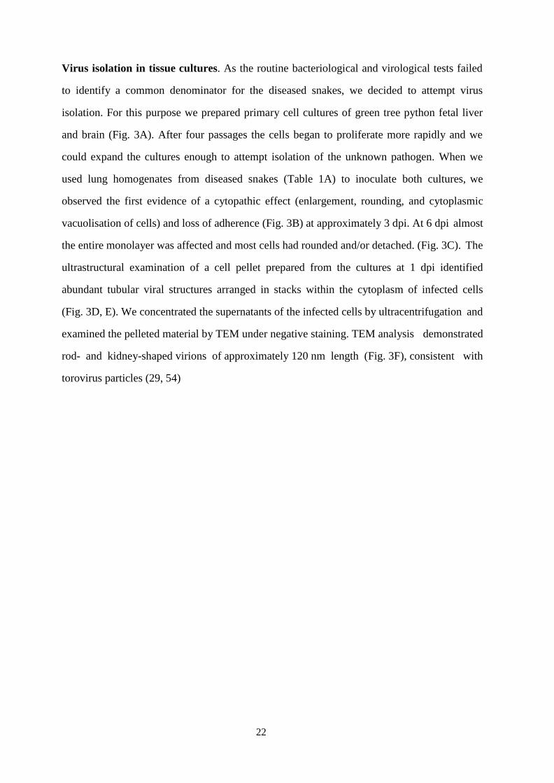
Virus isolation in tissue cultures. As the routine bacteriological and virological tests failed
to identify a common denominator for the diseased snakes, we decided to attempt virus
isolation. For this purpose we prepared primary cell cultures of green tree python fetal liver
and brain (Fig. 3A). After four passages the cells began to proliferate more rapidly and we
could expand the cultures enough to attempt isolation of the unknown pathogen. When we
used lung homogenates from diseased snakes (Table 1A) to inoculate both cultures, we
observed the first evidence of a cytopathic effect (enlargement, rounding, and cytoplasmic
vacuolisation of cells) and loss of adherence (Fig. 3B) at approximately 3 dpi. At 6 dpi almost
the entire monolayer was affected and most cells had rounded and/or detached. (Fig. 3C). The
ultrastructural examination of a cell pellet prepared from the cultures at 1 dpi identified
abundant tubular viral structures arranged in stacks within the cytoplasm of infected cells
(Fig. 3D, E). We concentrated the supernatants of the infected cells by ultracentrifugation and
examined the pelleted material by TEM under negative staining. TEM analysis demonstrated
rod- and kidney-shaped virions of approximately 120 nm length (Fig. 3F), consistent with
torovirus particles (29, 54)

23
Identification of nidovirus by NGS and confirmation of the findings by real-time RT-
PCR. In parallel to our attempts at virus isolation, we decided to employ NGS for the
identification of the causative agent(s) for the pneumonia. We isolated RNA from lung
homogenates of three animals (A2, B1, D1) and performed RNA sequencing. For two snakes
(B1, D1) only contigs matching bacterial genomes were identified. We interpreted these
bacterial sequences to most likely represent either contamination during post mortem sample
collection or, more likely, secondary bacterial infection, as both had also shown moderate
diffuse heterophil infiltration in the lung. From the third snake (A2), however, we obtained a
single ~21.000 nt contig and several shorter contigs matching the previously identified “Ball
python nidovirus (BPNV)” and “Python nidovirus (PNV)” (~85% identical to both) (9, 8). In
total we obtained ~28.000 nt (~75-80%) of the genome from the lung sample. In addition to
the novel nidovirus isolate, we identified a contig matching endogenous retrovirus genes with
very high coverage, which we describe in a separate report (Hepojoki et al., in preparation).
However, the retrovirus was also present in the supernatants of M. viridis cell cultures
inoculated with lung homogenate. We decided to attempt producing a nidovirus preparation
devoid of the contaminating retrovirus. To do this we inoculated boid kidney cells with
supernatant collected from Morelia viridis liver cell culture inoculated with lung
homogenates, since Huder et al. previously reported species-restricted growth of an
endogenous python retrovirus (55). Using this approach we obtained a pure nidovirus isolate
as confirmed by retrovirus-specific RT-PCR. We then performed end-point titration to
quantify the number of infectious units in the MVNV stock on M. viridis brain cells, and
could detect 2.25*1010
focus forming units per 1 ml of cell culture supernatant. RNA
extracted from the pure nidovirus preparation was subjected to another NGS run, which
yielded almost a full-length genome in a single contig. We then performed a reference
assembly using BPNV as the template to recover the missing (some 150 nt in total) genome
24
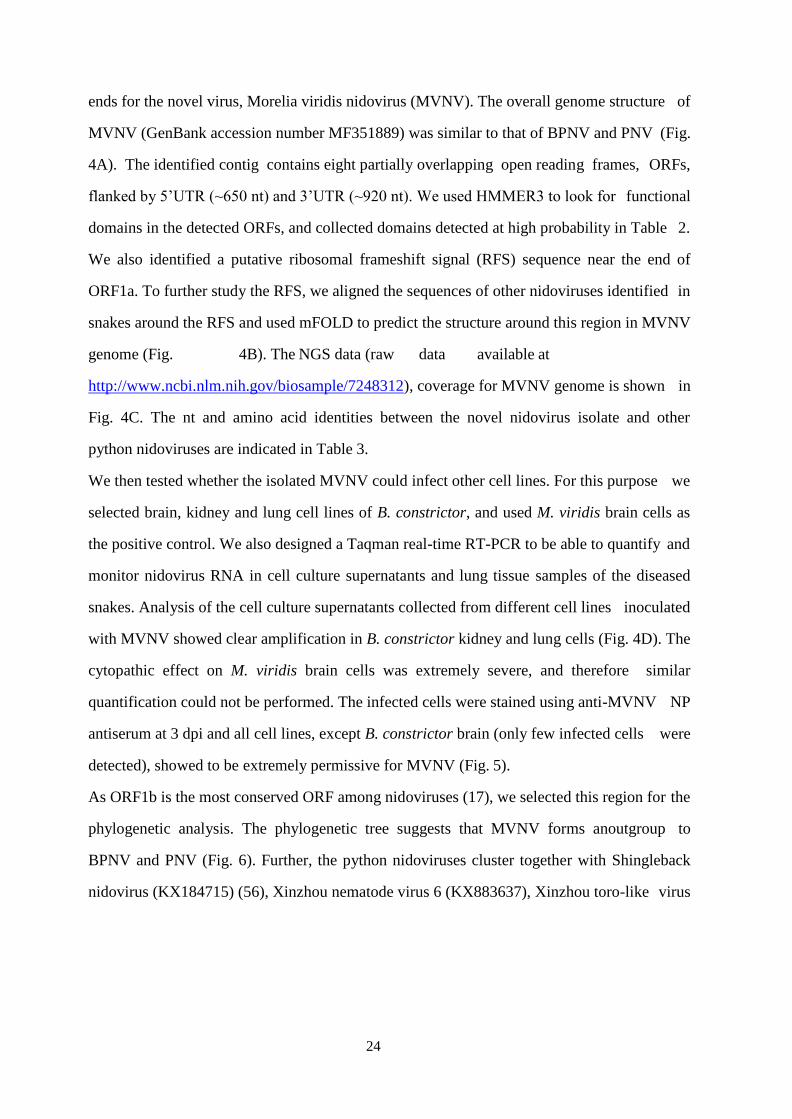
ends for the novel virus, Morelia viridis nidovirus (MVNV). The overall genome structure of
MVNV (GenBank accession number MF351889) was similar to that of BPNV and PNV (Fig.
4A). The identified contig contains eight partially overlapping open reading frames, ORFs,
flanked by 5’UTR (~650 nt) and 3’UTR (~920 nt). We used HMMER3 to look for functional
domains in the detected ORFs, and collected domains detected at high probability in Table 2.
We also identified a putative ribosomal frameshift signal (RFS) sequence near the end of
ORF1a. To further study the RFS, we aligned the sequences of other nidoviruses identified in
snakes around the RFS and used mFOLD to predict the structure around this region in MVNV
genome (Fig. 4B). The NGS data (raw data available at
http://www.ncbi.nlm.nih.gov/biosample/7248312), coverage for MVNV genome is shown in
Fig. 4C. The nt and amino acid identities between the novel nidovirus isolate and other
python nidoviruses are indicated in Table 3.
We then tested whether the isolated MVNV could infect other cell lines. For this purpose we
selected brain, kidney and lung cell lines of B. constrictor, and used M. viridis brain cells as
the positive control. We also designed a Taqman real-time RT-PCR to be able to quantify and
monitor nidovirus RNA in cell culture supernatants and lung tissue samples of the diseased
snakes. Analysis of the cell culture supernatants collected from different cell lines inoculated
with MVNV showed clear amplification in B. constrictor kidney and lung cells (Fig. 4D). The
cytopathic effect on M. viridis brain cells was extremely severe, and therefore similar
quantification could not be performed. The infected cells were stained using anti-MVNV NP
antiserum at 3 dpi and all cell lines, except B. constrictor brain (only few infected cells were
detected), showed to be extremely permissive for MVNV (Fig. 5).
As ORF1b is the most conserved ORF among nidoviruses (17), we selected this region for the
phylogenetic analysis. The phylogenetic tree suggests that MVNV forms anoutgroup to
BPNV and PNV (Fig. 6). Further, the python nidoviruses cluster together with Shingleback
nidovirus (KX184715) (56), Xinzhou nematode virus 6 (KX883637), Xinzhou toro-like virus

25
(NC_033700), and bovine nidovirus (NC_027199) (31). Consistent with the previous reports,
these form a clade that is separate from both the genus Bafinivirus and the genus Torovirus
(Fig. 6).
Finally, we used RNA isolated from cell cultures inoculated with lung homogenates of an
infected snake as the positive and RNA isolated from a Boa constrictor as the negative
control in the real-time RT-PCR. All diseased snakes were tested positive for nidovirus RNA
in the lungs. In contrast, the four snakes without gross and/or histological evidence of
pneumonia were negative.
Nidoviruses are associated with pneumonia in M. viridis. The histological examination
confirmed that all diseased snakes had suffered from a chronic pneumonia with epithelial
thickening in trachea and lung (Fig. 2) and excess mucus in the lumen of lungs and airway.
The inflammatory component was represented by mild to moderate multifocal interstitial
infiltration of lymphocytes, plasma cells and/or heterophils (Fig. 2E). The mucus filling the
faveolar space often contained heterophils and cell debris. In three of the nine tracheas
examined, we also observed a variable degree of mixed infiltration (heterophils, macrophages,
lymphocytes). Over its entire length, the lung epithelium exhibited numerous epithelial cells
that contained mucus as indicated by the PAS-Alcian Blue staining (Fig. 7A, C, E). In region
1, at the trabeculae, the mucus containing cells had the ultrastructural features of secretory
cells (50) (Fig. 7B). In regions 2 and 3 (faveolar epithelium), where the increase of these cells
was most striking, they exhibited the morphology of type II pneumocytes (Fig. 7D, F) (51);
their number varied in affected snakes and ranged from occasional patchy aggregates to
diffuse lining of the entire faveolae, associated with an almost complete absence of type I
pneumocytes (Fig. 2E). Ultrastructurally, these cells exhibited serous/mucous granules instead
of the lamellar bodies that are characteristic for type II pneumocytes (Fig. 7B, D, F); they
have recently been described as “transformed” type II pneumocytes (58).
26
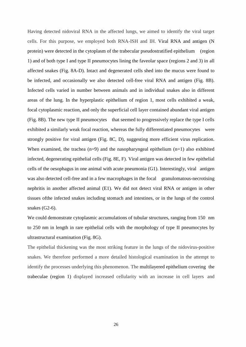
Having detected nidoviral RNA in the affected lungs, we aimed to identify the viral target
cells. For this purpose, we employed both RNA-ISH and IH. Viral RNA and antigen (N
protein) were detected in the cytoplasm of the trabecular pseudostratified epithelium (region
1) and of both type I and type II pneumocytes lining the faveolar space (regions 2 and 3) in all
affected snakes (Fig. 8A-D). Intact and degenerated cells shed into the mucus were found to
be infected, and occasionally we also detected cell-free viral RNA and antigen (Fig. 8B).
Infected cells varied in number between animals and in individual snakes also in different
areas of the lung. In the hyperplastic epithelium of region 1, most cells exhibited a weak,
focal cytoplasmic reaction, and only the superficial cell layer contained abundant viral antigen
(Fig. 8B). The new type II pneumocytes that seemed to progressively replace the type I cells
exhibited a similarly weak focal reaction, whereas the fully differentiated pneumocytes were
strongly positive for viral antigen (Fig. 8C, D), suggesting more efficient virus replication.
When examined, the trachea (n=9) and the nasopharyngeal epithelium (n=1) also exhibited
infected, degenerating epithelial cells (Fig. 8E, F). Viral antigen was detected in few epithelial
cells of the oesophagus in one animal with acute pneumonia (G1). Interestingly, viral antigen
was also detected cell-free and in a few macrophages in the focal granulomatous-necrotising
nephritis in another affected animal (E1). We did not detect viral RNA or antigen in other
tissues ofthe infected snakes including stomach and intestines, or in the lungs of the control
snakes (G2-6).
We could demonstrate cytoplasmic accumulations of tubular structures, ranging from 150 nm
to 250 nm in length in rare epithelial cells with the morphology of type II pneumocytes by
ultrastructural examination (Fig. 8G).
The epithelial thickening was the most striking feature in the lungs of the nidovirus-positive
snakes. We therefore performed a more detailed histological examination in the attempt to
identify the processes underlying this phenomenon. The multilayered epithelium covering the
trabeculae (region 1) displayed increased cellularity with an increase in cell layers and

27
irregular arrangement, indicating hyperplasia (Fig. 2E). The epithelial hyperplasia extended to
the upper respiratory tract and was also observed in the trachea, larynx and nasal cavity. The
mid and basal areas of the faveolae (regions 2 and 3), where the epithelium is unilayered,
showed nuclear crowding which also suggested hyperplasia (Fig. 2E). The thickening
appeared to result from an increase in individual cell height, associated with a more columnar
appearance of the cells (Fig. 2E, F). Additionally, we noted an increase in septal connective
tissue (interstitial fibrosis). The described changes resulted in thickening of the septa and
narrowing of the faveolar lumen (Fig. 2D).
In the attempt to quantify the epithelial hyperplasia we measured the average total epithelial
height in the three defined regions (Figs. 2 and 9) in all diseased, i.e. RT-PCR-positive
animals in comparison to the RT-PCR-negative animals without pneumonia (controls). In all
locations, the epithelium was significantly (P <0.05 higher in the diseased animals than in the
control snakes (Fig. 10A; Table 4). Hyperplasia was confirmed in both lung (regions 1, 2 and
3) and trachea, where the average number of nuclei in the epithelial layer was significantly
higher in the diseased snakes (Fig 10B; Table 4).
Epithelial hyperplasia was associated with increased proliferative activity, as up to 90% of
epithelial cells in all lung regions and in the trachea were found to express PCNA in the
diseased animals (Fig. 11A). Alongside this, a moderate number of epithelial cells were found
to undergo apoptosis, based on cleaved caspase-3 staining (Fig. 11C). In comparison, control
animals exhibited rare PCNA-positive type II pneumocytes in all layers (Fig. 11B) as well as
scattered apoptotic (cleaved caspase-3 positive) type I and type II pneumocytes (Fig. 11D),
likely representing the physiological turnover of the epithelium.

28
Discussion
We initiated the present study by the urge to identify the causative agent for a fatal pneumonia
observed in green tree pythons (Morelia viridis), characterised by the accumulation of mucoid
material in the airways and a histologically notable thickening of the lung epithelium. NGS
revealed the presence of a novel nidovirus, MVNV, which by phylogenetic analysis groups
with toroviruses. RT-PCR, immunohistology and RNA-ISH served to confirm its association
with the disease and to identify the viral target cells, i.e. epithelial cells in airways and the
luminal trabeculae, as well as the faveolar type I and II pneumocytes. We were able to isolate
and grow MVNV in both M. viridis and B. constrictor cell cultures. MVNV induced a
cytopathic effect both in vitro and in vivo. However, we also found nidovirus infection to
associate with generalised hyperplasia of the airway and lung epithelium which exhibited a
distinct proliferative activity and a degree of apoptotic cell death. Together these findings
would suggest nidovirus infection to increase the turnover of the epithelium. The mucus
accumulation in the air conducting space was accompanied by a significant increase in
secretory epithelial cells at the trabeculae and in (transformed) type II pneumocytes in the
faveolae, indicating increased mucus and/or surfactant production.
These results indicate that MVNV infects and damages the differentiated respiratory and
faveolar epithelium, but then persists and induces increased turnover of the infected epithelial
cells. The observed type II pneumocyte hyperplasia is obviously not specific to nidovirus
infection in the python, since it has been described in snakes as a consequence of pneumocyte
injury in a range of infectious diseases of viral, bacterial and mycotic nature (51, 58). It might
therefore represent an exaggerated regenerative attempt. Also in mammals type II
pneumocyte hyperplasia is seen as part of a lung defense mechanism to various insults (59–
61). In our cases, the hyperplastic epithelial cells exhibited abundant cytoplasmic
serous/mucous granules instead of the lamellar bodies that represent surfactant (50). This
suggests excess mucus and reduced surfactant production and release and would explain the
clinical findings (50, 58). Previous studies have shown that the surfactant of snakes, due to its
differing phospholipid composition, is likely less important for airway stabilisation, but rather
functions as an anti-adherent (also known as an "anti-glue") factor and an anti-oedemic factor
in the faveolar space (62); its reduction could therefore have added to the respiratory distress
29
observed in the affected snakes.
Increased turnover and/or hyperplasia of the epithelium has been described for several
coronaviridal diseases, such as infectious bronchitis in chickens, Breda virus infection in
calves, and coronavirus infection in rats (63–65). So far, however, the mechanisms underlying
this process have not been explained. Members of the subfamily Torovirinae are also known
to induce epithelial cell apoptosis (54, 66, 67); however, though our findings point towards
this, further studies are required to elucidate whether this also applies to MVNV and other
snake nidoviruses.
In our study, only fatal cases were examined. The pathological findings suggest that death
was mainly due to impaired gas exchange as a consequence of the type I pneumocyte loss. In
the healthy snake lung, thin cytoplasmic extensions of the type I pneumocytes cover the
capillary walls, forming the gas-blood barrier (50). Their replacement by mucus/surfactant-
secreting type II pneumocytes with their excessive height due to the cell crowding is unlikely
to allow effective gas exchange. With an average thickness of 24.40 µm (region 2) and 17.43
µm (region 3), the barrier was more than 1.7 x 10⁴
times thicker than the normal blood-gas
barrier in the reptilian lung which ranges between 0.4 and 1 nm (50).
Further anatomical peculiarities of the Morelia viridis (or in general boid snake) lung could
have contributed to the fatal outcome of the disease (51, 68–70). Boidae have a well-
developed right lung and a rudimental left lung. The right lung displays two anatomically
distinct regions, the anterior region, which contains the profusely compartmented gas
exchange tissue, and the posterior saccular region, which is devoid of respiratory tissue and
has therefore been referred to as “air sac” (71, 72). The combination of elongated lungs and
caudal air sacs may contribute considerably to the outcome of the disease, as they create a
“cul-de-sac” that impairs removal of the mucus and thereby significantly reduces the air filled
space and the gas exchange capacity.
Though toroviruses mainly associate with enteric diseases, recent studies have shown that
they can be both entero- and pneumotropic (31, 73, 74). New nidoviruses were recently
identified in the lungs of cattle and wild shingleback lizards with pneumonia, though their
direct association with disease has so far not been examined (31, 56). We found MVNV-

30
associated lesions almost exclusively in the airways and lung, similar to previous reports on
nidovirus infections in other python species (8, 9, 15). The detection of viral RNA by PCR in
other tissues, such as liver, spleen, kidney and intestine, however, indicated that the viruses
spread systemically. We found further evidence of that, and of its pathogenicity, as we
detected nidovirus NP within a focal granulomatous-necrotising nephritis in one animal. We
also detected viral antigen in epithelial cells of the cranial oesophagus in one affected animal;
however, the oesophagus carries ciliated epithelium in M. viridis (data not shown), a feature
also known for other snake species (75, 76); infection could therefore be due to an overspill
from the trachea and nasal cavity. We did not detect viral antigen in any cells in stomach and
intestine, and neither did a previous study find viral RNA by ISH, suggesting that the python
nidoviruses are primarily respiratory (8). Spreading via the expelling of mucus from the nasal
cavity would be a likely route of transmission.
Acknowledgements
We are grateful to the technical staff of the Histology Laboratory and the Electron
Microscopy Unit, Institute of Veterinary Pathology, and to Elisabeth M. Schraner, Institute of
Virology and Institute of Veterinary Anatomy, Vetsuisse Faculty, University of Zurich, for
excellent technical support. We also thank our colleagues at the Institute of Veterinary
Bacteriology, Vetsuisse Faculty, University of Zurich, for performing the bacteriological
examinations. Particular thanks are due to the breeders who submitted their snakes for
diagnostic purposes.

31
References
1. McDiarmid RW, Campbell JA, Touré T. 1999. Snake Species of the World: A Taxonomic and
Geographic Reference., Vol.1. Herpetologists' League, Washington, DC.
2. More G, Pantchev N, Herrmann DC, Vrhovec MG, Ofner S, Conraths FJ, Schares G. 2014.
Molecular identification of Sarcocystis spp. helped to define the origin of green pythons
(Morelia viridis) confiscated in Germany. Parasitology 141:646–651.
3. Nijman V, Shepherd CR. 2009. Wildlife trade from ASEAN to the EU: Issues with the trade
in captive-bred reptiles from Indonesia. TRAFFIC Europe Report for the European Commission,
Brussels, Belgium.
4. Rawlings LH, Donnellan SC. 2003. Phylogeographic analysis of the green python, Morelia
viridis, reveals cryptic diversity. Molecular Phylogenetics and Evolution 27:36–44.
5. Aqrawi T, Stöhr AC, Knauf-Witzens T, Krengel A, Heckers KO, Marschang RE. 2015.
Identification of snake arenaviruses in live boas and pythons in a zoo in Germany. Tierärztl
Prax (K) 43:239–247.
6. Shaw G (ed.). 1802. General Zoology, Volume III, London.
7. Linné Cv, Salvius L. 1758. Systema naturae, Vol. 1. Holmiae:Impensis Direct. Laurentii
Salvii.
8. Bodewes R, Lempp C, Schurch AC, Habierski A, Hahn K, Lamers M, Dornberg K von,
Wohlsein P,
Drexler JF, Haagmans BL, Smits SL, Baumgartner W, Osterhaus, A. D. M. E. 2014. Novel divergent
nidovirus in a python with pneumonia. Journal of General Virology 95:2480–2485.
9. Stenglein MD, Jacobson ER, Wozniak EJ, Wellehan JFX, Kincaid A, Gordon M, Porter BF,
Baumgartner W, Stahl S, Kelley K, Towner JS, DeRisi JL. 2014. Ball Python Nidovirus. A Candidate
Etiologic Agent for Severe Respiratory Disease in Python regius. mBio 5:e01484-14-e01484-14.
10. Sun H, Lan D, Lu L, Chen M, Wang C, Hua X. 2014. Molecular characterization and
phylogenetic analysis of the genome of porcine torovirus. Archives of virology 159:773–778.
11. Draker R, Roper RL, Petric M, Tellier R. 2006. The complete sequence of the bovine
torovirus genome. Virus Research 115:56–68.
12. Fagerland JA, Pohlenz JFL, Woode GN. 1986. A Morphological Study of the Replication of
Breda Virus (Proposed Family Toroviridae) in Bovine Intestinal Cells. Journal of General Virology
67:1293–1304.
13. Hoet AE, Cho K-O, Chang K-O, Loerch SC, Wittum TE, Saif LJ. 2002. Enteric and nasal
shedding of bovine torovirus (Breda virus) in feedlot cattle. Am J Vet Res 63:342–348.
14. Woode GN, Reed DE, Runnels PL, Herrig MA, Hill HT. 1982. Studies with an unclassified
virus isolated from diarrheic calves. Veterinary microbiology 7:221–240.
15. Uccellini L, Ossiboff RJ, Matos REC de, Morrisey JK, Petrosov A, Navarrete-Macias I, Jain K,
Hicks AL, Buckles EL, Tokarz R, McAloose D, Lipkin W. 2014. Identification of a novel nidovirus
in an outbreak of fatal respiratory disease in ball pythons (Python regius). Virol J 11:144.
16. Groot RJ de, Cowley JA, Enjuanes L, Faaberg KS, Perlman S, Rottier PJ, Snijder EJ, Ziebuhr J,
Gorbalenya AE. 2011. Order nidovirales. Virus taxonomy:785–795.

32
17. Gorbalenya AE, Enjuanes L, Ziebuhr J, Snijder EJ. 2006. Nidovirales. Evolving the largest
RNA virus genome. Virus Research 117:17–37.
18. Nga PT, del Carmen Parquet M, Lauber C, Parida M, Nabeshima T, Yu F, Thuy NT, Inoue S,
Ito T, Okamoto K. 2011. Discovery of the first insect nidovirus, a missing evolutionary link in the
emergence of the largest RNA virus genomes. PLoS Pathog 7:e1002215.
19. Lauber C, Ziebuhr J, Junglen S, Drosten C, Zirkel F, Nga PT, Morita K, Snijder EJ, Gorbalenya
AE. 2012. Mesoniviridae: a proposed new family in the order Nidovirales formed by a single
species of mosquito-borne viruses. Archives of virology 157:1623–1628.
20. Lauber C, Gorbalenya AE. 2012. Toward Genetics-Based Virus Taxonomy. Comparative
Analysis of a Genetics-Based Classification and the Taxonomy of Picornaviruses. Journal of
Virology 86:3905–3915.
21. Walker PJ, Winton JR. 2010. Emerging viral diseases of fish and shrimp. Veterinary
Research 41:51.
22. Snijder EJ, Kikkert M, Fang Y. 2013. Arterivirus molecular biology and pathogenesis. J Gen
Virol 94:2141–2163.
23. To KKW, Hung IFN, Chan JFW, Yuen K-Y. 2013. From SARS coronavirus to novel animal and
human coronaviruses. Journal of Thoracic Disease 5 Suppl 2:8.
24. Baird A, Faisal M. 2016. Fathead minnow nidovirus infects spotfin shiner Cyprinella
spiloptera and golden shiner Notemigonus crysoleucas. Dis. Aquat. Org. 119:37–44.
25. McVey DS, Kennedy M, Chengappa MM (ed.). 2013. Veterinary Microbiology, 3. Aufl.
Wiley- Blackwell, New Jersey.
26. Beards GM, Campbell AD, Cottrell NR, Peiris JS, Rees N, Sanders RC, Shirley JA, Wood HC,
Flewett TH. 1984. Enzyme-linked immunosorbent assays based on polyclonal and monoclonal
antibodies for rotavirus detection. Journal of clinical microbiology 19:248–254.
27. Weiss M, Steck F, Horzinek MC. 1983. Purification and partial characterization of a new
enveloped RNA virus (Berne virus). Journal of General Virology 64:1849–1858.
28. Cann AJ. 2001. Chapter 2-Particles. The function and formation of virus particles, p. 25–
27. In Cann AJ (ed), Principles of Molecular Virology (Standard Edition). Elsevier, Academic
Press, London.
29. Cornelissen LA, van Woensel PA, Groot RJ de, Horzinek MC, Visser N, Egberink HF. 1998.
Cell culture-grown putative bovine respiratory torovirus identified as a coronavirus. Vet Rec
142:683–686.
30. Vanopdenbosch E, Wellemans G, Petroff K. 1991. Breda virus associated with respiratory
disease in calves. Vet Rec 129:203.
31. Tokarz R, Sameroff S, Hesse RA, Hause BM, Desai A, Jain K, Lipkin WI. 2015. Discovery of a
novel nidovirus in cattle with respiratory disease. J Gen Virol 96:2188–2193.
32. Smits SL, Lavazza A, Matiz K, Horzinek MC, Koopmans MP, de Groot RJ. 2003.
Phylogenetic and evolutionary relationships among torovirus field variants: evidence for
multiple intertypic
recombination events. Journal of Virology 77:9567–9577.
33. Petric M. 2003. Section VI. Other viruses causing gastroenteritis. Epidemiology of
toroviruses. In Desselberger U, Gray J (ed), Viral Gastroenteritis, 9th ed. Elsevier, Amsterdam.
34. Snijder EJ, Ederveen J, Spaan WJ, Weiss M, Horzinek MC. 1988. Characterization of Berne
virus genomic and messenger RNAs. J Gen Virol 69:2135–2144.
35. Hetzel U, Sironen T, Laurinmaki P, Liljeroos L, Patjas A, Henttonen H, Vaheri A, Artelt A,
Kipar A, Butcher SJ, Vapalahti O, Hepojoki J. 2013. Isolation, identification, and characterization
of novel arenaviruses, the etiological agents of boid inclusion body disease. Journal of Virology
87:10918–10935.

33
36. Li H, Durbin R. 2010. Fast and accurate long-read alignment with Burrows-Wheeler
transform. Bioinformatics 26:589–595.
37. Okonechnikov K, Golosova O, Fursov M. 2012. Unipro UGENE: a unified bioinformatics
toolkit. Bioinformatics 28:1166–1167.
38. Tamura K, Stecher G, Peterson D, Filipski A, Kumar S. 2013. MEGA6. Molecular
Evolutionary Genetics Analysis Version 6.0. Molecular Biology and Evolution 30:2725–2729.
39. Drummond AJ, Suchard MA, Xie D, Rambaut A. 2012. Bayesian phylogenetics with BEAUti
and the BEAST 1.7. Molecular Biology and Evolution 29:1969–1973.
40. Rambaut A., Suchard MA., Xie D., Drummond AJ. 2014. Tracer v1.6. Beast Bio.
http://beast.bio.ed.ac.uk/Tracer.
41. Zuker M. 2003. Mfold web server for nucleic acid folding and hybridization prediction.
Nucleic Acids Res 31:3406–3415.
42. Edgar RC. 2004. MUSCLE: multiple sequence alignment with high accuracy and high
throughput. Nucleic Acids Res 32:1792–1797.
43. Finn RD, Clements J, Eddy SR. 2011. HMMER web server. Interactive sequence similarity
searching. Nucleic Acids Res 39:W29-W37.
44. Hepojoki J, Kipar A, Korzyukov Y, Bell-Sakyi L, Vapalahti O, Hetzel U. 2015. Replication of
boid inclusion body disease-associated arenaviruses is temperature sensitive in both boid and
mammalian cells. J. Virol. 89:1119–1128.
45. Korzyukov Y, Hetzel U, Kipar A, Vapalahti O, Hepojoki J. 2016. Generation of Anti-Boa
Immunoglobulin Antibodies for Serodiagnostic Applications, and Their Use to Detect Anti-
Reptarenavirus Antibodies in Boa Constrictor. PLoS One 11:e0158417.
46. Ressel L, Ward S, Kipar A. 2015. Equine Cutaneous Mast Cell Tumours Exhibit Variable
Differentiation, Proliferation Activity and KIT Expression. J Comp Pathol 153:236–243.
47. Antoine DJ, Williams DP, Kipar A, Laverty H, Park BK. 2010. Diet Restriction Inhibits
Apoptosis and HMGB1 Oxidation and Promotes Inflammatory Cell Recruitment during
Acetaminophen Hepatotoxicity. Molecular Medicine 16:479–490.
48. Swadzba E, Rupik W. 2010. Ultrastructural studies of epidermis keratinization in grass
snake embryos Natrix natrix L. (Lepidosauria, Serpentes) during late embryogenesis. Zoology
(Jena) 113:339–360.
49. Wang F, Flanagan J, Su N, Wang L-C, Bui S, Nielson A, Wu X, Vo H-T, Ma X-J, Luo Y. 2012.
RNAscope: a novel in situ RNA analysis platform for formalin-fixed, paraffin-embedded tissues.
J Mol Diagn 14:22–29.
50. Pastor LM. 1995. Chapter 5- The histology of the reptilian lung, p. 127–155. In Pastor LM
(ed), Histology, ultrastructure and immunohistochemistry of the respiratory organs in non
mammalian vertebrates, 1st ed. Secretariado de Publicaciones de la Univesidad de Murcia,
Murcia.
51. Starck JM, Weimer I, Aupperle H, Muller K, Marschang RE, Kiefer I, Pees M. 2015.
Morphological Pulmonary Diffusion Capacity for Oxygen of Burmese Pythons (Python molurus):
a Comparison of Animals in Healthy Condition and with Different Pulmonary Infections. J
Comp Pathol 153:333–351.
52. Maina JN. 1989. The morphology of the lung of the black mamba Dendroaspis polylepis
(Reptilia: Ophidia: Elapidae). A scanning and transmission electron microscopic study. Journal of
Anatomy 167:31–46.
53. Perry SF. 1983. 1. Introduction. Descriptive Classification of Lung Types. Histological
Structure, p. 1–3. In Perry SF (ed), Reptilian lungs: functional anatomy and evolution. Springer
Verlag, Berlin.
54. Kuwabara M, Wada K, Maeda Y, Miyazaki A, Tsunemitsu H. 2007. First Isolation of
34
Cytopathogenic Bovine Torovirus in Cell Culture from a Calf with Diarrhea. Clinical and Vaccine
Immunology 14:998–1004.
55. Huder JB, Boni J, Hatt J-M, Soldati G, Lutz H, Schupbach J. 2002. Identification and
characterization of two closely related unclassifiable endogenous retroviruses in pythons
(Python molurus and Python curtus). J. Virol. 76:7607–7615.
56. O’Dea MA, Jackson B, Jackson C, Xavier P, Warren K. 2016. Discovery and Partial Genomic
Characterisation of a Novel Nidovirus Associated with Respiratory Disease in Wild Shingleback
Lizards (Tiliqua rugosa). PLoS One 11:e0165209.
57. Shi W, Zhang Z, Ling C, Carr MJ, Tong Y, Gao GF. 2016. Increasing genetic diversity of Zika
virus in the Latin American outbreak. Emerg Microbes Infect 5:e68. doi:10.1038/emi.2016.68.
58. Jacobson ER, Adams HP, Geisbert TW, Tucker SJ, Hall BJ, Homer BL. 1997. Pulmonary
Lesions in Experimental Ophidian Paramyxovirus Pneumonia of Aruba Island Rattlesnakes,
Crotalus unicolor. Veterinary Pathology 34:450–459.
59. Ward HE, Nicholas TE. 1984. Alveolar type I and type II cells. Aust N Z J Med 14:731–734.
60. Fehrenbach H, Kasper M, Tschernig T, Pan T, Schuh D, Shannon JM, Muller M, Mason RJ.
1999. Keratinocyte growth factor-induced hyperplasia of rat alveolar type II cells in vivo is
resolved by differentiation into type I cells and by apoptosis. European Respiratory Journal
14:534–544.
61. Caswell JL, Williams KJ. 2016. Chapter 5 - Respiratory System, 465-591. In Maxie MG (ed),
Jubb, Kennedy & Palmer's Pathology of Domestic Animals, 6th ed., vol. 2. Elsevier, St. Louis,
Missouri.
62. Veldhuizen R, Nag K, Orgeig S, Possmayer F. 1998. The role of lipids in pulmonary
surfactant.
Biochimica et Biophysica Acta (BBA) - Molecular Basis of Disease 1408:90–108.
63. Grgiæ H, Hunter DB, Hunton P, Nagy É. 2008. Pathogenicity of infectious bronchitis virus
isolates from Ontario chickens. Canadian Journal of Veterinary Research 72:403–410.
64. Schunk MK, Percy DH, Rosendal S. 1995. Effect of time of exposure to rat coronavirus and
Mycoplasma pulmonis on respiratory tract lesions in the Wistar rat. Canadian Journal of
Veterinary Research 59:60–66.
65. Koopmans M, Horzinek MC. 1995. The Pathogenesis of Torovirus Infections in Animals
and Humans, p. 403–413. In Siddell SG (ed), The Coronaviridae. Springer US, Boston, MA.
66. Maestre AM, Garzon A, Rodriguez D. 2011. Equine torovirus (BEV) induces caspase-
mediated apoptosis in infected cells. PLoS One 6:e20972.
67. Ding X, Xu F, Chen H, Tesh RB, Xiao S-Y. 2005. Apoptosis of Hepatocytes Caused by Punta
Toro Virus (Bunyaviridae. Phlebovirus) and Its Implication for Phlebovirus Pathogenesis. The
American Journal of Pathology 167:1043–1049.
68. Stinner JN. 1987. Gas exchange and air flow in the lung of the snake, Pituophis
melanoleucus. Journal of Comparative Physiology B 157:307–314.
69. van Wallach. 1998. The lungs of snakes. Biology of the Reptilia 19:93–295.
70. Perry SF, Bauer AM, Russell AP, Alston JT, Maloney JE. 1989. Lungs of the gecko
Rhacodactylus leachianus (reptilia: Gekkonidae): A correlative gross anatomical and light and
electron microscopic study. J. Morphol. 199:23–40.
71. McDonald HS. 1959. Respiratory functions of the ophidian air sac. Herpetologica 15:193–
198.
72. Stinner HN. 1982. Ventillation, gas exchange and blood gases in the snake, Pituophis
melanoleucus. Respiration physiology 47:279–298.
73. Saif LJ, Bohl EH. 1986. Transmissible gastroenteritis, 255-274. In Leman AD, Straw B, Glock
RD (ed.), Diseases of swine, 6th ed. Iowa State University Press, Iowa.

35
74. Koopmans M, Horzinek MC. 1994. Toroviruses of animals and humans. A review. Adv
Virus Res 43:233–273.
75. Imai M, Shibata T, Moriguchi K. 1991. Pepsinogen granules in the esophageal epithelium
of the rock snake. Okajimas Folia Anat Jpn 68:231–234.
76. Cundall D, Tuttman C, Close M. 2014. A model of the anterior esophagus in snakes, with
functional and developmental implications. Anat Rec (Hoboken) 297:586–598.
36
Figure Legends
Figure 1: Gross findings in a green tree python (Morelia viridis) with nidovirus-associated
proliferative pneumonia (animal E1). Both trachea (A) and lungs (B) are filled with abundant
mucoid material (arrows). H - heart.
Figure 2: Histological findings in Morelia viridis with nidovirus-associated proliferative
pneumonia. A-C. Uninfected control case (animal G2). Representative photomicrographs of a
healthy lung (cross section). Thin pulmonary septa form the faveolar spaces. At the luminal
end, the septa exhibit bundles of smooth muscle cells (myoelastic bundles) that form
contractile trabeculae. For detailed assessments, three regions (1, 2, 3) were identified. The
trabeculae are covered by a multilayered pseudostratified “bronchial-type” epithelium,
dominated by ciliated cells (B1, C1). In regions 2 (B2, C2) and 3 (B3, C3), the faveolae are
covered by a single-layered “gas exchange epithelium” comprised of flat type I pneumocytes
(arrowheads); surfactant producing type II pneumocytes (arrows) are less abundant. D-F.
Snake with nidovirus infection (animal B4). Representative photomicrograph of a diseased
lung (cross section). In the entire lung, the epithelial layer is thickened due to hyperplasia.
The faveolar space (3) is filled with proteinaceous material and the interstitium is broadened
due to a mixed inflammatory infiltrate (E1, 2). In region 1 (E1, F1), the pseudostratified
epithelium covering the trabeculae exhibits increased cellularity with an increase in cell layers
and irregular arrangement (hyperplasia) as well as a loss of cilia and several apoptotic
epithelia cells (arrowheads). In regions 2 (E2, F2) and 3 (E3, F3), type I pneumcytes are
almost entirely replaced by large, columnar type II pneumocytes. A, B, D, E: HE stain; C, F:
IH for cytokeratin, haematoxylin counterstain. Bars = 100 µm (A, D) and 10 µm (B, C, E, F).
Figure 3: Virus isolation in a primary Morelia viridis foetal brain cell culture. A. Uninfected
control cells. B, C. Cells after inoculation with lung homogenate from a diseased snake
(animal C2). First evidence of a cytopathic effect is seen after 2 days (B), as enlargement,

37
rounding, and cytoplasmic vacuolisation as well as loss of adherence (arrows). After 6 days
(C), only few intact cells have remained. Most cells are detached and necrotic. D, E.
Ultramicrographs of a cell pellet prepared at 1 dpi. There are abundant tubular structures
arranged in stacks within the cytoplasm of infected cells. F. Concentrated supernatant of the
infected cells, negative staining. Rod- and kidney-shaped virions of approximately 120 nm
length. Bar = 1 µm (D) and 100 nm (E).
Figure 4. Morelia viridis nidovirus, MVNV, genome organization, NGS coverage, and
phylogenetic relationship to related viruses. A. A schematic representation of the untranslated
regions (UTRs) and open reading frames (ORFs) of the 32,414 nt ssRNA+ genome of
MVNV. B. Alignment around the ribosomal frameshift signal (RFS, indicated by an arrow in
the alignment and structure prediction) in nidoviruses identified in pythons with ORF1a stop
codon highlighted. A prediction for the RNA structure around the RFS in MVNV, the arrow
highlights a difference to the structure described by Bodewes et al. (8) C. The contig
coverage, y-axis shows the number of reads matching each nucleotide position (x-axis) of the
contig. Bowtie2 was used to map the reads to the contig generated by de novo assembly. D.
Virus growth in different cell lines. Cell culture supernatant of the indicated cell lines
inoculated with MVNV was analyzed by qRT-PCR at the indicated time points. The fold
increase indicates viral RNA increase in the cell culture supernatant as compared to the
amount detected at 0 dpi.
Figure 5. Immunofluorescence staining of M. viridis and B. constrictor cell lines with anti-
MVNV NP antiserum at 1:2,000 dilution. MVNV- or mock-infected cells were stained at 3
dpi, and recorded at 40x under inverted fluorescence microscope. Mock infected cells are
shown on the right and MVNV infected cells on the left. DAPI is shown in blue and MVNV
specific staining in red.

38
Figure 6. Maximum clade credibility tree constructed from ORF1b amino acid sequences of
the representatives of the subfamily Torovirinae. The phylogenetic tree was constructed using
Bayesian MCMC method with LG+G+I model of substitution. Posterior probabilities are
shown in each node.
Figure 7. Changes in the lung epithelium in Morelia viridis with nidovirus-associated
proliferative pneumonia. Representative photomicrographs (A, C, E, animal B4) and
ultramicrographs (B, D, F, animal A1) of diseased lungs (cross sections). A, B. Region 1. A.
The PAS/Alcian Blue stain highlights positive (arrowheads) secretory epithelial cells in all
cell layers of the hyperplastic multilayered trabecular epithelium. Abundant mucus fills the
lumen. B. The ultrastructural features of most cells (cytoplasmic mucous granules and short
microvilli) show that they are secretory cells (50). Only a few ciliated cells are seen (arrow).
C-F. In regions 2 (C, D) and 3 (E, F), the faveolae are diffusely lined by cuboidal cells
containing serous/mucous granules (C, E, arrowheads), consistent with type II pneumocytes.
Ultrastructurally, these cells resemble so-called “transformed” type II pneumocytes, as they
contain serous/mucous granules (arrows) and lack the lamellar bodies characteristic for
normal type II pneumocytes (51).
Figure 8. Viral target cells in lungs and airways of Morelia viridis with nidovirus-associated
proliferative pneumonia. A-D. Animals A1 (A) and A2 (B-D), lung. The pseudostratified
trabelular epithelium (region 1) exhibits abundant viral RNA mainly in superficial epithelial
cell layers (A). B. Viral antigen expression is seen in all cell layers. In basal and mid layers, it
is represented by a focal cytoplasmic reaction (arrowhead). The fully differentiated superficial
epithelial cells exhibit abundant viral antigen (arrow) and loss of cilia. Infected cells are also
found shed into the lumen. C. Region 2. In areas with intact faveolar epithelium, both type I
(arrowhead) and differentiated type II pneumocytes (arrows) exhibit strong viral antigen
expression. The new, hyperplastic type II pneumocytes in other areas display a weaker focal

39
cytoplasmic reaction (arrows). At the base of the faveolae (D), type I (arrowheads) and type II
(arrows) pneumocytes are also found to be infected. They are also shed into the faveolar
lumen. E. Animal A1, nasal cavity. The epithelium is thickened due to hyperplasia and
exhibits nidovirus infection mainly of the fully differentiated superficial epithelial cells
(arrow). C - cartilage. F. Animal A1, trachea. Bottom: The ciliated epithelium is largely intact
(arrow), but there are scattered individual or small groups of infected cells. Sloughed,
degenerate epithelial cells are also found to carry viral RNA (arrowhead). Top: Infected cells
are intact (arrowhead) or degenerate, shedding virus (arrow). C – tracheal ring cartilage.
RNA-ISH (RNAscope technology; A, E, F) and IH (HRP method; B-D) for MVNV N
protein, haematoxylin counterstain. Bars = 20 µm (A, E, F bottom) and 10 µm (B-D, F top).
G. Ultramicrograph of a type II pneumocyte with staples of tubular structures of 100 – 150
nm length (arrows). Bar = 1 µm.
Figure 9. Illustration of the morphometric approach taken to measure the epithelial height and
number of epithelial cell nuclei in cytokeratin-stained cross sections of the lungs. A. Three
regions were defined in the proximal, bronchiolar lung: 1) The luminal part of the primary
trabeculae, covered by a multilayered pseudostratified ciliated epithelium (1). 2) The faveolae
at mid level (2) and at the base (3), covered by a single layered epithelium. B. Region 1,
measurement of epithelial height. The blue lines represent 10 measurements taken over a 300
µm wide epithelial segment, determining the distance between the apical epithelial cell border
and the basal membrane. 200-fold magnification. C. Region 1, determination of cell number.
The total amount of epithelial nuclei (x) counted in a 200 µm wide epithelial segment. 400-
fold magnification. D, E. Region 2 (D) and 3 (E), measurement of epithelial height. The blue
lines represent 10 measurements taken over a 300 µm wide epithelial segment, determining
the distance between the apical epithelial cell border and the basal membrane. 200-fold
magnification.

40
Figure 10.The average epithelial height measured in three lung regions and the trachea of
infected animals (white) was significantly higher than in non-infected (control) animals
(gray). Box plots illustrate the third quartile (Q3) and first quartile (Q1) range of the data,
filled circles represent data outliers (>1.5x interquartile range). Boxplots were created by
the SPSS Statistics software. A Mann-Whitney U test for independent samples (t-test) was
used for statistical analysis, and a p-value of less than 0.05 was considered significant
Figure 11. Increased epithelial turnover in the lungs of Morelia viridis with nidovirus-
associated proliferative pneumonia. Trabeculae (region 1). A, C. Diseased animal (C1). The
hyperplastic pseudostratified epithelium is proliferating, as confirmed by the PCNA
expression in almost all epithelial cells. Infiltrating lymphocytes (arrow; A) are also positive.
There are also abundant, cleaved caspase 3-positive apoptotic epithelial cells (C). A
proportion of infiltrating leukocytes is also found to undergo apoptosis (arrow; C). B, D.
Control animal (G3). In the control animal, only scattered individual basal epithelial cells are
found to express PCNA (arrowhead; B). Similarly, only rare epithelial cells are found to
undergo apoptosis (arrowhead; D). Apoptosis (i.e. cleaved caspase-3 expression) is also seen
in occasional lymphocytes (arrow) of the lymphoid aggregates. IH for PCNA (A, B) and
cleaved caspase-3 (C, D), HRP method, haematoxylin counterstain. Bars = 20 µm.
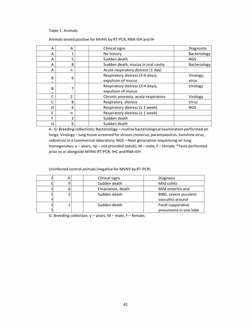
41
Table 1. Animals
Animals tested positive for MVNV by RT-PCR, RNA-ISH and IH
Animal
Age (y)
Sex
Clinical signs Diagnostic tests* A
1 1 M No history Bacteriology
A2
5 F Sudden death NGS
A3
8 M Sudden death, mucus in oral cavity Bacteriology
A4
np
F Acute respiratory distress (1 day)
B1
6 M Respiratory distress (3-4 days),
expulsion of mucus
Virology,
virus
isolation,
NGS B2
7 F Respiratory distress (3-4 days),
expulsion of mucus
Virology
C1
2.5
M Chronic anorexia, acute respiratory distress
Virology
C2
8 M Respiratory distress Virus isolation D
1 6 F Respiratory distress (≥ 1 week) NGS
E1
np
F Respiratory distress (≥ 1 week)
F1
2 F Sudden death
G1
6 F Sudden death
A - G: Breeding collections; Bacteriology – routine bacteriological examination performed on
lungs; Virology – lung tissue screened for viruses (reovirus, paramyxovirus, Sunshine virus,
nidovirus) in a commercial laboratory; NGS – Next generation sequencing on lung
homogenates; y – years; np – not provided (adult); M – male, F – female; *Tests performed
prior to or alongside MVNV RT-PCR, IHC and RNA-ISH
Uninfected control animals (negative for MVNV by RT-PCR)
Animal
Age (y)
Sex
Clinical signs Diagnosis
G2
9 M Sudden death Mild colitis
G3
6 F Emaciation, death Mild enteritis and nephritis G
4 2 F Sudden death BIBD, severe purulent
vasculitis around
oesophagus and trachea G5
1 M Sudden death Focal suppurative
pneumonia in one lobe
G: Breeding collection; y – years; M – male, F – female;
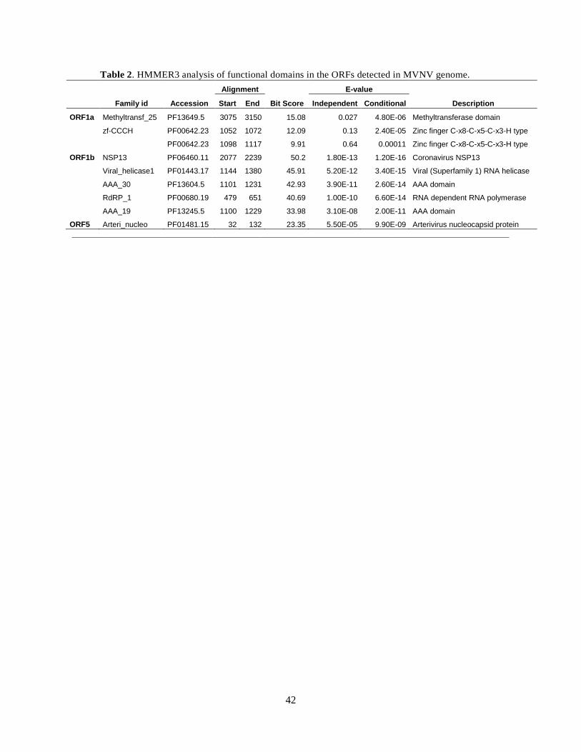
42
Table 2. HMMER3 analysis of functional domains in the ORFs detected in MVNV genome.
Alignment E-value
Family id Accession Start End Bit Score Independent Conditional Description
ORF1a Methyltransf_25 PF13649.5 3075 3150 15.08 0.027 4.80E-06 Methyltransferase domain
zf-CCCH PF00642.23 1052 1072 12.09 0.13 2.40E-05 Zinc finger C-x8-C-x5-C-x3-H type
PF00642.23 1098 1117 9.91 0.64 0.00011 Zinc finger C-x8-C-x5-C-x3-H type
ORF1b NSP13 PF06460.11 2077 2239 50.2 1.80E-13 1.20E-16 Coronavirus NSP13
Viral_helicase1 PF01443.17 1144 1380 45.91 5.20E-12 3.40E-15 Viral (Superfamily 1) RNA helicase
AAA_30 PF13604.5 1101 1231 42.93 3.90E-11 2.60E-14 AAA domain
RdRP_1 PF00680.19 479 651 40.69 1.00E-10 6.60E-14 RNA dependent RNA polymerase
AAA_19 PF13245.5 1100 1229 33.98 3.10E-08 2.00E-11 AAA domain
ORF5 Arteri_nucleo PF01481.15 32 132 23.35 5.50E-05 9.90E-09 Arterivirus nucleocapsid protein

43
Table 3. Nucleotide and amino acid identities comparing MVNV and the nidoviruses identified in
Python molurus (Indian python) and Python regius (ball python).
Nucleotide Amino acid
Python
nidovirus
S1536/13
(KJ935003)
Ball
python
nidovirus
07-53
(KJ54175
9)
Python
nidovirus
S1536/13
(KJ935003)
Ball
python
nidovirus
07-53
(KJ54175
9)
5´ UTR
85.1 87.0
ORF1a
72.8 72.8 69.5 69.6
ORF1b
86.5 86.3 91.0 90.5
ORF2
80.9 80.0 85.0 84.8
ORF3
69.5 67.0 64.7 65.4
ORF4
76.5 79.2 83.3 82.9
ORF5
75.1 74.0 74.2 74.8
ORF6
65.6 66.9 59.7 59.2
ORF7
77.1 78.6 68.8 71.9
3´ UTR
95.2 93.9
Complete
77.3 77.3 77.2 77.1
genome/CDS
In brackets: Genebank accession numbers.
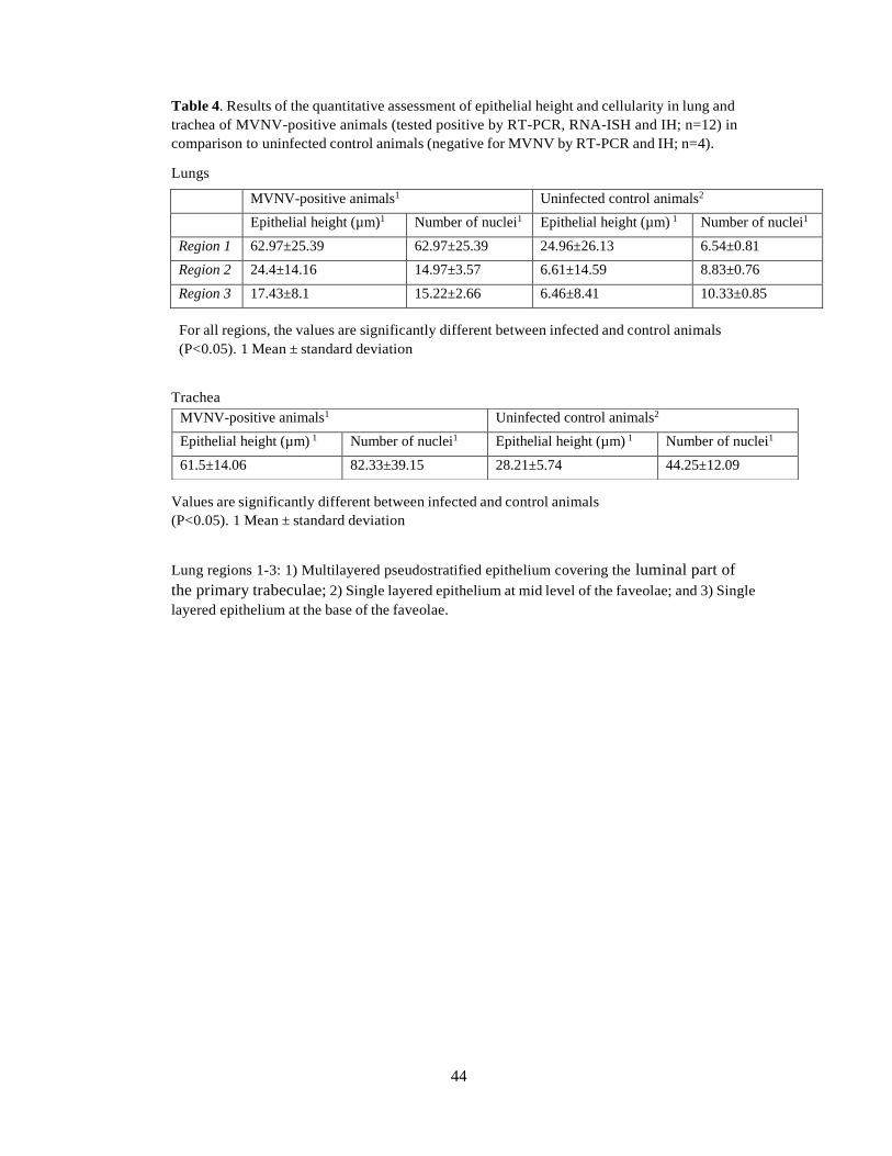
44
Table 4. Results of the quantitative assessment of epithelial height and cellularity in lung and
trachea of MVNV-positive animals (tested positive by RT-PCR, RNA-ISH and IH; n=12) in
comparison to uninfected control animals (negative for MVNV by RT-PCR and IH; n=4).
Lungs
For all regions, the values are significantly different between infected and control animals
(P<0.05). 1 Mean ± standard deviation
Trachea
Values are significantly different between infected and control animals
(P<0.05). 1 Mean ± standard deviation
Lung regions 1-3: 1) Multilayered pseudostratified epithelium covering the luminal part of
the primary trabeculae; 2) Single layered epithelium at mid level of the faveolae; and 3) Single
layered epithelium at the base of the faveolae.
MVNV-positive animals1 Uninfected control animals2
Epithelial height (µm)1 Number of nuclei1 Epithelial height (µm) 1 Number of nuclei1
Region 1 62.97±25.39 62.97±25.39 24.96±26.13 6.54±0.81
Region 2 24.4±14.16 14.97±3.57 6.61±14.59 8.83±0.76
Region 3 17.43±8.1 15.22±2.66 6.46±8.41 10.33±0.85
MVNV-positive animals1 Uninfected control animals2
Epithelial height (µm) 1 Number of nuclei1 Epithelial height (µm) 1 Number of nuclei1
61.5±14.06 82.33±39.15 28.21±5.74 44.25±12.09
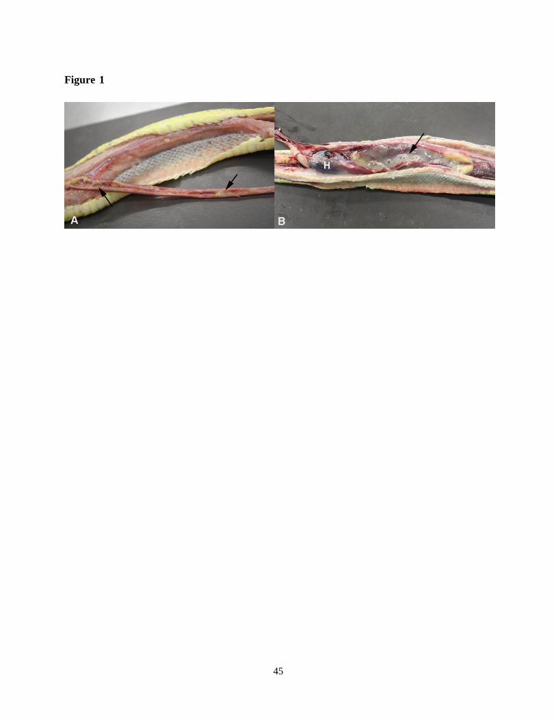
45
Figure 1

46
Figure 2

47
Figure 3

48
Figure 4
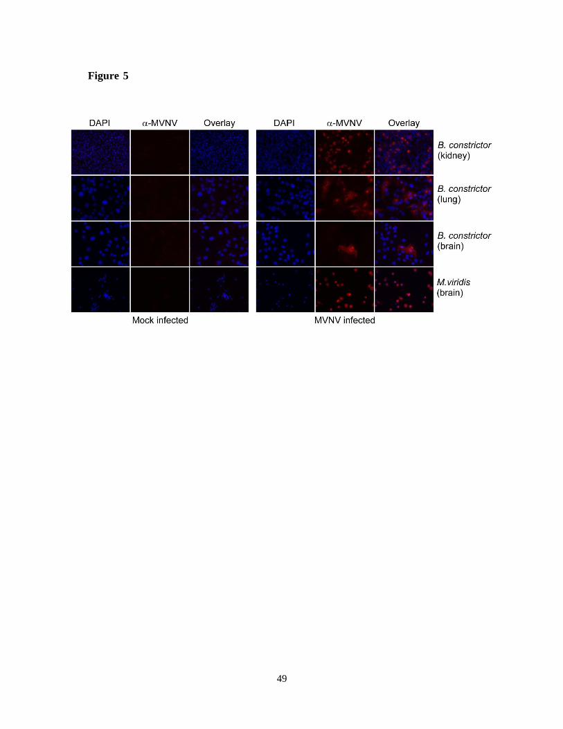
49
Figure 5
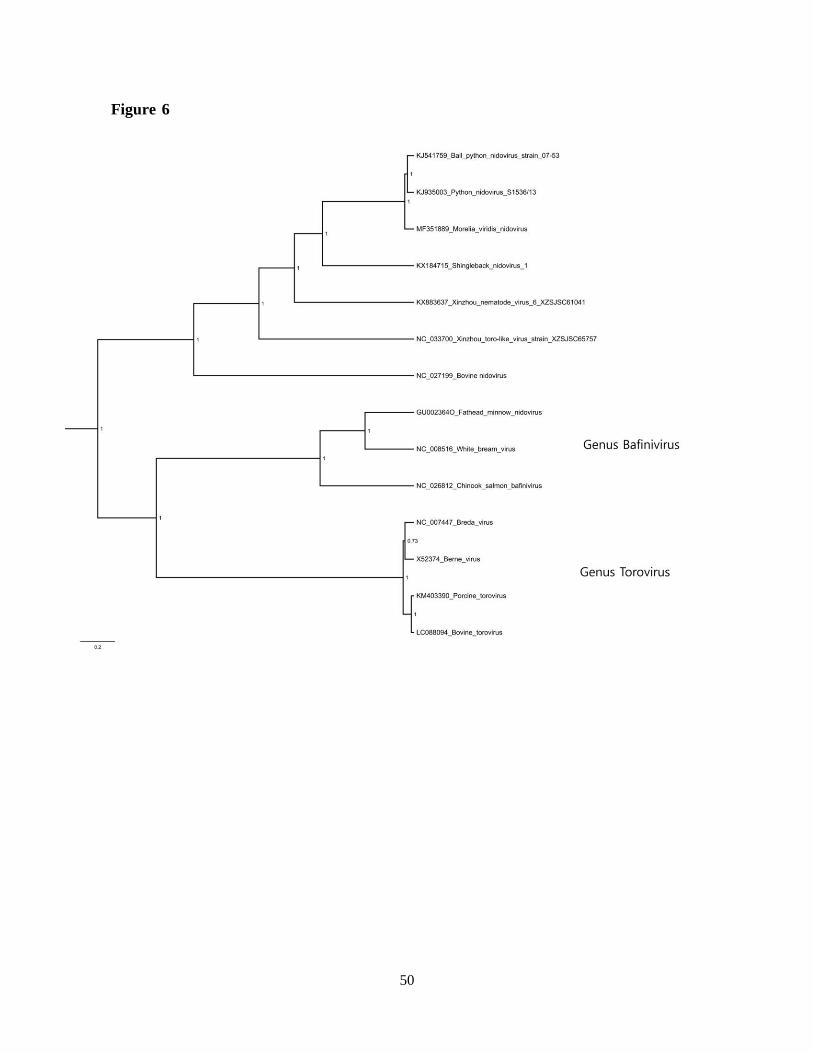
50
Figure 6

51
Figure 7

52
Figure 8

53
Figure 9

54
Figure 10

55
Figure 11

56
Danksagung
In erster Linie möchte ich mich bei meinen Betreuern (Prof. Anja Kipar, Dr. Dr. Udo
Hetzel) danken, dass sie mir die Möglichkeit gegeben haben, diese Arbeit anzugehen und
erfolgreich abzuschliessen.
Ein grosses Dankeschön geht ebenfalls an Fernando Romero, der mir die hohe Kunst der
In situ Hybridisierung und mir mit sehr viel Geduld die Tätigkeiten im Labor beigebracht
hat. Auch Jussi Hepojoki und seinem finnischen Team möchte ich für ihre stetige
Unterstützung aus der Ferne auf virologischer Ebene sehr danken. Ohne die oftmals
kryptischen Beiträge der Virologen wäre ein Grossteil dieses Werkes nicht vollbracht.
Von ebenso grosser Wichtigkeit war die Hilfe meiner BIBD-Arbeitskolleginnen (Dr. Saskia
Keller, Christine Jelinek, Dr. Pia Palamides), welche mir stets in guten und auch in
schweren Zeiten beigestanden und mir endlose Unterstützung auf allen Ebenen geliefert
haben.
Auch meinen restlichen Arbeitskollegen (Assistenten sowie auch Oberassistenten) möchte
ich danken, dass sie mir den oftmals Alltag verschönert und mich in schwierigen
Situationen ermutigt haben.
Ich möchte diese Arbeit meinen Eltern (Christina Tell und Dimitrios Dervas) und meiner
restlichen Familie widmen, ohne dich ich zu diesem Zeitpunkt nicht an diesem Punkt im
Leben stehen würde. Vielen Dank für die konstante moralische, seelische und auch
finanzielle Unterstützung seit so vielen Jahren, man könnte sich als Tochter nichts mehr als
das wünschen.
57
CURRICULUM VITAE
Vorname Name Eva Dervas
Geburtsdatum 14/09/1990
Geburtsort Athen
Nationalität Deutsch
Schulausbildung
09/1996-09/2002 Dimotiko Scholeio Agias Paraskevis, Athen, Griechenland
09/2002 – 09/2008 Deutsche Schule Athen (DSA), Athen, Griechenland
Höchster Schulabschluss
16/04/2008 Abitur, Deutsche Schule Athen (DSA), Athen, Griechenland
Studium
10/2008 – 10/2014 Veterinärmedizin, Veterinärmedizinische Universität Wien,
Wien, Österreich
10/11/2014 Abschlussprüfung vet.med.
Veterinärmedizinische Universität Wien, Wien, Österreich
01/2015 – 05/2017 Anfertigung der Dissertation
unter der Leitung von Prof. Dr.med.vet. Anja Kipar
am Institut für Veterinärpathologie
der Vetsuisse-Fakultät, Universität Zürich
Direktorin Prof. Dr. Anja Kipar
Fachrelevante Anstellungen nach Abschluss des veterinärmedizinischen Studiums
Seit Feb 2016 Residency in Veterinärpathologie am Institut für
Veterinärpathologie, Vetsuisse-Fakultät, Universität Zürich
01/2015-01/2016
Internship in Veterinärpathologie am Institut für
Veterinärpathologie, Vetsuisse-Fakultät, Universität Zürich
Seit Jan 2015 Doktorandin am Institut für Veterinärpathologie, Vetsuisse-
Fakultät, Universität Zürich