nih public access humans cogn neurosci - leopereeigiorni … · · 2014-09-21subjects' whole...
TRANSCRIPT

Cortical reactivity and effective connectivity during REM sleep inhumans
M Massimini2, F Ferrarelli1, MJ Murphy1, R Huber3, BA Riedner1, S Casarotto2, and GTononi1
1)Department of Psychiatry, University of Wisconsin, 6001 Research Park Blvd, Madison, WI 53719,USA 2)Department of Clinical Sciences, University of Milan, via G.B. Grassi 74, Milan 20157, Italy3)Children Hospital, University of Zurich, Steinwiesenstrasse 75, Zurich 8032, Switzerland
AbstractWe recorded the electroencephalographic (EEG) responses evoked by transcranial magneticstimulation (TMS) during the first rapid eye movement (REM) sleep episode of the night and wecompared them with the responses obtained during previous wakefulness and NREM sleep.Confirming previous findings, upon falling into NREM sleep, cortical activations became more localand stereotypical, indicating a significant impairment of the intracortical dialogue. During REMsleep, a state in which subjects regain consciousness but are almost paralyzed, TMS triggered morewidespread and differentiated patterns of cortical activation, that were similar to the ones observedin wakefulness. Similarly, TMS/hd-EEG may be used to probe the internal dialogue of thethalamocortical system in brain injured patients that are unable to move and communicate.
IntroductionAccording to a recent proposal (Tononi, 2004, 2008) consciousness depends critically not somuch on firing rates, synchronization at specific frequency bands, or sensory input per se, butrather on the brain's ability to integrate information, which is contingent on the effectiveconnectivity among functionally specialized regions of the thalamocortical system. Effectiveconnectivity refers to the ability of a set of neuronal elements to causally affect the firing ofother neuronal groups within a system (Lee, Harrison, & Mechelli, 2003). To test this proposal,our group has recently investigated whether the reduction of consciousness that occurs duringnon-rapid eye movement (NREM) sleep early in the night (Hobson & Pace-Schott, 2002;Stickgold, Malia, Fosse, Propper, & Hobson, 2001) may be associated with changes in corticaleffective connectivity (Massimini et al., 2005). To this aim, transcranial magnetic stimulation(TMS) was combined with high-density electroencephalography (hd-EEG) to explore how theactivation of one cortical area (the premotor area) is transmitted to the rest of the brain duringwakefulness and NREM sleep. Result showed that, during quiet wakefulness, TMS triggeredan initial response at the stimulation site that was followed by a sequence of fast waves (15–30 Hz) that moved to connected cortical areas several centimeters away. Upon entering NREMsleep stages 3/4, the brain's response to TMS became a single large positive-negative slowwave that rapidly extinguished and did not propagate beyond the stimulation site. Based onthis finding, we hypothesized that a significant breakdown of cortical effective connectivity
Correspondence should be addressed to: Giulio Tononi Department of Psychiatry, University of Wisconsin, Madison 6001 ResearchPark Blvd Madison, WI 53719, USA Tel. (608) 263 6063 Fax. (608) 265 2953 [email protected].
NIH Public AccessAuthor ManuscriptCogn Neurosci. Author manuscript; available in PMC 2011 September 1.
Published in final edited form as:Cogn Neurosci. 2010 September ; 1(3): 176–183. doi:10.1080/17588921003731578.
NIH
-PA Author Manuscript
NIH
-PA Author Manuscript
NIH
-PA Author Manuscript

may actually explain why subjects awakened during NREM sleep early in the night report little,or no, conscious experience.
Of course, blank reports upon awakening from sleep are not the rule, and many awakeningsyield dream reports (Casagrande, Violani, Lucidi, Buttinelli, & Bertini, 1996) (Stickgold et al.,2001) (Fagioli, 2002). Dreams can be at times as vivid and intensely conscious as wakingexperiences. Dream-like consciousness occurs during various phases of sleep, such as at sleeponset, during the last part of the night and, especially, during rapid eye movement (REM) sleep.Here we report the results of experiments in which we were able to record TMS-evokedpotentials during the first REM sleep episode of the night and to compare them with theresponses obtained during previous wakefulness and NREM sleep. During the transition fromNREM to REM, while subjects were behaviorally asleep, the brain's reaction to TMS recoveredfast oscillatory components and became similar to the one obtained during wakefulness,especially during the first 100–150 ms post-stimulus. The resurgence of fast waves was alsoassociated with a partial recovery of cortical effective connectivity. These results suggest thatdirectly measuring cortical reactivity and effective connectivity with TMS/hd-EEG may helpevaluating the brain's capacity for conscious experience in the absence of overt behavior, suchas in sleeping subjects or in non-communicative, brain injured patients.
MethodsSubjects
Ten subjects (age 21–34, 3 females) were involved in the study. All participants gave writteninformed consent and the experiment was approved by the University of Wisconsin HumanSubjects Committee. Prior to the experiment a neurological screening was performed toexclude potential adverse effects of TMS. The data presented here are from the five subjectsin whom at least 30 trials of TMS-evoked potentials could be recorded during REM sleep.
TMS targetingCortical TMS targets were identified on T1 weighted MR images (resolution 0.5 mm) of thesubjects' whole heads acquired with a 3T GE Signa scanner. In order to ensure precision andreproducibility of stimulation we employed a Navigated Brain Stimulation (NBS) system(Nexstim Ltd, Helsinki FI). The NBS device located (with an error <3 mm) the relativepositions of the subject's head and of the TMS coil by means of an optical tracking system.NBS also calculated the distribution and strength of the intracranial electric field induced byTMS. The coordinates of stimulation were input to a software aiming tool that ensuredthroughout the session the reproducibility of position, direction, and angle of the stimulator.We targeted the rostral part of the right premotor cortex (r-PM), 1 cm ahead of the anteriorcommissure and 2 cm lateral to the midline. This area is accessible through a central scalpposition, far from any major head or facial muscle.
Stimulation parametersStimulation was performed by means of a Magstim figure-of-eight coil (model P/N9925), witha wing diameter of 70 mm, connected to a Magstim Rapid biphasic stimulator. Resting motorthreshold (rMT) was measured prior to the experiment as the lowest stimulator output at whichat least 5 out of 10 pulses, delivered on the optimal hand motor area, resulted in a motor evokedpotential of 50 μV or greater in the abductor pollicis brevis (2). We stimulated r-PM at 110%of rMT, corresponding to a stimulator output between 54 and 67% and to a maximum estimatedelectric field on the target between 90 and 120 V/m. TMS was delivered with an interstimulusinterval jittering randomly between 2 and 2.3 s.
Massimini et al. Page 2
Cogn Neurosci. Author manuscript; available in PMC 2011 September 1.
NIH
-PA Author Manuscript
NIH
-PA Author Manuscript
NIH
-PA Author Manuscript
EEG recordingsWe recorded the spontaneous and TMS-evoked EEG by means of a 60 carbon electrodes capand a specifically designed TMS-compatible amplifier (Nexstim Ltd, Helsinki, FI). The artifactinduced by TMS was gated and saturation of the amplifier was avoided by means of aproprietary sample-and-hold circuit that kept the analog output of the amplifier constant from100 μs pre- to 2 ms post-stimulus (Virtanen, Ruohonen, Naatanen, & Ilmoniemi, 1999). Tofurther optimize TMS compatibility, the impedance at all electrodes was kept below 3 KΩ.The EEG signals, referenced to an additional electrode on the forehead, were filtered (0.1–500Hz) and sampled at 1450 Hz with 16 bit resolution. Four extra sensors were used to record theelectrooculogram (EOG) and the chin electromyogram (EMG). In most cases, no signs of TMSinduced magnetic artifact were detected and in all cases the EEG signals were artifact-freefrom <10 ms post-stimulus. At the end of each experiment, a pen visible to the infrared camerawas used to digitise the EEG electrode positions on the subject's head. During EEG recording,subject's perception of the clicks produced by TMS coil's discharge was eliminated by meansof inserted earplugs continuously playing a masking noise. Volume of the masking noise(always below 90 dB) was adjusted immediately prior to the experiment until the subjectsreported that the TMS click was not perceptible. A thin layer of foam was placed between coiland scalp (resulting in less than 1 mm thickness when coil was pressed against the head) inorder to attenuate bone conduction. As previously demonstrated, this procedure effectivelyprevented any contamination of EEG signals by auditory potentials elicited by TMS-associatedclicks (Massimini et al., 2005) (Massimini et al., 2007).
General experimental proceduresSubjects were invited to the lab in the evening (9:30 PM) and were requested to stay awakeuntil 1:00 AM. After setting-up the EEG cap and selecting the TMS target, recordings startedat around 2:00 AM. We chose this timing in order to extend the recordings to a circadian phase(early morning) that is more favorable to the occurrence of REM sleep (Dijk & Czeisler,1995). During the experiment subjects were laying eyes closed on a reclining chair with a head-rest that allowed a comfortable and stable head position. Participants were requested to stayawake while at least 200 trials were recorded. Afterward, we allowed the subjects to fall asleepwhile the spontaneous EEG was monitored and TMS was continuously delivered. In order toavoid overheating of the coil, we interrupted and restarted the stimulation according to the EEGpattern of interest. Responses during NREM sleep were collected in 7 out of 10 subjects. Fiveof these subjects subsequently entered REM sleep for at least 2 minutes and were included inthe analysis. Throughout the recording session the stability of stimulation coordinates werecontinuously monitored. If a movement of >5 mm occurred, the session was interrupted andthe coil was repositioned. At the end of the experiment, the stimulation coordinates wererecorded and the electrode positions were digitized.
Dream reportsIn order to evaluate subjective experience we collected a dream report in all the subjects (n=5)who entered REM sleep. Two subjects awakened spontaneously during REM sleep while theremaining three were awakened as soon as the REM sleep episode was judged to be over, basedon the on-line inspection of the EEG, EOG and EMG traces. Upon awakening subjects wereasked to remember what was going in their mind in the time prior to waking (Stickgold et al.,2001). All reports were collected by dictation into a portable digital audio recorder (OlympusDS-50). Recordings were transcribed and subsequently edited for Total Recall Count, or TRC(Antrobus, 1983), removing extraverbal utterances, nonwords, repeated words and secondaryelaborations. TRCs were obtained using the word-count function of Microsoft Word.
Massimini et al. Page 3
Cogn Neurosci. Author manuscript; available in PMC 2011 September 1.
NIH
-PA Author Manuscript
NIH
-PA Author Manuscript
NIH
-PA Author Manuscript

Data analysisData analysis was performed in Matlab (The Matworks, MA, USA) and Curry 5.0 (PhilipsGmbH, Germany). Sleep stages were scored off-line according to Rechtschaffen and Kales(1968) on two EEG channels (C3, C4) re-referenced to the mastoid, one EOG channel and oneEMG channel. We classified the single trials according to the stages during which they werecollected. Trials containing noise, muscle activity or eye movements were rejected. The trialscollected during wakefulness, NREM sleep stage 3–4 and REM sleep were averagedseparately. The averaged signals were baseline corrected and band-pass filtered between 2 and100 Hz. In all subjects, the averaged TMS-evoked potentials was characterized, duringwakefulness, by two consecutive positive peaks (peak 1 and peak 2) occurring at around 20and 60 ms, respectively (fig. 2). We characterized changes in the shape of the early EEGresponse to TMS by detecting significant changes of the amplitude of peak 1 and peak 2 acrossstates of vigilance. To this aim, we measured, on each single-trial response, the maximumvoltage at around (±10 ms) the latency of peak 1 and peak 2 (as detected during wakefulness).After that, we performed a Student's T-test on the single-trial amplitude distributions obtainedin the different conditions.
In order to perform source modeling, the averaged responses, the MRI sets and the electrodecoordinates were input to the software package Curry 5.0. Following a semi-automaticsegmentation of the individual MRI images we implemented a Boundary Element Model(BEM) of the head having 3 compartments of fixed conductivities (scalp: 0.33 S/m; skull:0.0042 S/m; brain: 0.33 S/m). The cortical surface was also reconstructed with a 6 mmresolution and modeled with approximately 14,000 rotating dipoles. Next, the electrodepositions were projected onto the skin surface and the lead field matrix was calculated. Weestimated the current density on the cortical surface using the Minimum Norm Least Squares(L2 Norm) method (weighted for depth bias removal) (Hamalainen & Ilmoniemi, 1994). Toidentify the cortical areas involved by TMS-evoked activity we proceeded as follows: i) wecalculated the global mean field power GMFP (Hamalainen & Ilmoniemi, 1994) as the meanof the absolute voltage recorded from electrodes in average reference ii) to identify the latencieswere TMS evoked a significant response, we detected the time intervals where the post-stimulus amplitude of the GMFP was 3 standard deviations larger than pre-stimulus activity;iii) within these intervals, we detected the time of the local maxima of the GMFP; iv) at thesetime points, we estimated the location of maximum neural activity using the L2 norm. Thus,we estimated the location of the maximum activation (top 10%) at each significant time sample,using Matlab 7.0, the identified sources were then plotted on the individual cortical surfaceand color-coded according to the latency of their involvement.
ResultsWhile TMS was delivered, the following percentages of sleep stages were obtained across thefive subjects who entered REM sleep: wakefulness: 45.4%; stage 1: 9.1%; stage 2: 15.6%;stages 3–4: 22.6%; REM 7.3%. In all cases, at least 150 single-trial evoked responses couldbe retained during wakefulness and NREM sleep. In four subjects, all the trials averaged in theNREM sleep condition were collected during stages 3 and 4, while in one subject trials collectedduring NREM stages 2, 3 and 4 had to be averaged together in order to obtain a stable NREMresponse. TMS/hd-EEG measurements during REM sleep were more challenging. Indeed, thissleep stage tends to occur unpredictably and is rather unstable, especially during the first sleepepisode. As a consequence, periods of clear-cut REM sleep were much shorter. Specifically,we obtained, across all subjects, the following amount of seconds (and of TMS-evoked singletrials) during REM sleep: 132 s (42 trials), 163 s (53 trials), 169 s (52 trials), 230 s (92 trials),348 s (145 trials). While TMS-evoked potentials have an excellent signal-to-noise ratio, alimited number of trials hampers the reliability of the average response, especially at late
Massimini et al. Page 4
Cogn Neurosci. Author manuscript; available in PMC 2011 September 1.
NIH
-PA Author Manuscript
NIH
-PA Author Manuscript
NIH
-PA Author Manuscript

latencies, where intertrial variability increases. Thus, to assess the reliability of the overallscalp-recorded average response at different latencies, we detected the time-samples where theGMFP was significant (post-stimulus amplitude > of 3 standard deviations of pre-stimulus;see also Methods and (Massimini et al., 2005)). In each subject, this test was repeated for eachcondition (including wakefulness, NREM sleep) using the maximum number of trials collectedduring REM sleep. In all subjects, a significant response could be detected during the first 100–150 ms, while in the subject in whom 145 single trials could be analyzed the significant responseextended to 300 ms. For this reason, the analysis of TMS-evoked potentials was restricted tothe first 150 ms in all cases except for this subject, in whom source modeling was alsoperformed. All subjects, prompted upon awakening from REM sleep, provided a dream report.TWC varied largely across subjects (ranging from 45 words to 237 words) and tended tocorrelate (p<0.1) with the length of the period that subjects spent in REM sleep.
In all vigilance states, TMS elicited a time-locked response that could be appreciated on asingle-trial basis. Fig. 1A displays the single-trial responses recorded from one electrodelocated under the stimulator (corresponding to FC2, in the 10–20 system) during a transitionfrom wakefulness through stage 1 and NREM to REM sleep. In this representation, signals areband-passed from 15 to 100 Hz, in order to highlight the presence of fast oscillations in thesingle-trial responses. Fig. 1B shows the corresponding averages (filtered 2–100 Hz) calculatedin the four vigilance states. During wakefulness, TMS triggered a sustained response made ofa sequence of time-locked, high-frequency (20–35 Hz) oscillations in the first 100 ms. As soonas the subject transitioned into stage 1, the amplitude of the first positive component (between0 and 40 ms) increased by 40%, while the subsequent waves were markedly dampened. DuringNREM sleep the response changed markedly; the first positive component became larger(120% increase), slower and was followed by a negative rebound after which the responseextinguished. Upon transitioning to REM sleep the cortical response to TMS recoveredwakefulness-like fast oscillations, despite the subject being behaviorally asleep.
Figure 2 shows that the resumption of fast oscillations during REM sleep was reproducibleacross subjects and, at the same time, it highlights significant differences between the REMsleep and the wakefulness response. In REM sleep, compared to wakefulness, the first positivecomponent (peak1) was larger (p<0.05) and the second (peak 2) was smaller (p<0.05) in eachsubject (Student's T-test, comparing single-trial amplitude distributions). During NREM sleep,peak 1 was larger compared to both REM sleep (p<0.001) and wakefulness (p<0.001), whilepeak 2 was replaced by a negative wave.
In one subject, we were able to collect a sufficient number of trials to perform source modelingand to compare REM sleep to NREM sleep and wakefulness also at longer latencies (Fig. 3).This analysis revealed that the complex pattern of long-lasting, long-range activation triggeredby TMS during wakefulness broke down during NREM sleep and that it partially recovered,within the first 150 ms post-stimulus, during REM sleep. Fig. 3 also shows that long-latencycomponents that were present in wakefulness were obliterated during REM sleep.Unfortunately, due to the short duration of clear-cut REM sleep periods, the long latencyresponse could not be analyzed in the other subjects.
DiscussionThe level and quality of conscious experience can vary dramatically across the sleep-wakecycle. During NREM sleep early in the night, consciousness can nearly vanish (Pivik &Foulkes, 1968) (Suzuki et al., 2004) (Hobson & Pace-Schott, 2002) despite persistent neuralactivity in the thalamocortical system (Steriade, Timofeev, & Grenier, 2001). Why is it so?The present work confirms the results of previous measurements (Massimini et al., 2005) byshowing that, during NREM sleep stages 3 and 4 early in the night, thalamocortical circuits
Massimini et al. Page 5
Cogn Neurosci. Author manuscript; available in PMC 2011 September 1.
NIH
-PA Author Manuscript
NIH
-PA Author Manuscript
NIH
-PA Author Manuscript
remain active and reactive but lose their ability to interact and to produce complex, integratedresponses. Indeed, in this state, TMS failed to trigger a sustained, long-range pattern ofactivation and instead evoked a simple positive-negative wave that remained local.Interestingly, this stereotypical response has been shown to share fundamental characteristicswith the slow waves that occur spontaneously during NREM sleep (Massimini et al., 2007).This evidence suggests that the mechanisms underlying the generation of slow waves, mayalso be responsible for blocking the emergence of specific long-range responses during NREMsleep (Tononi & Massimini, 2008) (Massimini, Tononi, & Huber, 2009). Upon falling asleep,due to a dampening of brainstem noradrenergic, serotoninergic and cholinergic activatingsystems (Steriade, 2004), cortical neurons become bistable and inevitably tend to fall into asilent, hyperpolarized state (down-state) after a period of activation (up-state) (Timofeev,Grenier, & Steriade, 2001). This mechanism provides the mechanism for the slow oscillationsof sleep, where large populations of cortical neurons spontaneously alternate between up anddown-states (Hill & Tononi, 2005). In addition, bistability may also be contributed for by ashift in the balance of synaptic excitation and inhibition toward inhibition due to changes inthe neuromodulatory milieu (Esser, Hill, & Tononi, 2009). In any case, bistability may preventthe emergence of sustained, complex thalamocortical interactions. Thus, in this condition, anylocal activation, whether occurring spontaneously or induced by a stimulus (like TMS), willconverge into a silent neuronal down-state and into a stereotypical EEG slow wave. While thisseems to occur at least during deep NREM sleep early in the night (Tononi & Massimini,2008), it is hard to predict what would happen during REM sleep. In this state, whilenoradrenergic and serotoninergic arousing systems remain silent, brainstem cholinergicneurons come back to activity (Pace-Schott & Hobson, 2002), spontaneous slow wavesdisappear and the EEG becomes, at least superficially, similar to the one of wakefulness.However, despite this apparent resemblance with wakefulness, it is difficult to infer the degreeof underlying thalamocortical bistability during REM sleep based on the presence of a low-voltage EEG alone. Indeed, it was shown that, during NREM sleep, TMS delivered duringshort stretches of low-amplitude EEG sleep was still able to trigger full-fledged slow waves(Massimini et al., 2007). The present experiments demonstrate that, during REM sleep, TMSevokes a response that is more similar to the one observed in wakefulness, namely, a sequenceof fast oscillations (Fig. 1 and 2) occurring in the first 150 ms. This evidence suggests that thedischarge of mesopontine cholinergic neurons alone may represent a major excitatory inputthat can largely prevent the emergence of thalamocortical bistability during REM sleep.
Despite obvious similarities, the REM and the wakefulness responses also showed consistentdifferences; in REM sleep, the first positive component (peak 1) was always larger and thesecond one (peak 2) was always smaller compared to wakefulness (Fig. 2). In this sense, theREM response tended to share some features with the NREM sleep response, where peak 1reached its maximum amplitude and peak 2 disappeared. Notably, the REM sleep responserecorded in the present experiment strongly resembled the TMS-evoked potential obtainedduring sleep stage 1 in a previous work (Massimini et al., 2005), suggesting that stage 1 andREM sleep may be supported by a similar degree of cortical activation.
Source modeling revealed that, as in wakefulness, the resumption of fast oscillations duringREM sleep was associated with a pattern of activation that was more complex and widespreadcompared to one of NREM sleep. This observation, although limited to a single subject,corroborates the hypothesis that cortical effective connectivity may play a role in the shifts ofconscious experience that occur during sleep. Indeed, the subject in Fig. 3 spent the longesttime in REM sleep (348 s) and reported a long dream recall (237 words) upon awakening. Thepersistence, to some degree, of long-range cortico-cortical effective connectivity has been alsoreported during stage 1 (see Fig S2 in (Massimini et al., 2005), another sleep stage associatedwith frequent and long dream reports (Foulkes, 1966). Fig. 3 also shows that long-latencycomponents that were present in wakefulness were obliterated during REM sleep. This finding,
Massimini et al. Page 6
Cogn Neurosci. Author manuscript; available in PMC 2011 September 1.
NIH
-PA Author Manuscript
NIH
-PA Author Manuscript
NIH
-PA Author Manuscript

is consistent with the notion that late component of peripherally evoked potentials are alsodampened during REM sleep(Goff, Allison, Shapiro, & Rosner, 1966;Wesensten & Badia,1988). In future works it would be interesting to systematically collect TMS/hd-EEG measuresof thalamocortical bistability and effective connectivity during the whole night and to correlatethem with dream reports. This approach, might represent a valid attempt to study the neuralcorrelates of consciousness during sleep on a finer time scale, beyond the REM/NREM sleepdichotomy and beyond traditional sleep staging. Similarly, exploring brain activity on a finerspatial scale with functional neuroimaging already shed light on the neural mechanismsunderlying changes in the quality of consciousness across the sleep-wake cycle (Maquet et al.,1996) (Maquet et al., 2005).
Besides their possible relevance for sleep physiology, the present experiments demonstratethat TMS/hd-EEG may represent an effective way to probe the internal dialogue of thethalamocortical system in the absence of any behavioral cue. TMS triggered more widespreadand differentiated patterns of EEG activation, just as it does during normal wakefulness, uponentering REM sleep, a state in which subjects are conscious but almost paralyzed. Hence, inthe future, this technique may be employed as an aid to evaluate the brain's capacity forconsciousness in brain injured patients who are unable to move and communicate (Massimini,Boly, Casali, Rosanova, & Tononi, 2009).
AcknowledgmentsThis work was supported by a National Institutes of Health Pioneer Award (to G.T.) and by the European Union(LSHM-CT-2005-518189 to M.M.).
ReferencesAntrobus J. REM and NREM sleep reports: comparison of word frequencies by cognitive classes.
Psychophysiology 1983;20(5):562–568. [PubMed: 6635096]Casagrande M, Violani C, Lucidi F, Buttinelli E, Bertini M. Variations in sleep mentation as a function
of time of night. International Journal of Neuroscience 1996;85(1–2):19–30. [PubMed: 8727679]Dijk DJ, Czeisler CA. Contribution of the circadian pacemaker and the sleep homeostat to sleep
propensity, sleep structure, electroencephalographic slow waves, and sleep spindle activity in humans.Journal of Neuroscience 1995;15(5 Pt 1):3526–3538. [PubMed: 7751928]
Esser SK, Hill S, Tononi G. Breakdown of effective connectivity during slow wave sleep: investigatingthe mechanism underlying a cortical gate using large-scale modeling. Journal of Neurophysiology2009;102(4):2096–2111. [PubMed: 19657080]
Fagioli I. Mental activity during sleep. Sleep Medical Review 2002;6(4):307–320.Goff WR, Allison T, Shapiro A, Rosner BS. Cerebral somatosensory responses evoked during sleep in
man. Electroencephalography and Clinical Neurophysiology 1966;21(1):1–9. [PubMed: 4165361]Hamalainen MS, Ilmoniemi RJ. Interpreting magnetic fields of the brain: minimum norm estimates.
Medical and Biological Engineering and Computing 1994;32(1):35–42. [PubMed: 8182960]Hill S, Tononi G. Modeling sleep and wakefulness in the thalamocortical system. Journal of
Neurophysiology 2005;93(3):1671–1698. [PubMed: 15537811]Hobson JA, Pace-Schott EF. The cognitive neuroscience of sleep: neuronal systems, consciousness and
learning. Nature Review Neuroscience 2002;3(9):679–693.Lee L, Harrison LM, Mechelli A. A report of the functional connectivity workshop, Dusseldorf 2002.
Neuroimage 2003;19(2 Pt 1):457–465. [PubMed: 12814594]Maquet P, Peters J, Aerts J, Delfiore G, Degueldre C, Luxen A, et al. Functional neuroanatomy of human
rapid-eye-movement sleep and dreaming. Nature 1996;383(6596):163–166. [PubMed: 8774879]Maquet P, Ruby P, Maudoux A, Albouy G, Sterpenich V, Dang-Vu T, et al. Human cognition during
REM sleep and the activity profile within frontal and parietal cortices: a reappraisal of functionalneuroimaging data. Progress in Brain Research 2005;150:219–227. [PubMed: 16186026]
Massimini et al. Page 7
Cogn Neurosci. Author manuscript; available in PMC 2011 September 1.
NIH
-PA Author Manuscript
NIH
-PA Author Manuscript
NIH
-PA Author Manuscript

Massimini M, Boly M, Casali A, Rosanova M, Tononi G. A perturbational approach for evaluating thebrain's capacity for consciousness. Progress in Brain Research 2009;177:201–214. [PubMed:19818903]
Massimini M, Ferrarelli F, Esser SK, Riedner BA, Huber R, Murphy M, et al. Triggering sleep slowwaves by transcranial magnetic stimulation. Proceedings of the National Academy of Sciences U SA 2007;104(20):8496–8501.
Massimini M, Ferrarelli F, Huber R, Esser SK, Singh H, Tononi G. Breakdown of cortical effectiveconnectivity during sleep. Science 2005;309(5744):2228–2232. [PubMed: 16195466]
Massimini M, Tononi G, Huber R. Slow waves, synaptic plasticity and information processing: insightsfrom transcranial magnetic stimulation and high-density EEG experiments. European Journal ofNeuroscience 2009;29(9):1761–1770. [PubMed: 19473231]
Pace-Schott EF, Hobson JA. The neurobiology of sleep: genetics, cellular physiology and subcorticalnetworks. Nature Review Neuroscience 2002;3(8):591–605.
Pivik T, Foulkes D. NREM mentation: relation to personality, orientation time, and time of night. Journalof Consulting and Clinical Psychology 1968;32(2):144–151. [PubMed: 5654189]
Steriade M. Acetylcholine systems and rhythmic activities during the waking--sleep cycle. Progress inBrain Research 2004;145:179–196. [PubMed: 14650916]
Steriade M, Timofeev I, Grenier F. Natural waking and sleep states: a view from inside neocorticalneurons. Journal of Neurophysiology 2001;85(5):1969–1985. [PubMed: 11353014]
Stickgold R, Malia A, Fosse R, Propper R, Hobson JA. Brain-mind states: I. Longitudinal field study ofsleep/wake factors influencing mentation report length. Sleep 2001;24(2):171–179. [PubMed:11247053]
Suzuki H, Uchiyama M, Tagaya H, Ozaki A, Kuriyama K, Aritake S, et al. Dreaming during non-rapideye movement sleep in the absence of prior rapid eye movement sleep. Sleep 2004;27(8):1486–1490.[PubMed: 15683138]
Timofeev I, Grenier F, Steriade M. Disfacilitation and active inhibition in the neocortex during the naturalsleep-wake cycle: an intracellular study. Proceedings of the National Academy of Sciences U S A2001;98(4):1924–1929.
Tononi G. An information integration theory of consciousness. BMC Neuroscience 2004;5:42. [PubMed:15522121]
Tononi G. Consciousness as integrated information: a provisional manifesto. Biological Bulletin2008;215(3):216–242. [PubMed: 19098144]
Tononi G, Massimini M. Why does consciousness fade in early sleep? Annals of the New York Academyof Sciences 2008;1129:330–334. [PubMed: 18591492]
Virtanen J, Ruohonen J, Naatanen R, Ilmoniemi RJ. Instrumentation for the measurement of electric brainresponses to transcranial magnetic stimulation. Medical and Biological Engineering and Computing1999;37(3):322–326. [PubMed: 10505382]
Wesensten NJ, Badia P. The P300 component in sleep. Physiology and Behavior 1988;44(2):215–220.[PubMed: 3237827]
Massimini et al. Page 8
Cogn Neurosci. Author manuscript; available in PMC 2011 September 1.
NIH
-PA Author Manuscript
NIH
-PA Author Manuscript
NIH
-PA Author Manuscript

Fig.1.(A) Single trial TMS-evoked responses are recorded from one channel located under thestimulator (FC2) while a subject transitions from wakefulness (W), through sleep stage 1 (S1)and NREM sleep (NREM) to REM sleep. The red line marks the time of TMS. Single-trialEEG data are band-pass filtered (15 to 100 Hz) and color coded for voltage (red for positive,blue for negative). (B) The averaged responses (filtered from 2 to 100 Hz) calculated in thefour vigilance states are depicted. The onset of REM sleep is associated with a resumption ofTMS-evoked fast oscillations.
Massimini et al. Page 9
Cogn Neurosci. Author manuscript; available in PMC 2011 September 1.
NIH
-PA Author Manuscript
NIH
-PA Author Manuscript
NIH
-PA Author Manuscript
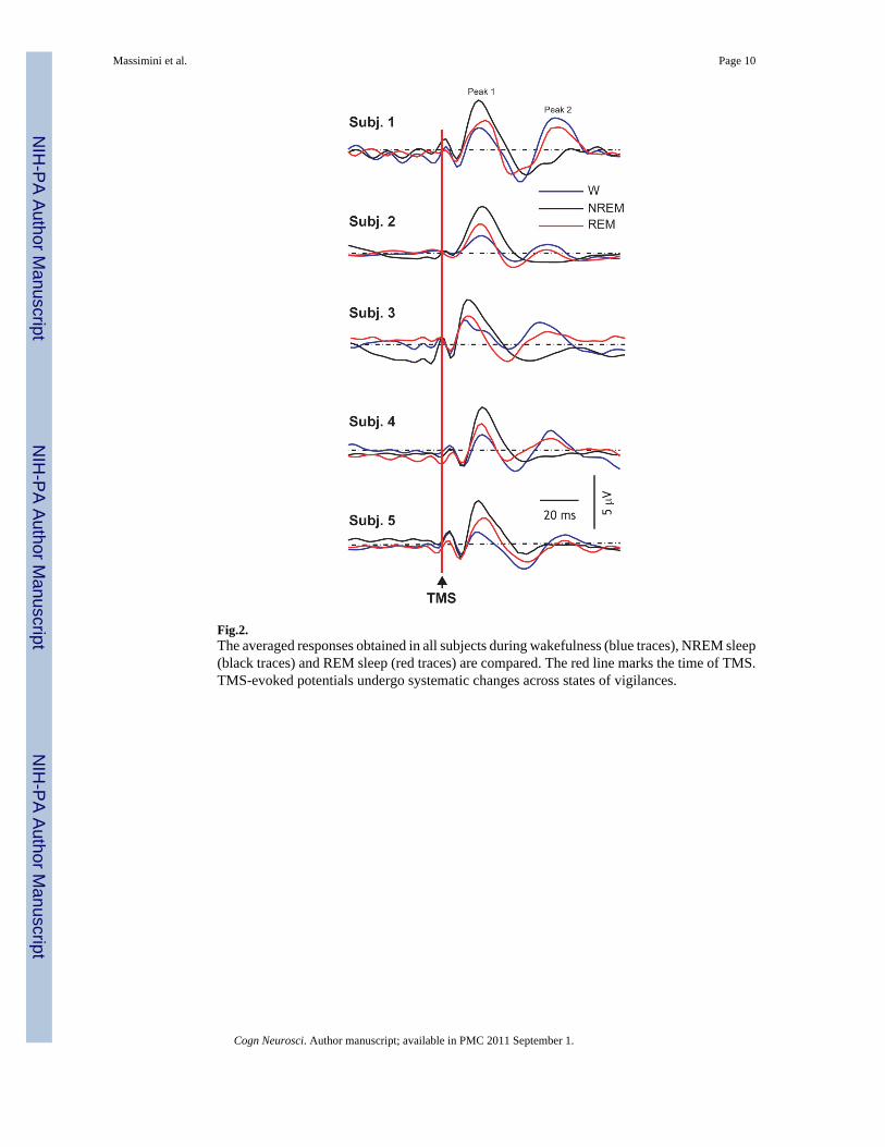
Fig.2.The averaged responses obtained in all subjects during wakefulness (blue traces), NREM sleep(black traces) and REM sleep (red traces) are compared. The red line marks the time of TMS.TMS-evoked potentials undergo systematic changes across states of vigilances.
Massimini et al. Page 10
Cogn Neurosci. Author manuscript; available in PMC 2011 September 1.
NIH
-PA Author Manuscript
NIH
-PA Author Manuscript
NIH
-PA Author Manuscript

Fig. 3.(A) The TMS-evoked potentials recorded from one subject, in whom a long stretch of REMsleep could be recorded, are displayed (blue: wakefulness, black: NREM sleep, red: REMsleep). The traces were recorded from the channels indicated by red dots in the upper left panel,where the site of stimulation on the subject's MRI is also indicated by (red arrow). (B)Spatiotemporal cortical maps of TMS-evoked cortical activation during wakefulness, NREMand REM sleep. For each significant time sample, maximum current sources were plotted andcolor-coded according to their latency of activation (light blue, 0 milliseconds; red, 300milliseconds). The yellow cross marks the TMS target on the cortical surface. During REMsleep, the resumption of TMS-evoked fast oscillations was associated with a partial recoveryof cortical effective connectivity.
Massimini et al. Page 11
Cogn Neurosci. Author manuscript; available in PMC 2011 September 1.
NIH
-PA Author Manuscript
NIH
-PA Author Manuscript
NIH
-PA Author Manuscript