nih public access mdx mice j diet suppl - wordpress.com · 2013-09-05 · mice on control diet. in...
TRANSCRIPT
The Dietary Supplement Protandim® Decreases PlasmaOsteopontin and Improves Markers of Oxidative Stress inMuscular Dystrophy Mdx Mice
Muhammad Muddasir Qureshi, MD, MPH,Department of Pediatrics, Texas Tech University Health Sciences Center, Paul L. Foster School ofMedicine, El Paso, TX. Earlier, he was associated with Department of Neurology, MassachusettsGeneral Hospital, Harvard Medical School, Boston, MA.
Warren C. McClure, MS,Department of Math and Science, Otero Junior College, CO. Earlier, he was associated withDepartment of Cell and Developmental Biology, University of Colorado Denver Health SciencesCenter (UCDHSC), Aurora, CO.
Nicole L. Arevalo, MA,Department of Cell and Developmental Biology, University of Colorado Denver Health SciencesCenter (UCDHSC), Aurora, CO.
Rick E. Rabon, BA,Department of Cell and Developmental Biology, University of Colorado Denver Health SciencesCenter (UCDHSC), Aurora, CO.
Benjamin Mohr,Department of Medicine, University of Colorado Denver Health Sciences Center (UCDHSC),Aurora, CO.
Swapan K. Bose, BS, BPharm,Department of Medicine, University of Colorado Denver Health Sciences Center (UCDHSC),Aurora, CO.
Joe M. McCord, PhD, andDepartment of Medicine, University of Colorado Denver Health Sciences Center (UCDHSC),Aurora, CO.
LifeVantage Corporation, San Diego, CA.
Brian S. Tseng, MD, PhDDepartment of Neurology, Division of Child Neurology, Massachusetts General Hospital, HarvardMedical School, 55 Fruit St ACC 708, Boston, MA 02114
AbstractTherapeutic options for Duchenne muscular dystrophy (DMD), the most common and lethalneuromuscular disorder in children, remain elusive. Oxidative damage is implicated as a pertinentfactor involved in its pathogenesis. Protandim® is an over-the-counter supplement with the ability
© 2010 by Informa Healthcare USA, Inc. All rights reserved.Address correspondence to: Brian S. Tseng, MD, PhD, ([email protected]).DISCLOSUREThe authors report J.M.M. is a consultant to LifeVantage Corporation and has a financial interest in the company.
NIH Public AccessAuthor ManuscriptJ Diet Suppl. Author manuscript; available in PMC 2010 August 24.
Published in final edited form as:J Diet Suppl. 2010 June 1; 7(2): 159–178. doi:10.3109/19390211.2010.482041.
NIH
-PA Author Manuscript
NIH
-PA Author Manuscript
NIH
-PA Author Manuscript

to induce antioxidant enzymes. In this study we investigated whether Protandim® provided benefitusing surrogate markers and functional measures in the dystrophin-deficient (mdx)mouse model ofDMD. Male 3-week-old mdx mice were randomized into two treatment groups: control (receivingstandard rodent chow) and Protandim®-supplemented standard rodent chow. The diets werecontinued for 6-week and 6-month studies. The endpoints included the oxidative stress markerthiobarbituric acid-reactive substances (TBARS), plasma osteopontin (OPN), plasma paraoxonase(PON1) activity, H&E histology, gadolinium-enhanced magnetic resonance imaging (MRI) of legmuscle and motor functional measurements. The Protandim® chow diet in mdx mice for 6 monthswas safe and well tolerated. After 6 months of Protandim®, a 48% average decrease in plasma TBARSwas seen; 0.92 nmol/mg protein in controls versus 0.48 nmol/mg protein in the Protandim® group(p = .006). At 6 months, plasma OPN was decreased by 57% (p = .001) in the Protandim®-treatedmice. Protandim® increased the plasma antioxidant enzyme PON1 activity by 35% (p = .018). After6 months, the mdx mice with Protandim® showed 38% less MRI signal abnormality (p = .07) thanmice on control diet. In this 6-month mdx mouse study, Protandim® did not significantly alter motorfunction nor histological criteria.
KeywordsDuchenne muscular dystrophy; protandim®; mdx mice; dystrophin; dystrophic muscle; paraoxonase;osteopontin; oxidative stress
INTRODUCTIONDuchenne muscular dystrophy (DMD) is an X-linked recessive disorder that occurs in 1 in3,500 live male births and is the most common fatal genetic disorder in children (Emery,1991). It is caused by loss-of-function mutations in the gene dystrophin that encodes a massivemuscle sub-sarcolemmal cytoskeletal protein. The pathogenesis of DMD is frequently studiedin the dystrophic mdx mouse model (Bulfield, Siller, Wight, & Moore, 1984; Sicinski et al.,1989). However, despite the complete deficiency of dystrophin protein, mdx mice have a near-normal life span. At approximately 3–6 weeks of postnatal age, mdx mice develop a crisis phaseof muscle necrosis followed by regeneration (McArdle et al., 1998). The mice are often studiedonly during the first 6 weeks of their lives before a stable regenerative phase ensues thatachieves a mild clinical adult mouse phenotype.
There is no cure for DMD and the only medications proven to favorably alter its natural historyare corticosteroids (Mendell et al., 1989). There is evidence from randomized controlled trialsthat glucocorticoid therapy in DMD improves muscle strength and function in the short-termperiod of 6 months to 2 years (Manzur, Kuntzer, Pike, & Swan, 2004). However, the long-termefficacy of corticosteroids in DMD with randomized placebo-controlled trials will likely neverbe studied given its orphan disease state and obvious side effects. There are a number of chroniccorticosteroid regimens (daily versus intermittent) and most recommended dose appears to beprednisone 0.75 mg/kg/day or deflazacort at 0.9 mg/kg/day. The use of corticosteroids isassociated with numerous side effects particularly weight gain, stunted height/growth,cataracts, osteoporosis, hypertension, diabetes, hirsutism, and mood/behavioral changes (Kellyet al., 2008; Manzur, Kuntzer, Pike, & Swan, 2004; Wong & Christopher, 2002). Thus, bettertreatments to augment or supplant corticosteroid use in DMD would be of immense value. Tofind a cocktail of other compounds that could lower the dose needed of corticosteroid wouldbe of tremendous clinical value to attenuate the chronic corticosteroid side-effect burden.
Oxidative stress is a significant pathologic factor in DMD. Skeletal muscles of mdx micedemonstrate increased quantities of oxidative damage markers including the by-products oflipid peroxidation and carbonyls (Haycock, Mac, & Mantle, 1998; Ragusa, Chow, & Porter,
Qureshi et al. Page 2
J Diet Suppl. Author manuscript; available in PMC 2010 August 24.
NIH
-PA Author Manuscript
NIH
-PA Author Manuscript
NIH
-PA Author Manuscript

1997). The dystrophin-deficient myotubes are highly susceptible to cellular injury, particularlyloss of membrane integrity, when exposed to reactive oxygen species (Disatnik, Chamberlain,& Rando, 2000; Rando, Disatnik, Yu, & Franco, 1998). Several co-morbidities in DMD,including muscle fatigue and cardiomyopathy, are associated with increased oxidative stress(Bia et al., 1999; Chi et al., 1987; Mohr, Hallak, de Boitte, Lapetina, & Brune, 1999).
Protandim® (LifeVantage Corp., San Diego, CA) is a dietary supplement available as an over-the-counter herbal supplement (once daily capsule of 675 mg) (Nelson, Bose, Grunwald,Myhill, & McCord, 2006). It is composed of the following phtyochemicals: (1) Bacopamonniera extract (45% bacosides), 150 mg; (2) Silybum marianum extract (70%–80%silymarin), 225 mg; (3) Withania somnifera (Indian ginseng) powder, 150 mg; (4) green teaextract (Camellia sinensis, 98% polyphenols and 45% epigallocatechin-3 gallate), 75 mg; and(5) curcumin (95%) from Curcuma longa, 75 mg. Individual ingredients of Protandim® arewell-known antioxidants that cause induction of SOD and catalase in rodents and diminishcellular lipid peroxidation (Joe, Vijaykumar, & Lokesh, 2004; Kishore & Singh, 2005; Lang,Deak, Muzes, Pronai, & Feher, 1993; Mandel, Weinreb, Amit, & Youdim, 2004; Rasool &Varalakshmi, 2007).
The effect of a single dose per day (675 mg) of Protandim®, for 30–120 days, has been testedon 29 healthy human volunteers ranging in age from 20 to 78 years (Nelson, Bose, Grunwald,Myhill, & McCord, 2006). Erythrocytes were assayed for superoxide dismutase (SOD) andcatalase, and plasma was assayed for thiobarbituric acid-reacting substances (TBARS). Beforesupplementation, the levels of TBARS showed a strong age-dependent increase. After 30 daysof supplementation, TBARS declined by an average of 40% and the age-dependent increasewas eliminated. By 120 days, erythrocyte SOD increased by 30% and catalase by 54%.
The mechanism of action of Protandim® has been shown to be through activation of thetranscription factor nuclear factor E2-related factor 2 (Nrf2) by a mechanism involving multiplesignaling pathways and substantial synergy among the five active ingredients (Velmurugan,Alam, McCord, & Pugazhenthi, 2009). Nrf2 is known to induce many antioxidant enzymesvia the antioxidant response element in their promoters, including enzymes involved in thesynthesis and metabolism of glutathione. The synergy obtained in the composition enablesactivation of Nrf2 at very low concentrations of the individual active ingredients (Velmurugan,Alam, McCord, & Pugazhenthi, 2009). Protandim® provided substantial chemoprevention ina two-stage skin carcinogenesis study in the mouse. Protandim®-supplemented mice showeda 33% reduction in skin tumor incidence and a 57% reduction in tumor multiplicity (Liu et al.,2009). Protandim® has also been shown to induce the antioxidant transcription factor Nrf2 thatprevents cardiac oxidative stress (Bogaard et al., 2009). Nrf2 preserves the expression ofvascular endothelial growth factor and prevents right ventricular failure without modifyinglung angioproliferation.
The by-products of free radical damage to polyunsaturated fatty acids react with thiobarbituricacid to form products that may be assayed as an index of oxidative damage and lipidperoxidation (Armstrong & Browne, 1994). The concentration of TBARS has been found tobe significantly elevated in skeletal muscle of boys with DMD as well as mdx mice and is areliable indicator of oxidative stress levels in dystrophic muscle (Faist, Koenig, Hoeger, &Elmadfa, 1998; Jackson, Jones, & Edwards, 1984; Kar & Pearson, 1979). In the present work,we evaluated the 6-week and 6-month use of Protandim® in mdx mice using a placebo-controlled design. The primary endpoint of the study was to assess the impact of Protandim®-enriched diet versus control diet on mdx mice plasma and skeletal muscle homogenate levelsof TBARS. Secondary endpoints included a comparison of the plasma levels of profibroticfactor osteopontin (OPN, also known as SPP1) and antioxidant enzyme paraoxonase (PON1)as arylesterase activity, Hematoxylin and Eosin (H&E) muscle histology, region-of-interest
Qureshi et al. Page 3
J Diet Suppl. Author manuscript; available in PMC 2010 August 24.
NIH
-PA Author Manuscript
NIH
-PA Author Manuscript
NIH
-PA Author Manuscript
(ROI)-quantitative measurement of percentage gadolinium-enhanced muscle areas usingmagnetic resonance imagine (MRI) (Voisin et al., 2005) and functional measures includingvoluntary running (cumulative distances), time (min) to exhaustion running downhill andspontaneous cage activity (beam break counts).
MATERIALS AND METHODSAnimals, Specimen Collection, and Preparation
Adult mdx (C57BL/10ScSn-mdx) mice were originally obtained from Jackson Laboratory(JAXR®, BarHarbor, ME). All mdx mice were housed and handled in accordance withguidelines and procedures approved by the Institutional Animal Care and Use Committee.Mdx mice were kept at a research lab at the University of Colorado Health Sciences Centerinside cages in a traffic-free, quiet, and dim environment. Male mdx mice with confirmatoryPCR genotyped from tail DNA (data not shown) were utilized for these studies.
Treatment ProtocolDiets were provided ad lib to either adult mdx mice or breeder females. Protandim® wasformulated into standard Harlan Teklad 2018S rodent chow (custom order through ResearchDiets, Inc., New Brunswick, NY) for the intervention group while the control group ate thesame chow without Protandim®. The mice received Protandim® at a dosage of approximately457 mg/m2, calculated according to Reagan-Shawet al. (2008), which is nearly equivalent tothe manufacturers recommended human dose of 675 mg per day for a 60 kg adult, or 422 mg/m2. For the 6-week study, the treatment was given to breeder females so that the 6-weekmdx mice group was exposed to Protandim® through placental absorption. After birth, the pupswere kept on the Protandim® diet to 6 weeks of age when they were euthanized.
The chow was provided at 3–6 weeks of postnatal age during known muscle necrosis phase inmdx mouse. During this stage, the mice do not appear overtly crippled and are active, but theirmuscle tissues demonstrate most marked features of histopathology. In the second part of thestudy, Protandim® chow diet was provided for 6 months to mdx mice greater than 8 weeks ofage where more histopathology was exacerbated by having mdx mice run downhill (5% grade)on treadmills which increases eccentric damage to muscles. The mdx mice at this age havestably regenerated muscles with minor markers of past regeneration events particularly centralnuclei instead of peripheral muscle nuclei.
Blood CollectionBlood (50–100 µl) was collected via retro-orbital eye bleed of time of harvest. Isofluraneinhalant via a rodent anesthesia funnel mask was given to the mice for approximately 2 minprior to the retro-orbital eye bleed. Additional local anesthesia of preparacaine eyedrops wasgiven before blood sample was collected. Each mouse was pinch-tested to verify adequateanesthesia. The entire procedure took less than 5 min. The mice woke up usually within 2–3min after removal of isoflurane. Blood samples allowed serum creatine kinase (CK), plasmaTBARS, OPN, and PON1 analyses.
Tissue CollectionAfter completion of the dietary period, mice were euthanized with a mixture containingketamine and xylazine. Cervical dislocation was performed and then skeletal muscles,including gastrocnemius, tibialis anterior, rectus femoris and hamstring, from the one leg weredissected and harvested for histological studies using quick freezing technique. Thecontralateral leg muscles were harvested for biochemical and western blot assays.
Qureshi et al. Page 4
J Diet Suppl. Author manuscript; available in PMC 2010 August 24.
NIH
-PA Author Manuscript
NIH
-PA Author Manuscript
NIH
-PA Author Manuscript
Mdx mice were randomized into the two groups. Blood samples, tissue samples, and imageswere collected, coded, and subsequently analyzed in a blinded fashion to laboratory personnelso that dietary group was unknown until studies were completed then code revealed.
Thiobarbituric Acid-Reacting Substances Assay (TBARS)—Thiobarbituric acid-reacting substances were measured by a method described previously (Ohkawa, Ohishi, &Yagi, 1978, 1979). The reagents included thiobarbituric acid reagent (0.8% w/v), sodiumdodecyl sulfate (SDS) reagent (8.1% w/v), acetic acid reagent (20% v/v), n-butanol/pyridinemixture (15:1, v/v), and malonaldehyde standard (100 nmol/ml).
The reaction mixture was comprised of 50 µl of 8.1% SDS, 0.375 ml of 20% acetic acid reagent,and 0.375 ml of 0.8% thiobarbituric acid. Distilled water was added so that the total samplefrom plasma or muscle homogenate/water volume became 200 µl and the total reaction volume1.0 ml. This mixture was heated in boiling water for 60 min and then cooled under tap water.Distilled water (0.250 ml) and 1.25 ml of n-butanol/pyridine mixture were added to the mixture.Then, the mixture was shaken vigorously and centrifuged at 500–1000 g for 10 min. Theamount of color formation at an absorbance wavelength of 532 nm (A532) was measuredagainst a reaction mixture blank. The A532 was plotted against the malonaldehyde standardsolution (nmol) to determine the plasma TBARS level.
Assessment of Plasma Osteopontin (OPN)—Plasma samples from mdx micemaintained for 6 months on control (n = 11) or Protandim®-supplemented (n = 15) diets weresubjected to Western blot analysis for OPN. Plasma samples (1 µl) were chromatographed on4%–20% SDS-PAGE (BioRad, Hercules, CA). Blots were probed with anti-OPN (aka SPP1)(mouse monoclonal anti-SPP1 antibody diluted 1:2,000, Millipore, Billerica, MA). Two bandswere visualized: band 1 at 50 kDa and band 2 at 25 kDa. The bands were scanned and digitallyintegrated using a Kodak Image Station 440CF and 1D Image Analysis Software (EastmanKodak, Rochester, NY).
Plasma Paraoxonase (PON1) Activity—PON1 arylesterase activity was measured inplasma from mdx mice at 6 weeks of age and at 6 months, on control diet and Protandim-supplemented diet. Arylesterase activity was measured spectrophotometrically as described byEckerson et al. (1983) using phenylacetate (Sigma, St. Louis, MO) as substrate. The reactionmixture contained 1 mM phenylacetate, 9 mM of Tris/HCL, and 0.9 mM of CaCl2 at pH 8.0.The increase in absorbance at 270 nm was read using a molar extinction coefficient of 1,310M−1 cm−1. Arylesterase activity is expressed in U ml−1 plasma.
Assessment of Disease ProgressionHistology—Areas of degeneration or regeneration (DRG) were measured and compared withthe total area of the examined muscle using H&E staining. The H&E staining is used to detectabnormal variation in fiber size, degenerating and regenerating fibers, immune cell infiltration,and increased fibrosis in mdx muscles. Control mice muscles do not have these pathologicfeatures (data not shown).
Gadolinium-Enhanced MRI of Skeletal Muscle—Gadolinium-enhanced MRI imagesof the gastrocnemius and rectus muscles of the anesthetized mdx mice were obtained. A customhome-built nonmagnetic mouse holder was used to keep each mouse stabilized with goodairway protection while anesthetized for imaging. Imaging was performed in 4.7 Tesla BrukerMRI at the University of Colorado Cancer Center Core Facility Bioimaging Suite. Region-of-Interest ROI-quantitative measurements of percentage gadolinium-enhanced areas,representing excessive muscle cell permeability, in the Protandim® and control diet groupswere obtained. Scar perfusion and vascularity was obtained using dynamic contrast-enhanced
Qureshi et al. Page 5
J Diet Suppl. Author manuscript; available in PMC 2010 August 24.
NIH
-PA Author Manuscript
NIH
-PA Author Manuscript
NIH
-PA Author Manuscript
MRI using gadolinium-based contrast agent as a diffusible extracellular tracer. The procedureincluded an injection of gadolinium-DTPA bismethylamide (gadodiamide, OMNISCANR®)into the tail vein of the mdx mice. OMNISCANR® (Amersham Health) is an FDA approvedinjectable, nonionic MRI contrast agent which is broadly used in human clinical MRI. Controlmuscles do not have gadolinium-enhancing lesions (data not shown).
Measures of Muscle Performance—Voluntary exercise performance was assessed onmouse running wheels (Hara et al., 2002). The mdx mice were housed with 4.5 inch runningwheels (Super Pet Mini Run-Around) adapted with Sigma Bicycle odometers (Sigma SportBC 401) that records speeds and cumulative distances run. Weekly running distances (km)were recorded over the treatment period.
Forced 5% downhill treadmill running was performed on motorized treadmill. The treadmillhad a 45-degree slope at 10 m/min. Mice ran 7.5 min for 7.5 m/min pace, then 7.5 additionalmin at 10 m/min pace. This protocol results in eccentric muscle damage to exacerbate themdx mouse skeletal muscle phenotype. With treadmill run to exhaustion at speeds of up to 10m/min, all mice eventually stop and the time to exhaustion (minutes) is recorded.
Motion beam detectors: To quantify spontaneous locomotor activity, experimental mice andcontrol littermates were placed in individual automated photocell activity cages (29 × 50 cm)with twelve 2 cm high infrared beam detectors in a 4 × 8 grid (San Diego Instruments, SanDiego, CA). Mdx mice were habituated and recordings were then made (i) during their activenocturnal dark 12-hour cycle for overnight baseline activity, and (ii) during their normal sleepcycle for post-downhill run recovery.
StatisticsOn the basis of the effect size projected from other published mouse data, we anticipated a sizeof effect “variance” of 25%. Given this size of effect, our preliminary power analysis (alpha< .05) required at least 9.4 mdx mice per group and time point. In some of our experiments wehad multiple time points with some attempts to minimize number of mdx mice needed by doingnonterminal studies such as imaging with the MRI. However, for satisfactory statistics, whenthe sizes of effects (variance) were modest we aimed for a larger sample size of up to 10 miceper group to raise our power of analysis.
Group or pairwise parametric or nonparametric comparisons were done using NCSS software(NCSS, Kaysville, UT, USA). A p-value of <.05 was considered significant. Average plasmaTBARS, muscle TBARS, plasma PON1, plasma OPN, and running distances were determinedfor each mdx mouse. These values were averaged for Protandim® and control mice for anoverall value. Statistical differences between means were analyzed using Student’s t-test.
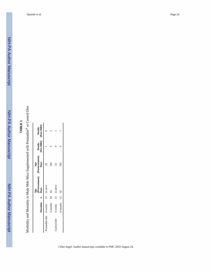
RESULTSMorbidity and mortality comparisons in mdx mice supplemented with Protandim® or controlrodent diets are summarized in Table 1. The average body weight between the Protandim®
(n = 13) and control (n = 12) rodent diet groups increased (at equivalent rates) during the 22-day period, in which body weight and health were monitored. There were no observations ofadverse events or growth impairments in the Protandim®-treated mice. In fact, one interestingincidental observation was the glossier sheen of fur seen in all mdx mice on 6 months ofProtandim diet (not shown). This incidental finding did make our blinding efforts of the twogroups important to ensure sample identifiers were coded and then blinded to subsequentanalysis. Protandim® treated mice demonstrated no difference in serum CK (data not shown).
Qureshi et al. Page 6
J Diet Suppl. Author manuscript; available in PMC 2010 August 24.
NIH
-PA Author Manuscript
NIH
-PA Author Manuscript
NIH
-PA Author Manuscript
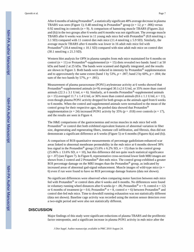
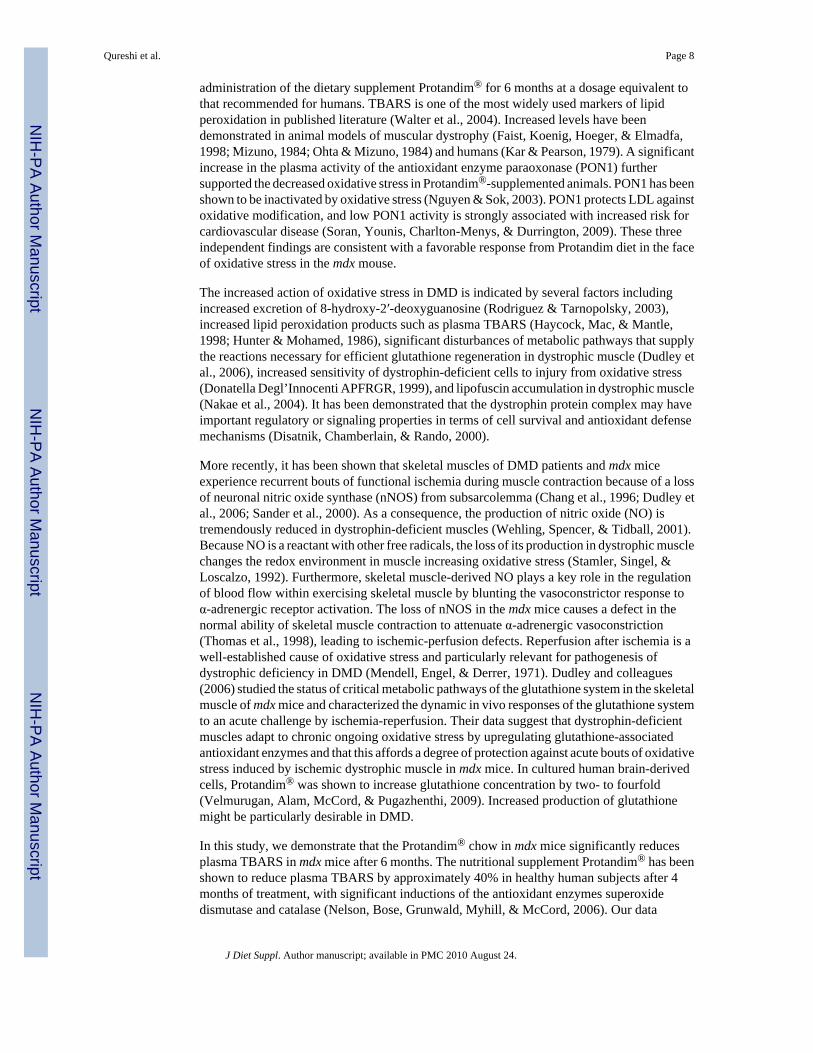
After 6 months of taking Protandim®, a statistically significant 48% average decrease in plasmaTBARS was seen (Figure 1); 0.48 nmol/mg in Protandim® group (n = 12; p = .006) versus0.92 nmol/mg in controls (n = 9). A comparison of hamstring muscle TBARS (Figures 2(a)and (b)) in the two groups after 6 weeks and 6 months was not significant. The average muscleTBARS after 6 weeks was lower in 11 young mdx mice fed with Protandim® (8.8 nmol/mg ±3.1 SD) compared with 11 control diet mdx mice (11.4 nmol/mg ± 5.9 SD). Similarly, theaverage muscle TBARS after 6 months was lower in 16 adult mdx mice fed withProtandim® (18.4 nmol/mg ± 10.1 SD) compared with nine adult mdx mice on control diet(30.1 nmol/mg ± 21.3 SD).
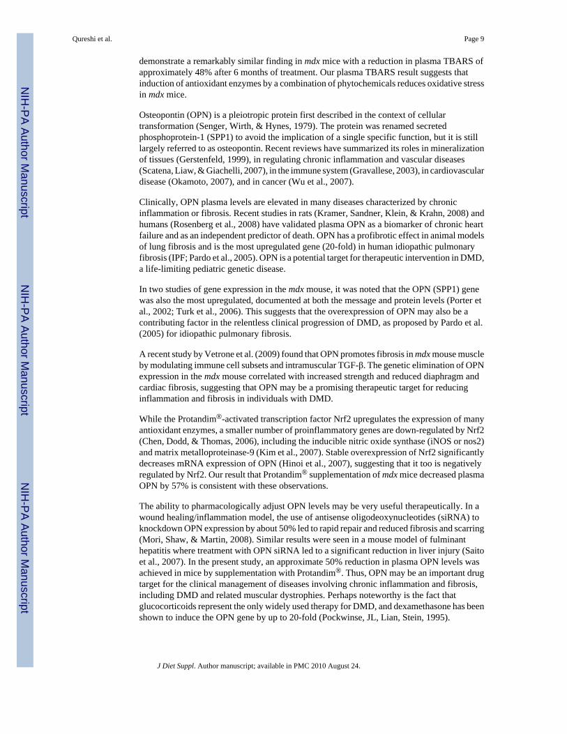
Western blot analysis for OPN in plasma samples from mdx mice maintained for 6 months oncontrol (n = 11) or Protandim®-supplemented (n = 15) diets revealed two bands: band 1 at 50kDa and band 2 at 25 kDa. The bands were scanned and digitally integrated, and the resultsare shown in Figure 3. Both bands were reduced in intensity by Protandim® supplementationand to approximately the same extent (band 1 by 53%, p = .007; band 2 by 60%, p = .004; thesum of the two bands by 57%, p = .001).
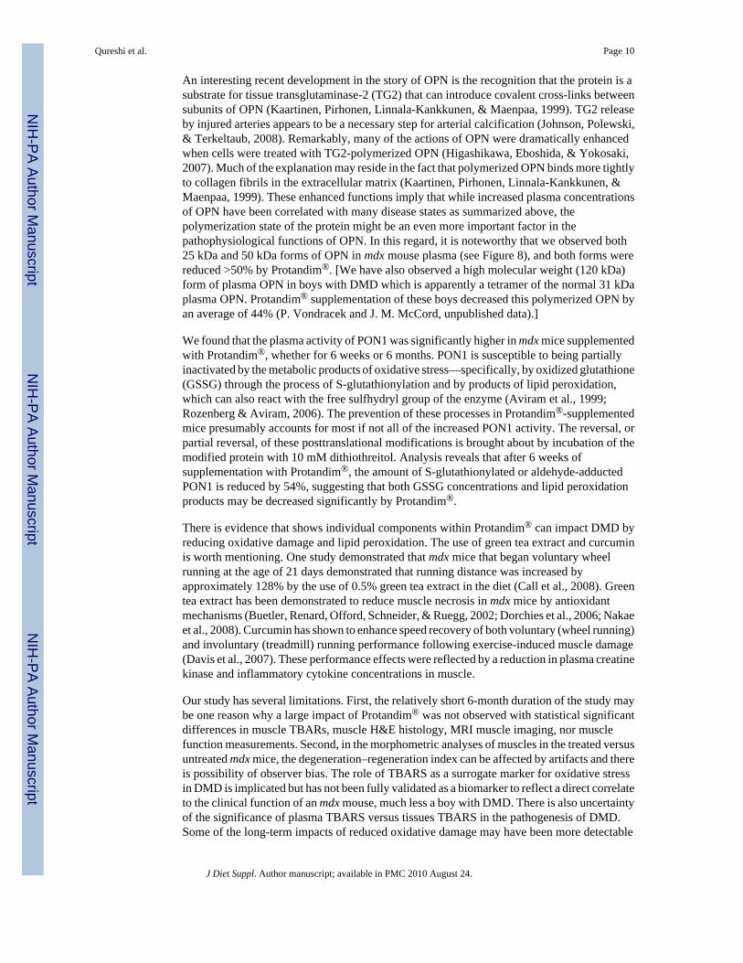
Measurement of plasma paraoxonase (PON1) arylesterase activity at 6 weeks showed thatProtandim®-supplemented animals (n=9) averaged 30.1±2.6 U/ml, or 35% more than controlanimals (22.3 ± 3.1 U/ml, n = 6). Similarly, at 6 months Protandim®-supplemented animals(n = 15) averaged 11.7 ± 1.4 U/ml, or 36% more than control animals (8.6 ± 1.1 U/ml, n = 11),even though plasma PON1 activity dropped for both groups as the animals aged from 6 weeksto 6 months. When the control and supplemented animals were normalized to the mean of thecontrol group for their respective ages, the pooled data showed that Protandim®
supplementation (n = 24) increased PON1 activity by 35% (p = .018) over controls (n = 17),and the results are seen in Figure 4.
The H&E comparisons of the gastrocnemius and rectus muscles in mdx mice fed withProtandim® or control diet both exhibited equivalent features of abnormal variation in fibersize, degenerating and regenerating fibers, immune cell infiltration, and fibrosis, thus did notdemonstrate a significant difference at 6 weeks (Figure 5) or 6 months (Figures 6(a) and (b)).
A comparison of ROI-quantitative measurements of percentage gadolinium-enhanced scarareas linked to abnormal membrane permeability in the mdx mice at 6 months showed 38%less signal in the Protandim® group (15.6% ± 6.2% SD, n = 15) than in the control group(25.06% ± 13.6% SD, n = 10), but this difference did not quite reach statistical significance(p = .07) (see Figure 7). In Figure 8, representative cross-sectional lower limb MRI images areshown from 2 control and 2 Protandim® diet mdx mice. The control group exhibited a greaterROI percentage damage on the MRI images than the Protandim® group, as indicated byincreased areas of abnormal gad-signal enhancement. Muscle images of wild-type mice (n =6) even if run were found to have no ROI percentage damage features (data not shown).
No significant differences were observed when comparing motor function between mdx micefed with Protandim® or control diets after 6 weeks and 6 months. No differences were foundin voluntary running wheel distances after 6 weeks (p = .06; Protandim® n = 9, control n = 12)or 6 months of treatment (p = 0.6; Protandim® n = 8, control n = 6) between Protandim® andcontrol diet-fed mdx mice. Time to downhill running exhaustion was not statistically different(data not shown). Baseline cage activity was recorded using the motion sensor detectors overa two-night period and were also not statistically different.
DISCUSSIONMajor findings of this study were significant reductions of plasma TBARS and the profibroticfactor osteopontin, and a significant increase in plasma PON1 activity in mdx mice after the
Qureshi et al. Page 7
J Diet Suppl. Author manuscript; available in PMC 2010 August 24.
NIH
-PA Author Manuscript
NIH
-PA Author Manuscript
NIH
-PA Author Manuscript
administration of the dietary supplement Protandim® for 6 months at a dosage equivalent tothat recommended for humans. TBARS is one of the most widely used markers of lipidperoxidation in published literature (Walter et al., 2004). Increased levels have beendemonstrated in animal models of muscular dystrophy (Faist, Koenig, Hoeger, & Elmadfa,1998; Mizuno, 1984; Ohta & Mizuno, 1984) and humans (Kar & Pearson, 1979). A significantincrease in the plasma activity of the antioxidant enzyme paraoxonase (PON1) furthersupported the decreased oxidative stress in Protandim®-supplemented animals. PON1 has beenshown to be inactivated by oxidative stress (Nguyen & Sok, 2003). PON1 protects LDL againstoxidative modification, and low PON1 activity is strongly associated with increased risk forcardiovascular disease (Soran, Younis, Charlton-Menys, & Durrington, 2009). These threeindependent findings are consistent with a favorable response from Protandim diet in the faceof oxidative stress in the mdx mouse.
The increased action of oxidative stress in DMD is indicated by several factors includingincreased excretion of 8-hydroxy-2′-deoxyguanosine (Rodriguez & Tarnopolsky, 2003),increased lipid peroxidation products such as plasma TBARS (Haycock, Mac, & Mantle,1998; Hunter & Mohamed, 1986), significant disturbances of metabolic pathways that supplythe reactions necessary for efficient glutathione regeneration in dystrophic muscle (Dudley etal., 2006), increased sensitivity of dystrophin-deficient cells to injury from oxidative stress(Donatella Degl’Innocenti APFRGR, 1999), and lipofuscin accumulation in dystrophic muscle(Nakae et al., 2004). It has been demonstrated that the dystrophin protein complex may haveimportant regulatory or signaling properties in terms of cell survival and antioxidant defensemechanisms (Disatnik, Chamberlain, & Rando, 2000).
More recently, it has been shown that skeletal muscles of DMD patients and mdx miceexperience recurrent bouts of functional ischemia during muscle contraction because of a lossof neuronal nitric oxide synthase (nNOS) from subsarcolemma (Chang et al., 1996; Dudley etal., 2006; Sander et al., 2000). As a consequence, the production of nitric oxide (NO) istremendously reduced in dystrophin-deficient muscles (Wehling, Spencer, & Tidball, 2001).Because NO is a reactant with other free radicals, the loss of its production in dystrophic musclechanges the redox environment in muscle increasing oxidative stress (Stamler, Singel, &Loscalzo, 1992). Furthermore, skeletal muscle-derived NO plays a key role in the regulationof blood flow within exercising skeletal muscle by blunting the vasoconstrictor response toα-adrenergic receptor activation. The loss of nNOS in the mdx mice causes a defect in thenormal ability of skeletal muscle contraction to attenuate α-adrenergic vasoconstriction(Thomas et al., 1998), leading to ischemic-perfusion defects. Reperfusion after ischemia is awell-established cause of oxidative stress and particularly relevant for pathogenesis ofdystrophic deficiency in DMD (Mendell, Engel, & Derrer, 1971). Dudley and colleagues(2006) studied the status of critical metabolic pathways of the glutathione system in the skeletalmuscle of mdx mice and characterized the dynamic in vivo responses of the glutathione systemto an acute challenge by ischemia-reperfusion. Their data suggest that dystrophin-deficientmuscles adapt to chronic ongoing oxidative stress by upregulating glutathione-associatedantioxidant enzymes and that this affords a degree of protection against acute bouts of oxidativestress induced by ischemic dystrophic muscle in mdx mice. In cultured human brain-derivedcells, Protandim® was shown to increase glutathione concentration by two- to fourfold(Velmurugan, Alam, McCord, & Pugazhenthi, 2009). Increased production of glutathionemight be particularly desirable in DMD.
In this study, we demonstrate that the Protandim® chow in mdx mice significantly reducesplasma TBARS in mdx mice after 6 months. The nutritional supplement Protandim® has beenshown to reduce plasma TBARS by approximately 40% in healthy human subjects after 4months of treatment, with significant inductions of the antioxidant enzymes superoxidedismutase and catalase (Nelson, Bose, Grunwald, Myhill, & McCord, 2006). Our data
Qureshi et al. Page 8
J Diet Suppl. Author manuscript; available in PMC 2010 August 24.
NIH
-PA Author Manuscript
NIH
-PA Author Manuscript
NIH
-PA Author Manuscript
demonstrate a remarkably similar finding in mdx mice with a reduction in plasma TBARS ofapproximately 48% after 6 months of treatment. Our plasma TBARS result suggests thatinduction of antioxidant enzymes by a combination of phytochemicals reduces oxidative stressin mdx mice.
Osteopontin (OPN) is a pleiotropic protein first described in the context of cellulartransformation (Senger, Wirth, & Hynes, 1979). The protein was renamed secretedphosphoprotein-1 (SPP1) to avoid the implication of a single specific function, but it is stilllargely referred to as osteopontin. Recent reviews have summarized its roles in mineralizationof tissues (Gerstenfeld, 1999), in regulating chronic inflammation and vascular diseases(Scatena, Liaw, & Giachelli, 2007), in the immune system (Gravallese, 2003), in cardiovasculardisease (Okamoto, 2007), and in cancer (Wu et al., 2007).
Clinically, OPN plasma levels are elevated in many diseases characterized by chronicinflammation or fibrosis. Recent studies in rats (Kramer, Sandner, Klein, & Krahn, 2008) andhumans (Rosenberg et al., 2008) have validated plasma OPN as a biomarker of chronic heartfailure and as an independent predictor of death. OPN has a profibrotic effect in animal modelsof lung fibrosis and is the most upregulated gene (20-fold) in human idiopathic pulmonaryfibrosis (IPF; Pardo et al., 2005). OPN is a potential target for therapeutic intervention in DMD,a life-limiting pediatric genetic disease.
In two studies of gene expression in the mdx mouse, it was noted that the OPN (SPP1) genewas also the most upregulated, documented at both the message and protein levels (Porter etal., 2002; Turk et al., 2006). This suggests that the overexpression of OPN may also be acontributing factor in the relentless clinical progression of DMD, as proposed by Pardo et al.(2005) for idiopathic pulmonary fibrosis.
A recent study by Vetrone et al. (2009) found that OPN promotes fibrosis in mdx mouse muscleby modulating immune cell subsets and intramuscular TGF-β. The genetic elimination of OPNexpression in the mdx mouse correlated with increased strength and reduced diaphragm andcardiac fibrosis, suggesting that OPN may be a promising therapeutic target for reducinginflammation and fibrosis in individuals with DMD.
While the Protandim®-activated transcription factor Nrf2 upregulates the expression of manyantioxidant enzymes, a smaller number of proinflammatory genes are down-regulated by Nrf2(Chen, Dodd, & Thomas, 2006), including the inducible nitric oxide synthase (iNOS or nos2)and matrix metalloproteinase-9 (Kim et al., 2007). Stable overexpression of Nrf2 significantlydecreases mRNA expression of OPN (Hinoi et al., 2007), suggesting that it too is negativelyregulated by Nrf2. Our result that Protandim® supplementation of mdx mice decreased plasmaOPN by 57% is consistent with these observations.
The ability to pharmacologically adjust OPN levels may be very useful therapeutically. In awound healing/inflammation model, the use of antisense oligodeoxynucleotides (siRNA) toknockdown OPN expression by about 50% led to rapid repair and reduced fibrosis and scarring(Mori, Shaw, & Martin, 2008). Similar results were seen in a mouse model of fulminanthepatitis where treatment with OPN siRNA led to a significant reduction in liver injury (Saitoet al., 2007). In the present study, an approximate 50% reduction in plasma OPN levels wasachieved in mice by supplementation with Protandim®. Thus, OPN may be an important drugtarget for the clinical management of diseases involving chronic inflammation and fibrosis,including DMD and related muscular dystrophies. Perhaps noteworthy is the fact thatglucocorticoids represent the only widely used therapy for DMD, and dexamethasone has beenshown to induce the OPN gene by up to 20-fold (Pockwinse, JL, Lian, Stein, 1995).
Qureshi et al. Page 9
J Diet Suppl. Author manuscript; available in PMC 2010 August 24.
NIH
-PA Author Manuscript
NIH
-PA Author Manuscript
NIH
-PA Author Manuscript
An interesting recent development in the story of OPN is the recognition that the protein is asubstrate for tissue transglutaminase-2 (TG2) that can introduce covalent cross-links betweensubunits of OPN (Kaartinen, Pirhonen, Linnala-Kankkunen, & Maenpaa, 1999). TG2 releaseby injured arteries appears to be a necessary step for arterial calcification (Johnson, Polewski,& Terkeltaub, 2008). Remarkably, many of the actions of OPN were dramatically enhancedwhen cells were treated with TG2-polymerized OPN (Higashikawa, Eboshida, & Yokosaki,2007). Much of the explanation may reside in the fact that polymerized OPN binds more tightlyto collagen fibrils in the extracellular matrix (Kaartinen, Pirhonen, Linnala-Kankkunen, &Maenpaa, 1999). These enhanced functions imply that while increased plasma concentrationsof OPN have been correlated with many disease states as summarized above, thepolymerization state of the protein might be an even more important factor in thepathophysiological functions of OPN. In this regard, it is noteworthy that we observed both25 kDa and 50 kDa forms of OPN in mdx mouse plasma (see Figure 8), and both forms werereduced >50% by Protandim®. [We have also observed a high molecular weight (120 kDa)form of plasma OPN in boys with DMD which is apparently a tetramer of the normal 31 kDaplasma OPN. Protandim® supplementation of these boys decreased this polymerized OPN byan average of 44% (P. Vondracek and J. M. McCord, unpublished data).]
We found that the plasma activity of PON1 was significantly higher in mdx mice supplementedwith Protandim®, whether for 6 weeks or 6 months. PON1 is susceptible to being partiallyinactivated by the metabolic products of oxidative stress—specifically, by oxidized glutathione(GSSG) through the process of S-glutathionylation and by products of lipid peroxidation,which can also react with the free sulfhydryl group of the enzyme (Aviram et al., 1999;Rozenberg & Aviram, 2006). The prevention of these processes in Protandim®-supplementedmice presumably accounts for most if not all of the increased PON1 activity. The reversal, orpartial reversal, of these posttranslational modifications is brought about by incubation of themodified protein with 10 mM dithiothreitol. Analysis reveals that after 6 weeks ofsupplementation with Protandim®, the amount of S-glutathionylated or aldehyde-adductedPON1 is reduced by 54%, suggesting that both GSSG concentrations and lipid peroxidationproducts may be decreased significantly by Protandim®.
There is evidence that shows individual components within Protandim® can impact DMD byreducing oxidative damage and lipid peroxidation. The use of green tea extract and curcuminis worth mentioning. One study demonstrated that mdx mice that began voluntary wheelrunning at the age of 21 days demonstrated that running distance was increased byapproximately 128% by the use of 0.5% green tea extract in the diet (Call et al., 2008). Greentea extract has been demonstrated to reduce muscle necrosis in mdx mice by antioxidantmechanisms (Buetler, Renard, Offord, Schneider, & Ruegg, 2002; Dorchies et al., 2006; Nakaeet al., 2008). Curcumin has shown to enhance speed recovery of both voluntary (wheel running)and involuntary (treadmill) running performance following exercise-induced muscle damage(Davis et al., 2007). These performance effects were reflected by a reduction in plasma creatinekinase and inflammatory cytokine concentrations in muscle.
Our study has several limitations. First, the relatively short 6-month duration of the study maybe one reason why a large impact of Protandim® was not observed with statistical significantdifferences in muscle TBARs, muscle H&E histology, MRI muscle imaging, nor musclefunction measurements. Second, in the morphometric analyses of muscles in the treated versusuntreated mdx mice, the degeneration–regeneration index can be affected by artifacts and thereis possibility of observer bias. The role of TBARS as a surrogate marker for oxidative stressin DMD is implicated but has not been fully validated as a biomarker to reflect a direct correlateto the clinical function of an mdx mouse, much less a boy with DMD. There is also uncertaintyof the significance of plasma TBARS versus tissues TBARS in the pathogenesis of DMD.Some of the long-term impacts of reduced oxidative damage may have been more detectable
Qureshi et al. Page 10
J Diet Suppl. Author manuscript; available in PMC 2010 August 24.
NIH
-PA Author Manuscript
NIH
-PA Author Manuscript
NIH
-PA Author Manuscript
with other outcome measures including cardiac, respiratory function, grip strength-testing, andtotal lifespan, which were not defined in this study. Finally, the mdx mouse may not be themost affected model to test a potential DMD interventional agent such as Protandim, whichappears to exert effects through antioxidant pathways.
Despite these limitations, the study provides evidence of reduced oxidative damage, andperhaps of a reduced profibrotic state, with the use of Protandim® in mdx mice. The use ofProtandim® did not cause any untoward side effects and overall the compound appeared safe.Pharmacokinetic studies and long-term trials of Protandim® in mdx mice and humans withDMD are required to determine its impact on DMD disease progression and survival.
AcknowledgmentsThis work was conducted at the University of Colorado Denver Health Sciences Center (UCDHSC). We thank theParent Project Muscular Dystrophy (PPMD), The Sharp Family Foundation, The Lu Foundation, and The JettFoundation for research support; LifeVantage Corporation for providing Protandim® and research support; Dr. SallyNelson for technical expertise; and Dr. Paul Maclean (UCDHSC) for providing the motorized rodent treadmill. Wealso thank Dr. Natalie Serkova and Kendra Hasebrook of the University of Colorado Cancer Center Core FacilityBioimaging Suite for the muscle MRI imaging expertise. This work was supported by a grant from the National Instituteof Arthritis and Musculoskeletal and Skin Diseases (AR052308) to B.S.T.
REFERENCESArmstrong D, Browne R. The analysis of free radicals, lipid peroxides, antioxidant enzymes and
compounds related to oxidative stress as applied to the clinical chemistry laboratory. Adv Exp MedBiol 1994;366:43–58. [PubMed: 7771281]
Aviram M, Rosenblat M, Billecke S, Erogul J, Sorenson E, Bisgaier C, et al. Human serum paraoxonase(PON 1) is inactivated by oxidized low density lipoprotein and preserved by antioxidants. Free RadicBiol Med 1999;26:892–904. [PubMed: 10232833]
Bia B, Cassidy P, Young M, Rafael J, Leighton B, Davies K, et al. Decreased myocardial nNOS, increasediNOS and abnormal ECGs in mouse models of Duchenne muscular dystrophy. J Mol Cell Cardiol1999;31:1857–1862. [PubMed: 10525423]
Bogaard H, Natarajan R, Henderson S, Long C, Kraskauskas D, Smithson L, et al. Chronic pulmonaryartery pressure elevation is insufficient to explain right heart failure. Circulation 2009;120:1951–1960.[PubMed: 19884466]
Buetler TM, Renard M, Offord EA, Schneider H, Ruegg UT. Green tea extract decreases muscle necrosisin mdx mice and protects against reactive oxygen species. Am J Clin Nutr 2002 April 1;75(4):749–753. 2002. [PubMed: 11916763]
Bulfield G, Siller WG, Wight PA, Moore KJ. X chromosome-linked muscular dystrophy (mdx) in themouse. Proc Natl Acad Sci USA 1984;81(4):1189–1192. [PubMed: 6583703]
Call J, Voelker K, Wolff A, McMillan R, Evans N, Hulver M, et al. Endurance capacity in maturingmdx mice is markedly enhanced by combined voluntary wheel running and green tea extract. J ApplPhysiol 2008;105(3):923–932. [PubMed: 18583385]
Chang WJ, Iannaccone ST, Lau KS, Masters BS, McCabe TJ, McMillan K, et al. Neuronal nitric oxidesynthase and dystrophin-deficient muscular dystrophy. Proc Natl Acad Sci USA 1996;93(17):9142–9147. [PubMed: 8799168]
Chen X, Dodd G, Thomas S, Zhang X, Wasserman MA, Rovin BH, et al. Activation of Nrf2/ARE pathwayprotects endothelial cells from oxidant injury and inhibits inflammatory gene expression. Am J PhysiolHeart Circ Physiol 2006;290:H1862–H1870. [PubMed: 16339837]
Chi M, Hintz C, McKee D, Felder S, Grant N, Kaiser K, et al. Effect of Duchenne muscular dystrophyon enzymes of energy metabolism in individual muscle fibers. Metabolism 1987;36:761–767.[PubMed: 3600288]
Davis J, Murphy E, Carmichael M, Zielinski M, Groschwitz C, Brown A, et al. Curcumin effects oninflammation and performance recovery following eccentric exercise-induced muscle damage. AmJ Physiol – Regul Integr Comp Physiol 2007;292(6):R2168–R2173. [PubMed: 17332159]
Qureshi et al. Page 11
J Diet Suppl. Author manuscript; available in PMC 2010 August 24.
NIH
-PA Author Manuscript
NIH
-PA Author Manuscript
NIH
-PA Author Manuscript
Disatnik M, Chamberlain J, Rando T. Dystrophin mutations predict cellular susceptibility to oxidativestress. Muscle & Nerve 2000;23(5):784–792. [PubMed: 10797403]
Donatella Degl’Innocenti APFRGR. Oxidative stress and calcium homeostasis in dystrophic skinfibroblasts. IUBMB Life 1999;48(4):391–396. [PubMed: 10632567]
Dorchies OM, Wagner S, Vuadens O, Waldhauser K, Buetler TM, Kucera P, et al. Green tea extract andits major polyphenol (−)-epigallocatechin gallate improve muscle function in a mouse model forDuchenne muscular dystrophy. Am J Physiol Cell Physiol 2006;290(2):C616–C625. [PubMed:16403950]
Dudley RWR, Khairallah M, Mohammed S, Lands L, Des Rosiers C, Petrof BJ. Dynamic responses ofthe glutathione system to acute oxidative stress in dystrophic mouse (mdx) muscles. Am J PhysiolRegul Integr Comp Physiol 2006;291(3):R704–R710. [PubMed: 16614063]
Eckerson HW, Wyte CM, LaDu BN. The human serum paraoxonase/arylesterase polymorphism. Am JHuman Fenet 1983;35(6):1126–1138.
Emery A. Population frequencies of inherited neuromuscular diseases: a world survey. Neuromusc Disord1991;1:19–29. [PubMed: 1822774]
Faist V, Koenig J, Hoeger H, Elmadfa I. Mitochondrial oxygen consumption, lipid peroxidation andantioxidant enzyme systems in skeletal muscle of senile dystrophic mice. Pflügers Arch Eur J Physiol1998;437(1):168–171.
Gerstenfeld L. Osteopontin in skeletal tissue homeostasis: an emerging picture of the autocrine/paracrinefunctions of the extracellular matrix. J Bone Miner Res 1999;14:850–855. [PubMed: 10352092]
Gravallese E. Osteopontin: a bridge between bone and the immune system. J Clin Invest 2003;112:147–149. [PubMed: 12865402]
Hara H, Nolan P, Scott M, Bucan M, Wakayama Y, Fischbeck K. Running endurance abnormality inmdx mice. Muscle & Nerve 2002;25(2):207–211. [PubMed: 11870688]
Haycock J, Mac N, Mantle D. Differential protein oxidation in Duchenne and Becker muscular dystrophy.Neuroreport 1998;9(10):2201–2207. [PubMed: 9694200]
Higashikawa F, Eboshida A, Yokosaki Y. Enhanced biological activity of polymeric osteopontin. FEBSLett 2007;581:2697–2701. [PubMed: 17531983]
Hinoi E, Takarada T, Fujimori S, Wang L, Lenato M, Uno K, et al. Nuclear factor E2 p45-related factor2 negatively regulates chondrogenesis. Bone 2007;40:337–344. [PubMed: 17029980]
Hunter MIS, Mohamed JB. Plasma antioxidants and lipid peroxidation products in Duchenne musculardystrophy. Clin Chim Acta 1986;155(2):123–131. [PubMed: 3698311]
Jackson M, Jones D, Edwards R. Techniques for studying free radical damage in muscular dystrophy.Med Biol 1984;62(2):135–138. [PubMed: 6471931]
Joe B, Vijaykumar M, Lokesh B. Biological properties of curcumin-cellular and molecular mechanismsof action. Crit Rev Food Sci Nutr 2004;44(2):97–111. [PubMed: 15116757]
Johnson K, Polewski M, Terkeltaub R. Transglutaminase 2 is central to induction of the arterialcalcification program by smooth muscle cells. Circ Res 2008;102:529–537. [PubMed: 18202319]
Kaartinen M, Pirhonen A, Linnala-Kankkunen A, Maenpaa P. Cross-linking of osteopontin by tissuetransglutaminase increases its collagen binding properties. J Biol Chem 1999;274:1729–1735.[PubMed: 9880554]
Kar N, Pearson C. Catalase, superoxide dismutase, glutathione reductase and thiobarbituric acid-reactiveproducts in normal and dystrophin human muscle. Clin Chim Acta 1979;94:277–280. [PubMed:466816]
Kelly HW, Van Natta ML, Covar RA, Tonascia J, Green RP, Strunk RC, et al. Effect of long-termcorticosteroid use on bone mineral density in children: a prospective longitudinal assessment in thechildhood asthma management program (CAMP) study. Pediatrics 2008;122(1):e53–e61. [PubMed:18595975]
Kim B, Jeon W, Hong H, Jeon KB, Hahn JH, Kim YM, et al. The anti-inflammatory activity of Phellinuslinteus (Berk. & M.A. Curt.) is mediated through the PKCdelta/Nrf2/ARE signaling to up-regulationof heme oxygenase-1. J Ethnopharmacol 2007;113:240–247. [PubMed: 17644290]
Kishore K, Singh M. Effect of bacosides, alcoholic extract of Bacopa monniera Linn. (brahmi), onexperimental amnesia in mice. Indian J Exp Biol 2005;43(7):640–645. [PubMed: 16053272]
Qureshi et al. Page 12
J Diet Suppl. Author manuscript; available in PMC 2010 August 24.
NIH
-PA Author Manuscript
NIH
-PA Author Manuscript
NIH
-PA Author Manuscript

Kramer F, Sandner P, Klein M, Krahn T. Plasma concentrations of matrix metalloproteinase-2, tissueinhibitor of metalloproteinase-1 and osteopontin reflect severity of heart failure in DOCA-salthypertensive rat. Biomarkers 2008;13:270–281. [PubMed: 18415800]
Lang I, Deak G, Muzes G, Pronai L, Feher J. Effect of the natural bioflavonoid antioxidant silymarin onsuperoxide dismutase (SOD) activity and expression in vitro. Biotechnol Ther 1993;4(3–4):263–270.[PubMed: 8292974]
Liu J, Gu X, Robbins D, Li G, Shi R, McCond JM, et al. Protandim, a fundamentally new antioxidantapproach in chemoprevention using mouse two-stage skin carcinogenesis as a model. PLoS ONE2009;4:e5284. [PubMed: 19384424]
Mandel S, Weinreb O, Amit T, Youdim M. Cell signaling pathways in the neuroprotective actions of thegreen tea polyphenol (−)-epigallocatechin-3-gallate: implications for neurodegenerative diseases. JNeurochem 2004;88(6):1555–1569. [PubMed: 15009657]
Manzur A, Kuntzer T, Pike M, Swan A. Glucocorticoid corticosteroids for Duchenne muscular dystrophy.Cochrane Database Syst Rev 2004;2:CD003725. [PubMed: 15106215]
McArdle A, Helliwell TR, Beckett GJ, Catapano M, Davis A, Jackson MJ. Effect of propylthiouracil-induced hypothyroidism on the onset of skeletal muscle necrosis in dystrophin-deficient mdx mice.Clin Sci 1998;95(1):83–89. [PubMed: 9662489]
Mendell JR, Engel WK, Derrer EC. Duchenne muscular dystrophy: functional ischemia reproduces itscharacteristic lesions. Science 1971;172(3988):1143–1145. [PubMed: 5574520]
Mendell JR, Moxley RT, Griggs RC, Brooke MH, Fenichel GM, Miller JP, et al. Randomized, double-blind six-month trial of prednisone in Duchenne’s muscular dystrophy. N Engl J Med 1989;320(24):1592–1597. [PubMed: 2657428]
Mizuno Y. Changes in superoxide dismutase, catalase, glutathione peroxidase, and glutathione reductaseactivities and thiobarbituric acid-reactive products levels in early stages of development in dystrophicchickens. Exp Neurol 1984;84(1):58–73. [PubMed: 6705887]
Mohr S, Hallak H, de Boitte A, Lapetina EG, Brune B. Nitric Oxide-induced S-Glutathionylation andInactivation of Glyceraldehyde-3-phosphate Dehydrogenase. J Biol Chem 1999;274(14):9427–9430.[PubMed: 10092623]
Mori R, Shaw T, Martin P. Molecular mechanisms linking wound inflammation and fibrosis: knockdownof osteopontin leads to rapid repair and reduced scarring. J Exp Med. 2008
Nakae Y, Hirasaka K, Goto J, Nikawa T, Shono M, Yoshida M, et al. Subcutaneous injection, from birth,of epigallocatechin-3-gallate, a component of green tea, limits the onset of muscular dystrophy inmdx mice: a quantitative histological, immunohistochemical and electrophysiological study.Histochem Cell Biol 2008;129(4):489–501. [PubMed: 18264714]
Nakae Y, Stoward P, Kashiyama T, Shono M, Akagi A, Matsuzaki T, et al. Early onset of lipofuscinaccumulation in dystrophin-deficient skeletal muscles of DMD patients and mdx mice. J Mol Histol2004;35(5):489–499. [PubMed: 15571326]
Nelson S, Bose S, Grunwald G, Myhill P, McCord J. The induction of human superoxide dismutase andcatalase in vivo: a fundamentally new approach to antioxidant therapy. Free Radic Biol Med 2006;40(2):341–347. [PubMed: 16413416]
Nguyen S, Sok D. Oxidative inactivation of paraoxonase1, an antioxidant protein and its effect onantioxidant action. Free Radic Res 2003;37:1319–1330. [PubMed: 14753756]
Ohkawa H, Ohishi N, Yagi K. Reaction of linoleic acid hydroperoxide with thiobarbituric acid. J LipidRes 1978;19(8):1053–1057. [PubMed: 103988]
Ohkawa H, Ohishi N, Yagi K. Assay for lipid peroxides in animal tissues by thiobarbituric acid reaction.Anal Biochem 1979;95(2):351–358. [PubMed: 36810]
Ohta K, Mizuno Y. Studies on pathogenesis of muscular dystrophy: levels of thiobarbituric acid-reactiveproducts in avian muscular dystrophy. No To Shinkei 1984;36(4):333–337. [PubMed: 6743404]
Okamoto H. Osteopontin and cardiovascular system. Mol Cell Biochem 2007;300:1–7. [PubMed:17136480]
Pardo A, Gibson K, Cisneros J, Richards TJ, Yang Y, Becerril C, et al. Up-regulation and profibrotic roleof osteopontin in human idiopathic pulmonary fibrosis. PLoS Med 2005;2:e251. [PubMed:16128620]
Qureshi et al. Page 13
J Diet Suppl. Author manuscript; available in PMC 2010 August 24.
NIH
-PA Author Manuscript
NIH
-PA Author Manuscript
NIH
-PA Author Manuscript
Pockwinse S, JL S, Lian J, Stein G. Developmental stage-specific cellular responses to vitamin D andglucocorticoids during differentiation of the osteoblast phenotype: interrelationship of morphologyand gene expression by in situ hybridization. Exp Cell Res 1995;216:244–260. [PubMed: 7813627]
Porter J, Khanna S, Kaminski H, Rao JS, Merriam AP, Richmonds CR, et al. A chronic inflammatoryresponse dominates the skeletal muscle molecular signature in dystrophin-deficient mdx mice. HumMol Genet 2002;11:263–272. [PubMed: 11823445]
Ragusa RJ, Chow CK, Porter JD. Oxidative stress as a potential pathogenic mechanism in an animalmodel of Duchenne muscular dystrophy. Neuromuscul Disord 1997;7(6–7):379–386. [PubMed:9327402]
Rando TA, Disatnik M-H, Yu Y, Franco A. Muscle cells from mdx mice have an increased susceptibilityto oxidative stress. Neuromuscul Disord 1998;8(1):14–21. [PubMed: 9565986]
Rasool M, Varalakshmi P. Protective effect of Withania somnifera root powder in relation to lipidperoxidation, antioxidant status, glycoproteins and bone collagen on adjuvant-induced arthritis inrats. Fundam Clin Pharmacol 2007;21(2):157–164. [PubMed: 17391288]
Reagan-Shaw S, Nihal M, Ahmad N. Dose translation from animal to human studies revisited. FASEB2008;22:659–661.
Rodriguez M, Tarnopolsky M. Patients with dystrophinopathy show evidence of increased oxidativestress. Free Radic Biol Med 2003;34(9):1217–1220. [PubMed: 12706502]
Rosenberg M, Zugck C, Nelles M, Jvenger C, Fronk D, Remppis A, et al. Osteopontin, a new prognosticbiomarker in patients with chronic heart failure. Circ Heart Fail 2008;1:43–49. [PubMed: 19808269]
Rozenberg O, Aviram M. S-Glutathionylation regulates HDL-associated paraoxonase 1 (PON1) activity.Biochem Biophys Res Commun 2006;351:492–498. [PubMed: 17070779]
Saito Y, Kon S, Fujiwara Y, Nakayama Y, Kurotaki D, Fukudo N, et al. Osteopontin small interferingRNA protects mice from fulminant hepatitis. Hum Gene Ther 2007;18:1205–1214. [PubMed:17988193]
Sander M, Chavoshan B, Harris SA, Iannaccone ST, Stull JT, Thomas GD, et al. Functional muscleischemia in neuronal nitric oxide synthase-deficient skeletal muscle of children with Duchennemuscular dystrophy. Proc Natl Acad Sci USA 2000;97(25):13818–13823. [PubMed: 11087833]
Scatena M, Liaw L, Giachelli C. Osteopontin: a multifunctional molecule regulating chronicinflammation and vascular disease. Arterioscler Thromb Vasc Biol 2007;27:2302–2309. [PubMed:17717292]
Senger D, Wirth D, Hynes R. Transformed mammalian cells secrete specific proteins andphosphoproteins. Cell 1979;16:885–893. [PubMed: 88265]
Sicinski P, Geng Y, Ryder-Cook AS, Barnard EA, Darlison MG, Barnard PJ. The molecular basis ofmuscular dystrophy in the mdx mouse: a point mutation. Science 1989;244(4912):1578–1580.[PubMed: 2662404]
Soran H, Younis N, Charlton-Menys V, Durrington P. Variation in paraoxonase-1 activity andatherosclerosis. Curr Opin Lipidol 2009;20:265–274. [PubMed: 19550323]
Stamler JS, Singel DJ, Loscalzo J. Biochemistry of nitric oxide and its redox-activated forms. Science1992;258(5090):1898–1902. [PubMed: 1281928]
Thomas GD, Sander M, Lau KS, Huang PL, Stull JT, Victor RG. Impaired metabolic modulation ofadrenergic vasoconstriction in dystrophin-deficient skeletal muscle. Proc Natl Acad Sci USA1998;95(25):15090–15095. [PubMed: 9844020]
Turk R, Sterrenburg E, Van Der Wees C, de Megjer EJ, de Menezes RX, Groh S, et al. Commonpathological mechanisms in mouse models for muscular dystrophies. FASEB 2006;20:127–129.
Velmurugan K, Alam J, McCord J, Pugazhenthi S. Synergistic induction of heme oxygenase-1 by thecomponents of the antioxidant supplement Protandim. Free Radic Biol Med 2009;46:430–440.[PubMed: 19056485]
Vetrone S, Montecino-Rodriguez E, Kudryashova E, Kranerovo I, Hoffman EP, Liv SD, et al.Osteopontin promotes fibrosis in dystrophic mouse muscle by modulating immune cell subsets andintramuscular TGF-beta. J Clin Invest 2009;119:1583–1594. [PubMed: 19451692]
Voisin V, Sébrié C, Matecki S, Yu H, Gillet B, Ramonatxo M, et al. l-arginine improves dystrophicphenotype in mdx mice. Neurobiol Dis 2005;20(1):123–130. [PubMed: 16137573]
Qureshi et al. Page 14
J Diet Suppl. Author manuscript; available in PMC 2010 August 24.
NIH
-PA Author Manuscript
NIH
-PA Author Manuscript
NIH
-PA Author Manuscript

Walter M, Jacob R, Jeffers B, Ghadanfar M, Preston G, Buch J, et al. Serum levels of thiobarbituric acidreactive substances predict cardiovascular events in patients with stable coronary artery disease: alongitudinal analysis of the PREVENT study. J Am Coll Cardiol 2004;44:1996–2002. [PubMed:15542282]
Wehling M, Spencer MJ, Tidball JG. Anitric oxide synthase transgene ameliorates muscular dystrophyin mdx mice. J Cell Biol 2001;155(1):123–132. [PubMed: 11581289]
Wong BLY, Christopher C. Corticosteroids in Duchenne muscular dystrophy: a reappraisal. J ChildNeurol 2002;17(3):183–190. [PubMed: 12026233]
Wu C, Wu M, Chiang E, Wu CC, Chen YJ, Chen CJ, et al. Elevated plasma osteopontin associated withgastric cancer development, invasion and survival. Gut 2007;(56):782–789. [PubMed: 17148500]
Qureshi et al. Page 15
J Diet Suppl. Author manuscript; available in PMC 2010 August 24.
NIH
-PA Author Manuscript
NIH
-PA Author Manuscript
NIH
-PA Author Manuscript

FIGURE 1.Plasma TBARS in 6 months Protandim® versus control rodent diet fed mdx mice show a 48%decrease; 0.92 nmol/mg in controls (n = 9) versus 0.48 nmol/mg in Protandim® group (n = 12;p = .006).
Qureshi et al. Page 16
J Diet Suppl. Author manuscript; available in PMC 2010 August 24.
NIH
-PA Author Manuscript
NIH
-PA Author Manuscript
NIH
-PA Author Manuscript

FIGURE 2.Muscle TBARS analysis of Protandim® versus control diet fed mdx mice demonstrates that(a) average muscle TBARS after 6 weeks was lower if fed Protandim® (n = 11) (8.8 nmol/mg± 3.1 SD) compared with control diet (n = 11) (11.4 nmol/mg ± 5.9 SD) (b) average muscleTBARS after 6 months was lower in mdx mice fed with Protandim® (n = 16) (18.4 nmol/mg± 10.1 SD) compared with control diet (n = 9) (30.1 nmol/mg ± 21.3 SD).
Qureshi et al. Page 17
J Diet Suppl. Author manuscript; available in PMC 2010 August 24.
NIH
-PA Author Manuscript
NIH
-PA Author Manuscript
NIH
-PA Author Manuscript

FIGURE 3.(a) Western blot analysis for OPN in 6 month mdx mice, showing three representative animalsfrom each group with total densities near the mean for their group. (b) The mean integratedrelative densities ± SEM for the two groups are shown for each band, and for the sum of thetwo bands for each animal. An internal standard was used to normalize multiple blots.
Qureshi et al. Page 18
J Diet Suppl. Author manuscript; available in PMC 2010 August 24.
NIH
-PA Author Manuscript
NIH
-PA Author Manuscript
NIH
-PA Author Manuscript
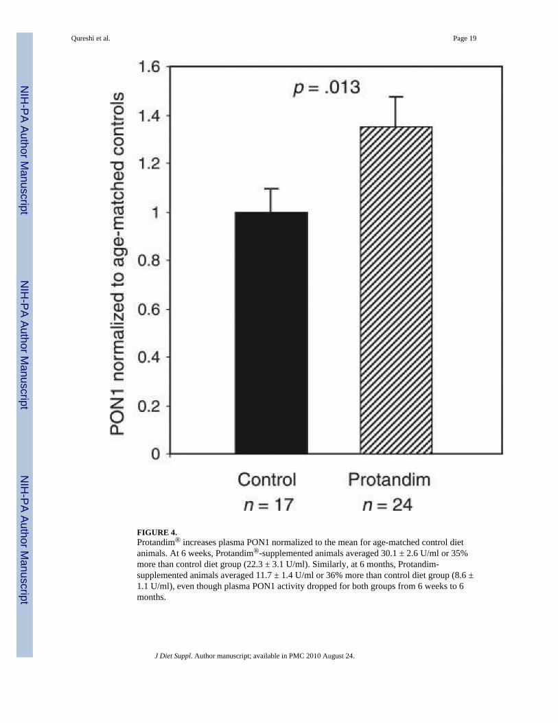
FIGURE 4.Protandim® increases plasma PON1 normalized to the mean for age-matched control dietanimals. At 6 weeks, Protandim®-supplemented animals averaged 30.1 ± 2.6 U/ml or 35%more than control diet group (22.3 ± 3.1 U/ml). Similarly, at 6 months, Protandim-supplemented animals averaged 11.7 ± 1.4 U/ml or 36% more than control diet group (8.6 ±1.1 U/ml), even though plasma PON1 activity dropped for both groups from 6 weeks to 6months.
Qureshi et al. Page 19
J Diet Suppl. Author manuscript; available in PMC 2010 August 24.
NIH
-PA Author Manuscript
NIH
-PA Author Manuscript
NIH
-PA Author Manuscript

FIGURE 5.H&E comparisons in the quadriceps muscles of Protandim® versus control rodent diet fed in6-week-old mdx mice demonstrate increased percentage degeneration–regeneration in thecontrol group (however statistically not significant).
Qureshi et al. Page 20
J Diet Suppl. Author manuscript; available in PMC 2010 August 24.
NIH
-PA Author Manuscript
NIH
-PA Author Manuscript
NIH
-PA Author Manuscript
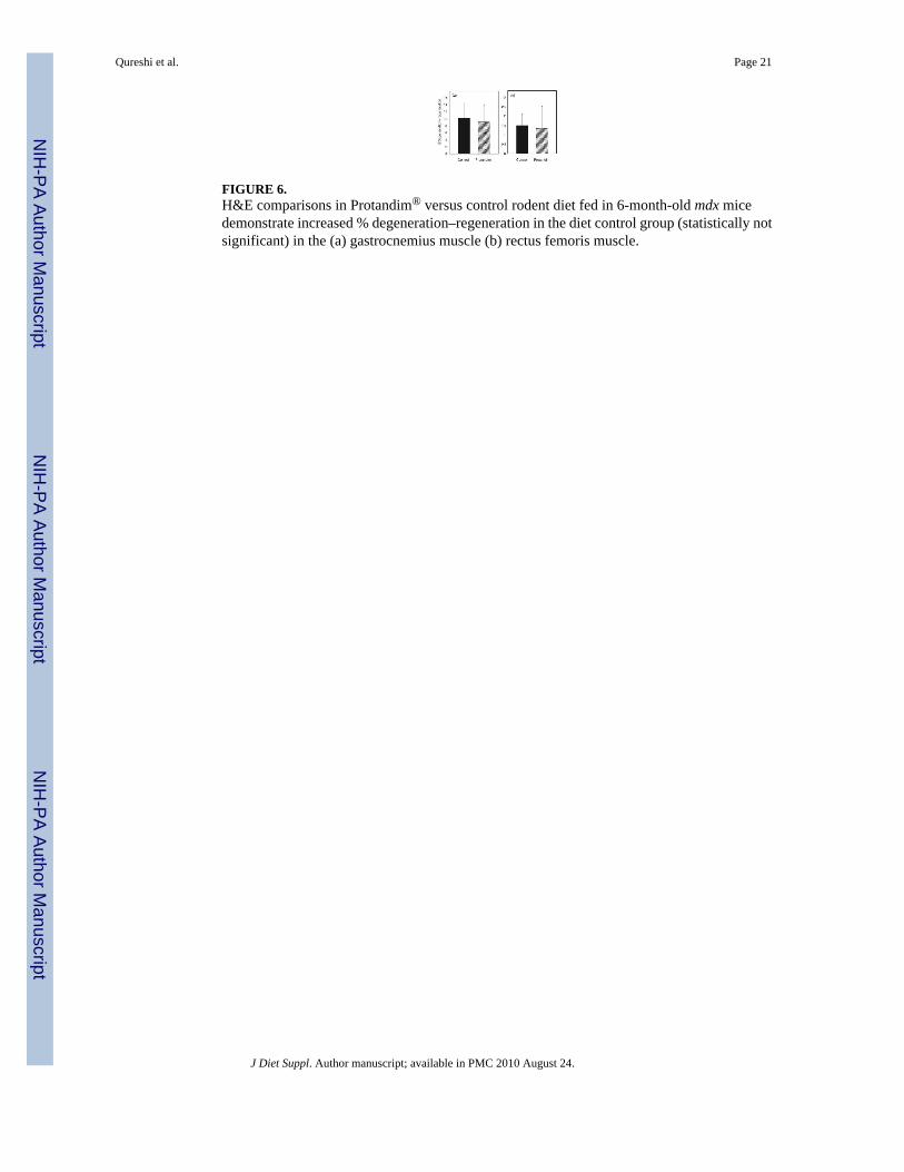
FIGURE 6.H&E comparisons in Protandim® versus control rodent diet fed in 6-month-old mdx micedemonstrate increased % degeneration–regeneration in the diet control group (statistically notsignificant) in the (a) gastrocnemius muscle (b) rectus femoris muscle.
Qureshi et al. Page 21
J Diet Suppl. Author manuscript; available in PMC 2010 August 24.
NIH
-PA Author Manuscript
NIH
-PA Author Manuscript
NIH
-PA Author Manuscript
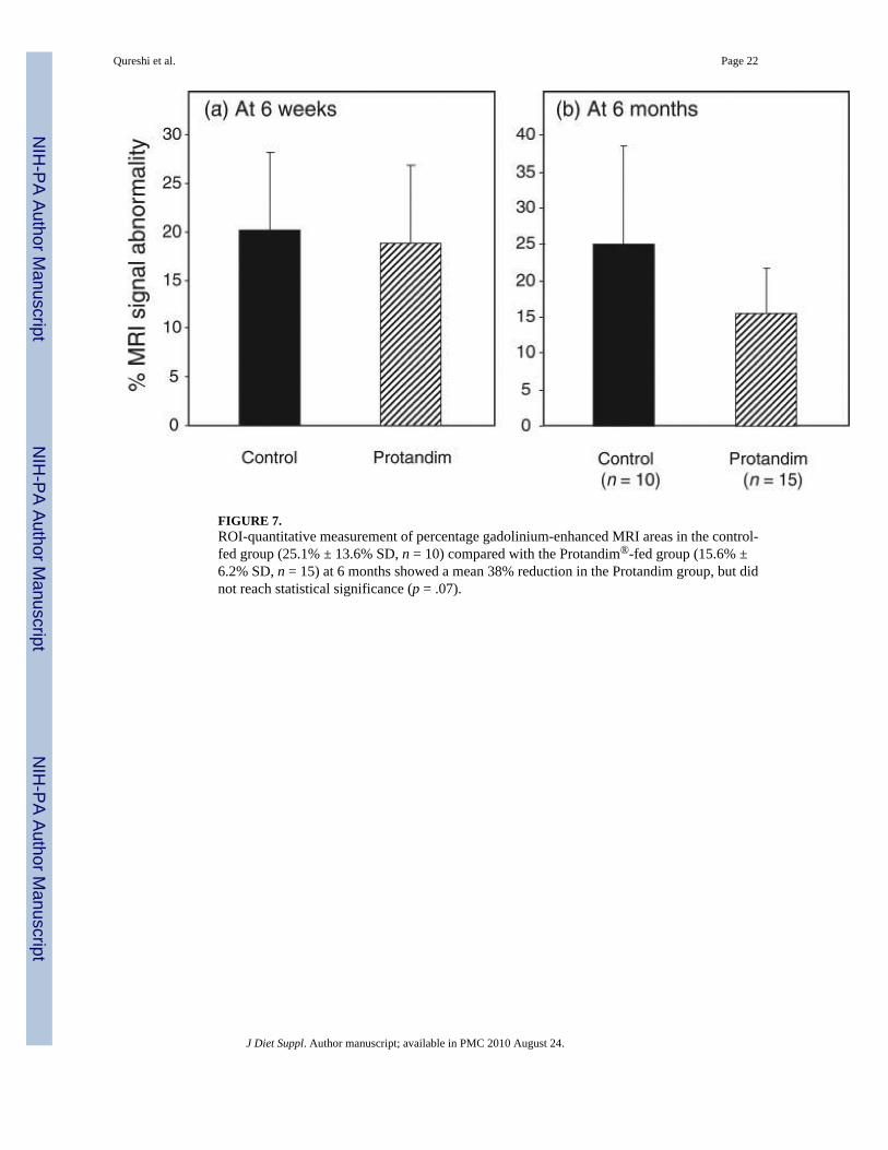
FIGURE 7.ROI-quantitative measurement of percentage gadolinium-enhanced MRI areas in the control-fed group (25.1% ± 13.6% SD, n = 10) compared with the Protandim®-fed group (15.6% ±6.2% SD, n = 15) at 6 months showed a mean 38% reduction in the Protandim group, but didnot reach statistical significance (p = .07).
Qureshi et al. Page 22
J Diet Suppl. Author manuscript; available in PMC 2010 August 24.
NIH
-PA Author Manuscript
NIH
-PA Author Manuscript
NIH
-PA Author Manuscript
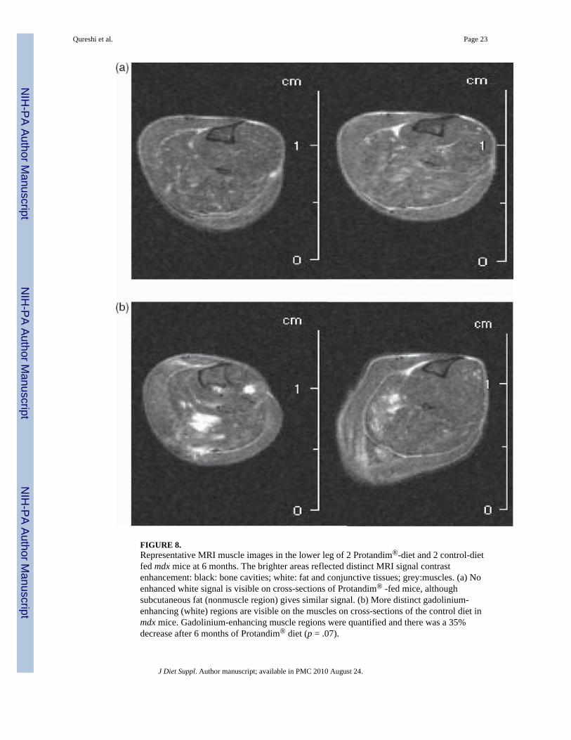
FIGURE 8.Representative MRI muscle images in the lower leg of 2 Protandim®-diet and 2 control-dietfed mdx mice at 6 months. The brighter areas reflected distinct MRI signal contrastenhancement: black: bone cavities; white: fat and conjunctive tissues; grey:muscles. (a) Noenhanced white signal is visible on cross-sections of Protandim® -fed mice, althoughsubcutaneous fat (nonmuscle region) gives similar signal. (b) More distinct gadolinium-enhancing (white) regions are visible on the muscles on cross-sections of the control diet inmdx mice. Gadolinium-enhancing muscle regions were quantified and there was a 35%decrease after 6 months of Protandim® diet (p = .07).
Qureshi et al. Page 23
J Diet Suppl. Author manuscript; available in PMC 2010 August 24.
NIH
-PA Author Manuscript
NIH
-PA Author Manuscript
NIH
-PA Author Manuscript
NIH
-PA Author Manuscript
NIH
-PA Author Manuscript
NIH
-PA Author Manuscript
Qureshi et al. Page 24
TAB
LE 1
Mor
bidi
ty a
nd M
orta
lity
in M
ale
Mdx
Mic
e Su
pple
men
ted
with
Pro
tand
im®
or C
ontro
l Die
t
Dur
atio
nn
Age
(Pre
trea
tmen
t)D
ays
Age
(Pos
ttrea
tmen
t)D
ays
Dea
ths
(Pre
-MR
I)D
eath
s(P
ost-M
RI)
Prot
andi
m d
iet
6 w
eeks
13in
ute
ro42
10
6 m
onth
s18
4218
20
2
Con
trol d
iet
6 w
eeks
12in
ute
ro42
01
6 m
onth
s12
4218
20
1
J Diet Suppl. Author manuscript; available in PMC 2010 August 24.