nih public access p. hemachandra reddy biochim … · is multiple sclerosis a mitochondrial...
TRANSCRIPT

Is multiple sclerosis a mitochondrial disease?
Peizhong Maoa and P. Hemachandra Reddya,baNeurogenetics Laboratory, Neuroscience Division, Oregon National Primate Research Center,West Campus, Oregon Health & Science University, 505 NW 185th Avenue, Beaverton, OR 97006bDepartment of Physiology and Pharmacology, Oregon Health & Science University, Portland, OR97201
AbstractMultiple sclerosis (MS) is a relatively common and etiologically unknown disease with no curetreatment. It is the leading cause of neurological disability in young adults, affecting over two millionpeople worldwide. Traditionally, MS has been considered a chronic, inflammatory disorder of thecentral white matter in which ensuing demyelination results in physical disability. Recently, MS hasbecome increasingly viewed as a neurodegenerative disorder in which axonal injury, neuronal loss,and atrophy of the central nervous system lead to permanent neurological and clinical disability. Inthis article, we discuss the latest developments on MS research, including etiology, pathology, geneticassociation, EAE animal models, mechanisms of neuronal injury and axonal transport andtherapeutics. In this article, we also focus on the mechanisms of mitochondrial dysfunction that areinvolved in MS, including mitochondrial DNA defects, and mitochondrial structural/functionalchanges.
KeywordsMultiple sclerosis; Experimental autoimmune encephalomyelitis; Mitochondria; Oxidative stress;Myelin; Neuroprotection; NO; Gender difference
1. IntroductionMultiple sclerosis (MS) is a chronic, potentially highly disabling disorder with considerablesocial impact and economic consequences. Onset of MS typically occurs during earlyadulthood, making MS the most common neurological disease affecting people under the ageof 30. It is the major cause of non-traumatic disability in young adults. The social costsassociated with MS are high because of its early age of onset, patients with MS experience anearly loss in productivity, they need assistance in performing activities of daily living, and theyrequire immunomodulatory treatments and multidisciplinary health care. Currently, nearly400,000 people are living with MS in the United States, and in the 1990s, there were at least250,000 patients with MS in the United States .
© 2009 Elsevier B.V. All rights reserved.Address for correspondence and reprint requests: P. Hemachandra Reddy, PhD, Neurogenetics Laboratory, Neuroscience Division,Oregon National Primate Research Center, West Campus, Oregon Health & Science University, 505 NW 185th Avenue, Beaverton, OR97006, Tel: 503 418 2625, Fax: 503 418 2501, E-mail: [email protected]'s Disclaimer: This is a PDF file of an unedited manuscript that has been accepted for publication. As a service to our customerswe are providing this early version of the manuscript. The manuscript will undergo copyediting, typesetting, and review of the resultingproof before it is published in its final citable form. Please note that during the production process errors may be discovered which couldaffect the content, and all legal disclaimers that apply to the journal pertain.
NIH Public AccessAuthor ManuscriptBiochim Biophys Acta. Author manuscript; available in PMC 2011 January 1.
Published in final edited form as:Biochim Biophys Acta. 2010 January ; 1802(1): 66–79. doi:10.1016/j.bbadis.2009.07.002.
NIH
-PA Author Manuscript
NIH
-PA Author Manuscript
NIH
-PA Author Manuscript

The clinical presentation of MS is heterogeneous. Main symptoms include impaired vision,extreme fatigue, spasms and paralysis of a variety of muscle systems. In the majority of cases,MS develops in an episodic fashion, with phases of clinical disease followed by recovery. Inthis form of MS, called relapsing-remitting MS (RRMS), white matter lesions can typicallydeteriorate to permanent tissue injury that is associated with neural loss and clinical disability.Over time, RRMS patients may develop chronic lesions that promote irreversible axonal injury,resulting in the conversion of RRMS to secondary progressive MS (SPMS). SPMS ischaracterized by minimal or no intermittent recovery of function. Cognitive impairment is alsocommon in MS, occurring at all stages of disease progression. Dysfunction in free recall fromlong-term memory, speed of information processing, working memory, and abstract reasoningare frequently observed in MS .
In recent years, basic research in MS has elucidated the mechanisms and processes underlyingthe disease, the development of imaging techniques (such as magnetic resonance imaging:MRI), and the development of immunomodulatory drugs which, for the first time, are alteringdisease outcome. However, basic research in MS has not help explain many disordersassociated with MS, such as depression, which is the most frequent psychiatric disorder in MSpatients. The cause of depression is multifactorial and is likely associated with psychosocialstress, focal demyelinating lesions, and immune dysfunction. Early intervention in depressioncan prevent a decline in quality of life that typically characterizes MS patients and has evenprevented suicide. Despite advances in reducing clinical symptoms in patients with MS throughthe use of immunomodulating pharmacotherapy, not all respond well to these treatments,especially when the patient is in SPMS, probably due to disease heterogeneity and multi-local,multi-cell damage throughout not only the white matter, but also the gray matter of the centralnervous system (CNS). Gray matter involvement has been detected in the earliest stages ofMS, and cortical gray matter atrophy has been found to occur at a faster rate than white matteratrophy early in disease progression, suggesting that other mechanisms may be involved inMS development and progression. This hypothesis challenges current research on MS that hasfocused on white matter. To deny or confirm this hypothesis, additional research is needed. Itargues for the development of new approaches and therapies.
The purpose of this article is 2-fold: 1) to review latest developments in MS research,particularly causal factors and therapeutic approaches, and 2) to review the mechanisms ofmitochondrial function/dysfunction including mitochondrial DNA defects, mitochondrialstructural and functional changes, mitochondrial DNA repair events, and mitochondrialtherapeutics that are involved in MS patients and EAE mouse models.
2. Etiology and Pathology of MSTo date, the exact cause of MS is still unclear, but it is believed to result from an abnormalresponse of the immune system to one or more myelin antigens that develops in geneticallysusceptible individuals after their exposure to an as-yet undefined causal agent. It has beencharacterized by an accumulation of macrophages (microglia in the brain) and lymphocytes inthe CNS (the white matter and the gray matter), leading to demyelination and destruction ofaxons. Figure 1 summarizes the possible causal factors of MS.
2.1 Genetics of MSThe identification and characterization of MS susceptibility genes likely define the basicetiology of the disease, to improve risk assessment, and to influence therapies. The past 10years have seen some progress in defining the genetic basis of MS.
The increased risk of occurrence within families indicates genetic factors may play a role inMS etiology. MS is more likely to strike siblings than the general population, and it is more
Mao and Reddy Page 2
Biochim Biophys Acta. Author manuscript; available in PMC 2011 January 1.
NIH
-PA Author Manuscript
NIH
-PA Author Manuscript
NIH
-PA Author Manuscript
likely to strike monozygotic compared to dizygotic twins. Recently, whole genome screenswere conducted in different populations and identified discrete chromosomal regionspotentially harboring MS susceptibility genes. However, with the exception of the majorhistocompatibility complex on 6p21, no single locus generated overwhelming evidence ofgenetic linkage. These results suggest a complex genetic etiology for MS, including multiplegenes of small to moderate effect and probable genetic heterogeneity. On the other hand, thehuman leukocyte antigen (HLA) was found to control immune response genes in MS, withHLA associations indicating the involvement of autoimmunity. Further, MS was one of thefirst diseases proven to be HLA-associated, primarily linked to HLA class II factors. The HLA-DRB1*1501 molecule may explain about 50% of MS cases. Furthermore, CD45 or proteintyrosine phosphatase receptor-type C (PTPRC) has been reported as a candidate in somefamilies with MS, 77C-->G PTPRC polymorphism is present and preferentially transmitted ina small subgroup of MS families, which may only be detected with complementary methodsof analysis .
Recently, large international research collaborations have provided strong evidence for theinvolvement of the polymorphism of two cytokine receptor genes in MS pathogenesis: theinterleukin 7 receptor alpha chain gene (IL7RA) on chromosome 5p13 and the interleukin 2receptor alpha chain gene (IL2RA (=CD25)) on chromosome 10p15. It is estimated that the Callele of a single nucleotide polymorphism, rs6897932, within the alternative spliced exon 6of IL7RA is involved in about 30% of MS cases. These investigations indicate that MS has astrong genetic component. Interestingly, some of these findings (such as HLA-DRB1 andIL2RA) were confirmed by recent pathway and network-based genome-wide associationstudies (GWAS). In GWAS, neural pathways, namely axon guidance and synaptic potentiation,were also over-represented in genes from MS patients. In addition to identifyingimmunological pathways previously identified, for the first time GWAS described the potentialinvolvement of neural pathways in MS susceptibility. For example, GWAS revealed morecomprehensive and extensive immune antigens, cell adhesion, and signaling moleculesassociated with MS, such as CD4, CD11b, CD58, CD82, ITGB2, and STAT3, as well asglutamate receptors, multimeric scaffold molecular DLG1, and DLG2. Using a pooling-based,genome-wide approach, and high-density, single-nucleotide polymorphisms arrays, GWASalso identified a novel risk locus for MS on chromosome 13, in addition of the HLA class IIgenes (such as HLA-DRB1) .
2.2 Virus infectionsA long-standing hypothesis about MS etiology is that MS is an infectious disease by a micro-organism. However, after decades of research, no specific infectious agents have beenidentified in MS, yet many neurologists and researchers still remain open to an infectious originfor MS. In particular, much interest has focused on a potential role for the Epstein-Barr virus(EBV). Recent findings from a population-based investigation support the implication of theEBV in MS susceptibility. It has been reported that a clinical history of infectiousmononucleosis conspicuously associated with increased MS susceptibility (). Other studieshave of progressive MS cases found the EBV present within B cells that infiltrate the meninges(membranes that envelop the CNS) and white matter - strong evidence for the involvement ofEBV in MS through B cells as triggers.
Another type of virus, corona viruses, has also been found in the brains of MS patients. Coronaviruses, important human and animal pathogens of the order Nidovirales, usually causerespiratory and gastrointestinal illnesses, including SARS (severe acute respiratory syndrome).However, their localization in the brains of MS patients indicate they may be a possible MSpathogen their neurotropism and immune system attack. Viruses have been found to induce
Mao and Reddy Page 3
Biochim Biophys Acta. Author manuscript; available in PMC 2011 January 1.
NIH
-PA Author Manuscript
NIH
-PA Author Manuscript
NIH
-PA Author Manuscript

demyelinating diseases in animals. That viruses can induce demyelinating diseases in animalsstrongly supports the hypothesis that MS may have a viral origin.
2.3 Gender Differences and Other Factors in MS SusceptibilityFemales, Caucasians, and people of northern European ancestry are at an increased risk forMS. The incidence of MS in persons of any of these 3 ancestries has considerably increasedover the last century, with the increase greatest in women. A large multicenter clinical trial ofglatiramer acetate in primary progressive multiple sclerosis indicates that there existdifferences in the rates of clinical diseases between men and women with MS. Sex dimorphismin MS may be explained by the effects of sex chromosomes and of sex steroid hormones onthe immune system, blood brain barrier, and parenchymal CNS cells. Both clinical andexperimental studies have found that sex steroid supplementation may be beneficial in MSpatients in order to reduce symptoms. Interestingly, beneficial neuroprotective effects of MSwere noted in clinical studies for elevated levels of hormones in both female and male hormones(estrogens, progesterone, and androgen), an elevation that could be related to anti-inflammatory actions on the immune system or the CNS or related to direct neuroprotectiveproperties. It should be mentioned that these actions can also be seen in estrogen receptorregulators in animal model. These observations may further stimulate current clinical studiesto determine the efficacy of and tolerance to sex steroid therapeutic approaches for MS as wellas other related diseases.
Interestingly, a gender-based method that uses sex-specific and genotype-specific primarycultures was recently established. Astrocytes, a main type of glial cells, showed sex differencesagainst oxygen-glucose deprivation (OGD). Wild-type female astrocytes were more resistantto OGD than were wild-type male cells, but this sex difference disappeared in aromataseknockout cells. In combination, these data suggest a critical role of the androgen-aromatase-estrogen network in protecting cells under stress conditions. However, sex differences inoligodendrocyte, another glial cell and the original target of MS, has not been reported.Therefore, a sex-specific oligodendrodyte study may help further our understanding of the roleof gender difference in MS etiology and in MS therapeutics.
The prevalence of MS was higher in Scandinavia, Iceland, the British Isles, and North America(∼1-2 per 1,000) than in southern Europe (with the notable exception of Sardinia). Accordingto some observers, this geographical distribution implicates an environmental disease pathogenthat may not be ubiquitously distributed. However, the geographic distribution of MS mightalso be explained, at least in part, by regional variations in genetic risk factors. Interestingly,residential or occupational exposure of MS patients to sunlight may be associated with a lowermortality rate from MS slower progression with vitamin D mediating this effect. Sinceultraviolet radiation is the principal catalyst for endogenous vitamin D3 synthesis in humans,and low levels of vitamin D3 are more common at northern latitudes than at southern latitudes,this may be another reason for persons in southern European countries having lower rates ofMS.
2.4 MS pathophysiology2.4.1 Autoimmune Attacks, Pre-active Lesions, and MS Lesions—Pro-inflammatory cytokines, such as interferon and tumor necrosis factor beta released by activatedTh1 cells may upregulate the expression of cell-surface molecules on neighboring lymphocytesand antigen-presenting cells. The binding of putative MS antigens may trigger an enhancedimmune response against the bound antigens. Such putative MS antigens include componentsof myelin, such as myelin basic protein, myelin-associated basic glycoprotein, myelinoligodendrocyte glycoprotein (MOG), proteolipid protein (PLP), and others in the trimolecular
Mao and Reddy Page 4
Biochim Biophys Acta. Author manuscript; available in PMC 2011 January 1.
NIH
-PA Author Manuscript
NIH
-PA Author Manuscript
NIH
-PA Author Manuscript

complex, the T cell receptor, and major histocompatibility complex class II molecules on APCsmay trigger either an enhanced immune response against the bound antigens .
In addition to the autoimmune response, oligodendrocyte death, axon damage, and evenneuronal loss have also been associated with MS inflammatory attacks on the CNS. However,the reason for these attacks is largely unknown, although genetic factors may influenceimmune-mediated inflammation as well as neuronal and glial survival by modulating the MSphenotype (). Therefore, autoreactive T cells are thought to be generated in response to theinterplay of (environmental) triggers and genetic susceptibility factors. Differentiation of suchCD4+ T cells results in pro-inflammatory Th1, Th17 cells and/or regulatory Th2 cells, all ofwhich produce cytokines such as interferon-gamma, IL-17, IL-4 and IL-10. After activation,myelin-specific T cells are able to cross the blood-brain- barrier via interaction of adhesionmolecules, such as vascular cell adhesion molecule-1. In the CNS, including the cerebralcortex, reactivation of these T cells involves local APCs. These APCs initiate a detrimentalcascade that typically involves the attraction of microglia, macrophages, CD8+ T cells, andplasma cells, which produce myelin-specific antibodies. It may be that, in MS, these tieredmechanisms in combination may lead to mitochondrial dysfunction, neuronal demyelination,and irreversible tissue damage characterized by axonal loss and gliosis. Recent evidenceshowed that myelin-specific T cells also recognize neuronal autoantigen in a mouse model ofMS, further indicating that multiple autoantigens may be involved in spontaneously developinghuman MS disease.
These features of tissue damage were found in brain and spinal cord tissue from classic MSlesions, termed reactive lesions. However, recently a new concept, termed preactive lesions,has been used to refer to early pathological changes that occur before the actual developmentof the reactive (active, demyelinating) lesion .
Indeed, focal disorder has been documented in normal-appearing white matter of MS patientsmonths to years before the appearance of gadolinium-enhancing lesions. Clusters of activatedmicroglia cells have been identified in these lesions through MRI and immunohistochemistry,notably in the absence of demyelination and clear leukocyte infiltration; distinguishing themfrom the traditional demyelinating active lesions and chronic active lesions [50-52]. Preactivelesions can also be seen in the grey matter, particularly in this part there may be variable degreesof demyelination, along with regions that will eventually become overtly lesion containing andareas of remyelination .
The activated state of microglia cells was also reflected by increased expression of humanleukocyte antigen-DR (HLA-DR) and CD68. In addition, foamy macrophages wereoccasionally found in some of the clusters. Together, these features strongly suggested that theprogression of MS may include a stage that actually precedes what has been termed thetraditional reactive MS lesion. Although events that give rise to preactive lesions are still to beidentified, oligodendrocyte abnormalities appear to be crucially involved (. Importantly,preactive lesions do not always develop into demyelinating lesions. Therefore, preactivelesions in MS may represent early stages in the development of MS lesions. As many of themspontaneously resolve, they are expected to hold important clues to halt the inflammatorydemyelinating process in MS. While the activation of pro-inflammatory mechanisms inmicroglia may favour disease progression, the upregulation of genes involved in anti-inflammatory and antioxidative mechanisms driven by oligodendrocytes and astrocytes mayprotect the CNS environment and thus limit lesion formation. Interestingly, a dysfunction ofmitochondria in lesions as well as in the normal-appearing white and grey matter is increasinglyrecognized in MS and could be an important determinant of axonal dysfunction anddegeneration. Together, these observations indicate that mitochondria and mitochondrial-
Mao and Reddy Page 5
Biochim Biophys Acta. Author manuscript; available in PMC 2011 January 1.
NIH
-PA Author Manuscript
NIH
-PA Author Manuscript
NIH
-PA Author Manuscript

targeted antioxidant agents may have the potential for the disease, in addition to anti-inflammatory.
2.4.2 Cellular Ionic Imbalance—Intracellular environments, especially ionic balance, arecritical for maintaining neuronal functions. Ionic imbalance has been hypothesized to be a keymechanism of MS pathophysiology. In the progression of MS, inflammatory mediators,including cytokines, oxidants, and nitric oxide, are released by microglia or are generated byhypoxia, which is secondary to tissue damage and which is believed to result in a malfunctionof oxidative metabolism in the demyelinated axon. These mediators deplete ATP and perturbmitochondrial function, causing failure of the Na+-K+ ATPase, the enzyme that is responsiblefor rapidly correcting Na+ and K+ levels and for extruding Na+ from the axon and preventinga pathological influx of Na+ in both resting and active axons .
Hypoxia is considered to be a physiological stress that induces a replication-associated DNAdamage response. It has been shown that hypoxia can inhibit Na+-K+ ATPase activity and ROSincreases Na+-K+ ATPase degredation. Even in normal appearing white matter in MS,microarray analysis revealed that transcription factor HIF-1alpha, a key regulator of hypoxia-induced gene regulation, and its downstream genes were significantly and consistentlyupregulated, indicating a hypoxia condition in this area. As shown in studies of anoxia, thehigh intra-axonal Na+ concentration that results from this failure will cause increased activityof the Na+-Ca2+ exchange channel, with the efflux of Na+ requiring a higher degree of Ca2+
influx. This, in turn, activates intra-axonal proteases, resulting in neurofilament fragmentationand perturbation of axon transport and integrity, ultimately leading to neuronal degeneration.In fact, Na+-K+ ATPase enzymatic activity and distribution were reduced or undetectable inchronic MS patients (. It appears that chronically demyelinated axons that lack Na+-K+ ATPasecannot exchange axoplasmic Na+ for K+ and are incapable of nerve transmission. Thereforeloss of axonal Na+-K+ ATPase is likely to be a major contributor to continuous neurologicaldecline in chronic stages of MS.
2.4.3 Dysfunction of cellular clearance systems—In experimental models ofdemyelinating disease in aged animals, as well as in multiple sclerosis, oligodendrocyteprecursor cells (OPCs) differentiation appears to be impaired. This is due, at least in part, tochanges in environmental signals governing remyelination. In particular, myelin debris withinlesions appears to contain powerful inhibitors of precursor cell differentiation. It has beenshown that the glycosaminoglycan hyaluronan (HA) accumulates in demyelinated lesions frompatients with MS and in mice with EAE, and that HA can prevent remyelination by inhibitingOPC maturation. Efficient removal of such molecules and myelin debris by macrophages(microglia) and other functional systems may thus facilitate OPCs differentiation and permitsuccessful remyelination of damaged axons. Interestingly, the elimination of myelin debris isextremely efficient in young animals, whereas old animals show very poor clearance of myelindebris. Systemic progesterone administration could reverse partially this age-associateddecline in CNS remyelination in male rats. These observations indicate that the inhibitors ofremyelination are increased and/or clearance systems are not efficient in aged animals andsteroid hormones and tissue/neurotrophic factors may be involved in this process. Furtheridentifying signaling molecules in this network (the myelin sheath and myelin debris) probablyrepresents very promising therapeutic targets for pharmacological strategies aimed atenhancing remyelination.
Autophagy is a newly recognized cellular functional system that delivers cytoplasmic materialsto lysosomes for degradation. The formation of autophagosomes is controlled by a specific setof autophagy-related genes, called atg genes. Autophagy is thought to be a major,evolutionarily conserved response to nutrient and bioenergetic stresses. Autophagy has beenhypothesized to remove aggregated proteins and damaged organelles, such as mitochondria.
Mao and Reddy Page 6
Biochim Biophys Acta. Author manuscript; available in PMC 2011 January 1.
NIH
-PA Author Manuscript
NIH
-PA Author Manuscript
NIH
-PA Author Manuscript
Recent studies have provided evidence that autophagy is another mechanism of programmedcell death, termed autophagic programmed cell death or secondary programmed cell death todistinguish from apoptosis, thereby possessing important housekeeping and quality-controlfunctions that contribute to health and longevity.
Autophagy also plays a role in innate and adaptive immunity, apoptosis, neurodegeneration,and aging, as well as the prevention of cancer. However, excessive or imbalanced inductionof autophagic recycling can actively contribute to neuronal atrophy, neurite degeneration, andcell death .
The role of autophagy in T cells was recently examined in mouse CD4+ T cells. Interestingly,resting naive CD4+ T cells do not contain detectable autophagosomes. Autophagy can beobserved in activated CD4+ T cells upon TCR stimulation, cytokine culturing, and prolongedserum starvation. Induction of autophagy in T cells requires JNK and the class III PI3K.Autophagy is inhibited by caspases and mammalian target of rapamycin in T cells and moreTh2 cells than Th1 cells undergo autophagy. Th2 cells become more resistant to growth factor-withdrawal cell death when autophagy is blocked using either chemical inhibitors 3-methyladenine, or by RNA interference knockdown of Atg7 and beclin. Therefore, autophagyis an important mechanism that controls homeostasis of CD4+ T cells .
Very recently, Alirezaei and colleagues examined the expression of Atg5 genes in T cells usingboth a mouse model of autoimmune demyelination as well as blood and brain tissues from MSpatients. Quantitative real-time PCR analysis of RNA isolated from blood samples of theexperimental autoimmune encephalitis (EAE) mice revealed a strong correlation between Atg5expression and clinical disability. Analysis of protein extracted from the T cells confirmed boththe upregulation and post-translational modification of Atg5 genes, the latter of which waspositively correlated with EAE severity. Analysis of RNA extracted from T cells isolated bynegative selection indicated that Atg5 expression was significantly elevated in patients withactive RRMS compared to non-diseased controls. Brain tissue sections from RRMS patients,examined by immunofluorescent histochemistry, suggested that encephalitogenic T cells maybe a source of Atg5 expression in MS brains. Together, these data suggest that increased T cellexpression of Atg5 may contribute to inflammatory demyelination in MS .
Another clearance machinery is the ubiquitin-proteasome system (UPS). The destruction ofproteins is as important as their synthesis for the maintenance of protein homeostasis in cells.In eukaryotes, the ubiquitin-proteasome system is responsible for most protein degradation:the small protein ubiquitin acts as a death warrant, tagging and targeting other proteins to thelarge proteolytic chamber of the proteasome. It is now known that ubiquitin-mediateddestruction plays a crucial part in many basic cell functions. Given the central role of UPS indiverse cellular processes, it is not surprising that its dysfunction contributes toneurodegenerative and immunological disorders, either as a primary cause or secondaryconsequence (. Importantly the proteolysis system is ATP-dependent. Recently it has beenshown that assembly of the proteasome base is a rapid yet highly orchestrated process, andproteasome regulatory particle is chaperone-mediated .
The autoimmune process of PLP139-151-induced relapsing experimental autoimmuneencephalomyelitis is regulated, in part, by the transcription factor nuclear factor (NF)-kappaB,which is activated via the UPS. Administration of PS-519, a selective inhibitor of the ubiquitin-proteasome pathway, during the remission phase of MS following an acute attack was effectivein significantly reducing the incidence of clinical relapses, CNS histopathology, and T cellresponses to both the initiating and the relapse-associated PLP epitopes. The inhibition ofclinical disease was dependent on a continuous administration of PS-519 in that recovery of Tcell function and onset of disease relapses developed within 10-14 days of drug withdrawal (.
Mao and Reddy Page 7
Biochim Biophys Acta. Author manuscript; available in PMC 2011 January 1.
NIH
-PA Author Manuscript
NIH
-PA Author Manuscript
NIH
-PA Author Manuscript

The data indicated that UPS is involved in relapsing EAE, and they suggested that targetingthe UPS, in particular the NF-kappaB, may offer a novel and efficacious approach fordecreasing progressive autoimmune diseases, including MS.
Some proteinases may be involved in the proteolysis of immune antigens and may be involvedin the progression of MS. It is generally accepted that the processing of MHC 1 antigens ismediated by UPS pathway. In addition, matrix metalloproteinase proteolysis plays a significantrole in the fragmentation of MBP. The classic MBP isoforms are predominantly expressed inthe oligodendrocytes of the CNS. A recent in vitro cleavage study determined that MBP, andits splice variants, are highly sensitive to redundant matrix metalloproteinases proteolysis.MT6-MMP (initially called leukolysin), however, was superior over all of the other MMPs incleaving the MBP isoforms. This study demonstrated that matrix metalloproteinase proteolysisof the MBP and its isoforms is a source of immunogenic peptides in autoimmune MS. If thisis the case in vivo, in some cases matrix metalloproteinase proteolysis may directly destroyMBP and initiate demyelination in MS or EAE.
Protease-activated receptors are G protein-coupled receptors that regulate the cellular responseto extracellular serine proteases. The PAR family consists of four members: PAR-1, -3, and -4as thrombin receptors, and PAR-2 as the trypsin/tryptase receptor. These four members areabundantly expressed in the brain throughout development of MS. The expression of PARs inthe brain is differentially upregulated or downregulated under pathological conditions inneurodegenerative disorders, including MS (. Noorbakhsh et al. found that PAR2 expressionwas significantly increased on astrocytes and infiltrating macrophages in human MS andmurine EAE CNS white matter. Indeed, PAR2 wild-type mice showed markedly greatermicroglial activation and T lymphocyte infiltration accompanied by worsened demyelinationand axonal injury in the CNS compared to their PAR2 knockout littermates. Enhancedneuropathological changes were associated with a more severe, progressive relapsing diseasephenotype in wild-type mice. These studies revealed pathogenic interactions between CNSPAR2 expression and neuroinflammation with ensuing demyelination and axonal injury.Therefore, PARs are capable of mediating either neurodegeneration or neuroprotection in MSas well as other neurodegenerative disorders, and they represent attractive therapeutic targetsfor treatment of these diseases.
3. Experimental autoimmune encephalitis model of MSCurrently there are no genetically engineered mouse models available to study MS progressionin mice. However, several induced mouse models have been generated, particularly EAE inmouse. The EAE can be induced by immunization of susceptible animals with a number ofmyelin antigens including myelin basic protein, PLP, and MOG. The origins of EAE date backto the 1920s, when Koritschoner and Schweinburg induced spinal cord inflammation in rabbitsby inoculating with tissue from a human spinal cord. Since then, EAE has been developed inmany different species, including rodents and primates. EAE is an animal model of MS thatexhibits the functional characteristics of human immune molecules in vivo. The ‘humanizedMS animal models allow the functional characterization of human immune molecules invivo. We emphasized that MOG although a minor component of the myelin sheath, is a potentencephalitogenic protein that induces EAE in many strains and species of experimentalanimals, particularly monkey model of MS that may closely mimic human disease, may providea unique experimental platform to understand the mechanisms of disease process, and also todevelop therapeutic strategies for MS .
It is clear that EAE can mimic many of the clinical, neuropathological, and immunologicalaspects of MS. In particular, EAE appears to mimic most closely the disability-related axonalloss seen in MS and may provide a convenient opportunity to study axon-damaging
Mao and Reddy Page 8
Biochim Biophys Acta. Author manuscript; available in PMC 2011 January 1.
NIH
-PA Author Manuscript
NIH
-PA Author Manuscript
NIH
-PA Author Manuscript

mechanisms of relevance to MS. More importantly, EAE has led directly to the developmentof three therapies approved for use in MS: glatiramer acetate (copaxone), mitoxantrone, andnatalizumab. Several new approaches to studying MS in clinical trials have also been basedon preclinical work relying on EAE.
There are a few limitations in using EAE as a research model for MS. First, MS is a spontaneousdisease, while EAE is induced by active sensitization with brain tissue antigens and strongimmune adjuvants. Second, genetic heterogeneity of MS in the human populations. Tounderstand the disease progression and pathology of MS in mice, it is important to studymultiple mouse models of EAE that may provide more human MS features. Therefore, whenused appropriately, the EAE model provides a crucial tool for improving our understanding ofand treatment of MS.
In contrast, the compelling MS in vitro model has not been developed thus far. However, a fewrelated systems were reported for the MS/EAE mechanism study in some degree usingoligodendrocyte or neuron co-culture with microglial cells .
4. Multiple sclerosis/experimental autoimmune encephalitis is aneurodegenerative disorder
Axonal loss occurs in MS and is responsible for the permanent disability characterizing thelater chronic progressive stages of the disease. Immunohistochemistry of brain tissues showedthat the expression of amyloid precursor protein, a sensitive marker of axonal damage, occursin axons within acute MS lesions and in the active borders of less acute lesions that had notbeen identified as MS. Recently, evidence for widespread axonal damage even at the earliestclinical stages of MS has been reported, leading to the hypothesis that MS is aneurodegenerative disorder in which axonal injury, neuronal loss, and atrophy of the CNSbegin in the earliest stages of the disease and then intensify over time, even axonal loss couldbe found in normal-appearing white matter in a patient with acute MS. Such evidence has calledinto question the previously long-held hypothesis that axonal pathology is the end-stage resultof repeated inflammatory events in MS and argues strongly in favor of early neuroprotectiveintervention .
Axon loss has also been found in animal models of MS, especially in the EAE model, and hasbeen found to correlate with permanent neurological disability in the animal models. Thischronic-relapsing EAE model provides an excellent platform for two critical researchobjectives: determining mechanisms of axon loss in MS and evaluating the efficacy ofneuroprotective therapies.
5. Mitochondria dysfunction and ROS as causes of neuronal degeneration inMS5.1 Mitochondria, neurodegenerative diseases and MS
Figure 2 summarizes the involvement of mitochondrial abnormalities in patients with MS andEAE mouse models. As shown, current research revealed that the following mitochondrialabnormalities are involved in the development and progression of multiple sclerosis: 1)mitochondrial DNA defects, 2) abnormal mitochondrial gene expression, 3) defectivemitochondrial enzyme activities, 4) deficient mitochondrial DNA repair activity 5) andmitochondrial dysfunction. We propose that abnormal mitochondrial dynamics (increasedfission and decreased fusion in neurons affected by MS). Further, we also propose thatmitochondrial abnormalities and mitochondrial energy failure may impact other cellular
Mao and Reddy Page 9
Biochim Biophys Acta. Author manuscript; available in PMC 2011 January 1.
NIH
-PA Author Manuscript
NIH
-PA Author Manuscript
NIH
-PA Author Manuscript

pathways, including increased demyelination and inflammation in neurons and tissues that areaffected by multiple sclerosis. The details are given below.
Mitochondria contain the respiratory chain where energy in the form of ATP is most efficientlyproduced. The mitochondrial respiratory chain is located in the inner mitochondrial membraneand consists of five complexes (complexes I-V); the fifth complex is directly involved in ATPsynthesis. The complexes of the mitochondrial respiratory chain are made up of multiplesubunits, and all contain proteins encoded by nuclear DNA and mtDNA, except for complexII, which is entirely encoded by nuclear DNA. Neurons are highly dependent on oxidativeenergy metabolism. Axons, in particular, consume significant amounts of ATP, which it usesprimarily to fuel the sodium/potassium ATPase, or sodium pump that functions to remove thesodium ions that enter the axon during impulse activity. Mitochondria are not only the energyfactory for cells but also the seat of a number of important cellular functions, including essentialpathways of intermediate metabolism, amino acid biosynthesis, fatty acid oxidation, steroidmetabolism, calcium handling and apoptosis. Of key importance is the role of mitochondria inoxidative energy metabolism. Oxidative phosphorylation generates most of the cell’s ATP,and any impairment of the organelle’s ability to produce energy can have catastrophicconsequences, not only due to the primary loss of ATP, but also due to indirect impairment ofdownstream events. Moreover, the production of superoxide occurs mostly within themitochondria, mainly in complexes I and III, TCA cycle and conditionally in complex II .
Deficient mitochondrial metabolism may generate more reactive oxygen species (ROS) thatcan wreak havoc in the cell. Therefore, mitochondrial dysfunction is an attractive candidatefor neuronal degeneration. Impairment of mitochondrial energy metabolism is the keypathogenic factor in a number of neurodegenerative disorders, such as Alzheimer’s diseaseand Parkinson’s disease. Hence, therapeutic approaches targeting mitochondrial dysfunctionand oxidative damage in neurodegenerative diseases, including MS have great promise .
Recently, several lines of evidence suggests that mitochondrial dysfunction is present inpatients with MS. Mitochondrial DNA alterations, mitochondrial structural changes, defectivemitochondrial DNA repair events, abnormal mitochondrial enzyme activities, mitochondrialgene expressions, increased free radical production and oxidative damage have been reportedin patients with MS and EAE mouse models (Figure 2).
5.2 Mitochondrial DNA alternations in MSAge-related decline of mtDNA copy number is associated with late-onset MS. mtDNAmutations may increase the risk of MS. SNP analysis has shown that genetic variants ofcomplex I genes may influence the response of tissues to inflammation in the CNS. Further,genetic alterations in uncoupling proteins are reported to be implicated in patients with MS.Uncoupling protein 2 (UCP2) is a member of the mitochondrial proton transport family thatuncouples proton entry to the mitochondria from ATP synthesis. Vogler and colleaguesreported that the UCP2 common -866G/A promoter polymorphism is associated withsusceptibility to MS in a German population. In a study of 1,097 MS patients and 462 controlsubjects, they found the common G allele associated with disease susceptibility (p = 0.0015).The UCP2 -866G allele was correlated with lower levels of UCP2 expression in vitro and invivo. Thus, UCP2 may contribute to MS susceptibility by regulating the level of UCP2 proteinin the CNS and/or in the immune system .
Defects in mtDNA have been associated with late onset MS. Ban and colleagues (2008)sequenced the mtDNA from 159 patients with MS and completed a haplogroup analysis of 835MS patients and 1,506 controls. They found a trend towards over-representation of super-haplogroup U as the only evidence for association with MS. In a parallel analysis of nuclear-encoded mitochondrial protein genes in the same subjects, they also found a trend towards
Mao and Reddy Page 10
Biochim Biophys Acta. Author manuscript; available in PMC 2011 January 1.
NIH
-PA Author Manuscript
NIH
-PA Author Manuscript
NIH
-PA Author Manuscript
association with the complex I gene, NDUFS2. Taken together, these studies have contributedto evidence suggesting that variations in mtDNA and nuclear-encoded mitochondrial proteingenes may contribute to disease susceptibility in MS.
A study of MS patients in Europe showed that a potentially functional mtDNA SNP, nt13708G/A, was significantly associated with an increased risk of MS (P = 0.0002). The studyidentified the nt13708A variant as a allele susceptible to MS, which may suggest a role in MSpathogenesis. Recently, Vyshkina et al. discovered an association among common variants ofthe mitochondrial ND2 and ATP6 genes with both MS and systemic lupus erythematosus. Thisfinding raises the possibility of a shared mitochondrial genetic background between these twoautoimmune diseases. On the other hand, an increasing number of case reports on Leber’shereditary optic neuropathy (LHON) associated mtDNA point mutations, and some patientswith MS and LHON share the same mtDNA mutation, suggesting that mitochondrialdeterminants may contribute to genetic susceptibility in MS and LHON .
In fact, only a very small subgroup of MS patients, usually with prominent optic neuritis, maycarry pathogenic LHON mutations. This overlap between the two diseases may be related tothe association of MS with an mtDNA haplotype (a set of mtDNA polymorphisms) withinwhich pathogenic LHON mutations preferentially occur. In a recent study, 58 unrelatedBulgarian patients with RRMS and 104 randomly selected healthy individuals were analyzedfor the presence of 14 mtDNA polymorphisms determining major European haplogroups aswell as three (4216, 14 798, 13 708) secondary LHON mutations. Restriction enzyme analysis,used to screen patients and controls for common haplogroup-associated polymorphisms,showed that each of these changes which occurred in MS patients at a similar rate to controlsubjects. However, 21 of the 58 patients (36.2%) were positive for the T4 216C mutation, whileonly 11.3% of the controls carried this mutation (P < 0.01; OR = 4.38), suggesting that the4216C base substitution may be a predisposing marker for MS. These findings also supportedthe hypothesis that particular mtDNA variants may contribute to the genetic susceptibility ofsome people with MS. To further study the relationship between LHON and MS, Hwang et al.tested 20 Korean MS patients for the presence of mtDNA mutations at nucleotide (nt) 11778,and nt 14484, 3460, and 15257. However, none of the MS patients exhibited any pathogenicLHON mtDNA mutations. This result is in agreement with that of Japanese MS patients. Itmay be the case that racial characteristics may influence the association.
5.3 Mitochondrial dysfunction in MSIncreasing evidence suggests that mitochondrial dysfunction is involved in MS. Ultrastructuralanalysis of demyelinated spinal cord lesions showed dramatically reduced numbers ofmitochondria and microtubules, and demonstrated Ca2+-mediated destruction of chronicallydemyelinated axons and axonal swelling. Further, the gene expression study showed anunbalanced gene expression in MS patients. As reduced energy production is a majorcontributor to Ca2+-mediated axonal degeneration, authors focused on changes in oxidativephosphorylation and inhibitory neurotransmission. Compared with controls, 488 transcriptswere decreased and 67 were increased in the MS cortex. Twenty-six nuclear-encodedmitochondrial genes and the functional activities of mitochondrial respiratory chain complexesI and III were decreased in the MS motor cortex. Reduced mitochondrial gene expression wasspecific for neurons. In addition, synaptic components of GABAergic neurotransmission andthe density of inhibitory interneuron processes also were decreased in the MS cortex.
In addition, recently a number of mitochondrial respiratory chain proteins in active lesionsfrom acute MS was analyzed using immunohistochemistry. Functionally important defects ofmitochondrial respiratory chain complex IV [cytochrome c oxidase (COX)] including itscatalytic component (COX-I) are present in some active MS lesions (Pattern III). The lack ofimmunohistochemically detected COX-I is apparent in oligodendrocytes, hypertrophied
Mao and Reddy Page 11
Biochim Biophys Acta. Author manuscript; available in PMC 2011 January 1.
NIH
-PA Author Manuscript
NIH
-PA Author Manuscript
NIH
-PA Author Manuscript

astrocytes and axons, but not in microglia. These findings suggest that hypoxia-like tissueinjury in Pattern III MS lesions may be initiated from mitochondrial impairment. On the otherhand, in inactive areas of chronic MS lesions the complex IV activity and mitochondrial mass,judged by porin immunoreactivity, are increased within approximately half of large chronicallydemyelinated axons compared with large myelinated axons in the brain and spinal cord. Theaxon-specific mitochondrial docking protein (syntaphilin) and phosphorylated neurofilament-H were increased in chronic lesions. These results clearly indicate an adaptive change ofmitochondrial function and morphology in chronic MS.
Recently, Regenold and colleagues investigated the relationship between disturbed CNSmitochondrial energy metabolism and MS disease progression by measuring cerebrospinalfluid (CSF) concentrations of sorbitol, fructose, and lactate, all metabolites of extra-mitochondrial glucose metabolism. They found that concentrations of all three metabolites,but not concentrations of glucose or myoinositol, were significantly increased in CSF fromsecondary progressive and, to a lesser degree, relapsing-remitting patients, compared to healthycontrols. Furthermore, CSF concentrations of sorbitol and fructose (polyol pathwaymetabolites), but not lactate (anaerobic glycolysis metabolite), correlated positively andsignificantly with Expanded Disability Status Scale (EDSS) score, an index of neurologicdisability in MS patients. These findings suggest that abnormal mitochondrial glucosemetabolism is increased in MS patients and is associated with disease progression .
Interestingly, analysis of mitochondrial enzymes on human muscle showed that in people withMS, there were fewer type I fibers, and that fibers of all types were smaller and had lowersuccinate dehydrogenase (SDH, component of the respiratory chain complex II) and SDH/alpha-glycerol-phosphate dehydrogenase (GPDH) but not GPDH activities, suggesting thatmuscle in this disease is smaller and relies more on anaerobic than aerobic-oxidative energysupply than does muscle of healthy individuals. Similar to brain, muscles are also highlydependent on mitochondrial oxidative energy metabolism, so it is reasonable that there is aweaker muscle in MS patients, indicating muscle is also one of the targets of MS. In some rarecases, MS could have a mitochondrial myopathy combination, in which MRI showedwidespread white matter lesions, muscle biopsy showed ragged red fibres and COX (complexIV) deficiency, Southern blot analysis revealed a large deletion of mtDNA. Probably the severemitochondrial genomic deletion is the key cause or initiation factor for this special case.
Another interesting key issue of mitochondria must be discussed below. The mitochondrialpermeability transition leads to mitochondrial swelling, outer membrane rupture and the releaseof apoptotic mediators. The mitochondrial permeability transition pore (PTP) is thought toconsist of the adenine nucleotide translocator, a voltage-dependent anion channel, andcyclophilin D (CyPD, the Ppif gene product), a prolyl isomerase located within themitochondrial matrix. CyPD is a key regulator of the PTP and they are required for mediatingCa2+- and oxidative damage-induced cell death. In experimental animal MS disease model,EAE mice lacking CyPD showed that neurons missing CyPD, are resistant to oxidative agentsthought to be the mediators of axonal degeneration observed in both EAE and MS and havemitochondria that are able to more effectively handle elevated Ca2+. Consistent with thisneuronal resistance, animals missing CyPD are able to recover, clinically, following theinduction of EAE. These results directly implicate pathological activation of the mitochondrialPTP in the axonal damage occurring during MS, in other word, PTP and mitochondria are thecritical target of EAE, perhaps multiple sclerosis.
5.4 Oxidative stress in MSReactive oxygen species (ROS) are the by-products of cellular metabolism. Excessive ROS oran imbalance between cellular production of ROS and the ability of cells to defend againstthem is referred to as oxidative stress. Oxidative stress can cause cellular damage and
Mao and Reddy Page 12
Biochim Biophys Acta. Author manuscript; available in PMC 2011 January 1.
NIH
-PA Author Manuscript
NIH
-PA Author Manuscript
NIH
-PA Author Manuscript

subsequent cell death because the ROS oxidize critical cellular components, such as lipids,proteins, and DNA especially mitochondrial DNA. In neurodegenerative andneuroinflammatory disorders, there is evidence for a primary contribution of oxidative stressin neuronal death, as opposed to other diseases where oxidative stress more likely plays asecondary or by-stander role .
There is increasing evidence that oxidative stress is an important component in the pathogenesisof MS. The inflammatory environment in demyelinating lesions is conducive to the generationof reactive oxygen species. Macrophages and microglia are known to express myeloperoxidase(MPO) and generate ROS during myelin phagocytosis in the white matter. Recent researchinvolving the cerebral cortex in MS indicates that microglial production of ROS is also likelyto be involved in cortical demyelination. Protein kinase C (PKC) could induce an increasedproduction of ROS in mononuclear cells of patients with MS compared to those of controls,and it was predominantly or exclusively generated by PKC activated NADPH oxidase. Theconcentrations of reactive oxygen and/or nitrogen species (e.g. superoxide, nitric oxide andperoxynitrite) can increase dramatically under conditions such as inflammation, and this canoverwhelm the inherent antioxidant defences within lesions. Such oxidative and/or nitrativestress can damage the lipids, proteins and nucleic acids of cells and mitochondria, potentiallycausing cell death. Oligodendrocytes are more sensitive to oxidative and nitrative stress invitro than are astrocytes and microglia, seemingly due to a diminished capacity for antioxidantdefence, and the presence of raised risk factors. Oxidative and nitrative stress might thereforeresult in selective oligodendrocyte death, and thereby demyelination in vivo. The reactivespecies may also damage the myelin sheath, promoting its attack by macrophages/microglia.
Evidence for the existence of oxidative and nitrative stress within inflammatory demyelinatinglesions includes the presence of both lipid peroxidation and protein peroxides (proteincarbonyls), and nitrotyrosine (a marker for peroxynitrite formation). When the ROS/RNS aregenerated in MS and animal models of MS, products such as superoxide and peroxynitrite areformed that are highly toxic to both glia and neuronal cells. Kalman’s group has determinedthe level of DNA damage in MS patients using 8-hydroxy-deoxy-guanosine (8-OH-dG) as anoxidative marker, they found that a significant increase in DNA oxidation within plaquescompared to NAWM specimens in MS cerebella. A tendency for increase of oxidative markersin normal appearing cortical tissues located in the proximity of MS plaques was also observedwhen compared to those in control cortical specimens. Also, oxidative damage to mitochondrialDNA and impaired activity of mitochondrial enzyme complexes in MS lesions suggest thatinflammation can affect energy metabolism, ATP synthesis, and viability of affected cells .
A recent report showed that oxidative stress occurs in progressive as well as benign MSpatients. For example, serum diene conjugate levels (a measure of lipid peroxidation) weresignificantly elevated in MS patients, especially patients with primary progressive phenotypes.However, serum total antioxidative activity and total antiradical activity were not differentbetween MS patients and healthy controls. On the other hand, the chemical composition ofhuman cerebrospinal fluid is considered to reflect brain metabolism, there is experimentalevidence of a decrease in sulfhydryl groups (antioxidants) and increased content of productsof lipid peroxidation, such as ultraweak chemiluminescence and liposoluble fluorescence,which was higher in the CSF and plasma of MS patients than in controls, clearly pointing outthe role of oxidative stress in the pathogenesis of MS .
Recent insights into the molecular pathogenesis of progression in MS also list oxidative stressas one of main mechanisms. More recently, Van Horssen J et al reported the presence ofextensive oxidative damage to proteins, lipids, and nucleotides occurring in activedemyelinating MS lesions, predominantly in reactive astrocytes and myelin-ladenmacrophages. It is reasonable that some of structurally-damaged myelin proteins are the targets
Mao and Reddy Page 13
Biochim Biophys Acta. Author manuscript; available in PMC 2011 January 1.
NIH
-PA Author Manuscript
NIH
-PA Author Manuscript
NIH
-PA Author Manuscript

of immune system, which are recognized as foreign antigens. Hence we consider that if notall, at lease for some MS patients, oxidative stress (including nitrative stress) may be theprimary mechanism in pathogenesis.
On the other hand, antioxidant enzymes, including superoxide dismutase 1 and 2, catalase, andheme oxygenase 1, are markedly upregulated in active demyelinating MS lesions compared tonormal-appearing white matter and white matter tissue from nonneurological control brains.Enhanced antioxidant enzyme production in inflammatory MS lesions may reflect an adaptivedefense mechanism to reduce ROS-induced cellular damage. These data and observationsstrongly indicate that antioxidant therapy may be a potential treatment for MS patients.
5.5 Nitric oxide and MSAs described above, human blood macrophages, astrocytes, and microglial cells make NO. NOis present at increased concentrations in acute MS lesions and is known to have a deleteriouseffect on mitochondria. The relationship between NO and cytochrome c oxidase (mt complexIV) has been investigated at different integration levels of the enzyme, including the in situstate, such as in mouse liver mitochondria or cultured human SY5Y neuroblastoma cells.Micromolar NO rapidly inhibits cytochrome c oxidase in turnover with physiologicalsubstrates. The respiratory chain is inhibited by NO, either supplied exogenously or producedendogenously via the NO synthase activation. Inhibition of respiration is reversible, althoughit remains to be clarified whether reversibility is always full and how it depends onconcentration of and time of exposure to NO. At least under hypoxic condition, NO irreversiblyinhibits cytochrome oxidase. NO and superoxide redical combine to form peroxynitrate(ONOO-), which breaks down to form the highly reactive radicals hydroxyl radical and nitrogendioxide. In other words NO can enhance the cellular toxicity of ROS.
Peroxynitrite and other reactive nitrogen oxide species exert a toxic effect on neurons, axonsand glia cells and enhance apoptosis. In addition, they increase the blood-brain-barrier (BBB)permeability and can therefore promote invasion of inflammatory cells into the CNS. On theother hand, uric acid, a purine metabolite and peroxynitrite scavenger inhibits blood-CNS-barrier permeability changes, CNS inflammation and tissue damage in EAE and in mice withspinal cord injury. More recently the concentrations of uric acid, purine profile and creatininein samples of cerebrospinal fluid and serum of MS patients were measured in detail by HPLC.The values of all compounds assayed were significantly higher in both biological fluids of MSpatients with respect to values measured in controls. In particular, serum hypoxanthine,xanthine, uric acid and sum of oxypurines were, respectively, 3.17, 3.11, 1.23 and 1.27-foldhigher in these patients than corresponding values recorded in controls. Though differentlyfrom what previously reported, these data clearly demonstrate that all purine compounds,including uric acid, are elevated in biological fluids of MS patients. Reinforced by the trendobserved for creatinine, this corroborates the notion of sustained purine catabolism, possiblydue to imbalance in ATP homeostasis, under these pathological conditions. As observed inother pathological states, uric acid, purine compounds and creatinine, can be consideredmarkers of metabolic energy imbalance rather than of reactive oxygen species, even in MS.
Nitric oxide synthases (NOS) also play an important role under physiological as well aspathological conditions. Active iNOS enzyme has been demonstrated in astrocytes in acuteand chronic-active MS lesions at the lesion edge where de myelination is occurring. QRT-PCRanalysis detected significant upregulation of the neuronal form of NOS (nNOS), in most of theMS normal-appearing white matter tissue samples, this change together with the upregulationof HIF-1 in oligodendrocytes and neurons supports the view of oligodendrocyte and/orneuronal dysfunction in this non-lesion containing tissue as a possible primary cause .
Mao and Reddy Page 14
Biochim Biophys Acta. Author manuscript; available in PMC 2011 January 1.
NIH
-PA Author Manuscript
NIH
-PA Author Manuscript
NIH
-PA Author Manuscript

6. Development of therapeutic approaches in MS6.1 immunomodulatory treatments
The major development of the past decades is that MS has changed from an untreatable to atreatable disease. Four immunomodulatory treatments (glatiramer acetate and three interferon-b preparations) and two immunosuppressants (natalizumab and mitoxantrone) are nowapproved for the treatment of MS and allow the frequency of attacks to be diminished and theprogression of disability to be slowed, at least in the shorten medium-term. In addition, anumber of new strategies were developed for the treatment of the disease, such as novelimmunomodulators (including antibodies/immunosuppressants), therapeutic strategiestargeting leukocyte differentiation molecules, costimulatory molecules, anti-adhesionmolecules, chemotaxis, autologous stem cell transplantation, anti-infectious therapies andstrategies for neuroprotection neurorepair and remyelination (for detail, see previous reviews).Furthermore, there are good scientific rationales for the use of combination therapy in MS, andthe clinical trials are also currently underway to establish the therapeutic efficacy and safetyof various combination therapies for MS patients. Recently a small scale, open-label, 7-monthtrial of combination therapy with intramuscular interferon beta-1a and oral doxycycline, apotent inhibitor of matrix metalloproteinases, in patients with RRMS was successfullycompleted. The combination of doxycycline and interferon beta-1a treatment resulted inreductions in contrast-enhancing lesion numbers and posttreatment Expanded Disability StatusScale values (P < 0.001 for both). Totally this combination treatment was effective, safe, andwell tolerated. To evaluate the efficacy and tolerability of this combination, more importantwork is double-blind, randomized, placebo-controlled clinical trials in larger cohorts of patientswith MS. This hopeful trial may encourage related reasonable combination study in the nearfuture. In EAE animal models, some novel combination or cocktail therapy studies alreadyshowed the therapeutic efficacy. For instance, multiple Ag peptides (MAPs) containing 8 PLP(139-151) peptides arranged around a dendrimeric branched lysine core were tested toinfluence the expression and development of relapsing EAE in SJL mice. The PLP (139-151)MAPs were very efficient agents in preventing the development of clinical disease whenadministered after immunization. The treatment effect with these MAPs was peptide specificand long lasted over a 60-day observation period. Such effective cocktail agents should betested in different EAE models (including non-human primate model) prior to clinical patienttrial. Hence evaluation of specific combination therapies in the controlled setting of preclinicalstudies and clinical trials should be a priority in MS research.
6.2 Complementary and alternative medicine for MSMS is a chronic, unpredictable neurological disease that mainly affects the CNS and has noknown cure. Because of this, many people with MS often seek complementary and alternativemedicine (CAM) therapies to manage their disease symptoms. Results from the literatureshowed that around 63% of people with MS reported that they used one or more CAM therapies.The major reasons for choosing CAM were as follows: conventional treatment was noteffective, anecdotal reports of CAM’s help, and doctor referral. Common CAM therapies thatpeople use include dietary modification, nutritional and herbal supplementation, and mind-body therapies, including acupuncture and massage. There is a revival of interest among MSresearchers about the therapeutic potential of low-fat diet and essential fatty acidsupplementation in MS. The efficacy of specific vitamin supplementation remains unclear.Recently, cannabis and yoga have been studied in more controlled studies and have providedevidence that they may have some benefit. The research on CAM therapies in MS is stillexploratory, but considering peoples’ interest and common use of these therapies, furtherresearch in this area is clearly warranted .
Mao and Reddy Page 15
Biochim Biophys Acta. Author manuscript; available in PMC 2011 January 1.
NIH
-PA Author Manuscript
NIH
-PA Author Manuscript
NIH
-PA Author Manuscript

Traditional Chinese medicine (TCM) has an intrinsic system for treatment of patients. It hasbeen used for several thousands of years and today it is still an important part of the Medicinein China. Interestingly, the Journal of the Integration of Chinese Medicine and WesternMedicine showed that the most MS patients could be divided to two main types by syndrometyping according to their clinical manifestations, the Gan-Shen yin-deficiency (GSYD) typeand the both yin-yang deficiency (YYD) type. They found that the age of first attack was later,level of MBP in cerebrospinal fluid was higher, in the YYD type than those in the GSYD type.Besides, the relapsing time in GSYD type, and the blood-brain barrier index and level of MBPin YYD type showed an ascending trend (P = 0.056, 0.074, 0.093, respectively). Thedistinguishing and classification of patients with MS is the first step for treatment of patientswith the disease by using TCM. However, more important issue is randomized, well-controlledclinical trial on the MS patients. This is a difficult task but it should be completed as soon aspossible. On the other hand, the existed immunological difference between the MS patients ofGSYD type and those of YYD type indicates that traditional Chinese medicine has scientificbasis even it is not fully understood. Recently, an Iranian herbal-marine medicine, MS14, wasreported to ameliorates experimental allergic encephalomyelitis in mice. Hence the CAMexploration of both basic and clinical study is widely performed and it would be expected toprovide more useful therapeutic agents for treatment of MS patients.
6.3 Mitochondria targeted approaches in MSAs described above, multiple sclerosis is very complex, it considered to be an autoimmune andneurodegenerative disease. Current treatments for MS include immunomodulatory agents butno neuroprotective or regenerative therapy is available. Especially not all patients with MSrespond well to treatment with these agents. From various animal models, we have learned thatremyelination in the CNS is a potent neuroprotective mechanism. The knowledge aboutoligodendrocyte biology and the process of remyelination has greatly increased in recent years;however, the precise mechanisms are far from being understood, and remyelination,neurogeneration as well as neuroprotection occur only in animal models. Althoughremyelination is, in principle, also possible in the diseased MS brain, it is not clear why it failsin many MS patients. The clinical trials performed so far either failed to show an effect or wereinsufficient in design. Thus, further knowledge about the molecular mechanisms of the repairprocesses and MS pathophysiology is required to achieve the ultimate goal of a neuroprotectiveand neuroregenerative treatment in MS.
As described above, CyPD knockout mouse studies have provided evidence of direct linksbetween mitochondrial function, Ca2+ overload, and axonal destruction during EAE, and byextension, MS. Hence, compounds that are able to inhibit the PTP by inactivation of CyPDspecifically or by other mechanisms that modulate the PTP, would seem to warrantinvestigation as neuroprotective therapies in MS. Intravenous mitoxantrone (novantrone)treatment improved neurological disability and delayed progression of MS in patients withworsening relapsing-remitting or secondary-progressive disease. Regarding the actionmechanisms of this approved immunosuppressant mitoxantrone, a synthetic analog ofanthraquinone, at least in tumor cells, the mitochondrial PTP targeted mechanism is involved .
Mitochondrial DNA repair is a new and important part for neuroprotection in aging relateddiseases including MS. Multiple pathways of DNA repair have been elucidated for nuclearDNA. However, it appears that only base excision repair is functioning in mitochondria. Thisrepair pathway is responsible for the removal of most endogenous damage including alkylationdamage and oxidative damage. Interestingly, Astrocytes exhibit efficient repair, whereas, otherglial cell types and neuronal cells exhibit a reduced ability to remove lesions from mtDNA. Astrategy of targeting DNA repair proteins to mitochondria to enhance mtDNA repair capacitywas developed. Enhancement of mtDNA repair in oligodendrocytes provided protection from
Mao and Reddy Page 16
Biochim Biophys Acta. Author manuscript; available in PMC 2011 January 1.
NIH
-PA Author Manuscript
NIH
-PA Author Manuscript
NIH
-PA Author Manuscript

reactive oxygen species- and cytokine-induced apoptosis. These experiments provide a novelstrategy for protecting sensitive CNS cells and thus provide new treatment options forneurodegenerative diseases, such as MS.
As described in Pathology section, ion channels and ionic imbalance are involved in the MSinitiation and development, some channel blockers should protect axons from inflammatorymediators. In fact, Na+ channel blocker tetrodotoxin, which can block Na+ influx throughoutthe anoxic period, has been shown to preserve ATP levels, concurrent with protecting whitematter axons from NO-induced injury. Another Na+ channel blockers phenytoin and flecainidehave been shown to be protective in mouse and rat models of EAE, and the protective effecton axons and clinical improvement persists for as long as 180 days in mice treated regularlywith phenytoin .
Neurotrophic factors such as insulin-like growth factor-I, platelet-derived growth factor(PDGF), fibroblast growth factor and ciliary neurotrophic factor (CNTF) are multifunctionalgrowth factors which are found in the CNS. In vitro and in vivo studies have shown thatneurotrophic factors influence proliferation, differentiation, survival, and regeneration ofmature oligodendrocytes and oligodendroglial precursors in favor of a myelin repair. Sincemyelin breakdown is often severe in MS, the possibility of growth factors use in the treatmentof MS has been considered and recently, some have been shown to reduce lesion severity andpromote myelin regeneration in EAE. To investigate the role of endogenous CNTF ininflammatory demyelinating disease, MOG-induced EAE in CNTF-deficient and wild-typeC57BL/6 mice was studied. Disease was more severe in CNTF-deficient mice and recoverywas poor, with a 60% decrease in the number of proliferating oligodendrocyte precursor cellsand a more than 50% increase in the rate of oligodendrocyte apoptosis. In addition, vacuolardystrophy of myelin and axonal damage were more severe in CNTF-deficient mice. Thesespecific pathological features could be prevented by treatment with an antiserum against tumornecrosis factor-alpha, suggesting that endogenous CNTF may counterbalance this effect ofTNF-α. Hence CNTF modulates, in an inflammatory environment, glial cell survival and is anoutcome determinant of EAE and it may be a hopeful agent for treatment of MS patients. Morerecently the neuroprotective role of CTNF was confirmed by overexpression of CNTF in EAEmice, possibly by exerting their immunoregulatory activity, inhibiting inflammation, reducingdemyelination, and stimulating oligodendrogenesis .
Oligodendroglia are the cells that form and maintain myelin sheaths, the oligodendrogliallineage targeted approaches would be very interesting. Notably remyelination is regulated bystimulators and inhibitors. Leucine-rich repeats and Ig domain-containing, neurite outgrowthinhibitor receptor-interacting protein-1 (LINGO-1) is a potent negative regulator of axonalmyelination. Loss of LINGO-1 function by Lingo1 gene knockout or by treatment with anantibody antagonist of LINGO-1 function leads to functional recovery from EAE. Thisremyelination role is associated with enhanced OPCs differentiation. Therefore antagonism ofLINGO-1 or its pathway is a promising approach for the treatment of MS .
Since oxidative damage has been known to be involved in inflammatory and autoimmune-mediated tissue destruction in which, modulation of oxygen free radical production representsa new approach to the treatment of inflammatory and autoimmune diseases. Although a fewantioxidants showed some efficacy in animal models, there is limited and conflicting evidenceof potential therapeutic effects of antioxidants such as vitamins C and E in treating MS.Otherwise, little information is available on the effect of treatments with some antioxidants inpatients with MS .
In the initial phase of MS lesion formation, ROS are known to mediate the transendothelialmigration of monocytes and induce a dysfunction of the BBB. The beneficial effect of the
Mao and Reddy Page 17
Biochim Biophys Acta. Author manuscript; available in PMC 2011 January 1.
NIH
-PA Author Manuscript
NIH
-PA Author Manuscript
NIH
-PA Author Manuscript

antioxidant alpha-lipoic acid (LA) on these phenomena has been investigated. Interestingly,LA dose-dependently prevented the development of clinical signs in a rat model for MS, andclinical improvement was coupled to a decrease in leukocyte infiltration into the CNS, inparticular monocytes. Live cell imaging assessed that ROS are produced within minutes uponthe interaction of monocytes with brain endothelium. Monocyte adhesion to an in vitro modelof the BBB subsequently induced enhanced permeability, which could be inhibited by LA.Hence LA has a protective effect on EAE not only by affecting the migratory capacity ofmonocytes, but also by stabilization of the BBB, making LA an attractive therapeutic agentfor the treatment of MS.
As discussed above, the mitochondrial-targeted antioxidants, particularly MitoQ, SS31, CARTmay have potential role in EAE model or MS disease. Recently we discovered a potential directinteraction between neuropeptide CART and subunit B of the mitochondrial enzyme succinatedehydrogenase (SDHB). We found that CART significantly increased SDH function, mtcomplex II activity and ATP generation in purified mitochondria and primary cultured neurons.Furthermore, pretreatment with CART enhanced mitochondrial mechanisms of neuronalsurvival and prevented the decline in SDH and CII activities and ATP production after OGD.The findings suggest that CART’s neuroprotective mechanism of action may be linked topreservation of mitochondrial function and prevention of energy failure after ischemia-reperfusion injury. Importantly, CART has a transcription activity and stimulates brain-derivedneurotrophic factor production in cultured neuronal cells. More recently we found that CARThas a protective role in a mouse EAE model (unpublished data). This observation furtherindicates that mitochondrial targeted neuroprotection benefit EAE and probably MS patients.
A double-blind, placebo-controlled, parallel group designed clinical trial of the effect ofGinkgo biloba, a popular herbal medicine in China and Japan, on functional measures in MShas been completed. The Ginkgo group (patients received 240 mg per day of a Ginkgo specialextract, EGb 761 had significantly more individuals showing improvement on four or moremeasures with improvements associated with significantly larger effect sizes on measures offatigue, symptom severity, and functionality. The Ginkgo group also exhibited less fatigue atfollow-up compared with the placebo group. This exploratory pilot study also showed that noadverse events or side effects were reported. Therefore Ginkgo exerted modest beneficialeffects on select functional measures (eg, fatigue) among some individuals with MS. Eventhough the study may need verification from a multiple-center and large scale study, theexciting functional data indicate CAM and traditional medicine are worth further exploring forthe treatment of MS, as well as other related diseases.
7. Conclusions and future directionsIn summary, MS is a complicated autoimmune and neurodegenerative disease and causalfactors are still unknown. Several cellular mechanisms have been proposed, including geneticfactors, viral infections, autoimmune attack, demyelination, mitochondrial dysfunction, freeradicals production, ionic imbalance and cellular clearance system dysfunction leading to finalneuron loss. These factors work either as a primary cause or secondary consequence, howeverin many cases they work together to cause the MS disease. On-going efforts across world insearching genetic loci that may cause vast majority of patients with MS may provide someimportant clues to understand the disease process. Given the central role of the mitochondriain many important cellular functions including energy production, it is reasonable that itsdysfunction is the key contributor to neurodegenerative process of this disease. We proposethat the inflammation may be initiated by infection or by intrinsic imbalance (such as cellularenergy failure and increased oxidative stress in neurons or oligodendrocytes), and tissueresponse to this inflammation that controlled by DNA defects (in nuclear and mitochondrialgenomes) in MS patients. Therefore, mitochondrial targeted and neuroprotective treatments,
Mao and Reddy Page 18
Biochim Biophys Acta. Author manuscript; available in PMC 2011 January 1.
NIH
-PA Author Manuscript
NIH
-PA Author Manuscript
NIH
-PA Author Manuscript

or combination of neuroprotection and immunomodulatory may represent new and correctapproach of MS therapy.
AcknowledgmentsThis work was supported by grants from NIH (AG028072 AG026051) and Vertex Pharmaceuticals to P.H.R. and byAmerican Heart Association Award 0565527Z to P.M.
Mao and Reddy Page 19
Biochim Biophys Acta. Author manuscript; available in PMC 2011 January 1.
NIH
-PA Author Manuscript
NIH
-PA Author Manuscript
NIH
-PA Author Manuscript

Figure 1.The factors that may contribute to the development and progression of multiple sclerosis. Theprecise causal factors of multiple sclerosis are unknown. However, it is possible that multiplefactors are involved in causing multiple sclerosis, including DNA defects in nuclear andmitochondrial genomes, viral infection, hypoxia and oxidative stress, lack of sunlight orsufficient levels of vitamin D, and increased macrophages and lymphocytes in the brain.
Mao and Reddy Page 20
Biochim Biophys Acta. Author manuscript; available in PMC 2011 January 1.
NIH
-PA Author Manuscript
NIH
-PA Author Manuscript
NIH
-PA Author Manuscript
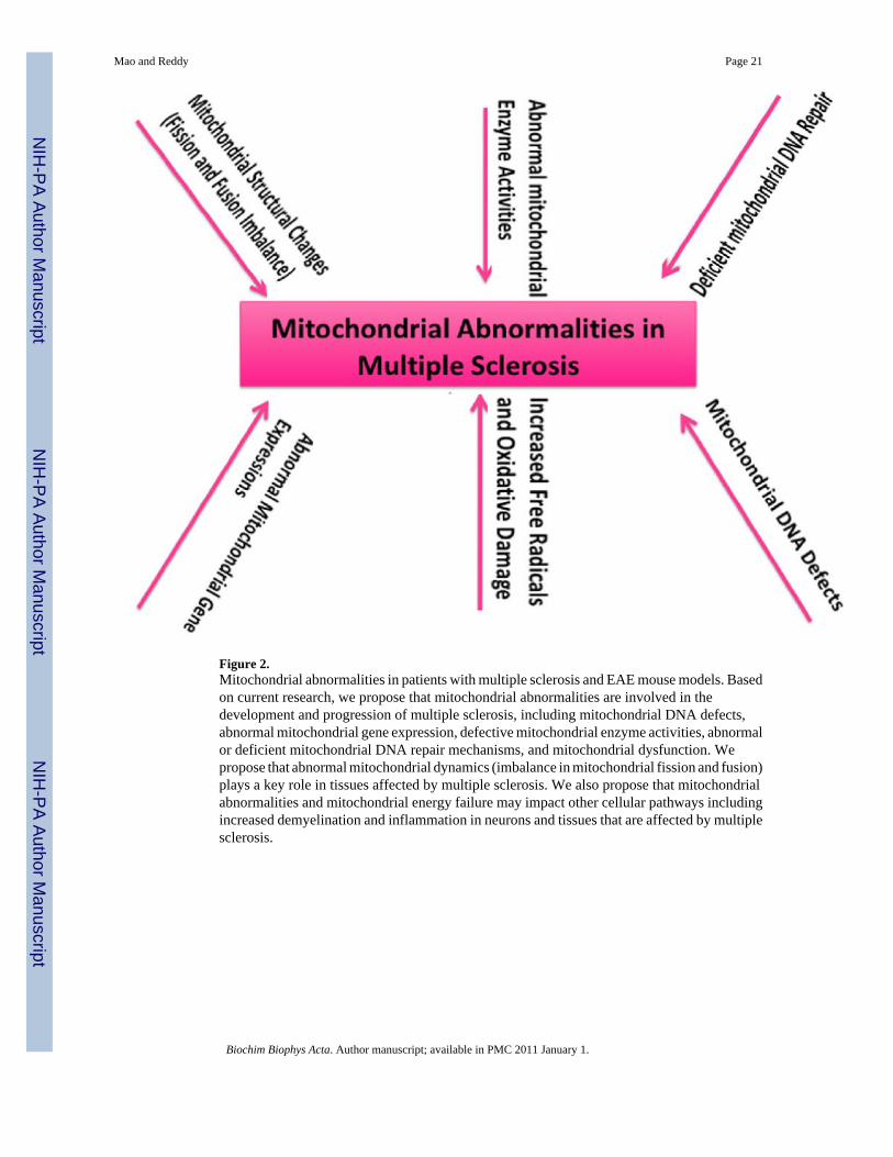
Figure 2.Mitochondrial abnormalities in patients with multiple sclerosis and EAE mouse models. Basedon current research, we propose that mitochondrial abnormalities are involved in thedevelopment and progression of multiple sclerosis, including mitochondrial DNA defects,abnormal mitochondrial gene expression, defective mitochondrial enzyme activities, abnormalor deficient mitochondrial DNA repair mechanisms, and mitochondrial dysfunction. Wepropose that abnormal mitochondrial dynamics (imbalance in mitochondrial fission and fusion)plays a key role in tissues affected by multiple sclerosis. We also propose that mitochondrialabnormalities and mitochondrial energy failure may impact other cellular pathways includingincreased demyelination and inflammation in neurons and tissues that are affected by multiplesclerosis.
Mao and Reddy Page 21
Biochim Biophys Acta. Author manuscript; available in PMC 2011 January 1.
NIH
-PA Author Manuscript
NIH
-PA Author Manuscript
NIH
-PA Author Manuscript