nlrp3/caspase-1 pathway-induced pyroptosis mediated ... · original article nlrp3/caspase-1...
TRANSCRIPT

ORIGINAL ARTICLE
NLRP3/Caspase-1 Pathway-Induced Pyroptosis MediatedCognitive Deficits in a Mouse Model of Sepsis-AssociatedEncephalopathy
Qun Fu,1,2 Jing Wu,3,4 Xiao-Yan Zhou,1 Mu-Huo Ji,5 Qing-Hong Mao,2 Qing Li,2
Man-Man Zong,1 Zhi-Qiang Zhou,1,6 and Jian-Jun Yang4,5,6
Abstract— Sepsis-associated encephalopathy (SAE) is a common complication thatleads to long-term cognitive impairments and increased mortality in sepsis survivors.The mechanisms underlying this complication remain unclear and an effective inter-vention is lacking. Accumulating evidence suggests the nucleotide-binding domain-likereceptor protein3 (NLRP3)/caspase-1 pathway is involved in several neurodegenerativediseases. Thus, we hypothesized that the NLRP3/caspase-1 pathway is involved inNLRP3-mediated pyroptosis, maturation and release of inflammatory cytokines, andcognitive deficits in SAE. We used the NLRP3 inhibitor MCC950 and the caspase-1inhibitor Ac-YVAD-CMK to study the role of the NLRP3/caspase-1 pathway inpyroptosis and cognitive deficits in a mouse model of SAE. Mice were randomlyassigned to one of six groups: sham+saline, sham+MCC950, sham+Ac-YVAD-CMK,cecal ligation and puncture (CLP)+saline, CLP+MCC950, and CLP+Ac-YVAD-CMK.Surviving mice underwent behavioral tests or had hippocampal tissues collected forhistochemical analysis and biochemical assays. Our results show that CLP-inducedhippocampus-dependent memory deficits are accompanied by increased NLRP3 andcaspase-1 positive cells, and augmented protein levels of NLRP3, caspase-1,gasdermin-D, and pro-inflammatory cytokines in the hippocampus. In addition, admin-istration of MCC950 or Ac-YVAD-CMK rescues cognitive deficits and amelioratesincreased hippocampal NLRP3-mediated neuronal pyroptosis and pro-inflammatorycytokines. Our results suggest that the NLRP3/caspase-1 pathway-induced pyroptosismediates cognitive deficits in a mouse model of SAE.
KEYWORDS: NLRP3; caspase-1; pyroptosis; pro-inflammatory cytokine; cognitive impairment.
Qun Fu and Jing Wu contributed equally to this work.
1 Department of Anesthesiology, Jinling Hospital, Medical College of Nan-jing Medical University, Nanjing, 210002, China
2 Department of Anesthesiology, Affiliated Hospital of Integrated Tradition-al Chinese and Western Medicine, Nanjing University of Chinese Medi-cine, Nanjing, 210028, China
3 Jiangsu Key Laboratory of Molecular Medicine, Medical School of Nan-jing University, Nanjing, 210093, China
4 Department of Anesthesiology, The first Affiliated Hospital of ZhengzhouUniversity, Zhengzhou, 450000, China
5 Department of Anesthesiology, ZhongdaHospital, Medical School, South-east University, Nanjing, 210009, China
6 To whom correspondence should be addressed to Zhi-Qiang Zhou atDepartment of Anesthesiology, Jinling Hospital, Medical College ofNanjing Medical University, Nanjing, 210002, China. E-mail:[email protected]; and Jian-Jun Yang at Department ofAnesthesiology, The first Affiliated Hospital of Zhengzhou University,Zhengzhou, 450000, China. E-mail: [email protected]
0360-3997/19/0100-0306/0 # 2018 The Author(s)
Inflammation, Vol. 42, No. 1, February 2019 (# 2018)DOI: 10.1007/s10753-018-0894-4
306

INTRODUCTION
Sepsis-associated encephalopathy (SAE), which ischaracterized by long-term cognitive impairments and psy-chiatric diseases in sepsis survivors, is associated withincreased morbidity and mortality [1–4]. Several mecha-nisms are involved in the pathogenesis of SAE, such asendothelial activation, disturbance of the blood-brain bar-rier, oxidative damage, neurotransmission disturbances,altered brain signaling, and neuronal apoptosis [5]. Thereis accumulating evidence that neuroinflammation plays akey role in the development of SAE [1, 6]. Nonetheless, themechanisms by which sepsis induces overactivated neuro-inflammation have yet to be elucidated.
Pyroptosis is an inflammatory form of programmedcell death and plays a protective role in host defenseduring infection. This kind of cell death is characterizedby cell swelling, lysis, and release of cytoplasmic con-tent. The initiation of pyroptosis leads to pore-mediatedcell lysis and IL-1β secretion [7]. However, excessivepyroptosis is harmful to normal tissues and cells. Toavoid host organism damage, pyroptosis is tightly regu-lated by the activation of inflammatory caspases, such ascaspase-1, caspase-4, caspase-5, and caspase-11. Thesecaspases found in the canonical and non-canonicalinflammasome signaling pathways lead to inflammatoryresponses [8–10]. However, it remains unclear how thesecaspases initiate pyroptosis.
The canonical inflammasome pathway is triggered byvarious cytoplasmic sensor proteins that recognize inflam-matory agents and multiple pathogens and recruit pro-caspase-1 monomers by the apoptosis-associated speck-like protein containing a CARD (ASC) and activatecaspase-1 through dimerization. The non-canonicalinflammasome pathway is activated by lipopolysaccharidemolecules in the cytoplasm of infected cells [11]. Thenucleotide-binding domain-like receptor protein 3(NLRP3) inflammasome is an intracellular protein com-plex that plays a crucial role in innate immune sensing.Moreover, activation of NLRP3 leads to the maturation ofcaspase-1, which mediates pyroptosis [12] and regulatesthe cleavage and maturation of pro-inflammatory cyto-kines, such as IL-1β and IL-18 [13]. However, how muchpyroptosis and increased inflammatory cytokines are in-volved in the cognitive impairments that occur during SAEremains unknown.
In light of these findings, we hypothesize that sepsistriggers NLRP3-mediated pyroptosis, neuro-inflammatoryresponses, and cognitive deficits through the NLRP3/caspase-1 pathway, and that administration of the NLRP3
inhibitor MCC950 and the caspase-1 inhibitor Ac-YVAD-CMK could attenuate hippocampal neuronal pyroptosisand neuroinflammation, reducing long-term cognitive def-icits and improving survival rates.
MATERIALS AND METHODS
Animals
One hundred and seventy-four C57BL/6 male miceaged 4 months were purchased from the Animal Center ofJinling Clinical Medical College at Nanjing Medical Uni-versity, Nanjing, China. All experimental procedures inthis study were performed according to the Guidelines forthe Care and Use of Laboratory Animals from the NationalInstitutes of Health, USA. Experiments began after micehad acclimated to the environment for 2 weeks. Mice werehoused in groups of five individuals per cage with a12:12 h light:dark cycle at a temperature of 23–25 °C withfood and water available ad libitum.
Surgical Procedures
Mice were subjected to cecal ligation and puncture(CLP) as previously described [14]. Briefly, mice wereintraperitoneally anesthetized using 2% sodium pentobar-bital (40 mg/kg; Sigma Chemical Co, St. Louis, MO,USA). Under aseptic conditions, the cecum was carefullyisolated and then ligated with 4.0 silk below the ileocecaljunction, approximately 1.2 cm from the distal end. Thececum was then perforated twice with a sterile 22-gaugeneedle and gently squeezed to extrude fecal contents intothe peritoneal cavity. After that, the cecum was returned tothe peritoneal cavity and the laparotomy incision closedwith 4.0 silk sutures. Mice subjected to sham operation hadthe cecum exposed in the same way as for CLP but it wasneither ligated nor punctured. Mice were revived immedi-ately after surgery by administering regular saline (30 ml/kg) subcutaneously and returned to their cages.
Drug Administrations
Mice were randomly divided into one of the follow-ing six groups: sham+saline (n = 18), sham+MCC950 (n =18), sham+Ac-YVAD-CMK (n = 18), CLP+saline (n =40), CLP+MCC950 (n = 40), and CLP+Ac-YVAD-CMK(n = 40). Saline, MCC950 (10 mg/kg, China Peptides Co.Ltd., China) [15], or Ac-YVAD-CMK (100 μg per mouse,Cayman Chemical Company, USA) [16] was intraperito-neally injected 30 min before surgery and on days 1, 2, 4,
307NLRP3/Caspase-1 Pathway-Induced Pyroptosis Mediated Cognitive

and 6 after surgery. Seven days after surgery, six mice ineach group were deeply anesthetized using 2% sodiumpentobarbital (60 mg/kg) and decapitated. The brain wasrapidly removed and separated into two halves for histo-chemical analysis and biochemical assays. The remainingmice in each group were used in behavioral tests 2 weeksafter surgery.
Hematoxylin and Eosin (HE) Staining
Half of each mouse brain harvested (n = 6 for eachgroup) underwent HE staining and immunohistochemicalanalysis. Brain tissues were immersed in 4% paraformal-dehyde and embedded using paraffin. The tissues weresliced into 4-mm sections until use.
HE staining was performed as follows: hematoxylinstaining for 5 min; 75% hydrochloric acid alcohol solutionfor 30 s decoloring; eosin staining for 5 min; and, 90%ethanol for 35 s decoloring. Normal neurons have a rela-tively large cell body that is rich in cytoplasm with one ortwo big round nuclei, while damaged cells show shrunkencell bodies, pyknotic nuclei, dark cytoplasm, and manyempty vesicles. Hippocampal neuronal damage was eval-uated using a standard semi-quantitative scale [17]. Inbrief, grade 0, no damage to any hippocampal subregion;grade 1, scattered neurons are damaged in the CA1 subre-gion; grade 2, moderate numbers of damaged neurons inthe CA1 subregion (< 50% neurons damaged); grade 3,severe damage (> 50% of cells affected) to pyramidal cellsin the CA1 subregion; and grade 4, extensive cell damagein all hippocampal regions. Four random high-power (×400) visual fields from each brain slice were checked.Evaluation of cells with nuclear pyknosis and morphologicabnormality was performed by two pathologists blind tothe treatment groups.
Immunohistochemistry
Paraffin sections were then deparaffinized and hydrat-ed using the following steps: 10 min in xylene twice; 5, 10,10, and 10 min in 100%, 95%, 85%, and 70% ethanol,respectively; and 5 min in PBS at room temperature re-peated three times. Antigen retrieval was achieved byboiling the sections in 10 mM sodium citrate for 10 minin a microwave oven. Sections were then washed with PBSthree times and treated with 3% H2O2-methanol for15 min. Immunostaining was performed by incubationwith antibodies against NLRP3 (1:100; Servicebio Tech-nology Co. Ltd., Wuhan, China) and caspase-1 (1:100;Servicebio Technology Co. Ltd., Wuhan, China); cellswith brownish yellow cytoplasm were recorded as positive
cells. The numbers of NLRP3 and caspase-1 immunoreac-tive cells in the hippocampal CA1 region were measuredby an investigator blind to group assignment.
Western Blotting Analysis
The hippocampus was harvested from the secondbrain half from each animal (n = 6 for each group) andhomogenized for biochemical assays. Proteins from eachhippocampus were electrophoretically separated andblotted onto nitrocellulose membranes. Protein levelswere estimated via incubation against antibodies ofNLRP3 (1:600; Servicebio Technology Co. Ltd.), ASC(1:200; Santa Cruz, USA), cleaved caspase-1 (1:500;Servicebio Technology Co. Ltd.), gasdermin-D(GSDMD, 1:500; Biorbyt, UK), IL-1β (1:200; SantaCruz, USA), IL-18 (1:200; Santa Cruz, USA), and β-actin (1:5000; Bioworld, USA). Protein bands were vi-sualized via enhanced chemiluminescence and quanti-fied using ImageQuant software (Syngene).
Enzyme-Linked Immunosorbent Assay
Hippocampal IL-1β and IL-18 levels were quantifiedusing an enzyme-linked immunosorbent assay (ELISA) kitaccording to the manufacturer’s directions (ServicebioTechnology Co. Ltd).
OpenField Test
Behavioral tests were conducted on surviving mice2 weeks after surgery in a sound-isolated room by a singleinvestigator that was blind to treatment group assignment.All tests were conducted between 14:00 and 17:00 h. Lo-comotor and exploratory activities of mice were measuredin an open-field apparatus [18]. Amouse was gently placedin the center of a white plastic chamber (50 × 50 × 30 cm)for 5 min and activities were automatically recorded usinga video tracking system (XR-XZ301, Shanghai XinruanSoft Information Technology Co. Ltd., Shanghai, China).The apparatus was cleaned with 75% alcohol between teststo remove any odor cues.
Fear Conditioning Test
Two hours after the open-field test, mice were trainedfor a fear conditioning test, as described previously [18].Eachmouse was gently placed into a conditioning chamber(XRXC404, Shanghai Soft Maze Information TechnologyCo., Ltd., Shanghai, China) and allowed to acclimate for3 min. A 30-s tone (75 dB, 3 kHz) was then deliveredfollowed by a 2-s foot shock (0.75 mA). The mouse was
308 Fu, Wu, Zhou, Ji, Mao, Li, Zong, Zhou, and Yang

kept in the chamber for another 30 s and then returned to itshome cage. A context test to evaluate hippocampus-dependent memory was performed 24 h after training.Each mouse was returned to the same test chamber for5 min without any stimulation. After the context test, eachmouse was placed for 390 s in a novel chamber altered inshape, color, and smell. The same tone was presented foranother 3 min without the foot shock to evaluatehippocampus-independent memory. Cognitive deficits inthe test was assessed by measuring the amount of time themouse demonstrated Bfreezing behavior,^which is definedas a completely immobile posture except for respiratoryefforts. Freezing behavior was automatically recorded by avideo tracking system.
Statistical Analysis
Statistical analyses were done using the software Sta-tistical Product for Social Sciences (SPSS; version 16.0,Chicago, IL, USA) and data plotted using GraphPad Prism5.0 Software (San Diego, CA, USA). Survival rate wasassessed by the Kaplan-Meier method and comparedamong treatment groups using the log-rank test. Differ-ences among groups were tested using a one-way analysisof variance followed by Tukey’s tests. Data are expressedas mean ± SEM. A p value < 0.05 was considered statisti-cally significant.
RESULTS
MCC950 or Ac-YVAD-CMK Improved Survival Rateof CLP Mice
Mouse mortality increased for 7 days after CLP,confirming the results of our previous study [14]. In thisstudy, survival rate increased with administration of intra-peritoneal MCC950 (77.5%) or Ac-YVAD-CMK (75%),compared to the CLP + saline group (52.5%; Fig. 1). Thisindicates that MCC950 and Ac-YVAD-CMK improve thesurvival rate of CLP mice.
MCC950 or Ac-YVAD-CMK AttenuatedHippocampus-Dependent Memory Impairments inSAE Mice
Mice recover with no signs of infection or motoralterations by 10 days post-CLP [19]. Thus, behavior testswere done 2 weeks after surgery. The total distance movedand time spent in the center of the arena were similaramong all six groups (Fig. 2a, b).
A fear conditioning test was used to evaluate wheth-er CLP-induced long-term memory impairments werepresent 24 h after training. The CLP+saline group had ashorter freezing time of context test than the sham+salinegroup, indicating the presence of a sepsis-induced
Fig. 1. MCC950 or Ac-YVAD-CMK improved survival rate in the first 7 days after CLP. Mice were randomly divided into six groups: sham+saline, sham+MCC950, sham+Ac-YVAD-CMK, CLP+saline, CLP+MCC950, and CLP+Ac-YVAD-CMK. Normal saline (0.25 ml per mouse), MCC950 (10 mg/kg), orAc-YVAD-CMK (100 μg per mouse) was administered to mice intraperitoneally 30 min before surgery and on days 1, 2, 4, and 6 after surgery. Survival rateto day 7 was evaluated (n = 18–40 mice/group). The asterisk indicates P < 0.05 versus the CLP+saline group.
309NLRP3/Caspase-1 Pathway-Induced Pyroptosis Mediated Cognitive

hippocampus-dependent long-term memory deficit.However, this memory deficit was ameliorated by ad-ministration of MCC950 or Ac-YVAD-CMK (Fig. 2c).No differences were observed in freezing time of the cuetest among the six groups (Fig. 2d), indicating thathippocampus-independent memory deficit was notfound in the SAE mice. Our results suggest thatMCC950 and Ac-YVAD-CMK have a therapeutic effecton CLP-induced cognitive deficits.
MCC950 or Ac-YVAD-CMK Rescued MorphologicalDamages of Hippocampus in SAE Mice
To evaluate sepsis-induced histological damageand the neuroprotective effects of MCC950 and Ac-YVAD-CMK, we did HE staining on brain sections ofthe hippocampus CA1 region 7 days after surgery. Noabnormalities in pyramidal cell morphology were ob-served in the sham-operated groups. The CLP+saline
Fig. 2. MCC950 or Ac-YVAD-CMK reversed CLP-induced learning and memory impairments. a Total ambulatory distance. b Time spent in the center. cThe contextual fear conditioning test. dThe cued fear conditioning test. Data are presented as themean ± SEM (n = 12–24mice/group). The asterisk indicatesP< 0.05 versus the sham+saline group; the pound sign, P< 0.05 versus the CLP+saline group. For the procedures of the open-field test and fear conditioningtest, see BMATERIALS AND METHODS.^
310 Fu, Wu, Zhou, Ji, Mao, Li, Zong, Zhou, and Yang
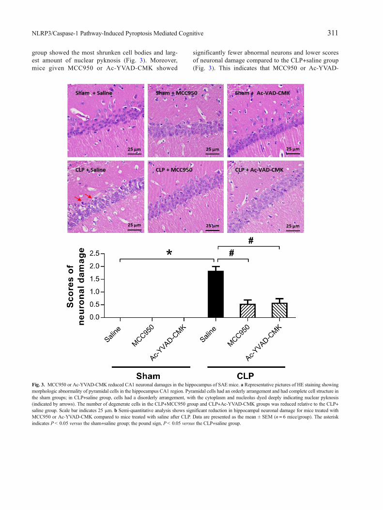
group showed the most shrunken cell bodies and larg-est amount of nuclear pyknosis (Fig. 3). Moreover,mice given MCC950 or Ac-YVAD-CMK showed
significantly fewer abnormal neurons and lower scoresof neuronal damage compared to the CLP+saline group(Fig. 3). This indicates that MCC950 or Ac-YVAD-
Fig. 3. MCC950 or Ac-YVAD-CMK reduced CA1 neuronal damages in the hippocampus of SAE mice. a Representative pictures of HE staining showingmorphologic abnormality of pyramidal cells in the hippocampus CA1 region. Pyramidal cells had an orderly arrangement and had complete cell structure inthe sham groups; in CLP+saline group, cells had a disorderly arrangement, with the cytoplasm and nucleolus dyed deeply indicating nuclear pyknosis(indicated by arrows). The number of degenerate cells in the CLP+MCC950 group and CLP+Ac-YVAD-CMK groups was reduced relative to the CLP+saline group. Scale bar indicates 25 μm. b Semi-quantitative analysis shows significant reduction in hippocampal neuronal damage for mice treated withMCC950 or Ac-YVAD-CMK compared to mice treated with saline after CLP. Data are presented as the mean ± SEM (n = 6 mice/group). The asteriskindicates P< 0.05 versus the sham+saline group; the pound sign, P< 0.05 versus the CLP+saline group.
311NLRP3/Caspase-1 Pathway-Induced Pyroptosis Mediated Cognitive

CMK can rescue hippocampal neurons from CLP-induced morphological damage.
MCC950 or Ac-YVAD-CMK Inhibited the Activationof NLRP3/Caspase-1 Pathway in the Hippocampus ofSAE Mice
The NLRP3 inflammasome, a component of the in-flammatory process, is highly expressed in various neuro-degenerative disorders [20–22]. CLP induces the activationof NLRP3 inflammasome in the hippocampus of SAEmice [14]. To explore whether MCC950 or Ac-YVAD-CMK inhibits the activation of the NLRP3/caspase-1 path-way, we measured levels of two markers of theinflammasome, NLRP3 and ASC, that may further activatecaspase-1 [12, 23]. Our results showed that the proteinlevels of NLRP3 and cleaved caspase-1 were higher at7 days post-CLP, and that this increase did not occur whenMCC950 was administered (Fig. 4). Ac-YVAD-CMKtreatment rescued the increase of cleaved caspase-1 but
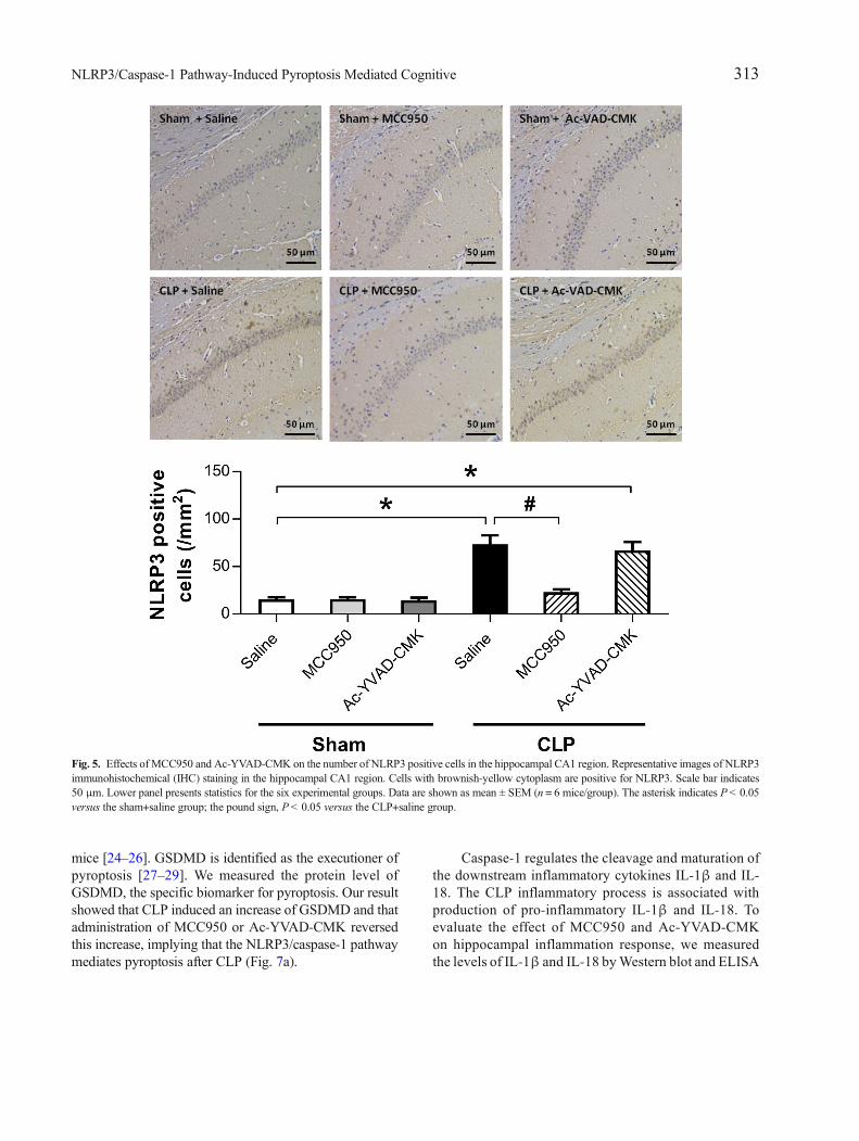
not that of NLRP3 (Fig. 4). No difference was observedin the levels of ASC among the six groups (Fig. 4). Immu-nohistochemical analysis revealed a significant increase inthe number of cells positive for NLRP3 and caspase-1 inthe CA1 region of the mouse brain at 7 days post-CLP, andthat this was reversed by administration of MCC950(Fig. 5). Ac-YVAD-CMK treatment rescued the increaseof caspase-1, but not that of NLRP3 (Fig. 6). These resultssuggest the CLP-induced activation of NLRP3/caspase-1pathway is inhibited by the specific inhibitors MCC950and Ac-YVAD-CMK.
MCC950 or Ac-YVAD-CMK Attenuated NLRP3/Caspase-1-Mediated Pyroptosis and InflammatoryCytokines in SAE Mice
The progressive dysfunction and death of neuronsprovide a molecular and cellular basis for memory deficitsin SAE. Thus, we further investigated the role of NLRP3/caspase-1-mediated pyroptosis in cognitive deficit of SAE
Fig. 4. MCC950 or Ac-YVAD-CMK inhibited the activation of NLRP3/caspase-1 pathway in the hippocampus of SAE mice. Representative Western blotand quantitative analysis of protein levels of NLRP3, ASC, and caspase-1 in hippocampal tissues. Data are shown as mean ± SEM (n = 6 mice/group). Theasterisk indicates P< 0.05 versus the sham+saline group; the pound sign, P< 0.05 versus the CLP+saline group.
312 Fu, Wu, Zhou, Ji, Mao, Li, Zong, Zhou, and Yang
mice [24–26]. GSDMD is identified as the executioner ofpyroptosis [27–29]. We measured the protein level ofGSDMD, the specific biomarker for pyroptosis. Our resultshowed that CLP induced an increase of GSDMD and thatadministration of MCC950 or Ac-YVAD-CMK reversedthis increase, implying that the NLRP3/caspase-1 pathwaymediates pyroptosis after CLP (Fig. 7a).
Caspase-1 regulates the cleavage and maturation ofthe downstream inflammatory cytokines IL-1β and IL-18. The CLP inflammatory process is associated withproduction of pro-inflammatory IL-1β and IL-18. Toevaluate the effect of MCC950 and Ac-YVAD-CMKon hippocampal inflammation response, we measuredthe levels of IL-1β and IL-18 byWestern blot and ELISA
Fig. 5. Effects of MCC950 and Ac-YVAD-CMK on the number of NLRP3 positive cells in the hippocampal CA1 region. Representative images of NLRP3immunohistochemical (IHC) staining in the hippocampal CA1 region. Cells with brownish-yellow cytoplasm are positive for NLRP3. Scale bar indicates50 μm. Lower panel presents statistics for the six experimental groups. Data are shown as mean ± SEM (n = 6 mice/group). The asterisk indicates P< 0.05versus the sham+saline group; the pound sign, P< 0.05 versus the CLP+saline group.
313NLRP3/Caspase-1 Pathway-Induced Pyroptosis Mediated Cognitive

in SAE mice. Levels of IL-1β and IL-18 were signifi-cantly higher in the CLP+saline group compared to thosein the sham+saline group, and administration ofMCC950 or Ac-YVAD-CMK reversed this increase(Fig. 7a, b). Thus, inhibition of the NLRP3/caspase-1pathway appears to alleviate inflammatory responses.
DISCUSSION
In this study, we show that administration of ei-ther the NLRP3-inhibitor MCC950 or the caspase-1inhibitor Ac-YVAD-CMK reduces mortality, reversescognitive impairments, and rescues neuronal damages
Fig. 6. Effects of MCC950 and Ac-YVAD-CMK on the number of caspase-1 positive cells in the hippocampal CA1 region. Representative images ofcaspase-1 immunohistochemical (IHC) staining in the hippocampal CA1 region. Cells with brownish-yellow cytoplasm are positive for caspase-1. Scale barindicates 50 μm. Lower panel presents statistics for the six experimental groups. Data are shown as mean ± SEM (n = 6 mice/group). The asterisk indicatesP< 0.05 versus the sham+saline group; the pound sign, P< 0.05 versus the CLP+saline group.
314 Fu, Wu, Zhou, Ji, Mao, Li, Zong, Zhou, and Yang

in mice that have undergone CLP. We further show theunderlying mechanism of these effects; namely, thesepsis-induced formation of NLRP3 inflammasome
leads to caspase-1 activation and triggers inflammatorycascades and pyroptosis. This supports the hypothesisthat MCC950 and Ac-YVAD-CMK inhibit the NLRP3/
Fig. 7. MCC950 or Ac-YVAD-CMK inhibited NLRP3/caspase-1-dependent pyroptosis and inflammatory cytokines in the hippocampus of SAE mice. aRepresentative Western blot and quantitative analysis of protein levels of GSDMD, IL-1β, and IL-18 in hippocampal tissues. b ELISA assays of IL-1β andIL-18 levels. Data are shown as mean ± SEM (n = 6 mice/group). The asterisk indicates P< 0.05 versus the sham+saline group; the pound sign, P< 0.05versus the CLP+saline group.
315NLRP3/Caspase-1 Pathway-Induced Pyroptosis Mediated Cognitive

caspase-1 pathway, alleviating pyroptosis and inflam-mation, and thus protecting mice from SAE.
Increasing evidence confirms that the brain canbe affected during sepsis development, and that sep-tic patients frequently suffer from cognitive impair-ments after discharge [3]. CLP appears to be a suit-able clinical sepsis model and is an important tool bywhich to study cognitive impairment and its mecha-nism after sepsis [30]. Our results demonstrate thatCLP decreases freezing time in the behavioral con-text test 24 h after training but not in the cue test,suggesting sepsis-induced hippocampus-dependentmemory impairment in a mouse model of CLP, con-sistent with our previous investigation [14]. Ourstudy also showed that sepsis does not induceanxiety-like behaviors in SAE mice, and this mightbe attributed to their full recovery without infectionor motor alterations [19].
Neuroinflammation has been proposed as a possiblepathogenic mechanism for SAE with long-term cogni-tive impairment [31, 32]. NLRP3 inflammasome is themost widely investigated inflammasome. It may activatecaspase-1 leading to the processing and secretion of pro-inflammatory IL-1β and IL-18, which are implicated inseveral metabolic and inflammatory diseases [24, 33]. Todate, NLRP3 inflammasome research has focused on thepathogenesis of a number of complex conditions, nota-bly autoinflammation and autoimmune disease, that canbe treated with the NLRP3-inhibitor MCC950 or thecaspase-1 inhibitor Ac-YVAD-CMK [24–26]. In agree-ment with this, we found that increases of NLRP3,cleaved caspase-1, IL-1β, and IL-18 in the hippocampiof mice after CLP were reversed by MCC950 and thatincreases of cleaved caspase-1, IL-1β, and IL-18 werereversed by Ac-YVAD-CMK, indicating that CLP-induced activation of the NLRP3/caspase-1 pathway isinhibited by administration of MCC950 or Ac-YVAD-CMK. Most importantly, MCC950 or Ac-YVAD-CMKtreatment prevented sepsis-induced neuronal damageand cognitive deficits in CLP mice, suggesting that theNLRP3/caspase-1 pathway is involved in the neurotox-icity and cognitive impairments observed in SAE. WhenNLRP3 is activated, sensor proteins oligomerize andrecruit the adaptor protein ASC which then binds withcaspase-1 to form the NLRP3 inflammasome [15]. Un-expectedly, CLP did not increase the expression of ASCin this model. It is possible that other proteins are in-volved in the formation of the NLRP3 inflammasome,such as NIMA-related kinases (NEK) [34] and proteinkinase D (PKD) [35].
Pyroptosis is an inflammatory form of pro-grammed cell death and thought to be involved inneuronal death in the hippocampus, an area of thebrain important for learning and memory, leading tocognitive impairments [36]. Excessive pyroptosiscauses sepsis and septic shock [27, 37]. Recent ad-vances demonstrate that pyroptotic cell death is medi-ated by the NLRP3 inflammasome-caspase-1 pathwayand that MCC950 and Ac-YVAD-CMK can inhibit theNLRP3 inflammasome response to prevent furtherpyroptosis [24–26]. GSDMD is the pivotal substrateof pyroptosis in sepsis [27, 37]. In this study, we showthat the hippocampus of mice surviving CLP hadhigher GSDMD expressions than sham-operatedgroups and that the increase of GSDMD was reversedby administration of MCC950 or Ac-YVAD-CMK,providing further support for the role of pyroptosis inthe pathogenesis of SAE. GSDMD lyses liposomes andforms pores on cell membranes; this, in turn, activatesthe NLRP3 inflammasome, driving caspase-1-dependent maturation of IL-1β, which is released fromthe cell upon membrane rupture [28, 38, 39]. Thus, wesuggest that interactions among these proteins andcytokines tightly regulate pyroptosis and neuroinflam-mation responses. While little information about thisexists, our data show that sepsis-triggered canonicalinflammasome depends on the NLRP3/caspase-1 path-way for the maturation and secretion of IL-1β and onGSDMD for the induction of pyroptosis [40]. Notably,restoration of this pathway by administration ofMCC950 or Ac-YVAD-CMK could reduce NLRP3-mediated overactivation of neuronal pyroptosis, down-regulate the expression of mature IL-1β and IL-18,rescue neuronal damage, and reduce cognitive impair-ments in SAE mice.
Some limitations must be acknowledged in thisstudy. First, no other specific biomarkers for neuronalpyroptosis in SAE were tested. Second, our experimen-tal model only considers 14 days after CLP, based on aprevious study [31], and long-term effects have not yetbeen considered. Finally, only five administration timepoints were used and no dose-response study forMCC950 or Ac-YVAD-CMK exists.
In summary, our data suggest the NLRP3/caspase-1signaling pathway may play a vital role in neuronalpyroptosis and cognitive impairments in the developmentof SAE secondary to a sepsis. Restoration of the signalingpathway could reverse neurobehavioral abnormities. Inhi-bition of NLRP3 or caspase-1 provides one possible strat-egy to prevent and treat SAE.
316 Fu, Wu, Zhou, Ji, Mao, Li, Zong, Zhou, and Yang

FUNDING INFORMATION
This work was supported by grants from the NationalNatural Science Foundation of China (Nos. 81471105 and81571083).
COMPLIANCE WITH ETHICAL STANDARDS
Conflict of Interest. The authors declare that they haveno conflict of interest.
Open Access This article is distributed under the terms ofthe Creative Commons Attribution 4.0 International Li-cense (http://creativecommons.org/licenses/by/4.0/),which permits unrestricted use, distribution, and reproduc-tion in any medium, provided you give appropriate creditto the original author(s) and the source, provide a link tothe Creative Commons license, and indicate if changeswere made.
REFERENCES
1. Ji, M.H., L.L. Qiu, H. Tang, L.S. Ju, X.R. Sun, H. Zhang, M. Jia,Z.Y. Zuo, J.C. Shen, and J.J. Yang. 2015. Sepsis-induced selectiveparvalbumin interneuron phenotype loss and cognitive impairmentsmay be mediated by NADPH oxidase 2 activation in mice. Journalof Neuroinflammation 12: 182.
2. Gofton, T.E., and G.B. Young. 2012. Sepsis-associated encephalop-athy. Nature Reviews. Neurology 8: 557–566.
3. Iwashyna, T.J., E.W. Ely, D.M. Smith, and K.M. Langa. 2010.Long-term cognitive impairment and functional disability amongsurvivors of severe sepsis. Journal of the American Medical Asso-ciation 304: 1787–1794.
4. Mina, F., C.M. Comim, D. Dominguini, O.J. Cassol-Jr, D.M. DallIgna, G.K. Ferreira, et al. 2014. Il1-beta involvement in cognitiveimpairment after sepsis. Molecular Neurobiology 49: 1069–1076.
5. Chaudhry, N., and A.K. Duggal. 2014. Sepsis associated encepha-lopathy. Advance Medical. 2014: 762320.
6. Michels, M., A.S. Vieira, F. Vuolo, H.G. Zapelini, B. Mendonca, F.Mina, et al. 2015. The role of microglia activation in the develop-ment of sepsis-induced long-term cognitive impairment. Brain, Be-havior, and Immunity 43: 54–59.
7. Jorgensen, I., and E.A. Miao. 2015. Pyroptotic cell deathdefends against intracellular pathogens. Immunological Re-views 265: 130–142.
8. Kayagaki, N., M.T. Wong, I.B. Stowe, S.R. Ramani, L.C. Gonzalez,S. Akashi-Takamura, K.Miyake, J. Zhang,W.P. Lee, A.Muszynski,L.S. Forsberg, R.W. Carlson, and V.M. Dixit. 2013. Noncanonicalinflammasome activation by intracellular LPS independent ofTLR4. Science 341: 1246–1249.
9. Kayagaki, N., S. Warming, M. Lamkanfi, L. Vande Walle, S. Louie,J. Dong, et al. 2011. Non-canonical inflammasome activation targetscaspase-11. Nature 479: 117–121.
10. Broz, P., and V.M. Dixit. 2016. Inflammasomes: mechanism ofassembly, regulation and signalling. Nature Reviews. Immunology16: 407–420.
11. Qiu, S., J. Liu, and F. Xing. 2017. ‘Hints’ in the killer proteingasdermin D: unveiling the secrets of gasdermins driving cell death.Cell Death and Differentiation 24: 588–596.
12. Schroder, K., and J. Tschopp. 2010. The inflammasomes. Cell 140:821–832.
13. Takeuchi, O., and S. Akira. 2010. Pattern recognition receptors andinflammation. Cell 140: 805–820.
14. Wu, J., M. Zhang, S. Hao, M. Jia, M. Ji, L. Qiu, X. Sun, J. Yang, andK. Li. 2015. Mitochondria-targeted peptide reverses mitochondrialdysfunction and cognitive deficits in sepsis-associated encephalop-athy. Molecular Neurobiology 52: 783–791.
15. Coll, R.C., A.A. Robertson, J.J. Chae, S.C. Higgins, R. Munoz-Planillo, M.C. Inserra, et al. 2015. A small-molecule inhibitor of theNLRP3 inflammasome for the treatment of inflammatory diseases.Nature Medicine 21: 248–255.
16. Chen, Y.L., G. Xu, X. Liang, J. Wei, J. Luo, G.N. Chen, X.D. Yan,X.P. Wen, M. Zhong, and X. Lv. 2016. Inhibition of hepatic cellspyroptosis attenuates CLP-induced acute liver injury. AmericanJournal of Translational Research 8: 5685–5695.
17. Tang, Y., X. Liu, J. Zhao, X. Tan, B. Liu, G. Zhang, L. Sun, D. Han,H. Chen, and M. Wang. 2016. Hypothermia-induced ischemic tol-erance is associated with Drp1 inhibition in cerebral ischemia-reperfusion injury of mice. Brain Research 1646: 73–83.
18. Gao, R., M.H. Ji, D.P. Gao, R.H. Yang, S.G. Zhang, J.J.Yang, and J.C. Shen. 2017. Neuroinflammation-induceddownregulation of hippocampacal neuregulin 1-ErbB4 signal-ing in the parvalbumin interneurons might contribute to cog-nitive impairment in a mouse model of sepsis-associatedencephalopathy. Inflammation 40: 387–400.
19. Comim CM, Cassol OJ, Jr., Abreu I, Moraz T, Constantino LS,Vuolo F, Galant LS, de Rochi N, dos Santos Morais MO, Scaini G,Barichello T, Streck EL, Quevedo J, Dal-Pizzol F Erythropoietinreverts cognitive impairment and alters the oxidative parameters andenergetic metabolism in sepsis animal model. Journal of NeuralTransmission (Vienna) 2012;119:1267–1274.
20. Heneka, M.T., M.P. Kummer, and E. Latz. 2014. Innate immuneactivation in neurodegenerative disease. Nature Reviews. Immunol-ogy 14: 463–477.
21. Gustin, A., M. Kirchmeyer, E. Koncina, P. Felten, S. Losciuto, T.Heurtaux, A. Tardivel, P. Heuschling, and C. Dostert. 2015. NLRP3inflammasome is expressed and functional in mouse brain microgliabut not in astrocytes. PLoS One 10: e0130624.
22. Heneka, M.T., M.P. Kummer, A. Stutz, A. Delekate, S. Schwartz, A.Vieira-Saecker, A. Griep, D. Axt, A. Remus, T.C. Tzeng, E. Gelpi,A. Halle, M. Korte, E. Latz, and D.T. Golenbock. 2013. NLRP3 isactivated in Alzheimer’s disease and contributes to pathology inAPP/PS1 mice. Nature 493: 674–678.
23. Gross, O., C.J. Thomas, G. Guarda, and J. Tschopp. 2011. Theinflammasome: an integrated view. Immunological Reviews 243:136–151.
24. Jiang, D.L., S. Chen, R.Y. Sun, X. Zhang, and D. Wang. 2018. TheNLRP3 inflammasome: role in metabolic disorders and regulationby metabolic pathways. Cancer Letters. 419: 8–19.
25. Dempsey, C., A. Rubio Araiz, K.J. Bryson, O. Finucane, C. Larkin,E.L. Mills, A.A.B. Robertson, M.A. Cooper, L.A.J. O'Neill, andM.A. Lynch. 2017. Inhibiting the NLRP3 inflammasome withMCC950 promotes non-phlogistic clearance of amyloid-beta andcognitive function in APP/PS1 mice. Brain, Behavior, and Immunity61: 306–316.
317NLRP3/Caspase-1 Pathway-Induced Pyroptosis Mediated Cognitive
26. Wu, D.D., P.H. Pan, B. Liu, X.L. Su, L.M. Zhang, H.Y. Tan, Z. Cao,Z.R. Zhou, H.T. Li, H.S. Li, L. Huang, and Y.Y. Li. 2015. Inhibitionof alveolar macrophage pyroptosis reduces lipopolysaccharide-induced acute lung injury in mice. Chinese Medical Journal 128:2638–2645.
27. Aglietti, R.A., and E.C. Dueber. 2017. Recent insights into themolecular mechanisms underlying pyroptosis and gasdermin familyfunctions. Trends in Immunology 38: 261–271.
28. Shi, J., Y. Zhao, K. Wang, X. Shi, Y.Wang, H. Huang, Y. Zhuang,T. Cai, F. Wang, and F. Shao. 2015. Cleavage of GSDMD byinflammatory caspases determines pyroptotic cell death. Nature526: 660–665.
29. Shi, J., W. Gao, and F. Shao. 2017. Pyroptosis: gasdermin-mediatedprogrammed necrotic cell death. Trends in Biochemical Sciences 42:245–254.
30. Granger, J.I., P.L. Ratti, S.C. Datta, R.M. Raymond, and M.R. Opp.2013. Sepsis-induced morbidity in mice: effects on body tempera-ture, body weight, cage activity, social behavior and cytokines inbrain. Psychoneuroendocrinology 38: 1047–1057.
31. Wu, J., L. Dong, M. Zhang, M. Jia, G. Zhang, L. Qiu, M. Ji, and J.Yang. 2013. Class I histone deacetylase inhibitor valproic acidreverses cognitive deficits in a mouse model of septic encephalop-athy. Neurochemical Research 38: 2440–2449.
32. Sui DM, Xie Q, Yi WJ, Gupta S, Yu XY, Li JB, et al. Resveratrolprotects against sepsis-associated encephalopathy and inhibits theNLRP3/IL-1beta axis in microglia. Mediators of Inflammation2016;2016:1045657.
33. Mangan, M.S.J., E.J. Olhava,W.R. Roush, H.M. Seidel, G.D. Glick,and E. Latz. 2018. Targeting the NLRP3 inflammasome in inflam-matory diseases. Nature Reviews. Drug Discovery 17: 588–606.
34. He, Y., M.Y. Zeng, D. Yang, B. Motro, and G. Nunez. 2016. NEK7is an essential mediator of NLRP3 activation downstream of potas-sium efflux. Nature 530: 354–357.
35. Zhang, Z., G. Meszaros, W.T. He, Y. Xu, H. de Fatima Magliarelli,L. Mailly, M. Mihlan, Y. Liu, M. Puig Gámez, A. Goginashvili, A.Pasquier, O. Bielska, B. Neven, P. Quartier, R. Aebersold, T.F.Baumert, P. Georgel, J. Han, and R. Ricci. 2017. Protein kinase Dat the Golgi controls NLRP3 inflammasome activation. The Journalof Experimental Medicine 214: 2671–2693.
36. Tan, M.S., L. Tan, T. Jiang, X.C. Zhu, H.F. Wang, C.D. Jia, and J.T.Yu. 2014. Amyloid-beta induces NLRP1-dependent neuronalpyroptosis in models of Alzheimer’s disease. Cell Death & Disease5: e1382.
37. Gao, Y.L., J.H. Zhai, and Y.F. Chai. 2018. Recent advances in themolecular mechanisms underlying pyroptosis in sepsis.Mediators ofInflammation 2018: 5823823.
38. Ding, J., K. Wang, W. Liu, Y. She, Q. Sun, J. Shi, H. Sun, D.C.Wang, and F. Shao. 2016. Pore-forming activity and structuralautoinhibition of the gasdermin family. Nature 535: 111–116.
39. Wallach, D., T.B. Kang, C.P. Dillon, and D.R. Green. 2016. Pro-grammed necrosis in inflammation: toward identification of theeffector molecules. Science 352: aaf2154.
40. Rathinam, V.A., and K.A. Fitzgerald. 2016. Inflammasome complexes:emerging mechanisms and effector functions. Cell 165: 792–800.
318 Fu, Wu, Zhou, Ji, Mao, Li, Zong, Zhou, and Yang