nmr assignment of the immune mapped protein 1 homologue
TRANSCRIPT

NMR assignment of the Immune Mapped Protein 1 homologue (IMP1) in
Plasmodium falciparum
Stefi Benjamin, Felix Williams, Louise Kerry and Steve Matthews.

Abbreviations
IMP1 Immune Mapped Protein 1
IPTG Isopropyl ‐D‐thiogalactopyranoside
LIC Ligation independent cloning
NOE Nuclear overhauser effect
NOESY Nuclear overhauser effect enhancement spectroscopy
TOCSY Total correlation spectroscopy
Abstract
Plasmodium falciparum is responsible for causing cerebral malaria in humans. IMP1 is an
immunogenic protein, present in the parasite, which has been shown to induce an immune
response against apicomplexan parasites in a species‐specific manner. Here, we report the
complete NMR assignments of PfIMP1.
Key word
IMP1, antigenic protein, Plasmodium falciparum, NMR

Background
Immune mapped protein 1 (IMP1) is an antigenic protein that is conserved across most
apicomplexan parasites (Blake et al, 2011). It was first identified in Eimeria maxima and was
found to raise protective immune response in hosts against the parasite (Blake et al, 2011).
Cui X et al (2012) have shown that mice immunised with a DNA vaccine of Toxoplasma
gondii IMP1 (TgIMP1) have a prolonged lifespan in comparison to control mice upon
infection with T. gondii. Studies by Yin G et al (2013) have also shown that a chimeric
subunit vaccine developed using truncated IMP1 from Eimeria tenella (EtIMP1) and a
molecular adjuvant raises protective immunity against E. tenella infection in chickens. These
studies show that IMP1 raises an antigenic response in hosts against parasites in a species
specific manner.
The localisation of IMP1 within apicomplexan parasites is ambiguous. Cui X et al (2012)
have shown that TgIMP1 is localised to the parasite membrane. Yin G et al (2013) suggest
that EtIMP1 could be a membrane protein based on localisation studies and bioinformatics
analysis. Although the function and localisation of IMP1 within the parasite is unknown, the
antigenic response raised by the protein in hosts makes it an interesting candidate for
structural studies. Hence, we perform an NMR analysis on an IMP1-like protein from P.
falciparum (PfIMP1) to understand the three dimensional structure of the protein in solution.
Here, we present the complete NMR assignments for IMP1 for PfIMP1.
Materials and Methods
PfIMP1 sequence (Accession number: XP_001349200) was synthesised following codon
optimisation for recombinant expression in E. coli. The codon optimised PfIMP1 sequence
was then amplified using primer set PfIMP1-NTH-F (5’ -

TACTTCCAATCCATGAGCGAAGAAAAAGGT - 3’) and PfIMP1-NTH-R (5’ –
TATCCACCTTTACTGTCAAAAGCTAATGCT - 3’). The PCR product was purified and
cloned into pET28-NTH vector by LIC cloning. The resulting plasmid pET28-NTH-PfIMP1
was transformed into Rosetta2 cells (NEB, UK). The recombinant strains were grown in M9
minimal media containing 0.07% 15NH4Cl and 0.2% 13C6-glucose (Sigma) for 15N/13C-
labeling of PfIMP1. Once OD600 = 0.6, expression was induced using 1mM IPTG. Following
overnight incubation at 18°C, the cells were harvested by centrifugation at 4500 rpm for 15
minutes. The cells were lysed by sonication and the lysate was clarified by centrifugation at
16000 rpm for 40 minutes. The clarified lysate was purified by affinity chromatography using
pre-packed Nickel column, 5 ml Histrap FF (GE Healthcare, UK) in the AKTAprime (GE
Healthcare, UK). The purified protein was cleaved with TEV protease to remove the N-
terminal His tag and further purified by Gel filtration using a Superdex 75 Hiload 16/600
column (GE Healthcare, UK) with AKTAprime (GE Healthcare, UK). The purified protein
was then concentrated and dialysed into 50 mM Hepes, 250 mM NaCl and 10mM DTT,
pH7.5. The final construct that was used to record NMR experiments contained a serine
residue left from the TEV cleavage, followed by the complete amino acid sequence of
PfIMP1. The molecular weight of this protein is 17.89 kDa. There are 159 residues in this
construct but the methionine following the first serine residue is labelled as residue 1
throughout this article.
NMR spectra were recorded at 303K on Bruker DRX600 and DRX800 spectrometers
equipped with cryo-probes. The Chemical shifts of 1HN, 15N, 13Cα, 13Cβ and 13CO cross
peaks were assigned using CBCA(CO)NH, HNCACB, HNCO and HN(CA)CO. The
aliphatic and aromatic side chain 1H and 13C assignments were obtained by using
HBHA(CBCACO)NH, HCCH-TOCSY, (H)CC(CO)NH-TOCSY and 1H-C13NOESY-
HMQC spectra.

NMR Assignments for PfIMP1
Backbone assignment of PfIMP1 was initially performed semi-automatically using MARS
(Jung and Zweckstetter, 2004) then subsequently confirmed and completed manually. Due to
the high number of lysines (13%) present in the PfIMP1 sequence, it was challenging to
assign them as these side chain resonances were heavily overlapped. In total, ~96 % of all
possible backbone atoms were assigned. Figure 1 shows the assigned 15N, 1H – HSQC
spectrum. ~94% of the amino acid side chain atoms have also been assigned. The secondary
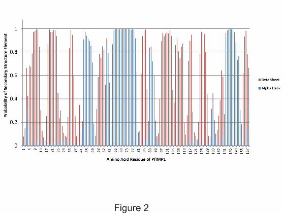
structure prediction (Figure 2) was obtained from Talos N (Shen and Bax, 2013). The amide
proton of I96 resonates at an unusual upfield-shifted position compared to the other
structured amides. It is located in a loop region between secondary structure elements and is
immediately preceded by an FP sequence. The proline residue within this loop likely
configures the backbone conformation such that amide proton of I96 experiences a significant
shielding ring current effect from F94.
References
BLAKE, D., BILLINGTON, K., COPESTAKE, S. & OAKES, R. 2011. Genetic mapping identifies novel highly protective antigens for an apicomplexan parasite. PLoS pathogens 7.
CUI, X., LEI, T., YANG, D., HAO, P., LI, B. & LIU, Q. 2012. Toxoplasma gondii immune mapped protein – 1 (TgIMP1) is a novel vaccine candidate against toxoplasmosis. Vaccine, 30, 2282-2287.
JUNG, Y. S. & ZWECKSTETTER, M. 2004. Mars -- robust automatic backbone assignment of proteins. J Biomol NMR, 30, 11-23.
YIN, G., QIN, M., LIU, X., SUO, J., TANG, X., TAO, G., HAN, Q., SUO, X. & WU, W. 2013. An Eimeria vaccine candidate based on Eimeria tenella immune mapped protein 1 and the TLR-5 agonist Salmonella typhimurium FliC flagellin. Biochem Biophys Res Commun, 440, 437-42.
SHEN, Y. & BAX, A. 2013. Protein backbone and sidechain torsion angles predicted from NMR chemical shifts using artificial neural networks. J Biomol NMR, 56, 227-41.

Figure Captions
Fig.1 Assigned 15N, 1H – HSQC spectrum of recombinant PfIMP1. The peaks are labelled with single letter amino acid code followed by their position in the recombinant PfIMP1 sequence. The underlined residues indicate aliased amide signals (namely G82, G51 and G149).
Fig. 2 Secondary structure determination using TALOS-N (Shen and Bax, 2013). The predicted secondary structure of this protein consists of four α-helices and eleven β strands. Helices are located between residues 43-49, 58-80, 88-92 and 142-151. β strands are located between residues 5-12, 17-24, 32-36, 53-57, 83-86, 96-104, 107-112, 115-118, 124-128, 139-140 and 154-157.