non-apoptotic roles of bcl-2 family: the calcium connection
TRANSCRIPT
Biochimica et Biophysica Acta 1833 (2013) 1755–1765
Contents lists available at SciVerse ScienceDirect
Biochimica et Biophysica Acta
j ourna l homepage: www.e lsev ie r .com/ locate /bbamcr
Review
Non-apoptotic roles of Bcl-2 family: The calcium connection☆
Benjamin Bonneau a,⁎, Julien Prudent a, Nikolay Popgeorgiev b, Germain Gillet a,⁎⁎a Université de Lyon, Université Lyon 1, Inserm U1052, CNRS UMR5286, Centre de Recherche en Cancérologie de Lyon, Centre Léon Bérard, Lyon, Franceb Unité de recherche sur les maladies infectieuses et tropicales émergentes, URMITE UM63, CNRS 7278, IRD 198, Inserm 1095, Aix-Marseille Université, Faculté de médecine,Marseille, France
☆ This article is part of a Special Issue entitled: 12th Eur⁎ Correspondence to: B. Bonneau, Centre de Recherche
Cheney D, Centre Léon Bérard, 28 rue Laennec 69008, L66 10.⁎⁎ Correspondence to: G. Gillet, Centre de RechercheCheney D, Centre Léon Bérard, 28 rue Laennec 69008, L66 56; fax: +33 4 69 16 66 60.
E-mail addresses: [email protected]@univ-lyon1.fr (G. Gillet).
0167-4889/$ – see front matter © 2013 Elsevier B.V. Allhttp://dx.doi.org/10.1016/j.bbamcr.2013.01.021
a b s t r a c t
a r t i c l e i n f oArticle history:Received 1 November 2012Received in revised form 11 January 2013Accepted 12 January 2013Available online 27 January 2013
Keywords:Bcl-2CalciumEndoplasmic reticulumMitochondriaNon-apoptoticApoptosis
The existence of the bcl-2 (B-cell lymphoma-2) gene was reported nearly 30 years ago. Yet, Bcl-2 familygroup of proteins still surprises us with their structural and functional diversity. Since the discovery of theBcl-2 family of proteins as one of the main apoptosis judges, the precise mechanism of their action remainsa hot topic of intensive scientific research and debates. Although extensive work has been performed on therole of mitochondria in apoptosis, more and more studies point out an implication of the endoplasmicreticulum in this process. Interestingly, Bcl-2 family proteins could be localized to both the mitochondriaand the endoplasmic reticulum highlighting their crucial role in apoptosis control. In particular, in theseorganelles Bcl-2 proteins seem to be involved in calcium homeostasis regulation although the mechanismsunderlying this function are still misunderstood. We now assume with high degree of certainty that themajority of Bcl-2 family members take part not only in apoptosis regulation but also in other processes im-portant for the cell physiology briefly denominated as “non-apoptotic” functions. Drawing a complete andcomprehensive image of Bcl-2 family requires the understanding of their implications in all cellular process-es. Here, we review the current knowledge on the control of calcium homeostasis by the Bcl-2 family at theendoplasmic reticulum and at the mitochondria. Then we focus on the non-apoptotic functions of the Bcl-2proteins in relation with the regulation of this versatile intracellular messenger. This article is part of a SpecialIssue entitled: 12th European Symposium on Calcium.
© 2013 Elsevier B.V. All rights reserved.
1. The Bcl-2 family of proteins and apoptosis
Historically, bcl-2 (B-cell lymphoma-2) gene was discovered at achromosomal translocation, t(14;18), associated with B-cell follicularlymphoma. This translocation places bcl-2 under the control of theimmunoglobulin gene enhancer leading to abnormal high-level expres-sion of Bcl-2 [1–3]. However, contrary to other oncogenes characterizedso far, Bcl-2 acts by inhibiting cell death rather than promoting cellproliferation [4]. Bcl-2 was thus initially characterized as a mitochon-drial resident protein that blocks apoptotic programmed cell death[5]. Since the discovery of Bcl-2, extensive work on various animalmodels, from worms to mammals, uncovered a family of proteins in-volved in the control of apoptosis and sharing structural homologywith the canonical member Bcl-2 [6]. In vertebrates, members of the
opean Symposium on Calcium.en Cancérologie de Lyon, Bat.
yon, France. Tel.: +33 4 69 16
en Cancérologie de Lyon, Bat.yon, France. Tel.: +33 4 69 16
r.fr (B. Bonneau),
rights reserved.
so called Bcl-2 family are globular proteins containing α-helixes andcharacterized by the presence of at least one Bcl-2 homology (BH)domain. According to their function in apoptosis, Bcl-2 family mem-bers are designated as anti- or pro-apoptotic. Anti-apoptotic proteins(e.g. Bcl-2, Bcl-xL, and Mcl-1) contain four BH domains and a hydro-phobic transmembrane (TM) domain essential for their localizationinto biological membrane. Pro-apoptotic proteins can be furthersubdivided into two groups. The effector proteins Bax and Bak containfour BH domains and a TM domainwhereas BH3-only proteins (e.g. Bad,Bid, and Bik) possess just one BH domain and no TM domain with theexception of Bik and Hrk [7,8].
The Bcl-2 family proteins play a crucial role in the control and theexecution of the intrinsic, or mitochondrial, pathway of apoptosis. Inthis process, some death stimuli (e.g. growth factor deprivation, andDNA damage) activate BH3-only proteins which in turn lead to theactivation of Bax and/or Bak either directly or by inhibiting anti-apoptoticmembers. ActivatedBax/Bak thenoligomerize at themitochon-dria to induce outer mitochondrial membrane (OMM) permeabilizationand release into the cytosol of apoptotic factors (e.g. cytochrome c andAIF) which promote caspase activation and subsequent apoptosis execu-tion. Anti-apoptotic members of Bcl-2 family are able to prevent OMMpermeabilization via a direct inhibitory interaction with pro-apoptoticmembers [8].

1756 B. Bonneau et al. / Biochimica et Biophysica Acta 1833 (2013) 1755–1765
2. Endoplasmic reticulum Ca2+ and apoptosis
Although the mitochondrion is a key organelle for promotingapoptosis, a growing body of evidence underlies the involvement ofendoplasmic reticulum (ER) in this process. The ER is the major siteof folding, modification, and trafficking of proteins as well as themain store for intracellular Ca2+. These functions of the ER are tightlyconnected as Ca2+ is required for protein synthesis in almost everycell type. In the ER, Ca2+ supports early protein processing andprotein glycosylation. In addition, Ca2+ may also be implicated insubunit assembly and protein folding [9]. Thus, the reduction of ERCa2+ level can induce the accumulation and aggregation of unfoldedproteins, a condition referred to as ER stress. To face this stress, cellsactivate a prosurvival response known as the Unfolded ProteinResponse (UPR) to reduce the accumulation of unfolded protein.However if the UPR's mechanisms are insufficient to decreaseunfolded protein load, the UPR activates apoptosis [10]. In this case,components of the UPR induce apoptosis via the inactivation of Bcl-2and the activation of several BH3-only proteins including Bim, Bad,Bik and Puma which then activate Bax and Bak to promote cytochromec release from the mitochondria. Interestingly, a cross-talk existsbetween the UPR and apoptosis as Bax/Bak have been proposed toparticipate in early events of the UPR [11].
Moreover a number of stresses, including ER stress, can lead to therelease of the ER Ca2+ store and subsequent increase of cytosolicCa2+ levels [10,12]. Cytosolic Ca2+ elevation could in turn induce apo-ptosis via several pathways independent or not from the mitochondria.
The first one is triggered by the caspase-12 independently of mito-chondria. This caspase localized at the ER [13,14] can be cleaved andactivated by the Ca2+-dependent protease, Calpain [15]. Activatedcaspase-12 then processes downstream caspases leading to apoptosisindependently of OMM permeabilization [16].
Secondly, [Ca2+] rise into the cytosol may lead to OMM perme-abilization through BH3-only protein activation. In particular, Calpain-mediated Bid cleavage into its active form tBid was reported to induceapoptosis in a Bax-dependent manner [17]. In addition, Calpain cleav-age of Bcl-2 and Bcl-xL N-terminus reduces their anti-apoptotic activityand even promotes Bcl-2 conversion into a pro-apoptotic proteinwhichpromotes cytochrome c release on its own [18]. Bad is another BH3-onlymember which could be activated in response to Ca2+ elevation. Underphysiological conditions, Bad is phosphorylated and sequesters in thecytosol by 14–3–3 proteins [19]. However, elevation of cytosolic Ca2+
levels activates the Ca2+-dependent phosphatase, Calcineurin (alsoknown as PP2B) which dephosphorylates Bad therefore activating theintrinsic mitochondrial pathway [20]. Interestingly, it has also beenshown that a slight increase of Ca2+ could, on the contrary, promoteBad phosphorylation and sequestration by 14–3–3 via the activationof the Ca2+/Calmodulin dependent protein kinase kinase (CaMKK)highlighting the versatility of Ca2+ signaling in cell fate [21].
Finally, massive Ca2+ release from the ER can directly triggercytochrome c release from the mitochondria. Indeed, the ER and themitochondria have long been known to be tightly associated [22,23].More recent research established that ER membranes are in closecontact with mitochondria, forming microdomains called MAM formitochondria-associated membranes [24]. MAM are enriched in trans-membrane proteins such as PACS-2 orMitofusin-2whichdirectly tetherthe ER and the mitochondria thus creating a physical interaction be-tween them [25,26]. These contact points are crucial for lipid andCa2+ exchanges between ER and mitochondria. Inositol 1,4,5-trisphos-phate receptors (IP3R) located at the ER membrane play an essentialrole in ER to mitochondria Ca2+ trafficking [27,28]. In MAM, IP3Rseems to be part of a protein complex with Grp75 and the mitochon-drial Voltage Dependent Anion Channel (VDAC) that promotes Ca2+
exchange between ER and mitochondria [29]. [Ca2+] in MAM canreach a very high level compared to the cytosolic concentration [30]which is consistent with a specialized function in Ca2+ transfer between
ER and mitochondria. Indeed, the recently identified MitochondrialCalcium Uniporter (MCU) [31,32] requires a high level of Ca2+ to opendue to its interaction with MICU1 which acts as a gatekeeper by settinga threshold for Ca2+ entry into mitochondria [33]. Thus, Ca2+ releasedthrough IP3R in MAM can be rapidly uptaken into the mitochondriaby VDAC and MCU to enter mitochondrial matrix [34]. Interestingly,IP3R has been acknowledged to be required for apoptosis in somecases [35–38]. Therefore, upon a stress inducing massive Ca2+ releasethrough IP3R, mitochondria face very high level of calcium and uptakeit to buffer cytosolic Ca2+. When Ca2+ accumulates in the mitochondriait binds to Cyclophilin D which induces the opening of the permeabilitytransition pore (PTP) and finally leads to cytochrome c release [39].
3. Regulation of ER Ca2+ homeostasis by Bcl-2 proteins
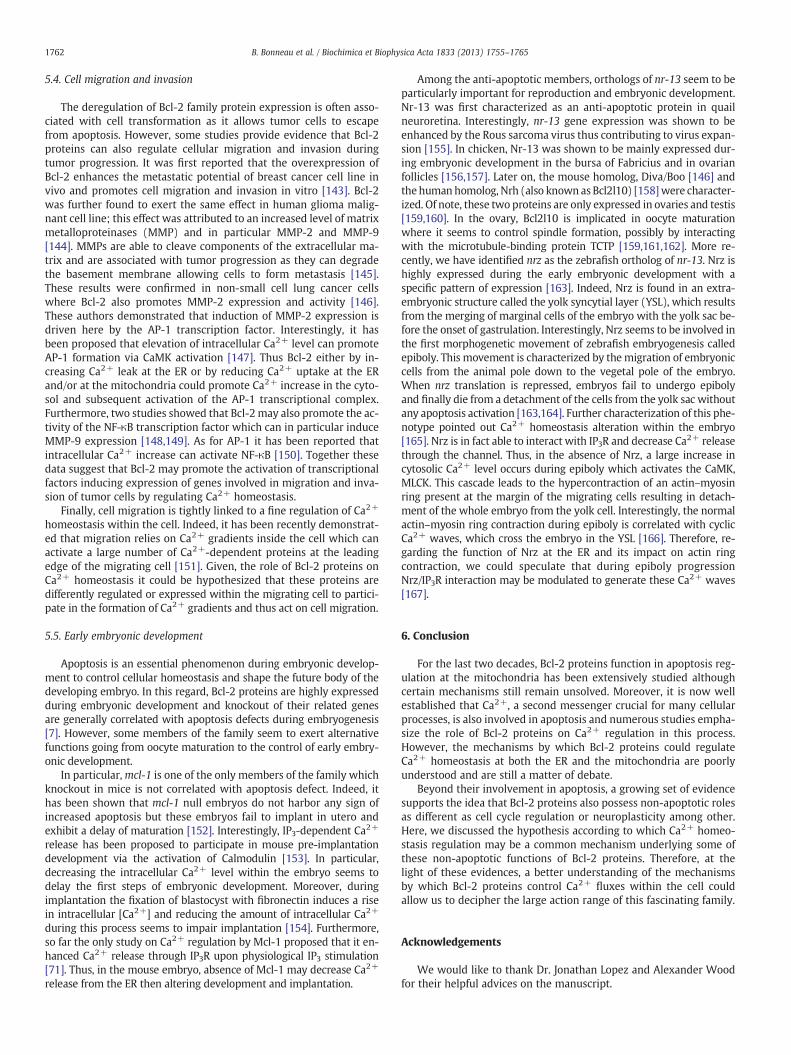
The concept that members of the Bcl-2 family could be localizedto the ER [40] and control apoptosis through ER Ca2+ homeostasisregulation, emerged almost two decades ago. However, in spite ofincreasing number of studies, the mechanisms by which the Bcl-2proteins exert this function are still unclear and subject to controversy(Fig. 1).
3.1. Regulation by anti-apoptotic proteins
The first observation suggesting a role for Bcl-2 on intracellular Ca2+
homeostasis was made in 1993 [41]. The authors demonstrated that, inInterleukin-3 (IL-3)-dependent hematopoietic cells, IL-3 withdrawalinduces apoptosis by a Ca2+ transfer from the ER to the mitochondria.In this model, Bcl-2 overexpression prevents apoptosis and blocks thisCa2+ flux. In this study, the thapsigargin-releasable Ca2+ pool beforeIL-3 withdrawal was not affected by Bcl-2 suggesting that Bcl-2 doesnot alter ER Ca2+ content but rather acts on Ca2+ release from the ER.The year after, two other studies concluded that Bcl-2 decreases Ca2+
release from the ER. The first showed that Bcl-2 prevents cytosolicCa2+ increase induced by serum withdrawal in NIH3T3 cells [42]. Thesecond reported that Bcl-2 was able to inhibit thapsigargin-inducedapoptosis [43]. Here the authors showed that Bcl-2 reduces Ca2+ effluxfrom the ER following thapsigargin treatment but that this effectwas not due to alteration in Ca2+ homeostasis within the ER lumen.The same effect was observed upon hydrogen peroxide treatmentsuggesting that Bcl-2 can reduce Ca2+ release from the ER to preventapoptosis induced by various stresses [44,45]. However, subsequentstudies showed that Bcl-2 is able to increase Ca2+ uptake in the ERand then enhanced ER Ca2+ load under certain conditions. He andcolleagues showed that overexpression of Bcl-2 maintains ER Ca2+
homeostasis when the sarco/endoplasmic reticulum Ca2+-ATPase(SERCA) is inhibited by thapsigargin or when the extracellular [Ca2+]is decreased [46]. Here, Bcl-2 does not seem to prevent SERCA inhibitionso the authors concluded that Bcl-2 could mediate Ca2+ uptake in theER. They proposed that Bcl-2, by preventing oxidative damage to theER membrane, could decrease Ca2+ leak through the ER and thus in-crease Ca2+ loading capacity of the ER. They also hypothesized thatthis effect of Bcl-2 could be due to the formation of cation-selectivechannel in lipid bilayer by Bcl-2 or Bcl-xL [47,48] thus allowing Ca2+
entry into the ER lumen. However, this hypothesis does not take intoaccount the existing calcium gradient across the ERmembrane, render-ing unlikely the passive calcium transport through Bcl-2 channels.Other studies also reported an increased Ca2+ load of the ER whenBcl-2 is overexpressed but in this case it was attributed to an increaselevel of SERCA expression [49,50]. Interestingly, the authors also de-scribed an interaction between Bcl-2 and SERCA which was proposedto enhance SERCA activity [44]. Such interaction was also suggestedin Xenopus oocytes where Bcl-2 was shown to desensitize SERCA tothapsigargin thanks to its BH4 domain [51]. In all these studies, it wasassumed that the anti-apoptotic effect of Bcl-2 was mediated by a
: IP3R
: SERCA
: Anti-apoptotic (Bcl-2, Bcl-xL or Mcl-1)
: Phosphorylation
: Pro-apoptotic (Bax or Bak)
: BH3-only
P
Bcl-2 reduces Ca2+ uptake
Bcl-2 increases Ca2+ leak
[Ca2+]ER
B
~ [Ca2+]ER
C
IP3
Anti-apoptotic enhanceIICR at low [IP3]
Anti-apoptotic decreaseIICR at high [IP3]
~ [Ca2+]ER
A
IP3
Bcl-2 decreases IICRBax/Bak promote Ca2+
release
Bcl-2 enhances Ca2+ uptake
: Bax Inhibitor-1
P
IP3
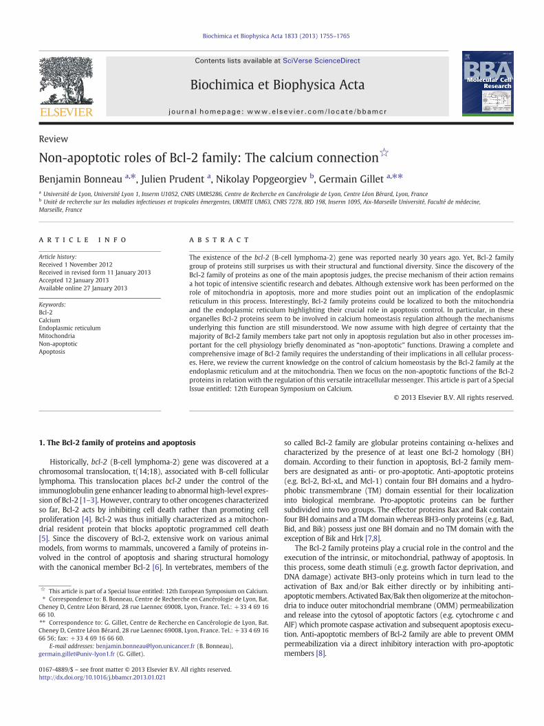
Fig. 1. Schematic representation of the different models for Bcl-2 family activity on ER Ca2+ homeostasis. (A) In this model, Bcl-2 reduces cytosolic [Ca2+] to prevent apoptosisinduction. Bcl-2 could act either by increasing SERCA activity or by reducing IICR. Bcl-2 may exert its inhibitory activity on IP3R via a direct interaction with the modulatory andtransducing domain of the receptor. Conversely, activated Bax and Bak are proposed to induce Ca2+ release from the ER. (B) In this model, Bcl-2 reduces ER Ca2+ content to preventmassive Ca2+ release which could trigger apoptosis. Bcl-2 is proposed to increase Ca2+ leak from the ER by promoting IP3R phosphorylation or by directly inducing Ca2+ leak aloneor in combination with a BH3-only protein. Bcl-2 could also reduce SERCA activity by a direct interaction. Bax and Bak may counteract the effect of Bcl-2 by a direct inhibitoryinteraction. Finally, Bcl-xL could act on BI-1 to increase Ca2+ leak from the ER with BI-1 acting as a Ca2+ channel or as an IP3R sensitizer. (C) In this model, anti-apoptotic proteinspromote survival by enhancing ER-mitochondria coupling. They are proposed to enhance IICR at low [IP3] to stimulate bioenergetics whereas at high [IP3] they could preventapoptosis by reducing IICR. This effect may be mediated by a direct interaction with the C-terminus of IP3R. Dashed lines represent a reduction of the calcium flux. Bold linesrepresent an increase of the calcium flux. Red and blue arrows represent activation and inhibition respectively. IP3R: inositol 1,4,5-trisphosphate receptor; SERCA: sarco/endoplasmicreticulum Ca2+-ATPase; BI-1: Bax-inhibitor-1; IICR: IP3-induced calcium release.
1757B. Bonneau et al. / Biochimica et Biophysica Acta 1833 (2013) 1755–1765
reduction of cytosolic Ca2+ level either by increasing ER Ca2+ load or byreducing Ca2+ release from the ER (Fig. 1A).
Nevertheless, in 2000 a new mechanism of action for Bcl-2 at theER emerged when two groups showed that Bcl-2 was able to decreaseER Ca2+ content [47,48]. In their study, Pinton and colleagues demon-strated that Bcl-2 overexpression reduces IP3-induced Ca2+ release(IICR) and attributed this effect to a lower ER Ca2+ content due toincreased Ca2+ leak from the ER [52]. Surprisingly, the authors did notobserve any variation in cytosolic [Ca2+] despite the detection ofdecreasedmitochondrial Ca2+ uptake. Foyouzi-Youssefi and colleaguesalso described a reduced ER Ca2+ content in Bcl-2 overexpressing cells[53]. As ER [Ca2+] decrease is a signal for capacitative Ca2+ entry,authors showed that Bcl-2 enhances unstimulated Ca2+ influx andthen induced elevated basal cytosolic [Ca2+]. As for the previous study,it was shown that decreased ER [Ca2+] is a consequence of increasedCa2+ leak from the ER rather than Ca2+ uptake reduction. In bothcases authors argued that formation of a cation-selective channel byBcl-2 may be responsible for the increased Ca2+ leak (Fig. 1B). Twosubsequent studies also concluded that Bcl-2 could decrease ER Ca2+
content. In the first it was demonstrated that Bcl-2 overexpressionincreases Ca2+ leak but also downregulates SERCA and Calreticulinexpression to reduce ER Ca2+ content [54]. In the second, using a newgenetically-encoded Ca2+ sensor, Palmer and colleagues measured[Ca2+] within the ER lumen. They found that Bcl-2 decreases ER Ca2+
content and, as a consequence, alters IICR [55].In thismodel it was proposed that reduction of Ca2+ levelwithin the
ERmediates the anti-apoptotic activity of Bcl-2 by reducing the amountof releasable Ca2+ available to induce apoptosis. Foyouzi-Youssefi andcolleagues also speculated that the increased cytosolic [Ca2+] they
observed could promote cell survival by the CaMKK-dependent phos-phorylation of Bad.
However, the idea according to which this effect is mediated byBcl-2 pore formation was challenged when Chami and colleaguesshowed that a Bcl-2 chimera protein containing the pore domain ofBax has the same effect on ER Ca2+ homeostasis as the wild-typeBcl-2 [56]. Another possible mechanism emerged from a studywhich showed that, in contrast to what was proposed earlier, Bcl-2can interact with and destabilize SERCA [57] (Fig. 1B). Another ideawas further put forward to explain the reduction of ER Ca2+ contentby Bcl-2. In their study, Bassik and colleagues proposed that Bcl-2phosphorylation within an unstructured loop between its BH4 andBH3 domains can modulate its ability to decrease ER Ca2+ content[58]. They showed that in cells expressing a non-phosphorylatablemutant of Bcl-2 (Bcl-2 AAA) the ER Ca2+ level is lower than in Bcl-2expressing cells due to higher Ca2+ leak. In this regard Bcl-2 AAA ismore efficient at protecting cells from Ca2+-dependent apoptoticstimuli than the wild-type protein. At last, they demonstrated thatphosphorylation of Bcl-2 impairs its interaction with Bax and BH3-only proteins and provided evidence suggesting that Bcl-2 interactionwith the BH3-only proteins was required to promote the reduction ofER Ca2+ content (Fig. 1B). However how a complex between Bcl-2and a BH3-only can induce Ca2+ leak through the ER membrane isnot discussed and remains elusive.
Finally, it has been proposed that Bax Inhibitor-1 (BI-1) may bethe downstream target of Bcl-xL to lower ER [Ca2+]. Indeed, Xu andcolleagues demonstrated that BI-1 is required for ER calcium contentreduction induced by Bcl-xL overexpression [59] (Fig. 1B). BI-1 wasshown to increase calcium leak from the ER in a pH-dependent

1758 B. Bonneau et al. / Biochimica et Biophysica Acta 1833 (2013) 1755–1765
manner [60] and it may act either by forming a Ca2+-permeablechannel pore with its C-terminus [61] or by interacting with IP3R tosensitize the channel to low [IP3] [62].
Thus, so far, the molecular mechanism by which Bcl-2 can reduceCa2+ level within the ER is still a matter of debate and should befurther investigated.
Finally, the group of C.W. Distelhorst proposed that Bcl-2 couldregulate calcium homeostasis at the ER by directly interacting withIP3R. In a work published in 2004 they demonstrated that Bcl-2 isable to decrease IICR without affecting ER calcium level. In a convinc-ing manner they showed that Bcl-2 directly interacts with IP3R,forming a complex with the receptor. As a consequence, they de-scribed that Bcl-2 is able to reduce the channel open probability ofIP3R [63]. These results were confirmed by another study where theauthors notably showed that ER-targeted Bcl-2 has the same effectas wild-type Bcl-2 on Ca2+ homeostasis [64]. Interaction betweenBcl-2 and IP3R was further characterized and it was demonstratedthat Bcl-2 binds to the modulatory and transducing domain of IP3Rbetween the residues 1347–1426 in the middle of the protein [65](Fig. 1A). Furthermore, Bcl-2 interacts with IP3R via its BH4 domainand it has been shown that the BH4 domain alone was sufficient todecrease IICR [66]. At last, a recent study comparing the BH4 domainsof Bcl-2 and Bcl-xL has identified a crucial residue within Bcl-2 whichseems essential for its regulatory activity on IICR [67]. Contrary toBcl-2, the BH4 domain of Bcl-xL was able neither to bind to the mod-ulatory and transducing domain nor to reduce Ca2+ release throughIP3R. The authors found the Lys17 residue of Bcl-2 to be critical forthe activity of Bcl-2 on IP3R since its mutation in aspartate, as in theBH4 domain of Bcl-xL, abolishes the activity of Bcl-2 BH4 domain onCa2+ signaling.
However, Bcl-xL was also shown to interact with IP3R but at a dif-ferent site as it binds the C-terminus of IP3R. This interaction wasshown to sensitize IP3R to low concentrations of IP3 thus enhancingCa2+ oscillation in the cytosol. IP3R sensitization stimulates mito-chondrial bioenergetics allowing the authors to propose that Bcl-xLcould promote survival by enhancing ER-mitochondria coupling andcellular metabolism [68]. In this case, Bcl-xL was also reported todecrease ER Ca2+ content but this effect does not seem to be relevant.Indeed, the same team showed two years later that Bcl-xL interactswith the three IP3R isoforms but reduces ER Ca2+ content only incells expressing IP3R3. However, Bcl-xL sensitizes all IP3R isoformsto IP3 and enhances cytosolic Ca2+ oscillation and apoptosis resis-tance whatever isoform is expressed [69]. Therefore it is more likelythat Bcl-xL action is mediated rather by its interaction with IP3Rthan by decreasing ER Ca2+ content (Fig. 1C).
These differences between Bcl-xL and Bcl-2 could be attributed, asexposed above, to the differences between the BH4 domains of theseproteins. Thus, depending on their BH4 domain, anti-apoptotic mem-bers could interact at different sites within IP3R thus regulating thechannel activity in a different manner (for extensive discussion onthe topic see [70]).
Nevertheless, Bcl-2 and Mcl-1 seem also able to bind to theC-terminus region of IP3R and to regulate Ca2+ homeostasis in a man-ner similar to Bcl-xL highlighting the complexity of Bcl-2 family me-diated regulation of IP3R [71,72] (Fig. 1C).
3.2. Regulation by pro-apoptotic members
Pro-apoptotic proteins are also able to localize at the ER and act onER Ca2+ homeostasis. In 2001, Pan and colleagues first proposed alink between Bax and ER Ca2+ signaling since apoptosis induced byoverexpression of Bax in CHO cells was correlated to a depletion ofER Ca2+ store [73]. However they argued that the depletion of the ERCa2+ store rather than cytosolic [Ca2+] increase is responsible forapoptosis induction in their model. The year after Bax and Bak wereshown to localize at the ER where they induce ER Ca2+ depletion and
subsequent cytochrome c release due to Ca2+ accumulation in the mi-tochondria [74]. Bax seems to be required to induce Ca2+-dependentapoptosis as Bax-null cells did not elicit ER Ca2+ pool reduction andsubsequent apoptosis induced by the staurosporine [75]. Moreover,the group of C. Thompson found that ER stress can induce Bax andBak oligomerization at the ER leading to caspase-12 activation [76].Interestingly, they showed that the targeted expression of Bak was suf-ficient to trigger ER Ca2+ depletion and subsequent caspase-12 activa-tion even in the absence of endogenous Bax and Bak. A more recentstudy is consistent with an activity of Bak at the ER as it was shownthat ER-targeted Bak is able to activate BH3-only proteins and subse-quent cytochrome c release in part via ER Ca2+ release [77]. All thesestudies therefore assess that once activated Bax and Bak could triggerCa2+ release from the ER to induce apoptosis (Fig. 1A). However, asfor the anti-apoptotic proteins contradictory results concerning therole of pro-apoptotic proteins at the ER exist. Indeed, the group of S.Korsmeyer found that in MEF cells deficient for Bax and Bak, ER Ca2+
content was significantly reduced suggesting that Bax and Bak act atthe ER by increasing [Ca2+] in the lumen of the ER [78]. A few yearslater, they proposed a model to explain the effect of Bax and Bak onER Ca2+ content. They showed that in cells lacking Bax and Bak IP3R1is hyperphosphorylated on Ser 1755. This PKA-dependent phosphoryla-tion renders IP3R sensitive to basal IP3 concentration thus increasingCa2+ leak from the ER. They also found that in the absence of Bax andBak the interaction between Bcl-2 and IP3R was enhanced and thatbcl-2 knockdown reduces IP3R phosphorylation in these cells [79]. Sothis group proposed that at the ER, Bcl-2 might induce IP3R phosphory-lation thus promoting Ca2+ leak and ER Ca2+ content reductionwhere-as Bax and Bak, by interacting with Bcl-2, could counteract IP3Rphosphorylation (Fig. 1B).
Finally, besides the multidomain proteins of the Bcl-2 family,the BH3-only protein Bik is also localized at the ER, thanks to itsC-terminus TM domain, where it can induce cytochrome c releasefrom the mitochondria [80]. Surprisingly, in this first study the authorsargued that Ca2+ was not involved in this process. However, in a morerecent study, they showed that Bik can induce Ca2+ release from the ERby promoting Bax and Bak localization and oligomerization at the ER[81] (Fig. 1A).
At the light of these conflicting results, it is difficult to decipher acommonmechanism of action for the Bcl-2 family at the ER. Differencesin the cell models aswell as the detectionmethods used by the differentgroups should be taken into account explaining partially the divergencein the proposed models. Despite these discrepancies, it remains clearthat multiple members of Bcl-2 proteins may act as key regulators ofcalcium homeostasis at the ER. Indeed, all studies at least agree on thefact that anti-apoptotic proteins are able to reduce the amount of calci-um released from the ER upon apoptotic stress whereas pro-apoptotichas the opposite effect.
4. Regulation of mitochondrial Ca2+ homeostasis byBcl-2 proteins
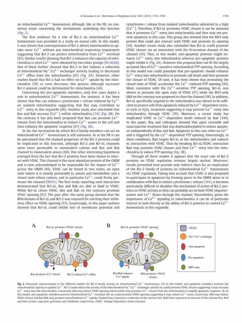
Although ER is the main store of Ca2+ in the cell, mitochondriaalso play a critical role in Ca2+ homeostasis. As mentioned inSection 2, mitochondria can rapidly uptake Ca2+ present in the cyto-sol acting as a buffer when cytosolic [Ca2+] increases. In mitochon-dria Ca2+ enhances substrate uptake, the rate of NADH productionand the activity of the ATP synthase as well as those of the pyruvate,ketoglutarate and isocitrate dehydrogenases of the TCA cycle thus pro-moting mitochondrial metabolism and ATP production [82]. However,Ca2+ overload is detrimental as it impairs mitochondrial respirationand leads to membrane potential drop, mitochondria swelling andfinally cytochrome c release. Thus, given the role of the Bcl-2 familyon the control of cell fate it can be assumed that this group of proteinscan also modulate Ca2+ homeostasis directly at the mitochondria.Indeed, several studies have revealed that Bcl-2 proteins could act
1759B. Bonneau et al. / Biochimica et Biophysica Acta 1833 (2013) 1755–1765
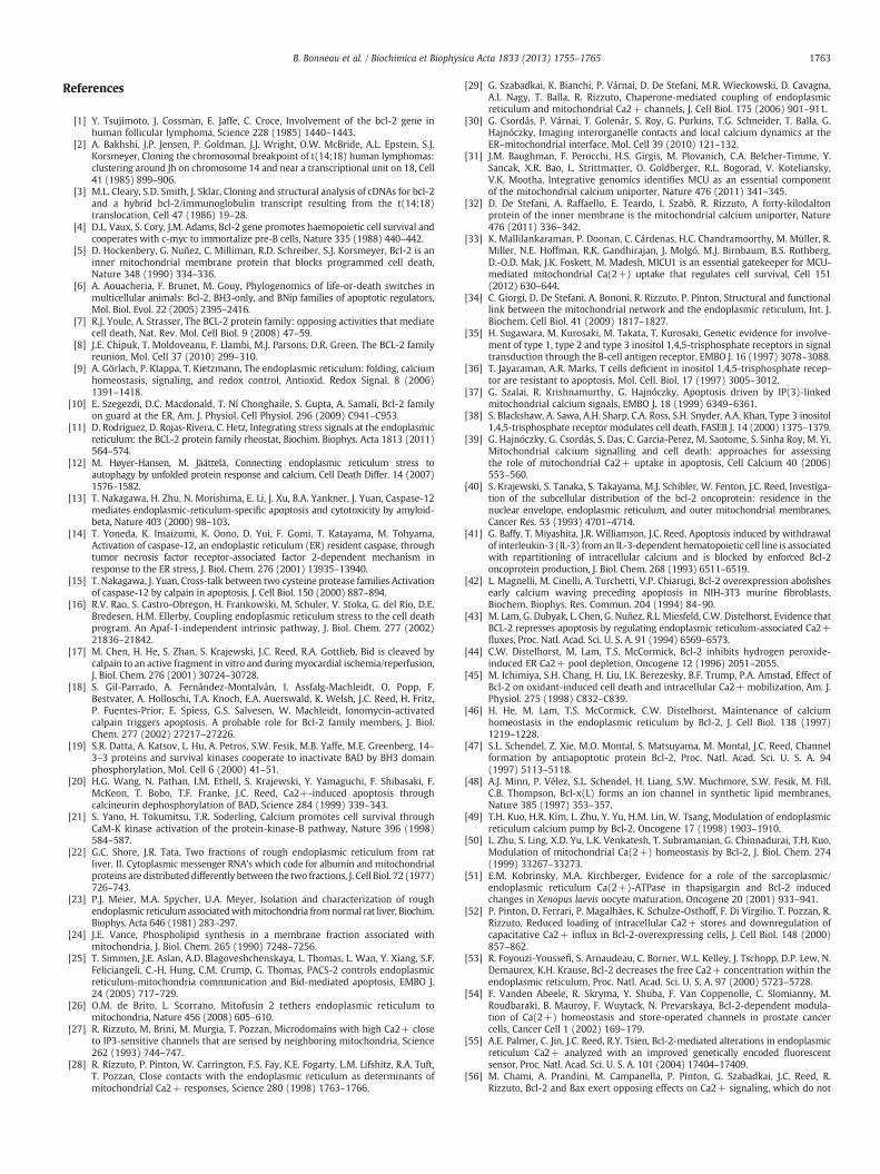
on mitochondrial Ca2+ homeostasis although, like at the ER, no con-sensus exists concerning the mechanisms underlying this function(Fig. 2).
The first evidence for a role of Bcl-2 on mitochondrial Ca2+
homeostasis was provided in a study on neural cells. In this model,it was shown that overexpression of Bcl-2 allows mitochondria to up-take more Ca2+ without any mitochondrial respiratory impairmentsuggesting that Bcl-2 can protect mitochondria from Ca2+ overload[83]. Similar results showing that Bcl-2 enhances the capacity of mito-chondria to store Ca2+ were obtained by two other groups [45,50,84].One of these further showed that Bcl-2 in fact induces a reductionof mitochondrial Na+/Ca2+ exchanger activity leading to decreasedCa2+ efflux from the mitochondria [85] (Fig. 2A). However, otherstudies found that Bcl-2 had no effect on Ca2+ uptake by the mito-chondria [58] or even decreases this process although excessiveBcl-2 amount could be detrimental for mitochondria [64].
Concerning the pro-apoptotic members, only few cases depict arole in mitochondrial Ca2+ homeostasis. For instance, it has beenshown that Bax can enhance cytochrome c release induced by Ca2+
on isolated mitochondria suggesting that Bax may contribute toCa2+ entry in this organelle [86]. Another study also concluded thatBax and Bak increase [Ca2+] within mitochondria [74] (Fig. 2B). Onthe contrary it has also been proposed that Bax can promote Ca2+
release from the mitochondria to induce Ca2+ waves in the cell andthen enhance the apoptotic response [87] (Fig. 2A).
So far, the mechanism by which Bcl-2 family members can act onmitochondrial Ca2+ homeostasis is still unknown. As at the ER it canbe speculated that the channel properties of the Bcl-2 proteins couldbe implicated in this function, although Bcl-2 and Bcl-xL channelsseem more permeable to monovalent cations and Bax and Bakchannel to monovalent anions [88]. One other interesting hypothesisemerged from the fact that Bcl-2 proteins have been shown to inter-act with VDAC. This channel is the most abundant protein of the OMMand is now acknowledged to be responsible for the import of Ca2+
across the OMM [89]. VDAC can be found in two states, an openstate where it is mainly permeable to anions and metabolites and aclosed state where cations, and in particular Ca2+, could freely per-meate the channel [90,91]. The first study reporting such interactiondemonstrated that Bcl-xL, Bax and Bak are able to bind to VDAC.While Bcl-xL closes VDAC, Bax and Bak on the contrary promoteVDAC opening [92]. The year after, the same group showed that theBH4 domain of Bcl-xL and Bcl-2 was required for exerting their inhib-itory effect on VDAC opening [93]. Surprisingly, in this paper authorsshowed that Bcl-xL could inhibit loss of membrane potential and
Bcl-xL promotes VDAC closure
Bcl-2 reduces Ca2+ effluxBax promotes Ca2+ release
Bcl-
Bax/Bak enCa2+ upt
BA
Fig. 2. Schematic representation of the different models for Bcl-2 family activity on mitomitochondrial capacity to uptake Ca2+. Bcl-2 could reduce the activity of the mitochondrial NCa2+ entry into the mitochondria. Conversely, Bad may induce VDAC opening whereas Bax mthis model, anti-apoptotic members prevent mitochondrial Ca2+ overload. Bcl-xL could proVDAC closure and Bax/Bak may promote mitochondrial Ca2+ uptake. Dashed lines representand blue arrows represent activation and inhibition respectively. VDAC: Voltage Dependen
cytochrome c release from isolated mitochondria submitted to a high[Ca2+]. Therefore, if Bcl-xL promotes VDAC closure it can be assumedthat it promotes Ca2+ entry into mitochondria and thus may not pre-vent apoptosis in this case. This group also showed that the BH3-onlyprotein Bim could also interact with VDAC and promote its opening[94]. Another recent study also concluded that Bcl-xL could promoteVDAC closure via an interaction with the N-terminus domain of thechannel [95]. Thus, in this model, anti-apoptotic proteins might en-hance Ca2+ entry into mitochondria whereas pro-apoptotic proteinsmight inhibit it (Fig. 2A). However this proposal does not fit the largelyaccepted idea of Ca2+-sensitive mitochondrial PTP opening. Accordingto this model one could postulate that pro-apoptotic should enhanceCa2+ entry into mitochondria to promote cell death and then promotethe closure of VDAC. Of note, it has been shown that promoting theclosed state of VDAC accelerates the Ca2+-induced PTP opening [96].More consistent with the Ca2+-sensitive PTP opening, Bcl-xL wasshown to promote the open state of VDAC [97] while the BH3-onlytBid on the contrarywas proposed to close the channel [98]. In addition,Bcl-xL specifically targeted to the mitochondria was shown to be suffi-cient to protect cells fromapoptosis induced by Ca2+-dependent stress-es such as H2O2 treatment suggesting it may prevent Ca2+ entry intomitochondria through VDAC [99]. Moreover, a recent study hasimplicated VDAC in Ca2+-dependent death induced by Bad [100].In this paper, Roy and colleagues showed that upon ceramide andstaurosporine treatment Bad was dephosphorylated to induce apopto-sis independently of Bax and Bak. Apoptosis in this case relies on Ca2+
and is triggered by the Ca2+-dependent PTP opening. Interestingly, inthese conditions, Bad targets Bcl-xL at the mitochondria and reducesits interaction with VDAC. Thus by breaking Bcl-xL/VDAC interactionBad may promote VDAC closure and then Ca2+ entry into the mito-chondria to induce PTP opening (Fig. 2B).
Through all these studies it appears that the exact role of Bcl-2proteins on VDAC regulation remains largely unclear. Moreover,results presented here provide only indirect clues for an implicationof the Bcl-2 family of proteins on mitochondrial Ca2+ homeostasisvia VDAC regulation. Taking into account that VDAC is also proposedto participate in apoptosis by forming pores in the OMM alone or incombination with Bax to induce cytochrome c release [101], it becomesparticularly difficult to decipher the mechanism of action of Bcl-2 pro-teins on VDAC activity as they can probably act on both VDAC oligomer-ization and Ca2+ fluxes through the channel. Nevertheless, given theimportance of Ca2+ signaling in mitochondria it can be of particularinterest to look directly at the ability of Bcl-2 proteins to control Ca2+
homeostasis in this organelle.
xL promotes VDAC opening
hanceake
tBid promotes VDAC closure
: VDAC
: Anti-apoptotic
: Pro-apoptotic
: BH3-only
: Na+/ Ca2+ exchanger
chondrial Ca2+ homeostasis. (A) In this model, anti-apoptotic members increase thea+/Ca2+ exchanger and Bcl-xL could promote VDAC closure suggesting it may increaseay promote Ca2+ release from the mitochondria to amplify apoptotic response. (B) In
mote VDAC opening suggesting it may reduce Ca2+ entry. Conversely, tBid may inducea reduction of the calcium flux. Bold lines represent an increase of the calcium flux. Redt Anion Channel.
1760 B. Bonneau et al. / Biochimica et Biophysica Acta 1833 (2013) 1755–1765
5. Calcium in non-apoptotic functions of Bcl-2 family
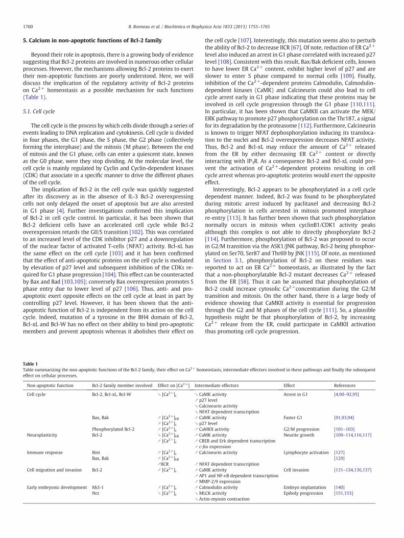
Beyond their role in apoptosis, there is a growing body of evidencesuggesting that Bcl-2 proteins are involved in numerous other cellularprocesses. However, the mechanisms allowing Bcl-2 proteins to exerttheir non-apoptotic functions are poorly understood. Here, we willdiscuss the implication of the regulatory activity of Bcl-2 proteinson Ca2+ homeostasis as a possible mechanism for such functions(Table 1).
5.1. Cell cycle
The cell cycle is the process by which cells divide through a series ofevents leading to DNA replication and cytokinesis. Cell cycle is dividedin four phases, the G1 phase, the S phase, the G2 phase (collectivelyforming the interphase) and the mitosis (M phase). Between the endof mitosis and the G1 phase, cells can enter a quiescent state, knownas the G0 phase, were they stop dividing. At the molecular level, thecell cycle is mainly regulated by Cyclin and Cyclin-dependent kinases(CDK) that associate in a specific manner to drive the different phasesof the cell cycle.
The implication of Bcl-2 in the cell cycle was quickly suggestedafter its discovery as in the absence of IL-3 Bcl-2 overexpressingcells not only delayed the onset of apoptosis but are also arrestedin G1 phase [4]. Further investigations confirmed this implicationof Bcl-2 in cell cycle control. In particular, it has been shown thatBcl-2 deficient cells have an accelerated cell cycle while Bcl-2overexpression retards the G0/S transition [102]. This was correlatedto an increased level of the CDK inhibitor p27 and a downregulationof the nuclear factor of activated T-cells (NFAT) activity. Bcl-xL hasthe same effect on the cell cycle [103] and it has been confirmedthat the effect of anti-apoptotic proteins on the cell cycle is mediatedby elevation of p27 level and subsequent inhibition of the CDKs re-quired for G1 phase progression [104]. This effect can be counteractedby Bax and Bad [103,105]; conversely Bax overexpression promotes Sphase entry due to lower level of p27 [106]. Thus, anti- and pro-apoptotic exert opposite effects on the cell cycle at least in part bycontrolling p27 level. However, it has been shown that the anti-apoptotic function of Bcl-2 is independent from its action on the cellcycle. Indeed, mutation of a tyrosine in the BH4 domain of Bcl-2,Bcl-xL and Bcl-W has no effect on their ability to bind pro-apoptoticmembers and prevent apoptosis whereas it abolishes their effect on
Table 1Table summarizing the non-apoptotic functions of the Bcl-2 family, their effect on Ca2+ homeffect on cellular processes.
Non-apoptotic function Bcl-2 family member involved Effect on [Ca2+] Inter
Cell cycle Bcl-2, Bcl-xL, Bcl-W ↘ [Ca2+]c ↘ Ca↗ p2↘ Ca↘ NF
Bax, Bak ↗ [Ca2+]ER ↗ Ca↗ [Ca2+]c ↘ p2
Phosphorylated Bcl-2 ↗ [Ca2+]c ↗ CaNeuroplasticity Bcl-2 ↘ [Ca2+]ER ↗ Ca
↗ [Ca2+]c ↗ CR↗ c-f
Immune response Bim ↗ [Ca2+]c ↗ CaBax, Bak ↗ [Ca2+]ER
↗IICR ↗ NFCell migration and invasion Bcl-2 ↗ [Ca2+]c ↗ Ca
↗ AP↗ M
Early embryonic development Mcl-1 ↗ [Ca2+]c ↗ CaNrz ↘ [Ca2+]c ↘ M
↘ Ac
the cell cycle [107]. Interestingly, this mutation seems also to perturbthe ability of Bcl-2 to decrease IICR [67]. Of note, reduction of ER Ca2+
level also induced an arrest in G1 phase correlated with increased p27level [108]. Consistent with this result, Bax/Bak deficient cells, knownto have lower ER Ca2+ content, exhibit higher level of p27 and areslower to enter S phase compared to normal cells [109]. Finally,inhibition of the Ca2+-dependent proteins Calmodulin, Calmodulin-dependent kinases (CaMK) and Calcineurin could also lead to cellcycle arrest early in G1 phase indicating that these proteins may beinvolved in cell cycle progression through the G1 phase [110,111].In particular, it has been shown that CaMKII can activate the MEK/ERK pathway to promote p27 phosphorylation on the Thr187, a signalfor its degradation by the proteasome [112]. Furthermore, Calcineurinis known to trigger NFAT dephosphorylation inducing its transloca-tion to the nuclei and Bcl-2 overexpression decreases NFAT activity.Thus, Bcl-2 and Bcl-xL may reduce the amount of Ca2+ releasedfrom the ER by either decreasing ER Ca2+ content or directlyinteracting with IP3R. As a consequence Bcl-2 and Bcl-xL could pre-vent the activation of Ca2+-dependent proteins resulting in cellcycle arrest whereas pro-apoptotic proteins would exert the oppositeeffect.
Interestingly, Bcl-2 appears to be phosphorylated in a cell cycledependent manner. Indeed, Bcl-2 was found to be phosphorylatedduring mitotic arrest induced by paclitaxel and decreasing Bcl-2phosphorylation in cells arrested in mitosis promoted interphasere-entry [113]. It has further been shown that such phosphorylationnormally occurs in mitosis when cyclinB1/CDK1 activity peaksalthough this complex is not able to directly phosphorylate Bcl-2[114]. Furthermore, phosphorylation of Bcl-2 was proposed to occurin G2/M transition via the ASK1/JNK pathway, Bcl-2 being phosphor-ylated on Ser70, Ser87 and Thr69 by JNK [115]. Of note, as mentionedin Section 3.1, phosphorylation of Bcl-2 on these residues wasreported to act on ER Ca2+ homeostasis, as illustrated by the factthat a non-phosphorylatable Bcl-2 mutant decreases Ca2+ releasedfrom the ER [58]. Thus it can be assumed that phosphorylation ofBcl-2 could increase cytosolic Ca2+concentration during the G2/Mtransition and mitosis. On the other hand, there is a large body ofevidence showing that CaMKII activity is essential for progressionthrough the G2 and M phases of the cell cycle [111]. So, a plausiblehypothesis might be that phosphorylation of Bcl-2, by increasingCa2+ release from the ER, could participate in CaMKII activationthus promoting cell cycle progression.
eostasis, intermediate effectors involved in these pathways and finally the subsequent
mediate effectors Effect References
MK activity Arrest in G1 [4,90–92,95]7 levellcineurin activityAT dependent transcriptionMK activity Faster G1 [91,93,94]7 levelMKII activity G2/M progression [101–103]MK activity Neurite growth [109–114,116,117]EB and Erk dependent transcriptionos expressionlcineurin activity Lymphocyte activation [127]
[129]AT dependent transcriptionMK activity Cell invasion [131–134,136,137]1 and NF-κB dependent transcriptionMP-2/9 expressionlmodulin activity Embryo implantation [140]LCK activity Epiboly progression [151,153]tin-myosin contraction

1761B. Bonneau et al. / Biochimica et Biophysica Acta 1833 (2013) 1755–1765
5.2. Neural plasticity
Neural plasticity, also known as neuroplasticity, refers to thechanges in structure, function and organization of the neuronal net-work. This phenomenon has long been thought to be restricted tothe developing nervous system but it is now accepted that it occursin processes such as learning or memory as well as in recovery afterbrain injury. Neuroplasticity is driven in part by the formation ofnew synapses and thus requires the extension of neurites to reachand form these new synapses.
Bcl-2 and Bcl-xL are expressed in the nervous system during em-bryonic development as well as during the whole lifespan but theyharbor different spatial and temporal patterns of expression. Bcl-2and Bcl-xL are both expressed in the developing central nervous sys-tem (CNS). However, postnatally, Bcl-xL expression increases in theCNS while Bcl-2 expression is reduced with aging, being almost re-stricted to the peripheral nervous system (PNS) [116,117]. Whilebcl-x knockout mice exhibit massive death of developing neuronsfollowed by embryonic death at day 13 [118], bcl-2 knockout micedo not show any reduction in neuron number following the periodof naturally occurring cell death [119], suggesting that Bcl-xL is themain regulator of neuronal apoptosis. On the other hand, it hasbeen shown that Bcl-2 can promote neurite outgrowth and regenera-tion suggesting that Bcl-2 may play a role in neuroplasticity. Consis-tently, Bcl-2 is strongly expressed in the developing CNS whenaxons elongate and its expression declines as neurons lose their abil-ity to generate new axons [120]. In adults its expression is maintainedin regions where postnatal neurogenesis takes place, such as the den-tate gyrus of the cortex [116]. Thus, a number of studies have demon-strated that Bcl-2 overexpression in CNS neurons promotes axonalgrowth [121] while axonal elongation is reduced in neurons fromBcl-2-deficient mice [122,123]. Interestingly, Jiao and colleaguesdemonstrated that the effect of Bcl-2 on axon elongation is dependenton its ER localization and is mediated by the activation of Erk andCREB transcriptional programs [124]. These programs appear tobe essential for the regulation of neurite extension and it is wellestablished that they can be activated by cytosolic [Ca2+] increase,in part via CaMKs-dependent phosphorylation [125]. These authorsshowed that in neurons, Bcl-2 reduces ER Ca2+ uptake, probably byacting on SERCA, to enhance cytosolic Ca2+ level and then promoteaxon elongation. Consistent with this result, measurement of intracel-lular Ca2+ store content in PC12 cells by another group also revealedthat Bcl-2 decreases ER uptake, suggesting that, in CNS neurons, Bcl-2may increase cytosolic Ca2+ level to promote axon elongation [84].Furthermore, another publication strengthens the idea that Bcl-2mediates axon elongation through CREB activation as Bcl-2 could in-crease c-fos gene expression, one of the target genes of CREB requiredfor axonal elongation [126]. Finally, the effect of Bcl-2 on Ca2+
homeostasis may have a number of other consequences than CREBactivation as many components of the machinery required for axonalelongation are Ca2+-sensitive [127]. In contrast with the results inCNS neurons, Hansen and colleagues found that Bcl-2 overexpressionin spiral ganglion neurons inhibits neurites outgrowth [128]. More-over, in a later study, they showed that this effect could be triggeredby Bcl-2 targeted either at the ER or at the mitochondria [129]. Theauthors argued that this difference could rely on the fact that eachtype of neuron has a different optimal intracellular [Ca2+] requiredto promote neurite growth [127]. Thus, in spiral ganglion neuron,Bcl-2, by acting on Ca2+ homeostasis at both ER and mitochondria,may alter the intracellular [Ca2+] in a way that inhibits neuritegrowth.
Of note, neural plasticity seems to be impaired in some patholo-gies such as anxiety or bipolar disorders and some studies revealthat Bcl-2 may be related to these pathologies [130,131]. In particular,bipolar disorder is associated with perturbations in intracellular Ca2+
homeostasis and it was recently showed that a single nucleotide
polymorphism (SNP), which decreases Bcl-2 expression, is associatedwith bipolar disorder [132,133]. Both studies demonstrate that thisSNP leads to alterations in intracellular Ca2+ homeostasis associatedwith abnormal Ca2+ release through IP3R.
Thus, Bcl-2 appears to play a crucial role in neuroplasticity althoughthe underlying mechanism is unclear. However, Bcl-2 seems to beimplicated in the tight regulation of Ca2+ fluxes required for neuriteelongation by acting at the ER, on both IP3R and SERCA, but also possiblyat the mitochondria.
5.3. Lymphocyte activation
Ca2+ is a crucial second messenger for lymphocyte activation andproliferation following antigen binding to the T-Cell receptor (TCR) orB-Cell receptor (BCR) [134]. Indeed, antigen binding on TCR and BCR,forming the immune synapse, first leads to IP3 generation and subse-quent Ca2+ release through IP3R. This release induces depletion of theER Ca2+ store and then promotes the influx of extracellular Ca2+
through the opening of the plasma membrane Ca2+-release activatedCa2+ channels (CRAC) by a pathway commonly referred to as store-operated Ca2+ entry (SOCE) [134,135]. It has also been shown thatmitochondria play a fundamental role in intracellular Ca2+ homeo-stasis after immune synapse formation by moving to the plasmamembrane and uptaking some amount of Ca2+ to prevent high cyto-solic [Ca2+] which leads to CRAC inactivation [136,137]. Finally, thisfine intracellular Ca2+ regulation leads, among other, to the activa-tion of Calcineurin which dephosphorylates and activates NFAT, acritical step for T-cell activation and proliferation.
Beyond their known function in lymphocyte selection and devel-opment by controlling apoptosis [138], Bcl-2 proteins might be in-volved in Ca2+ homeostasis regulation in the immune cells thanksto their ER and/or mitochondria localization. Indeed, recent studiespointed out the role of Bcl-2 proteins in Ca2+ release from the ERafter T cell receptor activation. Thus the BH3-only protein, Bim, wasshown to be crucial for T-cell activation and proliferation [139]. Inthis study authors demonstrated that T-cells from Bim−/− mice ex-hibit lower ER Ca2+ release and subsequent NFAT dephosphorylation.These severe defects seem to be correlated with an increase of theBcl-2/IP3R complex number, leading to partial ER depletion becauseof the suppression of Bim destabilization effect on Bcl-2/IP3R interac-tion. Also, a second team has shown that Bcl-2 could modulate Ca2+
release from the ER after strong T-cell activation, but not weak activa-tion, suggesting that Ca2+ responses may have different require-ments for the IP3R [140]. Thus, it is possible that Bcl-2 might beimplicated in these two different regulation pathways.
Moreover, the group of C. Thompson nicely demonstrated thenon-apoptotic role of Bax and Bak in the control of T-cell proliferationby their ability to regulate ER Ca2+ release after TCR engagement[141]. Their data indicated that T-cells lacking both Bax and Bak dis-play defects in TCR- and IP3-inducible Ca2+ fluxes from the ER. Theyargued that decreased IICR is a consequence of ER Ca2+ store reduc-tion in Bax−/− Bak−/− T cells. They also showed that Ca2+ dereg-ulation leads to a decrease of ROS production which could also beinvolved in T-cell proliferation.
Finally, another group demonstrated that Bcl-2 could regulateplasma membrane Ca2+ ATPases (PMCA) function in pancreatic aci-nar cells [142]. PMCA has a fundamental role in Ca2+ homeostasiscontrol near the CRAC after lymphocyte activation, avoiding CRACinactivation and thus permitting the SOCE process. We may thenhypothesize that during TCR activation, Bcl-2 might also regulateCa2+ extrusion by modulating PMCA function in T-cells.
Thus many studies have provided evidence that Ca2+ is crucial forlymphocyte activation with the ER and the mitochondria playing cen-tral roles. Given their ability to interact with IP3R and VDAC, Bcl-2proteins might be important for IICR and mitochondrial Ca2+ buffer-ing during lymphocyte activation.
1762 B. Bonneau et al. / Biochimica et Biophysica Acta 1833 (2013) 1755–1765
5.4. Cell migration and invasion
The deregulation of Bcl-2 family protein expression is often asso-ciated with cell transformation as it allows tumor cells to escapefrom apoptosis. However, some studies provide evidence that Bcl-2proteins can also regulate cellular migration and invasion duringtumor progression. It was first reported that the overexpression ofBcl-2 enhances the metastatic potential of breast cancer cell line invivo and promotes cell migration and invasion in vitro [143]. Bcl-2was further found to exert the same effect in human glioma malig-nant cell line; this effect was attributed to an increased level of matrixmetalloproteinases (MMP) and in particular MMP-2 and MMP-9[144]. MMPs are able to cleave components of the extracellular ma-trix and are associated with tumor progression as they can degradethe basement membrane allowing cells to form metastasis [145].These results were confirmed in non-small cell lung cancer cellswhere Bcl-2 also promotes MMP-2 expression and activity [146].These authors demonstrated that induction of MMP-2 expression isdriven here by the AP-1 transcription factor. Interestingly, it hasbeen proposed that elevation of intracellular Ca2+ level can promoteAP-1 formation via CaMK activation [147]. Thus Bcl-2 either by in-creasing Ca2+ leak at the ER or by reducing Ca2+ uptake at the ERand/or at the mitochondria could promote Ca2+ increase in the cyto-sol and subsequent activation of the AP-1 transcriptional complex.Furthermore, two studies showed that Bcl-2 may also promote the ac-tivity of the NF-κB transcription factor which can in particular induceMMP-9 expression [148,149]. As for AP-1 it has been reported thatintracellular Ca2+ increase can activate NF-κB [150]. Together thesedata suggest that Bcl-2 may promote the activation of transcriptionalfactors inducing expression of genes involved in migration and inva-sion of tumor cells by regulating Ca2+ homeostasis.
Finally, cell migration is tightly linked to a fine regulation of Ca2+
homeostasis within the cell. Indeed, it has been recently demonstrat-ed that migration relies on Ca2+ gradients inside the cell which canactivate a large number of Ca2+-dependent proteins at the leadingedge of the migrating cell [151]. Given, the role of Bcl-2 proteins onCa2+ homeostasis it could be hypothesized that these proteins aredifferently regulated or expressed within the migrating cell to partici-pate in the formation of Ca2+ gradients and thus act on cell migration.
5.5. Early embryonic development
Apoptosis is an essential phenomenon during embryonic develop-ment to control cellular homeostasis and shape the future body of thedeveloping embryo. In this regard, Bcl-2 proteins are highly expressedduring embryonic development and knockout of their related genesare generally correlated with apoptosis defects during embryogenesis[7]. However, some members of the family seem to exert alternativefunctions going from oocyte maturation to the control of early embry-onic development.
In particular,mcl-1 is one of the only members of the family whichknockout in mice is not correlated with apoptosis defect. Indeed, ithas been shown that mcl-1 null embryos do not harbor any sign ofincreased apoptosis but these embryos fail to implant in utero andexhibit a delay of maturation [152]. Interestingly, IP3-dependent Ca2+
release has been proposed to participate in mouse pre-implantationdevelopment via the activation of Calmodulin [153]. In particular,decreasing the intracellular Ca2+ level within the embryo seems todelay the first steps of embryonic development. Moreover, duringimplantation the fixation of blastocyst with fibronectin induces a risein intracellular [Ca2+] and reducing the amount of intracellular Ca2+
during this process seems to impair implantation [154]. Furthermore,so far the only study on Ca2+ regulation by Mcl-1 proposed that it en-hanced Ca2+ release through IP3R upon physiological IP3 stimulation[71]. Thus, in the mouse embryo, absence of Mcl-1 may decrease Ca2+
release from the ER then altering development and implantation.
Among the anti-apoptotic members, orthologs of nr-13 seem to beparticularly important for reproduction and embryonic development.Nr-13 was first characterized as an anti-apoptotic protein in quailneuroretina. Interestingly, nr-13 gene expression was shown to beenhanced by the Rous sarcoma virus thus contributing to virus expan-sion [155]. In chicken, Nr-13 was shown to be mainly expressed dur-ing embryonic development in the bursa of Fabricius and in ovarianfollicles [156,157]. Later on, the mouse homolog, Diva/Boo [146] andthe humanhomolog, Nrh (also known as Bcl2l10) [158]were character-ized. Of note, these two proteins are only expressed in ovaries and testis[159,160]. In the ovary, Bcl2l10 is implicated in oocyte maturationwhere it seems to control spindle formation, possibly by interactingwith the microtubule-binding protein TCTP [159,161,162]. More re-cently, we have identified nrz as the zebrafish ortholog of nr-13. Nrz ishighly expressed during the early embryonic development with aspecific pattern of expression [163]. Indeed, Nrz is found in an extra-embryonic structure called the yolk syncytial layer (YSL), which resultsfrom the merging of marginal cells of the embryo with the yolk sac be-fore the onset of gastrulation. Interestingly, Nrz seems to be involved inthe first morphogenetic movement of zebrafish embryogenesis calledepiboly. This movement is characterized by themigration of embryoniccells from the animal pole down to the vegetal pole of the embryo.When nrz translation is repressed, embryos fail to undergo epibolyand finally die from a detachment of the cells from the yolk sac withoutany apoptosis activation [163,164]. Further characterization of this phe-notype pointed out Ca2+ homeostasis alteration within the embryo[165]. Nrz is in fact able to interact with IP3R and decrease Ca2+ releasethrough the channel. Thus, in the absence of Nrz, a large increase incytosolic Ca2+ level occurs during epiboly which activates the CaMK,MLCK. This cascade leads to the hypercontraction of an actin–myosinring present at the margin of the migrating cells resulting in detach-ment of the whole embryo from the yolk cell. Interestingly, the normalactin–myosin ring contraction during epiboly is correlated with cyclicCa2+ waves, which cross the embryo in the YSL [166]. Therefore, re-garding the function of Nrz at the ER and its impact on actin ringcontraction, we could speculate that during epiboly progressionNrz/IP3R interaction may be modulated to generate these Ca2+ waves[167].
6. Conclusion
For the last two decades, Bcl-2 proteins function in apoptosis reg-ulation at the mitochondria has been extensively studied althoughcertain mechanisms still remain unsolved. Moreover, it is now wellestablished that Ca2+, a second messenger crucial for many cellularprocesses, is also involved in apoptosis and numerous studies empha-size the role of Bcl-2 proteins on Ca2+ regulation in this process.However, the mechanisms by which Bcl-2 proteins could regulateCa2+ homeostasis at both the ER and the mitochondria are poorlyunderstood and are still a matter of debate.
Beyond their involvement in apoptosis, a growing set of evidencesupports the idea that Bcl-2 proteins also possess non-apoptotic rolesas different as cell cycle regulation or neuroplasticity among other.Here, we discussed the hypothesis according to which Ca2+ homeo-stasis regulation may be a common mechanism underlying some ofthese non-apoptotic functions of Bcl-2 proteins. Therefore, at thelight of these evidences, a better understanding of the mechanismsby which Bcl-2 proteins control Ca2+ fluxes within the cell couldallow us to decipher the large action range of this fascinating family.
Acknowledgements
We would like to thank Dr. Jonathan Lopez and Alexander Woodfor their helpful advices on the manuscript.
1763B. Bonneau et al. / Biochimica et Biophysica Acta 1833 (2013) 1755–1765
References
[1] Y. Tsujimoto, J. Cossman, E. Jaffe, C. Croce, Involvement of the bcl-2 gene inhuman follicular lymphoma, Science 228 (1985) 1440–1443.
[2] A. Bakhshi, J.P. Jensen, P. Goldman, J.J. Wright, O.W. McBride, A.L. Epstein, S.J.Korsmeyer, Cloning the chromosomal breakpoint of t(14;18) human lymphomas:clustering around Jh on chromosome 14 and near a transcriptional unit on 18, Cell41 (1985) 899–906.
[3] M.L. Cleary, S.D. Smith, J. Sklar, Cloning and structural analysis of cDNAs for bcl-2and a hybrid bcl-2/immunoglobulin transcript resulting from the t(14;18)translocation, Cell 47 (1986) 19–28.
[4] D.L. Vaux, S. Cory, J.M. Adams, Bcl-2 gene promotes haemopoietic cell survival andcooperates with c-myc to immortalize pre-B cells, Nature 335 (1988) 440–442.
[5] D. Hockenbery, G. Nuñez, C. Milliman, R.D. Schreiber, S.J. Korsmeyer, Bcl-2 is aninner mitochondrial membrane protein that blocks programmed cell death,Nature 348 (1990) 334–336.
[6] A. Aouacheria, F. Brunet, M. Gouy, Phylogenomics of life-or-death switches inmulticellular animals: Bcl-2, BH3-only, and BNip families of apoptotic regulators,Mol. Biol. Evol. 22 (2005) 2395–2416.
[7] R.J. Youle, A. Strasser, The BCL-2 protein family: opposing activities that mediatecell death, Nat. Rev. Mol. Cell Biol. 9 (2008) 47–59.
[8] J.E. Chipuk, T. Moldoveanu, F. Llambi, M.J. Parsons, D.R. Green, The BCL-2 familyreunion, Mol. Cell 37 (2010) 299–310.
[9] A. Görlach, P. Klappa, T. Kietzmann, The endoplasmic reticulum: folding, calciumhomeostasis, signaling, and redox control, Antioxid. Redox Signal. 8 (2006)1391–1418.
[10] E. Szegezdi, D.C. Macdonald, T. Ní Chonghaile, S. Gupta, A. Samali, Bcl-2 familyon guard at the ER, Am. J. Physiol. Cell Physiol. 296 (2009) C941–C953.
[11] D. Rodriguez, D. Rojas-Rivera, C. Hetz, Integrating stress signals at the endoplasmicreticulum: the BCL-2 protein family rheostat, Biochim. Biophys. Acta 1813 (2011)564–574.
[12] M. Høyer-Hansen, M. Jäättelä, Connecting endoplasmic reticulum stress toautophagy by unfolded protein response and calcium, Cell Death Differ. 14 (2007)1576–1582.
[13] T. Nakagawa, H. Zhu, N. Morishima, E. Li, J. Xu, B.A. Yankner, J. Yuan, Caspase-12mediates endoplasmic-reticulum-specific apoptosis and cytotoxicity by amyloid-beta, Nature 403 (2000) 98–103.
[14] T. Yoneda, K. Imaizumi, K. Oono, D. Yui, F. Gomi, T. Katayama, M. Tohyama,Activation of caspase-12, an endoplastic reticulum (ER) resident caspase, throughtumor necrosis factor receptor-associated factor 2-dependent mechanism inresponse to the ER stress, J. Biol. Chem. 276 (2001) 13935–13940.
[15] T. Nakagawa, J. Yuan, Cross-talk between two cysteine protease families Activationof caspase-12 by calpain in apoptosis, J. Cell Biol. 150 (2000) 887–894.
[16] R.V. Rao, S. Castro-Obregon, H. Frankowski, M. Schuler, V. Stoka, G. del Rio, D.E.Bredesen, H.M. Ellerby, Coupling endoplasmic reticulum stress to the cell deathprogram. An Apaf-1-independent intrinsic pathway, J. Biol. Chem. 277 (2002)21836–21842.
[17] M. Chen, H. He, S. Zhan, S. Krajewski, J.C. Reed, R.A. Gottlieb, Bid is cleaved bycalpain to an active fragment in vitro and duringmyocardial ischemia/reperfusion,J. Biol. Chem. 276 (2001) 30724–30728.
[18] S. Gil-Parrado, A. Fernández-Montalván, I. Assfalg-Machleidt, O. Popp, F.Bestvater, A. Holloschi, T.A. Knoch, E.A. Auerswald, K. Welsh, J.C. Reed, H. Fritz,P. Fuentes-Prior, E. Spiess, G.S. Salvesen, W. Machleidt, Ionomycin-activatedcalpain triggers apoptosis. A probable role for Bcl-2 family members, J. Biol.Chem. 277 (2002) 27217–27226.
[19] S.R. Datta, A. Katsov, L. Hu, A. Petros, S.W. Fesik, M.B. Yaffe, M.E. Greenberg, 14–3–3 proteins and survival kinases cooperate to inactivate BAD by BH3 domainphosphorylation, Mol. Cell 6 (2000) 41–51.
[20] H.G. Wang, N. Pathan, I.M. Ethell, S. Krajewski, Y. Yamaguchi, F. Shibasaki, F.McKeon, T. Bobo, T.F. Franke, J.C. Reed, Ca2+-induced apoptosis throughcalcineurin dephosphorylation of BAD, Science 284 (1999) 339–343.
[21] S. Yano, H. Tokumitsu, T.R. Soderling, Calcium promotes cell survival throughCaM-K kinase activation of the protein-kinase-B pathway, Nature 396 (1998)584–587.
[22] G.C. Shore, J.R. Tata, Two fractions of rough endoplasmic reticulum from ratliver. II. Cytoplasmic messenger RNA's which code for albumin and mitochondrialproteins are distributeddifferently between the two fractions, J. Cell Biol. 72 (1977)726–743.
[23] P.J. Meier, M.A. Spycher, U.A. Meyer, Isolation and characterization of roughendoplasmic reticulumassociatedwithmitochondria fromnormal rat liver, Biochim.Biophys. Acta 646 (1981) 283–297.
[24] J.E. Vance, Phospholipid synthesis in a membrane fraction associated withmitochondria, J. Biol. Chem. 265 (1990) 7248–7256.
[25] T. Simmen, J.E. Aslan, A.D. Blagoveshchenskaya, L. Thomas, L. Wan, Y. Xiang, S.F.Feliciangeli, C.-H. Hung, C.M. Crump, G. Thomas, PACS-2 controls endoplasmicreticulum-mitochondria communication and Bid-mediated apoptosis, EMBO J.24 (2005) 717–729.
[26] O.M. de Brito, L. Scorrano, Mitofusin 2 tethers endoplasmic reticulum tomitochondria, Nature 456 (2008) 605–610.
[27] R. Rizzuto, M. Brini, M. Murgia, T. Pozzan, Microdomains with high Ca2+ closeto IP3-sensitive channels that are sensed by neighboring mitochondria, Science262 (1993) 744–747.
[28] R. Rizzuto, P. Pinton, W. Carrington, F.S. Fay, K.E. Fogarty, L.M. Lifshitz, R.A. Tuft,T. Pozzan, Close contacts with the endoplasmic reticulum as determinants ofmitochondrial Ca2+ responses, Science 280 (1998) 1763–1766.
[29] G. Szabadkai, K. Bianchi, P. Várnai, D. De Stefani, M.R. Wieckowski, D. Cavagna,A.I. Nagy, T. Balla, R. Rizzuto, Chaperone-mediated coupling of endoplasmicreticulum and mitochondrial Ca2+ channels, J. Cell Biol. 175 (2006) 901–911.
[30] G. Csordás, P. Várnai, T. Golenár, S. Roy, G. Purkins, T.G. Schneider, T. Balla, G.Hajnóczky, Imaging interorganelle contacts and local calcium dynamics at theER–mitochondrial interface, Mol. Cell 39 (2010) 121–132.
[31] J.M. Baughman, F. Perocchi, H.S. Girgis, M. Plovanich, C.A. Belcher-Timme, Y.Sancak, X.R. Bao, L. Strittmatter, O. Goldberger, R.L. Bogorad, V. Koteliansky,V.K. Mootha, Integrative genomics identifies MCU as an essential componentof the mitochondrial calcium uniporter, Nature 476 (2011) 341–345.
[32] D. De Stefani, A. Raffaello, E. Teardo, I. Szabò, R. Rizzuto, A forty-kilodaltonprotein of the inner membrane is the mitochondrial calcium uniporter, Nature476 (2011) 336–342.
[33] K. Mallilankaraman, P. Doonan, C. Cárdenas, H.C. Chandramoorthy, M. Müller, R.Miller, N.E. Hoffman, R.K. Gandhirajan, J. Molgó, M.J. Birnbaum, B.S. Rothberg,D.-O.D. Mak, J.K. Foskett, M. Madesh, MICU1 is an essential gatekeeper for MCU-mediated mitochondrial Ca(2+) uptake that regulates cell survival, Cell 151(2012) 630–644.
[34] C. Giorgi, D. De Stefani, A. Bononi, R. Rizzuto, P. Pinton, Structural and functionallink between the mitochondrial network and the endoplasmic reticulum, Int. J.Biochem. Cell Biol. 41 (2009) 1817–1827.
[35] H. Sugawara, M. Kurosaki, M. Takata, T. Kurosaki, Genetic evidence for involve-ment of type 1, type 2 and type 3 inositol 1,4,5-trisphosphate receptors in signaltransduction through the B-cell antigen receptor, EMBO J. 16 (1997) 3078–3088.
[36] T. Jayaraman, A.R. Marks, T cells deficient in inositol 1,4,5-trisphosphate recep-tor are resistant to apoptosis, Mol. Cell. Biol. 17 (1997) 3005–3012.
[37] G. Szalai, R. Krishnamurthy, G. Hajnóczky, Apoptosis driven by IP(3)-linkedmitochondrial calcium signals, EMBO J. 18 (1999) 6349–6361.
[38] S. Blackshaw, A. Sawa, A.H. Sharp, C.A. Ross, S.H. Snyder, A.A. Khan, Type 3 inositol1,4,5-trisphosphate receptor modulates cell death, FASEB J. 14 (2000) 1375–1379.
[39] G. Hajnóczky, G. Csordás, S. Das, C. Garcia-Perez, M. Saotome, S. Sinha Roy, M. Yi,Mitochondrial calcium signalling and cell death: approaches for assessingthe role of mitochondrial Ca2+ uptake in apoptosis, Cell Calcium 40 (2006)553–560.
[40] S. Krajewski, S. Tanaka, S. Takayama, M.J. Schibler, W. Fenton, J.C. Reed, Investiga-tion of the subcellular distribution of the bcl-2 oncoprotein: residence in thenuclear envelope, endoplasmic reticulum, and outer mitochondrial membranes,Cancer Res. 53 (1993) 4701–4714.
[41] G. Baffy, T. Miyashita, J.R. Williamson, J.C. Reed, Apoptosis induced by withdrawalof interleukin-3 (IL-3) from an IL-3-dependent hematopoietic cell line is associatedwith repartitioning of intracellular calcium and is blocked by enforced Bcl-2oncoprotein production, J. Biol. Chem. 268 (1993) 6511–6519.
[42] L. Magnelli, M. Cinelli, A. Turchetti, V.P. Chiarugi, Bcl-2 overexpression abolishesearly calcium waving preceding apoptosis in NIH-3T3 murine fibroblasts,Biochem. Biophys. Res. Commun. 204 (1994) 84–90.
[43] M. Lam, G. Dubyak, L. Chen, G. Nuñez, R.L. Miesfeld, C.W. Distelhorst, Evidence thatBCL-2 represses apoptosis by regulating endoplasmic reticulum-associated Ca2+fluxes, Proc. Natl. Acad. Sci. U. S. A. 91 (1994) 6569–6573.
[44] C.W. Distelhorst, M. Lam, T.S. McCormick, Bcl-2 inhibits hydrogen peroxide-induced ER Ca2+ pool depletion, Oncogene 12 (1996) 2051–2055.
[45] M. Ichimiya, S.H. Chang, H. Liu, I.K. Berezesky, B.F. Trump, P.A. Amstad, Effect ofBcl-2 on oxidant-induced cell death and intracellular Ca2+ mobilization, Am. J.Physiol. 275 (1998) C832–C839.
[46] H. He, M. Lam, T.S. McCormick, C.W. Distelhorst, Maintenance of calciumhomeostasis in the endoplasmic reticulum by Bcl-2, J. Cell Biol. 138 (1997)1219–1228.
[47] S.L. Schendel, Z. Xie, M.O. Montal, S. Matsuyama, M. Montal, J.C. Reed, Channelformation by antiapoptotic protein Bcl-2, Proc. Natl. Acad. Sci. U. S. A. 94(1997) 5113–5118.
[48] A.J. Minn, P. Vélez, S.L. Schendel, H. Liang, S.W. Muchmore, S.W. Fesik, M. Fill,C.B. Thompson, Bcl-x(L) forms an ion channel in synthetic lipid membranes,Nature 385 (1997) 353–357.
[49] T.H. Kuo, H.R. Kim, L. Zhu, Y. Yu, H.M. Lin, W. Tsang, Modulation of endoplasmicreticulum calcium pump by Bcl-2, Oncogene 17 (1998) 1903–1910.
[50] L. Zhu, S. Ling, X.D. Yu, L.K. Venkatesh, T. Subramanian, G. Chinnadurai, T.H. Kuo,Modulation of mitochondrial Ca(2+) homeostasis by Bcl-2, J. Biol. Chem. 274(1999) 33267–33273.
[51] E.M. Kobrinsky, M.A. Kirchberger, Evidence for a role of the sarcoplasmic/endoplasmic reticulum Ca(2+)-ATPase in thapsigargin and Bcl-2 inducedchanges in Xenopus laevis oocyte maturation, Oncogene 20 (2001) 933–941.
[52] P. Pinton, D. Ferrari, P. Magalhães, K. Schulze-Osthoff, F. Di Virgilio, T. Pozzan, R.Rizzuto, Reduced loading of intracellular Ca2+ stores and downregulation ofcapacitative Ca2+ influx in Bcl-2-overexpressing cells, J. Cell Biol. 148 (2000)857–862.
[53] R. Foyouzi-Youssefi, S. Arnaudeau, C. Borner, W.L. Kelley, J. Tschopp, D.P. Lew, N.Demaurex, K.H. Krause, Bcl-2 decreases the free Ca2+ concentration within theendoplasmic reticulum, Proc. Natl. Acad. Sci. U. S. A. 97 (2000) 5723–5728.
[54] F. Vanden Abeele, R. Skryma, Y. Shuba, F. Van Coppenolle, C. Slomianny, M.Roudbaraki, B. Mauroy, F. Wuytack, N. Prevarskaya, Bcl-2-dependent modula-tion of Ca(2+) homeostasis and store-operated channels in prostate cancercells, Cancer Cell 1 (2002) 169–179.
[55] A.E. Palmer, C. Jin, J.C. Reed, R.Y. Tsien, Bcl-2-mediated alterations in endoplasmicreticulum Ca2+ analyzed with an improved genetically encoded fluorescentsensor, Proc. Natl. Acad. Sci. U. S. A. 101 (2004) 17404–17409.
[56] M. Chami, A. Prandini, M. Campanella, P. Pinton, G. Szabadkai, J.C. Reed, R.Rizzuto, Bcl-2 and Bax exert opposing effects on Ca2+ signaling, which do not
1764 B. Bonneau et al. / Biochimica et Biophysica Acta 1833 (2013) 1755–1765
depend on their putative pore-forming region, J. Biol. Chem. 279 (2004)54581–54589.
[57] E.S. Dremina, V.S. Sharov, K. Kumar, A. Zaidi, E.K. Michaelis, C. Schöneich,Anti-apoptotic protein Bcl-2 interacts with and destabilizes the sarcoplasmic/endoplasmic reticulum Ca2+-ATPase (SERCA), Biochem. J. 383 (2004) 361–370.
[58] M.C. Bassik, L. Scorrano, S.A. Oakes, T. Pozzan, S.J. Korsmeyer, Phosphorylation ofBCL-2 regulates ER Ca2+homeostasis andapoptosis, EMBO J. 23 (2004) 1207–1216.
[59] C. Xu, W. Xu, A.E. Palmer, J.C. Reed, BI-1 regulates endoplasmic reticulum Ca2+homeostasis downstream of Bcl-2 family proteins, J. Biol. Chem. 283 (2008)11477–11484.
[60] H.-R. Kim, G.-H. Lee, K.-C. Ha, T. Ahn, J.-Y. Moon, B.-J. Lee, S.-G. Cho, S. Kim, Y.-R. Seo,Y.-J. Shin, S.-W. Chae, J.C. Reed, H.-J. Chae, Bax inhibitor-1 is a pH-dependent regu-lator of Ca2+ channel activity in the endoplasmic reticulum, J. Biol. Chem. 283(2008) 15946–15955.
[61] G. Bultynck, S. Kiviluoto, N. Henke, H. Ivanova, L. Schneider, V. Rybalchenko, T.Luyten, K. Nuyts, W. De Borggraeve, I. Bezprozvanny, J.B. Parys, H. De Smedt, L.Missiaen, A. Methner, The C terminus of Bax inhibitor-1 forms a Ca2+-permeablechannel pore, J. Biol. Chem. 287 (2012) 2544–2557.
[62] S. Kiviluoto, L. Schneider, T. Luyten, T. Vervliet, L. Missiaen, H. De Smedt, J.B.Parys, A. Methner, G. Bultynck, Bax inhibitor-1 is a novel IP3 receptor-interacting and -sensitizing protein, Cell Death Dis. 3 (2012) e367.
[63] R. Chen, I. Valencia, F. Zhong, K.S. McColl, H.L. Roderick, M.D. Bootman, M.J.Berridge, S.J. Conway, A.B. Holmes, G.A. Mignery, P. Velez, C.W. Distelhorst,Bcl-2 functionally interacts with inositol 1,4,5-trisphosphate receptors to regu-late calcium release from the ER in response to inositol 1,4,5-trisphosphate,J. Cell Biol. 166 (2004) 193–203.
[64] C.J. Hanson, M.D. Bootman, C.W. Distelhorst, R.J.H. Wojcikiewicz, H.L. Roderick,Bcl-2 suppresses Ca2+ release through inositol 1,4,5-trisphosphate receptorsand inhibits Ca2+ uptake by mitochondria without affecting ER calcium storecontent, Cell Calcium 44 (2008) 324–338.
[65] Y.-P. Rong, A.S. Aromolaran, G. Bultynck, F. Zhong, X. Li, K. McColl, S. Matsuyama,S. Herlitze, H.L. Roderick, M.D. Bootman, G.A. Mignery, J.B. Parys, H. De Smedt,C.W. Distelhorst, Targeting Bcl-2-IP3 receptor interaction to reverse Bcl-2'sinhibition of apoptotic calcium signals, Mol. Cell 31 (2008) 255–265.
[66] Y.-P. Rong, G. Bultynck, A.S. Aromolaran, F. Zhong, J.B. Parys, H. De Smedt, G.A.Mignery, H.L. Roderick, M.D. Bootman, C.W. Distelhorst, The BH4 domain ofBcl-2 inhibits ER calcium release and apoptosis by binding the regulatory andcoupling domain of the IP3 receptor, Proc. Natl. Acad. Sci. U. S. A. 106 (2009)14397–14402.
[67] G. Monaco, E. Decrock, H. Akl, R. Ponsaerts, T. Vervliet, T. Luyten, M. De Maeyer,L. Missiaen, C.W. Distelhorst, H. De Smedt, J.B. Parys, L. Leybaert, G. Bultynck,Selective regulation of IP(3)-receptor-mediated Ca(2+) signaling and apoptosisby the BH4 domain of Bcl-2 versus Bcl-Xl, Cell Death Differ. (2011) 1–15.
[68] C. White, C. Li, J. Yang, N.B. Petrenko, M. Madesh, C.B. Thompson, J.K. Foskett, Theendoplasmic reticulum gateway to apoptosis by Bcl-X(L) modulation of theInsP3R, Nat. Cell Biol. 7 (2005) 1021–1028.
[69] C. Li, X. Wang, H. Vais, C.B. Thompson, J.K. Foskett, C. White, Apoptosis regula-tion by Bcl-x(L) modulation of mammalian inositol 1,4,5-trisphosphate receptorchannel isoform gating, Proc. Natl. Acad. Sci. U. S. A. 104 (2007) 12565–12570.
[70] G. Monaco, T. Vervliet, H. Akl, G. Bultynck, The selective BH4-domain biology ofBcl-2-family members: IP(3)Rs and beyond, Cell Mol. Life Sci. (2012).
[71] E.F. Eckenrode, J. Yang, Apoptosis protection by Mcl-1 and Bcl-2 modulation ofinositol 1,4,5-trisphosphate receptor-dependent Ca2+ signaling, J. Biol. Chem.285 (2010) 13678–13684.
[72] G. Monaco, M. Beckers, H. Ivanova, L. Missiaen, J.B. Parys, H. De Smedt, G.Bultynck, Profiling of the Bcl-2/Bcl-X(L)-binding sites on type 1 IP(3) receptor,Biochem. Biophys. Res. Commun. 428 (2012) 31–35.
[73] Z. Pan, M.B. Bhat, A.L. Nieminen, J. Ma, Synergistic movements of Ca(2+) andBax in cells undergoing apoptosis, J. Biol. Chem. 276 (2001) 32257–32263.
[74] L.K. Nutt, A. Pataer, J. Pahler, B. Fang, J. Roth, D.J. McConkey, S.G. Swisher, Baxand Bak promote apoptosis by modulating endoplasmic reticular and mitochon-drial Ca2+ stores, J. Biol. Chem. 277 (2002) 9219–9225.
[75] L.K. Nutt, J. Chandra, A. Pataer, B. Fang, J.A. Roth, S.G. Swisher, R.G. O'Neil, D.J.McConkey, Bax-mediated Ca2+ mobilization promotes cytochrome c releaseduring apoptosis, J. Biol. Chem. 277 (2002) 20301–20308.
[76] W.-X. Zong, C. Li, G. Hatzivassiliou, T. Lindsten, Q.-C. Yu, J. Yuan, C.B. Thompson,Bax and Bak can localize to the endoplasmic reticulum to initiate apoptosis, J. CellBiol. 162 (2003) 59–69.
[77] M. Klee, K. Pallauf, S. Alcalá, A. Fleischer, F.X. Pimentel-Muiños, Mitochondrialapoptosis induced by BH3-only molecules in the exclusive presence of endoplas-mic reticular Bak, EMBO J. 28 (2009) 1757–1768.
[78] L. Scorrano, S.A. Oakes, J.T. Opferman, E.H. Cheng, M.D. Sorcinelli, T. Pozzan, S.J.Korsmeyer, BAX and BAK regulation of endoplasmic reticulum Ca2+: a controlpoint for apoptosis, Science 300 (2003) 135–139.
[79] S.A. Oakes, L. Scorrano, J.T. Opferman, M.C. Bassik, M. Nishino, T. Pozzan, S.J.Korsmeyer, Proapoptotic BAX and BAK regulate the type 1 inositol trisphosphatereceptor and calcium leak from the endoplasmic reticulum, Proc. Natl. Acad. Sci.U. S. A. 102 (2005) 105–110.
[80] M. Germain, J.P. Mathai, G.C. Shore, BH-3-only BIK functions at the endoplasmicreticulum to stimulate cytochrome c release from mitochondria, J. Biol. Chem.277 (2002) 18053–18060.
[81] J.P. Mathai, M. Germain, G.C. Shore, BH3-only BIK regulates BAX, BAK-dependentrelease of Ca2+ from endoplasmic reticulum stores and mitochondrial apopto-sis during stress-induced cell death, J. Biol. Chem. 280 (2005) 23829–23836.
[82] T.E. Gunter, L. Buntinas, G. Sparagna, R. Eliseev, K. Gunter, Mitochondrial calciumtransport: mechanisms and functions, Cell Calcium 28 (2000) 285–296.
[83] A.N. Murphy, D.E. Bredesen, G. Cortopassi, E. Wang, G. Fiskum, Bcl-2 potentiatesthe maximal calcium uptake capacity of neural cell mitochondria, Proc. Natl.Acad. Sci. U. S. A. 93 (1996) 9893–9898.
[84] H. Hirata, G.S. Lopes, A. Jurkiewicz, L. Garcez-do-Carmo, S.S. Smaili, Bcl-2 modu-lates endoplasmic reticulum and mitochondrial calcium stores in PC12 cells,Neurochem. Res. 37 (2012) 238–243.
[85] L. Zhu, Y. Yu, B.H. Chua, Y.S. Ho, T.H. Kuo, Regulation of sodium–calcium exchangeand mitochondrial energetics by Bcl-2 in the heart of transgenic mice, J. Mol. Cell.Cardiol. 33 (2001) 2135–2144.
[86] V. Gogvadze, J.D. Robertson, B. Zhivotovsky, S. Orrenius, Cytochrome c releaseoccurs via Ca2+-dependent and Ca2+-independent mechanisms that are regu-lated by Bax, J. Biol. Chem. 276 (2001) 19066–19071.
[87] A.C.P. Carvalho, J. Sharpe, T.R. Rosenstock, Bax affects intracellular Ca2+ storesand induces Ca2+ wave propagation, Cell Death Differ. 11 (2004) 1265–1276.
[88] P.H. Schlesinger, A. Gross, X.M. Yin, K. Yamamoto, M. Saito, G. Waksman, S.J.Korsmeyer, Comparison of the ion channel characteristics of proapoptotic BAXand antiapoptotic BCL-2, Proc. Natl. Acad. Sci. U. S. A. 94 (1997) 11357–11362.
[89] R. Rizzuto, S. Marchi, M. Bonora, P. Aguiari, A. Bononi, D. De Stefani, C. Giorgi, S.Leo, A. Rimessi, R. Siviero, E. Zecchini, P. Pinton, Ca(2+) transfer from the ERto mitochondria: when, how and why, Biochim. Biophys. Acta 1787 (2009)1342–1351.
[90] V. Shoshan-Barmatz, D. Gincel, The voltage-dependent anion channel: charac-terization, modulation, and role in mitochondrial function in cell life anddeath, Cell Biochem. Biophys. 39 (2003) 279–292.
[91] W. Tan, M. Colombini, VDAC closure increases calcium ion flux, Biochim.Biophys. Acta 1768 (2007) 2510–2515.
[92] S. Shimizu, M. Narita, Y. Tsujimoto, Bcl-2 family proteins regulate the release ofapoptogenic cytochrome c by the mitochondrial channel VDAC, Nature 399(1999) 483–487.
[93] S. Shimizu, A. Konishi, T. Kodama, Y. Tsujimoto, BH4 domain of antiapoptoticBcl-2 family members closes voltage-dependent anion channel and inhibitsapoptotic mitochondrial changes and cell death, Proc. Natl. Acad. Sci. U. S. A.97 (2000) 3100–3105.
[94] T. Sugiyama, S. Shimizu, Y. Matsuoka, Y. Yoneda, Y. Tsujimoto, Activation ofmitochondrial voltage-dependent anion channel by apro-apoptotic BH3-onlyprotein Bim, Oncogene 21 (2002) 4944–4956.
[95] N. Arbel, D. Ben-Hail, V. Shoshan-Barmatz, Mediation of the antiapoptotic activityof Bcl-xL protein upon interaction with VDAC1 protein, J. Biol. Chem. 287 (2012)23152–23161.
[96] A. Tikunov, C.B. Johnson, P. Pediaditakis, N. Markevich, J.M. Macdonald, J.J.Lemasters, E. Holmuhamedov, Closure of VDAC causes oxidative stress andaccelerates the Ca(2+)-induced mitochondrial permeability transition in ratliver mitochondria, Arch. Biochem. Biophys. 495 (2010) 174–181.
[97] M.G. Vander Heiden, X.X. Li, E. Gottleib, R.B. Hill, C.B. Thompson, M. Colombini,Bcl-xL promotes the open configuration of the voltage-dependent anion channeland metabolite passage through the outer mitochondrial membrane, J. Biol.Chem. 276 (2001) 19414–19419.
[98] T.K. Rostovtseva, B. Antonsson, M. Suzuki, R.J. Youle, M. Colombini, S.M.Bezrukov, Bid, but not Bax, regulates VDAC channels, J. Biol. Chem. 279 (2004)13575–13583.
[99] C.O. Eno, E.F. Eckenrode, K.E. Olberding, G. Zhao, C. White, C. Li, Distinct rolesof mitochondria- and ER-localized Bcl-xL in apoptosis resistance and Ca2+homeostasis, Mol. Biol. Cell 23 (2012) 2605–2618.
[100] S.S. Roy, M. Madesh, E. Davies, B. Antonsson, N. Danial, G. Hajnóczky, Bad targetsthe permeability transition pore independent of Bax or Bak to switch betweenCa2+-dependent cell survival and death, Mol. Cell 33 (2009) 377–388.
[101] K.S. McCommis, C.P. Baines, The role of VDAC in cell death: friend or foe?Biochim. Biophys. Acta 1818 (2012) 1444–1450.
[102] G.P. Linette, Y. Li, K. Roth, S.J. Korsmeyer, Cross talk between cell death and cellcycle progression: BCL-2 regulates NFAT-mediated activation, Proc. Natl. Acad.Sci. U. S. A. 93 (1996) 9545–9552.
[103] L.A.O. Reilly, D.C.S. Huang, A. Strasser, The cell death inhibitor Bcl-2 and itshomologues influence control of cell cycle entry, EMBO J. 15 (1996) 6979–6990.
[104] C. Greider, A. Chattopadhyay, C. Parkhurst, E. Yang, BCL-x(L) and BCL2 delayMyc-induced cell cycle entry through elevation of p27 and inhibition of G1cyclin-dependent kinases, Oncogene 21 (2002) 7765–7775.
[105] Y.M. Janumyan, C.G. Sansam, A. Chattopadhyay, N. Cheng, E.L. Soucie, L.Z. Penn,D. Andrews, C.M. Knudson, E. Yang, Bcl-xL/Bcl-2 coordinately regulates apoptosis,cell cycle arrest and cell cycle entry, EMBO J. 22 (2003) 5459–5470.
[106] H.J. Brady, G. Gil-Gómez, J. Kirberg, A.J. Berns, Bax alpha perturbs T cell develop-ment and affects cell cycle entry of T cells, EMBO J. 15 (1996) 6991–7001.
[107] D.C. Huang, L.A. O'Reilly, A. Strasser, S. Cory, The anti-apoptosis function of Bcl-2can be genetically separated from its inhibitory effect on cell cycle entry, EMBOJ. 16 (1997) 4628–4638.
[108] Y.J. Chen, J.K. Lin, S.Y. Lin-Shiau, Proliferation arrest and induction of CDK inhib-itors p21 and p27 by depleting the calcium store in cultured C6 glioma cells, Eur.J. Cell Biol. 78 (1999) 824–831.
[109] Y. Janumyan, Q. Cui, L. Yan, C.G. Sansam, M. Valentin, E. Yang, G0 function ofBCL2 and BCL-xL requires BAX, BAK, and p27 phosphorylation byMirk, revealinga novel role of BAX and BAK in quiescence regulation, J. Biol. Chem. 283 (2008)34108–34120.
[110] C.R. Kahl, A.R. Means, Regulation of cell cycle progression by calcium/calmodulin-dependent pathways, Endocr. Rev. 24 (2003) 719–736.
[111] K.A. Skelding, J.A.P. Rostas, N.M. Verrills, Controlling the cell cycle: the role ofcalcium/calmodulin-stimulated protein kinases I and II, Cell Cycle 10 (2011)631–639.

1765B. Bonneau et al. / Biochimica et Biophysica Acta 1833 (2013) 1755–1765
[112] N. Li, C. Wang, Y. Wu, X. Liu, X. Cao, Ca(2+)/calmodulin-dependent proteinkinase II promotes cell cycle progression by directly activating MEK1 and subse-quently modulating p27 phosphorylation, J. Biol. Chem. 284 (2009) 3021–3027.
[113] Y.H. Ling, C. Tornos, R. Perez-Soler, Phosphorylation of Bcl-2 is a marker of Mphase events and not a determinant of apoptosis, J. Biol. Chem. 273 (1998)18984–18991.
[114] C.D. Scatena, Z.A. Stewart, D. Mays, L.J. Tang, C.J. Keefer, S.D. Leach, J.A. Pietenpol,Mitotic phosphorylation of Bcl-2 during normal cell cycle progression andTaxol-induced growth arrest, J. Biol. Chem. 273 (1998) 30777–30784.
[115] K. Yamamoto, H. Ichijo, S.J. Korsmeyer, BCL-2 is phosphorylated and inactivatedby an ASK1/Jun N-terminal protein kinase pathway normally activated at G2/M,Mol. Cell. Biol. 19 (1999) 8469–8478.
[116] D.E. Merry, D.J. Veis, W.F. Hickey, S.J. Korsmeyer, bcl-2 protein expression iswidespread in the developing nervous system and retained in the adult PNS,Development 120 (1994) 301–311.
[117] M. González-García, I. García, L. Ding, S. O'Shea, L.H. Boise, C.B. Thompson, G.Núñez, Bcl-X is expressed in embryonic and postnatal neural tissues and functionsto prevent neuronal cell death, Proc. Natl. Acad. Sci. U. S. A. 92 (1995) 4304–4308.
[118] N. Motoyama, F. Wang, K.A. Roth, H. Sawa, K. Nakayama, I. Negishi, S. Senju, Q.Zhang, S. Fujii, Massive cell death of immature hematopoietic cells and neuronsin Bcl-x-deficient mice, Science 267 (1995) 1506–1510.
[119] T.M. Michaelidis, M. Sendtner, J.D. Cooper, M.S. Airaksinen, B. Holtmann, M.Meyer, H. Thoenen, Inactivation of bcl-2 results in progressive degeneration ofmotoneurons, sympathetic and sensory neurons during early postnatal develop-ment, Neuron 17 (1996) 75–89.
[120] K.-S. Cho, L. Yang, B. Lu, H. Feng Ma, X. Huang, M. Pekny, D.F. Chen,Re-establishing the regenerative potential of central nervous system axons inpostnatal mice, J. Cell Sci. 118 (2005) 863–872.
[121] Y.J. Oh, B.C. Swarzenski, K.L. O'Malley, Overexpression of Bcl-2 in a murine dopa-minergic neuronal cell line leads to neurite outgrowth, Neurosci. Lett. 202(1996) 161–164.
[122] M. Hilton, G. Middleton, A.M. Davies, Bcl-2 influences axonal growth rate inembryonic sensory neurons, Curr. Biol. 7 (1997) 798–800.
[123] D. Chen, G. Schneider, J. Martinou, S. Tonegawa, Bcl-2 promotes regeneration ofsevered axons in mammalian CNS, Nature 385 (1997) 434–439.
[124] J. Jiao, X. Huang, R.A. Feit-Leithman, R.L. Neve, W. Snider, D.A. Dartt, D.F. Chen,Bcl-2 enhances Ca(2+) signaling to support the intrinsic regenerative capacityof CNS axons, EMBO J. 24 (2005) 1068–1078.
[125] R. Waltereit, M. Weller, Signaling from cAMP/PKA to MAPK and synaptic plastic-ity, Mol. Neurobiol. 27 (2003) 99–106.
[126] A. Gal, K. Pentelenyi, V. Remenyi, E.A. Wappler, G. Safrany, J. Skopal, Z. Nagy,Bcl-2 or bcl-XL gene therapy increases neural plasticity proteins nestin andc-fos expression in PC12 cells, Neurochem. Int. 55 (2009) 349–353.
[127] J. Henley, M. Poo, Guiding neuronal growth cones using Ca2+ signals, TrendsCell Biol. 14 (2004) 320–330.
[128] M.R. Hansen, P.C. Roehm, N. Xu, S.H. Green, Overexpression of Bcl-2 or Bcl-xLprevents spiral ganglion neuron death and inhibits neurite growth, Dev. Neurobiol.3 (2006) 316–325.
[129] J.P. Renton, N. Xu, J.J. Clark, M.R. Hansen, Interaction of neurotrophin signalingwith Bcl-2 localized to the mitochondria and endoplasmic reticulum on spiralganglion neuron survival and neurite growth, J. Neurosci. Res. 88 (2010) 2239–2251.
[130] L. Rondi-Reig, Y. Lemaigre Dubreuil, J.C. Martinou, N. Delhaye-Bouchaud, J.Caston, J. Mariani, Fear decrease in transgenic mice overexpressing bcl-2 inneurons, NeuroReport 8 (1997) 2429–2432.
[131] R. Lien, S. Flaisher-Grinberg, C. Cleary, M. Hejny, H. Einat, Behavioral effectsof Bcl-2 deficiency: implications for affective disorders, Pharmacol. Rep. 60(2008) 490–498.
[132] R. Machado-Vieira, N.B. Pivovarova, R.I. Stanika, P. Yuan, Y. Wang, R. Zhou, C.A.Zarate, W.C. Drevets, C.A. Brantner, A. Baum, G. Laje, F.J. McMahon, G. Chen, J.Du, H.K. Manji, S.B. Andrews, The Bcl-2 gene polymorphism rs956572AAincreases inositol 1,4,5-trisphosphate receptor-mediated endoplasmic reticulumcalcium release in subjects with bipolar disorder, Biol. Psychiatry 69 (2011)344–352.
[133] T. Uemura, M. Green, T.W. Corson, T. Perova, P.P. Li, J.J. Warsh, Bcl-2 SNPrs956572 associates with disrupted intracellular calcium homeostasis in bipolarI disorder, Bipolar Disord. 13 (2011) 41–51.
[134] M.M. Winslow, J.R. Neilson, G.R. Crabtree, Calcium signalling in lymphocytes,Curr. Opin. Immunol. 15 (2003) 299–307.
[135] M.J. Berridge, P. Lipp, M.D. Bootman, The versitality and universality of calciumsignalling, Nat. Rev. Mol. Cell Biol. 1 (2000).
[136] A. Quintana, E.C. Schwarz, C. Schwindling, P. Lipp, L. Kaestner, M. Hoth,Sustained activity of calcium release-activated calcium channels requires trans-location of mitochondria to the plasma membrane, J. Biol. Chem. 281 (2006)40302–40309.
[137] A. Quintana, M. Hoth, Mitochondrial dynamics and their impact on T cell func-tion, Cell Calcium 52 (2012) 57–63.
[138] J.C. Rathmell, C.B. Thompson, Pathways of apoptosis in lymphocyte develop-ment, homeostasis, and disease, Cell 109 (Suppl.) (2002) S97–S107.
[139] M.W. Ludwinski, J. Sun, B. Hilliard, S. Gong, F. Xue, R.J. Carmody, J. DeVirgiliis,Y.H. Chen, Critical roles of Bim in T cell activation and T cell-mediated autoim-mune inflammation in mice, J. Clin. Invest. 119 (2009) 1706–1713.
[140] F. Zhong, M.C. Davis, K.S. McColl, C.W. Distelhorst, Bcl-2 differentially regulatesCa2+ signals according to the strength of T cell receptor activation, J. Cell Biol.172 (2006) 127–137.
[141] R.G. Jones, T. Bui, C. White, M. Madesh, C.M. Krawczyk, T. Lindsten, B.J. Hawkins,S. Kubek, K.A. Frauwirth, Y.L. Wang, S.J. Conway, H.L. Roderick, M.D. Bootman, H.Shen, J.K. Foskett, C.B. Thompson, The proapoptotic factors Bax and Bak regulateT cell proliferation through control of endoplasmic reticulum Ca(2+) homeo-stasis, Immunity 27 (2007) 268–280.
[142] P.E. Ferdek, A novel role for bcl-2 in regulation of cellular calcium extrusion,Curr. Biol. 22 (2012) 1241–1246.
[143] D. Del Bufalo, A. Biroccio, C. Leonetfi, G. Zupi, Bcl-2 overexpression enhances themetastatic potential of a human breast cancer line, FASEB J. 11 (1997) 947–953.
[144] W. Wick, S. Wagner, S. Kerkau, J. Dichgans, T.C. Jörg, M. Weller, BCL-2 promotesmigration and invasiveness of human glioma cells, FEBS Lett. 440 (1998)419–424.
[145] B. Bauvois, New facets of matrix metalloproteinases MMP-2 and MMP-9 as cellsurface transducers: outside-in signaling and relationship to tumor progression,Biochim. Biophys. Acta 1825 (2012) 29–36.
[146] J. Choi, K. Choi, E.N. Benveniste, S.B. Rho, Y.-S. Hong, J.-H. Lee, J. Kim, K. Park, Bcl-2promotes invasion and lung metastasis by inducing matrix metalloproteinase-2,Cancer Res. 65 (2005) 5554–5560.
[147] A. Schaefer, M. Magócsi, A. Fandrich, H. Marquardt, Stimulation of theCa2+-mediated egr-1 and c-fos expression in murine erythroleukaemia cellsby cyclosporin A, Biochem. J. 335 (Pt 3) (1998) 505–511.
[148] X. Wang, K. Belguise, N. Kersual, K.H. Kirsch, N.D. Mineva, F. Galtier, D. Chalbos,G.E. Sonenshein, Oestrogen signalling inhibits invasive phenotype by repressingRelB and its target BCL2, Nat. Cell Biol. 9 (2007) 470–478.
[149] Z. Wang, W. Song, A. Aboukameel, M. Mohammad, G. Wang, S. Banerjee, D.Kong, S. Wang, F.H. Sarkar, R.M. Mohammad, TW-37, a small-molecule inhibitorof Bcl-2, inhibits cell growth and invasion in pancreatic cancer, Int. J. Cancer 123(2008) 958–966.
[150] H.L. Pahp, P.A. Baeuerle, Activation of NF-κB by ER stress requires both Ca2+and reactive oxygen intermediates as messengers, FEBS Lett. 392 (1996)129–136.
[151] C. Wei, X. Wang, M. Zheng, H. Cheng, Calcium gradients underlying cell migra-tion, Curr. Opin. Cell Biol. 24 (2012) 254–261.
[152] J. Rinkenberger, S. Horning, Mcl-1 deficiency results in peri-implantation embryoniclethality, Genes Dev. 14 (2000) 23–27.
[153] J.J. Stachecki, D.R. Armant, Transient release of calcium from inositol 1,4,5-trisphosphate-specific stores regulates mouse preimplantation development,Development 122 (1996) 2485–2496.
[154] J. Wang, L. Mayernik, D.R. Armant, Integrin signaling regulates blastocyst adhe-sion to fibronectin at implantation: intracellular calcium transients and vesicletrafficking in primary trophoblast cells, Dev. Biol. 245 (2002) 270–279.
[155] G. Gillet, M. Guerin, A. Trembleau, G. Brun, A Bcl-2-related gene is activated in aviancells transformed by the Rous sarcoma virus, EMBO J. 14 (1995) 1372–1381.
[156] A.L. Johnson, J.T. Bridgham, J.P. Witty, J.L. Tilly, Expression of bcl-2 and nr-13 inhen ovarian follicles during development, Biol. Reprod. 57 (1997) 1096–1103.
[157] R.M. Lee, G. Gillet, J. Burnside, S.J. Thomas, P. Neiman, Role of Nr13 in regulationof programmed cell death in the bursa of Fabricius, Genes Dev. 13 (1999)718–728.
[158] A. Aouacheria, E. Arnaud, S. Venet, P. Lalle, M. Gouy, D. Rigal, G. Gillet, Nrh, ahuman homologue of Nr-13 associates with Bcl-Xs and is an inhibitor of apoptosis,Oncogene 20 (2001) 5846–5855.
[159] Y. Guillemin, P. Lalle, G. Gillet, J.F. Guerin, S. Hamamah, A. Aouacheria, Oocytesand early embryos selectively express the survival factor BCL2L10, J. Mol. Med.87 (2009) 923–940.
[160] N. Inohara, T.S. Gourley, R. Carrio, M. Muñiz, J. Merino, I. Garcia, T. Koseki, Y. Hu,S. Chen, G. Núñez, Diva, a Bcl-2 homologue that binds directly to Apaf-1 andinduces BH3-independent cell death, J. Biol. Chem. 273 (1998) 32479–32486.
[161] S.-J. Yoon, E.-Y. Kim, Y.S. Kim, H.-S. Lee, K.-H. Kim, J. Bae, K.-A. Lee, Role ofBcl2-like 10 (Bcl2l10) in regulating mouse oocyte maturation, Biol. Reprod. 81(2009) 497–506.
[162] M.K. Jaglarz, F. Bazile, K. Laskowska, Z. Polanski, F. Chesnel, E. Borsuk, M. Kloc, J.Z.Kubiak, Association of TCTP with centrosome and microtubules, Biochem. Res.Int. 2012 (2012) 541906.
[163] E. Arnaud, K.F. Ferri, J. Thibaut, Z. Haftek-Terreau, A. Aouacheria, D. Le Guellec, T.Lorca, G. Gillet, The zebrafish bcl-2 homologue Nrz controls development duringsomitogenesis and gastrulation via apoptosis-dependent and -independentmechanisms, Cell Death Differ. 13 (2006) 1128–1137.
[164] N. Popgeorgiev, J. Prudent, B. Bonneau, G. Gillet, The yolk cell of the zebrafishblastula harbors functional apoptosis machinery, Commun. Integr. Biol. (2011)549–551.
[165] N. Popgeorgiev, B. Bonneau, K.F. Ferri, J. Prudent, J. Thibaut, G. Gillet, The apoptoticregulator Nrz controls cytoskeletal dynamics via the regulation of Ca(2+) traffickingin the zebrafish blastula, Dev. Cell 20 (2011) 663–676.
[166] S.E. Webb, A.L. Miller, Calcium signalling during embryonic development, Nat.Rev. Mol. Cell Biol. 4 (2003) 539–551.
[167] B. Bonneau, N. Popgeorgiev, J. Prudent, G. Gillet, Cytoskeleton dynamics in earlyzebrafish development: a matter of phosphorylation? BioArchitecture 1 (2011)216–220.